EFFECT OF INSECTICIDE DIETHYLTOLUAMIDE (DEET) AND CO- APPLIED SUNSCREENS ON PERCUTANEOUS ABSORPTION By NATHALY CRISTINA MARTOS GIBAILE A thesis submitted to the Graduate School—New Brunswick Rutgers, The State University of New Jersey In partial fulfillment of the requirements For the degree of Master of Science Graduate Program in Pharmaceutical Science Written under the direction of Professor Bozena Michniak-Kohn, Ph.D. And approved by _________________________ _________________________ _________________________ New Brunswick, New Jersey May, 2016
Transcript
EFFECT OF INSECTICIDE DIETHYLTOLUAMIDE (DEET) AND CO-
APPLIED SUNSCREENS ON PERCUTANEOUS ABSORPTION
By NATHALY CRISTINA MARTOS GIBAILE
A thesis submitted to the Graduate School—New Brunswick
Rutgers, The State University of New Jersey
In partial fulfillment of the requirements
For the degree of
Master of Science
Graduate Program in Pharmaceutical Science
Written under the direction of
Professor Bozena Michniak-Kohn, Ph.D.
And approved by
_________________________
_________________________
_________________________
New Brunswick, New Jersey
May, 2016
ii
ABSTRACT OF THE THESIS
EFFECT OF INSECTICIDE DIETHYLTOLUAMIDE (DEET) AND CO-
APPLIED SUNSCREENS ON PERCUTANEOUS ABSORPTION
By NATHALY CRISTINA MARTOS GIBAILE
Thesis Advisor: Bozena Michniak-Kohn, Ph.D.
The combination of sunscreens and insect repellents is widely used by
the population, in all regions of the globe. Several published papers reported
that the concomitant use of oxybenzone and N,N-diethyl-m-toluamide (DEET),
common actives present in such products, can enhance the percutaneous
permeation of each of the actives which is an undesirable outcome. In this
study, we evaluated the effects of the insecticide DEET on the permeation of
sunscreens octyl methoxycinnamate and octyl salicylate. Several combinations
of the UV absorbers and the insect repellent were tested and percutaneous
permeation of all actives was compared when they were co-applied on human
skin, in vitro. The outcomes of these studies suggest that DEET did not
enhance the skin permeability of octyl salicylate and octyl methoxycinnamate.
However, the UV absorbers can be potential enhancers when mixed with
DEET, because when the sunscreen actives were used in combination with
DEET, the resulting skin permeation of the insect repellent was higher than the
control.
iii
ACKNOWLEDGEMENTS
I would like to thank everyone who supported me during my studies.
First, I would like to thank Dr. Bozena Michniak-Kohn, who was extremely
patient, guiding me with kindness and supporting me through all the way. She
showed me how to improve my academic stills and she helped me to become
a better student. Also, I would like to thank everyone in Dr. Michniak’s
laboratory, especially Dina W. Ameen, Tannaz Ramezanli and Pei-Chin Tsai
for teaching me pretty much everything that I needed to know to be able to
perform this study and for making me feel welcomed and part of the group.
Also, I would like to say a special thank you to Dr. Minko and Dr. You for willing
to be part of my thesis committee.
Besides, I would like to gratefully acknowledge CAPES scholarship
(Science without Boarders Program) for the financial support.
Finally, I would like to thank my family and friends for their unconditional
love and encouragement.
iv
DEDICATION
I would like to dedicate this to my mom, Simone, for supporting my
decisions to pursuit a better education so far away from home. Thank you
mom, for having faith in me, for giving me all the right principles to be who I
am today and for allowing me to follow my dreams. I would never have done it
without you.
v
TABLE OF CONTENTS
ABSTRACT OF THE THESIS II
ACKNOWLEDGEMENTS III
DEDICATION IV
INTRODUCTION 1
BACKGROUND 3
2.1 SKIN PHYSIOLOGY 3
2.2 PERMEABILITY OF THE HUMAN SKIN 5
2.3 EFFECTS OF UV LIGHT ON THE HUMAN SKIN 7
2.3.1 TYPES OF UV LIGHT 7
2.3.2 TUMOR DEVELOPMENT 8
2.4 SUNSCREENS 9
2.5 INSECT REPELLENTS 11
2.6 CO-APPLICATION OF SUNSCREENS AND INSECT REPELLENTS 12
vi
METHODS 15
3.1 HUMAN SKIN PREPARATION 15
3.2 DIFFUSION STUDY 15
3.3 HPLC ASSAY DEVELOPMENT 16
3.4 PREPARATION OF STANDARDS SOLUTIONS 17
3.5 HPLC METHOD VALIDATION 18
3.6 DATA ANALYSIS 23
RESULTS 25
4.1 DETERMINATION OF CONTROL AND TEST GROUPS 25
4.1.1 OCTYL METHOXYCINNAMATE 25
4.1.2 OCTYL SALICYLATE 28
4.1.3 DEET 31
4.2 PERMEATION OF OCTYL METHOXYCINNAMATE CO-APPLIED WITH DEET THROUGH
HUMAN SKIN 36
4.3 PERMEATION OF OCTYL SALICYLATE CO-APPLIED WITH DEET THROUGH HUMAN SKIN 38
4.4 PERMEATION OF DEET CO-APPLIED WITH OCTYL METHOXYCINNAMATE THROUGH
HUMAN SKIN 40
vii
4.5 PERMEATION OF DEET CO-APPLIED WITH OCTYL SALICYLATE THROUGH HUMAN SKIN 42
4.6 PERCUTANEOUS PERMEATION OF OCTYL METHOXYCINNAMATE, OCTYL SALICYLATE
AND DEET 44
DISCUSSION 47
CONCLUSION 50
REFERENCES 51
viii
LIST OF FIGURES
FIGURE 1 - OCTYL METHOXYCINNAMATE ABSORBANCE (200-400 NM). – THE HIGHEST ABSORBANCE WAS OBSERVED IN
Figure 2 - Calibration curve of absorbance (mAu*S) x concentration (µg/mL) of DEET.
Linearity for octyl salicylate was observed with an R2 value of 0.9997 and
the equation obtained was y = 44,1x - 33,318 – Table 2 and Figure 3.
Table 2 - Standard concentrations, average of the peak, standard deviation and the percentage of relative standard deviation (%RSD) of octyl salicylate.
Figure 3 - Calibration curve of absorbance (mAu*S) x concentration (µg/mL) of octyl salicylate.
Linearity for octyl salicylate was observed with an R2 value of 0,9997 and
the equation obtained was y = 79,567x - 28,286 – Table 3 and Figure 4.
Table 3 - Standard concentrations, average of the peak, standard deviation and the percentage of relative standard deviation (%RSD) of octyl methoxycinnamate.
Figure 4 - Calibration curve of absorbance (mAu*S) x concentration (µg/mL) of octyl methoxycinnamate.
Precision
The method is precise when the test is applied repeatedly to the same
sample and the results are reproducible. The repeatability of this method was
obtained by %RSD of eight replicates at concentrations of 10, 50 and 100
µg/ml. All the results had a %RSD lower than the required 2% - Table 4.
Table 4 - Analytical performance of the method: precision values of DEET, octyl methoxycinnamate and octyl salicylate. SD is standard deviation and %RSD is relative standard deviation.
The intermediate precision is described according to the variability of the
intra and inter-day precisions. Inter-day precision was based on the comparison
of three days curves and the intra-day precision was obtained comparing three
y = 79,567x - 28,286R² = 0,9997
0
1000
2000
3000
4000
5000
6000
7000
8000
9000
0 20 40 60 80 100 120
Are
a u
nd
er t
he
pea
k (m
Au
*S)
Concentration (ug/mL)
Average Calibration Curve
22
curves from the same day. The same concentrations were utilized in this
analysis – Table 5 and 6.
Table 5 - Analytical performance of the method: intraday precision of DEET, octyl methoxycinnamate and octyl salicylate. SD is standard deviation and %RSD is relative standard deviation.
Table 6 - Analytical performance of the method: interday precision of DEET, octyl methoxycinnamate and octyl salicylate. SD is standard deviation and %RSD is relative standard deviation.
The results are presented as mean ± standard deviation. The data was
analyzed to determine if the difference between groups was significant.
Student- t test and ANOVA were performed (GraphPad Prism 6.0), and a p-
value<0.05 was considered significant.
25
RESULTS
Diffusion experiments were performed to obtain the transdermal
permeation of the actives: octyl methoxycinnamate (OM), octyl salicylate (OS)
and DEET.
Each active was tested individually or in combination in order to obtain
all the possible outcomes. The intention was to determine if the order of the
application can interfere with the final cumulative permeation of the active and
its flux.
4.1 DETERMINATION OF CONTROL AND TEST GROUPS
For each control and test group, two experiments were performed on
different days to test the reproducibility of the data. The means together with
the standard deviations of groups (n=6 each) were used for final comparisons
between the groups.
4.1.1 OCTYL METHOXYCINNAMATE
Two groups that had only octyl methoxycinnamate (7.5% v/v) applied
onto the skin were compared. The objective was to use the mean of the results
and consider all the possible differences and the variabilities. The flux and the
cumulative permeation of the groups OM A and OM B, n=6 each, after 10 hours
were compared (Table 10). There was no significant difference between the
groups (p value = 0.07 for the flux (J) and p value = 0.42 for the cumulative
permeation (Q10) (Figure 5).
26
Table 10 - Flux (J) and Cumulative permeation (Q10) of OM A, OM B and OM control.
Flux(J) (µg/cm²/hr)
Mean (SD)
Cumulative permeation over 10h (Q10) (µg/cm²)
Mean (SD)
OM A 0.13 ± 0.045 2.02 ± 0.39 OM B 0.17 ± 0.033 2.18 ± 0.26
OM Control 0.14 ± 0.031 2.10 ± 0.32
Figure 5 - A. Flux of Octyl Methoxycinnamate across human skin over 10h B (n=6). Cumulative permeation of Octyl Methoxycinnamate across human skin over 10h.
For the second group, the skin was previously treated with DEET at 15%
v/v for 1h and then exposed to the sunscreen active at 7.5% v/v. Two
experiments (OM pretreated with DEET A and OM pretreated with DEET B;
n=6 each) were performed and the mean of both (OM pretreated with DEET)
was used for comparison (Table 11). The results presented in Figure 6
correspond to the active Octyl Methoxycinnamate at 7.5% v/v. The difference
between the flux of both groups was not statistically different (p value = 0.48),
however, the cumulative permeation after 10h was significantly higher in group
A (p value = 0.0003). This difference was expected since there is high biological
variability between skin samples obtained from human donors. . The mean of
both groups was used as the group “OM pretreated with DEET”.
27
Table 11 - Flux (J) and Cumulative permeation (Q10) of OM pretreated with DEET A, OM pretreated with DEET B and OM pretreated with DEET.
Flux(J) (µg/cm²/hr)
Mean (SD)
Cumulative permeation over 10h (Q10) (µg/cm²)
Mean (SD)
OM pretreated with DEET A
0.22 ± 0.01778 3.48 ± 0.23
OM pretreated with DEET B
0.22 ± 0.007927 2.76 ± 0.08
OM pretreated with DEET 0.22 ± 0.009062 3.12 ± 0.41
Figure 6 - A. Flux of Octyl Methoxycinnamate pretreated with DEET across human skin over 10h (n=6). B. Cumulative permeation of Octyl Methoxycinnamate pretreated with DEET across human skin over 10h.
Finally, the last experiment octyl methoxycinnamate was mixed with the
sunscreen absorber at 7.5% v/v with DEET at 15% v/v. The mean of the flux
and permeation of the groups OM mixed with DEET A and OM mixed with
DEET B, n=6 each, were used for the further comparisons (Table 12). The flux
of the actives between the two experiments was statistically different (p value
= 0.0149). This difference was not considered an issue since as mentioned
above, it represents the variability inherent in different types of skin. The
cumulative permeation was significantly similar after 10h (p value = 0.23)
(Figure 7).
28
Table 12 - Flux (J) and Cumulative permeation (Q10) of OM mixed with DEET A, OM mixed with DEET B and OM mixed with DEET.
Flux (J) (µg/cm²/hr)
Mean (SD)
Cumulative permeation over 10h (Q10) (µg/cm²)
Mean (SD)
OM mixed with DEET A 0.2084 ± 0.05 3.073 ± 0.7057 OM mixed with DEET B 0.1367± 0.03 2.385 ± 0.1834 OM mixed with DEET 0.1700 ± 0.03 2.729 ± 0.6090
Figure 7 - A. Flux of Octyl Methoxycinnamate mixed with DEET across human skin over 10h (n= 6). B. Cumulative permeation of Octyl Methoxycinnamate mixed with DEET across human skin over 10h.
4.1.2 OCTYL SALICYLATE
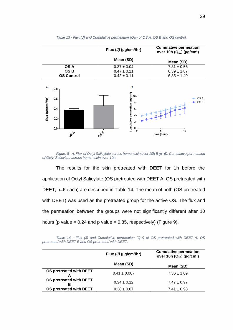
Two groups that had only Octyl Salicylate (5% v/v) applied onto the skin
were compared. The flux and the cumulative permeation of the groups OS A
and OS B, n=6 each, after 10 hours are described in Table 13. There was no
significant difference between the groups (p value = 0.31) for the flux (J) and
for the cumulative permeation (Q10) (p value = 0.29) (Figure 8).
29
Table 13 - Flux (J) and Cumulative permeation (Q10) of OS A, OS B and OS control.
Flux (J) (µg/cm²/hr)
Mean (SD)
Cumulative permeation over 10h (Q10) (µg/cm²)
Mean (SD)
OS A 0.37 ± 0.04 7.31 ± 0.56 OS B 0.47 ± 0.21 6.39 ± 1.87
OS Control 0.42 ± 0.11 6.85 ± 1.40
Figure 8 - A. Flux of Octyl Salicylate across human skin over 10h B (n=6). Cumulative permeation of Octyl Salicylate across human skin over 10h.
The results for the skin pretreated with DEET for 1h before the
application of Octyl Salicylate (OS pretreated with DEET A, OS pretreated with
DEET, n=6 each) are described in Table 14. The mean of both (OS pretreated
with DEET) was used as the pretreated group for the active OS. The flux and
the permeation between the groups were not significantly different after 10
hours (p value = 0.24 and p value = 0.85, respectively) (Figure 9).
Table 14 - Flux (J) and Cumulative permeation (Q10) of OS pretreated with DEET A, OS pretreated with DEET B and OS pretreated with DEET.
Flux (J) (µg/cm²/hr)
Mean (SD)
Cumulative permeation over 10h (Q10) (µg/cm²)
Mean (SD)
OS pretreated with DEET A
0.41 ± 0.067 7.36 ± 1.09
OS pretreated with DEET B
0.34 ± 0.12 7.47 ± 0.97
OS pretreated with DEET 0.38 ± 0.07 7.41 ± 0.98
30
Figure 9 - A. Flux of octyl salicylate pretreated with DEET across human skin over 10h (n=6). B. Cumulative permeation of octyl salicylate pretreated with DEET across human skin over 10h.
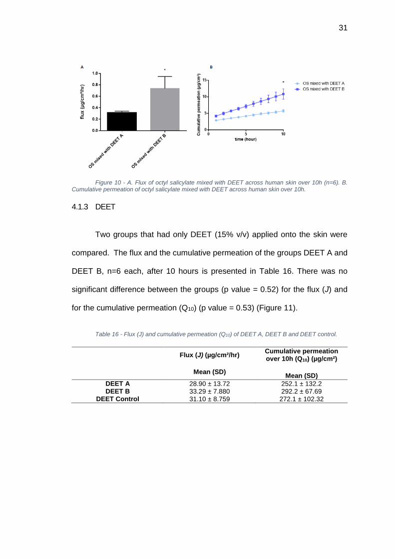
The mix of octyl salicylate at 5% v/v and DEET at 15% v/v is presented
in Table 15. The flux between the groups OS mixed with DEET A and OS mixed
with DEET B, n=6 each, was statistically different (p value = 0.0042) and the
concentration that permeated after 10h was also significantly different (p value
= 0.0003) (Figure 10). These differences were not considered an issue, since
as mentioned above they represent the high variability that is found in the skin
Table 15 - Flux (J) and cumulative permeation (Q10) of OS mixed with DEET A, OS mixed with DEET B and OS mixed with DEET.
Flux (J) (µg/cm²/hr)
Mean (SD)
Cumulative permeation over 10h (Q10) (µg/cm²)
Mean (SD)
OS mixed with DEET A 0.31 ± 0.028 5.74 ± 0.39 OS mixed with DEET B 0.74 ± 0.21 10.84 ± 1.55 OS mixed with DEET 0.53 ± 0.10 8.29 ± 2.87
31
Figure 10 - A. Flux of octyl salicylate mixed with DEET across human skin over 10h (n=6). B. Cumulative permeation of octyl salicylate mixed with DEET across human skin over 10h.
4.1.3 DEET
Two groups that had only DEET (15% v/v) applied onto the skin were
compared. The flux and the cumulative permeation of the groups DEET A and
DEET B, n=6 each, after 10 hours is presented in Table 16. There was no
significant difference between the groups (p value = 0.52) for the flux (J) and
for the cumulative permeation (Q10) (p value = 0.53) (Figure 11).
Table 16 - Flux (J) and cumulative permeation (Q10) of DEET A, DEET B and DEET control.
Flux (J) (µg/cm²/hr)
Mean (SD)
Cumulative permeation over 10h (Q10) (µg/cm²)
Mean (SD)
DEET A 28.90 ± 13.72 252.1 ± 132.2 DEET B 33.29 ± 7.880 292.2 ± 67.69
DEET Control 31.10 ± 8.759 272.1 ± 102.32
32
Figure 11 - A. Flux of DEET across human skin over 10h (n=6). B. Cumulative permeation of DEET across human skin over 10h.
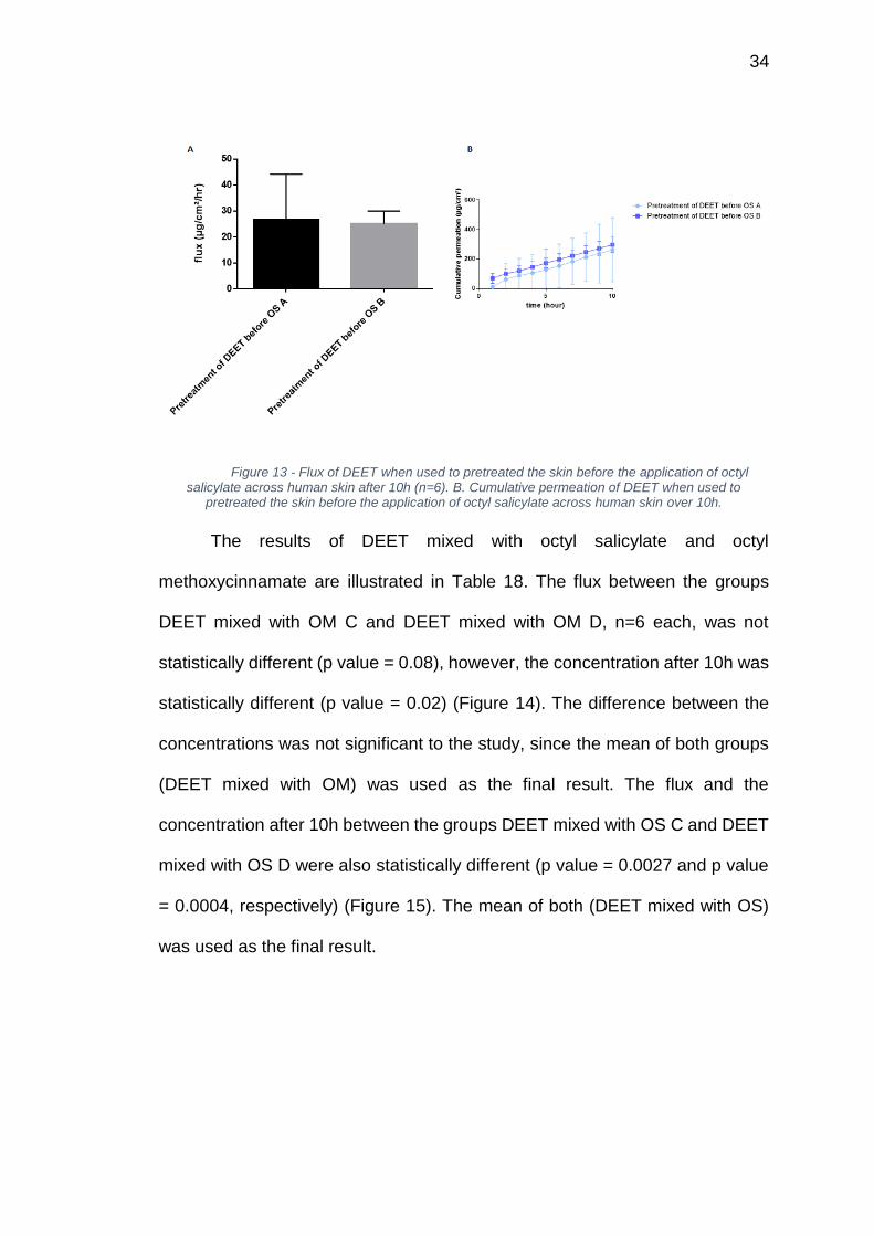
DEET at 15% v/v was used to pretreat the skin for 1h before the
application of the sunscreen actives. The results for octyl methoxycinnamate
and octyl salicylate are describe in Table 17. As mentioned before, the mean
of the groups was used as the final flux and cumulative permeation. There was
no difference on the flux and the final concentration after 10h (Q10) between
the groups Pretreatment of DEET before OM A and pretreatment of DEET
before OM B (p value = 0.20)(Figure 12). The groups exposed to octyl salicylate
also did not exhibit any difference between the flux (p value = 0.85) and the
final concentration (p value = 0.73) (Figure 13).
33
Table 17 - Flux (J) and cumulative permeation (Q10) of Pretreatment of DEET before OM (A, B and control) and Pretreatment of DEET before OS (A, B and control).
Flux (J) (µg/cm²/hr)
Mean (SD)
Cumulative permeation over 10h (Q10) (µg/cm²)
Mean (SD)
Pretreatment of DEET before OM A
35.81 ± 8.62 506.50 ± 170.70
Pretreatment of DEET before OM B
42.76 ± 8.72 462.10 ± 126.40
Pretreatment of DEET before OM
39.29 ± 6.16 484.30 ± 145.10
Pretreatment of DEET before OS A
26.48 ± 17.65 263.50 ± 217.30
Pretreatment of DEET before OS B
25.02 ± 4.90 296.60 ± 50.43
Pretreatment of DEET before OS
25.75 ± 8.76 280.00 ± 151.40
Figure 12 - Flux of DEET when used to pretreated the skin before the application of octyl methoxycinnamate across human skin over 10h (n=6). B. Cumulative permeation of DEET when used to pretreated the skin before the application of octyl methoxycinnamate across human skin over 10h.
34
Figure 13 - Flux of DEET when used to pretreated the skin before the application of octyl salicylate across human skin after 10h (n=6). B. Cumulative permeation of DEET when used to
pretreated the skin before the application of octyl salicylate across human skin over 10h.
The results of DEET mixed with octyl salicylate and octyl
methoxycinnamate are illustrated in Table 18. The flux between the groups
DEET mixed with OM C and DEET mixed with OM D, n=6 each, was not
statistically different (p value = 0.08), however, the concentration after 10h was
statistically different (p value = 0.02) (Figure 14). The difference between the
concentrations was not significant to the study, since the mean of both groups
(DEET mixed with OM) was used as the final result. The flux and the
concentration after 10h between the groups DEET mixed with OS C and DEET
mixed with OS D were also statistically different (p value = 0.0027 and p value
= 0.0004, respectively) (Figure 15). The mean of both (DEET mixed with OS)
was used as the final result.
35
Table 18 - Flux (J) and cumulative permeation (Q10) of OM mixed with DEET (C, D and control) and OS mixed with DEET (C, D and control).
Flux (J) (µg/cm²/hr)
Mean (SD)
Cumulative permeation over 10h (Q10) (µg/cm²)
Mean (SD)
DEET mixed with OM C 72.86 ± 15.53 809.70 ± 195.70 DEET mixed with OM D 58.25 ± 8.205 554.00 ± 79.46
DEET mixed with OM 65.55 ± 10.12
681.90 ± 195.20
DEET mixed with OS C 36.52 ± 16.60 348.20 ± 153.40 DEET mixed with OS D 73.85 ± 16.13 665.70 ± 140.30 DEET mixed with OS 55.19 ± 10.85 507.00 ± 217.10
Figure 14 - A. Flux of DEET mixed with OM across human skin over 10h (n=6). B. Cumulative permeation of DEET mixed with OM across human skin over 10h.
Figure 15 - A. Flux of DEET mixed with OS across human skin over 10h (n=6). B. Cumulative permeation of DEET mixed with OS across human skin over 10h.
36
4.2 PERMEATION OF OCTYL METHOXYCINNAMATE CO-APPLIED
WITH DEET THROUGH HUMAN SKIN
The purpose of this experiment was to mimic the application of a
sunscreen after the use of an insect repellent. The skin was pretreated with an
infinite dose of DEET at 15% v/v for 1h and before the application of octyl
methoxycinnamate at 7.5% v/v, the skin was cleaned with a cotton swab, in
order to remove all the residues of DEET. The other experiment was designed
to mimic the concomitant application of a sunscreen and an insect repellent, so
DEET at 15% v/v was previously mixed with OM at 7.5% v/v and the resulting
solution was applied onto the skin for 10h.
The control, containing the results of the application of octyl
methoxycinnamate alone on the human skin was compared to the results of the
skin pretreated for 1h with DEET followed by exposure to the sunscreen active
and with the results of the sunscreen mixed with DEET, n=12 each (Table 19).
The flux and the Q10 between the control and the group mixed with DEET were
not significantly different (Figure 16), however, the group that was pretreated
with DEET before the application of OM had a higher cumulative permeation
when compared to the other groups (p value = 0.0003), indicating that DEET
does not affect directly this UV absorber when co-applied with it, but when this
insect repellent is used to pretreat the skin, it can increase the flux significantly
(Figure 17). When the final concentration of OM is compared after 10h, it is
observed that both groups that were exposed to DEET had a higher cumulative
permeation at the end of the experiment, when compared to the control,
37
indicating that a higher concentration of the active penetrates the skin when
used in combination (p value < 0.0001). Also, the difference between the
groups exposed to DEET is not significantly different.
Table 19 - Flux (J) and cumulative permeation (Q10) of OM Control, OM pretreated with DEET and OM mixed with DEET.
Flux (J) (µg/cm²/hr)
Mean (SD)
Cumulative permeation over 10h (Q10) (µg/cm²)
Mean (SD)
OM Control 0.14 ± 0.04 2.10 ± 0.32 OM pretreated with DEET 0.22 ± 0.01 3.12 ± 0.41
OM mixed with DEET 0.17 ± 0.03 2.73 ± 0.61
OM
co
ntr
ol
OM
mix
ed
wit
h D
EE
T
OM
pre
treate
d w
ith
DE
ET
0 .0 0
0 .0 5
0 .1 0
0 .1 5
0 .2 0
0 .2 5
flu
x (
µg
/cm
²/h
r)
*
Figure 16 - Flux of octyl methoxycinnamate co-applied with DEET across human skin over 10h (n=12). The group pretreated with DEET has a higher flux when compared to the control and the group
mixed with DEET (p value = 0.0003).
38
0 5 1 0
0
1
2
3
4
t im e (h o u r )
Cu
mu
lati
ve
pe
rm
ea
tio
n (
µg
/cm
²)
O M c o n tro l
O M m ix e d w ith D E E T
O M p re tre a te d w ith D E E T
*
Figure 17 - Cumulative permeation of octyl methoxycinnamate co-applied with DEET across human skin over 10h (n=12). The groups exposed to DEET had a higher concentration when compared
to the control (p value < 0.0001).
4.3 PERMEATION OF OCTYL SALICYLATE CO-APPLIED WITH DEET
THROUGH HUMAN SKIN
The control, consisting of the application of octyl salicylate (5% v/v) alone
on the human skin was compared to the results of the skin pretreated for 1h
with DEET (15% v/v) and then exposed to the sunscreen active and with the
active mixed with DEET, n=12 each (Table 20). The flux of the group mixed
with DEET was higher than the group pretreated with the insect repellent after
10 hours, but both groups exposed to DEET were not significantly different from
the control (p value = 0.04) (Figure 18). The concentration of octyl salicylate
found in the receptor compartment was similar in all three experiments,
indicating that DEET does not affect the final cumulative permeation of the OS
(p value = 0.1981) (Figure 19).
39
Table 20 - Flux (J) and cumulative permeation (Q10) of OS Control, OM pretreated with DEET and OM mixed with DEET.
Flux (J) (µg/cm²/hr)
Mean (SD)
Cumulative permeation over 10h (Q10) (µg/cm²)
Mean (SD)
OS Control 0.42 ± 0.11 6.85 ± 1.40
OS pretreated with DEET 0.38 ± 0.07 7.41 ± 0.98
OS mixed with DEET 0.53 ± 0.10 8.29 ± 2.87
OS
co
ntr
ol
OS
pre
treate
d w
ith
DE
ET
OS
mix
ed
wit
h D
EE
T
0 .0
0 .2
0 .4
0 .6
0 .8
flu
x (
µg
/cm
²/h
r)
*
Figure 18 - Flux of octyl salicylate co-applied with DEET across human skin after 10h (n=12). The group mixed with DEET had a higher flux when compared to the pretreated group (p value =
0.0401).
40
0 5 1 0
0
5
1 0
1 5
t im e (h o u r )
Cu
mu
lati
ve
pe
rm
ea
tio
n (
µg
/cm
²)
O S c o n tro l
O S m ix e d w ith D E E T
O S p re tre a te d w ith D E E T
Figure 19 - Cumulative permeation of octyl salicylate co-applied with DEET across human skin over 10h. (n=12). No difference between the groups was observed (p value = 0.1981).
4.4 PERMEATION OF DEET CO-APPLIED WITH OCTYL
METHOXYCINNAMATE THROUGH HUMAN SKIN
The control was compared to the flux and the cumulative permeation
after 10h of three different groups, DEET mixed with OM, pretreatment of the
skin with DEET for 1h before the application of OM and pretreatment of the skin
with OM for 2 hours before the exposure to the insect repellent. The results of
each experiment are presented in Table 21. The flux of the group that was
exposed to the mix of DEET and OM has higher, when compared to all the
other groups (p value <0.0001) (Figure 20). All the groups exposed to octyl
methoxycinnamate had a higher concentration at the end of the experiment
when compared to the control, however, they were not statistically different from
each other (Figure 21) (p value =0.0004).
41
Table 21 - Flux (J) and cumulative permeation (Q10) of DEET Control, Pretreatment of DEET before OM, DEET mixed with OM and DEET pretreated with OM.
Flux (J) (µg/cm²/hr)
Mean (SD)
Cumulative permeation over 10h (Q10) (µg/cm²)
Mean (SD)
DEET Control 31.10 ± 8.759 272.1 ± 102.32
Pretreatment of DEET before OM
39.29 ± 6.160
484.3 ± 145.10
DEET mixed with OM 65.55 ± 10.12 681.9 ± 195.20
DEET pretreated with OM 31.58 ± 7.505 524.3 ± 168.0
DE
ET
co
ntr
ol
Pre
treatm
en
t o
f D
EE
T b
efo
re O
M
DE
ET
mix
ed
wit
h O
M
DE
ET
pre
treate
d w
ith
OM
0
2 0
4 0
6 0
8 0
flu
x (
µg
/cm
²/h
r)
*
Figure 20 - Flux of DEET across human skin over 10h (n=12). The flux of DEET mixed with OM was higher than the control and all the other groups (p<0.0001).
42
0 5 1 0
0
2 0 0
4 0 0
6 0 0
8 0 0
1 0 0 0
t im e (h o u r )
Cu
mu
lati
ve
pe
rm
ea
tio
n (
µg
/cm
²)
D E E T c o n tro l
P re tre a tm e n t o f D E E T
b e fo re O M
D E E T m ixe d w ith O M
D E E T p re tre a te d w ith O M
*
Figure 21 - Cumulative permeation of DEET across human skin over 10h. (n=12). All the groups exposed to OM have a higher transdermal permeation than the control after 10h (p = 0.0004).
4.5 PERMEATION OF DEET CO-APPLIED WITH OCTYL SALICYLATE
THROUGH HUMAN SKIN
The data for DEET (15% v/v) with and without octyl salicylate (5% v/v)
are illustrated in Table 22. The control, containing only DEET alone on the
human skin was compared to the results of the skin pretreated for 1h with DEET
and then exposed to the sunscreen active and the skin pretreated with OS for
2 hours before the application of DEET, n=12 each. The flux of DEET when it
was mixed with OS was higher than for all the other groups, including the control
(p value = 0.0001) (Figure 22). The cumulative concentration that permeated
after 10 hours was higher in all groups exposed to OS when compared with
control (p value = 0.002) (Figure 23).
43
Table 22 - Flux (J) and cumulative permeation (Q10) of DEET Control, Pretreatment of DEET before OS, DEET mixed with OS and DEET pretreated with OS.
Flux (J) (µg/cm²/hr)
Mean (SD)
Cumulative permeation over 10h (Q10) (µg/cm²)
Mean (SD)
DEET Control 31.10 ± 8.76 272.1 ± 102.32 Pretreatment of DEET
before OS 25.75 ± 8.76 280.0 ± 151.4
DEET mixed with OS 55.19 ± 10.85 507.0 ± 217.1 DEET pretreated with OS 38.11 ± 8.766 384.0 ± 91.19
Figure 22 - Flux of DEET across human skin over 10h (n=12). The flux of DEET mixed with OS was higher than the control and all the other groups (p=0.0001).
44
5 1 0
-2 0 0
0
2 0 0
4 0 0
6 0 0
8 0 0
t im e (h o u r )
Cu
mu
lati
ve
pe
rm
ea
tio
n (
µg
/cm
²)
D E E T c o n tro l
P re tre a tm e n t o f D E E T b e fo re O S
D E E T m ix e d w ith O S
D E E T p re tre a te d w ith O S*
Figure 23 - Cumulative permeation of DEET across human skin over 10h. (n=12). The concentration over 10h of DEET when mixed with OS was higher than the control group and the group pretreated with DEET (p value = 0.0021).
4.6 PERCUTANEOUS PERMEATION OF OCTYL METHOXYCINNAMATE,
OCTYL SALICYLATE AND DEET
All experiments were compared to evaluate which combination had the
highest flux and cumulative permeation after 10 hours (Q10). The fluxes of octyl
salicylate and octyl methoxycinnamate when applied alone onto the skin are
not statistically different when compared to all the other groups. The same was
observed when these sunscreen actives were mixed with DEET or even when
the skin was previously pretreated with the insect repellent before the
application of OM or OS. The fluxes corresponding to DEET control, DEET
when it was applied to the skin after the pretreatment with OM and OS and
DEET when it pretreated the skin before the addition of OM and OS were higher
than the results of OM and OS (mentioned above) and lower than the two final
groups. DEET mixed with OS (55.19 ± 10.85 µg/cm²/hr) and DEET mixed with
OM (65.55 ± 10.12 µg/cm²/hr) resulted in the highest flux of all the groups. The
45
lowest flux found was for OM control (0.1437 ± 0.03089 µg/cm²/hr) and the
difference between the lowest and the highest concentration observed was
significantly different (p value < 0.0001) (Figure 24). The highest cumulative
concentration permeating the skin after 10 hours was recorded for DEET when
it was mixed and co-applied with OM (681.9 ± 195.2 µg/cm²) and the difference
between this group and DEET pretreated with OM was not significantly
different. The lowest concentration was recorded for OM control (2.103 ±
0.3243) and again the difference between these two groups was significantly
different (p value < 0.0001) (Figure 25).
OM
co
ntr
ol
OS
co
ntr
ol
DE
ET
co
ntr
ol
OM
pre
treate
d w
ith
DE
ET
OM
mix
ed
wit
h D
EE
T
OS
pre
treate
d w
ith
DE
ET
OS
mix
ed
wit
h D
EE
T
Pre
treatm
en
t o
f D
EE
T b
efo
re O
M
DE
ET
mix
ed
wit
h O
M
DE
ET
pre
treate
d w
ith
OM
Pre
treatm
en
t o
f D
EE
T b
efo
re O
S
DE
ET
mix
ed
wit
h O
S
DE
ET
pre
treate
d w
ith
OS
0
2 0
4 0
6 0
8 0
1 0 0
flu
x (
µg
/cm
²/h
r)
Figure 24 - Flux of octyl methoxycinnamate, octyl salicylate and DEET across human skin over 10h (n=12). The fluxes of DEET mixed with OS and DEET mixed with OM were higher than for all the
other groups (p<0.0001).
46
0 5 1 0
0
2 0 0
4 0 0
6 0 0
8 0 0
t im e (h o u r )
Cu
mu
lati
ve
pe
rm
ea
tio
n (
µg
/cm
²)
O M c o n tro l
O S c o n tro l
D E E T c o n tro l
O M p re tre a te d w ith D E E T
O M m ix e d w ith D E E T
O S p re tre a te d w ith D E E T
O S m ix e d w ith D E E T
O M p re tre a te d w ith D E E T
D E E T m ixe d w ith O M
D E E T p re tre a te d w ith O M
O S p re tre a te d w ith D E E T
D E E T m ix e d w ith O S
D E E T p re tre a te d w ith O S
*
Figure 25 - Cumulative permeation of octyl methoxycinnamate, octyl salicylate and DEET across human skin after 10h (n=12). The concentration over 10h of DEET when mixed with OM was higher than for all the other groups - p value < 0.0001).
47
DISCUSSION
Previous studies demonstrated that sunscreens actives and DEET have
an ability to permeate the skin, even when applied alone [4]. When these
actives are used in combination, it was observed that the insect repellent can
decrease the efficacy of the sunscreen [9] and they can act as enhancers to
each other [67, 68, 76]. The most used active to test this interaction is
oxybenzone, and all in vitro and in vivo studies observed that the percutaneous
permeation of both actives increased when they were co-applied [4, 77].
However, all the results showed that the ability to permeate of DEET is higher
than the sunscreen and even when other factor is responsible for the increased
permeation, the penetration of the insect repellent is always more affected [67,
68].
The interactions associated with the permeation of sunscreens in the
presence of insect repellents are still not completely understood and more
studies are needed to elucidate the skin transport effects between these two
commonly used actives. In this study, it was observed that even when these
actives are applied individually onto the human skin in vitro, there are some
clear differences that were recorded. The sunscreen actives octyl
methoxycinnamate and octyl salicylate have similar fluxes in human skin in vitro
and the concentrations that permeate the skin are also not significantly different
from each other. However, the permeation of DEET is higher even when
applied alone, when compared to the sunscreens. Since DEET is applied by a
large population globally over large surfaces of the body and in also in children,
48
many scientists have been concerned about the outcomes. Since it is also
known to act as a skin “penetration enhancer” which means that it is able to
facilitate the skin transport of co-administered actives into the skin [78], the
concern is magnified (potentially) when such insecticides are applied onto skin
with products containing sunscreens. Little work is reported in this area
however, the FDA warns against using such combinations. In this study we
tested some commonly used FDA approved sunscreens to test out what
actually happens with these combinations in human skin in vitro and whether
the concerns are justified or not.
It was found that with octyl salicylate a low permeation through skin was
observed even when it was co-applied with DEET. The differences found
between the fluxes of the group that had OS mixed with DEET and the control
was insignificant and the final concentration found in the receptor compartment
was not different from that for all the other groups. The same was observed for
octyl methoxycinnamate, which had a little difference between the group mixed
with DEET and the control. These findings indicate that for some sunscreen
actives, even the co-application with an enhancer, like DEET, does not affect
the ability of the sunscreen molecule to permeate the skin. It can be higher, but
when compared to the control, the differences are not particularly high and in
vivo would probably be insignificant.
However, when permeation of DEET was studied, several interesting
points were observed. When DEET was co-applied with octyl salicylate, the flux
observed was higher than for the control or the other treatment groups and the
49
concentration that permeated after 10 hours was almost double when
compared to control. This is an indication that OS can act as a weak enhancer
of DEET in vitro. On the other hand, experiments with the mixture of DEET and
octyl methoxycinnamate produced even more interesting data. The flux for the
mix of DEET plus OM was almost two times the flux of the control group and
the cumulative permeation recorded after 10 hours was almost three times the
Q10 of control. Therefore, the concomitant application of OM and DEET, in vitro,
did show significant enhancement, and based on these data, the use of DEET
and OM together cannot be recommended, since OM will enhance the
penetration of the insect repellent into the skin. Since the over-exposition to
DEET is connected to many toxic effects, this co-application, based on the
results of this study, should not be recommended [53, 59].
In summary, this thesis presents an overview of some of the possible
interactions between two of the most commonly used sunscreen absorbers in
the U.S. and the most popular insect repellent. It was clear that the ability to
permeate the skin is very different between the two UV absorbers utilized in this
study, especially when all the results obtained were compared. DEET mixed
with OM and DEET mixed with OS produced the highest transdermal
permeation in vitro and the lowest was obtained with OM applied alone onto the
skin.
50
CONCLUSION
In conclusion, DEET does not act as an enhancer in skin in vitro when
used in combination with octyl salicylate and octyl methoxycinnamate. The skin
permeation of these sunscreens actives were not significantly affected by the
insecticide. Furthermore, it is possible to acknowledge that the UV absorbers
used on this study can be potential enhancers when co-applied with DEET.
When both sunscreen actives were mixed with DEET, the resulting skin
permeation was higher than the control, however, the differences found in vitro
can be insignificant in vivo, so more studies are necessary to determine if this
interaction can be considered dangerous to the population and what can be
done to minimize the possible effects.
51
REFERENCES
1. Battie, C., et al., New insights in photoaging, UVA induced damage and skin types. Experimental Dermatology, 2014. 23: p. 7-12.
2. Manaia, E.B., et al., Inorganic UV filters. Brazilian Journal of Pharmaceutical Sciences, 2013. 49: p. 201-209.
3. Rai, R., S.C. Shanmuga, and C.R. Srinivas, Update on Photoprotection. Indian Journal of Dermatology, 2012. 57(5): p. 335-342.
4. Kasichayanula, S., et al., Percutaneous characterization of the insect repellent DEET and the sunscreen oxybenzone from topical skin application. Toxicology and Applied Pharmacology, 2007. 223(2): p. 187-194.
5. Gonzalez, H., et al., Percutaneous absorption of the sunscreen benzophenone-3 after repeated whole-body applications, with and without ultraviolet irradiation. British Journal of Dermatology, 2006. 154(2): p. 337-340.
6. Gu, X., et al., In vitro evaluation of concurrent use of commercially available insect repellent and sunscreen preparations. Br J Dermatol, 2005. 152(6): p. 1263-7.
7. Chen, T., et al., Percutaneous permeation comparison of repellents picaridin and DEET in concurrent use with sunscreen oxybenzone from commercially available preparations. Pharmazie, 2010. 65(11): p. 835-9.
8. Montemarano, A.D., et al., Insect repellents and the efficacy of sunscreens. Lancet, 1997. 349(9066): p. 1670-1.
9. Murphy, M.E., et al., The effect of sunscreen on the efficacy of insect repellent: a clinical trial. J Am Acad Dermatol, 2000. 43(2 Pt 1): p. 219-22.
10. Mouret, S., A. Forestier, and T. Douki, The specificity of UVA-induced DNA damage in human melanocytes. Photochem Photobiol Sci, 2012. 11(1): p. 155-62.
11. Elçioğlu, H.K.b. and A.D. Sezer, Nanoparticle Insulin Drug Delivery — Applications and New Aspects, in "Application of Nanotechnology in Drug Delivery", A.D. Sezer, Editor. 2014.
12. Bos, J.D. and M.M. Meinardi, The 500 Dalton rule for the skin penetration of chemical compounds and drugs. Exp Dermatol, 2000. 9(3): p. 165-9.
13. McGrath, J.A. and J. Uitto, Anatomy and Organization of Human Skin, in Rook's Textbook of Dermatology. 2010, Wiley-Blackwell. p. 1-53.
14. Mescher, A., Junqueira's Basic Histology: Text and Atlas, Twelfth Edition: Text and Atlas, Twelfth Edition. 12 ed. 2013: McGraw-Hill Education.
52
15. Koster, M.I., Making an epidermis. Annals of the New York Academy of Sciences, 2009. 1170: p. 7-10.
16. Kumamoto, J., et al., External negative electric potential accelerates exocytosis of lamellar bodies in human skin ex vivo. Experimental Dermatology, 2013. 22(6): p. 421-423.
17. Kanerva, L., et al., Handbook of Occupational Dermatology. Volume 1 ed. 2013: Springer Berlin Heidelberg.
18. Huzil, J.T., et al., Drug delivery through the skin: molecular simulations of barrier lipids to design more effective noninvasive dermal and transdermal delivery systems for small molecules, biologics, and cosmetics. Wiley Interdiscip Rev Nanomed Nanobiotechnol, 2011. 3(5): p. 449-62.
19. Desai, P., R.R. Patlolla, and M. Singh, Interaction of nanoparticles and cell-penetrating peptides with skin for transdermal drug delivery. Mol Membr Biol, 2010. 27(7): p. 247-59.
20. Wang F, C.Y., Benson HAE, Formulation of nano and micro PLGA particles of the model peptide insulin: preparation, characterization, stability and deposition in human skin. Open Drug Delivery J, 2008(2): p. 1-9.
21. Madison, K.C., Barrier Function of the Skin: [ldquo]La Raison d'Etre[rdquo] of the Epidermis. J Investig Dermatol, 2003. 121(2): p. 231-241.
22. Bouwstra, J.A., et al., Structure of the skin barrier and its modulation by vesicular formulations. Prog Lipid Res, 2003. 42(1): p. 1-36.
23. Jakasa, I., et al., Altered Penetration of Polyethylene Glycols into Uninvolved Skin of Atopic Dermatitis Patients. J Invest Dermatol, 2006. 127(1): p. 129-134.
24. Florence, A.T. and D. Attwood, Physicochemical Principles of Pharmacy. 2006: Pharmaceutical Press.
25. Paudel, K.S., et al., Challenges and opportunities in dermal/transdermal delivery. Therapeutic delivery, 2010. 1(1): p. 109-131.
26. Ng, S.-F., et al., Validation of a Static Franz Diffusion Cell System for In Vitro Permeation Studies. AAPS PharmSciTech, 2010. 11(3): p. 1432-1441.
27. Martin, A.N., Physical Pharmacy: Physical Chemical Principles in Pharmaceutical Science. 1966: Henry Kimpton.
28. Holzle, E. and H. Honigsmann, [UV-radiation--sources, wavelength, environment]. J Dtsch Dermatol Ges, 2005. 3 Suppl 2: p. S3-10.
29. Hussein, M.R., Ultraviolet radiation and skin cancer: molecular mechanisms. J Cutan Pathol, 2005. 32(3): p. 191-205.
53
30. Natarajan, V.T., et al., Multifaceted pathways protect human skin from UV radiation. Nat Chem Biol, 2014. 10(7): p. 542-51.
31. Fourtanier, A., D. Moyal, and S. Seite, UVA filters in sun-protection products: regulatory and biological aspects. Photochem Photobiol Sci, 2012. 11(1): p. 81-9.
32. Goodsell, D.S., The molecular perspective: ultraviolet light and pyrimidine dimers. Oncologist, 2001. 6(3): p. 298-9.
33. Murphy, G., et al., The molecular determinants of sunburn cell formation. Experimental Dermatology, 2001. 10(3): p. 155-160.
34. Samarasinghe, V., V. Madan, and J.T. Lear, Focus on Basal cell carcinoma. J Skin Cancer, 2011. 2011: p. 328615.
35. Alam, M., et al., The use of brachytherapy in the treatment of nonmelanoma skin cancer: a review. J Am Acad Dermatol, 2011. 65(2): p. 377-88.
36. Samarasinghe, V. and V. Madan, Nonmelanoma Skin Cancer. Journal of Cutaneous and Aesthetic Surgery, 2012. 5(1): p. 3-10.
37. Ramachandran, S., et al., Basal cell carcinomas: association of allelic variants with a high-risk subgroup of patients with the multiple presentation phenotype. Pharmacogenetics, 2001. 11(3): p. 247-54.
38. Warren, H., Cutaneous Melanoma: A Population Health Problem. Plast Surg Nurs, 2015. 35(4): p. 164-70.
39. Lo, J.A. and D.E. Fisher, The melanoma revolution: from UV carcinogenesis to a new era in therapeutics. Science (New York, N.Y.), 2014. 346(6212): p. 945-949.
40. Bald, T., et al., Ultraviolet-radiation-induced inflammation promotes angiotropism and metastasis in melanoma. Nature, 2014. 507(7490): p. 109-13.
41. Latha, M.S., et al., Sunscreening Agents: A Review. The Journal of Clinical and Aesthetic Dermatology, 2013. 6(1): p. 16-26.
42. Nordlund, T.M., Quantitative Understanding of Biosystems: An Introduction to Biophysics. 2011: CRC Press.
43. Shaath, N., Sunscreens: Regulations and Commercial Development. 2005: Taylor & Francis.
44. States), F.a.D.A.U., Re: Tentative Final Monograph for OTC Sunscreen. 1998-09-11. Retrieved 2009-09-25.
.
54
45. Morabito, K., et al., Review of sunscreen and the emergence of non-conventional absorbers and their applications in ultraviolet protection. Int J Cosmet Sci, 2011. 33(5): p. 385-90.
46. Schalka, S., et al., Brazilian Consensus on Photoprotection. Anais Brasileiros de Dermatologia, 2014. 89(6 Suppl 1): p. 1-74.
47. Lademann, J., et al., Penetration of titanium dioxide microparticles in a sunscreen formulation into the horny layer and the follicular orifice. Skin Pharmacol Appl Skin Physiol, 1999. 12(5): p. 247-56.
48. Miquel-Jeanjean, C., et al., Penetration study of formulated nanosized titanium dioxide in models of damaged and sun-irradiated skins. Photochem Photobiol, 2012. 88(6): p. 1513-21.
49. Sharma, V., et al., Zinc oxide nanoparticle induced genotoxicity in primary human epidermal keratinocytes. J Nanosci Nanotechnol, 2011. 11(5): p. 3782-8.
50. Donaldson, K., et al., Nanotoxicology. Occup Environ Med, 2004. 61(9): p. 727-8.
51. Xu, P., et al., Mosquito odorant receptor for DEET and methyl jasmonate. Proceedings of the National Academy of Sciences of the United States of America, 2014. 111(46): p. 16592-16597.
52. Brown, M. and A.A. Hebert, Insect repellents: an overview. J Am Acad Dermatol, 1997. 36(2 Pt 1): p. 243-9.
53. Katz, T.M., J.H. Miller, and A.A. Hebert, Insect repellents: historical perspectives and new developments. J Am Acad Dermatol, 2008. 58(5): p. 865-71.
54. Kuklenyik, P., et al., On-line solid phase extraction-high performance liquid chromatography–isotope dilution–tandem mass spectrometry approach to quantify N,N-diethyl-m-toluamide and oxidative metabolites in urine. Analytica chimica acta, 2013. 787: p. 267-273.
55. Abu-Qare, A.W. and M.B. Abou-Donia, High performance liquid chromatographic determination of diazinon, permethrin, DEET (N, N-diethyl-m-toluamide), and their metabolites in rat plasma and urine. Fresenius J Anal Chem, 2001. 370(4): p. 403-7.
56. Olsson, A.O., et al., A liquid chromatography--tandem mass spectrometry multiresidue method for quantification of specific metabolites of organophosphorus pesticides, synthetic pyrethroids, selected herbicides, and deet in human urine. Anal Chem, 2004. 76(9): p. 2453-61.
57. Chen-Hussey, V., R. Behrens, and J.G. Logan, Assessment of methods used to determine the safety of the topical insect repellent N,N-diethyl-m-toluamide (DEET). Parasites & Vectors, 2014. 7: p. 173-173.
55
58. McGready, R., et al., Safety of the insect repellent N,N-diethyl-M-toluamide (DEET) in pregnancy. Am J Trop Med Hyg, 2001. 65(4): p. 285-9.
59. Briassoulis, G., M. Narlioglou, and T. Hatzis, Toxic encephalopathy associated with use of DEET insect repellents: a case analysis of its toxicity in children. Hum Exp Toxicol, 2001. 20(1): p. 8-14.
60. Roberts, M.S., Dermal Absorption and Toxicity Assessment, Second Edition. 2007: CRC Press.
61. Pawar, A.P., et al., Formulation and Evaluation of Optimized Oxybenzone Microsponge Gel for Topical Delivery. Journal of Drug Delivery, 2015. 2015: p. 261068.
62. Kunisue, T., et al., Analysis of five benzophenone-type UV filters in human urine by liquid chromatography-tandem mass spectrometry. Analytical Methods, 2010. 2(6): p. 707-713.
63. Krause, M., et al., Sunscreens: are they beneficial for health? An overview of endocrine disrupting properties of UV-filters. Int J Androl, 2012. 35(3): p. 424-36.
64. Kunz, P.Y. and K. Fent, Estrogenic activity of UV filter mixtures. Toxicol Appl Pharmacol, 2006. 217(1): p. 86-99.
65. Kim, S. and K. Choi, Occurrences, toxicities, and ecological risks of benzophenone-3, a common component of organic sunscreen products: a mini-review. Environ Int, 2014. 70: p. 143-57.
66. Gupta, V.K., J.L. Zatz, and M. Rerek, Percutaneous absorption of sunscreens through micro-yucatan pig skin in vitro. Pharm Res, 1999. 16(10): p. 1602-7.
67. Gu, X., et al., In-vitro permeation of the insect repellent N,N-diethyl-m-toluamide (DEET) and the sunscreen oxybenzone. J Pharm Pharmacol, 2004. 56(5): p. 621-8.
68. Wang, T., S. Kasichayanula, and X. Gu, In vitro permeation of repellent DEET and sunscreen oxybenzone across three artificial membranes. Int J Pharm, 2006. 310(1-2): p. 110-7.
69. Wang, T. and X. Gu, In vitro percutaneous permeation of the repellent DEET and the sunscreen oxybenzone across human skin. J Pharm Pharm Sci, 2007. 10(1): p. 17-25.
70. Wang, T., et al., Evaluation of percutaneous permeation of repellent DEET and sunscreen oxybenzone from emulsion-based formulations in artificial membrane and human skin. Acta Pharmaceutica Sinica. B, 2014. 4(1): p. 43-51.
71. Janjua, N.R., et al., Systemic absorption of the sunscreens benzophenone-3, octyl-methoxycinnamate, and 3-(4-methyl-benzylidene) camphor after whole-body topical application and reproductive hormone levels in humans. J Invest Dermatol, 2004. 123(1): p. 57-61.
56
72. Gu, X. and T. Chen, In vitro permeation characterization of repellent picaridin and sunscreen oxybenzone. Pharm Dev Technol, 2009. 14(3): p. 332-40.
73. Peruchi, L.M. and S. Rath, Development and application of a HPLC method for eight sunscreen agents in suncare products. Int J Cosmet Sci, 2012. 34(3): p. 226-33.
74. de Souza de Bustamante Monteiro, M.S., et al., Evaluation of octyl p-methoxycinnamate included in liposomes and cyclodextrins in anti-solar preparations: preparations, characterizations and in vitro penetration studies. International Journal of Nanomedicine, 2012. 7: p. 3045-3058.
75. Register, F., International Conference on the Harmonization. Draft guideline on validation of analytical procedures: definition and terminology. 2006.
76. Ross, E.A., et al., Insect repellent [correction of repellant] interactions: sunscreens enhance DEET (N,N-diethyl-m-toluamide) absorption. Drug Metab Dispos, 2004. 32(8): p. 783-5.
77. Puglia, C., et al., Evaluation of percutaneous absorption of the repellent diethyltoluamide and the sunscreen ethylhexyl p-methoxycinnamate-loaded solid lipid nanoparticles: an in-vitro study. J Pharm Pharmacol, 2009. 61(8): p. 1013-9.
78. Kaushik, D., A. Costache, and B. Michniak-Kohn, Percutaneous penetration modifiers and formulation effects. Int J Pharm, 2010. 386(1-2): p. 42-51.