ORIGINAL ARTICLE Effect of intra-articular injection of mesenchymal stem cells in cartilage repair in experimental animals Nadia Salah Kamel a , Mona Mahmoud Arafa a , Amr Nadim d , Hnaa Amer b , Irene Raouf Amin a, * , Naglaa Samir c , Amina Salem a a Department of Physical Medicine, Rheumatology and Rehabilitation, Faculty of Medicine, Ain Shams University, Egypt b Department of Clinical Pathology, Faculty of Medicine, Ain Shams University, Egypt c Department of Pathology, Faculty of Medicine, Ain Shams University, Egypt d Department of Obstetrics and Gynecology, Faculty of Medicine, Ain Shams University, Egypt Received 10 February 2014; accepted 8 March 2014 KEYWORDS Intra articular injection; Mesenchymal stem cells (MSCs); Cartilage repair; Osteoarthritis; Experimental animals Abstract Osteoarthritis (OA) is characterized by degeneration of the articular cartilage and, ultimately, joint destruction. Human umbilical cord blood (hUCB) may prove to be a new source of mesenchymal stem cells (MSCs) for cellular therapeutics used for cartilage repair. Aim of the work: This study was carried out over a nine-month period of time, to study the effect of intra-articular injection of hUCB MSCs in cartilage repair by histopathological and ultra structural assessment. Materials and methods: We conducted our study on 20 adult rats, which were subjected to the induction of cartilaginous defect in both knee joints. This was followed by injection of MSCs suspended in Hyaluronic acid solution in the right knee of each rat while the left knee served as a control. Histopathological and electron microscopic studies were performed. Results: The present study revealed: In the injected knees; In 73% of the cases, the tissue was typical of fibrohyaline cartilage and appeared more cellular than fibrous. In 27% of the cases the repaired tissue appeared more fibrous than hyaline. In the control knees; the newly formed tissue was an undifferentiated connective tissue and the cells were covered with a thin layer of fibrous tissue. The electron microscopic pictures of the injected knees showed mitotic chondrocyte activity. The pictures indicated a repaired fibrohyaline cartilage. * Corresponding author. Tel.: +20 226223833. E-mail address: [email protected](I.R. Amin). Peer review under responsibility of Egyptian Society for Joint Diseases and Arthritis. Production and hosting by Elsevier The Egyptian Rheumatologist (2014) xxx, xxx–xxx Egyptian Society for Joint Diseases and Arthritis The Egyptian Rheumatologist www.rheumatology.eg.net www.sciencedirect.com 1110-1164 Ó 2014 Production and hosting by Elsevier B.V. on behalf of Egyptian Society for Joint Diseases and Arthritis. http://dx.doi.org/10.1016/j.ejr.2014.03.001 Please cite this article in press as: Kamel NS et al. Effect of intra-articular injection of mesenchymal stem cells in cartilage repair in experimental animals, The Egyptian Rheumatologist (2014), http://dx.doi.org/10.1016/j.ejr.2014.03.001
Transcript
The Egyptian Rheumatologist (2014) xxx, xxx–xxx
Egyptian Society for Joint Diseases and Arthritis
The Egyptian Rheumatologist
www.rheumatology.eg.netwww.sciencedirect.com
ORIGINAL ARTICLE
Effect of intra-articular injection of mesenchymal
Peer review under responsibility of Egyptian Society for Joint Diseases
and Arthritis.
Production and hosting by Elsevier
1110-1164 � 2014 Production and hosting by Elsevier B.V. on behalf of Egyptian Society for Joint Diseases and Arthritis.
http://dx.doi.org/10.1016/j.ejr.2014.03.001
Please cite this article in press as: Kamel NS et al. Effect of intra-articular injection of mesenchymal stem cells in cartilage repair in experanimals, The Egyptian Rheumatologist (2014), http://dx.doi.org/10.1016/j.ejr.2014.03.001
Nadia Salah Kamela, Mona Mahmoud Arafa
a, Amr Nadim
d,
Hnaa Amer b, Irene Raouf Amin a,*, Naglaa Samir c, Amina Salem a
a Department of Physical Medicine, Rheumatology and Rehabilitation, Faculty of Medicine, Ain Shams University, Egyptb Department of Clinical Pathology, Faculty of Medicine, Ain Shams University, Egyptc Department of Pathology, Faculty of Medicine, Ain Shams University, Egyptd Department of Obstetrics and Gynecology, Faculty of Medicine, Ain Shams University, Egypt
Received 10 February 2014; accepted 8 March 2014
KEYWORDS
Intra articular injection;
Mesenchymal stem cells
(MSCs);
Cartilage repair;
Osteoarthritis;
Experimental animals
Abstract Osteoarthritis (OA) is characterized by degeneration of the articular cartilage and,
ultimately, joint destruction. Human umbilical cord blood (hUCB) may prove to be a new source
of mesenchymal stem cells (MSCs) for cellular therapeutics used for cartilage repair.
Aim of the work: This study was carried out over a nine-month period of time, to study the effect
of intra-articular injection of hUCB MSCs in cartilage repair by histopathological and ultra
structural assessment.
Materials and methods: We conducted our study on 20 adult rats, which were subjected to the
induction of cartilaginous defect in both knee joints. This was followed by injection of MSCs
suspended in Hyaluronic acid solution in the right knee of each rat while the left knee served as
a control. Histopathological and electron microscopic studies were performed.
Results: The present study revealed: In the injected knees; In 73% of the cases, the tissue was
typical of fibrohyaline cartilage and appeared more cellular than fibrous. In 27% of the cases the
repaired tissue appeared more fibrous than hyaline. In the control knees; the newly formed tissue
was an undifferentiated connective tissue and the cells were covered with a thin layer of fibrous
tissue. The electron microscopic pictures of the injected knees showed mitotic chondrocyte activity.
The pictures indicated a repaired fibrohyaline cartilage.
Please cite this article in press as: Kameanimals, The Egyptian Rheumatologist
Conclusion: We can conclude that the intra-articular injection of hUCB MSCs is an effective
method for cartilage repair in rats. This makes it a very promising tool for the treatment of patients
with OA.
� 2014 Production and hosting by Elsevier B.V. on behalf of Egyptian Society for Joint Diseases and
Arthritis.
1. Introduction
Osteoarthritis (OA) is a degenerative joint disease that is char-acterized by erosion of the articular cartilage, growth of boneat the margins i.e., osteophytes, subchondral sclerosis and arange of biochemical and morphologic alterations of the syno-
vial membrane and joint capsule [1]. The primary tissueaffected is the thin rim of hyaline articular cartilage interposedbetween the two articulating bones [2].
In early OA, the articular cartilage surface becomes irregu-lar, and superficial clefts within the tissue become apparent. Asthe condition worsens, the clefts deepen, surface irregularities
increase and the articular cartilage eventually ulcerates, expos-ing the underlying bone [3]. Chondrocytes in areas surroundingan injured zone are unable to migrate, proliferate, repopulate,regenerate or repair tissue with similar structure, function,
and biomechanical properties of normal hyaline cartilage [4].The burden of OA is exacerbated by the inadequacies of
current therapies. Nonpharmacologic and pharmacologic
treatments are used for early and moderately early cases ofOA [5].
Mesenchymal stem cells (MSCs), which have the ability to
differentiate into cells of the chondrogenic lineage, are verypromising candidates to develop new cell-based articular carti-lage repair strategies; this strategy entails the use of MSCs as
trophic producers of bioactive factors to initiate endogenousregenerative activities in the OA joint [6]. Autologous bonemarrow (BM) represents the main source of MSCs for bothexperimental and clinical studies; however as the number of
MSCs and their differentiation capacity decline with age, theirtherapeutic potential might be diminished as well [7]. As sev-eral ethical and practical issues arise from the use of BM
and fetal stem cells, umbilical cord blood (UCB) has turnedout to be an excellent alternative source of MSCs for clini-cal-scale allogeneic transplantation [8].
It was postulated that hyaluronic acid might facilitate themigration and adherence of MSCs to the defect, which mightexplain the occurrence of partial healing at 6 weeks in animals
that were treated with hyaluronic acid alone. The repairedtissue in animals treated with hyaluronic acid alone was ofinferior quality and was shown to deteriorate further after12 weeks, so the combination between MSCs and hyaluronic
acid will result in synergistic effect [9].Aim of the Work: This study was carried out over a nine-
month period of time, to study the effect of intra-articular
injection of human umbilical cord blood (hUCB) mesenchy-mal stem cells (MSCs) in cartilage repair by histopathologicaland ultra structural assessment.
2. Materials and methods
The study was carried out on twenty albino rats of Wistar
strain (adult males) during a nine-month period of time. They
l NS et al. Effect of intra-articular(2014), http://dx.doi.org/10.1016/j
were brought from the medical research center, faculty ofmedicine, animal house of Ain Shams University. This study
was approved by the local ethical committee.The animals were maintained under conditions of
controlled humidity, they were fed with commercial rat pellets
and water. The animal house staff detected the age and weightof the animals and supervised their feeding. All animals werehealthy and had no joint problems.
2.1. All animals were subjected to the following
Induction of cartilaginous defect in both knee joints byscratching the cartilage using a sterile needle [10,11]. In
maximal flexion, longitudinal and diagonal grooves were madeon the weight-bearing parts of femoral condyles withoutdamaging the subchondral bone. The latter was checked by
histology in 2 of the rats which were sacrificed; one of themafter 1 week and the other after 4 weeks from the scratchingwhich was done in the beginning of the experiment in order
to ensure development of osteoarthritis (OA).
2.2. Preparation of MSCs from human umbilical cord blood(UCB)
2.2.1. Collection of UCB
Umbilical cord blood was obtained from the labor room of
Obstetrics and Gynecology Department, Faculty of Medicine,Ain Shams University after written consent from the mothers.
4 UCB samples from full-term deliveries were collected
from the unborn placenta using complete aseptic techniquein sterile 15 ml Falcon tubes (Nunclon, Germany) containing2 ml of acid citrate dextrose (ACD) anticoagulant (Lonza,
Switzerland). The samples were stored at 22 ± 4 �C beforeprocessing [12]. Isolation and culture of MSCs were carriedout in the medical research center, Faculty of Medicine, AinShams University as follows.
2.2.2. Isolation of mononuclear cells (MNCs)
Under complete asepsis each UCB sample was diluted 1:1 with
phosphate-buffered saline (PBS) (lonza) and was carefullyloaded onto Ficoll–Hypaque solution 2:1 ratio. After densitygradient centrifugation at 2000 rpm for 30 min at roomtemperature, MNCs were removed from the interphase (the
Buffy coat) and were washed three times with PBS; each timewere centrifuged at 1500 rpm for 5 min, a clear cell pallet wasformed in the bottom of the tube [13].
2.2.3. Culture of mesenchymal stem cells
The cells were cultured in complete culture medium;Dulbecco’s modified Eagle’s medium (DMEM) (Lonza, Swit-
zerland) containing 12% fetal calf serum, 1% antibiotics –antimycotic; 100 units/ml of penicillin, 100 lg/ml ofstreptomycin and 250 lg/ml Amphotericin B. Cells were
injection of mesenchymal stem cells in cartilage repair in experimental.ejr.2014.03.001
Effect of intra articular injection of mesenchymal stem cells in cartilage repair in experimental animals 3
placed in 25 ml Falkon flasks (Nunclon, Germany). The flaskswere incubated at 37 �C in 5% CO2 (NuAire, USA).
On the fifth day of culture, non-adherent cells were
discarded and adherent cells were examined microscopicallyfor morphological evaluation of spindle shaped cells. The com-plete medium was changed and the flasks were reincubated.
Flasks were examined every other day for MSCs (spindleshaped, fibroblast like cells).
After 2 weeks of culture, the adherent cells were almost
confluent. Adherent cells were harvested using 0.25% trypsinfor 5 min at 37 �C, 5 ml of medium was added to deactivateit. Cells were then counted with a haemocytometer. Cells werethen collected in a 15 ml Falcon tube and centrifuged at
2000 rpm for 10 min. The supernatant discarded and thesediment washed twice with PBS, and then centrifuged1500 rpm for 5 min [14].
2.2.4. Preparing MSCs for injection
After isolating MSCs we suspended them in 2 ml ofHyaluronic acid solution (Hyalgan�, Sanofi Aventis, USA)
at a density of 1.2–1.5 · 106 cells/ml.
2.3. Injection of animals
Injection of MSCs suspended in Hyaluronic acid solution wasdone in the right knee of each rat after sterilization withBetadine solution. The left knee served as a control [15].
2.4. Preparation of histological sections of the knee joint
Animals were sacrificed by intraperitoneal injection of a lethal
dose of thiopental (50 mg/kg) after 6 weeks of injection andknee joints were removed. Histopathological study was doneto examine chondrogenic regenerative changes of the animalarticular cartilage after injection [16].
The total knee joints were removed and fixed for 4 days in10% neutral formalin, decalcified by 5% formic acid indistilled water. The decalcifying solution was renewed every
48 h until softening of the tissues. The decalcified specimenswere washed and dehydrated in ascending grades of alcohol,cleared in xylene and embedded in paraffin. From each
paraffin block, longitudinal sections (5 lm) were cut andstained with hematoxylin & eosin and Masson’s trichrome(for detection of collagen fibers).
2.5. Experimental assessment
Our experimental assessment was done to detect the develop-ment of OA in the knees of the studied rats and this depended
upon subjective observational parameters as limping and/oraggressive behavior denoting pain.
2.6. Histopathological assessment
Each stained section was examined with a light microscope toassess the following criteria for cartilage repair: (1) Tissue
morphology (2) Surface architecture (3) Clustering of chon-drocytes (4) Thickening of repaired cartilage (5) Synovial cellproliferation and inflammation.
Please cite this article in press as: Kamel NS et al. Effect of intra-articularanimals, The Egyptian Rheumatologist (2014), http://dx.doi.org/10.1016/j
The control slides were examined to assess the followingcriteria for osteoarthritic changes: (1) Surface irregularities(2) Organization and hypertrophy of chondrocytes and (3)
The degree of proliferation and inflammation of synovial cells.
2.7. Preparation for electron microscopic (EM) study
The right (injected) knee joint of one of the knees was openedanteriorly by cutting through the ligamentum patellae. Thearticular cartilage surface was immediately washed with saline.
Washing continued while the knee was disarticulated. The softtissue surrounding the femur was reflected proximally and thelimb amputated at the middle of femoral shaft. Specimen thus
consisted of the distal half of the femur.
2.8. EM preparation of the sample
First, the specimen was immersed in freshly prepared glutaral-
dehyde, buffered with 0.1 M sodium cocodylate at pH 7.3.Thespecimen was then left for 4 h at a temperature of four degreescentigrade. The specimen was then washed for 2 h in the three
changes of the same buffer. Then, it was post-fixed in 1%osmium tetroxide for two hours in the refrigerator. The speci-men was then washed in three changes of the same buffer for
half an hour in each change. After post-fixation, dehydrationof the specimen was carried out through ascending grades ofethanol alcohol at four degrees centigrade as follows: 30 minin each of the following concentrations of ethanol alcohol,
50%, 70%, 80% and 95%. Then, we used three changes alco-hol for 30 min. At room temperature, the specimen was treatedwith three changes of propylene oxide over a period of one and
a half hours. The specimen was impregnated in a mixture ofequal parts of epoxy resin mixture and propylene oxide for8 h. Finally, specimen was embedded in pure resin and was left
for 48 h in an oven at 60 �C for polymerization of the resin.Section cutting was done on an LKB ultramicrotome. Ultrathin sections were obtained and picked up on coated copper
grids. The ultrathin sections were stained with urenyl acetatefor 30 min, and then followed by lead citrate for 15 min. Thestained ultrathin sections were examined under transmissionelectron microscope; Sumy electron optics (TEM-100 SEO)
Ukraine, at 60 kV accelerating voltage.Electron microscopic study was done to visualize mitotic
pictures (newly formed chondrocytes and chondroblasts), also
to assess the collagen fibers and the matrix arrangement.
2.9. Statistical analysis
Data collected were presented in the form of numbers andpercentages.
3. Results
This study was carried out on twenty albino rats of Wistarstrain (adult males). Their age ranged from 6 to 12 months
and their weight ranged from 180 to 220 g. All the animalswere subjected to induction of cartilaginous defect in bothknee joints by scratching the cartilage using a sterile needlein order to develop OA. Experimental assessment, in the form
of observation of limping and/or aggressive behavior denoting
injection of mesenchymal stem cells in cartilage repair in experimental.ejr.2014.03.001
Figure 1 (a) Control knees: Granulation tissue at the site of
articular injury (H&E ·400).
4 N.S. Kamel et al.
pain, revealed development of OA in all the studied rats. The 2rats which were sacrificed after 1 and 4 weeks from the scratch-ing maneuver showed evidence of development of OA in the
form of fibrillation of the surface, the chondrocytes weresmaller and lying in parallel longitudinal rows, the synovialcells showed mild proliferation and mild infiltration by
inflammatory cells.After induction of OA by scratching, one of the animals
died before being injected. After 6 weeks of scratching; injec-
tion of MSCs suspended in hyaluronic acid solution was donein the right knee of 17 rats and the left knee served as a control.After injection none of the animals was limping or aggressive.
The animals were killed after 6 weeks of injection and knee
joints were removed. Two of the animal samples were used inthe electron microscopic study and the rest of the 15 animalsamples were used for the histopathological assessment.
3.1. Microscopic findings
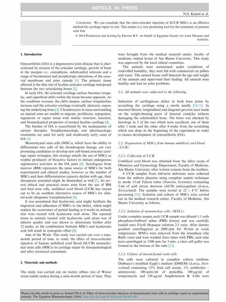
In the control group (left knees) (Table 1): the area of the
defect was partially filled with a fibrovascular granulationtissue, which united the wound edges (Fig. 1a). The newlyformed tissue was composed of undifferentiated connective
tissue cells covered with a thin layer of fibrous tissue. It wasgrossly distinguishable from the surrounding normal tissue.Cartilaginous sequestra surrounded by organized fibrous tissuewere seen (Fig. 1b). All the control knees did not show any
signs of cartilage repair.In the injected group (right knees) (Table 2): no signs of OA
such as osteophytes, cysts formation, cartilage erosion or syno-
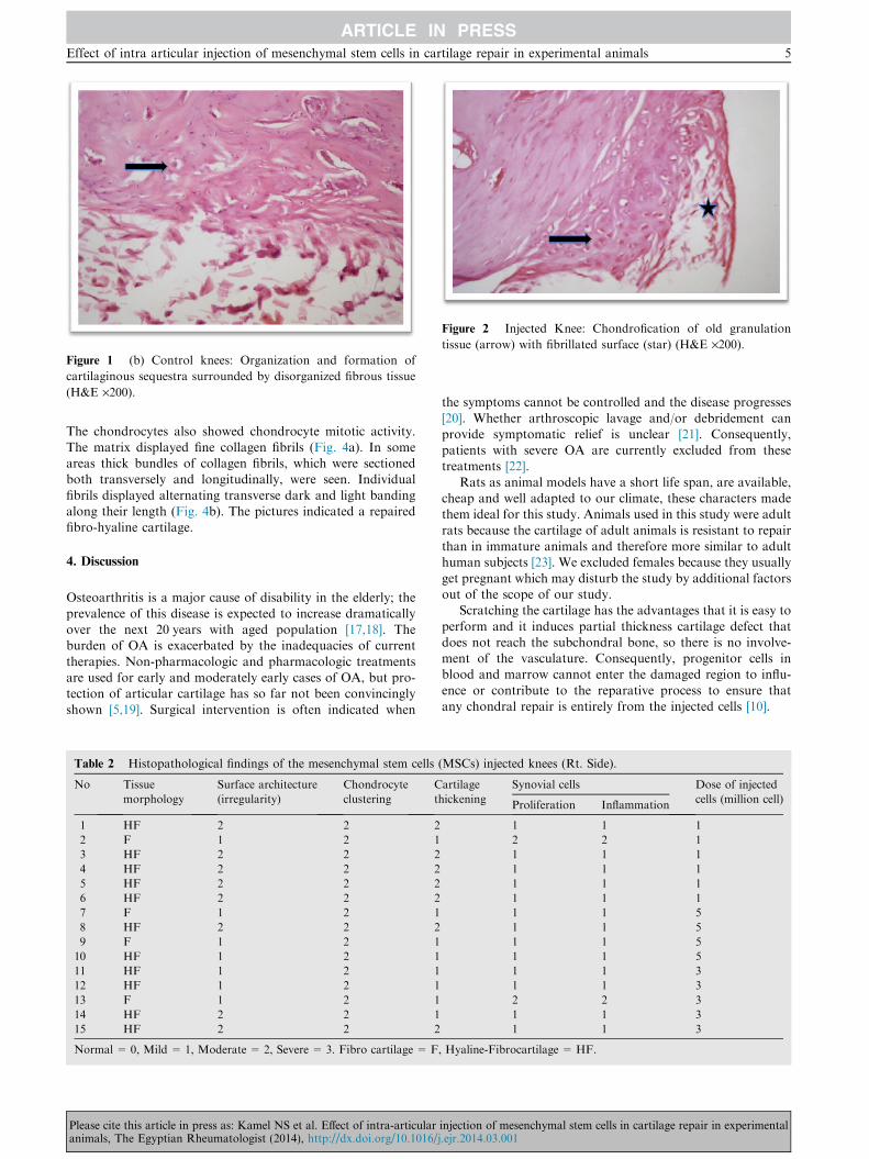
vial proliferation were observed in any of the knees; fibrocar-tilaginous mass was detected at the region of the defect byMasson’s trichrome stain in 4 treated knees (27%). The tissue
appeared more fibrous than hyaline. The surface became mod-erately fibrillated, the chondrocytes were smaller and lying inparallel longitudinal rows, the subjacent matrix was densely
fibrous and the synovial cells showed mild proliferation andmild infiltration by inflammatory cells (Fig. 2). In theremaining 11 treated knees (73%): the tissue was typical of
Table 1 Histopathological findings of the control knees (Lt. side).
No Joint surface Chondrocytes
Irregularity Disorganization
1 2 1
2 1 1
3 2 2
4 1 1
5 2 1
6 1 1
7 2 2
8 1 2
9 1 1
10 1 1
1 1 1
12 2 2
13 1 1
14 2 2
15 2 2
Normal = 0, Mild = 1, Moderate = 2, Severe = 3.
Please cite this article in press as: Kamel NS et al. Effect of intra-articularanimals, The Egyptian Rheumatologist (2014), http://dx.doi.org/10.1016/j
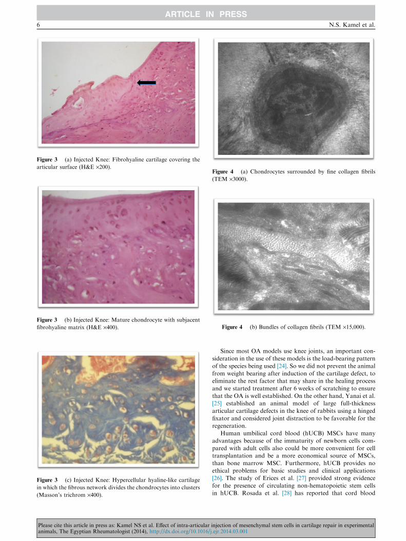
fibro-hyaline cartilage and appeared more cellular thanfibrous. These findings were confirmed by Masson’s trichromestain that demonstrated pink nuclei subjacent to the pale blue
staining matrix and fine fibrous tissue network showing intensemetachromasia that divided the chondrocytes into clusters(Figs. 3a–c).
3.2. Electron microscopic findings
As regards the EM study of the MSC-injected knees; it showed
a repaired fibro-hyaline cartilage demonstrated by manychondrocytes and chondroblasts. The chondrocytes hadcentrally positioned nuclei surrounded by a rich roughendoplasmic reticulum (rER) and numerous mitochondria.
Synovial cells
Hypertrophy Proliferation Inflammation
1 2 2
1 2 2
1 1 1
1 2 2
1 2 2
1 1 1
1 2 2
2 2 2
1 1 1
1 1 1
1 1 1
1 2 2
1 1 1
2 2 2
2 2 2
injection of mesenchymal stem cells in cartilage repair in experimental.ejr.2014.03.001
Figure 1 (b) Control knees: Organization and formation of
cartilaginous sequestra surrounded by disorganized fibrous tissue
(H&E ·200).
Figure 2 Injected Knee: Chondrofication of old granulation
tissue (arrow) with fibrillated surface (star) (H&E ·200).
Effect of intra articular injection of mesenchymal stem cells in cartilage repair in experimental animals 5
The chondrocytes also showed chondrocyte mitotic activity.The matrix displayed fine collagen fibrils (Fig. 4a). In someareas thick bundles of collagen fibrils, which were sectionedboth transversely and longitudinally, were seen. Individual
fibrils displayed alternating transverse dark and light bandingalong their length (Fig. 4b). The pictures indicated a repairedfibro-hyaline cartilage.
4. Discussion
Osteoarthritis is a major cause of disability in the elderly; the
prevalence of this disease is expected to increase dramaticallyover the next 20 years with aged population [17,18]. Theburden of OA is exacerbated by the inadequacies of current
therapies. Non-pharmacologic and pharmacologic treatmentsare used for early and moderately early cases of OA, but pro-tection of articular cartilage has so far not been convincingly
shown [5,19]. Surgical intervention is often indicated when
Table 2 Histopathological findings of the mesenchymal stem cells (
No Tissue
morphology
Surface architecture
(irregularity)
Chondrocyte
clustering
C
th
1 HF 2 2 2
2 F 1 2 1
3 HF 2 2 2
4 HF 2 2 2
5 HF 2 2 2
6 HF 2 2 2
7 F 1 2 1
8 HF 2 2 2
9 F 1 2 1
10 HF 1 2 1
11 HF 1 2 1
12 HF 1 2 1
13 F 1 2 1
14 HF 2 2 1
15 HF 2 2 2
Normal = 0, Mild = 1, Moderate = 2, Severe = 3. Fibro cartilage = F,
Please cite this article in press as: Kamel NS et al. Effect of intra-articularanimals, The Egyptian Rheumatologist (2014), http://dx.doi.org/10.1016/j
the symptoms cannot be controlled and the disease progresses
[20]. Whether arthroscopic lavage and/or debridement canprovide symptomatic relief is unclear [21]. Consequently,patients with severe OA are currently excluded from thesetreatments [22].
Rats as animal models have a short life span, are available,cheap and well adapted to our climate, these characters madethem ideal for this study. Animals used in this study were adult
rats because the cartilage of adult animals is resistant to repairthan in immature animals and therefore more similar to adulthuman subjects [23]. We excluded females because they usually
get pregnant which may disturb the study by additional factorsout of the scope of our study.
Scratching the cartilage has the advantages that it is easy to
perform and it induces partial thickness cartilage defect thatdoes not reach the subchondral bone, so there is no involve-ment of the vasculature. Consequently, progenitor cells inblood and marrow cannot enter the damaged region to influ-
ence or contribute to the reparative process to ensure thatany chondral repair is entirely from the injected cells [10].
MSCs) injected knees (Rt. Side).
artilage
ickening
Synovial cells Dose of injected
cells (million cell)Proliferation Inflammation
1 1 1
2 2 1
1 1 1
1 1 1
1 1 1
1 1 1
1 1 5
1 1 5
1 1 5
1 1 5
1 1 3
1 1 3
2 2 3
1 1 3
1 1 3
Hyaline-Fibrocartilage = HF.
injection of mesenchymal stem cells in cartilage repair in experimental.ejr.2014.03.001
in which the fibrous network divides the chondrocytes into clusters
(Masson’s trichrom ·400).
Figure 3 (b) Injected Knee: Mature chondrocyte with subjacent
fibrohyaline matrix (H&E ·400).
Figure 4 (a) Chondrocytes surrounded by fine collagen fibrils
(TEM ·3000).
Figure 3 (a) Injected Knee: Fibrohyaline cartilage covering the
articular surface (H&E ·200).
Figure 4 (b) Bundles of collagen fibrils (TEM ·15,000).
6 N.S. Kamel et al.
Please cite this article in press as: Kamel NS et al. Effect of intra-articularanimals, The Egyptian Rheumatologist (2014), http://dx.doi.org/10.1016/j
Since most OA models use knee joints, an important con-sideration in the use of these models is the load-bearing pattern
of the species being used [24]. So we did not prevent the animalfrom weight bearing after induction of the cartilage defect, toeliminate the rest factor that may share in the healing process
and we started treatment after 6 weeks of scratching to ensurethat the OA is well established. On the other hand, Yanai et al.[25] established an animal model of large full-thickness
articular cartilage defects in the knee of rabbits using a hingedfixator and considered joint distraction to be favorable for theregeneration.
Human umbilical cord blood (hUCB) MSCs have manyadvantages because of the immaturity of newborn cells com-pared with adult cells also could be more convenient for celltransplantation and be a more economical source of MSCs,
than bone marrow MSC. Furthermore, hUCB provides noethical problems for basic studies and clinical applications[26]. The study of Erices et al. [27] provided strong evidence
for the presence of circulating non-hematopoietic stem cellsin hUCB. Rosada et al. [28] has reported that cord blood
injection of mesenchymal stem cells in cartilage repair in experimental.ejr.2014.03.001
Effect of intra articular injection of mesenchymal stem cells in cartilage repair in experimental animals 7
multilineage cells are slower to establish in culture, have alower precursor frequency and a lower level of bone antigenexpression, and lack constitutive expression of neural antigens
when compared with bone marrow, suggesting a more primi-tive population. The hUCB is easy to obtain and to store,and its use for hematopoietic stem cell transplantation does
not require a close human leucocyte antigen match as reportedby Gluckman [29].
Human UCB stem cells are xenograft cells. Rat UCB is dif-
ficult to obtain and the available quantity of rat UCB is verylimited; however, hUCB is easy to obtain and is plentiful. Inour experiment, we expected to evaluate only the possibilityof UCB stem cells differentiated into chondrocytes in vivo.
So, we used hUCB in the rat model. Umbilical cord bloodmay prove to be a new source of cells for cellular therapeuticsfor stromal, bone, and potentially, cartilage repair. Also it is a
very promising source in Egypt because of the high birth rate.For all the above-mentioned reasons, cord blood MSCs wereconsidered in our study as the most promising cell source for
cartilage repair.Due to the specific structure and functions of placenta,
human extra-embryonic MSCs, including MSCs derived from
the UCB represent stem cell types, which combine someproperties of pluripotent embryonic stem cells with other prop-erties of multipotent MSCs. They have immunoprivilegedcharacteristics, posses a broader plasticity, and proliferate
faster than adult MSCs [30].The hUCB MSCs were delivered in this study by intra-
articular injection, there are 2 different approaches to this.
One is to implant cells directly with or without a suitablematrix or scaffold seeded with chondroprogenitor cells and sig-naling substances as in the study of Jackson and Simon [31].
The alternative is to differentiate stem cells in vitro andimplant a mature construct. The ability of stem cells to differ-entiate and adhere to scaffolds such as matrices of hyaluronan
derivatives and gelatin-based resorbable sponge matrices hasbeen investigated and proven by Solchaga et al. [32], and Pon-ticello et al. [33] respectively.
Most studies presume that scaffolds are required for the
regeneration of cartilage. It is ventured that loads and fluidmovements would simply prevent cells from thriving wherethey are needed. However Barry [34] has shown that MSCs
can survive and thrive without a scaffold and injected stemcells have been recovered in viable form in a goat knee withsimulated arthritis.
In the present study hUCB MSCs were delivered by intra-articular injection in a suspension of hyaluronic acid (HA)solution without the use of a solid biomatrix 6 weeks afterscratching the cartilage. Lee et al. [9] postulated that HA might
facilitate the migration and adherence of MSCs to the defect.Our choice of experimental animals to be rats, limited our
ability to experimental assessment. It was limited to subjective
observational parameters such as limping or aggressive behav-ior denoting pain, so we chose the histopathological assess-ment and ultrastructural study that were based upon
histological objectives. Samples were stained with hematoxylin& eosin and Masson’s trichrome.
In the present study, the repair of lesions was a variable mix-
ture of fibrous tissue, fibrocartilage and hyaline-like cartilage.At 6 weeks at the control group; the area of the defect was filledwith a fibrovascular granulation tissue, the newly formed tissuewas an undifferentiated connective tissue covered with a thin
Please cite this article in press as: Kamel NS et al. Effect of intra-articularanimals, The Egyptian Rheumatologist (2014), http://dx.doi.org/10.1016/j
layer of fibrous tissue. Cartilaginous sequestra surrounded byorganized fibrous tissue were seen. All control knees did notshow any signs of repair (differentiated cartilage) denoted that
there is no systemic parenteral trafficking through systemic cir-culation from the injected right knee to the other left knee.These findings coincide with those reported by Yanai et al.
[25], Bos et al. [35], and Lee et al. [9]. However in this studythe tissue appeared more fibrous than cartilaginous.
At the injected group; no signs of OA such as osteophytes,
cysts formation, cartilage erosion or synovial proliferation,were observed in any of the knees these findings match withthat of Lee et al. [9].
After 6 weeks of injection fibrocartilaginous mass was
detected in 27% of cases. The tissue appeared more fibrousthan cartilaginous and the surface layers and the cells weremore typically fibrocartilaginous than hyaline. The surface
became moderately fibrillated, the chondrocytes are smallerand lying in parallel longitudinal rows, the subjacent matrixwas densely fibrous and the synovial cells showed mild prolif-
eration and mild infiltration by inflammatory cells. In 73% ofcases; the tissue appeared more cellular than fibrous. The thinfibrillated surface and the cells were typical of fibro-hyaline
cartilage; these findings match with those of Yanai et al. [25],Yan and Yu [36], and Lee et al. [9].
Regarding the EM study; it showed a repaired fibrohyalinecartilage demonstrated by many chondrocytes and chondro-
blasts. The pictures indicated a repaired fibrohyaline cartilage.A recent study conducted by Nam and his colleagues [37],
showed a superior role of injecting autologous bone marrow
derived mesenchymal stem cells (BM-MSCs) to hUCB MSCs.This study demonstrated hyaline-like cartilage regeneration inthe knees of goats receiving BM-MSCs after two full-thickness
chondral 5 mm diameter defects were created. They suggestedthat supplementing intra-articular injections of BM-MSCs fol-lowing bone marrow stimulation (BMS) knee surgery provides
superior cartilage repair outcomes.We can conclude from these results that the intra-articular
injection of hUCB MSCs is an effective method for cartilagerepair in rats. This makes it a very promising tool for the treat-
ment of patients with OA.
Conflict of interest
None.
Acknowledgement
The authors would like to express their gratitude to Awatef
Mohamed (Veterinary Fellow of Biochemistry) and LarissaInge (Electron Microscopy) without whom this work wouldnot have been accomplished.
References
[1] Herndon JH, Davidson SM, Apazidis A. Recent socioeconomic
trends in orthopedic practice. J Bone Joint Surg
2001;83A:1097–105.
[2] Felson DT. Osteoarthritis of the knee. N Engl J Med
2006;354:841–8.
[3] Bollet AJ. The biology of degenerative joint disease. Arthritis
Rheum 1971;14(1):144.
injection of mesenchymal stem cells in cartilage repair in experimental.ejr.2014.03.001
Griffin MR, et al. Guidelines for the medical management of
osteoarthritis. Part II. Osteoarthritis of the knee. American
College of Rheumatology. Arthritis Rheum 1995;38:1541–6.
[20] Gunther KP. Surgical approaches for osteoarthritis. Best Pract
Res Clin Rheumatol 2001;15:627–43.
Please cite this article in press as: Kamel NS et al. Effect of intra-articularanimals, The Egyptian Rheumatologist (2014), http://dx.doi.org/10.1016/j