Ent. exp. & appl, 2 (I959): 82--99. North-Holland Publishing Co., Amsterdam EFFECT OF PARASITIZAT1ON BY APHIDIUS PLATENSIS BRETHES ON THE DEVELOPMENTAL PHYSIOLOGY OF ITS HOST, APHIS CRACCIVORA KOCH BY BRUCE JOHNSON Waite Agricultural Research Institute, University of Adelaide, Australia Parasitization of nymphs of Aphis craccivora by Aphidius platensis results in interference with their normal developmental physiolog'y. Different stages of growth of the parasite have opposite influences on the host: unhatched parasite eggs exert a "juvenilizing" effect, producing metathetely; whereas parasite larvae sometimes cause the premature appearance of adult characters (prothetely). When winged nymphs of the host are parasitized, the dif- ferentiation of alatiform structures is inhibited. This results in the production of aphids intermediate in form between normal alatae and apterae; the extent of development of alatiform structures is determined by their degree of differentiation at the time of para- sitization and whether one or more parasite eggs are deposited. It is suggested that parasitic metathetely in aphids is brought about by interference with the titre of juvenile hormone, and that alate-apterous polymorphism in aphids generally is controlled by the same hormone system as is concerned in moulting and metamorphosis. When the immature stages of some insects and crustacea are parasitized, the ensuin~ adults have been found to develop certain structural abnormalities. "Parasitic castration" of some crabs is caused by isopod and rhizopod infections: male crabs tend structurally towards the female type and female crabs retain some juvenile characters (CaULLERY, 1952). In insects, parasitization of various Hymenoptera and Homoptera by Streps~ptera causes the production of "intersexes", the particular sex that is parasitized developing some of the characteristics of the opposite sex (CLAUSEN, 1940). Mermis infections of larvae of worker ants result in the development of ocelli and other characteristic queen structures (WHEELER, 1937). l~4ermis can also cause restriction in wing size in the alate sexual forms of ants (MRAZEK, 1908). Structural deformities of alate ants, in- cluding complete suppression of the wings, are brought about by parasitization by the chalcid wasp Orasema (WHEELER, 1910). In simuliid larvae STRICKLAND (1911), showed that parasitization by Merm;s resulted in suppression of the development of the imaginal discs. This phenomenon of the suppression of adult structures only, he termed metathetely. Instances of metathetely have been found by HALCROW (1954) in Anopheles larvae parasitized with Glaucoma pyriformis (Ehrenberg) and by FINLAYSON cox; WALTERS (1957) in saturniid moths infected with Nosema. It has been suggested for cases of parasite-induced metathetely that the hormone system of the host has been in some way upset by the parasites (WIGGLESWORTH, I954; FINLAYSON & WALTERS, I957). In the present paper an account is given of the influence of parasitization by the braconid wasp Aphidius platensis Br&hes on the morphology of its host Aphis
E F F E C T O F P A R A S I T I Z A T 1 O N B Y APHIDIUS P L A T E N S I S
BRETHES O N THE DEVELOPMENTAL PHYSIOLOGY O F I T S H O S T , APHIS CRACCIVORA K O C H
B Y
BRUCE J O H N S O N Waite Agricultural Research Institute, University of Adelaide, Australia
Parasitization of nymphs of Aphis craccivora by Aphidius platensis results in interference with their normal developmental physiolog'y. Different stages of growth of the parasite have opposite influences on the host: unhatched parasite eggs exert a "juvenilizing" effect, producing metathetely; whereas parasite larvae sometimes cause the premature appearance of adult characters (prothetely). When winged nymphs of the host are parasitized, the dif- ferentiation of alatiform structures is inhibited. This results in the production of aphids intermediate in form between normal alatae and apterae; the extent of development of alatiform structures is determined by their degree of differentiation at the time of para- sitization and whether one or more parasite eggs are deposited. It is suggested that parasitic metathetely in aphids is brought about by interference with the titre of juvenile hormone, and that alate-apterous polymorphism in aphids generally is controlled by the same hormone system as is concerned in moulting and metamorphosis.
When the immature stages of some insects and crustacea are parasitized, the ensuin~ adults have been found to develop certain structural abnormalities. "Parasitic castration" of some crabs is caused by isopod and rhizopod infections: male crabs tend structurally towards the female type and female crabs retain some juvenile characters (CaULLERY, 1952). In insects, parasitization of various Hymenoptera and Homoptera by Streps~ptera causes the production of "intersexes", the particular sex that is parasitized developing some of the characteristics of the opposite sex (CLAUSEN, 1940). Mermis infections of larvae of worker ants result in the development of ocelli and other characteristic queen structures (WHEELER, 1937). l~4ermis can also cause restriction in wing size in the alate sexual forms of ants (MRAZEK, 1908). Structural deformities of alate ants, in- cluding complete suppression of the wings, are brought about by parasitization by the chalcid wasp Orasema (WHEELER, 1910). In simuliid larvae STRICKLAND (1911), showed that parasitization by Merm;s resulted in suppression of the development of the imaginal discs. This phenomenon of the suppression of adult structures only, he termed metathetely. Instances of metathetely have been found by HALCROW (1954) in Anopheles larvae parasitized with Glaucoma pyriformis (Ehrenberg) and by FINLAYSON cox; WALTERS (1957) in saturniid moths infected with Nosema. It has been suggested for cases of parasite-induced metathetely that the hormone system of the host has been in some way upset by the parasites (WIGGLESWORTH, I954; FINLAYSON & WALTERS, I957) .
In the present paper an account is given of the influence of parasitization by the braconid wasp Aphidius platensis Br&hes on the morphology of its host Aphis
PARASITIZATION OF APHIS CRACC1VORA 83
craccivora Koch. A brief account of the work has already been published (JOHN- SON, 1958). The bionomics of wasp parasites of aphids have been studied by a number of workers but there does not appear to be any mention in the literature of an influence of the parasite on the morphology of the host, apart from a brief remark by WEBSTER & PHILLIPS (1912) that the wings of aphids parasitized after the second moult are often imperfect. VEVaI (1942) noted that the "sessiles" or "mummies" of dead parasitized aphids are nearly always apterous, but possibly because he had already four~d that wasps oviposie more readily in apterous than in alate adult aphids he did not look for the cause.
M A T E R I A L S AND M E T H O D S
The aphids used in this study were alienicolae of Aphis craccivora Koch which were reared on broadbean, Vicia faba L. Batches of aphids of uniform known age were obtained by keeping a number of adults on bean cuttings overnight and re- moving them the next morning and rearing the progeny which had been deposited on the cutting. Alatiform nymphs were obtained by putting apterous parents on cuttings, bearing only or~e mature leaf from old bean plants. The cuttings were kept in jars of water. When reared under these conditions the progeny of apterae nearly all developed into alates. The results of five such rearings are given in Table I.
TABLE I
Form of progeny of apterous parents when reared an mature bean leaf cuttings in water
Experiment Total No. % No. of larvae alatae
1 391 98 2 296 97 3 t26 83 4 174 88 5 391 92
Apteriform nymphs were obtained as the progeny of young alate parents born within 24 hours after the parents had settled on a host plant: these progeny invariably developed into apterae.
The parasite used was Aphidius platensis Br&thes, adults of which were kindly identified by Dr. C. F. W. Muesebeck of the National Museum, Washington, D.C., U.S.A.
Aphids to be parasitized were put on a leaf and kept in a small glass cage containing a number of wasps. When parasitization with a single wasp egg was required, the aphids were placed individually on a leaf and removed as soon as a wasp had thrust at one of them with its ovipositor: dissections of a number of aphids treated in this way failed to reveal the presence of more than one egg. Aphids left in the cage were repeatedly oviposited in by the wasps: the maximum number of eggs found in an aphid which had been left in the cage overnight was fifty-seven. This is in accord with the observations of a number of authors who have found that braconid wasps will oviposit in aphids in which they or other wasps have already deposited eggs.
84 BRUCE JOHNSON
The parasitized aphids were reared on bean cuttings in water. This provided an adequate nutrition source for the aphids to attain the same speed of development and size as aphids reared on seedlings.
The morphological structures of the aphids were examined in the intact aphids immersed in 70 % alcohol, and where a more detailed study of their cuticular structures was required the aphids were later cleared in chloral phenol and mounted in gum arabic on microscope slides.
LIFE CYCLE OF APHIDIUS PLATENSIS BRETHES
The life cycles of several species of Aphidius have been studied by a number of authors (SPENCER, 1926; ULLYETT, 1938; VEVAI, 1942, etc.). The biology of A. p&tensis does not appear to differ in any significant way from the accounts of other species which have been published.
An egg tapering at both ends and measuring about 20 • 50/, is deposited in the body cavity of the aphid by the female wasp. Development of the egg of A. platensis resembles closely the description of the development of eggs of Aphi- diinae given by SPENCER (1926). The parasite eggs begin to swell soon after being laid, and ten hours after oviposition they contain many nuclei. At twenty-four hours at 20~ the eggs have increased to about 80/~ in diameter and the embryo resembles the "morula" stage of vertebrate embryos. At forty-eight hours the egg measures about 160-200/~ in diameter and contains an undifferentiated elongated worm-like embryo curled about on itself but which when straightened out measures about 500p. in length. During the third day the size of the head increases, external segmentation of the body occurs and tissue differentiation is initiated: for the first time the nervous system, alimentary canal and salivary glands can be recog- nized as distinct structures. At the time of hatching the egg measures about 250> in diameter. After hatching, the cells of the serosa become greatly enlarged and continue their existence as free-living cells in the aphid until they are finally consumed by the parasite. The parasite larva has well developed mandibles but apparently obtains most of its food from imbibing the host's haemolymph, as it is rarely found with its mandibles imbedded in host tissues and the host tissues rarely show evidence of physical injury. The first and second larval instars, which can be identified by the length of the caudal process, last for only about one day each at 20 ~ C. Then follow about three further instars (VEvAI, 1941) in which the appearance of the parasite does not change appreciably from one instar to the next except that it becomes more curved about on itself; these last for a total of 3-4 days. Soon after the last larval stage is reached, the larva changes its feeding habits and voraciously cleans out the entire body contents of the host; this is facilitated by the secretion of cytolytic enzymes which cause breakdown of the host tissues. The parasite then spins a cocoon inside the aphid skin and pupates.
When more than one egg is deposited in an aphid, all hatch in about the same time as a single egg, but all but one of the resultin/~ parasite larvae die within a few days. The larvae that die appear to be killed by chemical means rather than by physical injury caused by the surviving parasites since their cuticle frequently remains intact well after the body contents have completely lost their structure.
PARASITIZATION OF APHIS CRACCIVORA 85
N O R M A L D E V E L O P M E N T OF THE H O S T
There are four nymphal instars in both alatiform and apteriform nymphs of Aphis craccivora Koch. When reared on healthy bean plants at 20 ~ C the approxi- mate duration of the successive instars in apteriform nymphs is 1.5, 1.5, 2.0, and 2.0 days; in alatiform nymphs, the last stadium is typically one day longer.
Externally, nymphs which will develop into alatae do not differ from those which will develop into apterae until the third nymphal cuticle is laid down. In third instar alatiform nymphs, small wingpads are present and the thorax is distinctly alatiform in character: the mesothorax is lengthened and broadened and tlm metathorax shortened, giving the thorax a more box-like appearance than in apteriform nymphs, where the segments remain approximately the same length and taper in width towards the head. In the fourth instar the difference between the two forms is more pronounced: apteriform nymphs are merely larger but not different in body shape from the previous instar, whereas in alatiform nympEs, the wingpads are much larger and the thorax is more conspicuously different in its shape and dimensions from that of apteriform nymphs.
Adult aphids differ from nymphs principally in that they are reproductively mature. There are a number of structural features which distinguish nymphs from adults: the length and proportions of the appendages, the shape of tee cauda and the presence in the adult only of a genital aperture and genital plate~ Areas of pigmentation also differ on the two forms. The whole of the dorsal surface of the head capsule is pigmented in adults, whereas in nymphs, the pigment is confined to two broad bands running longitudinally along it; in adults the dorsal surface of the abdomen is pigmented, whereas in nymphs, it is unpigmented.
Adult alate aphids differ structurally from apterous adults in a number of features, among which are the presence in alatae and absence in apterae of wings, specialized pterothoracic sclerites, ocelli, and placoid sensoria on the third segments of the antennae. The antennae also differ in the two forms in the relative lengths of the 2nd and 3rd segments, and in their degree of pigmentation: in alatae, the antennae are pigmented along their whole length, whereas in apterae, they are pigmented at the ends only, the intermediate region being unpigmented. The pigmentation of the abdomen of the two forms also differs: in apterae, there is a dorsal pigmented "shield" covering the entire abdomen and thorax, whereas in alatae there are discrete bands of pigment across of the abdominal sclerites.
R E L A T I V E RATES OF D E V E L O P M E N T OF H O S T AND PARASITE AT 2 0 ~ C
At 20 ~ C the parasite eg& hatches on the third day after oviposition. Thus eggs deposited in aphid wmphs do not hatch until the nymphs have undergone consider- able growth and have moulted once or twice since oviposition. In Fig. 1 a schematic representation is given of the relative rates of development of host and parasite at 20 ~ C when the aphid is kept on a host plant that favours optimum devdopment. On less favourable host plants, the speed of development of the aphids is retarded but parasite development is not affected.
No detailed study has been made of the effects of different temperatures on the
86 BRUCE JOHNSON
relative rates of development of host and parasite, but incidental observations suggest that they are not very extensive at least at temperatures within the range of 15-25 ~ C.
WEBSTER &: PHILLIPS (1908) and ULLYETT (1938) found that when aphid
,j HOST 1"sT INS R . ]]" NSTA• Tff,~ I~ P, lgT. 1~15 R (AP'f/
O~ H~
(AL~
Fig. 1. Relative rates of development of host and parasite at 20 ~ C. The heavy horizontal lines indicate the approximate duration of the moult from the beginning of endocrine
activity. O: oviposition; H: hatching of the parasite egg.
nymphs were parasitized before the first or second moult, both aphids and parasites failed to survive. In the present study, adult wasps emerged from all aphids that were parasitized, regardless of the instar of the host at the time of parasitization, provided the aphids received adequate nutrition. All nymphs that were parasitized in the first instar failed to moult into adults and were mostly killed by the parasite in the fourth instar. The number of moults aphids under- went after oviposition and before they were killed by the parasite larvae was determined by the nutritional status of the host plants they were reared on. Inade- quate nutrition for aphid development, within limits, did not interfere with parasite growth. When parasitized nymphs were kept on inferior host plants, the parasites frequently completed their development and killed the aphids in the third and fourth instars even when oviposition occurred as late as the second and third instars respectively; on suitable host plants aphids parasitized in these instars developed to maturity, before they were killed.
PARASITIZATION OF APTERIFORM NYMPHS
Apteriform nymphs parasitized at all stages continued their development nor- mally without undergoing any major structural changes. Those which were para- sitized in the first instar were killed and "mummified" by the parasite in the fourth instar and a number of them showed some premature development of adult characters (prothetely), having the cauda intermediate in shape between nymphal and adult types, and patches of pigment on the abdomen at the lateral muscle insertions.
Nymphs parasitized in the second and subsequent instars developed to maturity, and the only apparent external effects of parasitization in the resulting adults was suppression of pigment deposition on the dorsal surface of the abdomen and the retention of slightly juvenile pigmentation pattern of the head capsule.
PARASITIZATIOIXl OF APHIS CRACCIVORA 87
P A R A S I T I Z A T I O N O F F I R S T I N S T A R A L A T I F O R M N Y M P H S
The influence of parasitization on first instar alatiform nymphs was demonstrated in the following experiment. Five bean leaves bearing batches of first instar alatiform nymphs which had been born to apterous parents in the preceding over-
TABLE II
Effect of parasitization on the form of aphids parasitized in the 1st lnstar
night period were put in the parasite cage for six hours. The leaves were then removed from the parasite cage and the aphids were reared in another cage at an average temperature of 15 o C with a control series of unparasitized nymphs. All the parasitized aphids were killed by the parasites when they were in the fourth nymphal instar. At the time they died they had no alatiform structures, although the control series consisted almost entirely of normal alatiform nymphs
TABLE III
Numbers of aphids pa, asitized in the Ist instar showing different degrees of prothetely in the 4th instar
Total number of aphids Normal apteriform nymphs Nymphs with pigmented muscle insertions Nymphs with intermediate shaped cauda Nymphs with pigmented areas on tergites of abdomen
Number of aphids 363 147 216
58 25
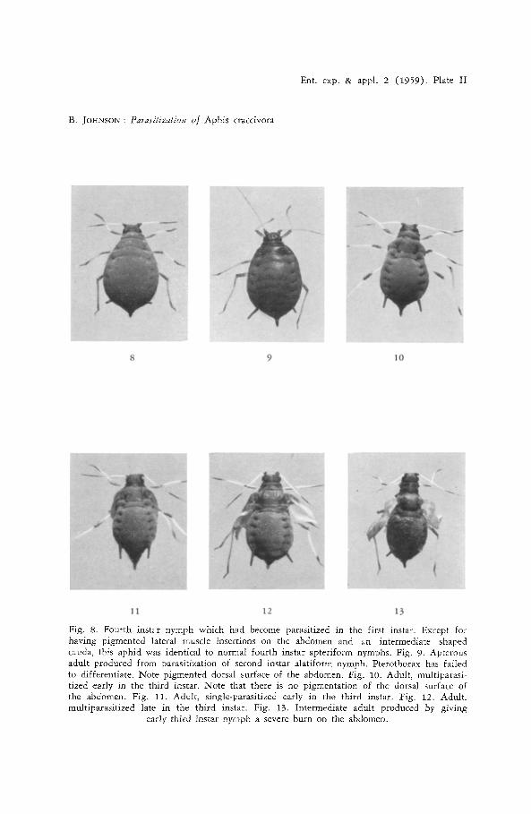
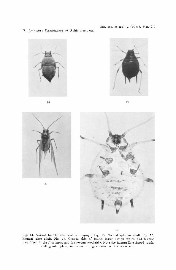
(Table II). The parasitized nymphs were indistinguishable from normal unpara- sitized apteriform nymphs except that some of them showed some degree of prothetely (Table III). Thus in many of them, the lateral abdominal muscle attachments were pigmented, and in a few there was a rudimentary, cleft genital plate, a cauda intermediate in size and shape between nymph and adult (fig. 2), and some pigmentation of the dorsal surface of the abdomen (fig. 17).
VF i ii iii iv v vi
Fig. 2. Cauda and anal plate of i) normal fourth instar nymph; i i - -v) parasitized fourth instar nymphs which had become parasitized in the first instar; vi) normal adult.
88 BRUCE JOHNSON
Several parasitized nymphs were dissected just prior to casting their third instar cuticle and they were found to contain second instar parasite larvae that had apparently only recently moulted from the first instar as they were still en- veloped by the first instar cuticle.
P A R A S I T I Z A T I O N OF SECOND I N S T A R A L A T I F O R M N Y M P H S
A number of alatiform nymphs which had moulted into the second instar overnight were placed the following morning in the parasite cage and left there for six hours. They were then kept in another cage with a control series of unparasitized aphids and reared to maturity. The aphids were collected and examined as they moulted to adults. A few of them were killed by the parasites while still in the fourth instar. All the parasitized aphids that survived to become adults developed characters intermediate between alate and apterous, but in general appearance they were closer to the apterous form. All of them had rudimentary ocelli, and the antennal sensoria were either lacking, or, when present were frequently very reduced in size and number. The degree of wing development varied: in some aphids there was no trace of pterothorax differ- entiation, in others the shoulders of the mesothorax were squared and small rudimentary wing evaginations were present. The head capsule was of the fully pigmented adult type and most of the aphids had some pigmentation of the dorsal surface of the abdomen (fig. 18; Table IV) .
TABLE IV
Characters of intermediates formed from parasitization of 2nd instar alatiform nymphs
Characters Number of aphids Total number of aph,ds 327 Number killed m 4th instar 19 Intermediates with ocelli but no antennal sensoria 48 Intermediates with ocdli and antennal sensoria 247 Intermediates with patches of pigment on abdominal tergites 220
The development of the wing rudiments was followed throug~ the various instars in several aphids which had become parasitized in the early second instar. At the time they were parasitized, there were no external signs of the wing buds. In the third instar, wing evaginations were formed but they were smalier than in normal third instar nymphs. In the fourth instar of some of the aphids, the evaginations appeared to be no more than exaggerated folds in the cuticle, and in the adult stage they were even less conspicuous. It was apparent that, from the second moult, the wingpads failed to differentiate any further and as the cuticle became stretched in each subsequent instar they became flattened out and were reduced or lost by the time the aphids reached the adult stage.
P A R A S I T I Z A T I O N OF T H I R D I N S T A R ALATIFORM N Y M P H S
Aphids parasitized in the third instar showed a considerable variation in the extent to which alatiform structures were suppressed when they became adult. They all had ocelli, antennal sensoria and wing rudiments, but the size of the
PARASITIZATION OF APHIS CRACCIVORA 89
wing rudiments and the degree of differentiation of the pterothorax varied according to the time during the instar that the aphids were parasitized and the number of parasite eggs that were deposited in them. All aphids that were parasitized in the third instar failed to develop pigment on the dorsal surface of the abdomen when they became adult, and most of them retained some of the nymphal pigmentation pattern of the head capsule.
Time of parasitizatio,~ A number of newly moulted third instar alatiform nymphs were collected
and were divided into two batches; one batch was immediately placed in the parasite cage for three hours to be multi-parasitized and the other was put in the parasite cage for three hours on the following day. The aphids were kept at 20 ~ C and, after moulting to the fourth instar, they were examined for effects of parasitization on development of the wingpads. The aphids which were parasitized at the beginning of the third instar all had smaller and more drooped win&pads than normal in the fourth instar; whereas the wingpads of the aphids parasitized later in the third instar were normal in the fourth instar. In both cases the wingpads failed to differentiate much further at the final moult (figs 10, 12).
Effect of single and multi-parasitization One batch of newly moulted third instar nymphs was kept in the parasite
cage for six hours to be multi-parasitized; in another batch the aphids were introduced individually to the parasites to have only one egg deposited in them. Both lots of aphids were then kept at 20 ~ C and were examined when they moulted to the fourth instar. Only in the multi-parasitized aphids was there any effect on the wingpads in the fourth instar. When they became adults, all the multi-parasitized aphids (n = 14) had small rudimentary wingpads (fig. 10); of the single parasitized aphids, seven developed imperfect wings, about half the length of normal wings (fig. 12), and four had win&pads of the same size as those of fourth instar nymphs (fig. 11).
P A R A S I T I Z A T I O N OF F O U R T H I N S T A R A L A T I F O R M N Y M P H S
The effects on the structure of the adult cuticle caused by parasitization of fourth instar nymphs that were kept on healthy host plants were far less marked than the effects of earlier parasitization. No matter how early in the fourth instar they were parasitized or how many eggs were deposited in them, the aphids went on to develop distinctly alate structures, although these were sometimes imperfect.
The earlier in the instar the aphids were parasitized, the more extensive were the effects of parasitization. At 15 ~ C, the fourth instar lasts for six days. A number of newly moulted fourth instar nymphs were collected and kept at 15 ~ C and each day several of them were placed in the parasite cage for three hours. Aphids which were parasitized after the third day all became normal Mate adults. In the aphids parasitized before the third day, the wings
90 BRUCE JOHNSON
of several failed to expand fully; the antennae had typical apterous-type pig- mentation; the tergites of all segments of the thorax failed to become fully sclerotized and pigmented; and in the mesothorax, the postnotum was not clearly differentiated from the notum.
As in third instar nymphs, multi-parasitization of fourth instar nymphs pro- duced greater effects than single parasitization. Sixteen newly moulted fourth instar nymphs were oviposited in once, and sixteen were multi-parasitized. In the former group, there was only a slight thinning of the pigmentation along the median line of the postnotum and the prothorax; in the multi-parasitized aphids there were larger unpigmented and unsderotized areas in these regions, and the wings ware generally smaller than in normal alatae; in nine of them the wings failed to expand evenly and became deformed.
EFFECT OF RETARDING DEVELOPMENT OF THE HOST BY PARTIAL STARVATION
From the experiments described above it appeared that the extent of the effect of parasitization was related to the a m o u n t of parasite material inside the aphid at the time of formation of the new cuticle, and that this was a mani- festation of both numbers of eggs present and the leng~th of time the eggs had been in the host and consequently the amount of development they had undergone. If this were so, then by delaying moulting of the host by partial starvation and thus enabling the parasite eggs to attain a greater size before the host moulted, it should be possible to increase the effects of parasitization. Accordingly, a number of newly moulted fourth instar nymphs were put on a leaf in the parasite cage for six hours. The aphids were then removed from the leaf and for the next five days they were kept in a 20 ~ C incubator, spending some of the time in a petri dish containing a wet filter paper, and some on an old, wilted host plant leaf. The duration of the fourth instar was extended in this way for two to three days. Out of an original 15 aphids, 5 died, 4 survived and were killed by the parasites while still in the fourth instar, and 6 moulted into adults. All the aphids which became adults had wing rudiments which were no larger than normal fourth instar wingpads; they had normal antennal sensoria, slightly reduced ocelli, no abdominal pigmentation, and the head capsule had a pattern of pigmentation that was more nymphal than adult in character (fig. 19). The extent of the suppression of general adult and alate structures was thus considerably increased by delaying the moult.
EFFECTS OF I N J U R I E S ON THE D E V E L O P M E N T OF ALATIFORM STRUCTURES
The effects of parasitization became apparent in the aphid nymphs within a few days of the parasite egg being deposited in them and before the egg had actually hatched. It seemed possible that the effect may have been a response to injuries caused to the host by the parasite. Such injuries could have been caused by oviposition by the wasp or by toxic substances secreted by the wasp embryo.
Physical injuries were inflicted on aphid nymphs of different instars to
PARASITIZATION OF APHIS CRACCIVORA 91
ascertain whether injury alone could produce the effect of suppressing the development of alatiform structures. There was a high mortality of first instar nymphs which were pricked in the abdomen with a fine needle and, out of an original thirty-five which were injured, only nine survived: these all developed into apparently normal apterae, although in a control series of 362 aphids there were only 4% apterae. There had apparently been a complete reversal of form as a result of the treatment. Puncturing the abdomen of third instar nymphs had no effect on their subsequent development, but when a number of newly moulted third instar nymphs were given a severe burn injury to the abdomen with a microcautery, six out of eleven survivors developed into inter- mediates. They had normal alate ocelli and antennal sensoria but the pterothorax was imperfectly differentiated, the wings were only half expanded, and the antennae had apterous type pigmentation. Thus although the burn had been applied very early in the third instar, these aphids were very similar in structure to aphids which had been parasitized late in the third instar. However the dorsal surface of the abdomen of the burnt aphids was fully pigmented as in normal apterous aphids except for the site of the burn injury which was unpigmented, and the head pigmentation was adult in character. The burn injury thus affected only the alate characters and did not result, as did para- sitization, in the retention of juvenile head and abdomen pigmentation. Severe injuries were also inflicted on fourth instar nymphs but in no case was the development of the normal alate structures interfered with.
S U P P R E S S I O N O F D I F F E R E N T A L A T I F O R M S T R U C T U R E S
1. Ocelli
In intermediates produced by the parasitization of aphids of different ages, the ocelli showed various degrees of suppression. In normal alate aphids there are three ocelli, the lateral ones situated immediately above the compound eyes and set back a short distance from the bases of the antennae and measuring
i ii iii iv Fig. 3. Head, showing the size and position of the ocelli in i) normal alate adult;
ii--iv) parasitized intermediates.
about 25/, in diameter. In aphids with imperfect ocellar development, the lateral ocelli were moved forward and away from the compound eyes and were reduced in size (fig. 3). The smallest perfect ocellus found was only 5/, in diameter. Further stages in the suppression of the ocelli were found in some
92 B R U C E J O H N S O N
aphids which had been parasitized in the second instar: the ocelli were reduced to a raised ridge around a crater about 5/z in diameter but with no lens, in others slightly darker pigment in the vicinity of the ocelli was all that remained to indicate that the aphid was in fact an intermediate.
The median ocellus is situated in an awkward position for observation, and it could not be studied in as close detail as the lateral ocdli; however it was found in all the aphids with lateral ocelli which were examined.
2. Antennal sensoria The placoid sensoria on the antennae showed various degrees of suppression
in size and number in intermediates. The diameter of the sensoria was reduced in some intermediates that had been parasitized in the second instar, from 12-15/, to 5-6/,, and the number of sensoria on a single antenna was decreased from an average of five in the normal alate adult, through all intermediates to one
0
r J O]
o
o
I I
ii iii
8
0
v vi
V"
J iv vii
Fig. 4. Third antennal segment showing placoid sensoria in i) normal alate adult; i i --vi i) parasitized intermediates.
or none (fig. 4). There was no apparent suppression of the sensoria in aphids parasitized in the third and fourth instars.
The relative lengths of the third and fourth antennal segments of alate and
~0. /.
m
~ ' 3 7
--i ....
n* gK r l = 6"P--
04" 1"001; I'00"l I'0 0.6 I'0 ANTENHAL RAT ! OS
i. i i. iii. iv. Fig. 5. Ratios of length of third to fourth antennal segments, i) normal apterous adults;
ii) intermediates without antennal sensorial iii) intermediates with antennal sensoria, iv) normal alate adults.
P A R A S I T I Z A T I O N O F A P H I S C R A C C 1 V O R A 93
apLerous aphids differ. In alatae, the sensoria-bearing third segment is shorter in relation to the fourth segment. In intermediate aphids, the antennal ratios were found to be intermediate between alatae and apterae: in intermediates without antennal sensoria (but with ocelli), the ratios tended to be closer to apterae, while in intermediates with antennal sensoria they tended to be closer to alatae (Fig. 5).
The fact that complete suppression of the antennal sensoria and the ocelli could be brought about by parasitizafion only in the second instar and not in later instars suggested that these structures become determined at that time, but do not go on to differentiate until the final moult. Their differentiation occurred at the final moult whether or not the aphid at that time was producing other alatiform structures. From this it might be argued that the differentiation of the antennal sensoria and ocelli is not necessarily controlled by the same mechanism as controls the differentiation of other alatiform structures. However it was possible to demonstrate that the antennal sensoria once determined (presumably in the second instar) go on to differentiate at the last moult regardless of whether they are then supplied by the moulting factor by an
alatiform or an apteriform nymph. Antennae from newly moulted fourth instar alatiform and apteriform nymphs were implanted into one another and after the "host" aphid had moulted they were dissected out and examined. Sensoria always developed on antennae from alatiform nymphs but never on antennae from apteriform nymphs regardless of the form of the nymph into which they were implanted. It appears therefore that the differentiation of these sensoria is not dependent upon an alatiform type of humoral environment at the time of their formation as cuticular structures, although it does depend upon such an environment at the time of their determination in the second instar. The same appears to be true for ocelli, but to a lesser extent, as the ocelli, unlike the antennal sensoria, were considerably reduced in size in aphids that were parasitized in the third and fourth instars.
i IL
w,ae V iV
Fig. 6. Differentiation of the pterothorax, i) Normal apterous adult; ii) adult aphid which was parasitized in the second instar; iii) adult aphid multi-parasitized in early third instar; iv) adult aphid multi-parasitized in late third instar; v) normal alate aphid, pt: prothorax;
3. Pterothorax A continuous series of degrees of differentiation of the thorax from the
normal apterous thorax to the pterothorax of the alate was obtained in parasitized aphids, the extent of differentiation depending upon the age of the aphids at the time they were parasitized and consequently the extent of differentiation the pterothorax had already undergone, and whether one or many parasite egg,s were deposited in them. In some aphids which had been parasitized in the second instar there was no trace at all of wingpads or other alatiform modi- fication of the thorax, but later parasitization always resulted in intermediate pterothorax development (fig. 6).
i ii iiJ iV Fig. 7. Different degrees of development of the furca; i) apterous adult; if, iii) parasitized
intermediates; iv) alate adult.
Inside the thorax of intermediates, the flight muscles, the apodemes supporting them, and the furca were reduced corresponding to the degree of differentiation of the pterothorax (fig. 7).
In describing interinediates of Aphis fabae Scop. which he found in laboratory cultures, DAVIDSON (1927) claimed to have observed wingpads inside aphids which had very small ocelli and antennal sensoria. In the present investigation, the aphids were carefully examined before and after clearing and before as well as after mounting on slides and no evidence was ever found of the presence of wingpads inside the thorax. Intermediates with a comparable develop- ment of the ocelli and antennal sensoria to that described by Davidson were all found to have normal external wing-rudiments. As Davidson examined only cleared mounted skins of the aphids, it seems probable that he was mistaken in believing the wing-rudiments to be inside the thorax.
4. Other structures The genital plate is normally present only in adult aphids. Its differentiation
was influenced by parasitization in two different ways. In aphids which were parasitized in the first instar, it was induced to differentiate prematurely in the fourth instar; and in aphids which were parasitized in the fourth instar and then starved, its differentiation was partly suppressed. In both cases the result was a cleft genital plate. As the cleft condition was produced both in nymphs showing prothetely and in adults showing metathetely, it would seem that the humoral conditions normally required for its differentiation probably lie between those required to produce the adult and those required to produce the juvenile form.
The dorsal surface of the head capsule is always completely sclerotized in adult alate aphids. Apterous adults, on the other hand, sometimes have small
Ent. exp. & appl. 2 (1959). Plate II
B. JOHNSON : Paras#izafion of Aphis craccivora
8 9 10
11 12 13
Fig. 8. Fourth instar nymph which had become parasitized in the first instar. Except for having pigmented lateral muscle insertions on the abdomen and an intermediate shaped cauda, this aphid was identical to normal fourth instar apteriform nymphs. Fig. 9. Apterous adult produced from parasitization of second instar alatiform nymph. Pterothorax has failed to differentiate. Note pigmented dorsal surface of the abdomen. Fig. 10. Adult, multiparasi- tized early in the third instar. Note that there is no pigmentation of the dorsal surface of the abdomen. Fig. 11. Adult, single-parasitized early in the third instar. Fig. 12. Adult, multiparasitized late in the third instar. Fig. 13. Intermediate adult produced by giving
early third instar nymph a severe burn on the abdomen.
B. JOHNSON: Parasitization of Aphis craccivora Ent. exp. & appl. 2 ( i959) . Plate III
14 15
16
17
Fig. 14. Normal fourth instar alatiform nymph. Fig. 15. Normal apterous adult. Fig. 16. Normal alate adult. Fig. 17. Cleared skin of fourth instar nymph which bad become parasitized in the first instar and is showing prothetely. Note the intermediate-shaped cauda,
cleft genital plate, and areas of pigmentation on the abdomen.
S. JOHNSON: Pa*'asitizatiov of Aphis cracdvora
Ent. exp. & appl. 2 (1959). Plate IV
o ~ ] - ~ ~ .~_
~ . ~
S a2N.~
m e~
u . . Q o
r~
~ 8
PARASITIZATION OF APHIS CRACCIVORA 95
unpigmented areas behind the eyes and in the middle of the posterior margin of the head capsule. In Brevico*7,e brasdcae L. all stages of intermediates between the adult and the nymphal type of head capsule sclerotization are normally found in apterous adults; in normal Aphis cracch,ora, however, these unpigmented regions are frequently lacking and when they are present they are always very small. Adults of A. craccivora which had become parasitized in the second instar, and which therefore contained parasite larvae when they moulted into adults, all developed complete or almost complete adult type of head sclerotization; but aphids which had become parasitized in the third or fourth instars, and which therefore contained only unhatched parasite eggs at the time the adult cuticle was being formed, developed intermediate types of head capsules that were much more juvenile in character than is normally found in apterae of this species (fig. 19). Aphids which had become parasitized in the second instar also developed some dorsal abdominal pigmentation, whereas those parasitized later failed to develop any pigmentation of the abdomen.
D I S C U S S I O N
A physiological interpretation of polymorphism in insects has been proposed by WIGGL~SWORTH (1954). The epidermal ceils are envisaged as havin~ multi- potentials with regard to the type of cuticle and cuticular structures they produce. Which of these potentials is realized depends upon the availability of a specific raw material, and this may be obtained directly from the food the animal ingests or it may be produced by an endocrine gland and circulate in the form of a hormone. Such a hormone is the juvenile hormone, or neotenin, which has been demonstrated in a number of insects and the presence and titre of which at the time of new cuticle formation influence the type of cuticle that is formed.
Polymorphism with respect to wing development occurs in a large number of different species of insects, but in none has the physiological factor which controls the extent of development of the wings been determined. Inadequate nutrition in males of Gelis corruptor (Gray.) was found by SALT (1952) to result in the production of apterous individuals, but whether the epidermal cells were reacting directly to a deficiency of a specific nutrient, or to modified hormone balance is not known. WIGGLESWORTH (1958) found that the appli- cation of juvenile hormone to the wing-rudiments of Rhodnius resulted in their failure to differentiate further. This and previous published accounts of the effect of juvenile hormone on wing differentiation (see WIGGLESWORTH, 1954) suggest that the various types of incomplete wing development that are found in a number of insects (aptery, microptery, brachyptery) could all be produced by upsets in the hormone balance of the insect in favour of the juvenile hormone. The extent of wing development that is found in the insects could be influenced by both the extent of upset of the hormone balance, and the stage of development of the insect at the time it is upset.
In aphids there are four different kinds of polymorphism. Besides the juvenile-adult and male-female forms that are found in most insects, there also
96 B~UCE JOHNSON
occur apterous-alate and parthenogenetic-gamic forms. In Aphis craccivora sexual forms have not been found and this study is restricted to the agamic forms. K~NNZDY (1956) drew attention to the dual attributes of adults of terrestrial animals generally: their possession of specialized sensori-motor apparatus and their ability to reproduce. Structurally, adult apterous aphids resemble young nymphs in that the sensori-motor apparatus as found in Mate adults has failed to differentiate during their growth. In the sense that they do not possess these typically adult structures they are still juvenile, althoug~h they are adult in the sense that they are capable of reproduction and they possess several adult features such as head and abdomen pigmentation that are not obviously associated with either reproduction or locomotion. They may therefore be regarded as naturally occurring neotenics.
The results of parasitization of aphids described above support the theory that apterous-alate polymorphism in these insects is controlled by the same endocrine system as has been shown in other insects to control metamorphosis. The presence of parasite eggs in aphids resulted not only in the suppression of the development of the sensori-motor apparatus peculiar to alatae, but also in the retention of some nymphal characters, namely head and abdomen pigmentation. Metathetely was thus produced in a number of different structures, some of which were not only associated with alate adults. These effects of parasitization are very similar to effects produced in other insects by experi- mentally upsetting the hormone balance in favour of the juvenile hormone (WIGGLESWORTH, 1954); and it is suggested that such an upset is brought about in parasitized aphids.
If the hormone balance in aphids is upset by the presence of parasite eggs, how is this brought about? It could be caused by either the host modifying its own endocrine activity as a response to the presence of the parasite, or by the parasite directly influencin& the titre of hormone in the blood of the host in much the same way as ichneumon parasites affect their syrphid hosts as demonstrated by SCHNHDER (1950). It was found that injuries to alatiform aphid nymphs could result in their developing into apterae or intermediates instead of alatae: in young nymphs a complete switchover was effected, but in older nymphs it became increasingly difficult to interfere with the differen- tiation of alatiform structures, and other adult characters such as head pig- mentation were not affected at all. Parasitization, however, was effective in suppressing general adult as well as specialized alatiform structures. Thus although it is possible that the effects of parasitization could in part be due to the presence of the parasite causing the host to modify its own endocrine activity, to account for the extensive and regular influence of parasitization, especially in late instar nymphs, on the differentiation of adult structures, it is difficult to escape the conclusion that the parasite egg directly affects the hormone balance in the blood of the host. The finding that the extent of the suppression of Mate structures was affected by the number of parasite eggs that were laid, and the time that had elapsed between oviposition and moulting of the host, fits in well with this theory.
From the experiments in which aphids were parasitized at different times during
PARASITIZATION OF APHIS CRACCIVORA 97
the third and fourth instars it was evident that the parasites were exerting an effect on the host within a day or two of oviposition, although the eggs did not hatch until the third day. After oviposition and before hatching the eggs increase greatly in size and extensive cell division occurs as the embryos develop. NOVAK (1951) has shown that in late embryos of O~zcopeltus the corpus allatum is actively producing juvenile hormone. It is suggested here that the developing embryos of ADhidius produce juvenile hormone and that some of it diffuses into the blood of the host, there increasing the titre and resulting in the observed effects on the cuticle. If this account is true, then production of juvenile hormone by the parasite embryo must occur well before differentiation of the corpus allatum, as until the third day after oviposition it is not possible to distinguish any differentiated structures in the embryo.
There is good evidence that the influence of the parasite that results in sup- pression of slate and other adult characters is exerted by the unhatched parasite embryo only, and not by the parasite larva. In aphids that were parasitized in the second instar, the differentiation of alatiform structures was inhibited, but at the final moult they developed typical adult head capsule and abdominal pigmentation, indicating that at the time of the last moult the parasite was not exerting so strong a juvenile influence as in aphids parasitized later in development, when these adult characters are suppressed by parasitization. It will be seen from fig. I that in aphids parasitized in the second instar the parasite egg hatches well before the initiation of endocrine activity preceding the final moult, whereas in aphids parasitized in later instars hatching occurs closer to or after this period. Further evidence that the juvenile influence not only ceases when the egg I hatches but that after hatching the parasite larva can exert an opposite, namely adult, effect on the host is obtained from aphids parasitized in the first instar. These aphids developed prothetely as wall as metathetely when they reached the fourth instar, the different effects being produced by exposure during their development to different conditions. During the first and second moults, they contained parasite eggs which were producing a juvenile influence and suppressing wing anlagen development, while at the third moult, they contained a parasite larva which was no longer exerting a juvenile influence but was possibly itself moulting and producing an adult in- fluence by tipping1 the hormone balance in the opposite direction. The fact that at the third moult no alatiform structures developed was probably because for the production of alatiform structures the epidermis requires an alate-type humoral environment throughout development and not only at the last moult.
The failure of aphids that were parasitized in the first instar to develop any further than the fourth instar is worth comment. At the time that the aphids would be expected to complete their moult into the adult, the parasites were in the third instar, and while they occupied much of the abdomen of the host, they had not yet begun to attack any of the vital organs of the aphid, and would not have caused any physical damage to the host's endocrine organs. As a number of these nymphs developed prothetelv, it is possible that the endocrine conditions in all aphids parasitized in the first instar may have been modified at the third moult sufficiently in the direction of the adult to have resulted in the thoracic glands
98 BRUCE JOHNSON
responding to this moult as they normally would to the last moult, by breaking down. The thoracic glands of aphids have not yet been positively identified and this theory remains to be tested.
Although metathetely is known to be produced in a number of insects as a result of parasitization by various nematodes and protozoa, there appear to be no other instances (with the possible exception of WHEELER~ 1910, quoted above) of its being caused by parasitization by an endoparasitic insect. The reasons for the apparent absence of this phenomenon in other insects are several, as are the con- ditions which must be fulfi l led for it to occur. If the host insect is large, secretions from the tiny parasite eggs may become too diluted to be effective; the host insect must become parasitized in the juvenile stages and must then pass through at least one moult before the parasite egg hatches (aphids frequently pass through two moults); the effect is more likely to be produced by parasites which will deposit a number of eggs in the same host than by those which will oviposit in a host only once; and it is more likely to be found in hemimetabolous than holome- tabolous insects, and especially in those which exhibit different morphological forms of adults. There are probably very few host-parasite relationships which possess all these qualities.
Acknowledgements. I am indebted to Miss M. Szokoloczi for her assistance during this work and to Mr. K. P. Phillips for taking the photographs on the figures 8-16.
ZUSAMMENFASSUNG
DIE V/IRKUNG DER PARASITIERUNG DURCH APHIDIUS PLATENSIS BRETHES AUF DIE ENTIVICKLUNGSPHYSIOLOGIE SEINES IVIRTES,
APHIS CRACCIVORA KOCH.
Die normale Entwicklungsphysiologie der Junglarven yon Aphis craccivora wird dutch die Bestlftung mit Eiem von Aphidius platensis gehemmt. Dabei haben verschiedene Ent- wicklungsstadien des Parasiten gegens~ttzliche Wirkungen auf den Wirt: nichtgeschliipfte Parasiteneier haben eine Verjiingungswirkung mit Metatelie als Endeffekt, w~ihrend Jung- larven des Parasiten manchmal eine verfriihte Ausbildung imaginaler Eigenschaften (Prote- lie) bei den Aphiden verursachen. W.erden junge Nymphen des Wirtes parasitiert, so wird die kutikulare Differenzierung ihrer Fliigelanlagen gehemmt. Daraus entstehen Imagines, die Intermedi.~irformen zwischen gefl.tigelten und ungeftiigelten Morphen darstellen. Das Stadium der Differenzierung der kutikularen Strukturen bei der Belegung und die Anzahl clef Parasiteneier bestimmen dabei den endgiiltigen Ausbildungsgrad dieser Anlagen.
Es wird angenommen, dab die Blattl/iuse metatelische Entwicklung zeigen, wenn ihr Juvenilhormontitre gest6rt wird, und dab der Fliigelpolymorphismus durch das gleiche Hormonsystem kontrolliert wird wie Hfiutung und Metamorphose.
REFERENCES
CAULLERY, M. (1952), Parasitism and Symbiosis. London. Sidgewick & Jackson. 340 pp. CLAUSEN, C. P. (1940), Entomophagous Insects. New York. McGraw-Hill. 688 pp. DAVmSON, J. (1927), On the occurrence of intermediates in Aphis rumicis L. and their
relatmn to the alate and apterous v,viparous females. ]. Llnn. Soc. Lond., 36: 467-- 477.
FINLAYSON, I~. H. & V. k. WALTERS (1957), Abnormal metamorphosis in Saturniid moths infected by a microsporidian. Natu~'e LoJad., 180: 713--714.
PARASITIZATtON OF APHIS CRACCIVORA 99
HALCROW, J. G. (1954), The phenomenon of metathetely, with notes on the parasites of some Mauritian mosquitoes. Entomologist, 87 : 122--123.
JOHNSON, B. (1958), Influence of parasitization on form determination in aphids. Nature Lond'., 181: 205--206.,
KENNEDY, J. S. (1956), Phase transformation in locust biology. Biol. Revs, 31: 349--370. MRAZEK, A1. (1908), Myrmekologick~ Pozn~imky Ill. Acta, Soc. Ent, Bohemice Vol. 4 :
139--146. [quoted by STRICKLAND, 1911.] NOVAK, V. J. A. (1951), The metamorphosis hormones and morphogenesis in Oncopeltus
fasciatus Dal. M~m. Sac. zool. tch&osl. 15 : 1--47. SALT, G. (1952), Trimorphism in the Ichneumonid parasite Gelis corruptor. Quart. J. micros.
Sci. 93 : 453--474. SCHNEIDER, F. (1950), Die Entwicklung des Syrphidenparasiten Diplazon ]issorius Gray.
(Hym. Ichneum.) in uni-, oligo-, und polyvoltinen Wirten und sein VerhaIten bei parasit~irer Aktivierung der Diapauselarven durch Diplazon pectoratorius Gray., Mitt. schweiz. Ent. Ges. 23 : 155--194.
SPENCER, H. (1926), Biology of the parasites and hyperparasites of aphids. Ann. ent. Soc. Amer. 19 : 119--157.
STRICKLAND, E. H. (1911), Some parasites of Simulium larvae and their effects on the development of the host. Biol. Bull., IVoods Hole, 21 : 302--338.
ULLYETT, G. C. (1938), The species of Aphidius (Aphidiinae: Braconidae) as parasites of aphids in South Africa. Sci. Bull. Dept. Agric. S. Aft. No. 178. 28 pp.
VEVAI, E. J. (1942), On the bionomics of Aphidius matrzcariae Hal., a braconid parasite of Myzus persicae Sulz. Parasitology 34 : 141--151.
WEBSTER, F. M. & W. J. PHILLIPS (1912), The spring grain-aphis or "Green Bug". U.S. Dept. Agr. Bureau Entomology. Bull. 110. 153 pp.
WHEELER, W. M. (1910), Ants. Their Structure, Development, and Behaviour. New York. Columbia University Press. 663 pp.
- - (1937), Mosaics and other anomalies anaong ants. Cambridge Mass., Harvard Univ. Press, 95 pp.
W][GGLESWORTH, V. B. (1954), The Physiology of Insect Metamorphosis. Cambridge Univer- sity Press. 152 pp.
(1958), Some methods for assaying extracts of the juvenile hormone in insects. J. Insect Physiol. 2 : 73--84.