Page 1
1
Title: Enhanced task related brain activation and resting perfusion in healthy older adults after chronic
blueberry supplementation
Authors: 1Joanna L. Bowtell, 1Zainie Aboo-Bakkar, 2Myra Conway, 3Anna-Lynne R. Adlam,
4Jonathan Fulford.
Corresponding author:
Assoc Prof Joanna Bowtell, Sport and Health Sciences, St Luke’s Campus, University of Exeter,
Heavitree Rd, Exeter, EX1 2LU, UK; Email: [email protected] ; Tel: +44 1392 262869
Affiliations:
Zainie Aboo-Bakkar, Sport and Health Sciences, University of Exeter, UK; Email:
[email protected]
Prof Myra Conway, Department of Applied Sciences, University of the West of England, Bristol, UK;
Email: [email protected]
Anne-Lynne Adlam, Psychology Department, University of Exeter, EX4 4QG, UK;
[email protected]
Jonathan Fulford, Medical School, University of Exeter, EX1 2LU, UK; Email: [email protected]
Page 2
2
Abstract
Blueberries are rich in flavonoids, which possess antioxidant and anti-inflammatory properties. High
flavonoid intakes attenuate age-related cognitive decline, but data from human intervention studies are
sparse. We investigated whether 12 weeks of blueberry concentrate supplementation improved brain
perfusion, task-related activation and cognitive function in healthy older adults. Participants were
randomised to consume either 30 ml blueberry concentrate providing 387 mg anthocyanidins (5 female,
7 male; age 67.5±3.0 y; BMI, 25.9±3.3 kg.m-2) or isoenergetic placebo (8 female, 6 male; age 69.0 ±3.3
y; BMI, 27.1±.4.0 kg.m-2). Pre- and post-supplementation, participants undertook a battery of cognitive
function tests and a numerical Stroop test within a 1.5T MRI scanner while functional magnetic
resonance images (fMRI) were continuously acquired. Quantitative resting brain perfusion was
determined using an arterial spin labelling (ASL) technique, and blood biomarkers of inflammation and
oxidative stress were measured. Significant increases in brain activity were observed in response to
blueberry supplementation relative to the placebo group within Brodmann areas 4/6/10/21/40/44/45,
precuneus, anterior cingulate, and insula/thalamus (p<0.001), as well as significant improvements in
grey matter perfusion in the parietal (5.0±1.8 vs -2.9±2.4 %, p=0.013) and occipital (8.0±2.6 vs -
0.7±3.2 %, p=0.031) lobes. There was also evidence suggesting improvement in working memory (two
back test) after blueberry versus placebo supplementation (p=0.05). Supplementation with an
anthocyanin rich blueberry concentrate improved brain perfusion and activation in brain areas
associated with cognitive function in healthy older adults.
Keywords: Polyphenols, cognitive function, cerebral perfusion, fMRI, blueberry, aging.
Page 3
3
Introduction
Epidemiological studies demonstrate that risk of dementia is reduced by higher fruit and vegetable
intake, and cognitive function is better in healthy older adults with a diet rich in plant-based foods
(Nurk et al. 2010; Lamport et al. 2014). A group of polyphenols known as flavonoids, which are highly
abundant in plants (Manach et al. 2005), are likely to be important bioactive components contributing
to these favourable effects. Flavonoids encompass a range of sub-classes including anthocyanins,
flavonones, flavanols, flavonols, flavones, isoflavones and proanthocyanidins that possess in vivo anti-
oxidant and anti-inflammatory properties (Williamson and Manach 2005; Maraldi 2013; Del Rio et al.
2013). Higher flavonoid intake is associated with an attenuated rate of cognitive decline over a 10-year
period in healthy adults (Letenneur et al. 2007).
In a meta-analysis, flavonoid supplementation relative to a placebo control group improved cognitive
performance in 9 of 15 randomised controlled trials (RCTs) in response to (Macready et al. 2009).
Subsequent studies found that 12 weeks of wild blueberry juice supplementation (~7 mg
anthocyanins.kg-1.d-1) improved memory function (paired associative learning and word list recall) in
adults with mild cognitive impairment (Krikorian et al. 2010). Whereas, consumption of high flavanol
cocoa (≥ 900 mg.d-1) improved cognitive function in healthy older adults after 8 weeks in comparison
to a low flavanol cocoa supplement (Mastroiacovo et al. 2015). Brickman et al. (2014) found that 12
weeks of high flavanol cocoa consumption enhanced task related activation of the dentate gyrus and
associated cognitive function. Similarly, 4 weeks of supplementation with pomegranate, which are rich
in the polyphenols from the elligitannins family, increased task-related brain activation and cognitive
function in healthy older adults (Bookheimer et al. 2013). Most recently, 8 weeks of consumption of
Page 4
4
flavanone-rich orange juice improved global cognitive function in healthy older participants in a
crossover RCT (Kean et al. 2015). The mechanisms most likely to be responsible for these effects
include interaction with signalling cascades responsible for neurogenesis, synaptic plasticity, and
neuronal repair. In addition, polyphenol supplementation may increase antioxidant capacity and hence
improve redox balance and vascular function, as well as reduce neuroinflammation (for review see
Rendeiro et al. 2015).
Acute and chronic supplementation with cocoa polyphenols enhances endothelium-dependent
vasodilation measured as brachial artery flow mediated dilatation (FMD) (Hooper et al. 2012). Local
perfusion of brain tissue is controlled by the neurovascular unit comprising neurons, astrocytes,
pericytes and endothelial cells. Therefore endothelial cell function plays an important role in regulating
cerebral blood flow through the release of vasoactive substances including nitric oxide (NO), whilst
microvascular pressure is maintained through dilation of the larger upstream arteries via endothelial
dependent mechanisms and vasomotor responses (for review see, Cipolla 2009). It is plausible,
therefore, that interventions which enhance peripheral vascular function may also improve cerebral
perfusion and hence cognitive function. In support of this hypothesis, Sorond et al. (2013) found a
strong correlation between neurovascular coupling and cognitive function in elderly participants with
vascular risk factors, both of which were improved after 30 days of high flavanol cocoa consumption
(Sorond et al. 2013).
Acute and chronic supplementation with fruit polyphenols has also been shown to improve peripheral
vascular function. Rodriguez-Mateos and colleagues (2013) showed that a range of blueberry
polyphenol doses (0.3 – 1.88 g polyphenols) acutely increased FMD in healthy men peaking at 1 h
Page 5
5
post-ingestion. A number of studies have also found improvements in endothelial-dependent
vasodilatation after chronic supplementation with fruit polyphenols, especially amongst study
populations with impaired cardiovascular function (Coimbra et al. 2005; Chaves et al. 2009; Poreba et
al. 2009; Khan et al. 2014). Despite this evidence of improved peripheral vascular function after either
acute or chronic supplementation with fruit flavonoids, the chronic effects of fruit polyphenol
supplementation on cerebral perfusion have not yet been investigated.
We hypothesised that cerebrovascular function will improve in response to blueberry supplementation
in healthy older adults due to favourable changes in redox balance and hence NO bioavailability. To
test this hypothesis resting cerebral perfusion and task-related brain activation were measured in
healthy older adults before and after 12 weeks of supplementation with placebo or blueberry
concentrate providing 387 mg.d-1 anthocyanins. Cognitive function was assessed, as a secondary
outcome, although the study was not sufficiently powered to detect changes in cognitive performance.
Page 6
6
Materials and Methods
Participants
Twenty-six participants (Table 1) completed this double blind randomized controlled trial, which was
approved by the Sport and Health Sciences Ethics Committee at the University of Exeter and
performed in accordance with the Declaration of Helsinki. At an initial visit, after providing their
written informed consent, participants completed the Addenbrooke’s Cognitive Examination III
questionnaire (ACE-III, version A), and MRI safety screening questionnaire to ensure their suitability
for the trial, prior to completing the cognitive function tasks to familiarize themselves with the test
procedures. The ACE-III questionnaire developed at Neuroscience Australia, assesses five cognitive
domains: attention, memory, verbal fluency, language and visuospatial abilities. The test took
approximately 15 min to administer, and scoring was performed after the visit following the validated
protocol (Hsieh et al., 2013). A cut-off score of 88/100 was adopted to indicate cognitive impairment as
recommended for a research context requiring high sensitivity (1.00) and lower specificity (0.96)
(Hsieh et al., 2013). Exclusion criteria were cognitive impairment) any contraindications to MRI,
consuming more than 5 portions of fruit per day, and an age of less than 65 years. No participants were
excluded on the basis of their ACE-III score.
Experimental Design
Participants were pair-matched for ACE-III score, and randomised to a group, with participants and
investigators blind to treatment. Measurements were taken before and after 12 weeks of
Page 7
7
supplementation with either BlueberryActive (CherryActive Ltd, Sunbury, UK) or an isoenergetic
placebo. Participants were instructed to consume their habitual diet throughout.
Supplements
Thirty ml of blueberry concentrate was consumed once per day for 12 weeks and provided 387 mg
anthocyanidins (34 mg malvidin, 108 mg cyanidin, 41 mg pelargonidin, 63 mg peonidin, 86 mg
delphinidin, and 55 mg petunidin; analysed by high performance liquid chromatography; Chandra et al.
2001) and 25.5 g carbohydrate. The placebo was a synthetic blackcurrant and apple cordial (Robinsons
cordial, Britvic Ltd, Hemel Hempstead, UK) with sugar added to match blueberry energy content.
Participants and researchers were unaware of the condition to which they were randomized and
blinding was maintained since participants were informed that the aim of the study was simply to test
the effects of fruit concentrates on brain function.
Experimental protocol
Participants completed a battery of cognitive function tests (CogState Ltd, New Haven, USA) in a quiet
room, after which a venous blood sample was taken prior to completing MRI testing. Participants were
subsequently given 12 x 210 ml bottles of the appropriate liquid supplement that were identical in
appearance for placebo and blueberry conditions, and instructed to take a 30 ml dose and diluted to 240
ml total volume with tap water every morning for the following 12 weeks. A measuring cup was
provided for this purpose. Participants returned 12 weeks later to repeat all measurements, and returned
the bottles to allow assessment of compliance with the supplementation regime.
Cognitive Function
Page 8
8
The battery of tests (CogState Ltd, New Haven, USA) selected for the present study took
approximately 35 minutes to complete and assessed a range of cognitive domains that have previously
been shown to be affected by polyphenol supplementation: psychomotor function, visual processing,
executive function, verbal and spatial memory and working memory (for review see Lamport et al.
2014). Specifically the tests comprised a detection task (has a card turned over, reaction time) to assess
psychomotor function, the Groton maze timed chase test (chasing a visual target, moves per second) to
assess speed of visual processing; the Groton maze learning test with a delayed recall component (find
a hidden pathway through the maze and then recall after a delay, number of errors and duration) to
assess executive function and delayed recall; identification task (is a card red, reaction time) to assess
attention; international shopping list task with delayed recall (learn and recall shopping list items,
number of correct responses) to assess verbal learning and delayed recall; and one back and two back
memory tasks (is the card the same as one or two back, reaction time and correct responses proportion)
to assess working memory. The speed and accuracy of responses were quantified.
The cognitive task undertaken while acquiring fMRI data was a numerical Stroop test (Kaufmann et al.
2008), chosen to challenge attention and memory. The task was selected based upon its independence
on knowledge, training, practice or educational level, particularly in terms of language or mathematical
ability. In addition, the task only required the pressing of a single button of a pair of MRI compatible
response boxes (Cedrus, San Pedro, USA), one held in each hand, to ensure ease of response within the
scanner environment. The Stroop task involved the presentation of a series of single digit number pairs
shown side by side with participants requested to press the button held in the same hand as the side of
the screen the larger number of the pair appeared. The definition of ‘larger’ as applied to the number
Page 9
9
pairs varied during the course of the experiment, with classification based upon either numerical value
or physical size and the experiment split up into blocks with each block begun with the presentation of
a classification guide: either the word ‘Numerical’ or ‘Physical’ being presented indicating which
parameter should be considered when deciding which number was ‘bigger’. The test was run using E-
Prime version 2 software (Psychology Software Tools Inc, Sharpsburg, USA) with participant
responses recorded allowing an assessment of accuracy and reaction times. In all cases participants
practiced the task once they had been positioned within the scanner to ensure full familiarity with the
test, the environment and the method of responding. Responses were monitored and the test proper not
begun until participants were consistently responding correctly.
Serum Analysis
For protein carbonylation detection, serum proteins were denatured using 12% SDS and derivatised
using 1X 2,4-dinitrophenylhydrazine (DNPH) solution at room temperature for 15 minutes and
neutralised. Carbonylated proteins were subsequently detected using Western blot analysis (anti
DNPH: 1/2000 and anti GAPDH: 1/1000, respectively) as described in Hull et al. (2015). 4-
hydroxynonenal was detected using anti-HNE, 1/500 and secondary anti:rabbit (1/2000). Serum
glutathione was assayed using a commercially available GSH/GSSG ratio detection assay kit (Abcam,
Cambridge, MA, USA). Serum samples were also analysed for BDNF by commercially available
enzyme-linked immunosorbent assay (ELISA, Chemikine, Darmstadt, Germany) and for C reactive
protein (CRP) using a turbidometric assay.
Magnetic Resonance Imaging
Page 10
10
All magnetic resonance imaging was undertaken in a 1.5 T Philips Gyroscan scanner with an eight
element head coil. Within the head coil a mirror assembly was mounted, such that with their head
within the coil, participants were able to see a screen at the end of the scanner bed onto which visual
based cognitive tasks could be projected. The assessment of brain activity during a cognitive task was
undertaken using a standard single shot echo-planar dynamic imaging (EPI) sequence (Repetition time
TR=3 s, Echo time TE=45 ms, resolution 2.5 x 2.5 x 3.5 mm, 39 contiguous transverse-oblique slices,
field of view 230 x 230 mm, 64 x 64 within-plane matrix, 220 dynamics) while undertaking a
numerical Stroop test (Kaufmann et al. 2008) (144 trials) where image presentation was alternated with
the presentation of a fixation cross. This was followed by the acquisition of a high resolution whole
brain T1-weighted anatomical scan (resolution 0.9 x 0.9 x 0.9 mm) and a pseudo-continuous arterial
spin labelling (ASL) sequence to assess any modifications in brain perfusion at rest, consisted of
labelling tag of 1800 ms applied to the carotid artery 20 mm inferior to a stack of 14 acquisition slices
covering the whole brain. Data were recorded for five different delay times (400, 800, 1200, 1600,
2000 ms) with 30 pairs of control-tag images acquired for each delay time.
MRU Data pre-processing and analysis
The assessment of brain activity during the cognitive task was undertaken, by monitoring alterations in
local blood delivery characteristics linked to task performance relative to that associated with the
fixation cross by examining changes in image signal intensity. Analysis was undertaken using SPM8
software (The Wellcome Department of Cognitive Neurology, University College London) a suite of
MATLAB functions and subroutines. Pre-processing included slice time correction, spatial processing
to correct for head movement and size, and warping to the Montreal Neurological Institute template
Page 11
11
(MNI305). Images were then convolved with a 3D Gaussian filter with an 8 mm full-width-at-half-
maximum (FWHM). The fMRI data was analyzed based on massunivariate (voxel-by-voxel) testing
within the general linear model framework over the whole brain, treating each participant separately
and constructing individual maps comparing the differences in response between the two visits.
Statistical testing took place examining the differences in group responses for overall activation, size of
hemodynamic response and timing of hemodynamic activation with significant brain activation defined
as arising within a region where the differences between groups in signal intensity or the time to signal
peak gave rise to a P-value < 0.001 after no corrections had been made for multiple comparisons and
the cluster size of the activated region was equal or greater than 10 voxels. For ASL analysis, ASL
images were initially registered to the structural images within the FSL software package (FMRIB
Software Library v5.0, Oxford University, UK) after which the structural images were registered into
MNI space and the generated warping matrix applied to the ASL data so it was also present within
MNI space. Difference images were obtained from the control-tag pairs followed by averaging over the
30 acquisitions at each delay time, such that a single difference image was obtained for each delay
time. Quantitative perfusion calculations were then undertaken using the BASIL toolbox (Bayesian
inference for arterial spin labelling MRI) (Chappell et al. 2010; Chappell et al. 2009) within FSL.
Regions of interest corresponding to the grey matter of the parietal, frontal and occipital lobes were
defined based upon the MNI structural atlas and the average perfusion values within each region
determined.
Page 12
12
Statistical Analysis
Sample size calculations were based on change in ROI based fMRI signal with a population SD
(<0.5%) (Zandbelt et al, 2008), although accurate calculations were hampered by the limited available
data on the effects of blueberry polyphenols on fMRI. A sample size of 12 participants per group, was
estimated based on a moderate effect size of 0.5 with 80% power and α level of 0.05. Data are reported
as mean±SEM for baseline and post-supplementation. Cognitive function, cerebral perfusion (ASL) as
well as serum BDNF and CRP concentration data were analysed by two way mixed model ANOVA
(treatment vs time) to determine whether there were any statistically significant effects of time or
treatment. The changes in cognitive performance and fMRI data from pre to post supplementation were
compared across groups using two-tailed independent sample t-tests with equal variance.
Page 13
13
Results
Participants
There were no significant differences between groups for any of the baseline characteristics (Table 1).
Magnetic Resonance Imaging
No significant performance differences were seen between pre- versus post-supplementation visits or
between groups for the number of correct responses while undertaking the numerical Stroop test. Over
all cases, group accuracy percentages ranged between 98.1 and 98.8%. However, significant increases
in brain activation responses were found in a number of task-associated regions following blueberry
compared to placebo supplementation relative to the baseline visits (Brodman areas 4, 6, 10, 21, 40, 44,
45, precuneus, anterior cingulate, insula and thalamus, all p<0.001, Figure 1). In contrast, no significant
increases in brain activity were observed following placebo compared to blueberry supplementation
relative to baseline.
Resting state quantitative perfusion of the gray matter increased significantly after blueberry
supplementation but not placebo supplementation in the parietal (p=0.013, Figure 2b) and occipital
lobes (p=0.031, Figure 2c). There was no change in resting state perfusion in the frontal lobes for either
condition (Figure 2a).
Page 14
14
Serum Measurements
There was a significant decrease in serum glutathione concentration in the both conditions (placebo:
from 73.7±3.6 to 64.4±2.6; blueberry: from 70.0±4.0 to 65.1±3.6 µM; main time effect p<0.001),
which tended to be smaller in the blueberry condition (placebo: -11.7±2.8 %; blueberry: -6.5±2.4 %;
p=0.09). However, an overall change in protein carbonylation HNE adduct or malonaldehyde
formation between these groups was not evident (data not shown). There was no significant change
over time in either serum hsCRP (placebo: 1.7±0.6 to 1.4±0.4; blueberry: 1.4±0.5 to 1.4±0.4 mg.L-1) or
BDNF (placebo: 103.7±10.1 to 98.8±9.0; blueberry: 88.6±7.3 to 97.4±9.6 ng.ml-1) concentration, nor
was there any difference between conditions.
Cognitive Function
Performance of the Groton maze learning task (accuracy, p=0.005), international shopping list task
(p=0.002) and ISL with delayed recall (p=0.004, Table 2) improved over time but there was no
significant difference in this improvement between groups. Performance of the one back test tended to
improve to a greater extent in the blueberry group but this was not statistically significant (speed,
p=0.094).
The percentage change in performance of the two back tests showed weak evidence for improvement in
the blueberry versus placebo groups (reaction time: placebo: 0.4 ± 0.4 % vs blueberry: -1.0 ± 0.7 %;
p=0.09; accuracy: placebo: -3.8 ± 2.5 %; blueberry: 3.6 ± 2.7 %; group by time interaction effect:
p=0.05). The change in performance for the other cognitive function tests were not significantly
different between placebo or blueberry supplementation
Page 15
15
Discussion
Chronic supplementation with blueberry concentrate providing 387 mg anthocyanins per day exerted
favourable effects on cerebrovascular and cognitive function in healthy older adults. Specifically,
resting state perfusion in the gray matter of the parietal and occipital lobes and brain activation in a
number of task-related different areas, increased from baseline levels after 12 weeks of blueberry
supplementation but not after placebo supplementation.
Improvements in task-related brain activation may reflect either increased cognitive effort due to better
focus upon the numerical Stroop task, or greater increases in task-related blood flow. Two other fruit
polyphenol intervention studies also found increased task-specific brain activation after
supplementation. Bookheimer et al. (2013) found that 4 weeks supplementation with pomegranate
juice increased verbal memory and task-related brain activation during verbal and visual memory tasks
in healthy older adults. In addition, Krikorian et al. (2012) found that activation in right anterior and
posterior cortical regions was increased when performing n-back tests during fMRI after 16 weeks of
Concord grape juice supplementation (260 mg anthocyanins per day). In both cases, the authors
attributed this response to improved vascular function, despite the absence of any direct measures of
vascular function or perfusion. Similarly, 12 weeks of high flavanol cocoa consumption in a healthy
older adult population improved task-related activation of the dentate gyrus, which is susceptible to
aging-related functional deterioration, as well as performance of cognitive tests that rely upon the
contribution of dentate gyrus (DG) (Brickman et al. 2014). In the present study, we observed increased
task-related brain activation and increased resting state cerebral perfusion in the parietal and occipital
Page 16
16
lobes after chronic blueberry supplementation. This seems to provide further support for the concept
that the increased task-related brain activation observed by us and others (Bookheimer et al, 2013;
Krikorian et al, 2012; Brickman et al, 2014) after chronic polyphenol supplementation can be attributed
to improved cerebrovascular function.
Acute consumption of blueberry polyphenols (0.3-1.88 g) has been shown to increase endothelium
dependent vasodilatation in the brachial artery, with the response peaking 1 hour after ingestion
(Rodriguez-Mateos et al. 2013). In addition, a number of studies have found that chronic
supplementation with fruit polyphenols improves peripheral vascular function, especially amongst
study populations with impaired cardiovascular function (Coimbra et al. 2005; Poreba et al. 2009).
Most recently, Khan et al. (2014) found that blackcurrant polyphenol supplementation for 6 weeks (815
mg polyphenols including 143 mg anthocyanins per day) improved FMD in healthy adults consuming
2 or less portions of fruit and vegetables per day. However, the present study is the first to directly
measure changes in cerebral perfusion in response to chronic fruit supplements, using arterial spin
labelling. The mechanism of these effects is likely to be related to improved availability of the potent
vasodilator, NO, in the vasculature. There is evidence from in vitro studies that polyphenols induce
activation of endothelial nitric oxide synthase via signalling through Estrogen Receptor-α via G
protein, ERK and PI3K pathways (Chalopin et al. 2010). In addition, polyphenols have been shown to
inhibit NADPH oxidase, one of the key sources of superoxide production (Maraldi 2013), and to
induce signalling through Nrf2 thus increasing endogenous antioxidant capacity (Ramirez-Sanchez et
al. 2013); both of which will preserve NO bioavailability by reduced formation of peroxynitrite from
the reaction of NO and superoxide. In the present study, glutathione status declined in both conditions
across the 12 week study. Although this decline was slightly attenuated in the blueberry group, serum
Page 17
17
GSH status was not increased after blueberry supplementation as hypothesised given that glutathione is
a downstream target of the Nrf2-ARE pathway. Nor was there evidence of reduced oxidative
modification of proteins or lipids since serum protein carbonyls, malondialdehyde and HNE adducts
were not differentially affected by blueberry versus placebo. However, blood samples were not
collected in the fasted state, and the last supplement dose was consumed at least 24 hours prior to
measurements, so such effects may no longer be evident at least in the extracellular compartment.
We a priori hypothesised that blueberry-induced increases in cerebrovascular perfusion, would result
in neurogenesis of brain areas that retain capacity for neurogenesis into adulthood, such as the
hippocampus. However, serum BDNF concentration, a marker of neural synaptic plasticity, which has
been associated with long term memory improvements, was not affected by blueberry supplementation.
Again, however, blood samples were not taken in a fasted state which may introduce significant
variation and confounding, since feeding status has been shown to affect serum BDNF concentration
(Karczewska-Kupczewska et al. 2012). To our knowledge, the acute or chronic effects of fruit
polyphenol supplementation on human plasma BDNF have not been previously assessed and may
warrant further investigation, especially given consumption of the Mediterranean diet for 3 years has
been shown to elevate BDNF in those with depression (Sanchez-Villegas et al. 2011).
Although Kolehmainen et al. (2012) found that 8 weeks bilberry consumption, providing 1323 mg.d-1
anthocyanins (~ 400g.d-1), reduced hsCRP in middle-aged men and women with metabolic syndrome,
blueberry supplementation in the present study, providing 387 mg anthocyanins per day, did not affect
hsCRP concentration. The discrepancy between trials may relate to the restriction of dietary berry
consumption by Kolehmainen et al. (2012), the difference in participant characteristics, and the lower
Page 18
18
dose of anthocyanins in the present study. Regarding cognitive function, although the study was not
powered to detect significant effects, consistent with previous studies (Macready et al. 2009;
Mastroiacovo et al. 2015), there was some evidence of improvement in processing speed and working
memory following blueberry supplementation.
In conclusion, blueberry concentrate consumed once per day (30 ml, providing 387 mg anthocyanins)
for 12 weeks increased activation of brain areas associated with cognitive processes including memory
and executive function, which tend to deteriorate with age. These effects of blueberry appear to be
mediated by improved vascular function as suggested by the improved resting perfusion of gray matter
in the parietal and occipital lobes of the brain.
Page 19
19
Conflict of Interest Disclaimer
The authors report no conflicts of interest associated with this manuscript.
Acknowledgements
This work was supported in part by the Alzheimer’s charity BRACE and CherryActive Ltd. Jonathan
Fulford’s salary was supported via an NIHR grant and Zainie Aboo Bakkar was supported by a
studentship from the University of Kuala Lumpar.
Page 20
20
References
Bookheimer, S.Y., Renner, B.A., Ekstrom, A., Li, Z., Henning, S.M., Brown, et al. 2013. Pomegranate
juice augments memory and FMRI activity in middle-aged and older adults with mild memory
complaints. eCAM. 2013:946298-946298.
Brickman, A.M., Khan, U.A., Provenzano, F.A., Yeung, L-K., Suzuki, W., Schroeter, H., et al. 2014.
Enhancing dentate gyrus function with dietary flavanols improves cognition in older adults.
Nat. Neurosci. 17(12): 1798-1803. doi:10.1038/nn.3850
Chalopin, M., Tesse, A., Martinez, M.C., Rognan, D., Arnal, J-F., and Andriantsitohaina, R. 2010.
Estrogen receptor alpha as a key target of red wine polyphenols action on the endothelium. Plos
One 5 (1): doi:10.1371/journal.pone.0008554
Chandra, A., Rana, J., and Li, Y.Q. 2001. Separation, identification, quantification, and method
validation of anthocyanins in botanical supplement raw materials by HPLC and HPLC-MS. J
Ag. Food Chem. 49(8): 3515-3521.
Chappell, M.A., MacIntosh, B.J., Donahue, M.J., Guenther, M., Jezzard, P., and Woolrich, M.W. 2010.
Separation of macrovascular signal in multi-inversion time arterial spin labelling MRI. Mag.
Res. Med. 63(5): 1357-1365. doi:10.1002/mrm.22320
Chappell, M.A., Okell, T.W., Jezzard, P., and Woolrich, M.W. 2009. Vascular territory image analysis
using vessel encoded arterial spin labeling. MICCAI. 12(Pt 2):514-521.
Chaves, A.A., Joshi, M.S., Coyle, C.M., Brady, J.E., Dech, S.J., Schanbacher, B.L., et al. 2009.
Vasoprotective endothelial effects of a standardized grape product in humans. Vas. Pharmacol.
50(1-2): 20-26. doi:10.1016/j.vph.2008.08.004
Page 21
21
Cipolla M.J. 2009. Control of Cerebral Circulation. In: The Cerebral Circulation Morgan & Claypool
Life Sciences, San Rafael (CA, USA),
Coimbra, S.R., Lage, S.H., Brandizzi, L., Yoshida, V., and da Luz, P.L. 2005. The action of red wine
and purple grape juice on vascular reactivity is independent of plasma lipids in
hypercholesterolemic patients. Br. J. Med. Biol. Res. 38(9):1339-1347. doi:10.1590/s0100-
879x2005000900008
Del Rio, D., Rodriguez-Mateos, A., Spencer, J.P., Tognolini, M., Borges, G., and Crozier, A. 2013.
Dietary (poly)phenolics in human health: structures, bioavailability, and evidence of protective
effects against chronic diseases. Antiox. Redox Signal. 18(14):1818-1892
Hooper, L., Kay, C., Abdelhamid, A., Kroon, P.A., Cohn, J.S., Rimm, E.B., and Cassidy, A. (2012)
Effects of chocolate, cocoa, and flavan-3-ols on cardiovascular health: a systematic review and
meta-analysis of randomized trials. Am. J. Clin. Nutr. 95(3): 740-751.
Hsieh, S., Schubert, S., Hoon, C., Mioshi, E., and Hodges, J.R., Validation of the Addenbrooke's
Cognitive Examination III in frontotemporal dementia and Alzheimer's disease. Dement Geriatr
Cogn Disord 2013; 36: 242-250.
Hull, J., Patel, V., El Hindy, M., Lee, C., Odeleye, E., Hezwani, M., et al. 2015. Regional increase in
the expression of the BCAT proteins in Alzheimer's disease brain: implications in glutamate
toxicity. J. Alzheim. Dis. 45(3):891-905. doi:10.3233/jad-142970
Karczewska-Kupczewska, M., Kowalska, I., Nikolajuk, A., Adamska, A., Zielinska, M., Kaminska, N.,
et al. 2012. Circulating brain-derived neurotrophic factor concentration is downregulated by
intralipid/heparin infusion or high-fat meal in young healthy male subjects. Diab. Care
35(2):358-362. doi:10.2337/dc11-1295.
Page 22
22
Kaufmann, L., Ischebeck, A., Weiss, E., Koppelstaetter, F., Siedentopf, C., Vogel, S.E., et al. 2008. An
fMRI study of the numerical Stroop task in individuals with and without minimal cognitive
impairment. Cortex 44(9):1248-1255. doi:10.1016/j.cortex.2007.11.009.
Kean, R.J., Lamport, D.J., Dodd, G.F., Freeman, J.E., Williams, C.M., Ellis, J.A., et al. 2015. Chronic
consumption of flavanone-rich orange juice is associated with cognitive benefits: an 8-wk,
randomized, double-blind, placebo-controlled trial in healthy older adults. Am. J. Clin. Nutr.
101(3):506-514. doi:10.3945/ajcn.114.088518.
Khan, F., Ray, S., Craigie, A.M., Kennedy, G., Hill, A., Barton, K.L., et al. 2014. Lowering of
oxidative stress improves endothelial function in healthy subjects with habitually low intake of
fruit and vegetables: A randomized controlled trial of antioxidant- and polyphenol-rich
blackcurrant juice. Free Rad. Biol. Med 72:232-237. doi:10.1016/j.freeradbiomed.2014.04.006.
Kolehmainen, M., Mykkanen, O., Kirjavainen, P.V., Leppanen, T., Moilanen, E., Adriaens, M., et al.
2012. Bilberries reduce low-grade inflammation in individuals with features of metabolic
syndrome. Mol. Nutr. Food Res. 56 (10):1501-1510. doi:10.1002/mnfr.201200195
Krikorian, R., Boespflug, E.L., Fleck, D.E., Stein, A.L., Wightman, J.D., Shidler, M.D., et al. 2012.
Concord grape juice supplementation and neurocognitive function in human aging. J. Agric
Food Chem. 60(23): 5736-5742. doi:10.1021/jf300277g.
Krikorian, R., Shidler, M.D., Nash, T.A., Kalt, W., Vinqvist-Tymchuk, M.R., Shukitt-Hale, B. et al.
2010. Blueberry supplementation improves memory in older adults. J. Agric Food Chem. 58(7):
3996-4000. doi:10.1021/jf9029332.
Page 23
23
Lamport, D.J., Saunders, C., Butler, L.T., and Spencer, J.P.E. 2014 Fruits, vegetables, 100% juices, and
cognitive function. Nutr. Rev. 72(12): 774-789. doi:10.1111/nure.12149.
Letenneur, L., Proust-Lima, C., Le Gouge, A., Dartigues, J.F., and Barberger-Gateau, P. 2007.
Flavonoid intake and cognitive decline over a 10-year period. Am. J. Epidemiol. 165(12): 1364-
1371. doi:10.1093/aje/kwm036.
Macready, A.L., Kennedy, O.B., Ellis, J.A., Williams, C.M., Spencer, J.P.E., and Butler, L.T. 2009.
Flavonoids and cognitive function: a review of human randomized controlled trial studies and
recommendations for future studies. Gen. Nutr. 4(4): 227-242. doi:10.1007/s12263-009-0135-4.
Manach, C., Williamson, G., Morand, C., Scalbert, A., and Remesy, C. 2005. Bioavailability and
bioefficacy of polyphenols in humans. I. Review of 97 bioavailability studies. Am. J. Clin.
Nutr. 81(S1):230S-242S.
Maraldi, T. 2013. Natural compounds as modulators of NADPH oxidases. Ox. Med. Cell. Long. 2013:
271602. doi:10.1155/2013/271602.
Mastroiacovo, D., Kwik-Uribe, C., Grassi, D., Necozione, S., Raffaele, A., Pistacchio, L., et al. 2015.
Cocoa flavanol consumption improves cognitive function, blood pressure control, and
metabolic profile in elderly subjects: the Cocoa, Cognition, and Aging (CoCoA) Study-a
randomized controlled trial. Am. J. Clin. Nutr. 101(3): 538-548. doi:10.3945/ajcn.114.092189.
Nurk, E., Refsum, H., Drevon, C.A., Tell, G.S., Nygaard, H.A., Engedal, K., et al. 2010. Cognitive
performance among the elderly in relation to the intake of plant foods. The Hordaland Health
Study. Br. J. Nutr. 104(8): 1190-1201. doi:10.1017/s0007114510001807.
Poreba, R., Skoczynska, A., Gac, P., Poreba, M., Jedrychowska, I., Affelska-Jercha, A., et al. 2009.
Drinking of chokeberry juice from ecological farm Dzieciolowo and distensibility of brachial
artery in men with mild hypercholesterolemia. Ann. Ag. Env. Med. 16(2): 305-308.
Page 24
24
Ramirez-Sanchez, I., Taub, P.R., Ciaraldi, T.P., Nogueira, L., Coe, T., Perkins, G., et al. 2013. (-)-
Epicatechin rich cocoa mediated modulation of oxidative stress regulators in skeletal muscle of
heart failure and type 2 diabetes patients. Int. J. Cardiol. 168(4): 3982-3990.
doi:10.1016/j.ijcard.2013.06.089.
Rendeiro, C., Rhodes, J.S., and Spencer, J.P.E. 2015. The mechanisms of action of flavonoids in the
brain: direct versus indirect effects. Neurochem. Int. 89:126-139.
doi:10.1016/j.neuint.2015.08.002.
Rodriguez-Mateos, A., Rendeiro, C., Bergillos-Meca, T., Tabatabaee, S., George, T.W., Heiss, C., et al.
2013. Intake and time dependence of blueberry flavonoid-induced improvements in vascular
function: a randomized, controlled, double-blind, crossover intervention study with mechanistic
insights into biological activity. Am. J. Clin. Nutr. 98(5): 1179-1191.
doi:10.3945/ajcn.113.066639.
Sanchez-Villegas, A., Galbete, C., Angel Martinez-Gonzalez, M., Alfredo Martinez, J., Razquin, C.,
Salas-Salvado, J., et al. 2011. The effect of the Mediterranean diet on plasma brain-derived
neurotrophic factor (BDNF) levels: The PREDIMED-NAVARRA randomized trial. Nutr.
Neurosci. 14(5): 195-201. doi:10.1179/1476830511y.0000000011.
Sorond, F.A., Hurwitz, S., Salat, D.H., Greve, D.N., and Fisher, N.D.L. 2013. Neurovascular coupling,
cerebral white matter integrity, and response to cocoa in older people. Neurol. 81(10): 904-909.
Williamson., G, and Manach, C. 2005. Bioavailability and bioefficacy of polyphenols in humans. II.
Review of 93 intervention studies. Am. J. Clin. Nutr. 81(1): 243S-255S.
Zandbelt, B.B., Gladwin, T.E., Raemaekers, M., van Buuren, M., Neggers, S.F., Kahn, R.S. et al. 2008.
Within-subject variation in BOLD-fMRI signal changes across repeated measurements:
Page 25
25
Quantification and implications for sample size. Neuroimage 42(1): 196-206. doi:
10.1016/j.neuroimage.2008.04.183.
Page 26
26
Table 1 Baseline characteristics of participants
Condition Age (y) Weight Height BMI ACE-III
(kg) (m) (kg.m-2)
Placebo 69.0 ± 0.9 73.6 ± 4.9 1.65 ± 0.02 27.1 ± 1.7 95.5 ± 0.8
n=14
8 female
Blueberry 67.5 ± 0.9 72.9 ± 4.7 1.67 ± 0.03 25.9 ± 1.1 94.8 ± 1.1
n=12
5 female
Page 27
27
Table 2 Cognitive function test performance before and after supplementation, with speed measures
(lmn, mean of the log10 transformed reaction times for correct responses; mps, correct moves per
second) and accuracy measures (ter, total number of errors; cor, number of correct responses; and acc,
arcsine transformation of the square root of the proportion of correct answers). Data are presented as
mean ± SEM; * indicates main effect of time, p<0.05, there were no differences between conditions.
Measure Placebo Blueberry
Pre Post Pre Post
Identification Speed
(lmn) 2.721 ± 0.013 2.720 ± 0.019 2.727 ± 0.019 2.710 ± 0.022
Detection Speed (lmn) 2.520 ± 0.027 2.560 ± 0.032 2.522 ± 0.014 2.512 ± 0.028
Groton Maze Chase
Speed (mps) 1.062 ± 0.088 1.081 ± 0.056 1.025 ± 0.138 1.075 ± 0.086
Groton maze recall
Speed (mps) 0.726 ± 0.056 0.765 ± 0.057 0.673 ± 0.086 0.735 ± 0.091
Accuracy (ter) 8.1 ± 0.97 6.7 ± 0.7 8.8 ± 0.8 8.2 ± 1.3
Groton maze learning
Speed (mps)* 0.563 ± 0.037 0.598 ± 0.037 0.565 ± 0.051 0.601 ± 0.066
Accuracy (ter)* 51.8 ± 3.1 43.4 ± 3.0 58.5 ± 3.9 48.3 ± 4.6
Shopping list Accuracy
(cor)* 24.3 ± 1.5 25.9 ± 1.6 24.7 ± 1.2 27.0 ± 1.1
Shopping list recall
Accuracy (cor)* 7.9 ± 0.7 9.4 ± 0.6 7.8 ± 0.6 8.5 ± 0.6
One back test
Speed (lmn) 2.904 ± 0.019 2.917 ± 0.017 2.935 ± 0.025 2.905 ± 0.020
Accuracy (acc) 1.266 ± 0.066 1.283 ± 0.038 1.272 ± 0.035 1.298 ± 0.013
Two back test
Speed (lmn) 2.984 ± 0.020 2.988 ± 0.024 3.050 ± 0.031 3.017 ± 0.025
Accuracy (acc) 1.290 ± 0.034 1.279 ± 0.053 1.159 ± 0.024 1.227 ± 0.046
Page 28
28
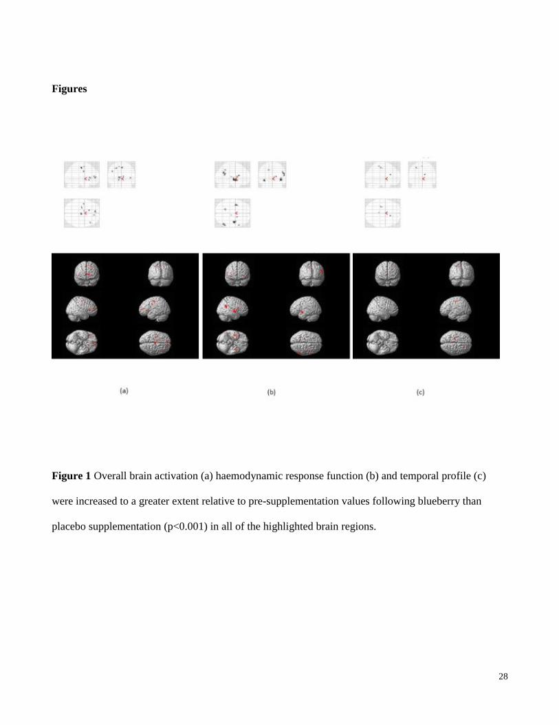
Figures
Figure 1 Overall brain activation (a) haemodynamic response function (b) and temporal profile (c)
were increased to a greater extent relative to pre-supplementation values following blueberry than
placebo supplementation (p<0.001) in all of the highlighted brain regions.
Page 29
29
Figure 2 Resting state quantitative perfusion of the gray matter in frontal (a), parietal (b), and occipital
(c) lobes of the brain, * - significantly different to pre-supplementation p<0.05.