Page 1
1
Effects of the avermectin, MK-243, on ovary development and
salivary gland degeneration in the ixodid tick, Amblyomma
hebraeum
Kevin J. Friesen, Reena Suri, and W. Reuben Kaufman*
Department of Biological Sciences, University of Alberta
Edmonton, Alberta, Canada T6G 2E9
Short Title: Physiological effects of avermectin MK-243 in ticks
*Author for correspondence: W. Reuben Kaufman, Department of Biological Sciences,
University of Alberta, Edmonton, Alberta, Canada T6G 2E9; Tel. 1-780-492-1279;
Fax. 1-780-492-9234;
E-mail: [email protected]
Submitted to Pesticide Biochemistry and Physiology, September 23, 2002.
Revised May 30, 2003.
Page 2
2
Abstract
Injection of the avermectin analogue, MK-243, into engorged female Amblyomma
hebraeum Koch resulted in reduced ovary weight, oocyte length, and ovary vitellin
content. There was no significant reduction in hemolymph vitellogenin concentration in
MK-243 treated ticks. Although MK-243 was previously shown to markedly reduce
hemolymph 20E-concentration, injection of 20E, the vitellogenic hormone in this tick,
did not reverse the effects of MK-243 on ovary development. These data suggest that
MK-243 may exert its inhibition of egg development more at the level of vitellogenin
uptake than vitellogenin synthesis. MK-243 also reversed salivary gland degeneration
slightly, probably via its inhibitory effect on 20E-synthesis.
Key Words: Avermectin, Vitellogenesis, 20-Hydroxyecdysone, Amblyomma hebraeum,
MK-243, ticks
Page 3
3
1. Introduction
In ixodid ticks, synthesis of the main yolk protein, vitellogenin (Vg), and its
uptake by oocytes, begins within a few days of engorgement on a blood meal. Until
recently, little was known about the hormonal regulation of Vg-synthesis in ticks. Current
evidence strongly suggests that an ecdysteroid is the vitellogenic hormone in the ixodid
ticks, Ixodes scapularis [1], Dermacentor variabilis [2], and Amblyomma hebraeum [3],
and the argasid tick, Ornithodoros moubata [4]. In A. hebraeum, the ecdysteroid, 20-
hydroxyecdysone (20E), stimulates Vg-synthesis, but appears insufficient on its own to
trigger Vg-uptake (Friesen and Kaufman, submitted manuscript).
Avermectins (AVMs) are a group of broad-spectrum anti-parasitic compounds
originally isolated from the bacterium Streptomyces avermitilis (reviewed in [5]). Early
research suggested that AVMs act as potentiators of -aminobutyric acid (GABA)
systems in nematodes and arthropods, possibly through an agonistic action on GABA-
mediated chloride channels (reviewed in [6]). However, more recent reports indicate that
the main mode of action is to modulate glutamate-gated ion channels [7, 8, 9], channels
which may also bind GABA [10].
Although treatment of host animals with AVM does not kill or cause detachment
of ticks, it does interfere with physiological functions such as molting, feeding to
engorgement, and reproduction in Amblyomma americanum [11]. AVMs also inhibit
oviposition when injected into A. hebraeum [12]. The mechanisms behind the latter
effects are not known. However, Lunke and Kaufman [13] observed a marked reduction
of hemolymph 20E-concentration and inhibition of ovarian development in engorged
female A. hebraeum following injection of the water-soluble AVM-analogue, MK-243.
Page 4
4
The effect of MK-243 might have been due to any combination of the following
inhibitions: (1) 20E-synthesis and/or release, (2) Vg-synthesis and/or release into the
hemolymph and, (3) uptake of Vg by the ovary. The purpose of this study was to clarify
the relative importance of these proposed mechanisms.
2. Materials and Methods
2.1 Ticks
A. hebraeum ticks were maintained in a laboratory colony at 27 C, >95%
humidity and in darkness. Ticks were allowed to feed on rabbits, as described by
Kaufman and Phillips [14], until they engorged and spontaneously detached.
2.2 Injections of MK-243 and 20E
MK-243 was a gift from Merck Sharp and Dohme Research Laboratories. A
stock solution of MK-243 (1 mg/ml) in 1.2% saline was stored at -20 C until needed.
Just prior to injection, this stock was diluted to 5, 10, and 15 µg/ml in 1.2% NaCl
(isosmotic to tick hemolymph). Injected at 1 µl/100 mg body weight (bw), these
concentrations of MK-243 corresponded to doses of 50, 100, and 150 ng/g bw. On the
day of detachment (day 0), ticks were weighed and MK-243 was injected into the
hemocoel through the camerostomal fold (articulation between the scutum and
capitulum), using an AGLA micrometer syringe apparatus (Wellcome Reagents Ltd).
Ticks were isolated in individual gauze-covered glass vials and stored under colony
conditions until 5 or 10 days after injection, at which time hemolymph and tissue samples
were collected. Control ticks were injected with 1.2% saline.
Page 5
5
To test whether injections of 20E could reverse the effect of MK-243, ticks were
treated with 150 ng MK-243/g bw on day 0, and this was followed by 3 bolus injections
of 20E (Simes, Milan) on days 1, 3 and 5. Multiple injections were considered necessary
because of the rapid rate of catabolism of 20E [15]. 20E stock solutions of (A) 5 mg/ml
and (B) 15 mg/ml were prepared in 70% ethanol (EtOH), and diluted to working
concentration in 1.2% NaCl immediately prior to injection. The concentration of EtOH
in the injected solution was 3%; after injection of 20 µl/g bw, the concentration of EtOH
in the body of the tick was estimated to be approximately 0.06%. The resulting doses
were 5 µg/g bw and 15 µg/g bw for each bolus injection; these concentrations of 20E are
known to stimulate Vg synthesis in partially-fed A. hebraeum [3]. Between each
injection, ticks were held in the colony incubator. Ticks were dissected on day 10 post-
engorgement, and samples of ovary (and hemolymph where possible) were collected.
2.3 Collection of hemolymph and ovary samples
On the day of dissection (day 5 or day 10 post-injection), ticks were secured
ventrally to a petri dish with cyanoacrylate glue and refrigerated for 15 min. Cooling
ticks prior to hemolymph collection inhibits gut contraction, thus reducing the chance of
breaking the gut and contaminating the hemolymph [16]. Incisions (1-2 mm long) were
made in the integument with a microscalpel. The exuding hemolymph was collected with
volumetric glass capillary tubes and diluted 1:4 in phosphate-buffered saline (PBS; 35
mM NaH2PO4, 60 mM Na2HPO4, 150 mM NaCl, pH 7.0). Any sample contaminated
with gut contents was discarded. Hemolymph samples were stored at -70 C until further
analyzed.
Page 6
6
Following hemolymph collection, ticks were flooded with a modified Hank‟s
balanced saline (200 mM NaCl, 8.9 mM D-glucose, 5.4 mM KCl, 1.3 mM CaCl2, 0.4
mM MgSO4, 0.44 mM KH2PO4, 0.35 mM Na2HPO4, 27 µM phenol red, pH 7.2), and the
dorsal cuticle removed. Ovaries were dissected out, and length of the long axis of the
eight apparently largest ovoid oocytes was measured using an ocular micrometer fitted to
a compound microscope. The mean value for the eight oocytes was recorded for each
tick. Ovaries were then gently blotted, weighed, and homogenized in 100 µl PBS per 30
mg ovary. Ovary homogenates were stored at -70 C until further analyzed.
2.4 ELISA for Vg and Vt
Hemolymph Vg and ovary vitellin (Vt) were quantified using an indirect
competitive ELISA as described by Friesen and Kaufman [3]. Partially purified Vt from
day 10 ovaries was used as the standard for determining the concentration of unknown
samples. Briefly, wells of a 96-well microtitre plate were coated with1 µg partially-
purified Vt, and a mixture of anti-Vg antibodies, plus either known concentrations of Vt
or unknown samples, added to each well. After incubation, the amount of antibody
binding to the plate-bound Vt was quantified using an alkaline phosphatase (AP)-linked
goat anti-rabbit secondary antibody (BioRad) and an AP colour substrate kit (p-
Nitropheynlphosphate and diethanolamine kit; BioRad). The colour reaction was
quantified by measuring the absorbance of each well at 405 nm using a microtitre plate
Page 7
7
reader (Bio-Tek). The sensitivity of this ELISA to A. hebraeum Vg is approximately 5 ng
of Vt-equivalents
2.5 Assay for salivary gland degeneration
As 20E triggers salivary gland degeneration [16], and because MK-243 reduces
hemolymph ecdysteroid concentration [13], we also measured salivary gland function in
this study using the technique of Harris and Kaufman [17]. Briefly, paired salivary
glands were excised from each tick 5 or 10 days post-engorgement, and the main ducts
ligated using very fine strands of surgical silk thread (Dermalon®; Davis and Geck). The
glands were gently blotted, weighed to the nearest 10 µg and incubated in medium TC
199 (Gibco) containing 10 µM dopamine (Sigma) for 10 min, blotted again, and weighed.
As dopamine stimulates salivary fluid secretion [17], the net weight increase is a direct
measure of fluid secretory competence; loss of fluid secretory competence compared to
controls is thus a quantitative measure of salivary gland degeneration. The wet, silk
thread weighed less than the sensitivity of the balance (10 µg).
2.6 Photography
Whole ticks, ovaries, and salivary glands were photographed using a Nikon
DXM1200 digital camera attached to a dissecting microscope. Digital images were re-
touched for publication using Adobe Photoshop 4.0 software.
Page 8
8
2.7 Statistical analysis
Results are reported as mean SEM (n). Statistical analysis was done using
Statview 4.02. Differences among treatments were analyzed using a one-way analysis of
variance (ANOVA). Statistical significance is indicated as follow: (*) 0.01<P<0.05; (**)
0.001<P<0.01; (***) P<0.001.
3. Results
3.1 Effects of MK-243 on engorged ticks
Except for a single tick at 50 ng MK-243/g bw, the doses of MK-243 used in this
study did not kill engorged ticks by day 5 (Table 1). By day 10 post-engorgement,
mortality increased only slightly with dose of MK-243, peaking at 11% at the highest
dose (150 ng MK-243/g bw). However, ticks treated with MK-243 appeared bloated, had
splayed legs, did not move, and displayed much shallower dorsal ridges than normal
healthy ticks (Fig. 1), suggesting that the major dorso-ventral muscles and leg muscles
were paralyzed.
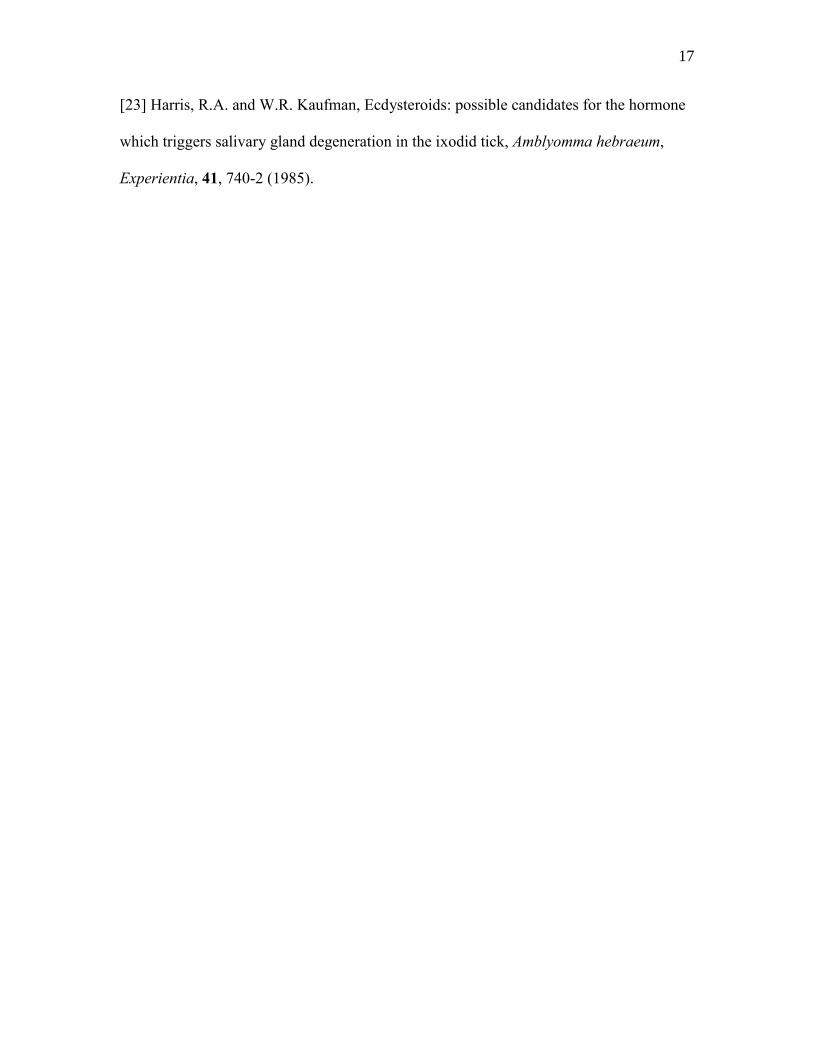
Ovary weight in control ticks rose 4.7-fold between days 5 and 10 (Fig. 2A,
control). In contrast, ovary weights of ticks treated with 150 ng MK-243/g bw were
significantly smaller, being only 32% of control on day 5 and 19% of control On day 10
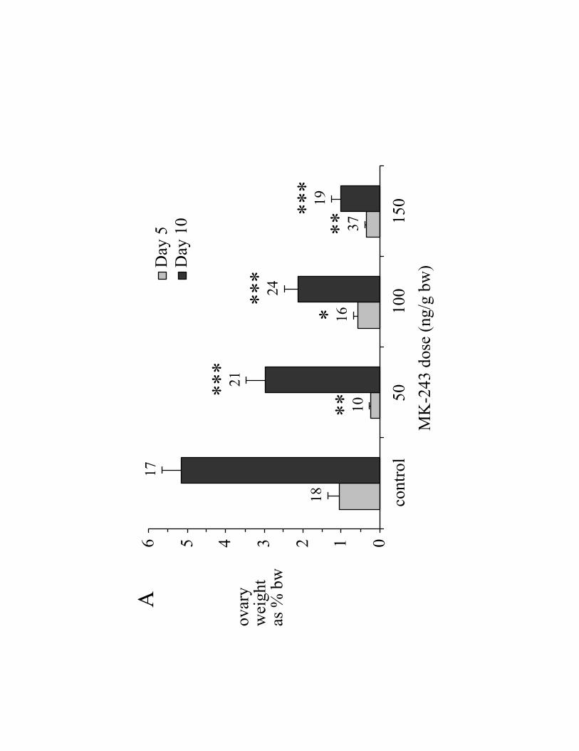
(Fig. 2A). On day 5, mean oocyte length of ticks treated with 150 ng MK-243/g bw was
60% of the control value (Fig. 2B); likewise, on day 10, oocyte length of ticks treated
with 150 ng MK-243/g bw was 51% of the control value.
Page 9
9
Total Vt content of the ovary in day 10 ticks treated with 150 ng MK-243/g bw
was reduced by 91% compared with saline injected control ticks (Fig. 2C, bars). Vt as %
ovary weight dropped 60% at 100 ng MK-243/g bw, with no further decline at 150 ng
MK-243/g bw (Fig. 2C, open circles). Hemolymph Vg concentration on days 5 and 10
was not significantly inhibited by MK-243 (150 ng/g bw) (Fig. 2D). For both days,
however, the variability was high.
Ovaries of day 5 ticks treated with MK-243 showed numerous regions where
oocytes were in the previtellogenic growth phase, but in general the oocytes were smaller
than those of controls, with few having begun Vg-uptake (Fig. 3A and 3B). By day 10,
MK-243 treated ovaries contained some clusters of oocytes at advanced stages of Vg-
uptake (Fig. 3D), whereas Vg-uptake in day 10 controls occurred in oocytes along the
entire length of the ovary (Fig. 3C).
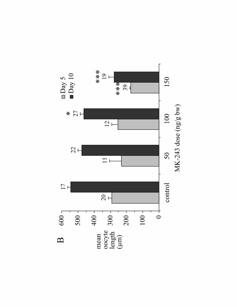
3.2 Effects of 20E on MK-243 treated ticks
Because MK-243 inhibits 20E hemolymph titers [13], and because 20E is
probably the vitellogenic hormone in A. hebraeum [3], we tested whether injections of
20E could reverse the inhibitory effects of MK-243 on the reproductive system. In
general, 20E (5 or 15 µg/g bw) did not reverse the inhibitory effect of MK-243 (150 ng/g
bw) on ovary weight (Fig. 4A), mean oocyte length (Fig. 4B), or ovary Vt-content (Fig.
4C). Note, however, that the mortality of ticks injected with 20E was substantially higher
than the mortality of ticks treated with MK-243 alone, reaching 40% at a dose of 15 µg
20E/g bw (Table 2).
Page 10
10
3.3 Effects of MK-243 on salivary gland weight and salivary fluid secretory competence
There were no significant differences in salivary gland weight of ticks treated
with MK-243 compared with control ticks on day 5 post-engorgement (Fig. 5A).
However, doses of 100 and 150 ng MK-243/g bw increased salivary gland weight
significantly by day 10 (Fig. 5A). Because the salivary glands degenerate significantly
over the first 4 days of engorgement [17], salivary fluid secretory competence was
generally low in both day 5 and 10 control ticks (Fig. 5B). Doses of 50 ng MK-243/g bw
and 150 ng MK-243/g bw, caused day 5 salivary glands to take up significantly more
fluid than control ticks; this trend was not seen on day 10, however (Fig. 5B).
Salivary glands of MK-243 treated ticks generally appeared more robust than
those of control ticks. Ten days after engorgement, salivary glands of controls were
extremely fragile, and had a wispy appearance under the dissecting microscope compared
to MK-243 treated tick salivary glands (Fig. 6).
4. Discussion
This study indicates that MK-243 inhibits egg development primarily at the level
of Vg-uptake by the oocyte. First, even though MK-243 reduces hemolymph ecdysteroid
titer by approximately 90% [13], multiple injections of 20E did not reverse the action of
MK-243 (Fig. 4). Second, MK-243 did not significantly reduce hemolymph Vg-
concentration (Fig. 2D). Finally, ovary weight (Fig. 2A) and vitellin content of the ovary
(Fig. 2C) were the most affected by MK-243. However, it is not yet possible to entirely
exclude an effect of MK-243 on Vg-synthesis. For example, if synthesis and uptake of
Page 11
11
Vg were inhibited to a similar degree, this would result in little or no change in Vg-
concentration in the hemolymph, as was observed here (Fig.2D). On the other hand,
treatment with 20E should then have resulted in an increase in Vg-concentration of the
hemolymph, but this was not the case (results not shown). This matter might be resolved
by measuring the effect of MK-243, with and without 20E, on radiolabelled amino acid
accumulation into Vg.
The effects of MK-243 on the ovary were much more noticeable on day 10 than
day 5. The bulk of ovary growth is due to Vg-uptake, which occurs between days 4 and
16 post-engorgement [3]. By day 5, most of oocyte growth is due to the previtellogenic
phase of development [18, 19]. This may explain why MK-243 showed less effect by day
5 than day 10 (Fig. 2B). Similar results were observed in mosquitoes, where oocyte
growth due to Vg-uptake was inhibited after ivermectin treatment [20].
Oocyte length was not an accurate measure for testing the effect of MK-243 in A.
hebraeum. MK-243 reduced the Vt content of ovaries by up to 81% (Fig. 2C), whereas
oocyte length was reduced by only 40% (Fig. 2B). It is clear from Fig. 3 that many fewer
oocytes accumulated Vg in MK-243 treated ticks compared to control ticks. But at least a
few clusters of oocytes accumulated Vg even at the highest dose of MK-243. The fact
that our index of oocyte size was based on measuring the eight apparently largest oocytes
accounts for why this index was less sensitive. It is interesting that those oocytes which
did accumulate Vg seemed to be clustered, rather than randomly distributed (Fig. 3D).
The reason for this is unknown, but might be that these oocytes were at a more advanced
stage of development at the time of treatment, or that some autosynthesis of Vg by the
Page 12
12
ovary occurred, as previously suggested for the ixodid tick, Rhipecephalus sanguineus
[21] or that, for some other reason, they escaped the effects of MK-243.
However, Because MK-243 inhibits 20E-synthesis [13], we hypothesized that it
might also inhibit salivary gland degeneration. MK-243 did cause a small increase in
fluid secretory competence, at least on day 5 (Fig. 5B), although secretory competence
did not approach the values expected for salivary glands from partially-fed A. hebraeum,
5-10 days post-removal (about 3 mg/gland /10 min [12, 22]). This is probably because
although MK-243 reduces hemolymph ecdysteroid concentration by approximately 90%
[13], the residual concentration (about 50 ng/ml) would still be above the threshold for
some degree of salivary gland degeneration (30 ng/ml; [23]).
In summary, this study indicates that MK-243 acts primarily by inhibiting Vg-
uptake by the oocytes. The mechanism of this inhibition remains to be determined.
Acknowledgments
We thank Ms. Rehka Chacko for her assistance with this project, and Dr. M.
Belosevic (Dept. of Biological Sciences, University of Alberta) for the use of his
microtitre plate reader. This research was generously supported by an operating grant to
W.R.K. from the Natural Sciences and Engineering Research Council (NSERC) of
Canada.
Page 13
13
References
[1] James, A.M., X.X. Zhu, and J.H. Oliver, Vitellogenin and ecdysteroid titers in Ixodes
scapularis during vitellogenesis, J. Parasitol., 83, 559-563 (1997).
[2] Sankhon, N., T. Lockey, R.C. Rosell, M. Rothschild, and L. Coons, Effect of
methoprene and 20-hydroxyecdysone on vitellogenin production in cultured fat bodies
and backless explants from unfed female Dermacentor variabilis, J. Insect Physiol., 45,
755-761 (1999).
[3] Friesen, K.J., and W.R. Kaufman, Quantification of vitellogenesis and its control by
20-hydroxyecdysone in the ixodid tick, Amblyomma hebraeum, J. Insect Physiol., 48,
773-782 (2002).
[4] Taylor, D., A. Moribayashi, N. Agui, T. Shono, and Y. Chinzei, Hormonal regulation
of vitellogenesis in the soft tick, Ornithodoros moubata. in “Proceedings of XIII
International Congress of Comparative Endocrinology. Yokohama, Japan” (S.
Kawashima, and S. Kikuyama, Eds.), pp. 213-220 (1997).
[5] Strong, L. and T.A. Brown, Avermectins in insect control and biology: a review,
Bull. Entomol. Res., 77, 357-389 (1987).
Page 14
14
[6] Campbell, W.C., M.H. Fisher, E.O. Stapley, G. Albers-Schönberg, and T.A. Jacob,
Ivermectin: a potent new antiparasitic agent, Science, 221, 823-828 (1983).
[7] Cully, D.F., D.K. Vassilatis, L.L. Liu, P.S. Paress, L.H.T. Vanderploeg, and J.M.
Schaeffer, Cloning of an avermectin-sensitive glutamate-gated chloride channel from
Caenorhabditis elegans, Nature, 371, 707-11 (1994).
[8] Arena, J.P., K.K. Liu, P.S. Paress, E.G. Frasier, D.F. Cully, H. Mrozik, and J.M.
Schaeffer, The mechanism of action of avermectins in Caenorhabditis elegans:
correlation between activation of glutamate-sensitive chloride current, membrane
binding, and biological activity, J. Parasitol., 81, 286-294 (1995)
[9] Martin, R.J., An electrophysiological preparation of Ascaris suum pharyngeal muscle
reveals a glutamate-gated chloride channel sensitive to avermectin analogue, milbemycin
D, Parasitology, 112, 247-52 (1996).
[10] Ludmerer, S.W., V.A. Warren, B.S. Williams, Y. Zheng, D.C. Hunt, M.B. Ayer,
M.A. Wallace, A.G. Caudhary, M.A. Egan, P.T. Meinke, D.C. Dean, M.L. Garcia, D.F.
Cully, and M.M. Smith, Ivermectin and Nodulisporic acid receptors in Drosophila
melanogaster contain both -aminobutyric acid-gated Rd1 and glutamate-gated GluCl
chloride channel subunits, Biochemistry, 41, 6548-6560 (2002).
Page 15
15
[11] Lancaster Jr., J.L., J.S. Simco, and R.L. Kilgore, Systematic efficacy of ivermectin
MK-933 against the Lone Star tick, J. Econ. Entomol., 75, 242-4 (1982).
[12] Kaufman, W.R., S.G. Ungarian, and A.E. Noga, The effect of avermectins on
feeding, salivary fluid secretion and fecundity in some ixodid ticks, Exp. Appl. Acarol.
21, 1-18 (1986).
[13] Lunke, M. and W.R. Kaufman, Effects of the avermectin analogue MK-243 on
vitellogenesis and reproduction in the ixodid tick, Amblyomma hebraeum, Exp. Appl.
Acarol, 13, 249-259 (1992).
[14] Kaufman, W.R., and J.E. Philips, Ion and water balance in the ixodid tick,
Dermacentor andersoni: I. Routes of ion and water excretion, J. Exp. Biol., 58, 523-536
(1973).
[15] Weiss, B.L., and W.R. Kaufman, The relationship between „critical weight; and 20-
hydroxyecdysone in the female ixodid tick, Amblyomma hebraeum, J. Insect Physiol., 47,
1261-1267 (2001).
[16] Kaufman, W.R., Correlation between haemolymph ecdysteroid titre, salivary gland
degeneration and ovarian development in the ixodid tick, Amblyomma hebraeum Koch, J.
Insect Physiol., 37, 95-99 (1991).
Page 16
16
[17] Harris, R.A., and W.R. Kaufman, Neural involvement in the control of salivary
gland degeneration in the ixodid tick, Amblyomma hebraeum, J. Exp. Biol., 109, 281-290
(1984).
[18] Balashov, Y.S., Bloodsucking ticks (Ixodoidea)—vectors of diseases of man and
animals, Misc. Publ. Entomol. Soc. Am., 8, 161-376 (1972).
[19] Diehl, P.A., A. Aeschlimann, and F.D. Obenchain, Tick reproduction: oogenesis and
oviposition, in “Physiology of Ticks” (F.D. Obenchain and R. Galun, Eds.), pp. 277-350,
Pergamon Press, Oxford, (1982)
[20] Mahmood, F., L.L. Walters, H. Guzman, and R.B. Tesh, Effect of ivermectin on the
ovarian development of Aedes aegypti (Diptera: Culicidae), J. Med. Entomol., 28, 701-
707 (1991).
[21] Araman, S.F, Protein digestion and synthesis in ixodid females, Recent Adv. Acarol.,
1, 385-395 (1979).
[22] Lomas L.O. and W.R. Kaufman, An indirect mechanism by which a protein from the
male gonad hastens salivary gland degeneration in the female ixodid tick, Amblyomma
hebraeum, Arch. Insect Biochem. Physiol., 21, 169-178 (1992).
Page 17
17
[23] Harris, R.A. and W.R. Kaufman, Ecdysteroids: possible candidates for the hormone
which triggers salivary gland degeneration in the ixodid tick, Amblyomma hebraeum,
Experientia, 41, 740-2 (1985).
Page 18
18
Fig. 1. Appearance of engorged female A. hebraeum 10 days after injection of (A) 1.2%
NaCl or (B) 150 ng MK-243/g bw. The cuticular ridges (white arrowheads) are caused
by the contraction of the dorso-ventral muscles. Note that the ridges are much shallower
in the MK-243 treated tick and that the legs are splayed (indices of paralysis) compared
to the control.
Fig. 2. Effect MK-243 on ovary development in A. hebraeum, 5 days (light grey bars) or
10 days (dark grey bars) post-engorgement. MK-243 was injected on the day of
engorgement (day 0). (A) Ovary weight as % engorged body weight as measured on day
0; (B) mean length of 8 of the largest oocytes; (C) total Vt-content of ovary; (D)
hemolymph Vg-concentration (reported as „Vt-equivalents‟, see Materials and Methods).
For all panels, data are reported as mean SEM (n). Statistical significance is indicated
as follows: (*) 0.01<P<0.05; (**) 0.001<P<0.01; (***) P<0.001.
Fig. 3. Effect of MK-243 on appearance of the ovary. Ovaries are from (A) day 5 post-
engorgement saline injected control, (B) day 5, 100 ngMK-243/g bw, (C) day 10, saline
injected control (D) day 10, 100 ng MK-243/g bw. Some of the regions containing
vitellogenic oocytes are indicated by white brackets. Asterisks (*) indicate a few regions
where oocytes are toward the end of the great cytoplasmic growth phase, but that have
not yet accumulated much Vg (see Discussion).
Fig. 4. Inability of 20E to reverse the effects of MK-243 in engorged A. hebraeum. MK-
243 (150 ng/g bw) or saline control was injected on day 0. Bolus injections of 20E were
Page 19
19
given on days 1, 3, and 5 post-engorgement, and ovaries collected on day 10. (Control):
ticks that received no injection; (EtOH): ticks that received EtOH (final concentration
0.06%) in 1.2% NaCl on days 1, 3, and 5; (MK-243): ticks that received 150 ng MK-
243/g bw followed by ethanol/saline injections (days 1, 3, and 5). (A) Ovary weight as %
bw; (B) mean length of 8 of the longest oocytes; (C) total Vt-content of the ovary. Data
are reported as mean SEM (n). Significant differences are indicated as in the legend to
Fig. 2.
Fig. 5. Effect of MK-243 on salivary gland (SG) weight (A) and fluid secretory
competence (B) 5 and 10 days post-engorgement. Ticks received a single injection of
1.2% NaCl (control) or MK-243 (50, 100, or 150 ng/g bw) on the day of engorgement.
Data are reported as mean SEM (n). Significant differences are indicated as in the
legend to Fig. 2.
Fig. 6. Effect of saline injection (A), or 150 ng MK-243g bw (B), on appearance of
salivary glands of A. hebraeum 10 days post-engorgement. Note the wispy appearance of
the control salivary gland versus the more robust appearance of the MK-243 treated
gland.
Page 21
0123456
cont
rol
5010
015
0M
K-2
43 d
ose
(ng/
g bw
)
ovar
y w
eigh
t as
% b
w
Day
5D
ay 1
0
A
18
17
10**16*
24***
19***
21***
37**
Page 22
0
100
200
300
400
500
600
cont
rol
5010
015
0M
K-2
43 d
ose
(ng/
g bw
)
mea
n oo
cyte
le
ngth
(µ
m)
Day
5D
ay 1
0B
20
17
11
22
12
27
39
19
*
*****
*
Page 23
0510152025
cont
rol
5010
015
0M
K-2
43 d
ose
(ng/
g bw
)
Vt
cont
ent
(mg/
ovar
y)
0369121518
Vt
as %
ov
ary wt
Vt (
mg/
ovar
y)V
t as %
ova
ry w
t
C
**
***
13
16
8
10
***
Page 24
0102030
cont
rol
5010
015
0M
K-2
43 d
ose
(ng/
gbw
)
hem
olym
ph
Vt-
equi
vale
nts
(mg/
ml)
Day
5D
ay 1
0
D
13
10
8
15
15
10
8
9
Page 26
02468
cont
rol
EtO
HM
K-2
43M
K-2
43+
5 µg
20E
per g
bw
MK
-243
+15
µg
20E
per g
bw
tre
atm
ent
ovar
y w
eigh
t as
% b
w
on d
ay 1
0
A10
10
11
9
2
***
*
***
***
Page 27
0
100
200
300
400
500
600
cont
rol
EtO
HM
K-2
43M
K-2
43+
5 µg
20E
per g
bw
MK
-243
+15
µg
20E
per g
bw
tre
atm
ent
mea
noo
cyte
le
ngth
on
da
y 10
(µ
m)
B10
11
1112
2
***
***
***
Page 28
0510152025
cont
rol
EtO
HM
K-2
43M
K-2
43+
5µg
20E
per
g bw
tre
atm
ent
ovar
y V
t con
tent
on
day
10
(mg/
ovar
y)
C
10
7
5
10
*
**
Page 29
0
1
2
3
control 50 100 150MK-243 dose (ng/g bw)
SG weight (mg)
Day 5Day 10
A44
28
21
46
4148
71
45***
***
Page 30
0
0.1
0.2
0.3
0.4
0.5
0.6
control 50 100 150MK-243 dose (ng/g bw)
SG fluid uptake (mg/gland/10 min.)
Day 5Day 10
B
23
9
15
18
23
16
41
22
**
Page 32
Table 1: Mortality of MK-243 treated engorged female A. hebraeum on day 5 or 10 post-engorgement. [MK-243] ng/g bw 0 50 100 150 %
mortality n %
mortalityn %
mortalityn %
mortalityn
Day 5 0 36 7.1 14 0 34 0 71
Day 10 3.7 27 6.4 47 7.9 38 10.9 55
Table 2: Mortality of MK-243/20E treated engorged female A. hebraeum on day 10 post-engorgement.
Treatment untreated EtOH MK-243
(150 ng/g bw) MK-243 +
20E (5 µg/g bw)MK-243 +
20E (15 µg/g bw)%
mortality n %
mortality n %
mortalityn %
mortality n %
mortality n
0 16 0 21 0 20 14.8 27 40 10


![The Ovary of the Teleost Fish Xenotoca Eiseni (Goodeidae ... · the ovary, called a gonoduct, connects the ovary to the exterior by a gonopore [7]. These unique features of the ovary](https://static.documents.pub/doc/80x56/5f5082c1c1cb78272c63e522/the-ovary-of-the-teleost-fish-xenotoca-eiseni-goodeidae-the-ovary-called-a.jpg)


