1 DATA SHEET Title: Effects of endocrine disrupters on the rat testis: quantitative and qualitative investigations with special focus on the Sertoli cell. Author: Majken Dalgaard. Publisher: Danish Veterinary and Food Administration Affiliation: Institute of Food Safety and Toxicology, Department of General Toxicology Adresse: Mørkhøj Bygade 19, DK-2860 Søborg, Denmark. Telephone: +45 33 95 65 60 Fax: +45 33 95 60 01 e-mail: [email protected]Abstract: The aim of the thesis was to study the effects of endocrine disrupters on the rat testis primarily by using various qualitative and quantitative methods. The thesis focuses on histomorphological investigations in order to investigate the effects on the number and function of Sertoli cells, as the Sertoli cells are of crucial importance to the production of well functioning spermatids in the adult male. In some industrial countries, there are indications of a decreasing trend in the sperm count of man. The aetiology, however, still remains uncertain. A declining sperm count could partly be caused by a decrease in the Sertoli cell number induced by exposure to environmental oestrogen-like compounds, as proposed by Sharpe and Skakkebæk (1993). The results presented here demonstrate that the total number of Sertoli cells, estimated with stereology-based methods, decreased in rats exposed in utero to the synthetic oestrogen diethylstilboestrol (DES). The Sertoli cell toxicant and antiandrogenic phthalate, mono-(2-ethylhexyl) phthalate (MEHP), induced a collapse of Sertoli cell vimentin in the acute phase of toxicity, without changing the level of apoptotic germ cells investigated by the terminal deoxy-nucleotidyl transferase-mediated nick end labelling (TUNEL) assay. In conclusion, immunostaining of Sertoli cell vimentin is a valuable marker in the acute phase of MEHP-induced Sertoli cell toxicity. The results obtained with stereology-based quantification will provide strong evidence of an effect in future studies of endocrine disrupters. The results in the in utero study of the synthetic oestrogen, DES, support the hypothesis by Sharpe and Skakkebæk (1993) that foetal exposure to environmental oestrogens may decrease the Sertoli cell number and perhaps result in a decreased sperm output. Key words: Sertoli cell, rat testis, endocrine disrupters, stereology, histology Please quote: Majken Dalgaard (2001). Effects of endocrine disrupters on the rat testis: quantitative and qualitative investigations with special focus on the Sertoli cell. ISBN: 87-91189-09-8 Front cover: Testis from an 11-day-old rat. Haematoxylin and eosin staining. INDEX
Transcript
1
DATA SHEET Title: Effects of endocrine disrupters on the rat testis: quantitative and qualitative investigations with special focus on the Sertoli cell. Author: Majken Dalgaard. Publisher: Danish Veterinary and Food Administration Affiliation: Institute of Food Safety and Toxicology, Department of General Toxicology Adresse: Mørkhøj Bygade 19, DK-2860 Søborg, Denmark. Telephone: +45 33 95 65 60 Fax: +45 33 95 60 01 e-mail: [email protected] Abstract: The aim of the thesis was to study the effects of endocrine disrupters on the rat
testis primarily by using various qualitative and quantitative methods. The thesis focuses on histomorphological investigations in order to investigate the effects on the number and function of Sertoli cells, as the Sertoli cells are of crucial importance to the production of well functioning spermatids in the adult male. In some industrial countries, there are indications of a decreasing trend in the sperm count of man. The aetiology, however, still remains uncertain. A declining sperm count could partly be caused by a decrease in the Sertoli cell number induced by exposure to environmental oestrogen-like compounds, as proposed by Sharpe and Skakkebæk (1993). The results presented here demonstrate that the total number of Sertoli cells, estimated with stereology-based methods, decreased in rats exposed in utero to the synthetic oestrogen diethylstilboestrol (DES). The Sertoli cell toxicant and antiandrogenic phthalate, mono-(2-ethylhexyl) phthalate (MEHP), induced a collapse of Sertoli cell vimentin in the acute phase of toxicity, without changing the level of apoptotic germ cells investigated by the terminal deoxy-nucleotidyl transferase-mediated nick end labelling (TUNEL) assay. In conclusion, immunostaining of Sertoli cell vimentin is a valuable marker in the acute phase of MEHP-induced Sertoli cell toxicity. The results obtained with stereology-based quantification will provide strong evidence of an effect in future studies of endocrine disrupters. The results in the in utero study of the synthetic oestrogen, DES, support the hypothesis by Sharpe and Skakkebæk (1993) that foetal exposure to environmental oestrogens may decrease the Sertoli cell number and perhaps result in a decreased sperm output.
Key words: Sertoli cell, rat testis, endocrine disrupters, stereology, histology Please quote: Majken Dalgaard (2001). Effects of endocrine disrupters on the rat testis: quantitative and qualitative investigations with special focus on the Sertoli cell. ISBN: 87-91189-09-8 Front cover: Testis from an 11-day-old rat. Haematoxylin and eosin staining. INDEX
2
PART ONE List of abbreviations ........................................................................................................................ 2 Preface............................................................................................................................................... 3 Acknowledgement ............................................................................................................................ 4 Summary........................................................................................................................................... 5 Sammendrag (Summary in Danish)............................................................................................... 7 List of papers included .................................................................................................................... 9 Introduction.................................................................................................................................... 10 The hypothalamic-pituitary-testicular axis ................................................................................. 11 The steroid hormone receptors and other mechanisms of action.............................................. 12 The Sertoli cell................................................................................................................................ 13 Histopathology of the seminiferous epithelium........................................................................... 15 Germ cell apoptosis........................................................................................................................ 16 Sertoli cell toxicants ....................................................................................................................... 17 Sertoli cell number, diameter and length of the seminiferous tubules ..................................... 18 Specific studies ............................................................................................................................... 21 Discussion and main conclusions.................................................................................................. 23 References ....................................................................................................................................... 26 PART TWO.................................................................................................................................... 31
Paper I Toxicity study of di(2-ethylhexyl)phthalate (DEHP) in combination with acetone in rats.
Paper II The acute effects of mono(2-ethylhexyl)phthalate (MEHP) on testes of prepubertal Wistar rats.
Paper III Developmental toxicity of toluene in male rats: effects on semen quality, testis morphology, and apoptotic neuro-degeneration.
Paper IV In utero exposure to diethylstilbestrol or 4-n-nonylphenol in rats: Number of Sertoli cells, diameter and length of seminiferous tubules estimated by stereological methods.
Paper V In utero reproductive study in rats exposed to nonylphenol. Paper VI Quantification of antiandrogen effect determined by lightcycler technology.
List of abbreviations DDT dichlorodiphenyl trichloroethane DEHP di-(2-ethylhexyl)phthalate DES diethylstilboestrol FSH follicle stimulating hormone GD gestational day GnRH gonadotropin releasing hormone HE hematoxylin and eosin LH luteinising hormone MEHP mono-(2-ethylhexyl)phthalate PND postnatal day p,p´-DDE 1,1´-(2,2-dichloroethenylidene)-bis[4-chlorobenzene] TUNEL assay deoxy-nucleotidyl transferase-mediated nick end labelling assay.
3
Preface This project was carried out at the Danish Veterinary and Food Administration, Institute of Food Safety and Toxicology, Division of General Toxicology, Department of Pathology and is part of the investigations concerning endocrine disrupters led by head of division, Jens-Jørgen Larsen. The two supervisors involved were Ole Ladefoged, Institute of Food Safety and Toxicology, and Hanne Cathrine Bisgaard at Roskilde University, Department of Life Sciences and Chemistry. The thesis is divided into two parts. Part one is a general introduction to the laboratory work on which the thesis is based. Part two covers the results obtained, which are presented in form of five published papers and one paper in press.
4
Acknowledgement First of all I would like to thank my supervisor at the Institute of Food Safety and Toxicology, Ole Ladefoged, for his effort. I am impressed by his theoretical skill, tireless enthusiasm, and concern about the environmental pollutants and human health. This has been a great inspiration to me and it has certainly been a pleasure working with him. Another person of huge importance to this project is my creative technical assistant and loyal colleague Karen Roswall, without whom this project would never have succeeded. I also want to express my gratitude to all my colleagues at the Department of Pathology, Institute of Food Safety and Toxicology, the co-authors, and my supervisor at Roskilde University, Department of Life Sciences and Chemistry, Hanne Cathrine Bisgaard, for their personal involvement and good advice. Additionally, my two proof-readers Kirsten Pilegaard and Elsa Nielsen are thanked for their quick response and their linguistic enthusiasm. Finally, I want to thank my external supervisors in stereology Bente Pakkenberg, Bispebjerg Hospital, and Hans Jørgen Gundersen, and Jens Randel Nyengaard, University of Aarhus.
5
Summary In the beginning of the nineteen nineties it was hypothesised that the decreasing trend in sperm counts during the last 50 years may share aetiology with the increasing incidence of testicular cancer and genital malformations in man. In wildlife populations, developmental and reproductive disorders such as feminisation of the external genital organs, decreased fertility in mammals, and egg shell thinning in birds have been reported. It has been proposed that endocrine disrupters present in the environment and our food may be responsible for the disorders. During the last 10 years, the endocrine disrupters have achieved worldwide attention in the scientific and regulatory field as well as in the media. Enormous amounts of resources are used to assess the potential human risk. At the Institute of Food Safety and Toxicology, research on chemicals with endocrine disrupting effects has been initiated. The present thesis predominantly focuses on the investigations of the testicular toxicity related to this research. Six papers are included in which the histopathological, the immunohistochemical, and the quantitative investigations of the testis are described. The main issue was to investigate the effects on the Sertoli cell morphology and function as well as to investigate the total number of Sertoli cells in the rat testis. The Sertoli cell is of crucial importance to the proliferation and differentiation of mature spermatids in the testis as the Sertoli cell is responsible for the germ cells passing through stages of mitosis, meiosis, and differentiation until the germ cells are released into the lumen of the seminiferous tubules. In rats, the Sertoli cell population expands perinatally whereas the Sertoli cell maturation occurs postnatally and therefore, the Sertoli cells are considered to be sensitive to toxic insults during early life. The effects on the supporting capacity of Sertoli cells were studied by use of an immunohisto-chemical investigation of the cytoplasmatic structure filament, vimentin, after exposure to di-(2-ethylhexyl)phthalate (DEHP) (paper I), mono-(2-ethylhexyl)phthalate (MEHP) (paper II) and toluene (paper III). Additionally, the effects of MEHP on the testis were investigated by an immunohistochemical staining of the androgen receptor and by evaluating germ cell apoptosis with the terminal deoxy-nucleotidyl transferase-mediated nick end labelling (TUNEL) assay (II). In the papers III, IV and V, an in utero design was used in order to study the effects of chemicals during the most sensitive period of the development of the reproductive system. Three chemicals were investigated in such modified one-generation studies: 1) Toluene, a chemical affecting the neuronal development of the foetus (III), 2) 4-n-nonylphenol, and 3) the positive control, the synthetic oestrogen, diethylstilboestrol (DES) (IV, V). The following parameters were applied: weight of the reproductive organs, testis morphology, and estimation of the total number of Sertoli cells. The total number of Sertoli cells and length and diameter of the seminiferous tubules were investigated by stereological methods after exposure to DES or 4-n-nonylphenol during gestation (IV). In this study, a statistically significant decrease in the total number of Sertoli cells in 11-day-old male rats exposed to DES in utero was observed compared to the control group. No effect on the Sertoli cell number was observed after exposure to 4-n-nonylphenol. However, in utero exposure to 4-n-nonylphenol induced a dose related decrease in the right epididymal weight in adult rats (V). During the last years, much attention has been paid to other endocrine disrupters than the oestrogenic ones, especially to chemical substances with antiandrogenic effect. These chemicals exert their effect through binding to the androgen receptor or through indirect mechanisms e.g. by affecting the testosterone synthesis. In a short-term screening test, the Hershberger test, organ weights were combined with the investigation of hormone levels and gene-expression in the ventral
6
prostate. The usefulness of these new parameters in toxicology was evaluated by the method applied in paper VI, where the effect of the known antiandrogen, vinclozolin, was studied by the gene-expression of testosterone-repressed prostatic message 2 and prostate specific binding protein polypeptide C3 in the ventral prostate. Generally, the new methods applied open up for investigations of chemical substances with similar endocrine effects. Germ cell apoptosis evaluated by the TUNEL assay as well as vimentin and androgen receptor immunostaining are most valuable when mechanisms are investigated, but they are not necessarily sensitive parameters in the evaluation of toxic effects. From the investigations described in paper II, it can be concluded that the TUNEL assay was not able to detect effects of MEHP exposure at lower dose levels than described in the literature with other methods, whereas the displacement of the vimentin filaments in the Sertoli cell cytoplasm seems to be a relatively sensitive parameter in the acute stage of phthalate-induced Sertoli cell toxicity. The stereological estimation of the total number of Sertoli cells in the testis seems to be a valuable method to detect effects of DES in lower doses than are detectable with other testis parameters such as testis weight, histopathology, and length and diameter of the seminiferous tubules (IV). According to Sharpe and Skakkebæk (1993), a decrease in sperm counts may be related to a decrease in the number of Sertoli cells. By a stereological method it has been shown that the number of Sertoli cells can be decreased by exposure to the oestrogen DES (IV). The estimation of the total number of Sertoli cells is therefore considered to be a valuable parameter in the future testing of endocrine disrupters.
7
Sammendrag (Summary in Danish) I begyndelsen af 1990erne blev der fremsat en hypotese om, at det mulige fald i mænds sædkvalitet gennem de sidste 50 år og den øgede incidens af testikelkræft samt genitale abnormaliteter hos mænd muligvis kan have en fælles årsagssammenhæng. I vildtlevende dyr er der rapporteret om udviklings- og reproduktionsforstyrrelser som for eksempel feminisering af de eksterne kønsorganer, nedsat fertilitet hos pattedyr, og tyndskallede æg hos flere fuglearter. Det er blevet foreslået, at disse fund kan skyldes tilstedeværelsen af hormonforstyrrende stoffer i miljøet og i vore fødevarer. Igennem de sidste 10 år har der været en verdensomspændende forskningsmæssig, regulatorisk og mediemæssig fokusering på de hormonforstyrrende stoffer. Enorme ressourcer er anvendt til at belyse den eventuelle humane risiko. På Institut for Fødevaresikkerhed og Toksikologi er der ligeledes igangsat et forskningsprojekt omhandlende kemiske stoffer mistænkt for hormonforstyrrende effekter. Denne afhandling beskæftiger sig primært med de testikeltoksiske undersøgelser i dette forskningsprojekt. I afhandlingen indgår 6 artikler, hvori de patologiske, immunhistokemiske og kvantitative undersøgelser i testiklerne er beskrevet. Hovedvægten ligger især på metoder til belysning af effekter på Sertolicellens morphologi og funktion samt en undersøgelse af det totale antal af disse celler i testiklen hos rotter. Sertolicellen har afgørende betydning for udviklingen af modne og funktionsdygtige spermatider i testiklerne, idet den er ansvarlig for, at kønscellerne gennemgår stadier af mitose, meiose og modning, indtil de er udviklet til modne spermatider, der afstødes til lumen af de sæddannende tubuli. Hos rotter ekspanderer Sertolicellepopulationen sent i fostertilstanden og omkring fødslen, mens modningen af cellerne foregår postnatalt. Sertolicellerne må derfor formodes at være særligt følsomme overfor toksiske påvirkninger tidligt i rottens liv. Effekter på Sertolicellens morphologi og funktion som støttecelle er undersøgt efter eksponering med di-(2-ethylhexyl)phthalat (DEHP) (artikel I), mono-(2-ethylhexyl)phthalat (MEHP) (artikel II) og toluen (artikel III) ved en immunhistokemisk undersøgelse af det cytoplasmatiske strukturfilament vimentin. Desuden er MEHPs effekt på testiklerne undersøgt ved hjælp af en immunhistokemisk farvning af androgenreceptoren samt påvisning af apoptose i kønscellerne bestemt ved TUNEL metoden. I artiklerne (III, IV, V) er der anvendt et forsøgsdesign, hvor kemiske stoffer er undersøgt, når de indgives i fostertilstanden under den mest følsomme periode for udviklingen af fosterets reproduktionssystem. Tre stoffer er belyst i sådanne modificerede 1-generationsforsøg: 1) Toluen, et stof der påvirker nervesystemets udvikling (III), 2) 4-n-nonylphenol, samt 3) den positive kontrol, det syntetiske østrogen diethylstilbøstrol (DES) (IV,V). I studierne er følgende parametre anvendt til at belyse effekter på det hanlige reproduktionssystem: Vægten af de enkelte reproduktionsorganer, testikel morfologi samt kvantitative parametre i testiklerne, herunder estimering af antallet af Sertoliceller med en stereologisk metode. Antallet af Sertoliceller samt længde og diameter af tubuli seminiferi contorti er undersøgt med stereologiske metoder efter eksponering af rotter for DES eller 4-n-nonylphenol under drægtigheden (IV). I dette forsøg er der påvist en statistisk signifikant reduktion i det totale antal af Sertoliceller i testiklen hos rotter doseret med DES sammenlignet med kontroldyr, mens der ikke blev observeret forskelle i celleantallet hos rotter doseret med 4-n-nonylphenol. 4-n-Nonylphenol medførte dog en dosisrelateret reduktion i den absolutte levervægt hos unge rotter og en reduktion i bitestikelvægten hos voksne dyr (V).
8
I de seneste år har opmærksomheden været rettet mod andre hormonforstyrrende stoffer end de østrogenlignende, og især mod kemiske forbindelser, der kan tænkes at virke som antiandrogener enten ved binding til androgenreceptoren eller via indirekte mekanismer, f.eks. ved påvirkning af testosteronniveauet. I en in vivo korttidstest, Hershberger testen, er organvægte suppleret med hormonmålinger og molekylærbiologiske undersøgelser til påvisning af ændringer i genekspressionen i den ventrale del af prostata. Disse nye parametres anvendelighed i toksikologien er undersøgt ved eksponering med det kendte antiandrogen vinclozolin i en metodeartikel om genekspressionen af testosteron-repressed prostatic message 2 og prostate specific binding protein polypeptide C3 (VI). Generelt åbner metoderne op for nye indfaldsvinkler til undersøgelse af kemiske stoffer med lignende hormonvirkninger. Deoxy-nucleotidyl transferase-mediated nick end labelling assay (TUNEL)-farvningen til påvisning af apoptose og de immunhistokemiske metoder til undersøgelser af vimentin og androgenreceptoren er dog mere anvendelige til afklaring af mekanistiske sammenhænge end nødvendigvis sensitive effektparametre. Ud fra undersøgelserne beskrevet i artikel II kan det konkluderes, at det med TUNEL-metoden ikke har været muligt at påvise effekter af MEHP i lavere doser end beskrevet i litteraturen, hvorimod lokaliseringen af vimentinfilamenter i Sertolicellerne tilsyneladende er både en tidlig og relativt sensitiv parameter for phthalatinduceret Sertolicelletoksicitet. De stereologiske undersøgelser tyder på, at bestemmelse af det totale antal Sertoliceller i testiklerne er velegnet til påvisning af effekter af DES, i en lavere dosis, end det har været muligt at observere ved undersøgelse af andre parametre som testikelvægt, histopatologi, og længde- og diameter af tubuli seminiferi contorti (IV). Ifølge hypotesen fremsat af Sharpe og Skakkebæk (1993) kan et fald i sædcellekoncentrationen være en følge af nedsat antal Sertoliceller. Ved stereologiske metoder er det her påvist, at antallet af Sertoliceller kan nedsættes efter eksponering for det potente østrogen DES (IV). Antallet af Sertoliceller anses derfor for en anvendelig parameter i den videre forskning med hormonforstyrrende stoffer.
9
List of papers included The present thesis is based upon the following six papers, and hereafter referred to in the text by their Roman numerals. I Majken Dalgaard, Grete Østergaard, Henrik Rye Lam, Ernst V. Hansen, Ole Ladefoged.
Toxicity study of di(2-ethylhexyl)phthalate (DEHP) in combination with acetone in rats. Pharmacol. & Toxicol. 2000, 86, 92-100.
II Majken Dalgaard, Christine Nellemann, Henrik Rye Lam, Ilona Kryspin Sørensen, Ole
Ladefoged. The acute effects of mono(2-ethylhexyl)phthalate (MEHP) on testes of prepubertal Wistar rats. Toxicol. Lett. 2001, 122, 69-79.
III Majken Dalgaard, Alireza Hossaini, Karin S. Hougaard, Ulla Hass, Ole Ladefoged.
Developmental toxicity of toluene in male rats: effects on semen quality, testis morphology, and apoptotic neuro-degeneration. Arch. Toxicol. 2001, 75, 103-109.
IV Majken Dalgaard, Kirsten Pilegaard, Ole Ladefoged. In utero exposure to diethylstilbestrol or
4-n-nonylphenol in rats: Number of Sertoli cells, diameter and length of seminiferous tubules estimated by stereological methods. Phamacol. & Toxicol. 2002, 90, (in press).
V Alireza Hossaini, Majken Dalgaard, Anne Marie Vinggaard, Henrik Frandsen, Jens-Jørgen
Larsen. In utero reproductive study in rats exposed to nonylphenol. Reprod. Toxicol. 2001, 15, 537-543.
VI Christine Nellemann, Anne Marie Vinggaard, Majken Dalgaard, Alireza Hossaini, Jens-
Jørgen Larsen. Quantification of antiandrogen effect determined by lightcycler technology. Toxicology, 2001, 163, 29-38.
10
Introduction In wildlife populations, various adverse developmental and reproductive effects such as malformed external genitals in reptiles, amphibians, and bears, reduced fertility in mammals, and decreased hatchability in birds have been reported. These effects have been associated primarily with exposure to highly persistent organochlorine pollutants like polychlorinated biphenyls, polychlorinated dibenzo-p-dioxins and polychlorinated dibenzofurans (Vos et al., 2000). Many other chemicals are also recognised as endocrine disrupters e.g., the phthalates, alkylphenols, bisphenol A and pesticides such as dichlorodiphenyl trichloroethane (DDT), 1,1´-(2,2-dichloroethenylidene)-bis[4-chlorobenzene] (p,p´-DDE), methoxychlor, chlordedone and vinclozolin (Gray et al., 1997). Sharpe and Skakkebæk (1993) have proposed that the observed decrease in human sperm counts may share aetiology with an increasing incidence of testicular cancer and reproductive tract malformations in men observed in some industrialised countries during the last 50 years. Sharpe and Skakkebæk (1993) proposed that the increasing incidence of reproductive abnormalities in men may be related to an increased oestrogen exposure in utero. There is, however, controversy about whether the human sperm count is actually declining (Safe, 2000). Despite national as well as regional differences in sperm counts, a decreasing trend has been reported in some industrialised countries (Carlsen et al., 1992; Swan et al., 1997). The previously observed decreasing trend in sperm counts has recently been supported by Swan et al. (2000). They included another 47 studies to the previously reported studies by Carlsen et al., (1992). Additionally, Andersen (2000) has raised a concern about the low sperm concentration observed in the Danish men, especially in the population of young men. She observed that young Danish men from the general population had a mean sperm concentration of 57.4 mill/ml compared to partners of pregnant women, who had a mean sperm concentration of 77.0 mill/ml. In the population of young men, 48% had sperm concentrations below 40 mill/ml and 25% had sperm concentrations below 20 mill/ml. When this was compared to partners of pregnant women, 32% of the partners had sperm concentrations below 40 mill/ml, while 12% had sperm concentrations below 20 mill/ml. Today, the lower reference value for a normal sperm concentration is 20 mill/ml. No direct evidence is available associating specific substances at environmental exposure levels with these outcomes in humans, but a growing collection of animal data lend credence to the proposed hypothesis as exposure to endocrine acting chemicals during the critical window(s) of reproductive development in animals can lead to many of the genital malformations and other adverse reproductive responses that are highlighted as being increased in human populations (Foster and Gray, 2001). Such responses are observed after exposure to chemicals recognised as endocrine disrupters, e.g., the phthalates, alkylphenols, and pesticides such as DDT and its metabolite p,p´-DDE, methoxychlor, linuron, and vinclozolin (Kelce et al., 1997; Gray et al., 1999; Ashby and Lefevre, 2000; McIntyre et al., 2000). Due to the central role of the Sertoli cell to spermatogenesis, the main object of this thesis was to investigate whether exogenous administered oestrogens actually decrease the number of Sertoli cells as proposed by Sharpe and Skakkebæk (1993). The number of Sertoli cells was estimated with unbiased counting methods in rats after exposure to 4-n-nonylphenol or the synthetic oestrogen diethylstilboestrol (DES). Another aim was to study the supporting capacity of the Sertoli cells after in utero exposure to toluene and the phthalates, di-(2-ethylhexyl)phthalate (DEHP) and mono-(2-ethylhexyl)phthalate (MEHP) by use of an immunohistochemical investigation of the structure
11
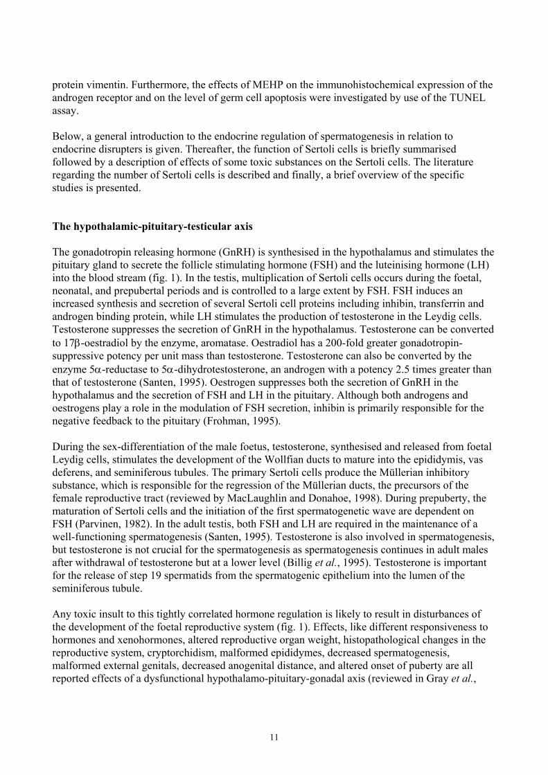
protein vimentin. Furthermore, the effects of MEHP on the immunohistochemical expression of the androgen receptor and on the level of germ cell apoptosis were investigated by use of the TUNEL assay. Below, a general introduction to the endocrine regulation of spermatogenesis in relation to endocrine disrupters is given. Thereafter, the function of Sertoli cells is briefly summarised followed by a description of effects of some toxic substances on the Sertoli cells. The literature regarding the number of Sertoli cells is described and finally, a brief overview of the specific studies is presented. The hypothalamic-pituitary-testicular axis The gonadotropin releasing hormone (GnRH) is synthesised in the hypothalamus and stimulates the pituitary gland to secrete the follicle stimulating hormone (FSH) and the luteinising hormone (LH) into the blood stream (fig. 1). In the testis, multiplication of Sertoli cells occurs during the foetal, neonatal, and prepubertal periods and is controlled to a large extent by FSH. FSH induces an increased synthesis and secretion of several Sertoli cell proteins including inhibin, transferrin and androgen binding protein, while LH stimulates the production of testosterone in the Leydig cells. Testosterone suppresses the secretion of GnRH in the hypothalamus. Testosterone can be converted to 17β-oestradiol by the enzyme, aromatase. Oestradiol has a 200-fold greater gonadotropin-suppressive potency per unit mass than testosterone. Testosterone can also be converted by the enzyme 5α-reductase to 5α-dihydrotestosterone, an androgen with a potency 2.5 times greater than that of testosterone (Santen, 1995). Oestrogen suppresses both the secretion of GnRH in the hypothalamus and the secretion of FSH and LH in the pituitary. Although both androgens and oestrogens play a role in the modulation of FSH secretion, inhibin is primarily responsible for the negative feedback to the pituitary (Frohman, 1995). During the sex-differentiation of the male foetus, testosterone, synthesised and released from foetal Leydig cells, stimulates the development of the Wollfian ducts to mature into the epididymis, vas deferens, and seminiferous tubules. The primary Sertoli cells produce the Müllerian inhibitory substance, which is responsible for the regression of the Müllerian ducts, the precursors of the female reproductive tract (reviewed by MacLaughlin and Donahoe, 1998). During prepuberty, the maturation of Sertoli cells and the initiation of the first spermatogenetic wave are dependent on FSH (Parvinen, 1982). In the adult testis, both FSH and LH are required in the maintenance of a well-functioning spermatogenesis (Santen, 1995). Testosterone is also involved in spermatogenesis, but testosterone is not crucial for the spermatogenesis as spermatogenesis continues in adult males after withdrawal of testosterone but at a lower level (Billig et al., 1995). Testosterone is important for the release of step 19 spermatids from the spermatogenic epithelium into the lumen of the seminiferous tubule. Any toxic insult to this tightly correlated hormone regulation is likely to result in disturbances of the development of the foetal reproductive system (fig. 1). Effects, like different responsiveness to hormones and xenohormones, altered reproductive organ weight, histopathological changes in the reproductive system, cryptorchidism, malformed epididymes, decreased spermatogenesis, malformed external genitals, decreased anogenital distance, and altered onset of puberty are all reported effects of a dysfunctional hypothalamo-pituitary-gonadal axis (reviewed in Gray et al.,
12
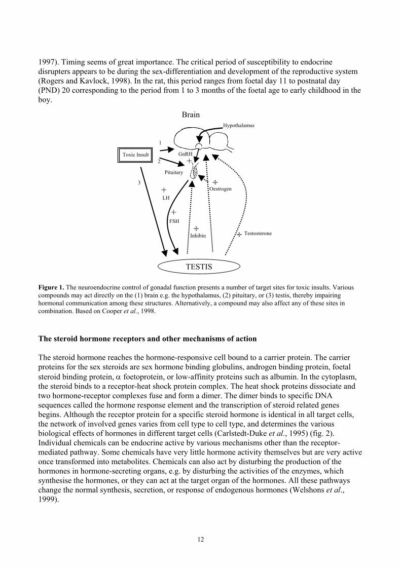
1997). Timing seems of great importance. The critical period of susceptibility to endocrine disrupters appears to be during the sex-differentiation and development of the reproductive system (Rogers and Kavlock, 1998). In the rat, this period ranges from foetal day 11 to postnatal day (PND) 20 corresponding to the period from 1 to 3 months of the foetal age to early childhood in the boy. Figure 1. The neuroendocrine control of gonadal function presents a number of target sites for toxic insults. Various compounds may act directly on the (1) brain e.g. the hypothalamus, (2) pituitary, or (3) testis, thereby impairing hormonal communication among these structures. Alternatively, a compound may also affect any of these sites in combination. Based on Cooper et al., 1998. The steroid hormone receptors and other mechanisms of action The steroid hormone reaches the hormone-responsive cell bound to a carrier protein. The carrier proteins for the sex steroids are sex hormone binding globulins, androgen binding protein, foetal steroid binding protein, α foetoprotein, or low-affinity proteins such as albumin. In the cytoplasm, the steroid binds to a receptor-heat shock protein complex. The heat shock proteins dissociate and two hormone-receptor complexes fuse and form a dimer. The dimer binds to specific DNA sequences called the hormone response element and the transcription of steroid related genes begins. Although the receptor protein for a specific steroid hormone is identical in all target cells, the network of involved genes varies from cell type to cell type, and determines the various biological effects of hormones in different target cells (Carlstedt-Duke et al., 1995) (fig. 2). Individual chemicals can be endocrine active by various mechanisms other than the receptor-mediated pathway. Some chemicals have very little hormone activity themselves but are very active once transformed into metabolites. Chemicals can also act by disturbing the production of the hormones in hormone-secreting organs, e.g. by disturbing the activities of the enzymes, which synthesise the hormones, or they can act at the target organ of the hormones. All these pathways change the normal synthesis, secretion, or response of endogenous hormones (Welshons et al., 1999).
Toxic Insult
TESTIS
1
2
3
LH
FSH
Hypothalamus
Pituitary
Inhibin
Oestrogen +
÷
÷
÷
GnRH
Brain
+
Testosterone
+
13
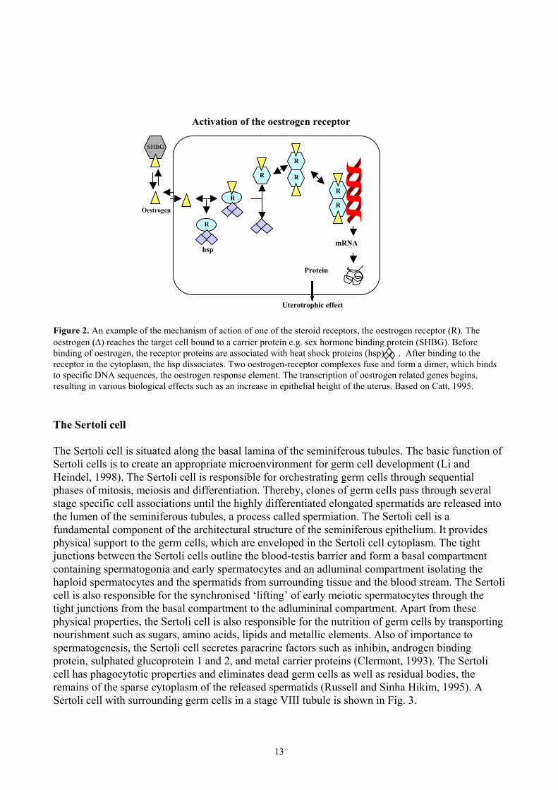
Figure 2. An example of the mechanism of action of one of the steroid receptors, the oestrogen receptor (R). The oestrogen (∆) reaches the target cell bound to a carrier protein e.g. sex hormone binding protein (SHBG). Before binding of oestrogen, the receptor proteins are associated with heat shock proteins (hsp) . After binding to the receptor in the cytoplasm, the hsp dissociates. Two oestrogen-receptor complexes fuse and form a dimer, which binds to specific DNA sequences, the oestrogen response element. The transcription of oestrogen related genes begins, resulting in various biological effects such as an increase in epithelial height of the uterus. Based on Catt, 1995. The Sertoli cell The Sertoli cell is situated along the basal lamina of the seminiferous tubules. The basic function of Sertoli cells is to create an appropriate microenvironment for germ cell development (Li and Heindel, 1998). The Sertoli cell is responsible for orchestrating germ cells through sequential phases of mitosis, meiosis and differentiation. Thereby, clones of germ cells pass through several stage specific cell associations until the highly differentiated elongated spermatids are released into the lumen of the seminiferous tubules, a process called spermiation. The Sertoli cell is a fundamental component of the architectural structure of the seminiferous epithelium. It provides physical support to the germ cells, which are enveloped in the Sertoli cell cytoplasm. The tight junctions between the Sertoli cells outline the blood-testis barrier and form a basal compartment containing spermatogonia and early spermatocytes and an adluminal compartment isolating the haploid spermatocytes and the spermatids from surrounding tissue and the blood stream. The Sertoli cell is also responsible for the synchronised ‘lifting’ of early meiotic spermatocytes through the tight junctions from the basal compartment to the adlumininal compartment. Apart from these physical properties, the Sertoli cell is also responsible for the nutrition of germ cells by transporting nourishment such as sugars, amino acids, lipids and metallic elements. Also of importance to spermatogenesis, the Sertoli cell secretes paracrine factors such as inhibin, androgen binding protein, sulphated glucoprotein 1 and 2, and metal carrier proteins (Clermont, 1993). The Sertoli cell has phagocytotic properties and eliminates dead germ cells as well as residual bodies, the remains of the sparse cytoplasm of the released spermatids (Russell and Sinha Hikim, 1995). A Sertoli cell with surrounding germ cells in a stage VIII tubule is shown in Fig. 3.
Uterotrophic effect
Activation of the oestrogen receptor
R
R
R
R
mRNA
Protein
hsp
Oestrogen
SHBG
R
R
R
14
Figure 3. The relationship of a Sertoli cell with the various germ cells of the rat seminiferous epithelium. The main functions of the Sertoli cell are listed. Labels: (SC) Sertoli cell; (ES) elongated spermatid (step 19); (RB) residual body; (RS) round spermatid; (P) pachytene spermatocyte; (PL) preleptothene spermatocytes; (A) apoptotic germ cell; (M) peritubular myoid cell. Based on Clermont, 1993. It is obvious that a well functioning Sertoli cell population is crucial for the development and maintenance of the spermatogenetic process at a normal-physiological level. Each Sertoli cell can only support a fixed number of germ cells during the development into spermatids. A toxic insult to a rat Sertoli cell may affect at least 10 elongated spermatids ‘nursed’ by this specific Sertoli cell (Russell and Peterson, 1984). Thus, a compound affecting the Sertoli cell population may result in a massive germ cell dysfunction and eventually lead to the death of these cells.
P
ESPhagocytosis
Barrier
Nutritive
Sustentacular
Secretion Spermiation
Lift
MPL
P
SC
RS
A
Receptor mediated endocytosis
RB
15
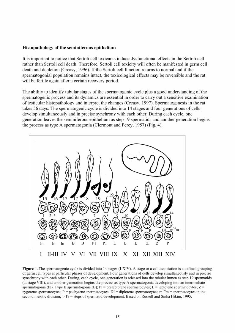
Histopathology of the seminiferous epithelium It is important to notice that Sertoli cell toxicants induce dysfunctional effects in the Sertoli cell rather than Sertoli cell death. Therefore, Sertoli cell toxicity will often be manifested in germ cell death and depletion (Creasy, 1996). If the Sertoli cell function returns to normal and if the spermatogonial population remains intact, the toxicological effects may be reversible and the rat will be fertile again after a certain recovery period. The ability to identify tubular stages of the spermatogenic cycle plus a good understanding of the spermatogenic process and its dynamics are essential in order to carry out a sensitive examination of testicular histopathology and interpret the changes (Creasy, 1997). Spermatogenesis in the rat takes 56 days. The spermatogenic cycle is divided into 14 stages and four generations of cells develop simultaneously and in precise synchrony with each other. During each cycle, one generation leaves the seminiferous epithelium as step 19 spermatids and another generation begins the process as type A spermatogonia (Clermont and Perey, 1957) (Fig. 4). Figure 4. The spermatogenic cycle is divided into 14 stages (I-XIV). A stage or a cell association is a defined grouping of germ cell types at particular phases of development. Four generations of cells develop simultaneously and in precise synschrony with each other. During, each cycle, one generation is released into the tubular lumen as step 19 spermatids (at stage VIII), and another generation begins the process as type A spermatogonia developing into an intermediate spermatogonia (In). Type B spermatogonia (B); Pl = preleptotene spermatocytes; L = leptotene spermatocytes; Z = zygotene spermatocytes; P = pachytene spermatocytes; DI = diplotene spermatocytes; m2 0m = spermatocytes in the second meiotic division; 1-19 = steps of spermatid development. Based on Russell and Sinha Hikim, 1995.
In In In B B P1 P1 LL L P ZZ
P m20m DIPP PP P P P P P P
1 2 -3 4 5 6 7 8 9 10 11 1312 14
15 16 17 17 18 1919
I II-III IV V VI VII VIII IX X XI XII XIII XIV
16
When a toxic effect in the testis is detected, then it may be appropriate to characterise the lesion further. Tubular staging will help characterising a lesion in its early phase of development. In the initial stages of testicular damage for example, the effect of a chemical may be restricted to a specific stage of the spermatogenic cycle and hence, the only observable change may be a specific cell type missing from the tubular epithelium. Another example is damage to the Sertoli cells by chemicals, which may prevent release of the spermatids in stage VIII, resulting in retention and phagocytosis during the subsequent stages or a delay in the spermatid release. The lesion is recognised by the presence of step 19 spermatids in stage IX –XIV tubules and may be the only detectable lesion (Creasy, 1997). Vacuoles in the cytoplasm of Sertoli cells are one of the most common histological manifestations of Sertoli cell toxicants and are observed after exposure to, e.g. DEHP, nitrobenzene and deltametrin (Poon et al., 1997; Shinoda et al., 1998; El-Gohary et al., 1999). Two forms of vacuoles exist, one form is characterised by multiple small vacuoles displacing the germ cells towards the lumen of the seminiferous tubules. This form is seen early in acute toxicity studies with MEHP (Richburg and Boekelheide, 1996). The other form consists of large clear vacuoles such as those seen following 2,5-hexanedione exposure (Larsen et al., 1991). Multinuclear giant cells are seen when germ cells fuse into an eosinophilic syncytium and are observed after exposure to 2,5-hexanedione (Larsen et al., 1991). The detachment of germ cells can be divided into two forms: a sloughing or a shedding of cells into the lumen of the seminiferous tubules. The sloughing of germ cells consists of clusters of germ cells adherent to fragments of the Sertoli cell cytoplasm, while the shedding is a release of germ cells without attached fragments of Sertoli cells (Boekelheide, 1993). Sloughing of germ cells into the lumen of the seminiferous tubules, germ cell degeneration and increased apoptosis are observed after exposure to several chemicals. Germ cell apoptosis Apoptosis is programmed cell death taking place in all organ systems, especially during development. Apoptosis is an active process regulated by molecular mechanisms that activate pre-programmed events. This process is essential for organ/tissue sculpturing, elimination of damaged or undesired structures, and control of cell number (Woolveridge and Morris, 2000). During spermatogenesis, apoptosis in testicular germ cells is recognised as an important physiologic mechanism to limit the germ cell population to numbers that the Sertoli cells can support (Billig et al., 1995). The mechanisms involved in germ cell apoptosis are numerous and are investigated for example by the removal of LH and FSH (Billig et al., 1995). Regulation of germ cell apoptosis in the normal testis is controlled by the Bcl-2 family, p53 and Fas-signalling pathway (Woolveridge and Morris, 2000). Effects of different chemicals on normal apoptotic processes are important and research into these topics is at its early beginning (Ladefoged et al., 2000). In toxicology, the effect of chemicals on the apoptotic frequency during development is of most concern. The most critical time window for studying chemically induced apoptosis may be when background apoptosis is at its highest. In the literature, no evidence of chemically induced Sertoli cell death by the apoptotic pathway exists in vivo. However, in vitro the oestrogen-like chemical, octylphenol, has been shown to induce Sertoli cell death (Raychoudhury et al., 1999). Increased germ cell apoptosis has been associated with
17
azoospermia, severe oligozoospermia and infertility in men (Lin et al. 1997; 1999). Ethnic differences, with higher incidences of spermatocytic apoptosis in Chinese men compared to Causcasian men, have been observed (Hikim et al., 1998). In rodents, several chemicals such as MEHP, 2,5-hexandione, nitrobenzene, deltamethrin, and hydroxyurea are reported to increase germ cell apoptosis (Lee et al., 1997; Shinoda et al., 1998; El-Gohary et al., 1999; Shin et al., 1999). Toxicants that injure or disrupt the Sertoli cell function can effectively reduce the supportive capacity of the cell, resulting in loss of germ cell attachment and increased elimination of germ cells via apoptosis (Richburg, 2000). Both the Fas receptor and the corresponding ligand are upregulated after administration of the two Sertoli cell toxicants, 2,5-hexanedione and MEHP, and the Sertoli cells up-regulate the Fas ligand in order to eliminate the Fas-receptor positive germ cells, which cannot be supported adequately (Lee et al., 1999; Richburg et al., 1999). Sertoli cell toxicants In the following, four of the most studied Sertoli cell toxicants are briefly addressed. In each case, there is strong evidence that the Sertoli cell is the primary target. Some very common phenomena such as inhibited spermiation, vacuolisation in the Sertoli cell cytoplasm and germ cell death have been related to several Sertoli cell toxicants. The mechanisms underlying these changes still remain unknown. However, altered germ cell attachment, blood-testis barrier defects, insufficient cytoskeletal support, metabolic insult, microtubule-dependent transport defects, and receptor and second messenger alterations are possibly mechanisms by which Sertoli cell toxicants can induce cell dysfunction and pathological changes (Boekelheide, 1993). The phthalates, DEHP and MEHP The phthalates are used extensively in consumer products and medical devices as plasticisers to impart flexibility to plastic materials (Li and Heindel, 1998). The testicular toxicity of phthalates is age-dependent and mature animals are less sensitive than immature animals. In 3-day-old rat pups a single dose of DEHP and MEHP decrease postnatal Sertoli cell proliferation (Li et al., 2000). Fourty eight hours after DEHP-treatment, a period of increased Sertoli cell proliferation followed. Li et al. did not observe any changes in the plasma level of FSH, suggesting that decreased FSH is not involved in the pathogenesis of DEHP in vivo as has been suggested from in vitro studies. Experiments in Sertoli cell cultures have demonstrated that MEHP inhibited FSH stimulated cAMP accumulation and altered FSH binding to Sertoli cell membranes (Heindel and Chapin, 1989; Grasso et al., 1993). Thus, the role of FSH in phthalate-induced testicular toxicity is not fully understood. Disrupted germ cell attachment caused by insufficient cytoskeletal support has been observed following exposure to phthalates, and studies by Richburg and Boekelheide (1996) suggest that the collapse of vimentin filaments in Sertoli cells exposed to MEHP results in loss of Sertoli cell-germ cell contact. 1,3-Dinitrobenzene 1,3-Dinitrobenzene is a chemical intermediate in the manufacture of azodyes, explosives, and several organic syntheses. Exposure to 1,3-dinitrobenzene results in cytoplasmic vacuolation in Sertoli cells. This toxic response is species and age specific. Mice are less sensitive than rats, and hamsters are insensitive to the testicular toxicity of 1,3-dinitrobenzene. Young animals are less sensitive to 1,3-dinitrobenzene than adults and this could be explained by metabolic differences since young animals metabolise 1,3-dinitrobenzene much faster than adult rats. In short term in vivo
18
studies in rats the testicular toxicity of 1,3-dinitrobenzene has also been shown to be stage-dependent beginning with the death of pachytene spermatocytes occurring first in stage VII and VIII tubules followed by focal degeneration and loss of pachytene spermatocytes, disorientation of elongated spermatids and further vacuolisation of Sertoli cell cytoplasm and vesiculation of the smooth endoplasmic reticulum in stages IX to X. A decrease in seminiferous tubule fluid 3 hours after dosing has also been reported in rat (Li and Heindel, 1998). Additionally, increased levels of androgen binding protein have been observed in rats (Suter et al., 1998). The production of lactate and pyruvate in the Sertoli cell are also altered by 1,3-dinitrobenzene (Williams and Foster, 1988). Ethylene glycol ethers Ethylene glycol ethers are a family of organic water-miscible solvents used in a wide variety of products including printing inks, textile dyes, epoxy resin coatings, and as anti-icing additive in jet fuels. 2-Methoxyethanol and 2-ethoxyethanol are the most potent of the ethylene glycol ethers in terms of testicular toxicity. The testis toxic metabolites are the alkoxyacetic acids, methoxyacetic acid and ethoxyacetic acid, respectively. They induce testicular atrophy, decreased sperm motility and increased abnormal sperm. Either the Sertoli cell or the cell-to-cell communication between Sertoli cells and germ cells is the initial target. Pachytene spermatocytes were the initial and major site of morphological damage (Creasy and Foster, 1984; Creasy et al., 1985). Although spermatocyte cell death has been shown to follow the apoptotic pathway across species, the morphology of the dying spermatocytes seems to be different in different species. 2-Methoxyethanol-induced spermatocytic cell death looks necrotic in rats, but apoptotic banding pattern is present in all the investigated laboratory species including rats. The germ cell degeneration is possibly mediated through initial Sertoli cell-based events such as changes in kinase activation/inactivation and disrupted cell cycle regulation (Li and Heindel, 1998). n-Hexane n-Hexane was a commonly used solvent but has now been phased out. It is a neurotoxicant, resulting in peripheral polyneuropathy. n-Hexane is metabolically converted into aliphatic ketones and alcohols. It has been shown that 2,5-hexanedione is the active testicular toxicant of n-hexane (Li and Heindel, 1998). Sertoli cell injury detected as vacuolisation leads to germ cell loss. The Sertoli cell vacuoles are found in stage I and stage XII-XIV of the spermatogenetic cycle after 3 weeks of dosing. Of the germ cells, the elongated spermatids are most susceptible to hexanedione-induced toxicity. Transport of the seminiferous tubule fluid is a microtubule-dependent process and disturbances in this process are observed in 2,5-hexanedione dosed rats. Additionally, type A spermatogonia continues to produce progeny cells, but the cells fail to differentiate beyond the spermatogonial stage and die through the apoptotic pathway, thereby reducing the size of the stem cell population resulting in development of a permanent atrophy (Allard and Boekelheide, 1996). Sertoli cell number, diameter and length of the seminiferous tubules The proliferation period of Sertoli cells in the rat begins at gestational day (GD) 17-19 and continues until PND 15-20 (Steinberger and Steinberger, 1971; Nagy, 1972; Orth, 1982). It is presumed that at the end of this proliferation period, the number of Sertoli cells will remain constant throughout life (Orth et al., 1988).
19
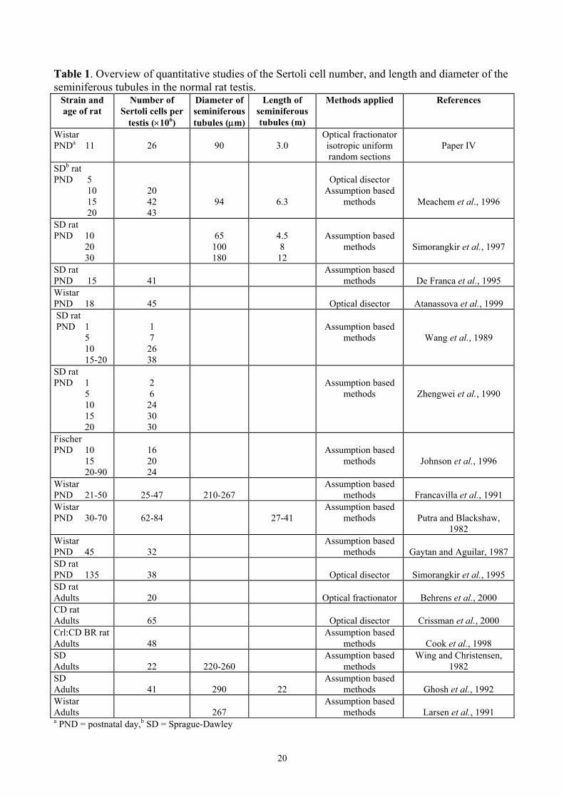
In the literature, several quantitative studies of the Sertoli cell number and diameter and length of the seminiferous tubules in the testis of normal rats have been reported, and these parameters have been recognised as sensitive parameters of toxic response (Larsen et al., 1991; Simorangkir et al., 1997; Wreford, 1995). Most of these quantitative studies have used assumption based methods that may confound the conclusions. The estimates of numerical density in the earlier studies have been obtained from two-dimensional images or probes and are potentially biased, because the probability that the profile in question appears in a section is related to its size, shape and the orientation of the tissue (West and Gundersen, 1990). In earlier studies, the seminiferous tubule diameter is estimated without considering the orientation of the structure or the shrinkage of the tissue. Table 1 gives an overview of the quantitative studies of the Sertoli cell number, diameter and length of the seminiferous tubules in the testis of normal rats. Only the studies estimating the Sertoli cell number at PND 1 to 20 are presented. Apart from two studies (Putra and Blackshaw, 1982; Francavilla et al., 1991), there are consistent results suggesting that the Sertoli cell proliferation in the rat ceases from PND day 15-20 (see table 1). According to the studies mentioned in table 1, the total Sertoli cell number in adult male rats ranges from 20 to 65 million per testis and the total Sertoli cell number in 10 to 11-day-old rats is around 16-26 million per testis. In one study, the diameter and length of the seminiferous tubules in 10-day-old rats have been estimated to 65 µm and 4.5 m, respectively (Simorangkir et al., 1997). These results were estimated from conventional cross sections and not from isotropic uniform random sections. Additionally, the shrinkage effect was not considered. In the literature, a few studies have estimated the total number of Sertoli cells after exposure to oestrogens or the oestrogen-like chemical octylphenol (Cook et al., 1998; Atanassova et al., 1999; Behrens et al., 2000; Sweeney et al., 2000). Behrens et al. (2000) estimated the number of Sertoli cells in adult Sprague-Dawley rats with the optical fractionator. They obtained a slightly lower number of Sertoli cells in the testis of adult control rats compared to the results presented in paper IV in 11-day-old Wistar rats. This may be explained by the use of different rat strains. Behrens et al. (2000) also estimated the number of Sertoli cells in adult rats exposed to DES in utero from gestational day 17-19 and the number of Sertoli cells tended to decrease; however, the decrease was not statistically significant. Petersen et al. (1999) investigated the number of Sertoli cells in man to be approximately 900×106.
20
Table 1. Overview of quantitative studies of the Sertoli cell number, and length and diameter of the seminiferous tubules in the normal rat testis.
Strain and age of rat
Number of Sertoli cells per
testis (×106)
Diameter of seminiferous tubules (µm)
Length of seminiferous tubules (m)
Methods applied References
Wistar PNDa 11
26
90
3.0
Optical fractionator isotropic uniform random sections
Paper IV
SDb rat PND 5 10 15 20
20 42 43
94
6.3
Optical disector
Assumption based methods
Meachem et al., 1996
SD rat
PND 10 20 30
65
100 180
4.5 8
12
Assumption based
methods
Simorangkir et al., 1997
SD rat PND 15
41
Assumption based methods
De Franca et al., 1995
Wistar PND 18
45
Optical disector
Atanassova et al., 1999
SD rat PND 1 5 10 15-20
1 7
26 38
Assumption based
methods
Wang et al., 1989
SD rat PND 1 5 10 15 20
2 6
24 30 30
Assumption based
methods
Zhengwei et al., 1990
Fischer PND 10 15 20-90
16 20 24
Assumption based
methods
Johnson et al., 1996
Wistar PND 21-50
25-47
210-267
Assumption based methods
Francavilla et al., 1991
Wistar PND 30-70
62-84
27-41
Assumption based methods
Putra and Blackshaw,
1982 Wistar PND 45
32
Assumption based methods
Gaytan and Aguilar, 1987
SD rat PND 135
38
Optical disector
Simorangkir et al., 1995
SD rat Adults
20
Optical fractionator
Behrens et al., 2000
CD rat Adults
65
Optical disector
Crissman et al., 2000
Crl:CD BR rat Adults
48
Assumption based methods
Cook et al., 1998
SD Adults
22
220-260
Assumption based methods
Wing and Christensen, 1982
SD Adults
41
290
22
Assumption based methods
Ghosh et al., 1992
Wistar Adults
267
Assumption based methods
Larsen et al., 1991
a PND = postnatal day,b SD = Sprague-Dawley
21
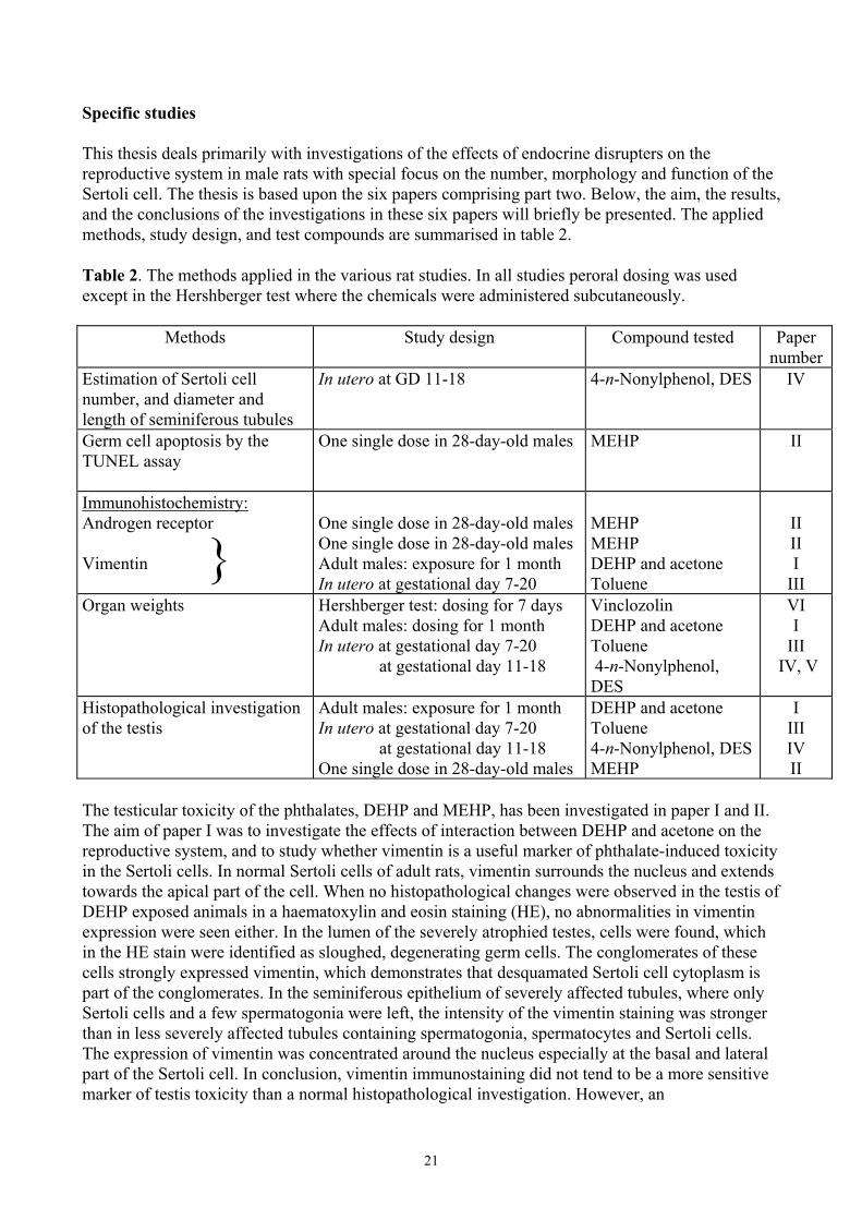
Specific studies This thesis deals primarily with investigations of the effects of endocrine disrupters on the reproductive system in male rats with special focus on the number, morphology and function of the Sertoli cell. The thesis is based upon the six papers comprising part two. Below, the aim, the results, and the conclusions of the investigations in these six papers will briefly be presented. The applied methods, study design, and test compounds are summarised in table 2. Table 2. The methods applied in the various rat studies. In all studies peroral dosing was used except in the Hershberger test where the chemicals were administered subcutaneously.
Methods Study design Compound tested Paper number
Estimation of Sertoli cell number, and diameter and length of seminiferous tubules
In utero at GD 11-18 4-n-Nonylphenol, DES IV
Germ cell apoptosis by the TUNEL assay
One single dose in 28-day-old males MEHP II
Immunohistochemistry: Androgen receptor Vimentin
One single dose in 28-day-old males One single dose in 28-day-old males Adult males: exposure for 1 month In utero at gestational day 7-20
MEHP MEHP DEHP and acetone Toluene
II II I
III Organ weights Hershberger test: dosing for 7 days
Adult males: dosing for 1 month In utero at gestational day 7-20 at gestational day 11-18
Vinclozolin DEHP and acetone Toluene 4-n-Nonylphenol, DES
VI I
III IV, V
Histopathological investigation of the testis
Adult males: exposure for 1 month In utero at gestational day 7-20 at gestational day 11-18 One single dose in 28-day-old males
DEHP and acetone Toluene 4-n-Nonylphenol, DESMEHP
I III IV II
The testicular toxicity of the phthalates, DEHP and MEHP, has been investigated in paper I and II. The aim of paper I was to investigate the effects of interaction between DEHP and acetone on the reproductive system, and to study whether vimentin is a useful marker of phthalate-induced toxicity in the Sertoli cells. In normal Sertoli cells of adult rats, vimentin surrounds the nucleus and extends towards the apical part of the cell. When no histopathological changes were observed in the testis of DEHP exposed animals in a haematoxylin and eosin staining (HE), no abnormalities in vimentin expression were seen either. In the lumen of the severely atrophied testes, cells were found, which in the HE stain were identified as sloughed, degenerating germ cells. The conglomerates of these cells strongly expressed vimentin, which demonstrates that desquamated Sertoli cell cytoplasm is part of the conglomerates. In the seminiferous epithelium of severely affected tubules, where only Sertoli cells and a few spermatogonia were left, the intensity of the vimentin staining was stronger than in less severely affected tubules containing spermatogonia, spermatocytes and Sertoli cells. The expression of vimentin was concentrated around the nucleus especially at the basal and lateral part of the Sertoli cell. In conclusion, vimentin immunostaining did not tend to be a more sensitive marker of testis toxicity than a normal histopathological investigation. However, an
}
22
immunohistochemical investigation of vimentin can be a valuable supplement when investigating Sertoli cell toxicants that may affect the supporting capacity of the Sertoli cells including the vimentin filaments. The aim of paper II was to investigate the MEHP-induced testicular toxicity in 28-day-old rats by histopathological, immunohistochemical, and apoptotic related measurements following a single oral dose. No histopathological changes were observed in the testis of the control group and in the MEHP exposed group sacrificed 3 hours following dosing. Slightly disorganised tubules, detachment, and sloughing of germ cells into the lumen of the seminiferous tubules were observed after 6 hours, and this effect was enhanced 12 hours after dosing, as only Sertoli cells and spermatogonia remained attached to the basal lamina. The sloughing of germ cells was apparently most pronounced in stage IX to XIV tubules. Three to 12 hours after administration of a single oral dose of MEHP, the vimentin filaments in the Sertoli cell cytoplasm collapsed, the apical extensions towards the lumen of the tubules disappeared, and a perinuclear condensation of vimentin was observed. The intensity of the androgen receptor immunostaining was most prominent in the peritubular myoid cells and in the Leydig cells and to a lesser extent in the Sertoli cells. In the Sertoli cells, the intensity of the reaction varied in a stage specific manner. This pattern did not change between the control group and the dosed groups. The TUNEL assay revealed that the level of apoptosis was unchanged in the MEHP dosed groups compared to the control group, when the number of apoptotic cells was counted in 100 randomly selected tubules per animal. However, a biochemical analysis demonstrated that the caspase-3 activity was increased in the MEHP dosed rats. In conclusion, vimentin localisation in the Sertoli cells and increased levels of caspase-3 activity in the testis appear to be sensitive and early markers of MEHP-induced testicular toxicity. However, the increased caspase-3 activity could not be confirmed by a semiquantitative investigation of TUNEL-positive and negative germ cells fulfilling the morphological chriteria of apoptosis. The effect of MEHP in testis is apparently not involving the androgen receptor. Similar to the immunohistochemical investigations presented in paper I and II, the effects of toluene on the Sertoli cell vimentin was investigated in male rats exposed to toluene in utero (paper III). Paired testes weight and histopathology were used as markers of testis toxicity at PND 11, 21 and 90. The absolute and relative testes weights were reduced in all three age groups, although not to a statistically significant degree. Histopathological examinations of the testis and immuno-expression of vimentin did not reveal any differences between toluene-exposed animals and control animals. In conclusion, toluene did not induce any effects on the parameters investigated. In a reproductive study rats were dosed with 4-n-nonylphenol or DES in utero (papers IV and V). A decrease in terminal body weight and a decrease in the absolute weight of right epididymis were observed in the DES-treated male rats sacrificed at PND 110 (paper V). At PND 110, the absolute right epididymal weight was decreased in the rats exposed to 75 and 15 mg/kg/day 4-n-nonylphenol, whereas the absolute weight of the left epididymis and the relative weight of the left and right epididymes were not changed. In the DES group (30 µg/kg/day), the absolute liver weight was decreased at PND 11 and 21. The absolute liver weight was decreased at PND 21 in the rats exposed to 75 mg/kg/day 4-n-nonylphenol and at PND 11 and 21 in the rats exposed to 15 mg/kg/day. 4-n-Nonylphenol did not decrease the absolute liver weight at PND 110. No changes in the relative liver weights were observed in any of the DES or 4-n-nonylphenol-treated groups. The absolute weights of the testes, seminal vesicles, prostate and adrenals were not affected in any of the treated groups. In conclusion, the body weight and the absolute weights of the liver and the right epididymis were decreased by exposure to both DES and 4-n-nonylphenol. The testis weight, testis histopathology, the diameter and length of the seminiferous tubules, and the
23
total number of Sertoli cells were investigated in 11-day-old rats exposed to 30 µg/kg/day DES or 75 mg/kg/day 4-n-nonylphenol in utero (paper IV). No differences in testis weight, histopathology, or length or diameter of the seminiferous tubules were observed in the DES and 4-n-nonylphenol exposed groups when compared to the control group. In the DES-treated group, a decrease in the number of Sertoli cells was observed when compared to the control group, whereas 4-n-nonylphenol had no effect. In conclusion, the results suggest that DES inhibits the Sertoli cell proliferation in the foetal testis and furthermore indicate that oestrogens may pose a risk to the reproductive capacity in sensitive species, including man. In the studies presented in paper VI, the well-known androgen receptor antagonist vinclozolin, was investigated in the classical Hershberger assay determining organ weight changes. The objective was to improve sensitivity of the assay by measuring hormone levels as well as determining changes in the expression of androgen-responsive genes. Compared to the testosterone (control) group, a decrease in the weight of the seminal vesicles was observed in the group dosed with 50 mg/kg/day of vinclozolin. The weights of the ventrale prostate and levator ani/bulbocavernosus muscles were not significantly decreased compared to the testosterone group. No change in the level of LH was observed, but gene-expressions of both testosterone-repressed prostatic message 2 and prostate specific binding protein polypeptide C3 were different from the expressions in the testosterone-treated group in spite of the use of a suboptimal dose of vinclozolin. Hence, gene expression can be a supplement as well as possibly a more sensitive parameter in the screening of chemicals with antiandrogenic effect. Discussion and main conclusions The methods The aim of the thesis was to study the effects of endocrine disrupters on the rat testis by using various qualitative and quantitative methods. The thesis particularly focused on methods investigating effects on the number, morphology and function of the Sertoli cells. As a marker of the supporting capacity of the Sertoli cells, the immunohistochemical investigation of vimentin can be a valuable parameter in the acute phase of toxicity. The change in the localisation and collapse of the vimentin filaments has been shown to be a convincing endpoint in our investigations of MEHP-induced Sertoli cell toxicity. Generally, when evaluating the effect of a chemical on a tissue stained with immunohistochemistry, a shift from a positively stained cell location to a negatively stained cell location or vice versa, is a convincing endpoint. However, alterations in the intensity of an immunohistochemical staining are more difficult to deal with in the subjective evaluation of the pathologist. The TUNEL assay is suitable when demonstrating germ cell apoptosis and chemically induced disturbed spermatogenesis in toxicity studies. It is, however, very important to be aware of the limitations of the method concerning sensitivity and specificity (Dalgaard, 2000). Paper II presents a semiquantitative investigation of apoptotic germ cells in rats exposed to MEHP. The results obtained are only indications of the factual level of apoptosis and with the huge physiological background level in germ cell apoptosis in mind as well as high inter-animal variation, many tubules with substantial amounts of apoptotic germ cells have to be counted before chemically induced effects can be detected. Furthermore, high dose levels are probably required in order to detect an effect. Hence, the TUNEL assay is valuable for studying mechanism of action in MEHP-induced testicular toxicity. However, it may not be a sensitive parameter. This experience will be brought forward in future testing of other chemicals. Unbiased stereological approaches can be applied in order to estimate cell numbers, volumes, surface area, and length. Several studies use potentially biased approaches by use of assumptions of shape, size, orientation, and shrinkage (Wreford, 1995). Therefore, the historical data of the Sertoli cell number in
24
assumption based studies should be interpreted with caution, and consequently a direct comparison across studies is not recommended. Quantification by use of stereological principles is technically a rather challenging and time-consuming procedure and therefore, some indications of a possible effect are preferable before investigations of testicular toxicity by stereological methods are initiated. Parameters such as an immunohistochemical staining of the vimentin filaments and the androgen receptor, the TUNEL-staining, and estimation of total Sertoli cell numbers increase the knowledge of the mechanisms involved in chemically induced testicular toxicity. Furthermore, an investigation of Sertoli cell vimentin is a promising parameter in the initial stage of Sertoli cell toxicity. Likewise, the number of Sertoli cells is promising as a sensitive parameter in the future testing of endocrine disrupters. It is however, very important always to combine these investigations with a careful histopathological investigation. The hypothesis It has been reported that rats with a decreased number of Sertoli cells have a decreased adult sperm count (Orth et al., 1988) and that neonatal secretion of FSH can be inhibited by exogenously administered oestrogens (Sharpe, 1993). Based on these data from studies in animals, Sharpe and Skakkebæk (1993) hypothesised that environmental oestrogens, via inhibition of the secretion of FSH, may decrease the daily sperm production in men by altering the multiplication of Sertoli cells during early life (Sharpe, 1993). The results presented in paper IV indicate that foetal exposure to a chemical such as the synthetic oestrogen DES decreases the rat Sertoli cell number. 4-n-Nonylphenol did not decrease the Sertoli cell number at the dose level tested. However, other weak oestrogen-like compounds may induce changes in the Sertoli cell number. The Sertoli cell toxicants, DEHP, MEHP, ethylene glycol ethers, and n-hexane induce various histopathological changes at fairly high doses. However, it could be interesting to examine whether foetal exposure to these chemicals, either individually or in combination, affect quantitative parameters such as the diameter and length of seminiferous tubules, and the Sertoli cell number at low dose levels closer to general human exposure levels. Last year, another hypothesis concerning the decrease in sperm count has been proposed by Crissman et al. (2000). The decrease in human sperm counts may be explained by the elimination of the juvenile iodine deficiency by iodination of salt since the mid-1920s. In young rats, iodine deficiency results in hypothyroidism, thereby increasing the Sertoli cell number by extending the cell’s proliferation period. The increased Sertoli cell population can ‘nurse’ more germ cells and thereby result in an increased spermatid output in adults. According to the authors, the improvement of the human juvenile thyroid status by iodised salt is thus a quite plausible cause for the decreasing sperm count observed during the last decades. Some chemicals can also induce hypothyroidism, increased sperm counts, and increased testis weight as have been observed in adult rats exposed neonatally to the PCB mixture, Aroclor 1242 and this may be due to an increased number of Sertoli cells (Cooke et al., 1996). Besides from increasing the number of Sertoli cells, hypothyroidism may also induce a periodically increase in germ cell degeneration as undifferentiated Sertoli cells are unable to support complete germ cell development until the Sertoli cell differentiation has completed (Chapin et al., 1996). Ethnic differences in sperm counts have also been proposed as an explanation for the discrepancy in sperm counts in some industrialised countries. Johnson et al. (1998) have observed that the daily sperm production, the Sertoli cell number, and the diameter of the seminiferous tubules are lower in Chinese men compared to Hispanic men and non-Hispanic Caucasian men. Thus, if there is a decline in the sperm count in some industrial countries, the aetiology still remains uncertain. However, the increased incidence of reproductive malformations and testicular cancer especially in Denmark probably have a foetal origin. This observation taken together with the reproductive disorders, induced after exposure to endocrine disrupters during the critical windows of reproductive development in animals, indicate
25
that either individual chemicals or chemicals in combination may be involved in the aetiology. Future perspectives In May 2001, a panel of academic, government and industry scientists in the US determined that there is "credible evidence" that some endocrine disrupters can affect test animals' bodily functions at very low levels – well below the "no effect" levels determined by traditional testing (National Toxicology Program’s Report of endocrine disrupters and low dose effects). Weak endocrine disrupters administered alone or in combination at dose levels close to general human exposure levels do probably not induce substantial and qualitative effects, which are detectable with conventional toxicological testing strategies. Hence, there is a need to develop new, sensitive and robust methods in order to detect endocrine disrupters individually, or in combination at low dose levels. Regarding endocrine disrupters and their possible adverse effects on the human reproductive function, quantitative methods as a supplement to qualitative investigations will be of high value. A quantitative estimation of the Sertoli cell number seems to be a sensitive parameter when studying chemicals with potential effects on the neuro-endocrine axis. Hence, there is a need for further studies of endocrine disrupters where valid and unbiased quantitative stereological methods will be highly relevant. Main conclusion Immunostaining of Sertoli cell vimentin is a valuable marker of Sertoli cell toxicity in the acute phase of toxicity. It is, however, very important always to use immunohistochemistry as well as the TUNEL-staining as a supplement to a careful histopathological investigation in order to avoid exclusion of valuable information. The estimation of the Sertoli cell number by use of valid and unbiased stereology-based study design will be highly relevant in future toxicological investigations of endocrine disrupters. The results obtained with stereology-based quantifications will provide strong evidence in future studies of endocrine disrupters. The results obtained in the in utero study of the synthetic oestrogen, DES, supports the hypothesis by Sharpe and Skakkebæk (1993) that foetal exposure to environmental oestrogens may decrease the Sertoli cell number and perhaps result in a decreased sperm output.
26
References Allard EK, Boekelheide K. 1996. Fate of germ cells in 2,5-hexanedione-induced testicular injury. II. Atrophy persists due to a reduced stem cell mass and ongoing apoptosis. Toxicol Appl Pharmacol 137:149-156. Andersen AG. 2000. Semen quality and reproductive hormones in normal young men and in partners of pregnant women. Ph.D. Thesis. University of Copenhagen, Rigshospitalet, Department of Growth and Reproduction and The Fertility Clinic. Ashby J, Lefevre PA. 2000. The peripubertal male rat assay as an alternative to the Hershberger castrated male rat assay for the detection of anti-androgens, oestrogens and metabolic modulators. J Appl Toxicol 20:35-47.
Atanassova N, McKinnell C, Walker M, Turner KJ, Fisher JS, Morley M, Millar MR, Groome NP, Sharpe RM. 1999. Permanent effects of neonatal estrogen exposure in rats on reproductive hormone levels, Sertoli cell number, and the efficiency of spermatogenesis in adulthood. Endocrinology 140:5364-5373.
Behrens GH, Petersen PM, Grotmol T, Sorensen DR, Torjesen P, Tretli S, Haugen TB. 2000. Reproductive function in male rats after brief in utero exposure to diethylstilboestrol. Int J Androl 23:366-371.
Billig H, Furuta I, Rivier C, Tapanainen J, Parvinen M, Hsueh AJ. 1995. Apoptosis in testis germ cells: developmental changes in gonadotropin dependence and localization to selective tubule stages. Endocrinology 136:5-12.
Blanco-Rodriguez J, Martinez-Garcia C. 1998. Apoptosis pattern elicited by several apoptogenic agents on the seminiferous epithelium of the adult rat testis. J Androl 19:487-497.
Boekelheide K. Sertoli cell toxicants. 1993. In: The Sertoli cell. Eds.: Russell LD and Griswold MD. Cache River Press 552-575. Carlsen E, Giwercman A, Keiding N, Skakkebæk NE. 1992. Evidence for decreasing quality of semen during past 50 years. BMJ 305:609-613.
Carlstedt-Duke J, Wright A, Göttlicher M, Okret S, Gustafsson JÅ. 1995. Molecular mechanisms of hormone action: regulation of target cell function by the steroid hormone receptor supergene family. In: Endocrinology and metabolism. Eds.: Felig P, Baxter JD, Frohman LA. McGraw-Hill.169-199. Catt KJ. 1995. Molecular mechanisms of hormone action: control of target cell function by peptide and catecholamine hormones. In: Endocrinology and metabolism. Eds.: Felig P, Baxter JD, Frohman LA. McGraw-Hill 91-199. Chapin RE, Stevens JT, Hughes CL, Kelce WR, Hess RA, Daston GP. 1996. Endocrine modulation of reproduction. Fundam Appl Toxicol 29:1-17.
Clermont Y. 1993. Introduction to the Sertoli cell. In: The Sertoli cell. Eds.: Russell LD and Griswold MD. Cache River Press xxii-xxv.
Clermont Y, Perey B. 1957. Quantitative study of the cell population of the seminiferous tubules in immature rats. Am J Anat 100:241-267.
Cook JC, Johnson L, O'Connor JC, Biegel LB, Krams CH, Frame SR, Hurtt ME. 1998. Effects of dietary 17 beta-estradiol exposure on serum hormone concentrations and testicular parameters in male Crl:CD BR rats. Toxicol Sci 44:155-168. Cooke PS, Zhao Y- D, Hansen LG. 1996. Neonatal polychlorinated biphenyl treatment increases adult testis size and sperm production in the rat. Toxicol Appl Pharmacol 136:112-117. Cooper RL, Goldman JM, Tyrey L. 1998. The hypothalamus and pituitary as targets for reproductive toxicants. Developmental Toxicology. In: Reproductive and developmental toxicology. Ed.: Korach KS. Marcel Dekker, Inc 195-210. Creasy DM, Flynn JC, Gray TJ, Butler WH. 1985. A quantitative study of stage-specific spermatocyte damage
27
following administration of ethylene glycol monomethyl ether in the rat. Exp Mol Pathol 43:321-336.
Creasy DM, Foster PM. 1984. The morphological development of glycol ether-induced testicular atrophy in the rat. Exp Mol Pathol 40:169-176.
Creasy DM. 1996. Spontaneous and chemically induced neoplasia of the testes. Proceedings at ILSI Histopathology seminar 11-13. September 35-41. Creasy DM. 1997. Evaluation of testicular toxicity in safety evaluation studies: the appropriate use of spermatogenic staging. Toxicol Pathol 25:119-131.
Crissman JW, Cooke PS, Hess RA, Marty MS, Liberacki AB. 2000. Postulated human sperm count decline may involve historic elimination of juvenile iodine deficiency: a new hypothesis with experimental evidence in the rat. Toxicol Sci 53:400-410.
Dalgaard M. 2000. In situ detection of apoptosis in young rat testis: an evaluation of the TUNEL method. In: Chemical induced apoptosis. Eds.: Ladefoged O, Lam HR, Sørensen IK, Thorup I, Pilegaard K, Edelfors S. Nordic Council of Ministers. TemaNord 2000 79-84.
El-Gohary M, Awara WM, Nassar S, Hawas S. 1999. Deltamethrin-induced testicular apoptosis in rats: the protective effect of nitric oxide synthase inhibitor. Toxicology 132:1-8.
Foster PM, Gray LE. 2001. Endocrine disruption and male reproductive development. Abstract at: Society of Toxicology 40th annual meeting. Toxicol Sci suppl 60, 1-3.
Franca de LR, Hess RA, Cooke PS, Russell LD. 1995. Neonatal hypothyroidism causes delayed Sertoli cell maturation in rats treated with propylthiouracil: evidence that the Sertoli cell controls testis growth. The Anatomical Record 242:56-69.
Francavilla S, Cordeschi G, Properzi G, Di Cicco L, Jannini EA, Palmero S, Fugassa E, Loras B, D'Armiento M. 1991. Effect of thyroid hormone on the pre- and post-natal development of the rat testis. J Endocrinol 129:35-42.
Frohman LA. 1995. Diseases of the anterior pituitary. In: Endocrinology and metabolism. Eds.: Felig P, Baxter JD, Frohman LA. McGraw-Hill. 289-383.
Gaytan F, Aguilar E. 1987. Quantitative analysis of Sertoli cells in neonatally oestrogen-treated rats. J Reprod Fert 79:589-598.
Ghosh S, Bartke A, Grasso P, Reichert LE,Russell LD. 1992. Structural manifestations of the rat Sertoli cell to hypophysectomy: a correlative morphometric and endocrine study. Endocrinology 131:485-497.
Grasso P, Heindel JJ, Powell CJ, Reichert LE, Jr. 1993. Effects of mono(2-ethylhexyl) phthalate, a testicular toxicant, on follicle-stimulating hormone binding to membranes from cultured rat Sertoli cells. Biol Reprod 48:454-459.
Gray LE, Kelce WR, Wiese T, Tyl R, Gaido K, Cook J, Klinefelter G, Desaulniers D, Wilson E, Zacharewski T, Waller C, Foster P, Laskey J, Reel J, Giesy J, Laws S, McLachlan J, Breslin W, Cooper R, Di Giulio R, Johnson R, Purdy R, Mihaich E, Safe S, Colborn T. 1997. Endocrine Screening Methods Workshop report: detection of estrogenic and androgenic hormonal and antihormonal activity for chemicals that act via receptor or steroidogenic enzyme mechanisms. Reprod Toxicol 11:719-750.
Gray LE, Wolf C, Lambright C, Mann P, Price M, Cooper RL, Ostby J. 1999. Administration of potentially antiandrogenic pesticides (procymidone, linuron, iprodione, chlozolinate, p,p'-DDE, and ketoconazole) and toxic substances (dibutyl- and diethylhexyl phthalate, PCB 169, and ethane dimethane sulphonate) during sexual differentiation produces diverse profiles of reproductive malformations in the male rat. Toxicol Ind Health 15:94-118.
Heindel JJ, Chapin RE. 1989. Inhibition of FSH-stimulated cAMP accumulation by mono(2- ethylhexyl) phthalate in primary rat Sertoli cell cultures. Toxicol Appl Pharmacol 97:377-385.
Hikim AP, Wang C, Lue Y, Johnson L, Wang XH, Swerdloff RS. 1998. Spontaneous germ cell apoptosis in humans:
28
evidence for ethnic differences in the susceptibility of germ cells to programmed cell death. J Clin Endocrinol Metab 83:152-156.
Johnson L, Barnard JJ, Rodriguez L, Smith EC, Wang XH, Wang C. 1998. Ethnic differences in testicular structure and spermatogenic potential may predispose testes of Asian men to a heightened sensitivity to steroidal contraceptives. J Androl 19:348-357.
Johnson L, Suggs LC, Norton YM, Zeh WC. 1996. Effect of developmental age or time after transplantation on Sertoli cell number and testicular size in inbred Fischer rats. Biol Reprod 54:948-959.
Kelce WR, Lambright CR, Gray LEJ, Roberts KP. 1997. Vinclozolin and p,p'-DDE alter androgen-dependent gene expression: in vivo confirmation of an androgen receptor-mediated mechanism. Toxicol Appl Pharmacol 142:192-200.
Ladefoged O, Lam HR, Sørensen IK, Thorup I, Pilegaard K, Edelfors S (eds). 2000. Chemical induced apoptosis, Nordic Council of Ministers. TemaNord 2000. Larsen JJ, Lykkegaard M, Ladefoged O. 1991. Infertility in rats induced by 2,5-hexanedione in combination with acetone. Pharmacol Toxicol 69:43-46.
Lee J, Richburg JH, Shipp EB, Meistrich ML, Boekelheide K. 1999. The Fas system, a regulator of testicular germ cell apoptosis, is differentially up-regulated in Sertoli cell versus germ cell injury of the testis. Endocrinology 140:852-858.
Lee J, Richburg JH, Younkin SC, Boekelheide K. 1997. The Fas system is a key regulator of germ cell apoptosis in the testis. Endocrinology 138:2081-2088.
Li L-H and Heindel JJ. 1998. Sertoli cell toxicants. In: Reproductive and developmental toxicology. Ed.: Korach KS. Marcel Dekker, Inc 655-691.
Li L-H, Jester WF, Laslett AL, Orth JM. 2000. A single dose of di-(2-ethylhexyl)phthalate in neonatal rats alters gonocytes, reduces Sertoli cell proliferation, and decreases cyclin D2 expression. Toxicol Appl Pharmacol 166:222-229. Lin WW, Lamb DJ, Lipshultz LI, Kim ED. 1999. Demonstration of testicular apoptosis in human male infertility states using a DNA laddering technique. Int Urol Nephrol 31:361-370.
Lin WW, Lamb DJ, Wheeler TM, Lipshultz LI, Kim ED. 1997. In situ end-labeling of human testicular tissue demonstrates increased apoptosis in conditions of abnormal spermatogenesis. Fertil Steril 68:1065-1069.
MacLaughlin DT, Donahoe PK. 1998. Müllerian-inhibiting substance activity and normal male sex determination. In: Reproductive and developmental toxicology. Ed.: Korach KS. Marcel Dekker, Inc. 1-10.
McIntyre BS, Barlow NJ, Wallace DG, Maness SC, Gaido KW, Foster PMD. 2000. Effects of in utero exposure to linuron on androgen-dependent reproductive development in the male Crl:CD(SD)BR rat. Toxicol Appl Pharmacol 167:87-99.
Meachem SJ, McLachlan RI, de Kretser DM, Robertson DM, Wreford NG. 1996. Neonatal exposure of rats to recombinant follicle stimulating hormone increases adult Sertoli and spermatogenic cell numbers. Biol Reprod 54:36-44.
Nagy F. 1972. Cell division kinetics and DNA synthesis in the immature Sertoli cells of the rat testis. J Reprod Fertil 28:389-395.
National Toxicology Program’s Report of the Endocrine Disruptors, Low Dose, Peer Review, 2001. Orth JM, Gunsalus GL, Lamperti AA. 1988. Evidence from Sertoli cell-depleted rats indicates that spermatid number in adults depends on numbers of Sertoli cells produced during perinatal development. Endocrinology 122:787-794.
29
Orth JM. 1982. Proliferation of Sertoli cells in fetal and postnatal rats: a quantitative autoradiographic study. Anat Rec 203:485-492.
Parvinen M. 1982. Regulation of the seminiferous epithelium. Endocr Rev 3:404-417.
Petersen PM, Giwercman A, Pakkenberg B. 1999. Stereological methods as efficient and unbiased tools to quantitate structures in the testis. Scand J Work Environ Health 25 suppl 1:31-33.
Poon R, Lecavalier P, Mueller R, Valli VE, Procter BG, Chu I. 1997. Subchronic oral toxicity of di-n-octyl phthalate and di(2-Ethylhexyl) phthalate in the rat. Food Chem Toxicol 35:225-239.
Putra DK, Blackshaw AW. 1982. Morphometric studies of compensatory testicular hypertrophy in the rat after hemicastration. Aust J Biol Sci 35:287-293.
Raychoudhury SS, Blake CA, Millette CF. 1999. Toxic effects of octylphenol on cultured rat spermatogenic cells and Sertoli cells. Toxicol Appl Pharmacol 157:192-202.
Richburg JH, Boekelheide K. 1996. Mono-(2-ethylhexyl) phthalate rapidly alters both Sertoli cell vimentin filaments and germ cell apoptosis in young rat testes. Toxicol Appl Pharmacol 137:42-50.
Richburg JH, Nanez A, Gao H. 1999. Participation of the Fas-signaling system in the initiation of germ cell apoptosis in young rat testes after exposure to mono-(2-ethylhexyl) phthalate. Toxicol Appl Pharmacol 160:271-278.
Richburg JH. 2000. The relevance of spontaneous- and chemically-induced alterations in testicular germ cell apoptosis to toxicology. Toxicol Lett 112-113 - 79-86.
Rogers JM, Kavlock RJ. 1998. Developmental Toxicology. In: Reproductive and developmental toxicology. Ed.: Korach KS. Marcel Dekker, Inc. 47-71. Russell E, Sinha Hikim C. 1995. Histological and histopathological evaluation of the testis. Vanguard Productions, Inc. Russell LD, Peterson RN. 1984. Determination of the elongate spermatid-Sertoli cell ratio in various mammals. J Reprod Fertil 70:635-641.
Safe SH. 2000. Endocrine disruptors and human health - Is there a problem? An update. Environ Health Perspect 108:487-493.
Santen RJ. The testis. 1995. In: Endocrinology and metabolism. Eds.: Felig P, Baxter JD, Frohman LA. McGraw-Hill. 885-972. Sharpe RM, Skakkebæk NE. 1993. Are oestrogens involved in falling sperm counts and disorders of the male reproductive tract? Lancet 341:1392-1395.
Sharpe RM. 1993. Declining sperm counts in men - Is there an endocrine cause? J Endocrinol 136:357-360.
Shin J-H, Mori C, Shiota K. 1999. Involvement of germ cell apoptosis in the induction of testicular toxicity following hydroxyurea treatment. Toxicol Appl Pharmacol 155:139-149.
Shinoda K, Mitsumori K, Yasuhara K, Uneyama C, Onodera H, Takegawa K, Takahashi M, Umemura T. 1998. Involvement of apoptosis in the rat germ cell degeneration induced by nitrobenzene. Arch Toxicol 72:296-302.
Simorangkir DR, de Kretser DM, Wreford NG. 1995. Increased numbers of Sertoli and germ cells in adult rat testes induced by synergistic action of transient neonatal hypothyroidism and neonatal hemicastration. J Reprod Fertil 104:207-213.
Simorangkir DR, Wreford NG, de Kretser DM. 1997. Impaired germ cell development in the testes of immature rats with neonatal hypothyroidism. J Androl 18:186-193.
Steinberger A, Steinberger E. 1971. Replication pattern of Sertoli cells in maturing rat testis in vivo and in organ
30
culture. Biol Reprod 4:84-87.
Suter L, Clemann N, Koch E, Bobadilla M, Bechter R. 1998. New and traditional approaches for the assessment of testicular toxicity. Reprod Toxicol 12:39-47.
Swan SH, Elkin EP, Fenster L. 1997. Have sperm densities declined? A reanalysis of global trend data. Environ Health Perspect 105:1228-1232.
Swan SH, Elkin EP, Fenster L. 2000. The question of declining sperm density revisited: an analysis of 101 studies published 1934-1996. Environ Health Perspect 108:961-966.
Sweeney T, Nicol L, Roche JF, Brooks AN. 2000. Maternal exposure to octylphenol suppresses ovine fetal follicle-stimulating hormone secretion, testis size, and Sertoli cell number. Endocrinology 141:2667-2673.
Vos JG, Dybing E, Greim HA, Ladefoged O, Lambré C, Tarazona JV, Brandt I, Vethaak AD. 2000. Health effects of endocrine-disrupting chemicals on wildlife, with special reference to the European situation. Crit Rev Toxicol 30:71-133.
Wang ZX, Wreford NGM, de Kretser DM. 1989. Determination of Sertoli cell numbers in the developing rat testis by stereological methods. Int J Androl 12:58-64.
Welshons WV, Nagel SC, Thayer KA, Judy BM, vom Saal F. 1999. Low-dose bioactivity of xenoestrogens in animals: fetal exposure to low doses of methoxychlor and other xenoestrogens increases adult prostate size in mice. Toxicol Ind Health 15:12-25.
West MJ, Gundersen HJG. 1990. Unbiased stereological estimation of the number of neurons in the human hippocampus. J Comp Neurol 296:1-22.
Williams J, Foster PMD. 1988. The production of lactate and pyruvate as sensitive indices of altered rat Sertoli cell function in vitro following the addition of various testicular toxicants. Toxicol Appl Pharmacol 94:160-170.
Wing T-Y, Christensen AK. 1982. Morphometric studies on rat seminiferous tubules. Am J Anat 165:13-25.
Woolveridge I and Morris ID. 2000. In: Apoptosis in toxicology. Ed.: Ruth Roberts. Taylor and Francis, New York 71-94. Wreford NG. 1995. Theory and practice of stereological techniques applied to the estimation of cell number and nuclear volume in the testis. Microsc Res Tech 32:423-436.
Zhengwei Y, Wreford NG, de Kretser DM. 1990. A quantitative study of spermatogonesis in the developing rat testis. Biology of Reproduction 43:629-635.
31
PART TWO
Paper I Majken Dalgaard, Grethe Østergaard, Henrik Rye Lam, Ernst V. Hansen, Ole Ladefoged. Toxicity study of di(2-ethylhexyl)phthalate (DEPH) in combination with acetone in rats. Pharmacol. & Toxicol. 2000, 86, 92-100. Paper II Majken Dalgaard, Christine Nellemann, Henrik Rye Lam, Ilona Kryspin Sørensen, Ole Ladefoged. The acute effects of mono(2-ethylhexyl)phthalate (MEPH) on testes of prepubertal Wistar rats. Toxicol. Lett. 2001, 122, 69-79. Paper III Majken Dalgaard, Alireza Hossani, Karin S. Hougard, Ulla Hass, Ole Ladefoged. Developmental toxicity of toluene in male rats: effects on semen quality, testis morphology, and apop-totic neuro-degeneration. Arch. Toxicol. 2001, 75, 103-109. Paper IV Majken Dalgaard, Kirsten Pilegaard, Ole Ladefoged. In utero exposure to diethylstilbestrol or 4-n-nonylphenol in rats: Number of Sertoli cells, diameter and length of seminiferous tubules estimated by stereological methods. Phamacol. & Toxicol. 2002, 90, (in press). Paper V Alireza Hossani, Majken Dalgaard, Anne Marie vinggaard, Henrik Frandsen, Jens-jørgen Larsen. In utero reproductive study in rats exposed to nonylphenol. Reprod. Tpxicol. 2001, 15, 537-543.