Page 1
1
1
Effects of endogenous salicylic acid on nodulation in the model legumes Lotus
japonicus and Medicago truncatula
Gary Stacey1,3,4, Crystal Bickley McAlvin1, Sung-Yong Kim3, José Olivares2, María
José Soto 2 1Department of Microbiology, University of Tennessee, Knoxville, TN, USA 2Estación Experimental del Zaidin, CSIC, Granada, Spain 3National Center for Soybean Biotechnology, Division of Plant Science, 4Division of
Biochemistry and Department of Molecular Microbiology and Immunology,
University of Missouri, Columbia, MO, USA
Corresponding author: Gary Stacey
Fax 573-884-4752; E-mail: [email protected]
Plant Physiology Preview. Published on June 23, 2006, as DOI:10.1104/pp.106.080986
Copyright 2006 by the American Society of Plant Biologists
www.plantphysiol.orgon November 23, 2018 - Published by Downloaded from Copyright © 2006 American Society of Plant Biologists. All rights reserved.
Page 2
2
2
ABSTRACT
Key Words: nodulation, salicylic acid, Lotus japonicus, rhizobium
The exogenous addition of salicylic acid (SA) was previously shown to inhibit
indeterminate but not determinate-type nodulation. We sought to extend these results by
modulating endogenous levels of SA through the transgenic expression of salicylate
hydroxylase (NahG) in both stably transformed Lotus japonicus and composite Medicago
truncatula plants. NahG expression in L. japonicus resulted in a marked reduction of SA
levels. This reduction correlated with an increase in the number of infections and mean
nodule number when compared to controls. However, a complicating factor was that
NahG expressing plants had greater root growth. Spot inoculations of NahG expressing L.
japonicus plants confirmed increased nodulation in these plants. Consistent with the
reported inhibitory effects of exogenous SA on indeterminate-type nodulation, NahG
expression in M. truncatula plants led to enhanced nodulation and infection. These data
point to an important role for SA-mediated plant defense pathways in controlling nodule
formation on both determinate and indeterminate nodule-forming hosts.
www.plantphysiol.orgon November 23, 2018 - Published by Downloaded from Copyright © 2006 American Society of Plant Biologists. All rights reserved.
Page 3
3
3
INTRODUCTION
Salicylic acid (SA) is a phenolic compound made throughout the plant kingdom
via the phenylpropanoid pathway. Research efforts over the past decade have studied this
molecule to elucidate its many roles in plant physiology. Many reports have
demonstrated that SA is a key molecule in plant disease resistance. Although the actual
mechanism of SA’s action is not understood, it is clear that SA is intimately involved in
the induction of both the hypersensitive response (HR) and systemic acquired resistance
(SAR) (Durner et al., 1997; Feys and Parker, 2000). Studies adding exogenous SA to
plants suggest that this compound can enhance defense gene induction, the production of
H2O2, and cell death (Draper, 1997). Moreover, certain Arabidopsis thaliana mutants
produce elevated levels of SA and show constitutive expression of PR genes and in some
cases HR lesion formation even in the absence of pathogen challenge (Shah et al, 2001).
Conversely, plants that express the bacterial nahG gene, encoding salicylate hydroxylase,
are unable to accumulate SA and are more susceptible to several pathogens (Gaffney et
al., 1993). SA levels can also affect the interaction of plants with symbiotic
microorganisms. For example, Medina et al (2003) found that tobacco plants expressing
NahG had enhanced mycorrhizal fungal infection, while plants constitutive for SA
expression exhibited reduced infection.
Plant-interacting microbes differ with respect to the nature of the responses that
they elicit in their respective hosts In an incompatible plant-pathogen interaction, the
host plant induces a defense response, either the HR or SAR or both, that limits pathogen
invasion and spread. However, in the case of symbiotic bacteria in the genera Rhizobium,
Bradyrhizobium, Allorhizobium, Mesorhizobium, and Sinorhizobium, an obvious defense
response is usually not elicited. Instead, a beneficial relationship is established that
results in nodule formation and atmospheric nitrogen fixation. Both partners benefit from
this relationship as the legume plant is provided with a ready source of fixed nitrogen and
the bacteria is, in turn, provided with a protected environment and usable carbon sources
(Stacey et al., 1995).
It was suggested that a defense response could be elicited during some rhizobial-
plant interactions and this could play an important role in determining host range or
regulating nodule formation (Mellor and Collinge, 1995). For example, Vasse et al.
www.plantphysiol.orgon November 23, 2018 - Published by Downloaded from Copyright © 2006 American Society of Plant Biologists. All rights reserved.
Page 4
4
4
(1993) reported that a plant defense response could be involved in the formation of
aborted infection threads during normal infection of alfalfa by S. meliloti. This was
supported by the microscopic observation of localized root cell necrosis and
accumulation of phenolic compounds at the site of infection thread arrest. It is well
established that only a small percentage of the infection sites that are initiated are
successful and it is possible that induction of a defense response could be responsible for
limiting successful infection (reviewed in Mellor and Collinge, 1995). In addition,
Santos et al. (2001) reported that alfalfa responds to wild-type S. meliloti by the transient
production of reactive oxygen species, termed the oxidative burst. Alfalfa roots
inoculated with rhizobial exopolysaccharide mutants, that are unable to fix nitrogen,
appear to exhibit a defense response (Niehaus et al., 1993). In this case, microscopy
revealed a thickening of the cell walls in contact with the EPS mutant rhizobia.
Moreover, pea roots inoculated with a lipopolysaccharide (LPS)-defective mutant showed
a phenotype reminiscent of the HR, including reduced nodule colonization by the mutant,
callose deposition leading to thickened host cell walls, and sporadic host cell death
(Perotto et al., 1994).
Martínez-Abarca et al. (1998) showed that SA accumulated in alfalfa roots
inoculated with a nodC mutant of S. meliloti unable to synthesize the lipo-chitin Nod
signal required for infection. This same report also showed that exogenous addition of
SA resulted in both reduced and delayed nodule formation on alfalfa roots inoculated
with wild-type S. meliloti. Subsequently, Bueno et al., (2001) showed a decrease in
antioxidant enzyme activities and an increase in H2O2 accumulation in alfalfa roots
following inoculation with a S. meliloti nodC mutant, as well as an increase in
lipoxygenase activity after inoculation with the wild-type strain.
Most recently, van Spronsen et al. (2003) reported that exogenous SA addition
inhibited indeterminate nodulation (e.g., with a persistent meristem) of vetch but not
determinate nodulation (e.g., with no persistent meristem) of Lotus japonicus. They
correlated this response with the fact that R. leguminosarum bv. viciae, which nodulates
vetch, produces a lipo-chitin nodulation signal with a 18:4 fatty acid. They postulated that
this fatty acid may be active in oxylipin signaling, which is known to be inhibited by SA.
Rhizobia that form determinate nodules produce Nod signals lacking polyunsaturated
www.plantphysiol.orgon November 23, 2018 - Published by Downloaded from Copyright © 2006 American Society of Plant Biologists. All rights reserved.
Page 5
5
5
fatty acids and, thus, these signals may act in a different way. However, the theory of van
Spronsen et al (2003) is inconsistent with two reports showing that the addition of
exogenous SA to soybean seedlings inhibited early nodulation (Sato et al., 2002; Lian et
al., 2000). Soybean forms determinate nodules.
In our current work, we initially set out to repeat the exogenous-SA studies of
Martínez-Abarca et al. (1998) and van Spronsen et al. (2003) using the symbiosis
between the model legume Lotus japonicus and Mesorhizobium loti, which results in
determinate nodules. However, we found that exogenous levels of SA that inhibited
nodulation also strongly reduced the growth of the bacterial symbiont. Since endogenous
SA levels would mediate any effect of SA on nodulation, we opted to modulate these
levels by construction of transgenic plants expressing salicylate hydroxylase, encoded by
the bacterial nahG gene. Decreased SA levels in the nahG plants correlated with a
significant increase in the number of infections and mean nodule number when compared
to wild-type controls. However, root growth of these plants was also enhanced. When this
factor was considered, no significant differences were apparent between the transgenic
and control L. japonicus plants. However, when we eliminated the effect of root growth
by spot inoculation, there was a significant increase in nodule formation on the NahG-
expressing roots. To examine the role of endogenous SA in indeterminate nodulation,
transformed, composite Medicago truncatula plants were constructed expressing NahG in
their roots. These NahG-expressing plants showed enhanced nodulation and infection.
Our data would suggest that SA levels impact nodulation on both determinate and
indeterminate nodule forming species.
RESULTS
Exogenous SA inhibits growth of Mesorhizobium loti.
Previously, Martínez-Abarca et al. (1998) reported that exogenously applied SA
at a level of 25 μM would inhibit nodulation of alfalfa. Addition of exogenous SA to
vetch roots showed a similar response with significant inhibition of nodulation occurring
at levels > 5μM (van Spronsen et al., 2003). In contrast to these results, our own results
and those of van Spronsen et al. (2003) showed that SA levels up to 100 μM had no
apparent effect on nodulation of Lotus japonicus roots. However, nodulation was
www.plantphysiol.orgon November 23, 2018 - Published by Downloaded from Copyright © 2006 American Society of Plant Biologists. All rights reserved.
Page 6
6
6
significantly reduced when >100 μM SA was added (data not shown). These nodulation
experiments were carried out in similar growth, nutrient, and pH conditions as those used
in the studies of Martínez-Abarca et al. (1998). Since these SA levels were in excess of
those used by Martínez-Abarca et al. (1998) and van Spronsen et al. (2003), experiments
were performed to test whether the response seen was due to detrimental effects on
symbiont growth or due to effects on the plant host. Initial experiments showed that the
addition of SA levels up to 1 mM had no detrimental effect on M. loti growth (data not
shown). However, these experiments were performed in rich medium (see Methods) and
may not reflect the nutritional environment of the root rhizosphere. Therefore, we
repeated these experiments by adding increasing levels of SA to M. loti growing in
minimal medium. Figure 1 shows that M. loti growth (as measured by O.D. 600) was
adversely affected at all of the SA concentrations tested. This strong effect of SA
addition on bacterial growth made it impossible to use this approach to accurately gauge
the possible role of SA in modulating the nodulation response.
Construction and genetic analysis of NahG transgenic Lotus japonicus
If SA is playing a role in nodulation, then the important parameter to be measured
is not exogenous SA levels but endogenous SA levels. Indeed, both Martínez-Abarca et
al. (1998) and Blilou et al., (1999) reported that endogenous SA levels significantly
increased in alfalfa and pea roots, respectively, when inoculated with non-compatible
rhizobia. However, this response was not found in vetch roots (van Spronsen et al.,
2003). In order to modulate endogenous SA levels, transgenic L. japonicus plants were
constructed by Agrobacterium –mediated hypocotyl transformation in which the bacterial
nahG gene, encoding salicylate hydroxylase, was constitutively expressed from the
strong CaMV 35S promoter.
Following transformation, primary (T1), NahG transgenic L. japonicus plants
were selfed and segregation of the transgene was analyzed in T2 and T3 generation seeds.
Lines GB8.10 T3 and GD9.6 T2 segregated 3:1 for the transgene and each had one T-
DNA insertion by Southern blotting. Segregation in lines GI3.8 T3 , GK2.8 T3 and GK6.7
T3 indicated that they were homozygous for the transgene. The first two of these lines had
one copy of the transgene, while line GK6.7 T3 had two copies. With the exception of the
www.plantphysiol.orgon November 23, 2018 - Published by Downloaded from Copyright © 2006 American Society of Plant Biologists. All rights reserved.
Page 7
7
7
nodulation assay, further analysis was conducted only with these lines showing either one
or two copies of the transgene.
NahG expressing transgenic Lotus japonicus plants have increased nodulation.
Transgenic plants, expressing NahG (as determined by Northern analysis, data not
shown), were analyzed for their nodulation phenotype four weeks after inoculation with
M. loti. Figure 2 presents the results from three independent experiments. The bars are
color coded to denote different statistical groups. Bars that are black, white, or black and
white checkered have mean nodule numbers that are statistically even, lower, or higher,
respectively, than the mean nodule numbers on wild-type controls. Fourteen of the
twenty-six lines tested exhibited a significantly higher mean nodule number, ranging
from 22% to 78% higher than the wild-type control. It is notable that all of the lines that
contained either a single or double copy of the nahG transgene were found in the
statistical group with greater nodulation than wild type. Many of the lines that
statistically fall in the even or lower groups were found to have two or more copies of the
transgene. Therefore, we interpret these results to suggest that increasing transgene copy
number was detrimental to nodulation. This effect may be the result of general
detrimental effects on plant metabolism, perhaps due to the toxic effects of
overproduction of catechol, the product of the salicylate hydroylase (van Wees and
Glazebrook, 2003). Moreover, pleiotropic effects due to NahG expression have been
noted before. For example, Heck et al. (2003) showed that not all of the effects on plant
disease resistance in NahG expression plants could be attributed solely to a reduction in
SA levels. In addition to the use of WT controls, the nodulation assays also included
plant lines that went through the tissue culture process but ended up having no insertion
of the transgene. These plants were essentially WT plants but had experienced the same
treatments as the generated transgenic NahG lines. These plants showed similar
nodulation results to the WT control plants, indicating that the tissue culture process itself
could not explain the enhanced nodulation phenotype.
www.plantphysiol.orgon November 23, 2018 - Published by Downloaded from Copyright © 2006 American Society of Plant Biologists. All rights reserved.
Page 8
8
8
Lotus japonicus NahG transgenics showing increased nodulation have reduced SA
content
NahG expression should correlate with a significant decrease in endogenous SA
content. High-performance liquid chromatography (HPLC) analysis confirmed that total
SA levels (free SA and SA conjugated with glucose) in NahG lines showing increased
nodulation were greatly reduced in both leaf and root tissues compared to wild type
plants (Table 1). Since the levels of free SA in wild type L. japonicus were found to be
very low (48 ± 17 ng g-1 FW) in roots and undetectable in leaves, analysis of free SA was
not performed for the transgenic lines. The finding that the NahG lines had low or
undetectable SA levels is consistent with the notion that reduced endogenous SA
correlates with higher nodulation. Those lines (i.e., GK1.9 T3, GB3.13 T3, GI1.1 T3 and
GH3 T2) shown in Figure 2 showing normal or reduced nodulation did not show a
significant change in SA levels (data not shown).
Lotus japonicus NahG lines have enhanced infection thread formation
As discussed, previous published reports suggested that a plant defense response
could lead to an arrest of infection thread development or growth. If SA is involved in
this phenomenon, then one would expect to see an increase in infection thread number or
growth in L. japonicus plants with reduced levels of SA. Alternatively, the NahG lines
might show the same number of total infections, but more of these infections could lead
to nodule formation due to the lack of a defense response. To examine these possibilities,
wild-type and NahG plants were inoculated with a M. loti strain carrying a hemA-lacZ
fusion (Schauser et al., 1998). Subsequent staining of roots infected with this strain using
5-bromo-4-chloro-3-indolyl-β-galactoside (X-Gal) allowed visualization of the bacterial
infection thread. Infection threads were counted on wild-type and NahG-expressing
plants using a stereomicroscope and the results are shown in Table 2. When counting
total infection threads/plant, the results showed that the NahG-expressing plants had a
significantly higher number of infection threads than the wild-type. In addition to
infection thread numbers, the infection zone (i.e., area above the root tip at the time of
inoculation, which is the primary area of infection on the root was measured; c.f. Calvert
et al., 1985) of NahG transgenic plants was significantly larger. In addition to the size of
www.plantphysiol.orgon November 23, 2018 - Published by Downloaded from Copyright © 2006 American Society of Plant Biologists. All rights reserved.
Page 9
9
9
the infection zone, the number of infection zones found on each plant was significantly
increased in the NahG lines.
Lotus japonicus NahG lines have enhanced root growth
Keeping in mind the diverse roles of SA in plant physiology, including its role as
a growth regulator (Lian et al., 2000; Gutiérrez-Coronado et al., 1998), we examined
general root growth parameters of the NahG expressing plants. Figure 3 shows that
representative plants from the NahG lines had significantly increased root size compared
to wild-type controls. This phenotype was apparent with plants grown in nitrogen-free
medium (allowing nodulation) and with 10 mM nitrate (where nodulation is inhibited).
Furthermore, Table 2 shows root size data from 3 independent experiments. Compared
to controls, the NahG lines had significantly increased tap root and lateral root length, as
well as increased lateral root numbers/plant. Relative to root growth, scored by the
number of infections/cm of total root (i.e., tap root and lateral roots), the NahG lines did
not have increased infection numbers. Similarly, the increase in root length could also
contribute to larger and more abundant infection zones on the NahG expressing plants.
However, it is important to remember that only a certain portion of the root can be
infected by rhizobia. Therefore, the infection zone length, and not total root length, is
perhaps a better parameter to measure when investigating the role of SA in nodulation.
By this measure, there was a slight, but significant, increase in infections in NahG
expressing roots (Table 2).
Spot inoculation of NahG-expressing Lotus japonicus
In order to remove the complication of enhanced root growth, the roots of the
various NahG-expressing were spot inoculated. The results clearly showed that the
NahG-expressing plants exhibited enhanced nodulation (Table 3). The rate of nodule
formation (as measured by the appearance of nodules) was not affected. Therefore, it
would appear that reduction of SA resulted in greater susceptibility to infection by M. loti.
Nodulation of NahG-expressing Medicago truncatula roots.
www.plantphysiol.orgon November 23, 2018 - Published by Downloaded from Copyright © 2006 American Society of Plant Biologists. All rights reserved.
Page 10
10
10
The data above indicates that NahG expression results in a small, but significant
increase in nodulation, suggesting that determinate nodulating plants, like L. japonicus,
may respond similarly to SA as indeterminate nodule forming plants, like M. truncatula.
To examine this more directly, we constructed composite M. truncatula transgenic plants
using transformation with Agrobacterium rhizogenes. Such plants have been used
commonly to examine nodulation, including analyzing phenotypes resulting from RNAi
expression (e.g., Boisson-Dernier et al., 2001; Kumagai and Kouchi, 2003).
Infection of M. truncatula expressing NahG with a S. meliloti strain expressing β-
galactosidase allowed measurement of both nodules and infections (Table 4). NahG
expression resulted in a doubling of the nodules/root and infections/root (Figure 4) with
no apparent affect on root growth.
In repeated experiments, the levels of SA in the M. truncatula transgenic roots
were quite variable and we were unable to demonstrate a significant reduction in either
free or total SA levels in these roots. We attribute this to the fact that A. rhizogenes
transformation of M. truncatula, unlike L. japonicus, results in the production of chimeric
roots (i.e., possessing both transformed and untransformed tissue). For example, Collier
et al. (2005) in a screen of several plant species found up to 23% chimeric, transformed
roots on M. truncatula, whereas other plants produced very few. The composite nature of
the NahG expressing plants and the likely presence of chimeric roots likely resulted in
wild-type tissue in our root preparations leading to the variability found in the SA
measurements.
DISCUSSION
In the Lotus japonicus-Mesorhizobium loti model system, the results of adding
exogenous SA clearly showed a detrimental effect on the bacterial symbionts. All SA
levels tested inhibited bacterial growth in minimal medium in a dose-dependent manner.
Therefore, in this system, addition of exogenous SA could not be used to examine the
role of this signal molecule in nodulation. In order to modulate endogenous SA levels,
transgenic L. japonicus plants expressing NahG were constructed. Consistent with the
postulated role of SA signaling in limiting nodulation, 14 out of 26 NahG transgenic lines
showed a significant increase in nodulation compared to wild-type controls. Since the
www.plantphysiol.orgon November 23, 2018 - Published by Downloaded from Copyright © 2006 American Society of Plant Biologists. All rights reserved.
Page 11
11
11
majority of the remaining lines had two or more transgene insertions, we interpret their
lack of nodulation response to the detrimental effects of multiple T-DNA insertions. For
example, multiple insertions could lead to silencing of the transgene consistent with the
fact that these plants showed normal SA levels. All of the single copy lines showed
significantly higher nodulation. As expected, these NahG transgenic plants showed
significantly lower levels of endogenous SA. The increased nodulation in the NahG
expressing plants could be due to a larger number of infection events and/or to an
increase in the number of successful infections. To examine these possibilities, we
visualized infection by X-gal staining of roots infected with a M. loti strain expressing a
hemA-lacZ fusion. These data showed that the NahG lines had roughly the same number
of successful vs. aborted infection threads. However, the total number of infection events
on the NahG plants was significantly higher than on wild-type plants. These results
support the hypothesis that a SA-mediated defense response may be involved in the
plant’s ability to autoregulate nodulation at the step of infection thread formation.
Importantly, the root infection zone was also significantly increased in the NahG lines,
suggesting that SA might play multiple roles in controlling nodulation. The increased
infection zone in the NahG plants suggests that the presence of SA in Lotus can reduce
the potential of the root for rhizobial infection.
It is widely accepted that plants have the ability to auto-regulate nodulation and it
is clear that the nodulation process can be auto-regulated at different stages of the
nodulation process (reviewed by Caetano-Anolles and Gresshoff, 1991). One possibility
is that a localized defense response may be involved in limiting nodulation. It has long
been known that few root hairs are infected and only a small percentage of those infected
result in nodule formation (Calvert et al. 1985). This indicates that the infection process,
including infection thread and nodule formation, is a highly regulated step. Cytological
studies in the alfalfa-S. meliloti symbioses showed that after initial nodule formation,
subsequent infections were aborted. The aborted infections were accompanied by a HR-
like defense response that included necrosis of infection thread cells and accumulation of
phenolics and PR proteins (Vasse et al., 1993). Keeping in mind the importance of SA in
the HR, the increased nodulation seen in the NahG expressing transgenic plants is
consistent with the hypothesis that a SA-mediated, defense response is involved in
www.plantphysiol.orgon November 23, 2018 - Published by Downloaded from Copyright © 2006 American Society of Plant Biologists. All rights reserved.
Page 12
12
12
limiting nodulation. Microscopy analysis would suggest that this SA-mediated response
limits infection thread initiation and not infection thread growth.
SA is a molecule that has a variety of effects in plant metabolism. For example,
SA has been shown to affect root and shoot growth in soybean (Lian et al., 2000 and
Gutiérrez-Coronado et al., 1998) and abscission in peach and pepper leaves (Ferrarese et
al., 1996). Therefore, we also examined the NahG expressing, transgenic L. japonicus
plants for general effects on plant growth. The NahG transgenic L. japonicus plants
clearly showed increased root growth compared to wild-type plants. If root size is taken
into account, then the number of infection events per cm of total root length was
statistically equivalent in the NahG transgenic plants when compared to the wild type, as
can be seen in Table 2. Similar root growth effects were not observed in experiments
with alfalfa (personal communication, Soto, M.J.) and no mention was made of SA
effects on root growth in vetch by van Spronsen et al. (2003). Since these papers dealt
exclusively with exogenous addition of SA, the work is not directly comparable with the
NahG transgenics where endogenous SA levels were affected.
To circumvent the issue of greater root growth in NahG-expressing plants, roots
were spot inoculated and nodule formation measured over time. The results of these
experiments are consistent with the notion that reduction of SA levels is responsible for
increased infection.
The results of these experiments led us to examine the effects of endogenous SA
levels on indeterminate nodulation of M. truncatula using roots transformed by A.
rhizogenes. The results obtained were qualitatively the same as those found using L.
japonicus. That is, both nodulation and infections were increased in the NahG-expressing
plants.
In summary, consistent with the variety of physiological effects reported for SA,
transgenic expression of NahG resulted in pleiotropic growth responses in L. japonicus.
However, even when these effects are considered, nodulation and infection was
significantly increased in plants expressing NahG. These results are consistent with the
notion that SA-mediated plant defense pathways are involved in modulating legume
infection both during indeterminate and determinate nodulation.
www.plantphysiol.orgon November 23, 2018 - Published by Downloaded from Copyright © 2006 American Society of Plant Biologists. All rights reserved.
Page 13
13
13
MATERIALS AND METHODS
Bacterial strains.
Bacterial strains used in this study are Mesorhizobium loti strain NZP2235
carrying a hemA-lacZ fusion (Schauser et al., 1998), S. meliloti ABS7M expressing an
ALA synthetase-lacZ fusion (Leong et al., 1985), Agrobacterium rhizogenes AR10
(Stiller, et al. 1997, Boisson-Dernier et al., 2001) and Agrobacterium tumefaciens strain
LBA4404 (Stiller, et al. 1997). M. loti NZP2235 was cultivated on yeast mannitol broth
(YMB) medium (Handberg et al., 1992) or B- minimal medium (Niwa et al., 2001) at
30°C. S. meliloti was grown on tryptone-yeast extract broth (TYC; 0.5% tryptone, 0.3%
yeast extract, 0.13% CaCl2 · 6H2O [pH 7.0]) at 30°C (Platzer et al., 1997). The
Agrobacterium strains were grown at 30°C on yeast extract peptone (YEP) medium
(Vervliet et al., 1975). Antibiotic concentrations were 2 μg/ml tetracycline and 100
μg/ml carbenicillin for M. loti NZP2235 and A. tumefaciens/ A. rhizogenes, respectively.
Plant material and growth conditions
Lotus japonicus ecotype “Gifu” seeds were originally provided by Dr. Jiri Stiller
and were propagated under greenhouse conditions. Lotus seeds were scarified by rubbing
briefly between two sheets of fine 150 grain sandpaper until the seed coat was visibly
roughened. Alternatively, the scarification process was performed by treating with
sulfuric acid for 3 min followed by washes with distilled water. The scarified seeds were
then soaked in 3% hydrogen peroxide (H2O2), 95% ethanol (EtOH) for fifteen minutes
with shaking at room temperature. The seeds were then germinated in square petri dishes
on a 0.5 cm stack of sterile Whatman #1 filter paper soaked in sterile, distilled, de-ionized
water. The petri dishes were then sealed with parafilm and placed in a Percival model
CU-32L (Percival Scientific, Boone, IA) incubator for one week with a light cycle of
eight hours, 22°C day and 16 hour, 20°C night. For segregation analysis, the seedlings
were transferred to ½ x B5 (Gamborg’s B5 medium; Gamborg and Shyluk, 1970) agar
plates containing 5 μg/ml G418 (Sigma, CA) and allowed to grow in the light for 4 weeks
before scoring for antibiotic resistance.
www.plantphysiol.orgon November 23, 2018 - Published by Downloaded from Copyright © 2006 American Society of Plant Biologists. All rights reserved.
Page 14
14
14
For infection and nodulation analysis, the germinated seedlings were transferred
to Leonard jars and four-inch diameter plastic pots containing sterile vermiculite,
respectively. Two-week-old plants were inoculated with 1ml each of a three day M. loti
(hemA-lacZ) culture washed with sterile water and diluted to an O.D.600 of 0.1. The
plants were allowed to grow as described above for an additional 2 weeks post-
inoculation before the roots were harvested for infection assays or for 4 weeks post-
inoculation for scoring nodulation. The plants were watered with B and D nutrient
solution as described by Broughton and Dilworth (1971). The plants were removed from
the Leonard jars by flooding gently with water. The roots were washed gently with water
and subsequently detached from the plant using a razor blade. The detached roots were
immediately fixed in glutaraldehyde and stained for LacZ (β-galactosidase) expression as
described by Boivin et al. (1990). Roots were stored in the dark in sterile, distilled, water
at 4°C until use.
The roots were measured and photographed using a stereoscope (Olympus
SZX12) equipped with a Nikon DXM1200 digital camera. The infection zone was
defined as an area on the root showing the most abundant infections (c.f. Calvert et al.,
1985). The edges of the infection zone were determined where no additional infections
were apparent within 3 mm.
Spot inoculations of L. japonicus roots were done as original described in Carlson
et al. ( 1993) for soybean roots. Five day old seedlings were placed in plastic pouches
containing 5 ml of B&D nutrient solution and allowed to acclimate for 2 days. At the
time of inoculation, the position of the smallest emergent root hairs, visible in a
dissecting microscope at magnification x 50, and the root tip were marked on the top face
of the plastic pouch. The top face of the plastic pouch was slit with a razor blade and
rolled back to expose the root. Prior to inoculation a single Amberlite bead was
transferred with forceps to a position above the root tip ~80% of the distance between the
root tip and the smallest emergent root hairs. Droplets containing M. loti in a volume of
30-50 nl were delivered by micropipette to the same position as the Amberlite bead. The
roots were shielded from the light using black paper and the plants were grown for 4
weeks in a growth chamber (see above). Roots were then examined for nodulation and
infection as described above.
www.plantphysiol.orgon November 23, 2018 - Published by Downloaded from Copyright © 2006 American Society of Plant Biologists. All rights reserved.
Page 15
15
15
. For SA analysis germinated Lotus seedlings were grown in hydroponic culture as
described by Olivares et al. (1980).
Medicago truncatula (ecotype A17) seeds were surface sterilized by soaking in
concentrated sulfuric acid for 10 mins., 20% bleach for 3 mins., and then washing
thoroughly in sterile H2O. Sterilized seeds were grown under continuous light at 25˚C on
B & D medium agar (Broughton and Dilworth, 1971). B & D medium was dispensed into
a 25 ml Nunc® plate (243 x 243 x 18 mm) with 6 % [w/v] Kalys agar (HP696-7470,
Kalys®, France).
Hairy root transformation of M. truncatula was performed as described by
Boisson-Dernier et al. (2001). Transgenic roots were inoculated with 1 ml of S. meliloti
strain ABS7M at 1 X 10⁸ cells per plant and grown in sterilized perlite (Hummert, MO)
with B&D nutrient solution for 3 weeks. The culture inoculant was manually applied to
each plant with a pipette. During harvesting, plants were washed with water and were
immediately fixed in glutaraldehyde and stained for LacZ (β-galactosidase) expression as
described by Boivin et al. (1990). Constitutive expression of GFP, encoded by the
pAKK1467B vector (C. Taylor, Danforth Plant Science Center, St. Louis, MO,
unpublished), was used to distinguish transformed roots from adventitious roots.
Phenotypic data was only scored from roots showing GFP expression.
Construction of NahG transgenic Lotus japonicus
The nahG gene, originally from Pseudomonas putida, encodes salicylate
hydroxylase, which catabolizes SA to catechol. The nahG construct was kindly provided
by Dr. Kay Lawton (Syngenta Biotechnology Inc., Research Triangle Park, NC).
Construction of the plant binary vector pCIB200 expressing nahG behind the CaMV 35S
promoter was previously described (Gaffney et al, 1993). This construct was used to
transform L. japonicus ecotype “Gifu” via Agrobacterium-mediated hypocotyl
transformation as described in Stiller et al. (1997).
Construction of composite M. truncatula plants expressing NahG.
In the case of A. rhizogenes transformation, the nahG gene was PCR amplified
from pCIB200 by ExTaq polymerase (Takara, Shiga, Japan) using forward primer
www.plantphysiol.orgon November 23, 2018 - Published by Downloaded from Copyright © 2006 American Society of Plant Biologists. All rights reserved.
Page 16
16
16
(5’ GCC TCG AGA TGA AAA ACA ATA AAC TTG GCT TG 3’) and reverse primer
(5’ GAC TTA AGC TAC CAT TTT CCC AAC CCA G 3’). The PCR product was
inserted into the pGemTeasy vector (Promega, WI). The resulting plasmid was then
restricted using Sse8387I and the nahG gene inserted into binary vector pAKK1448B
(Collier et al., 2005). This vector contains the green fluorescence protein (GFP)
expressed from a CaMV35s promoter; thus, allowing the detection of transformed hairy
roots by GFP expression.
Analysis of SA content.
Leaf and root tissues from 4-week-old wild-type and transgenic nahG L.
japonicus, were collected weighed and frozen in liquid nitrogen. Between 5 to 15 plants
were used for each SA measurement. Hairy-root transgenic M. truncatula plants were
grown in perlite with B&D nutrient solution for 3 weeks before harvest. Approximately
12 plants were used for each SA measurement. For each sample, 0.1-0.3 g of the frozen
tissue was extracted and quantitated for free and total SA (free and SA β-glucoside),
essentially as described previously (Enyedi et al., 1992; Bowling et al., 1994). Ten
microliters from a total volume of the final 150/80 μl of leaf/root methanolic extracts,
were injected into a 5μm C18 column (4.6 x 150 mm; Varian, Harbor City, CA). SA was
separated isocratically with 30 % (v/v) methanol containing 1% (v/v) acetic acid at a flow
rate of 1 ml min-1. The temperature of the oven was 40ºC. Salicylic acid was detected
with a Varian Prostar fluorescence detector using an excitation wavelength of 313 nm
and an emission wavelength of 405 nm. Identification was determined by spiking a
sample with an authentic standard.
Acknowledgements
The work was supported by a grant (DBI-0421620) to G. Stacey from the National
Science Foundation, Plant Genome Program. M. J. Soto was supported by an MEC
contract.
References
www.plantphysiol.orgon November 23, 2018 - Published by Downloaded from Copyright © 2006 American Society of Plant Biologists. All rights reserved.
Page 17
17
17
Blilou, I, Ocampo, J.A., García-Carrido, J.M. 1999. Resistance of pea roots to
endomycorrhizal fungus or Rhizobium correlates with enhanced levels of
endogenous salicylic acid. J. Exp. Bot. 50: 1663-1668.
Boisson-Dernier, A,, Chabaud, M, Garcia, F, Becard, G, Rosenberg, C, Barker, DG. 2001.
Agrobacterium rhizogenes-transformed roots of Medicago truncatula for the
study of nitrogen-fixing and endomycorrhizal symbiotic associations. Mol. Plant-
Microbe Int. 14: 695-700.
Boivin, C., Camut, S., Malpica, C., Truchet, G., Rosenberg, C. 1990. Rhizobium meliloti
genes encoding trignoellline are induced under symbiotic conditions. Plant Cell 2:
1157-1170.
Bowling, S. A., Guo, A., Cao, H. Gordon A. S., Klessig, D. F., and Dong, X. 1994. A
mutation in Arabidopsis that leads to constitutive expression of systemic acquired
resistance. Plant Cell 6: 1845-1857.
Broughton, W.J. and Dilworth, M.J. 1971. Control of leghaemoglobin synthesis in snake
beans. Biochem. J. 125: 1075-1080.
Bueno, P., Soto, M.J., Rodríguez-Rosales, M.P., Sanjuan, J., Olivares, J., Donaire, J.P.
2001. Time-course lipoxygenase, antioxidant enzyme actitivities and H2O2
accumulation during the early stages of Rhizobium-legume symbiosis. New
Phytologist. 152: 91-96.
Caetano-Anolles, G., Gresshoff, P.M. 1991. Plant Genetic-Control of Nodulation. Annu.
Rev. Microbiol 45: 345-382
Calvert, H.E., Pence, M.K., Pierce, M., Malik, N.S.A., Bauer, W.D. 1985. Anatomical
analysis of the development and distribution of rhizobium infections in soybean
roots. Canadian J. of Botany 62(11): 2375-2384.
Carlson, R.W., J. Sanjuan, U.R. Bhat, J. Glushka, H. Spaink, A.H.M. Wijfjes, A.A.N. van
Brussel, T. J.W. Stokkerman, K. Peters, and G. Stacey. 1993. The structures and
biological activities of the lipo-oligosaccharide nodulation signals produced by
type I and type II strains of Bradyrhizobium japonicum. J. Biol. Chem. 268:
18372-18381.
Collier, R., Fuchs, B., Walter, N., Lutke, W.K., Taylor, C.G. 2005. Ex vitro composite
plants: an inexpensive, rapid method for root biology. Plant J. 43: 449-457.
www.plantphysiol.orgon November 23, 2018 - Published by Downloaded from Copyright © 2006 American Society of Plant Biologists. All rights reserved.
Page 18
18
18
Draper, J. 1997. Salicylate, superoxide synthesis and cell suicide in plant defense. Trends
in Plant Science 2(5): 162-165
Durner, J., Shah, J., Klessig, D.F. 1997. Salicylic acid and disease resistance in plants.
Trends in Plant Science 2(7): 266-277
Enyedi, A. J., Yalpani, N., Silverman, P., and Raskin I. 1992. Localization, conjugation,
and function of salicylic acid in tobacco during the hypersensitive reaction to
tobacco mosaic virus. Proc. Natl. Acad. Sci. USA 89: 2480-2484.
Ferrarese, L., Moretto, P., Trainotti, L., Rascio, N., Casadoro, G. 1996. Cellulase
involvement in the abscission of peach and pepper leaves is affected by salicylic
acid. J. Exp. Botany. 47(295): 251-257.
Feys, B., Parker, J.E. 2000. Interplay of signaling pathways in plant disease resistance.
Trends in Genetics, 16(10): 449-455.
Gaffney, T., Friedrich, L., Vernooij, B., Negrotto, D., Nye, G., Uknes, S., Ward, E.,
Kessmann, H., Ryals, J. 1993. Requirement of salicylic acid for the induction of
systemic acquired resistance. Science 261: 754-756.
Gamborg, O.L., Shyluk, J.P. 1970. Culture of plant cells with ammonium salts as sole
nitrogen source. Plant Physiol. 45(5): 583-590.
Gutiérrez-Coronado, M.A., Trejo-López, C., Larqué-Saavedra, A. 1998. Effects of
salicylic acid on the growth of roots and shoots in soybean. Plant Physiol. and
Biochem. 36(8): 563-565.
Handberg, K., Stougaard, J. 1992. Lotus japonicus, an autogamous, diploid legume
species for classical and molecular genetics. Plant J. 2(4): 487-496.
Heck, S; Grau, T; Buchala, A; Metraux, JP; Nawrath, C. 2003. Genetic evidence that
expression of NahG modifies defence pathways independent of salicylic acid
biosynthesis in the Arabidopsis-Pseudomonas syringae pv. tomato interaction.
Plant J. 36 (3): 342-352.
Kumagai, H; Kouchi, H. 2003. Gene silencing by expression of hairpin RNA in Lotus
japonicus roots and root nodules. Mol. Plant-Microbe Int. 16: 663-668.
Leong, S. A., Williams, P. H., Ditta, G. S., 1985. Analysis of the 5’ regulatory region of
the gene for δ –aminolevulinic acid synthetase of Rhizobium meliloti. Nucleic.
Acids Res. 13: 5965-5976.
www.plantphysiol.orgon November 23, 2018 - Published by Downloaded from Copyright © 2006 American Society of Plant Biologists. All rights reserved.
Page 19
19
19
Lian, B., Zhou, X., Miransari, M., Smith, D.L. 2000. Effects of salicylic acid on the
development and root nodulation of soybean seedlings. J. Agron.Crop Sci.. 185:
187-192.
Martínez-Abarca, F., Herrera-Cervera, J.A., Bueno, P., Sanjuan, J., Bisseling, T.,
Olivares, J.. 1998. Involvement of salicylic acid in the establishment of the
Rhizobium meliloti-alfalfa symbiosis. Mol. Plant-Microbe Interact. 11(2): 153-
155.
Medina, MJH; Gagnon, H; Piche, Y; Ocampo, JA; Garrido, JMG; Vierheilig, H. 2003.
Root colonization by arbuscular mycorrhizal fungi is affected by the salicylic acid
content of the plant. Plant Sci. 164 (6): 993-998.
Mellor, R.B., and Collinge, D.B. 1995. A simple model based on known plant defense
reactions is sufficient to explain most aspects of nodulation. J. Exp. Botany
46(282):1-18
Niehaus, K., Kapp, D., Puhler, A. 1993. Plant defense and delayed infection of alfalfa
psuedonodules induced by an exopolysaccharide (EPS-I)-deficient Rhizobium
meliloti mutant. Planta 190(3): 415-425
Niwa, S., Kawaguchi M., Imaizumi-Anraku H, Chechetka SA, Ishizawa H., Ikuta A,
Kouchi, H.. 2001. Responses of a model legume Lotus japonicus to lipochitin
oligosaccharide nodulation factors purified from Mesorhizobium loti JRL501.
Mol. Plant-Microbe Interact. 14:848–856.
Olivares, J. Casadesus, J., and Bedmar E. J. 1980. Method for testing degree of infectivity
of Rhizobium meliloti strains. Appl. Environ. Microbiol. 39: 967-970.
Penmetsa, R.V., and Cook, D.R. 1997. A legume ethylene-insensitive mutant
hyperinfected by its rhizobial symbiont. Science. 275: 527-530.
Perotto, S., Brewin, N.J., Kannenberg, E.L. 1994. Cytological evidence for a host defense
response that reduces cell and tissue invasion in pea nodules by
lipopolysaccharide-defective mutants of Rhizobium leguminosarum strain 3841.
Mol. Plant-Microbe Interact. 7(1): 99-112.
Platzer J, Sterr W, Hausmann M, Schmitt R. 1997. Three genes of a motility operon and
their role in flagellar rotary speed variation in Rhizobium meliloti. J Bacteriol.
179:6391–6399.
www.plantphysiol.orgon November 23, 2018 - Published by Downloaded from Copyright © 2006 American Society of Plant Biologists. All rights reserved.
Page 20
20
20
Santos, R., Hérouart, D., Sigaud, S., Touati, D., Puppo, A. 2001. Oxidative burst in
alfalfa-Sinorhizobium meliloti symbiotic interaction. Mol. Plant-Microbe Interact.
14(1): 86-89.
Sato, T, Fujikake H, Ohtake N, Sueyoshi K, Takahashi, T, Sato, A., Ohyama, T. 2002.
Effect of exogenous salicylic acid supply on nodulation formation of
hypernodulating mutant and wild type of soybean. Soil Sci. Plant Nutr. 48: 413-
420.
Schauser, L., Handberg, K., Sandal, N., Stiller, J., Thykjaer, T., Pajuelo, E., Nielsen, A.,
Stougaard, J. 1998. Symbiotic mutants deficient in nodule establishment
identified after T-DNA transformation of Lotus japonicus. Mol. Gen. Genet. 259:
414-423
Shah, J., Kachroo, P., Nandi, A., Klessig, D.F. 2001. A recessive mutation in the
Arabidopsis SS12 gene confers SA- and NPR1-independent expression of PR
genes and resistance against bacterial and oomycete pathogens. Plant J. 25(5):
563-574.
Stacey, G., Sanjuán, J., Luka, S., Dockendorff, T., Carlson, R.W. 1995. Signal exchange
in the Bradyrhizobium-Soybean symbiosis. Soil Biol. Biochem. 27(4/5): 473-
483.
Stiller, J., Martirani, L., Túpale, Sl, Chian, R., Chiurazzi, M., Gresshoff, P.M. 1997.
High
frequency transformation and regeneration of transgenic plants in the model
legume Lotus japonicus. J. Exp. Botany. 48(312): 1357-1365
Van Spronsen, P.C., Tak, T., Rood, A.M.M., van Brussel, A.A.N., Kijne, J.W. Boot,
K.J.M. 2003. Salicylic acid inhibits indeterminate-type nodulation but not
determinate-type nodulation. Mol. Plant-Microb. Int. 16: 83-91.
van Wees, SCM; Glazebrook, J. 2003. Loss of non-host resistance of Arabidopsis NahG
to Pseudomonas syringae pv. phaseolicola is due to degradation products of
salicylic acid. Plant J. (4): 733-742.
Vasse, J., de Billy, F., Truchet, G. 1993. Abortion of infection during the Rhizobium
meliloti-alfalfa symbiotic interaction is accompanied by a hypersensitive reaction.
Plant J. 4: 555-566.
www.plantphysiol.orgon November 23, 2018 - Published by Downloaded from Copyright © 2006 American Society of Plant Biologists. All rights reserved.
Page 21
21
21
Vervliet, G., Holsters, H., Teuchy, H., Van Montagu, M., Schell, J. 1975.
Characterization of different plaque-forming and defective temperate phages in
Agrobacterium strains. J. Gen. Virol. 26(1): 33-48
www.plantphysiol.orgon November 23, 2018 - Published by Downloaded from Copyright © 2006 American Society of Plant Biologists. All rights reserved.
Page 22
22
22
Figure Legends.
Figure 1. Effect of salicylic acid on growth of Mesorhizobium loti in minimal media.
The growth of a M. loti culture in minimal medium was measured (O.D.600) in the
absence or presence of various levels of salicylic acid. Closed circles represent the
culture without added SA. Closed squares indicate the culture with 150μM SA. Closed
triangles represent the culture with 500μM SA. Finally, the open circles represent the
culture containing 1 mM SA.
Figure 2. Nodulation results of the NahG transgenic Lotus japonicus plants. Four-
week-old plants were inoculated with 1 ml of a M. loti culture at an O.D.600 of 0.1. The
plants were harvested four weeks post inoculation and nodule numbers enumerated.
Lines denoted a. WT, b. GK1.9 T3, c. GB8.9P T3, d. GB3.14P T3, e. GB3.13 T3, f. GK6.1
T3, g. GH3 T2, h. GD14 T2, i. GH6 T2, j. GB4.9 T3, k. GI1.1 T3, l. GI3.11 T3, m. GB3B
T3, n. GB3.8 T3, o. GI3.15 T3, p. GK6.6 T3, q. GK2.8 T3, r. GD9.6 T2, s. GB8.10 T3, t.
GB8.19 T3, u. GI3.8 T3, v. GK3.6 T3, w. GK6.7 T3. GK1.13 T3. It should be noted that
plant lines generated from the same callus, and thus have the same letter designation, can
vary genetically due to somoclonal variation and gene copy segregation. Plant lines
marked with black, white, and checkered bars are statistically different groups. The lines
with white bars have a mean nodule number significantly lower than the lines with black
bars, α=0.05 (using a Student t-test). The lines with black bars have a mean nodule
number significantly higher than the lines with white bars, but are still significantly lower
than the lines with checkered bars, α=0.05. And finally, the lines with checkered bars
have a mean nodule number statistically higher than the lines with black or white bars,
α=0.05. This figure represents data collected from 3 independent experiments where
N=30. Error bars represent (standard error) S.E.
Figure 3. NahG lines have increased root growth. Five-week-old wild-type and NahG
roots were harvested and measured for tap root size, lateral root number, and lateral root
size. A. is a representative wild-type root and B-D are representative NahG roots (B.
www.plantphysiol.orgon November 23, 2018 - Published by Downloaded from Copyright © 2006 American Society of Plant Biologists. All rights reserved.
Page 23
23
23
GI3.8, C. GK2.8, and D. GK6.7). Results are representative of three independent
experiments.
Figure 4. Nodulation of composite M. truncatula roots by S. meliloti. Top, NahG-
expressing and non-transgenic (control) plants growing side-by-side on an agar plate.
Middle: Left, composite roots expressing NahG and nodulated by S. meliloti; note
increased number of nodules in relation to the Middle, Right panel: control plants
nodulated by S. meliloti. Bottom: Left, transgenic roots and Right, non-transgenic roots.
Pictures show blue staining of β-galactosidase demonstrating images used to count the
number of infections on roots.
www.plantphysiol.orgon November 23, 2018 - Published by Downloaded from Copyright © 2006 American Society of Plant Biologists. All rights reserved.
Page 24
24
24
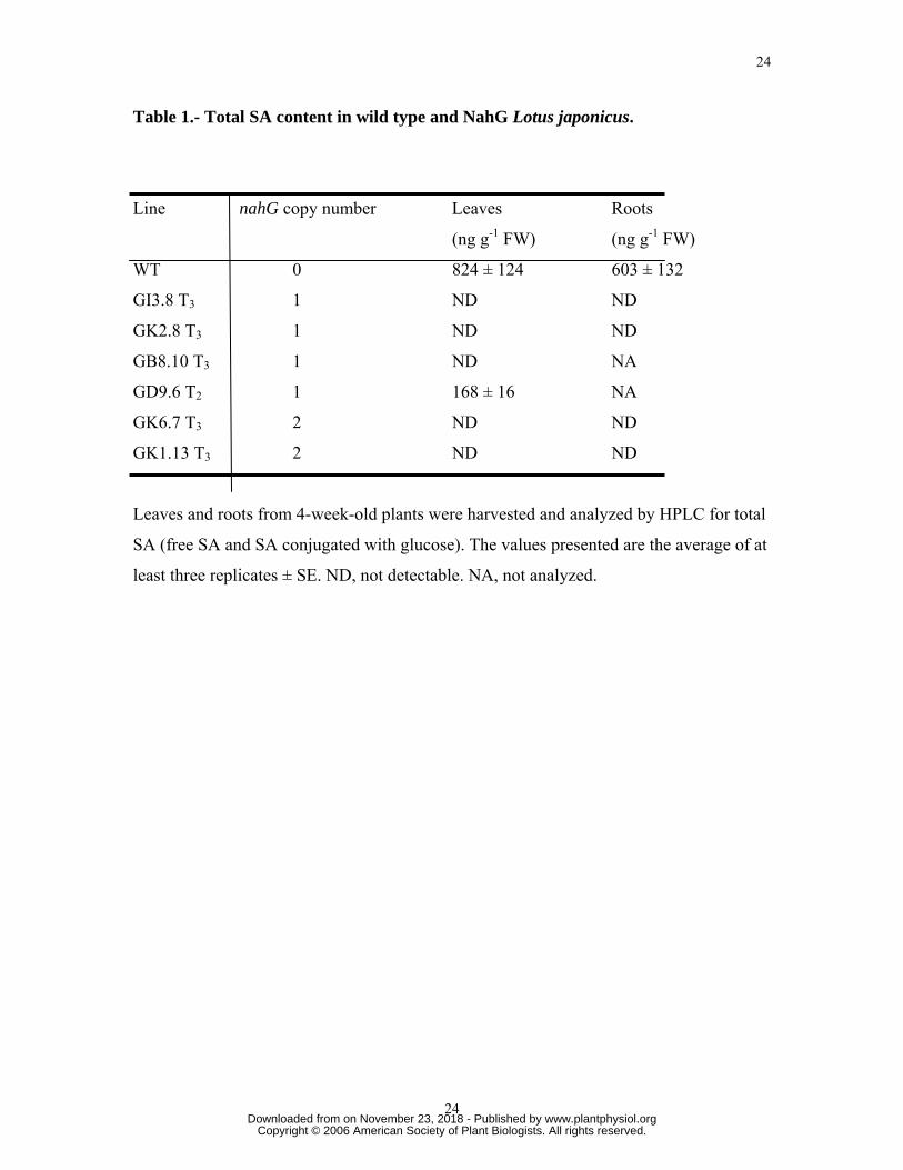
Table 1.- Total SA content in wild type and NahG Lotus japonicus.
Line nahG copy number Leaves Roots
(ng g-1 FW) (ng g-1 FW)
WT 0 824 ± 124 603 ± 132
GI3.8 T3 1 ND ND
GK2.8 T3 1 ND ND
GB8.10 T3 1 ND NA
GD9.6 T2 1 168 ± 16 NA
GK6.7 T3 2 ND ND
GK1.13 T3 2 ND ND
Leaves and roots from 4-week-old plants were harvested and analyzed by HPLC for total
SA (free SA and SA conjugated with glucose). The values presented are the average of at
least three replicates ± SE. ND, not detectable. NA, not analyzed.
www.plantphysiol.orgon November 23, 2018 - Published by Downloaded from Copyright © 2006 American Society of Plant Biologists. All rights reserved.
Page 25
25
25
Table 2. NahG lnfection events relative to L. japonicus root growth. Data taken from
10 plants per line. Plants were germinated 7 days and then planted for 14 days before
inoculation. Plants were harvested two weeks post inoculation and scored for infection
events. Groups marked (a),(b), or (c) were statistically significant at α=0.05 by a Student
t-test.
WT GI3.8 T3
(1 copy)
GK2.8
T3
(1 copy)
GK6.7 T3
(2 copies)
Avg. nodule number/ plant 8a 12b 11b 11b
Avg. number of infections/
plant 168a 219b 289c 275c
Avg. number of infections/
Avg. Total root length* (cm)
7a 6a 7a 7a
Avg. infection zone length
(mm) 6.1a 7.7b 7.7b 8.5c
Avg. number of infection
zones/ plant 3a 4b 5c 4b
Avg. number of infections/
infection zone length (mm) 27.5a 28.4a 37.5b 32.4b
Avg. tap root length (cm) 6.2a 8.4a 10.7c 12.7c
Avg. lateral root length (cm) 19.8a 26.7b 27.9b 24.3b
Avg. number of lateral roots/
plant 6a 9b 9b 12c
*Total root length=tap root + lateral roots
www.plantphysiol.orgon November 23, 2018 - Published by Downloaded from Copyright © 2006 American Society of Plant Biologists. All rights reserved.
Page 26
26
26
Table 3. Spot inoculation of NahG-expressing Lotus japonicus.
Inoculations were performed as described in methods. Data (average ± standard error)
were taken from 10 or more plants per line at the times shown.
Avg. nodule number/plant
Line Transgene
copy
number
5 dpi 6 dpi 7 dpi 10 dpi
wt
GA6T3
GB8.10
GD9.6
GI3.8
GK2.8
GK6.7
GB3B
0
0
1
1
1
1
2
3
0.65 ± 0.13
0.95 ± 0.31
1.46 ± 0.45a
1.37 ± 0.30a
0.77 ± 0.60
1.52± 0.58a
1.22 ± 0.27a
1.25 ± 0.57a
1.70 ± 0.28
2.1 ± 0.35
2.82 ± 0.44a
3.64 ± 0.48a
2.63 ± 0.45a
2.37 ± 0.29a
2.96 ± 0.15a
2.35 ± 0.32a
2.85 ± 0.36
2.18 ± 0.39
3.97 ± 0.47a
3.98 ± 0.47a
3.65 ± 0.65a
2.77 ± 0.54
4.28 ± 0.31a
3.37 ± 0.65a
3.1 ± 0.25
2.6 ± 0.29
4.2± 0.45a
3.99 ± 0.41a
3.78 ± 0.42
3.62 ± 0.36
4.35 ± 0.38a
4.38 ± 0.66a
a Values significantly different (α = 0.05) by Student t-test when compared to wild type at
the same dpi.
dpi= Days Post Inoculation
www.plantphysiol.orgon November 23, 2018 - Published by Downloaded from Copyright © 2006 American Society of Plant Biologists. All rights reserved.
Page 27
27
27
Table 4. Nodulation and infection of M. truncatula composite roots expressing NahG.
Genotype Number of
nodules/root
Number of
infections/root
Root Length-
inoculated
(cm)
Root Length-
uninoculated
(cm)
NahG
expressing
11.3±0.3 38±10 5.3±0.4 4±0.3
Empty vector
control
6±0.4 14±8 4.9±0.3 4±0.3
Data (average ± standard error) were taken from 10 plants
www.plantphysiol.orgon November 23, 2018 - Published by Downloaded from Copyright © 2006 American Society of Plant Biologists. All rights reserved.
Page 28
28
28
Figure 1.
Figure 2.
www.plantphysiol.orgon November 23, 2018 - Published by Downloaded from Copyright © 2006 American Society of Plant Biologists. All rights reserved.
Page 29
29
29
Figure 3.
www.plantphysiol.orgon November 23, 2018 - Published by Downloaded from Copyright © 2006 American Society of Plant Biologists. All rights reserved.
Page 30
NahG Control
www.plantphysiol.orgon November 23, 2018 - Published by Downloaded from Copyright © 2006 American Society of Plant Biologists. All rights reserved.


![Salicylic Acid-Dependent Plant Stress Signaling via ... · Salicylic Acid-Dependent Plant Stress Signaling via Mitochondrial Succinate Dehydrogenase1[OPEN] Katharina Belt, Shaobai](https://static.documents.pub/doc/80x56/5c9a845b09d3f2a06c8bfc9a/salicylic-acid-dependent-plant-stress-signaling-via-salicylic-acid-dependent.jpg)

