Accepted Manuscript Title: Effects of glyphosate and aminomethylphosphonic acid on an isogeneic model of the human blood-brain barrier Authors: Adriana Martinez, Abraham Jacob Al-Ahmad PII: S0378-4274(18)31904-0 DOI: https://doi.org/10.1016/j.toxlet.2018.12.013 Reference: TOXLET 10389 To appear in: Toxicology Letters Received date: 17 September 2018 Revised date: 10 December 2018 Accepted date: 28 December 2018 Please cite this article as: Martinez A, Al-Ahmad AJ, Effects of glyphosate and aminomethylphosphonic acid on an isogeneic model of the human blood-brain barrier, Toxicology Letters (2018), https://doi.org/10.1016/j.toxlet.2018.12.013 This is a PDF file of an unedited manuscript that has been accepted for publication. As a service to our customers we are providing this early version of the manuscript. The manuscript will undergo copyediting, typesetting, and review of the resulting proof before it is published in its final form. Please note that during the production process errors may be discovered which could affect the content, and all legal disclaimers that apply to the journal pertain.
Transcript
Accepted Manuscript
Title: Effects of glyphosate and aminomethylphosphonic acidon an isogeneic model of the human blood-brain barrier
Received date: 17 September 2018Revised date: 10 December 2018Accepted date: 28 December 2018
Please cite this article as: Martinez A, Al-Ahmad AJ, Effects of glyphosate andaminomethylphosphonic acid on an isogeneic model of the human blood-brain barrier,Toxicology Letters (2018), https://doi.org/10.1016/j.toxlet.2018.12.013
This is a PDF file of an unedited manuscript that has been accepted for publication.As a service to our customers we are providing this early version of the manuscript.The manuscript will undergo copyediting, typesetting, and review of the resulting proofbefore it is published in its final form. Please note that during the production processerrors may be discovered which could affect the content, and all legal disclaimers thatapply to the journal pertain.
Doxorubicin uptake assay was performed by pre-incubating iPSC-derived BMECs in
presence of GPH, AMPA or GLY at 100µM concentrations for 2 hours. Doxorubicin
(Sigma-Aldrich) was added a 5µM final concentration and allowed to incubate for 1
hour. Cells were homogenized as previously described and total fluorescence assessed
by fluorimetry (SynergyMX2). Total protein content obtained from cell homogenates
were determined using a BCA protein assay (Pierce, Thermofisher).
Statistics:
Cells were randomly assigned treatment conditions prior each experiment. Data are
represented as mean S.D. from three or more independent experiments. One-way
analysis of the variance (ANOVA) coupled with Dunnett (or Kruskal-Wallis) tests
analysis were performed using Prism 7.0 built-in package (GraphPad Software, La
Jolla, CA). A P<0.05 p-value was considered as indicative of a statistic difference
between one or more groups.
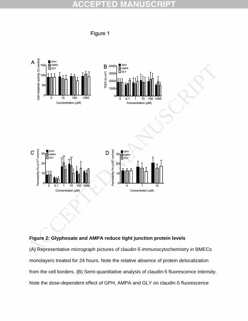
Results
Effects of glyphosate and AMPA on BMECs cell viability
We firstly assessed the impact of glyphosate (GPH) on BMECs cell monolayers viability
using a range of concentrations partially overlapping levels found in patients reported as
ACCEPTED MANUSCRIP
T
asymptomatic (17µM), minor and moderate (241µM and 428µM respectively) by
Roberts and colleagues (Roberts et al., 2010). Treatment with GPH for 24 hours at
concentrations ranging from 10µM to 1000µM resulted in no changes in cell metabolic
activity (Fig.1A). Similar outcomes were observed using AMPA or glycine (GLY). Taken
together, our data indicates that glyphosate or AMPA unlikely have toxicity towards the
blood-brain barrier.
Glyphosate and AMPA affect fluorescein permeability in BMECs monolayers
Next, we investigated changes in the barrier function in such BMECs monolayers using
TEER and fluorescein permeability (Fig.1B&C). In addition to the previous
concentrations, we included two concentrations (0.1 and 1µM) ranges aimed to reflect
average plasma concentrations reported in occupational exposure (Pan et al., 2016).
No changes in TEER were noted for any of the concentrations tested. Neither AMPA or
GLY showed effects on TEER. However, we noticed a biphasic response in our model.
At 0.1µM, we noted a slight decrease in fluorescein permeability for all three groups
(GPH, AMPA and GLY), followed by a significant increase for both GPH and AMPA at 1
and 10µM. Notably, higher concentrations resulted in permeability values similar to
controls. To confirm the increase in paracellular profile observed with GPH, we
investigated changes in paracellular permeability using [14C]-mannitol, an alternative
paracellular flux marker (Fig.1D). We observed a modest but significant increase in
mannitol permeability following treatment with 10µM GPH. In contrast, no significant
increase was noted following AMPA or GLY treatment. In summary, our data suggest
ACCEPTED MANUSCRIP
T
that GPH and AMPA treatment may increase the barrier permeability in BMECs
monolayers.
High levels of glyphosate, AMPA and glycine impair tight junction complexes integrity
To better understand the effect of GPH and AMPA on the barrier function, we
investigated changes in in tight junction complexes, in particular changes in claudin-5
and occludin, by immunocytochemistry (Figure 2). We did not observe changes in
claudin-5 immunolocalization following GPH or AMPA treatment (Fig.2A), however we
noted a dose-dependent decrease in claudin-5 relative expression (as quantified by
fluorescence intensity) in all groups (Fig.2B). Treatment with GPH resulted in decreased
claudin-5 fluorescence intensity at 100 and 1000µM concentrations, whereas treatment
with AMPA marked a significant decrease already by 10µM concentration. GLY
treatment showed a significant decrease at 100 and 1000µM concentrations.
To confirm such observation, we investigated changes in occludin levels (Fig.2C&D). As
expected, no changes in occludin localization occurred following treatment (Fig.2C).
However, we noted a significant decrease in occludin protein levels (as marked by
reduced fluorescence intensity) in all groups with the exception of the 10µM GLY
treatment group. Unlike claudin-5, such effect appeared dose-independent as we have
noted no changes between 10µM treatment and higher concentrations. Taken together,
our data suggest that GPH may increase paracellular permeability in BMECs
monolayers to fluorescein via partial disruption of tight junction complexes integrity.
ACCEPTED MANUSCRIP
T
Glyphosate diffuses across the blood-brain barrier via a transcellular route, modulates
glucose uptake in BMECs
In addition to its effect on paracellular permeability, we investigated the ability of GPH to
cross the BBB. Thus we investigated the diffusion profile of GPH following an acute
exposure at 100µM in the apical chamber and measured its diffusion for 2 hours
(Fig.3A&B) and quantified the amount of GPH present in the basolateral chamber using
the analytical method developed by Waiman and colleagues (Waiman et al., 2012). At 2
hours of diffusion, the amount of GPH capable to cross BMECs monolayers was about
1.670.31% of the injected dose (100µM). In addition, the permeability value of GPH
was significantly higher than fluorescein (18.673.55 x 10-6cm/min versus 10.59 x 10-
6cm/min) or mannitol (13.102.03 x 10-6 cm/min) in cells treated with the same amount
of GPH (Fig.3B). In conclusion, our data suggests that GPH may cross the BBB via a
transcellular mechanism. Next, we investigated the effect of GPH and AMPA on drug
efflux transporters using doxorubicin as a drug efflux substrate (Fig.3C). With the
exception of AMPA that showed a two-fold increase over control, treatment with 100µM
GPH or GLY for 24 hours showed no differences compared to controls. Finally, we
investigated changes in GLUT1 localization and expression (the main glucose
transporter at the blood-brain barrier) in BMECs following treatment by
immunocytochemistry (Fig.3D), as previous studies reported changes in glucose levels
in certain vertebrates following exposure to GPH and AMPA. (Becker et al., 2009;
Kondera et al., 2018). We noted changes in GLUT1 immunoreactivity following
treatment with GPH, with an apparent increase following treatment with 100µM. Similar
behavior was observed with AMPA albeit subtler. Notably, we noted a notable increase
ACCEPTED MANUSCRIP
T
in GLUT1 immunoreactivity following treatment with 100 and 1000µM GLY. Such
observation was confirmed by assessing GLUT1 fluorescence intensity in our
monolayers (Fig.3E). To ensure such differences was not inherent to the technique, we
performed a flow cytometry analysis (Fig.3F&G) and compared changes in mean
fluorescence indexes following treatment with 100µM for 24 hours. Notably, GPH
treatment yielded an increase in GLUT1 expression levels compared to control (Fig.3G).
Although AMPA showed no differences in GLUT1 expression, treatment with GLY
resulted in a significant decrease compared to control. Finally, we measured changes in
glucose uptake in BMECs monolayers, using [14C]-D-glucose (Fig.3H). We noted a
progressive increase in glucose uptake with a maximum uptake at 100µM
concentration. AMPA showed a similar pattern than GPH, resulting in a 3-fold increase
compared to control in the 100µM. GLY treatment resulted in the highest increase in
glucose uptake as noted at 100 and 1000 µM concentrations. Taken together, our data
suggest that exposure to high levels of GLY or AMPA may impair glucose uptake and
metabolism in BMECs monolayers via an alteration in GLUT1 expression and/or
activity.
Neurons co-cultured with brain microvascular endothelial cells display an impaired cell
metabolic activity
As GPH showed ability to cross the BBB and yielded to changes in GLUT1 expression
and glucose uptake in BMECs monolayers, we investigated the effects of GPH on
neurovascular coupling using a BMEC/neurons co-culture model (Fig.4) based on our
previous publication (Patel et al., 2017). We firstly, assessed the ability of such co-
ACCEPTED MANUSCRIP
T
cultures to yield to tighter barrier function by measuring differences in TEER between
BMECs monocultures and BMECs co-cultured with iPSC-derived neurons (Fig.4A). As
expected, co-cultures significantly up-regulated the barrier tightness, as we noted a 3-
fold increase in TEER in BMECs co-cultured with neurons compared to BMECs
maintained in monocultures. Next, these co-cultures to 100M GPH, AMPA or GLY by
adding these compounds on the apical (top) chamber for 24 hours. Addition of these
compounds on the apical chamber did not significantly alter the barrier tightness as we
noted no differences in TEER (Fig.4B). As expected, we noted a mild increase in
fluorescein permeability (Fig.4C) in GPH-treated group compared to control, but not in
the AMPA or GLY groups. Finally, we investigated the effect GPH incubation in the
apical chamber (100M) on the neurovascular coupling, by investigating changes in
neuronal cell metabolic activity by MTS (Fig.4D) following 6 hours exposure. Notably,
we noticed a significant decrease in cell metabolic activity in neurons co-cultured
compared to monocultures. Following incubation with GPH in the apical chamber, we
noticed an increase in cell metabolic activity compared to control. By contrast, neurons
directly exposed to monocultures showed no sign of activity. Interestingly, we observed
a similar increase in cell metabolic activity compared control. Such changes observed in
GPH was also observed in AMPA co-cultures but not in GLY co-cultures. Following
such incubation, we investigated changes in gross morphology of iPSC-derived neurons
colonies by immunocytochemistry against III-tubulin (Tuj1, Fig.4E). We did not observe
macroscopic changes in iPSC-derived neurons following incubation with GPH, AMPA or
GLY both in monocultures and co-cultures, suggesting that changes in cell metabolic
ACCEPTED MANUSCRIP
T
activity is unlikely due to cell death. In conclusion, our data suggest that exposure to
high amount of GPH (100µM) may impair neurovascular coupling.
GPH and AMPA effect on neuron progenitor cells
As we have demonstrated the ability of GPH to cross the BBB and to alter the
neurovascular unit, we investigated the effect of GPH on differentiating and
differentiated neurons (Fig.5) by exposing cells to a concentration considered
representative of the amount crossing the BBB (0.1-1M). We firstly investigated the
effect of acute exposure (24 hours) on the cellular metabolic activity of undifferentiated
NPCs by MTS assay (Fig.5A). Although we did not observe any differences in cell
metabolic activity at 0.1M, we noted a significant decrease in MTS activity at 1M in
both GPH and GLY groups, suggesting a possible deleterious effect on NPC cell
viability. Nevertheless, immunofluorescence analysis of these NPCs (Fig.5B) showed
no major alterations in the relative cell density and nestin (a cellular marker of neural
stem cells/progenitor cells) immunoreactivity. To further confirm the innocuity of GPH
and AMPA on neurogenesis, we treated differentiating NPCs continuously for 16 days
(by replacing cell medium every 48 hours) in presence of 0.1M GPH, AMPA or GLY.
Such concentration was chosen to be representative of plasma concentration reported
in occupational workers and 20 times higher than values reported in non-occupational
population (Conrad et al., 2017; Pan et al., 2016). Chronic exposure (16 days) to low
GPH levels (0.1µM) We did not observe significant changes in the cell metabolic activity
between the different groups compared to controls (Fig.5C). In addition to changes in
cell metabolic activity, we investigated changes in gross morphology in iPSC-derived
ACCEPTED MANUSCRIP
T
neurons colonies (Fig.5D). After 16 days, we observed the presence of a dense neurite
network. Treatment with GPH or AMPA did not display major changes in gross
morphology of iPSC-derived neuron colonies, as we still observed presence of dense
neurite network. In conclusion, chronic exposure to low-levels of GPH or AMPA failed to
show any signs of neurotoxicity.
Glyphosate and AMPA affects neurons cell metabolism without showing detrimental
effects on neurites density.
Finally, we investigated the effect of acute exposure by treating iPSC-derived neurons
seeded at low density (50’000 cells/cm2) in presence of GPH or AMPA for 24 hours at
concentrations ranging from 1M to 1000M (Fig.4E-H). Treatment with GPH and
AMPA showed a decrease in cell metabolic activity upon exposure to GPH to 10µM and
higher concentrations (Fig.5E). AMPA showed similar outcomes than GPH, albeit not
statistically significant. Treatment with GLY showed little effects on cell metabolic
activity. To better such changes in cell metabolic activity with cellular distress, we
investigated changes in cell density and neurites formation by immunocytochemistry
(Fig.5F). With the exception of GLY group, no depletion in neurites was observed in
both GPH and AMPA groups. Upon quantification of cell nuclei and neurites per surface
area (Fig.5G&H), we noted a progressive decrease in neuron density for both GPH and
AMPA (Fig.5G), with a significant decrease noted at 1000M. However, no differences
in terms of neurites density were noted in GPH and AMPA groups, with the exception of
very high amount of GLY (100 and 1000M treatment). Taken together, our data
ACCEPTED MANUSCRIP
T
suggest that low concentrations (<10M) of GPH and AMPA may not have detrimental
effects on iPSC-derived neurons.
Discussion
In this study, we investigated the toxicity of acute glyphosate poisoning on the blood-
brain barrier integrity by investigating its activity on the different cell types of the
neurovascular unit. To reflect the situation of a self-inflicted poisoning exposure, we
treated cells with glyphosate and AMPA concentration ranges from 10M to 1000M.
Such concentration ranges are within GPH plasma values reported by Roberts and
colleagues from patients with self-inflicted poisoning exposure and scored from
asymptomatic (17M) to fatal (8.12mM) at the time of admission in clinic (Roberts et al.,
2010). We consider it important that the meaning of such concentration such values are
put into context. According to the literature, the average GPH and AMPA levels
measured in urine following non-occupational exposure (residual pesticide exposure) in
North America were 0.28g/L and 0.30g/L (1.6nM and 2.7nM respectively)
respectively (McGuire et al., 2016); while a study by Conrad and colleagues (Conrad et
al., 2017) showed an average GPH and AMPA amount of GPH and AMPA of 0.78g/L
and 0.64g/L (4.6nM and 5.7nM respectively) in urine samples from a non-occupational
German cohort. By contrast, occupational workers showed higher values of GPH and
AMPA, with average plasma levels of 11.73g/L and 5.29g/L (69nM and 117nM) for
GPH and AMPA respectively (Pan et al., 2016). As a reference, normal plasma GLY
concentrations range from 125 to 450M, with values below 20M for CSF (Applegarth
et al., 1979; Van Hove et al., 1993). We noted an increase in fluorescein permeability
ACCEPTED MANUSCRIP
T
for both GPH and AMPA at 1M and 10M. We noted similar outcome for mannitol in
cells exposed to 1M GPH. Such observation is suggesting a possible detrimental effect
of GPH and AMPA on the barrier function. Although we did not observe major changes
in tight junction complexes localization, we noted a decrease in both claudin-5 and
occludin protein levels by immunoreactivity for all three groups. Such observations
suggest that GPH and AMPA may interfere with tight junction complexes integrity. Yet,
the interference of such compounds on tight junction proteins remains unclear. We
speculate that a possible mechanism explaining such down-regulation maybe occurring
via shedding of such proteins by matrix metalloproteinases (MMPs). MMPs are
proteases well documented to cleave claudin-5 and occludin during neurological
diseases (e.g. ischemic stroke)(Feng et al., 2011; Liu et al., 2012; Yang et al., 2007).
Thus, we will further investigate the effect of GPH and AMPA on tight junction proteins
expression and MMPs activity in BMECs.
An interesting observation done in our study was an increase in permeability following
GLY treatment. Although we did not observe a statistical significance, we sill noted a
50% increase in paracellular permeability following GLY treatment at 100 and 1000M.
Interestingly, a study recently reported the case report of an increased BBB permeability
in a patient suffering from glycine encephalopathy (also known as nonketotic
hyperglycinemia (NKH)) and presenting a meningitis (Scholl-Burgi et al., 2008). Our
data suggest that high levels of GLY may increase the permeability of the BBB and
disrupt tight junction complexes in such range. Therefore, assessing the barrier function
in BMECs derived from iPSCs obtained from these patients would provide important
insights on the effect of hyperglycinemia on the blood-brain barrier integrity. In addition
ACCEPTED MANUSCRIP
T
to changes in barrier function, we assessed GPH permeability in our BMECs
monolayers. We estimate that about 1% of the injected dose (100M) diffused across
BMECs monolayers (data not shown). However, the permeability for GPH was
significantly higher than fluorescein despite its high hydrophilicity (xLogP=-4.63).
Although its permeability value was higher than mannitol, we did not observe a
statistical difference. Thus, we speculate that GPH cross the BBB via a carrier-mediated
diffusion. A recent study suggested the possible involvement of system L-type amino
acid transporters 1 and 2 (LAT1/LAT2 encoded by SLC7A5 and SLC7A8 genes
respectively) in the uptake of GPH in mammalian cells (Xu et al., 2016), two receptors
expressed at the BBB (Kido et al., 2001). A major limitation of our study is the limited
access to LC-MS chromatography, limiting our scope to provide a further understanding
of the diffusion of GPH and AMPA. Therefore, a long-term goal will focus on detailing
the permeability of GPH and AMPA and the contribution of LAT1 and LAT2 in such
diffusion. Amongst the different cell types, neurons displayed the most important
changes in metabolic activity following exposure to GPH and AMPA. We noticed
significant changes in neuronal cell metabolic activity following treatment with GPH,
AMPA or GLY whereas we did not observe significant changes in BMECs. Yet, such
decrease in cell metabolic was unlikely reflective of a neurotoxicity, as we noted no
changes in neuronal cell density and neurites formation. Assessing the effects of GPH
and AMPA on iPSC-derived glycinergic neurons coupled with electrophysiological
methods would provide important insights on the ability of GPH and AMPA to act as
glycine mimetic molecules. A change in cell metabolic activity observed in our cells may
be due to changes in glucose metabolism, as we noted changes in glucose uptake in
ACCEPTED MANUSCRIP
T
BMECs, as well as some changes in GLUT1 expression levels. Glyphosate has been
associated with changes in glucose serum levels in various fish species (Becker et al.,
2009; Kondera et al., 2018), yet there are no reports in the presence of such effects in
mammalian species. Therefore, further studies investigating the effect of glyphosate
and AMPA on brain glucose uptake and changes in glucose and lactate CSF levels in
vivo is needed to confirm our observation. Taken together, our data demonstrate the
relative safety of glyphosate and AMPA on the blood-brain barrier during acute
accidental exposure. Yet, the presence of an active uptake and diffusion of glyphosate
across the blood-brain barrier suggests the need of an extensive brain-centered
pharmacokinetic studies to evaluate the pharmacokinetics and pharmacodynamics of
glyphosate on the central nervous system during acute exposure and in individuals
exposed to high amount of such pesticides. In conclusion, our study further supports the
relative safety of GPH with minimal effects observed at concentrations significantly
higher than baseline exposure levels, occupational and non-occupational alike.
Acknowledgements
This study was funded by Texas Tech University Health Sciences Center institutional
funds to A.A. The authors have no conflict of interests to disclose.
ACCEPTED MANUSCRIP
T
References
Alloisio, S., Nobile, M., Novellino, A., 2015. Multiparametric characterisation of neuronal network activity for in vitro agrochemical neurotoxicity assessment. Neurotoxicology 48, 152-165. Applegarth, D.A., Edelstein, A.D., Wong, L.T., Morrison, B.J., 1979. Observed range of assay values for plasma and cerebrospinal fluid amino acid levels in infants and children aged 3 months to 10 years. Clin Biochem 12, 173-178. Becker, A.G., Moraes, B.S., Menezes, C.C., Loro, V.L., Santos, D.R., Reichert, J.M., Baldisserotto, B., 2009. Pesticide contamination of water alters the metabolism of juvenile silver catfish, Rhamdia quelen. Ecotoxicol Environ Saf 72, 1734-1739. Birch, M., 1993. Toxicological investigation of CP 67573-3. In: Office of Prevention, P., and Toxic Substances, (Ed.). U.S. Government Printing Office, Washington, DC. Brewster, D.W., Warren, J., Hopkins, W.E., 2nd, 1991. Metabolism of glyphosate in Sprague-Dawley rats: tissue distribution, identification, and quantitation of glyphosate-derived materials following a single oral dose. Fundam Appl Toxicol 17, 43-51. Conrad, A., Schroter-Kermani, C., Hoppe, H.W., Ruther, M., Pieper, S., Kolossa-Gehring, M., 2017. Glyphosate in German adults - Time trend (2001 to 2015) of human exposure to a widely used herbicide. Int J Hyg Environ Health 220, 8-16. Coullery, R.P., Ferrari, M.E., Rosso, S.B., 2016. Neuronal development and axon growth are altered by glyphosate through a WNT non-canonical signaling pathway. Neurotoxicology 52, 150-161. Culbreth, M.E., Harrill, J.A., Freudenrich, T.M., Mundy, W.R., Shafer, T.J., 2012. Comparison of chemical-induced changes in proliferation and apoptosis in human and mouse neuroprogenitor cells. Neurotoxicology 33, 1499-1510. Efthymiou, A., Shaltouki, A., Steiner, J.P., Jha, B., Heman-Ackah, S.M., Swistowski, A., Zeng, X., Rao, M.S., Malik, N., 2014. Functional screening assays with neurons generated from pluripotent stem cell-derived neural stem cells. Journal of biomolecular screening 19, 32-43. Feng, S., Cen, J., Huang, Y., Shen, H., Yao, L., Wang, Y., Chen, Z., 2011. Matrix metalloproteinase-2 and -9 secreted by leukemic cells increase the permeability of blood-brain barrier by disrupting tight junction proteins. PLoS One 6, e20599. Foerch, C., Wunderlich, M.T., Dvorak, F., Humpich, M., Kahles, T., Goertler, M., Alvarez-Sabin, J., Wallesch, C.W., Molina, C.A., Steinmetz, H., Sitzer, M., Montaner, J., 2007. Elevated serum S100B levels indicate a higher risk of hemorrhagic transformation after thrombolytic therapy in acute stroke. Stroke 38, 2491-2495. Henderson, A.M., Gervais, J.A., Luukinen, B., Buhl, K., Stone, D., 2010. Glyphosate Technical Fact Sheet. National Pesticide Information Center. Oregon State University Extension Services. Hori, Y., Fujisawa, M., Shimada, K., Hirose, Y., 2003. Determination of the herbicide glyphosate and its metabolite in biological specimens by gas chromatography-mass spectrometry. A case of poisoning by roundup herbicide. J Anal Toxicol 27, 162-166. Kamijo, Y., Takai, M., Sakamoto, T., 2016. A multicenter retrospective survey of poisoning after ingestion of herbicides containing glyphosate potassium salt or other glyphosate salts in Japan. Clin Toxicol (Phila) 54, 147-151.
ACCEPTED MANUSCRIP
T
Kanner, A.A., Marchi, N., Fazio, V., Mayberg, M.R., Koltz, M.T., Siomin, V., Stevens, G.H., Masaryk, T., Aumayr, B., Vogelbaum, M.A., Barnett, G.H., Janigro, D., 2003. Serum S100beta: a noninvasive marker of blood-brain barrier function and brain lesions. Cancer 97, 2806-2813. Kapural, M., Krizanac-Bengez, L., Barnett, G., Perl, J., Masaryk, T., Apollo, D., Rasmussen, P., Mayberg, M.R., Janigro, D., 2002. Serum S-100beta as a possible marker of blood-brain barrier disruption. Brain Res 940, 102-104. Kido, Y., Tamai, I., Uchino, H., Suzuki, F., Sai, Y., Tsuji, A., 2001. Molecular and functional identification of large neutral amino acid transporters LAT1 and LAT2 and their pharmacological relevance at the blood-brain barrier. J Pharm Pharmacol 53, 497-503. Kondera, E., Teodorczuk, B., Lugowska, K., Witeska, M., 2018. Effect of glyphosate-based herbicide on hematological and hemopoietic parameters in common carp (Cyprinus carpio L). Fish Physiol Biochem. Lee, J.W., Choi, Y.J., Park, S., Gil, H.W., Song, H.Y., Hong, S.Y., 2017. Serum S100 protein could predict altered consciousness in glyphosate or glufosinate poisoning patients. Clin Toxicol (Phila) 55, 357-359. Lippmann, E.S., Al-Ahmad, A., Azarin, S.M., Palecek, S.P., Shusta, E.V., 2014. A retinoic acid-enhanced, multicellular human blood-brain barrier model derived from stem cell sources. Scientific reports 4, 4160. Lippmann, E.S., Azarin, S.M., Kay, J.E., Nessler, R.A., Wilson, H.K., Al-Ahmad, A., Palecek, S.P., Shusta, E.V., 2012. Derivation of blood-brain barrier endothelial cells from human pluripotent stem cells. Nat Biotechnol 30, 783-791. Liu, J., Jin, X., Liu, K.J., Liu, W., 2012. Matrix metalloproteinase-2-mediated occludin degradation and caveolin-1-mediated claudin-5 redistribution contribute to blood-brain barrier damage in early ischemic stroke stage. J Neurosci 32, 3044-3057. McGuire, M.K., McGuire, M.A., Price, W.J., Shafii, B., Carrothers, J.M., Lackey, K.A., Goldstein, D.A., Jensen, P.K., Vicini, J.L., 2016. Glyphosate and aminomethylphosphonic acid are not detectable in human milk. Am J Clin Nutr 103, 1285-1290. Nishiyori, Y., Nishida, M., Shioda, K., Suda, S., Kato, S., 2014. Unilateral hippocampal infarction associated with an attempted suicide: a case report. J Med Case Rep 8, 219. Pan, L., Xu, M., Yang, D., Wang, B., Zhao, Q., Ding, E.-M., Zhu, B., 2016. The association between coronary artery disease and glyphosate exposure found in pesticide factory workers. Public Health and Emergency 1, 9-9. Patel, R., Page, S., Al-Ahmad, A.J., 2017. Isogenic blood-brain barrier models based on patient-derived stem cells display inter-individual differences in cell maturation and functionality. J Neurochem 142, 74-88. Perriere, N., Demeuse, P., Garcia, E., Regina, A., Debray, M., Andreux, J.P., Couvreur, P., Scherrmann, J.M., Temsamani, J., Couraud, P.O., Deli, M.A., Roux, F., 2005. Puromycin-based purification of rat brain capillary endothelial cell cultures. Effect on the expression of blood-brain barrier-specific properties. J Neurochem 93, 279-289. Picetti, E., Generali, M., Mensi, F., Neri, G., Damia, R., Lippi, G., Cervellin, G., 2018. Glyphosate ingestion causing multiple organ failure: a near-fatal case report. Acta Biomed 88, 533-537.
ACCEPTED MANUSCRIP
T
Potrebic, O., Jovic-Stosic, J., Vucinic, S., Tadic, J., Radulac, M., 2009. [Acute glyphosate-surfactant poisoning with neurological sequels and fatal outcome]. Vojnosanit Pregl 66, 758-762. Roberts, D.M., Buckley, N.A., Mohamed, F., Eddleston, M., Goldstein, D.A., Mehrsheikh, A., Bleeke, M.S., Dawson, A.H., 2010. A prospective observational study of the clinical toxicology of glyphosate-containing herbicides in adults with acute self-poisoning. Clin Toxicol (Phila) 48, 129-136. Roy, N.M., Carneiro, B., Ochs, J., 2016. Glyphosate induces neurotoxicity in zebrafish. Environ Toxicol Pharmacol 42, 45-54. Scholl-Burgi, S., Korman, S.H., Applegarth, D.A., Karall, D., Lillquist, Y., Heinz-Erian, P., Davidson, A.G., Haberlandt, E., Sass, J.O., 2008. The relation of cerebrospinal fluid and plasma glycine levels in propionic acidaemia, a 'ketotic hyperglycinaemia'. Journal of inherited metabolic disease 31, 395-398. Talbot, A.R., Shiaw, M.H., Huang, J.S., Yang, S.F., Goo, T.S., Wang, S.H., Chen, C.L., Sanford, T.R., 1991. Acute poisoning with a glyphosate-surfactant herbicide ('Roundup'): a review of 93 cases. Hum Exp Toxicol 10, 1-8. Van Hove, J., Coughlin, C., II, Scharer, G., 1993. Glycine Encephalopathy. In: Adam, M.P., Ardinger, H.H., Pagon, R.A., Wallace, S.E., Bean, L.J.H., Stephens, K., Amemiya, A. (Eds.) GeneReviews((R)), Seattle (WA). Waiman, C.V., Avena, M.J., Garrido, M., Band, B.F., Zanini, G.P., 2012. A simple and rapid spectrophotometric method to quantify the herbicide glyphosate in aqueous media. Application to adsorption isotherms on soils and goethite. Geoderma 170, 154-158. Xu, J., Li, G., Wang, Z., Si, L., He, S., Cai, J., Huang, J., Donovan, M.D., 2016. The role of L-type amino acid transporters in the uptake of glyphosate across mammalian epithelial tissues. Chemosphere 145, 487-494. Yan, Y., Shin, S., Jha, B.S., Liu, Q., Sheng, J., Li, F., Zhan, M., Davis, J., Bharti, K., Zeng, X., Rao, M., Malik, N., Vemuri, M.C., 2013. Efficient and rapid derivation of primitive neural stem cells and generation of brain subtype neurons from human pluripotent stem cells. Stem Cells Transl Med 2, 862-870. Yang, Y., Estrada, E.Y., Thompson, J.F., Liu, W., Rosenberg, G.A., 2007. Matrix metalloproteinase-mediated disruption of tight junction proteins in cerebral vessels is reversed by synthetic matrix metalloproteinase inhibitor in focal ischemia in rat. J Cereb Blood Flow Metab 27, 697-709. Yu, G.C., Jian, X.D., Gao, B.J., 2017. [The clinical analytics of 10 patients with acute glyphosate poisoning]. Zhonghua Lao Dong Wei Sheng Zhi Ye Bing Za Zhi 35, 382-383. Yu, J., Vodyanik, M.A., Smuga-Otto, K., Antosiewicz-Bourget, J., Frane, J.L., Tian, S., Nie, J., Jonsdottir, G.A., Ruotti, V., Stewart, R., Slukvin, II, Thomson, J.A., 2007. Induced pluripotent stem cell lines derived from human somatic cells. Science 318, 1917-1920. Zouaoui, K., Dulaurent, S., Gaulier, J.M., Moesch, C., Lachatre, G., 2013. Determination of glyphosate and AMPA in blood and urine from humans: about 13 cases of acute intoxication. Forensic Sci Int 226, e20-25.
ACCEPTED MANUSCRIP
T
Figure Legends
Figure 1: Effects of glyphosate and AMPA on the blood-brain barrier integrity
Effect of prolonged exposure to glyphosate (GPH), AMPA and glycine (GLY) on the
barrier integrity in IMR90-derived BMECs monolayers. Cells were exposed for various
concentrations for 24 hours, followed by measurement of changes in cell metabolic
activity by MTS (A) and in the barrier function by TEER (B) and permeability to sodium
fluorescein (C) and mannitol (D). Note the quasi-absence of effect on cell metabolism
(A), whereas we noted an increased fluorescein permeability at 1M and 10µM for both
GPH and AMPA, whereas we noted an increase in mannitol permeability in 10µM GPH
treatment only. Scale bar = 50m. N=3 per group, * and ** denotes P<0.05 and P<0.01
versus control respectively.
ACCEPTED MANUSCRIP
T
Figure 2: Glyphosate and AMPA reduce tight junction protein levels
(A) Representative micrograph pictures of claudin-5 immunocytochemistry in BMECs
monolayers treated for 24 hours. Note the relative absence of protein delocalization
from the cell borders. (B) Semi-quantitative analysis of claudin-5 fluorescence intensity.
Note the dose-dependent effect of GPH, AMPA and GLY on claudin-5 fluorescence
ACCEPTED MANUSCRIP
T
intensity compared to control. (C) Representative micrograph immunofluorescence and
(D) semi-quantitative analysis of occludin localization and expression level. Scale bar =
50m. N=3 per group, * and ** denotes P<0.05 and P<0.01 versus control respectively.
Figure 3: Glyphosate permeability across BMECs monolayers and impact on
glucose uptake
(A) Percentage of injected dose (%ID) recovered in the donor (basolateral) chamber
after incubation with 100µM GPH in the apical chamber. (B) Permeability profiles of
ACCEPTED MANUSCRIP
T
GPH compared to fluorescein and mannitol. Note the relative higher Pe value observed
for GPH compared to two paracellular tracers, suggesting a possible transcellular
diffusion.
(C) Effect of 100µM GPH, AMPA and GLY on doxorubicin uptake. Cells were incubated
in presence of 100µM for 2 hours before incubation in presence of 10µM doxorubicin.
(D) GLUT1 expression profile in iPSC-derived BMECs following 24 hours treatment with
GPH, AMPA or GLY. Scale bar=50m. (E) Semi-quantitative analysis of GLUT1
fluorescence intensity from micrograph pictures. (F) Representative flow cytometry
histogram of GLUT1 expression by flow cytometry. Cells treated with mouse IgG isotype
served as negative control. (G) Quantitative analysis of GLUT1 mean fluorescence
index (MFI, geometric mean) in BMECs following treatment with 100µM GPH, AMPA or
GLY. Intrinsic fluorescence index from IgG control were removed from samples.
Cells were treated with 100µM for 24 hours, followed by cell detachment and
immunolabeling. Note the significant increase in GLUT1 expression (MFI) following
treatment with GPH compared to control, whereas GLY has significantly decreased
GLUT1 expression. (H) Glucose uptake following 24 hours treatment with GPH, AMPA
or GLY. Cells were incubated in presence of 0.4µCi [14C]-D-glucose for 1 hour. Cells
were homogenated, intracellular glucose amount was normalized to total protein content
by BCA. Note the increased uptake at high levels of AMPA and GLY.N=3, * and **
denote P<0.05 and P<0.01 versus controls. ACCEPTED MANUSCRIP
T
Figure 4: Glyphosate and AMPA induce neurovascular uncoupling in
BMECs/neurons co-cultures.
(A) Representative TEER values of iPSC-derived BMECs monocultures or co-cultured
with iPSC-derived neurons for 48 hours. Note the 3-fold increase in TEER compared to
control. (B) TEER values of co-cultures following 24 hours incubation in presence of
GPH, AMPA or GLY in the apical chamber. Both apical and basolateral medium were
ACCEPTED MANUSCRIP
T
replaced prior experiment. (C) Fluorescein permeability of co-cultures after 24 hours
incubation. Note the slight increase in GPH compared to control. (D) iPSC-derived
neurons cell metabolic activity following co-cultures with BMECs and compared to
monocultures. Untreated neurons monocultures served as control for calculation of the
cell metabolic activity. Note the increase cell metabolic activity in co-cultures following
treatment with 100µM GPH and AMPA treatment, but not observed in the GLY group.
N=3 per group, * and ** denote P<0.05 and P<0.01 versus control. (E) Representative
iPSC-derived neurons colonies monocultures and co-cultured with BMECs. Neurites
were stained against III-tubulin (TUJ1, green), cell nuclei were stained with DAPI
(blue). Note the dense neurites processes in both monocultures and co-cultures. GPH
or AMPA have no effect on gross morphology. Scale bar = 200µm.
ACCEPTED MANUSCRIP
T
Figure 5: Effect pf glyphosate and AMPA on neuron progenitor cells,
differentiating neurons and differentiated neurons.