Effects of Growth Hormone on the Rate of Disappearance of Insulin from Blood in Depancreatized and Houssay Dogs By JAMES CAMPBELL AND KRISHNA SUDHA HASTOGI The rate of disappearance of an intra- venous test dose of insulin was deter- mined by assay of serum immunoreactive insulin (IRI) in depancreatized dogs. The test was repeated at intervals, in the same animals, before and during the administration of bovine growth hor- mone. Disappearance of IRI from serum was an exponential function of time from 15 to 120 minutes after the injection of insulin. The rate constant for insulin dis- appearance was not altered appreciably by injection of growth hormone (2 mg./ Kg. of body weight/day) for 2 and 4 days. Similar results were obtained in a hypophysectomized-depancreatized dog given the same growth hormone treat- ment, but tested by intravenous injec- tion of glucose together with a lesser dose of insulin. The test dose of insulin caused rapid and parallel decreases in serum free fatty acids and glucose. In the Houssay dog, injections of growth hormone raised the concentrations of free and esterified fatty acids to about three- and fourfold the control values, respectively. In intact dogs (prior stud- ies), similar treatment with growth hor- mone increased serum IRI, five- to sevenfold, in the postabsorptive periods. Thus the results indicate that the rate of utilization of insulin in the body, which is dependent on serum insulin concentration, is greatly increased by growth hormone. The relations between hyperinsulinemia, and enhanced secre- tion and utilization of insulin, produced by growth hormone, are discussed. (Me- tabolism 16: No. 6, June, 562-571, 1967) f” ROWTH HORMONE has been shown to cause hyperinsulinemia in u man’,’ and experimental animals. 3*4 In the dog, serum immunoreactive insulin increased severalfold above normal during administration of growth hormone for 11 days. 4 This effect was associated with enhanced rate of secre- tion of insulin.” Since insulin concentration in serum is dependent on the balance between secretion and utilization, the present experiments (noted in a preliminary communication”) explore the effect of growth hormone on in- sulin utilization. The rates of disappearance and degradation of I l”l-labeled insulin have beerr studied in mani,8 and animals.“-” Elgee and Williams’” found that hypophy- sectomy in the rat reduced insulin degradation; however, little alteration was produced by the administration of growth hormone and/or hydrocortisone to intact and adrenalectomized animals. Initially, we investigated the metabolism of intravenously injected 11x’- labeled insulin, by determination of trichloroacetic acid-soluble and -insoluble From The Department of Physiology, and the Banting and Best Department of Medical Research, University of Toronto, Toronto, Canada. Aided by the Medical Research Council of Canada, Grant No. MT-1186. Received for publication Dec. 1, 1966. JAMES CAMPBELL, PH.D.: Professor. KHISHNA SUDHA RASTOGI, PH.D.: Research Associate, Department of Physiology, Uniuersity of Toronto. 562
Transcript
Effects of Growth Hormone on the Rate of Disappearance of Insulin from Blood in Depancreatized
and Houssay Dogs
By JAMES CAMPBELL AND KRISHNA SUDHA HASTOGI
The rate of disappearance of an intra- venous test dose of insulin was deter- mined by assay of serum immunoreactive insulin (IRI) in depancreatized dogs. The test was repeated at intervals, in the same animals, before and during the administration of bovine growth hor- mone. Disappearance of IRI from serum was an exponential function of time from 15 to 120 minutes after the injection of insulin. The rate constant for insulin dis- appearance was not altered appreciably by injection of growth hormone (2 mg./ Kg. of body weight/day) for 2 and 4 days. Similar results were obtained in a hypophysectomized-depancreatized dog given the same growth hormone treat- ment, but tested by intravenous injec- tion of glucose together with a lesser dose of insulin. The test dose of insulin
caused rapid and parallel decreases in serum free fatty acids and glucose. In the Houssay dog, injections of growth hormone raised the concentrations of free and esterified fatty acids to about three- and fourfold the control values, respectively. In intact dogs (prior stud- ies), similar treatment with growth hor- mone increased serum IRI, five- to sevenfold, in the postabsorptive periods. Thus the results indicate that the rate of utilization of insulin in the body, which is dependent on serum insulin concentration, is greatly increased by growth hormone. The relations between hyperinsulinemia, and enhanced secre- tion and utilization of insulin, produced by growth hormone, are discussed. (Me- tabolism 16: No. 6, June, 562-571, 1967)
f” ROWTH HORMONE has been shown to cause hyperinsulinemia in u man’,’ and experimental animals. 3*4 In the dog, serum immunoreactive insulin increased severalfold above normal during administration of growth hormone for 11 days. 4 This effect was associated with enhanced rate of secre- tion of insulin.” Since insulin concentration in serum is dependent on the balance between secretion and utilization, the present experiments (noted in a preliminary communication”) explore the effect of growth hormone on in- sulin utilization.
The rates of disappearance and degradation of I l”l-labeled insulin have beerr studied in mani,8 and animals.“-” Elgee and Williams’” found that hypophy- sectomy in the rat reduced insulin degradation; however, little alteration was produced by the administration of growth hormone and/or hydrocortisone to intact and adrenalectomized animals.
Initially, we investigated the metabolism of intravenously injected 11x’- labeled insulin, by determination of trichloroacetic acid-soluble and -insoluble
From The Department of Physiology, and the Banting and Best Department of Medical Research, University of Toronto, Toronto, Canada.
Aided by the Medical Research Council of Canada, Grant No. MT-1186. Received for publication Dec. 1, 1966. JAMES CAMPBELL, PH.D.: Professor. KHISHNA SUDHA RASTOGI, PH.D.: Research Associate,
Department of Physiology, Uniuersity of Toronto.
562
GROWTH HORMONE AND INSULIN DISAPPEARANCE 563
fractions of serum Pl. Due to difficulties of interpretation of the data, arising from variation between batches of labeled insulin and unknown factors of its processing in the body, this work was discontinued. The problems encountered have since been discussed and largely resolved by Berson et al.? and Scott et al.1° The design of the present experiments was to determine, by immuno- assay, the rate of disappearance of injected unlabeled insulin from the blood in dogs, following removal of the source of endogenous insulin, the pancreas.
MATERIALS AND METHODS
Adult, male. mongrel dogs were housed in metabolism cages, in a room at 20-22 C. Hy- pophysectomy by the transbuccal approach, and pancreatectomy were performed as de- scribed by Markowitz.14 The diet consisted of about 48 Gm. of cooked lean meats. 15 Gm. of ground chow (Ralston Purina Co.) and 15 Gm. of raw pancreas/Kg. of body weight/day, given as 2 meals at 10 a.m. and 4 p.m. The depancreatized clogs received maintenance doses of insulin (20-24 U/day of Insulin-Toronto. Connaught Medical Research Laboratories) subcutaneously, at mealtimes.
In depancreatized dogs, tests were performed 16-18 hours after a meal. and 24 hours after the last regular maintenance injection of insulin. Two initial samples of blood were withdrawn prior to intravenous injection of the test insulin. The stock solution of U.S.P. Reference Standard Insulin (10 U/ml. in U.S.P. medium. adjusted to pH 2.5 with 0.1 h‘ HCl) was freshly diluted to 0.5 or 1.0 U/ml. with 0.04 M phosphate buffer, pH 7.4. con- taining bovine plasma albumin 1 mg./ml. The volume injected was 0.5 ml/Kg. of body weight. U.S.P. insulin is a mixture of crystalline bovine and porcine insulins, which reacted in the immunoassay system to give comparable values. The Houssay dog was very sensitive to insulin; tests were therefore performed by intravenous injection of a solution containing 40 Gm. of glucose. 2 U of insulin and 0.9 Gm. of NaCl/lOO ml. The amount injected was 0.05 U of insulin and 1 Gm. of glucose (in 2.5 ml. solution)/&. of body weight.
Growth hormone (bovine, N. I. H.) solution was prepared fresh each day; 10 mg./ml. in saline, about pH 8.0. The dosage was constant. at 2 mg./Kg. of body weight/day, given in 2 subcutaneous injections at meal-times.
Glucose and free fatty acids (FFA) were determined by the Nelson-Somogyi,‘s and Dole and Meinertz methods.1” respectively, with minor modifications.17 Esterified fatty acids (EFA) were measured by the method of Stern and Shapiro.‘” Insulin in serum was deter- mined by the 2-antibody immunoassay method C of Hales and Randle.la with slight al- terations. as described previously.4 An I.B.M. computer No. 7094, was programmed for assay calculations. A computer program was also utilized in deriving the line of best fit (by the method of least squares), the slope and the intercept at time 0, for the regression of sermll immunoreactive insulin (IRI) following the intravenous injection of insulin.
RESULTS
Tests of the disappearance of intravenously injected insulin from the blood, hegun 70 and 90 days after pancreatectomy in dogs D.108 and D.1284, respec- tively, are shown in Figures l-3. The fall in serum IRI was relatively rapid in the first 15 minutes; this is considered to be an intermixing period.l’*lj During the following IO5 minutes, the change in the logarithm of the serum IRI with time was approximately linear. The IRI values in the initial samples of serum, (prior to injection of the test dose of insulin) were insignificant in dog D.1284, hut in dog D.108 were high and practically constant (235-281 PUIml.). The cause of this difference is not yet known. The high values were probably due in part to, insulin-antibody in the dog serum, producing excess of antiserum in the immunoassay reaction mixture. Since this appeared to be a constant factor
564 CAMPBELL AND RASTOGI
TIME FROM INJECTION OF INSULIN, min.
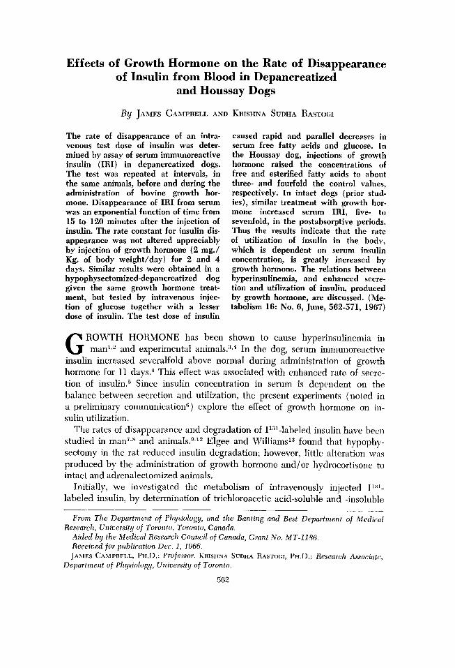
Fig. l.-Serum IRI, glucose and FFA following i.v. injection of insulin 0.50 U/Kg. at time 0, in depancreatized dog No. D. 108.
GROWTH HORMONE INJECTED DAILY DAYS: 0 2 4
0 so 100 0 50 loo 0 50 100 TIME FROM INJECTION OF INSULIN, min.
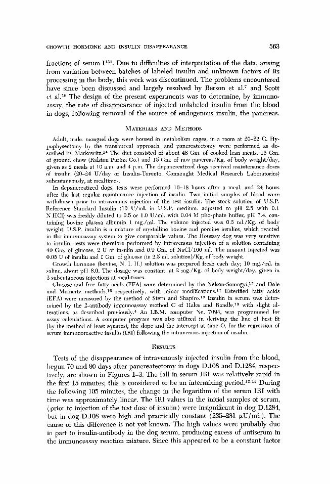
Fig. 2.-Serum IRI, glucose and FFA following i.v. injection of insulin 0.25 U/Kg. at time 0, in depancreatized dog No. D. 108. The test was repeated at intervals before and during the daily injection of growth hormone, 2 mg./Kg./day.
GROWTH HORMONE AND INSULIN DISAPPEARANCE 565
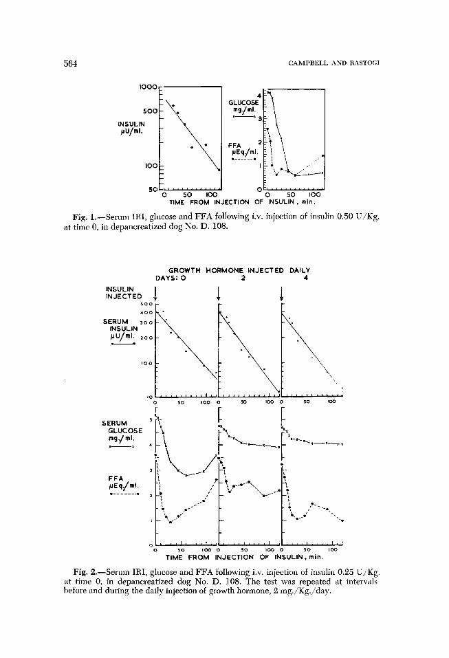
Fig. 3.-Serum IRI, glucose and FFA following i.v. injection of insulin, 0.25 U/‘Kg. at time 0 in depancreatized dog No. D. 1284. The test was repeated at in- tervals, before and during the daily injection of growth hormone, 2 mg./Kg./day.
GROWTH HORMONE INJECTED DAILY DAYS: 0
INSULIN INJECTED J i* i4
SERUM INSULIN
0 30 loo 0 so cm 0 10 100 TIME FROM INJECTION OF INSULIN, min.
in any one test, the initial value of serum IRI in dog D.108 was subtracted from the values obtained after the intravenous injection of insulin.
Inspection of the semilogarithm plots of serum IRI versus time reveals that disappearance of the hormone in 15-120 minutes approximately follows the well-known exponential course represented by equation 1:
C, = C,e -gt (1)
where Co is the initial concentration of serum IRI at time 0 (obtained by ex- trapolation of the fitted curve), Ct is the concentration at t minutes, and k is the disappearance rate constant in fraction/minute. The value of k was ob- tained by the equation:
k=- log, c, - log, c,
(2) t, - t,
566 CAMPBELL AND RASTOGI
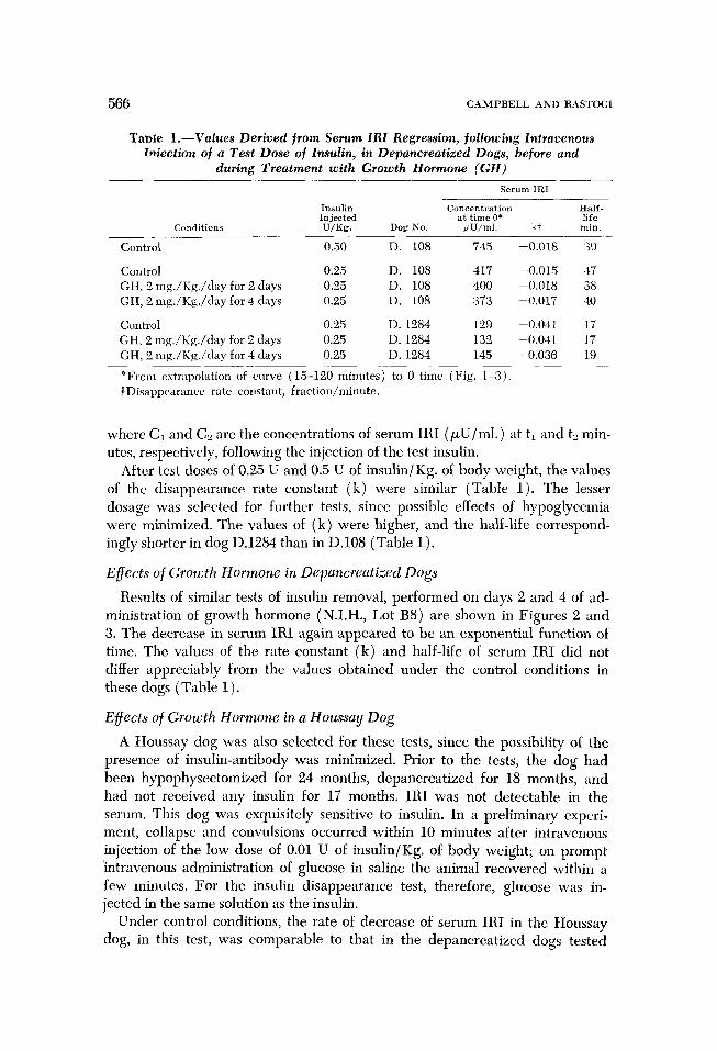
Tame l.-Values Derived from Serum ZRI Regression, following Intravenous Wection of a Test Dose of Insulin, in Depancreatized Dogs, before and
during Treatment with Growth Hormone (GH)
Serum IRI
Insulin Concentration Half- Injected at time 0’ life
Conditions U/Kg. Dog No. &U/4. kt min.
Control 0.50 D. 108 74s -0.018 39
Control 0.2.5 D. 108 417 -0.015 37 GH. 2 mg./Kg./day for 2 days 0.25 D. 108 400 -0.018 38 GH, 2 mg./Kg./day for 4 days 0.25 D. 108 373 -0.017 40
Control 0.25 D. 1284 129 -0.041 17 GH. 2 mg./Kg./day for 2 days 0.25 D. 1284 132 -0.041 17 GH, 3 mg./Kg./day for 4 days 0.25 D. 1284 145 -0.036 19
*From extrapolation of curve (15-120 minutes) to 0 time (Fig. l-3). fDisappearance rate constant, fraction/minute.
where C1 and C, are the concentrations of serum IRI (,uU/ml.) at ti and t:! min- utes, respectively, following the injection of the test insulin.
After test doses of 0.25 U and 0.5 U of insulin/Kg. of body weight, the values of the disappearance rate constant (k) were similar (Table 1). The lesser dosage was selected for further tests, since possible effects of hypoglycemia were minimized. The values of (k) were higher, and the half-life correspond- ingly shorter in dog D.1284 than in D.108 (Table 1).
Effects of Growth Hormone in Depancreatized Dogs
Results of similar tests of insulin removal, performed on days 2 and 4 of ad- ministration of growth hormone (N.I.H., Lot B8) are shown in Figures 2 and 3. The decrease in serum IRI again appeared to be an exponential function of time. The values of the rate constant (k) and half-life of serum IRI did not differ appreciably from the values obtained under the control conditions in these dogs (Table 1).
Effects of Growth Hormone in a Houssay Dog
A Houssay dog was also selected for these tests, since the possibility of the presence of insulin-antibody was minimized. Prior to the tests, the dog had been hypophysectomized for 24 months, depancreatized for 18 months, and had not received any insulin for 17 months. IRI was not detectable in the serum. This dog was exquisitely sensitive to insulin. In a preliminary experi- ment, collapse and convulsions occurred within 10 minutes after intravenous injection of the low dose of 0.01 U of insulin/Kg. of body weight; on prompt intravenous administration of glucose in saline the animal recovered within a few minutes. For the insulin disappearance test, therefore, glucose was in- jected in the same solution as the insulin.
Under control conditions, the rate of decrease of serum IRI in the Houssay dog, in this test, was comparable to that in the depancreatized dogs tested
GROWTH HORMONE AND INSULIN DISAPPEARANCE 567
GROWTH HORMONE INJECTED DAILY
DAYS: 0 2 4
INSULIN INJECTED J i &
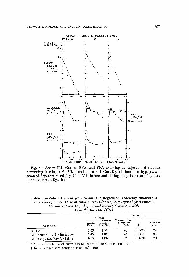
of solution Fig. 4.-Serum IRI, glucose, EFA, and FFA following i.v. injection containing insulin, 0.05 U/Kg. and glucose, 1 Gm./Kg. at time 0 in hypophysec- tomized-depancreatized dog No. 1251, before and during daily injection of growth hormone, 2 mg./Kg./day.
Table 2.-Values Derived from Serum IRI Regression, following Intravenous Injection of a Test Dose of Insulin with Glucose, in a Hypophysectomized-
Depancreatized Dog, before and during Treatment with Growth Hormone (GH)
Serum IRI Injection
concentration Insulin Glucose at time O+ Half-life
Condi!ions U/Kg. G&Kg. pU/ml. kt min.
Control 0.05 1.00 91 -0.029 24
GH, 2 mg./Kg./day for 2 days 0.05 1.00 147 -0.023 30
GH, 2 mg./Kg./day for 4 days 0.05 1.00 125 -0.034 20
*From extrapolation of curve (15 to 120 min.) to 0 time (Fig. 4). IDisappearance rate constant, fraction/minute.
568 CAMPBELL AND RASTOGI
with insulin alone. During the same growth hormone treatment (N.I.H., Lot R9) as was given to the depancreatized dogs, no alteration in the rate constant for disappearance of serum IRI was found (Fig. 4 and Table 2).
Effects on Serum Glucose and FFA
With the administration of the higher test dose of insulin in dog D.108 serum glucose fell rapidly, from the initial high level of 410, to 57 mg.1100 ml. in 1 hour. A similar sharp decrease occurred in serum FFA, followed by a rise at 2 hours (Fig. 1). The effects of insulin in uncontrolled diabetic patients are
comparable.“” The lesser test dose of insulin caused a similar sharp and pro- found fall in FFA, but less rapid decline in glucose (Fig. 2 and 3). During the administration of growth hormone in dog D.108, the fall in glucose was less than in the control period (Fig. 2 and 3), indicating an insulin-resistance effect.gl
In the Houssay dog, insulin given intravenously with glucose caused a fall in FFA to about half the initial value, without change in EFA (Fig. 4). Ad- ministration of growth hormone raised fasting levels of FFA about threefold, and of EFA about fourfold. Under these conditions, insulin with glucose in- travenously again decreased FFA to about half, but with greater net decrease, while EFA concentrations were relatively little affected.
DISCUSSION
The administration of growth hormone for several days did not alter ap- preciabIy the values of the disappearance rate constant for serum IRI (k) in depancreatized and Houssay dogs. The exponential relation for disappearance, or uptake of insulin has also been obtained in other investigations, through various approaches.7~10~22-2” Th e regression of serum IRI from the peak of the rise after intravenous injection of glucose in the normal dog is also exponen- tial’; it appears to be comparable physioIogicalIy to the removal of injected in- sulin in depancreatized dogs. The rate constants for insulin disappearance (k, fraction/minute) found in the present study (-0.015 to -0.041) were of the same magnitude as that for disappearance of insulin following glucose in- jection ( -0.031),-’ and were also comparable to that for regression of I’“‘-
labeled insulin in the rabbit (about -0.027, calculated from T1/ of about 26 minutes).7,10*11
The import of the results can be considered in the wider context of the mode of action of growth hormone on insulin secretion and utilization in the body. The observation that the values of the disappearance rate constant of injected insulin were not altered by growth hormone treatment in depancreatized dogs would appear, at first sight, to suggest that this treatment would not influence utilization of insulin in intact dogs. Evidence of an indirect nature indicates, however, that growth hormone can increase the rate of utilization of in- sulin.“C-2s This apparent conflict is resolved by the finding that in intact dogs, the same daily growth hormone treatment produced a very marked, persisting (fasting) hyperinsulinemia.” Assuming that the removal of endogenous insulin in the intact animal follows the same course as injected insulin, the rate of
GROWTH HORMONE AND INSULIN DISAPPEARANCE
DOG STATE
NORMAL
GROWTH HORMONE TREATED
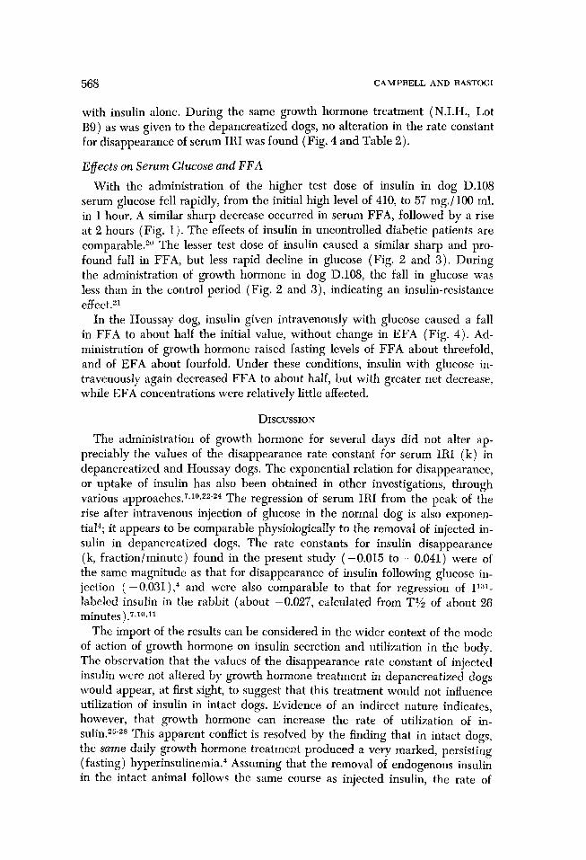
INSULIN SECRETION AND UTILIZATION
PANCREAS BETA CELLS PLASMA
EXTRAPANCREATIC TISSUES
569
Fig. S.-Proposed scheme for effects of growth hormone on insulin secretion and utilization in the dog. Growth hormone (GH) administered during 2-4 days in- creases the responsiveness of the pancreatic beta cells to glucose, causing enhanced rate of secretion of insulin at any particular level of glucose in blood.4.” Conse- quently, blood insulin concentration rises. The exponential rate constant of insulin disappearance (k) remains unaltered, but with elevated levels of insulin in blood, the rate of utilization of insulin in the body is enhanced.
utilization of insulin in the body, at a steady concentration of insulin in blood may be expressed asZO:
Iu = kcv (3)
where Iu is the rate of utilization in the body (mU/Kg./min.), k is the disap- pearance rate constant (fractionlmin.), c is the equilibrium concentration of insulin in serum (mu/L.), and v is the apparent volume of distribution of in- sulin (L./Kg. ). If it be assumed that in intact dogs the same growth hormone treatment did not alter the rate constant for removal of endogenous insulin (as of exogenous insulin in depancreatized dogs) this treatment in intact dogs, due to the sustained elevation in serum insulin concentration, must have greatly enhanced the rate of utilization of insulin (equation 3).
The extent of this increase in insulin utilization can be estimated with some degree of probability. In normal dogs under postabsorptive conditions, an ap- proximation to insulin utilization rate can be expressed as the product of the mean disappearance rate constant ( -0.031),4 the serum insulin equilibrium concentration (23 * 4 mU/L.),4 and the apparent volume of distribution of insulin (in man, 0.37 L./Kg.r), this value being 0.26 mU/Kg./minute (equa- tion 3). Estimates of insulin secretion rate in dogs under these conditions were of comparable value (about 0.43 mU/Kg./min.“). After 2 and 4 days of injec- tion of growth hormone in intact dogs, mean serum IRI concentrations were 160 * 55 and 226 rt 45 mu/L., respectively,” i.e., about five- and sevenfold the concentrations in the normal, postabsorptive state. Consequently, propor- tionate increases in the rate of utilization of insulin in the body can be expected, with values of Iu of about 1.3-1.8 mU/Kg./minute.
It may be noted, in comparison, that during this growth hormone treatment
570 CAMPBELL AND RASTOGI
in dogs, the rate of secretion of insulin was estimated to increase seven- to
nineteenfold the control level.5 The findings suggest the following explanation of the mode of action of
growth hormone. The rate of secretion of insulin is increased, due to enhanced
responsiveness of the pancreatic islet cells to glucose. A sustained rise in serum
insulin concentration results. Insulin utilization, being dependent on serum
insulin concentration, also rises, until a new equilibrium is established between
secretion and utilization of insulin at supernormal rates. These relations are
shown diagramatically in Figure 5.
ACKNOWLEDGMENTS
We wish to thank Dr. Charles H. Best for his interest in this investigation. We acknowl- edge with gratitude the expert assistance of Mrs. V. Lazdins and Mr. G. R. Green. the aid of Dr. C. C. Lucas and Dr. G. Hetenyi in the preparation of the manuscript, the computer programs prepared by Dr. R. Ninomiya and Dr. Hetenyi, the facilities provided by the Director and Staff of the Institute of Computer Science of this University. and the gift of growth hormone from the Endocrinology Study Section, National Institutes of Health, Bethesda, Maryland.
REFERENCES
1. Kipnis, D. M.: Growth hormone and insulin antagonism. In Leibel, B. S., and Wrenshall, G. A. (Eds.): Nature and Treatment of Diabetes. Amster- d am, Exerpta Medica Foundation, 1965, p. 258.
2. Luft, R., and Cerasi. E.: Effect of human growth hormone on insulin production in panhypopituitarism. Lancet 2:124, 1964.
3. Randle, P. J., and Young, F. G.: The influence of pituitary growth hormone on plasma insulin activity. J. Endo- crinol. 13:335, 1956.
4. Campbell, J., and Rastogi, K. S.: Growth hormone-induced diabetes and high levels of sermn insulin in dogs. Dia- betes 15:30, 1966.
5. -. and -: Augmented insulin secre- tion due to somatotropin. Stimulating effects of glucose and food in dogs. Diabetes 15: 749, 1966.
6. -. and -: Evidence of maintained in- crease in insulin secretion induced by growth hormone, with further augmentation after meals or infusion of glucose. in dogs with portal vein cannulae. Diabetes 14:444, 1965.
7. Berson, S. A.. Yalow, R. S., Bauman, A., Rothschild, M. A.. ancl Newerly, Ii.: Insulin-I’“1 metabolism in hu- man subjects: demonstration of in-
sulin binding globulin in the cir- culation of insulin treated subjects. J. Clin. Invest. 35: 170. 1956.
8. Bolinger, R. E., Morris, J. H., McKinght, F. G., and Diederich. D. A.: Dis- appearance of I1:3’-labelled insulin from plasma as a guide to manage- ment of diabetes. New Eng. J. Med. 270:767. 1964.
9. Elgee, N. J.. Williams, R. H., and Lee. N. D.: Distribution and degradation studies with insulin-Il”l. J. Clin. In- vest. 33:1252, 1954.
10. Scott, G. W., Prout. T. E., Weaver.
J. A.. and Asper, S. P.: A comparison of the behaviour of insulin and in- sulin labeled with Il”l in serunr. Diabetes 7:38, 1958.
11. Prout, T. E., and Evans, I. E.: De- termination of the rate of insulin destruction in vivo. Ann. N. Y. Acad. Sci. 74:530. 1959.
12. Beck. L. V.. Zaharko, D. S., Roberts. N., and King, C.: On insulin I-131 metabolism in mice. hlodifying effects of anti-insulin sermll and of total insulin dosage. Diabetes 15:336. 1966.
13. Elgee, N. J.. and Williams, R. II.: Pituitary and adrenal influences on insulin-I]~’ degradation. Amer. J. Physiol. 180:9, 1955.
li. 1larkowitz. J.: Textbook of Experi-
GROWTH HORMOKE AND INSULIN DISAPPEARANCE 571
mental Surgery. Baltimore, Wm.
Wood and Co., 1937. 15. Nelson, N.: A photometric adaptation
of the Somogyi method for determina- tion of glucose. J. Biol. Chem. 153: 375, 1944.
16. Dole, V. P.. and Meinertz, H.: Micro- determination of long-chain fatty acids in plasma and tissues. J. Biol. Chem. 235:2595,1960.
17. Campbell, J.. and Green. G. R.: Free fatty acid metabolism in Chinese hamsters. Canad. J. Physiol. Pharm. 44:47, 1966.
18. Stern, I.. and Shapiro, B.: A rapid and simple method for the determina- tion of esterified fatty acids and for total fatty acids in blood. J. Clin.
Path. 6158, 1953.
19. Hales, C. N., and Randle, P. J.: Im- munoassay of insulin with insulin- antibody precipitate. Biochem. J. 88: 137, 1963.
20. Bierman, E. L.. Dole, V. I’., and Roberts, T. N.: An abnormality of nonester- ified fatty acid metabolism in dia- betes mellitus. Diabetes 6:475, 1957.
21. DeBodo, R. C., Kurtz, M., Ancowitz, -4.. and Kiang, S. P.: Anti-insulin and diabetogenic actions of purified anterior pituitary growth hormone. Amer. J. Phvsiol. 163:310, 1950.
22. Samols, E., and Ryder, J. A.: Studies on tissue uptake of insulin in man using a differential immunoassay for endogenous and exogenous insulin. J. Clin. Invest. 40:2092, 1961.
23. Burgi. H., Kopetz, K., Schwarz, K., and Froesch, E. R.: Fate of rat in- sulin in rat-liver perfusion studies by adipose-tissue assay. Lancet 2:314, 1963.
24. Stimmler, L., and Elliott, R. B.: In- heritance of a diabetic-serum factor inhibiting normal utilization of in- sulin. Lancet 1:956, 1964.
25. Milman, A. E., DeMoor, P., and Lukens, F. D. W.: Relation of purified pituitary growth hormone and insulin in regu- lation of nitrogen balance. Amer. J. Physiol. 166354, 1951.
26. Campbell, J.: Diabetogenic actions of growth hormone. In Smith, R. W. Jr., Gaebler, 0. H., and Long, C. N. H. (eds.): The Hypophyseal Growth Hor- mone. Nature and Actions. New York, Blakiston Division, McGraw-Hill Co., 1955. p. 270.
27. Chaikof, L., and Campbell, J.: De- creased insulin requirement following growth hormone administration in diabetic dogs. Endocrinology 61:618, 1957.
28. Campbell, J., Chaikof, L., Wrenshall, G. A.. and Zemel, R.: Effects of growth hormone on metabolic and endocrine factors in hypophysecto- mized dogs. Canad. J. Biochem. Phys- iol. 37:1313, 1959.
29. Melick, R. A., Aurback, G. D., and Potts. J. T., Jr.: Distribution and half-life of r:siI-labeled parathyroid hormone in the rat. Endocrinology 77: 198. 1965.