Journal of Enzyme Inhibition and Medicinal Chemisty, August 2004 VOL. 19 (4). pp. 361-365 iTaylor&Francis healthsciences Effects of Some Antibiotics on Human Erythrocyte 6-Phosphogluconate Dehydrogenase: An in vitro and in vivo Study MEHMET AKWZ”, MUSTAFA ERATb, MEHMET ‘JFTCI”,‘r*, KENAN Gm$TEKINd and NURI BAKAN” aAtatiirk University, Medical Faculty, Department of Biochemistry, 25240 Erzurum, Turkey; bAtatiirk University, Biotechnology Application and Research Center, 25240 Erzurum, Turkey; ‘Atatiirk University, Arts and Science Faculty, Department of Chemisty, 25240 Erzurum, Turkey; dAtatiirk University, Medical Faculty, Department of Physiology, 25240 Erzurum, Turkey (Received 29 September 2003; Infinal form 20 December 2003) The in vitro and in vivo effects of some antibiotics on human erythrocyte 6-phosphogluconate dehydrogenase were investigated. Human erythrocyte 6-phosphogluco- nate dehydrogenase was purified with ammonium sulphate precipitation, Y,5’ ADP-Sepharose 4B affiity and gel filtration chromatography. Some antibiotics (netilmicin sulphate, cefepime, amikacin, isepamycin, chloramphenicol, ceftazidim, teicoplanin, ampicillin, ofloxacin, levofloxacin, cefotaxime, penicillin G, genta- micin sulphate, ciprofloxacin) inhibited enzyme activity in vitro but others (cefozin, decefin, streptomycin, combisid, and meronem) were devoid of inhibitory effects. For the drugs having low ICW values (netilmicin sulphate and cefepime), in vivo studies were performed in rats. Netilmicin sulphate at 15-mgkg inhibited enzyme activity significantly ( p < 0.001) 1 h, 2 h, and 3 h after dosing and cefepime at 200-mgkg very significantly (p -c 0.001) inhibited the enzyme 1 h and 2 h after dosing. Netilmicin sulphate and cefepime inhibited rat erythro- cyte 6-phosphogluconate dehydrogenase both in vivo and in-vitro. Keywords: 6-Phospogluconate dehydrogenase; Erythrocytes; Human; Antibiotics; Inhibitors INTRODUCTION 6-Phosphogluconate dehydrogenase (E.C.l.l. 1.44; 6PGD), the third enzyme in the pentose phosphate metabolic pathway, catalyzes the conversion of 6-PGA and NADP+, to ~-ribulose-5-phosphate and NADPH which protects the cell against oxidizin agents by producing reduced glutathione the synthesis of a number of biomolecules such as (GSH).l, f NADPH is also a coenzyme participating in fatty acids, steroids, and some amino acids.384 In the case of lack of NADPH, the concentration of GSH in living cells declines, resulting in cell death. GSH is indirectly produced by 6PGD, therefore, 6PGD can be defined as an indirect antioxidant enzyme.4f5 Many agents are known to activate or inhibit enzymes in vitro and in uiv0,6-~ so affecting meta- bolic pathways. Inhibition of 6PGD leads to decreased NADPH and GSH which will cause cell damage especially in older erythrocytes, resulting in some problems in living cells.6-8 No reports could be found in the literature on the in vitro and in vivo effects of some drugs on human erythrocyte 6PGD although vitamin C has been reported to stimulate 6PGD.l’ This study was aimed at purifying human erythrocyte GPGD, and to determination of effects of some commonly used antibiotics on human red blood cell GPGD activity. MATERIALS AND METHODS Materials 6-PGA, NADP*, Tris, and the other chemicals were from Sigma Chem. Com. and the drugs were purchased from Hoechst Marian Roussel (Turkey). Activity Determination Enzymatic activity was measured by Beutler’s method.” One unit of enzyme (EU) activity was Torresponding author. Tel.: f90-442-2311679. Fax: +90-442-2360948. E-mail: [email protected]ISSN 1475-6366 print/ISSN 1475-6374 online 8 2004 Taylor & Francis Ltd DOI: 10.1080/1475636041001667337 Journal of Enzyme Inhibition and Medicinal Chemistry Downloaded from informahealthcare.com by UB der LMU Muenchen on 03/17/13 For personal use only.
Transcript
Journal of Enzyme Inhibition and Medicinal Chemisty, August 2004 VOL. 19 (4). pp. 361-365 iTaylor&Francis
healthsciences
Effects of Some Antibiotics on Human Erythrocyte 6-Phosphogluconate Dehydrogenase: An in vitro and in vivo Study MEHMET AKWZ”, MUSTAFA ERATb, MEHMET ‘JFTCI”,‘r*, KENAN G m $ T E K I N d and NURI BAKAN”
aAtatiirk University, Medical Faculty, Department of Biochemistry, 25240 Erzurum, Turkey; bAtatiirk University, Biotechnology Application and Research Center, 25240 Erzurum, Turkey; ‘Atatiirk University, Arts and Science Faculty, Department of Chemisty, 25240 Erzurum, Turkey; dAtatiirk University, Medical Faculty, Department of Physiology, 25240 Erzurum, Turkey
(Received 29 September 2003; Infinal form 20 December 2003)
The in vitro and in vivo effects of some antibiotics on human erythrocyte 6-phosphogluconate dehydrogenase were investigated. Human erythrocyte 6-phosphogluco- nate dehydrogenase was purified with ammonium sulphate precipitation, Y,5’ ADP-Sepharose 4B affiity and gel filtration chromatography. Some antibiotics (netilmicin sulphate, cefepime, amikacin, isepamycin, chloramphenicol, ceftazidim, teicoplanin, ampicillin, ofloxacin, levofloxacin, cefotaxime, penicillin G, genta- micin sulphate, ciprofloxacin) inhibited enzyme activity in vitro but others (cefozin, decefin, streptomycin, combisid, and meronem) were devoid of inhibitory effects. For the drugs having low ICW values (netilmicin sulphate and cefepime), in vivo studies were performed in rats. Netilmicin sulphate at 15-mgkg inhibited enzyme activity significantly ( p < 0.001) 1 h, 2 h, and 3 h after dosing and cefepime at 200-mgkg very significantly ( p -c 0.001) inhibited the enzyme 1 h and 2 h after dosing. Netilmicin sulphate and cefepime inhibited rat erythro- cyte 6-phosphogluconate dehydrogenase both in vivo and in-vitro.
6-Phosphogluconate dehydrogenase (E.C.l.l. 1.44; 6PGD), the third enzyme in the pentose phosphate metabolic pathway, catalyzes the conversion of 6-PGA and NADP+, to ~-ribulose-5-phosphate and NADPH which protects the cell against oxidizin agents by producing reduced glutathione
the synthesis of a number of biomolecules such as (GSH).l, f NADPH is also a coenzyme participating in
fatty acids, steroids, and some amino acids.384 In the case of lack of NADPH, the concentration of GSH in living cells declines, resulting in cell death. GSH is indirectly produced by 6PGD, therefore, 6PGD can be defined as an indirect antioxidant enzyme.4f5 Many agents are known to activate or inhibit enzymes in vitro and in uiv0,6-~ so affecting meta- bolic pathways. Inhibition of 6PGD leads to decreased NADPH and GSH which will cause cell damage especially in older erythrocytes, resulting in some problems in living cells.6-8 No reports could be found in the literature on the in vitro and in vivo effects of some drugs on human erythrocyte 6PGD although vitamin C has been reported to stimulate 6PGD.l’
This study was aimed at purifying human erythrocyte GPGD, and to determination of effects of some commonly used antibiotics on human red blood cell GPGD activity.
MATERIALS AND METHODS
Materials
6-PGA, NADP*, Tris, and the other chemicals were from Sigma Chem. Com. and the drugs were purchased from Hoechst Marian Roussel (Turkey).
Activity Determination
Enzymatic activity was measured by Beutler’s method.” One unit of enzyme (EU) activity was
ISSN 1475-6366 print/ISSN 1475-6374 online 8 2004 Taylor & Francis Ltd DOI: 10.1080/1475636041001667337
Jour
nal o
f E
nzym
e In
hibi
tion
and
Med
icin
al C
hem
istr
y D
ownl
oade
d fr
om in
form
ahea
lthca
re.c
om b
y U
B d
er L
MU
Mue
nche
n on
03/
17/1
3Fo
r pe
rson
al u
se o
nly.
362 M. A K w Z et al.
defined as the amount of enzyme reducing 1 pmol NADP+ per 1 min at 25"C, pH 8.0.
Preparation of the Hemolysate and Hemoglobin Estimation
Fresh human blood samples were collected in tubes containing EDTA, then centrifuged (15 min, 2,500 X g) and plasma and buffy coat (leucocytes) were removed. The packed red cells were washed three times with KC1 (0.16M) and hemolyzed with 5 volume of ice-cold water and then centrifuged (4"C, 10,000 x g, for 30min) to remove the ghosts and intact cells. Hemoglobin (Hb) concentration in the hemolysate was determined by the cyan- methaemoglobin
Ammonium Sulphate Precipitation
The hemolyzate was subjected to precipitation with ammonium sulphate (between35% and 65%). Enzyme activity was determined both in the supernatant and in the precipitate for each respective precipitation. The precipitated enzyme was dissolved in phosphate buffer (50 mM; pH = 7.0). The resultant solution was clear, and contained partially purified enzyme.
Purification of the 6-PGD
For purification of 6-PGD, the ammonium sulphate fraction (35-65%) of the homogenate obtained previously was loaded onto a 2', 5'-ADP Sepharose 48 affinity column and the flow rate was adjusted to 20 ml/h. The column was then sequentially washed with a 25ml buffer of 0.1M K-acetate and 0.1M K-phosphate, (pH 6.0) and 25ml of a buffer of 0.1M K-acetate and 0.1M K-phosphate (pH 7.85). The wash with the second buffer was continued until the final absorbance difference became 0.05. Elution of the enzyme was carried out with a mixture containing 80 mM K-phosphate, 80 mM KC1, 5mM NADP+ and lOmM EDTA (pH 7.85). Enzyme activity was measured in all fractions, and the activity-containing fractions were pooled, then dialyzed in 50 mM K-acetate + 50 mM K-phosphate buffer (500ml, pH 7.0) for 2h with two chan es of buffer. All procedures were performed at 4°C. Pi14
Sephadex G-200 Gel Filtration Chromatography
5g of dry Sephadex G-200 was incubated in the distilled water at 90 " C for 5 hours. After removal of the air in the gel, it was loaded onto a column (2 X 50cm). Flow rate was adjusted to 15mL/h by means of peristaltic pump. The column was equili- brated with 50mM Tris-HC1, 50mM KC1 buffer, pH: 7.3 until the final absorbance difference became zero at 280 nm and pH value became same with that of equilibration buffer. The dialysed from affinity
chromatography column was mixed with glycerol at the ratio of 5%. The final sample was loaded onto the column and each elution was collected in Eppendorf tubes as 2mL. The absorbance values were determined at 280nm and at 340nm in each fractions. Active fractions were lyophilized and stored at - 85 "C for the use in in vitro studies.
Protein Determination
The protein content in all samples was quantified spectrophotometrically at 595 nm according to Bradford's method,15 using bovine serum albumin as standard.
SDS Polyacrylamide Gel Electrophoresis
The control of enzyme purity was carried out using Laemmli's procedure16 with 3% and 8% acrylamide concentrations for running and stacking gel, respect- ively. E . coli P-galactosidase (116,000), rabbit phosphorylase B (97,400), bovine albumin (66,000), chicken ovalbumin (45,000), and bovine carbonic anhydrase (29,000) were used as standards (Sigma:
(SDS-PAGE)
M W-SDS-200).
In vitro Drug Effect
In order to determine the effects of some antibiotics on human 6PGD, concentrations of gentamicin sulphate (4.15-33.20 mM), amikacin sulphate (4.26-14.20 mM), netilmicin sulphate (3.47-20.82 mM), isepamycin (21.90- 175.20 mM), chloramphenicol (10.30- 77.25 mM), ceftazidim (1.55-7.75 mM), teicoplanin (7.2-96mM), ampicillin (31.80-150 mM), ofloxacin (0.11 -0.412 mM), cefotaxime (1.50-15 mM), levoflox- acin (0.70-4.20mM), cefepime (1.04-10.39 mM), peni- cillin G (41.94-419.45 mM), and ciproflaxacin (0.12-1.20 mM), were added to the purified enzyme. The enzyme activity was measured and an experiment in the absence of drug was used as control (100% activity). The IC50 values were obtained from activity (%) us. drug concentration plots.
In vivo Drug Effect
In vivo studies were conducted only for those compounds having low ICs0 values. i.e. netilmicin sulphate and cefepime. Each drug-treated group comprised six rats (180 ? 20g) which were kept under special conditions (in a windowless room, at of 22"C, with light on for 14 h) for 2 months. Before intramuscular drug injection, control blood samples (0.5ml of whole blood with EDTA) were obtained from the animals. Then 15mg/kg of netilmicin sulphate was injected into one group and 200 mg/kg of cefepime into the other. Blood samples were
Jour
nal o
f E
nzym
e In
hibi
tion
and
Med
icin
al C
hem
istr
y D
ownl
oade
d fr
om in
form
ahea
lthca
re.c
om b
y U
B d
er L
MU
Mue
nche
n on
03/
17/1
3Fo
r pe
rson
al u
se o
nly.
EFFECTS OF ANTIBIOTICS ON 6-PGD 363
taken from each group 1, 2, and 3 h after injection. Hemolysates were prepared from all blood samples as mentioned above. Hemoglobin levels and 6-PGD activity were determined in these hemolysates.
Statistical Analysis
The mean values and standard deviations of the results were determined using student f-test for statistical evaluation of the difference, with p < 0.001 considered very significant.
RESULTS
The purification of the enzyme led to a specific activity of 1.886EU/mg proteins, a yield of 49.57% and a purification coefficient of 725 (Table I). Figure 1 shows the SDS-PAGE for the purity of the enzyme.
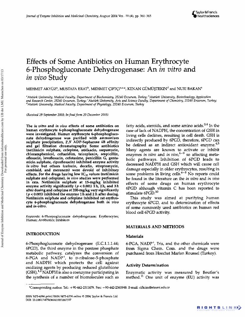
Figure 2 shows the in vitro inhibitory effects of four typical antibiotics (netilmicin sulphate, cefe- pime, amikacin, and isepamycin) on enzyme activity. The IC50 values were: netilmicin sulphate (3.372 mM), cefepime (4.213), amikacin (3.630), isepamycin (30.528), chloramphenicol (36.283), cef- tazidim (2.110), teicoplanin (21.521), ampicillin (36.282), ofloxacin (0.118), levofloxacin (2.142), cefotaxime (3.559), penicillin G (161.162), gentamicin sulphate (4.932), ciprofloxacine (0.271).
The results of the in vivo effects of netilmicin sulphate and cefepime are presented in Table II. In the netilmicin sulphate-treated group of animals, the control enzyme activity was 3.627 t 0.419EU/gHb, while the respective values determined 1, 2 and 3h after drug administration were 1.195 ? 0.115 EU/gHb, 0.354 2 0.056EU/gHb and of 1.821 t 0.123EU/gHb. In the cefepime-treated group of animals, the control enzyme activity was 3.893 ? 0.260EU/gHb, and 1, 2, and 3h after drug treatment it was 1.197 t 0.365EU/gHb, 0.362 t 0.077EU/gHb and 3.628 t 0.350EU/gHb respectively.
DISCUSSION
Human 6PGD from erythrocytes was purified in this study by hemolysate preparation, ammonium
FIGURE 1 SDS-PAGE bands of 6PGD (Lane 1: 6PGD enzyme, Lane 2, 3, 4: standard proteins; E.Coli P-galactosidase (116,000), rabbit phosphorylase B (97,400), bovine albumin (66,000), chicken ovalbumin (45,000), and bovine carbonic anhydrase (29,000) were used as standards (Sigma: MW-SDS-200).
sulphate precipitation, 2/,5/-ADP Sepharose 4B affinity chromatography and gel filtration chromato- graphy. The purified preparation was characterized with a specific activity of 1.886EU/mg protein, a yield of 49.57% and a purification coefficient of 725. These figures tend to validate the procedure used in the study. The SDSPAGE shows the high purity of the enzyme.
The antibiotics which were used this study are commonly used clinically throught the world. However, some are associated with side effects such as nephrotoxicity, ototoxicity, neurotoxicity, fever, bone marrow depression, fever, allerg epidermal eruption and hemolytic anemia. Ampicillin inhibits human red cells G6PD;' sheep liver G6PD,I9 rat 6PGD,9 and human CA?' netil- micin sulphate inhibits human red cells G6PD,I8 rat 6PGD9 and amikacin inhibits sheep liver G6PD:' sheep red blood cells G6PD," and rat 6PGD.9 In addition, it has been reported that ampicillin, netilmicin and amikacin inhibit the rat 6PGA and G6PD in v i v ~ . ~ ~ ~ ~ . We could not find any
K;
TABLE I Purification scheme for 6-phosphogluconate dehydrogenase from human erythrocytes
Total Specific Total Total Purification Activity volume Protein protein activity activity Yield Purification step (EU/ml) (ml) (mg/ml) (mg) (EU) (EU/mg) 0'0) factor
previous reports related to the effects of other drugs on 6PGD activity. In the present study, the effect of some commonly used drugs on 6PGD has been investigated in vitro and in an animal model in-vitro.
Amikacin, isepamycin, chloramphenicol, cefta- zidim, teicoplanin, ampicillin, ofloxacin, levo- floxacin, cefotaxime, penicillin G, gentamicin sulphate, and ciprofloxacin inhibit 6PGD in vitro (Figure 2) and netilmicin sulphate and cefepime are inhibitory both in vitro and in vivo (Table III).
From the in vitro studies, it was clear that 6PGD was more strongly inhibited by netilmicin sulphate, cefepime, amikacin, ceftazidim, ofloxacin, levo- floxacin, cefotaxime, gentamicin sulphate and cipro- floxacin of the drugs tested. Of these drugs,
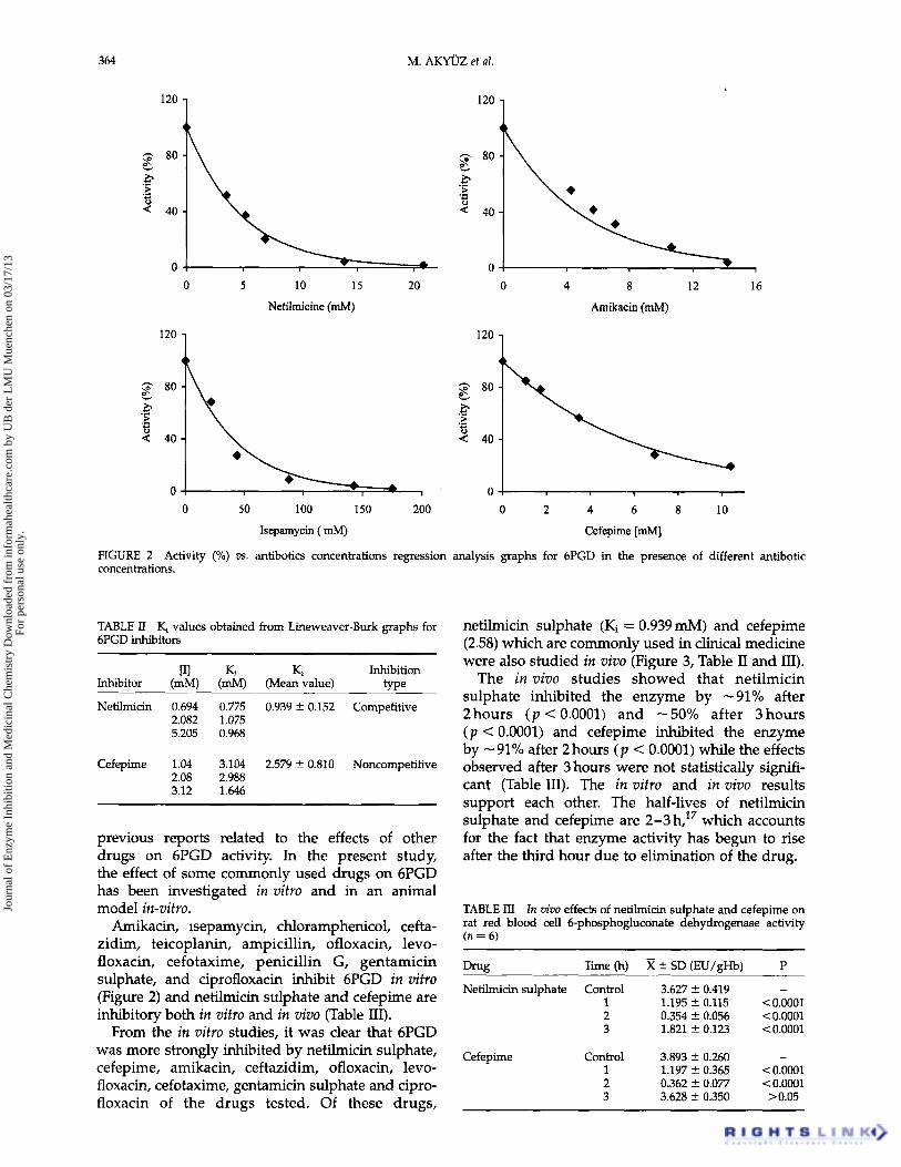
netilmicin sulphate (Ki = 0.939mM) and cefepime (2.58) which are commonly used in clinical medicine were also studied in vivo (Figure 3, Table I1 and III).
The in vivo studies showed that netilmicin sulphate inhibited the enzyme by -91% after 2hours ( p < 0.0001) and -50% after 3hours ( p < 0.0001) and cefepime inhibited the enzyme by - 91% after 2 hours ( p < 0.0001) while the effects observed after 3 hours were not statistically signifi- cant (Table 111). The in vitro and in vivo results support each other. The half-lives of netilmicin sulphate and cefepime are 2-3 h,17 which accounts for the fact that enzyme activity has begun to rise after the third hour due to elimination of the drug.
TABLE III In aivo effects of netilmicin sulphate and cefepime on rat red blood cell 6-phosphogluconate dehydrogenase activity (n = 6)
- Drug Time (hl X t SD (EU/eHb) P
Netilmicin sulphate Control 3.627 * 0.419 1 1.195 t 0.115 2 0.354 2 0.056 3 1.821 2 0.123
Cefepime Control 3.893 t 0.260 1 1.197 C 0.365 2 0.362 t 0.077 3 3.628 ? 0.350
- <0.0001 < 0.0001 < 0.0001
- < 0.0001 <0.0001 > 0.05
Jour
nal o
f E
nzym
e In
hibi
tion
and
Med
icin
al C
hem
istr
y D
ownl
oade
d fr
om in
form
ahea
lthca
re.c
om b
y U
B d
er L
MU
Mue
nche
n on
03/
17/1
3Fo
r pe
rson
al u
se o
nly.
EFFECTS OF ANTIBIOTICS ON 6-PGD 365
0 0,694 mM Net. A2,082 mM Net.
60
-0 -0 0,Ol 0,03 0,05 0,07 0,09 0,11
1/[6-PGA] (mM)-' -0,os 0,02 0,12 0,22
1/[6-PGA] (mM)-'
FIGURE 3 Lineweaver-Burk graphs using 3 different netilmicin and cefepime concentrations for determination of Ki.
The doses of netilmicin sulphate (15mg/kg) and cefepime (200 mg/kg) used here are consistent with previous studies, since the clinical doses of 37.5- 200mg/kg and 20-2000 mg/kg have been reported for netilmicin and cefepime respectively.22-28
The use of these antibiotics can cause serious adverse effects such as hemolytic effects, which may move fatal. If it is required to give netilmicin sulphate and cefepime to patients, their dosage should be very well controlled to decrease hemolytic and other side effects.
References [l] Bianchi, D., Bertrant, O., Haupt, K. and Coello, N. (2001)