Effects of Storage Duration and Temperature on the Values of Haematological Parameters in Bovine and Ovine Blood Samples Kirmizigul A.H. 1 *, Gokce E. 1 , Sozmen M. 2 * Kafkas University, Faculty of Veterinary Medicine, Department of Internal Medicine, 36100, Kars-TURKEY **Ondokuz Mayis University, Faculty of Veterinary Medicine, Department of Pathology, 55139, Samsun-TURKEY Corresponding Author: A.H. Kirmizigul, Kafkas University, Faculty of Veterinary Medicine, Department of Internal Medicine, 36100, Pasacayir, Kars,TURKEY E-mail: [email protected]Date of initial submission: 11.02.2016 Date of revised submission: 27.05.2016 Date of acceptance:01.06.2016 Research article Ερευνητικό άρθρο J HELLENIC VET MED SOC 2017, 68(2): 145 - 154 ΠΕΚΕ 2017, 68(2): 145 - 154 ABSTRACT. The purpose of the present study was to determine the alterations in haematological values at 0, 12, 24, 36, 48, 60, 72, 84 and 96 hours of storage in bovine and ovine venous blood samples stored at 4˚C and 24˚C in EDTA-coated tubes. Twenty healthy animals, including ten 4-year-old cattle and ten 2-year-old sheep constituted the study material. Bovine blood samples stored at 4˚C produced reliable results for the WBC, RBC, Hb, PCV, MCV, MCH and MCHC levels with the exception of PLT level as PLT levels decreased when the PLT stored 24 hours or longer times. On the other hand ovine blood samples stored at 4˚C for 24 hours or longer, all the parameters measured (WBC, RBC, Hb, PCV, MCV, MCH, MCHC and PLT levels) gave reliable results indicating that ovine blood parameters can be used effectively and safely. Furthermore, in the bovine blood samples, MCHC levels were decreased as from 60 hours stored at 24°C and WBC levels were decreased in the ovine blood samples stored 48 hours or longer at 24°C. However, the blood samples stored at 24°C, the measurement of the RBC, Hb, PCV, MCV, MCH and PLT levels produced reliable results. Keywords: Haemogram, sheep, storage temperature, cattle, storage duration
Transcript
Effects of Storage Duration and Temperature on the Values of Haematological Parameters in Bovine and Ovine Blood Samples
Kirmizigul A.H.1*, Gokce E.1, Sozmen M.2
*Kafkas University, Faculty of Veterinary Medicine, Department of Internal Medicine, 36100, Kars-TURKEY**Ondokuz Mayis University, Faculty of Veterinary Medicine, Department of Pathology,
55139, Samsun-TURKEY
Corresponding Author: A.H. Kirmizigul,Kafkas University, Faculty of Veterinary Medicine, Department of Internal Medicine, 36100, Pasacayir, Kars,TURKEYE-mail: [email protected]
Date of initial submission: 11.02.2016Date of revised submission: 27.05.2016Date of acceptance:01.06.2016
Research articleΕρευνητικό άρθρο
J HELLENIC VET MED SOC 2017, 68(2): 145 - 154ΠΕΚΕ 2017, 68(2): 145 - 154
ABSTRACT. The purpose of the present study was to determine the alterations in haematological values at 0, 12, 24, 36, 48, 60, 72, 84 and 96 hours of storage in bovine and ovine venous blood samples stored at 4˚C and 24˚C in EDTA-coated tubes. Twenty healthy animals, including ten 4-year-old cattle and ten 2-year-old sheep constituted the study material. Bovine blood samples stored at 4˚C produced reliable results for the WBC, RBC, Hb, PCV, MCV, MCH and MCHC levels with the exception of PLT level as PLT levels decreased when the PLT stored 24 hours or longer times. On the other hand ovine blood samples stored at 4˚C for 24 hours or longer, all the parameters measured (WBC, RBC, Hb, PCV, MCV, MCH, MCHC and PLT levels) gave reliable results indicating that ovine blood parameters can be used effectively and safely. Furthermore, in the bovine blood samples, MCHC levels were decreased as from 60 hours stored at 24°C and WBC levels were decreased in the ovine blood samples stored 48 hours or longer at 24°C. However, the blood samples stored at 24°C, the measurement of the RBC, Hb, PCV, MCV, MCH and PLT levels produced reliable results.
J HELLENIC VET MED SOC 2017, 68(2)ΠΕΚΕ 2017, 68(2)
146 KIRMIZIGUL A.H., GOKCE E., SOZMEN M.
INTRODUCTION
In veterinary medicine, the complete blood count is a routine diagnostic procedure (Furlanello et al., 2006).
In some cases, blood samples required for haematological analyses may be collected from animals raised at farms remotely located to the laboratory. In such cases, the analysis of these samples may be delayed (Hulme-Moir et al., 2006). Hence, the storage conditions of these blood samples during transport to the laboratory and until the time of analysis may affect test results. In fact, false and misleading results can be obtained with blood samples stored under inappropriate conditions (Ihedioha and Onwubuche, 2007). In some cases, blood samples may be required to be transported to the laboratory for several hours at room temperature, without being frozen. In some other cases, the sampling of a high number of ani-mals and the collective transport of the resulting samples may significantly delay the conduct of labo-ratory analysis (Furlanello et al., 2006; Ihedioha and Onwubuche, 2007).The purpose of the present study was to determine the alterations in the haematological values (WBC, RBC, Hb, PCV, MCV, MCH, MCHC and PLT levels) of bovine and ovine venous blood samples stored at 4˚C and 24˚C in EDTA-coated tubes, at 0, 12, 24, 36, 48, 60, 72, 84 and 96 hours of storage.
MATERIALS AND METHODSTwenty healthy animals, including ten 4-year-old cattle and ten 2-year-old sheep, which were raised at the Research and Practice Farm of Kafkas Uni-versity, Faculty of Veterinary Medicine, constituted the study material. Two blood samples were taken from the jugular vein of each animal, into EDTA-coated tubes. One of these samples was stored at 24°C and the other was stored at 4°C. Both samples were analyzed at 0, 12, 24, 36, 48, 60, 72, 84 and 96 hours of storage with the use of an automated hematology analyzer (MS4e, Melet Schloesing Labo-ratories) for White Blood Cell (WBC), Red Blood Cell (RBC), hemoglobin (Hb), Packed Cell Volume (PCV), Mean Corpuscular Volume (MCV), Mean Cell Hemoglobin (MCH), Mean Cell Hemoglobin Concentration (MCHC) and platelet (PLT) levels. Previous the analysis the device was calibrated with
control blood. The measurement is automatically made by the device according to formula provided below which can be applied all the species: “Meas-ured Value x Control Bench Adjustment Factor x Correction coefficient factor based on the type of ani-mals used= Target Value/Results”For haematological examination, blood smears were prepared with these samples at the time points indi-cated above. For this purpose, a drop of blood was spread along a clean slide. After being air-dried, the smear was fixed with methyl alcohol for two min-utes. Subsequently, the smears were stained with 10% Giemsa solution for 30 minutes. Following staining, the smears were washed with distilled water and air-dried. Finally, the blood smears stained with Giemsa solution were examined under a light micro-scope (Olympus BX51, Olympus Corp., Japan) at 100X10 magnifications. Statistical evaluation of the results was done using SPSS® (SPSS 20, IL, USA) software. The differences between the values obtained in this study, for the dif-ferent storage periods, were analyzed with analysis of variance (ANOVA), whereas the differences for the storage temperatures were analyzed with the t-test (Kuipers et al., 2007). Differences were consid-ered significant if the P value was less than 0.05.
RESULTSThe mean WBC, RBC, Hb, PCV, MCV, MCH, MCHC and PLT levels measured in the ovine and bovine blood samples stored at 4°C and 24°C are presented in Tables 1, 2, 3 and 4. The graphic repre-sentation of the alterations observed in these values is shown in Figures 1 and 2. Throughout the study, no statistically significant alteration was observed in the WBC, RBC, Hb, PCV, MCV, MCH and MCHC levels of the bovine blood samples stored at 4°C. On the other hand, the PLT values measured at 0 hour were determined to significantly differ from the PLT values measured at 24, 48 and 96 hours (p<0.05).Throughout the study, no statistically significant alteration was determined in the WBC, RBC, Hb, PCV, MCV, MCH and PLT levels of the bovine blood samples stored at 24°C (P>0.05). On the other hand, the MCHC values were decreased over the time, and the differences between the values meas-
J HELLENIC VET MED SOC 2017, 68(2)ΠΕΚΕ 2017, 68(2)
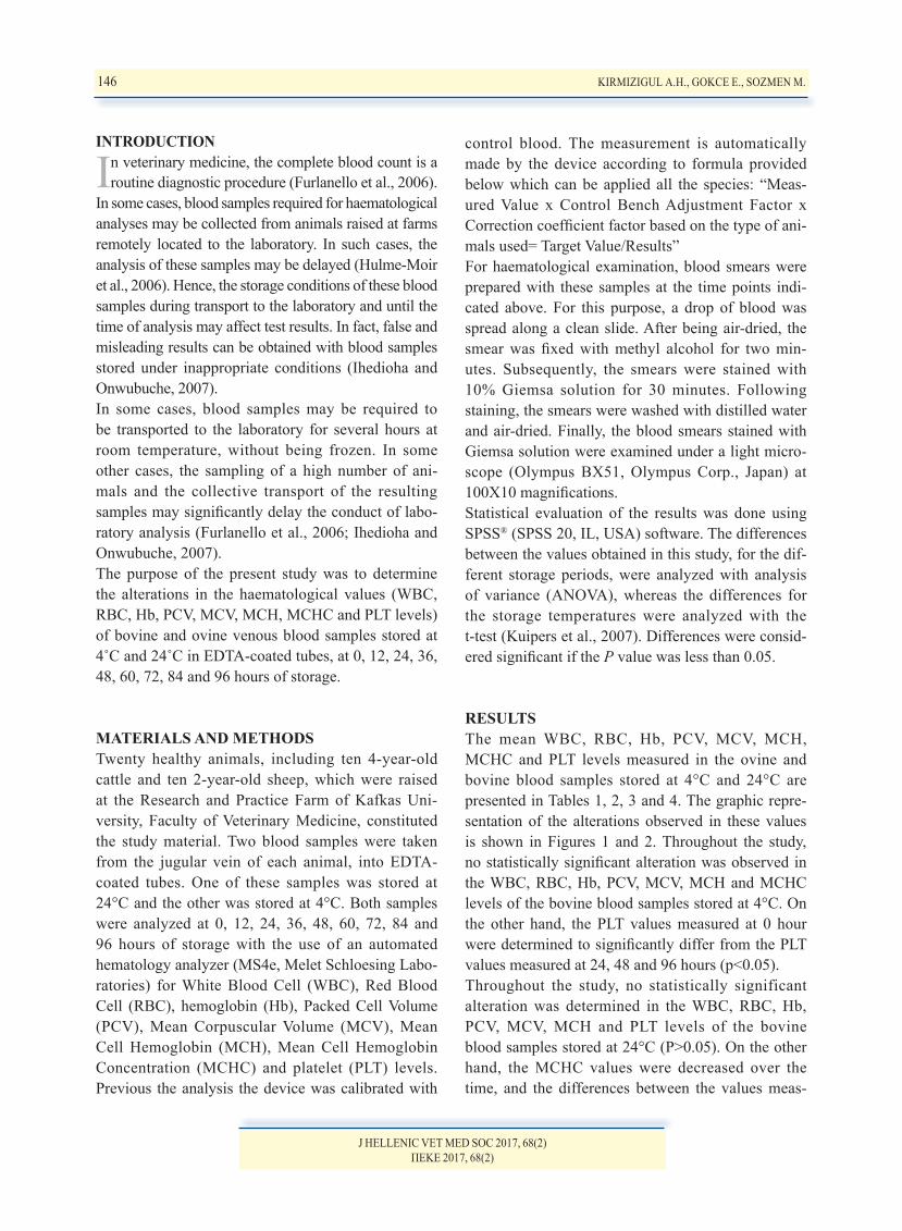
Table 1: The haematological values of the bovine blood samples stored at 4°C
Table 2: The haematological values of the bovine blood samples stored at 24°C
Table 3: The haematological values of the ovine blood samples stored at 4°C
Table 4: The haematological values of the ovine blood samples stored at 24°C
J HELLENIC VET MED SOC 2017, 68(2)ΠΕΚΕ 2017, 68(2)
Figure 1: Haematological changes in the bovine blood samples
148 KIRMIZIGUL A.H., GOKCE E., SOZMEN M.
J HELLENIC VET MED SOC 2017, 68(2)ΠΕΚΕ 2017, 68(2)
Figure 2: Haematological changes in the ovine blood samples
KIRMIZIGUL A.H., GOKCE E., SOZMEN M. 149
J HELLENIC VET MED SOC 2017, 68(2)ΠΕΚΕ 2017, 68(2)
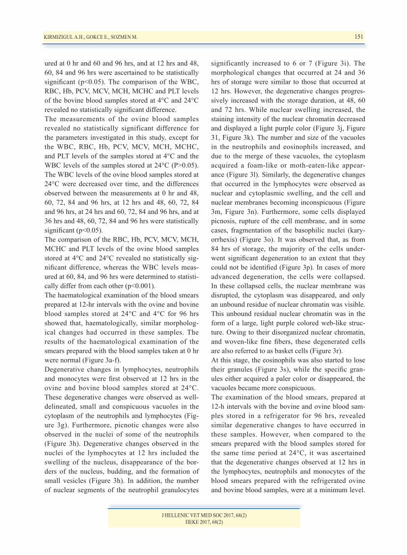
Normal blood from cow (a, b, c) and sheep (d, e, f).(a) A normal segmented neutrophil (upper left) and a medium lymphocyte (lower left) in blood from a cow. Bar= 8 µm. Giemsa.(b) A monocyte in blood from a cow with pleomorphic nucleus and slightly basophilic cytoplasm. Bar= 8 µm. Giemsa.(c) A medium to large lymphocyte (upper left) and an eosinophil (middle right) in blood from a cow, exhibiting numerous small round granules. Bar= 8 µm. Giemsa.(d) Two neutrophils (lower left) and a small lymphocyte (lower right) in blood from a sheep. Bar= 8 µm. Giemsa. (e) A monocyte in blood from a sheep with pleomorphic nucleus (upper right corner). Bar= 8 µm. Giemsa.(f) An eosinophil in blood from a sheep, exhibiting numerous small round granules. Bar= 8 µm. Giemsa.(g) Small, distinct, clear intracytoplasmic vacuole in the cytoplasm of a lymphocyte (short arrow). Sheep blood smear. 12 hours of storage at 24°C. Bar= 8 µm. Giemsa. (h) A neutrophil with four condensed nuclear chromatin material showing pyknotic change (short arrow). A lymphocyte with irregular cytoplasmic and nuclear contours exhibiting small vesicular like protrusions (long ar-rows). Sheep blood smear. 12 hours of stor-age at +4 ºC. Bar= 8 µm. Giemsa.(i) Hypersegmented neutrophilic granulocyte during degradation. Sheep blood smear. 24 hours of storage at +4 ºC. Bar= 8 µm. Giemsa. (j) Two neutrophils with distended, light purple colored nuclear chromatin. Three smudge cells are also seen. Sheep blood smear. 60 hours of storage at 24°C. Bar= 8 µm. Giemsa.(k) A neutrophil with distended, irregular, lightly colored nuclear chromatin. Nuclear chromatin contains a few, small, irregular, discrete vacuoles (arrow). Sheep blood smear. 60 hours of storage at 24°C. Bar= 8 µm. Giemsa.(l) An eosinophil with pyknotic nucleus. Cytoplasm contains numerous, various sized, irregularly shaped vacuoles giving the cytoplasm a foamy appearance. Sheep blood smear. 60 hours of storage at 24°C. Bar= 8 µm. Giemsa.(m) Cytoplasm of lymphocytes are expanded with presence of various sized vacuoles (ar-rows). Smooth nuclear contours are disrupt-ed. Sheep blood smear. 48 hours of storage at +4 ºC. Bar= 8 µm. Giemsa. (n) A lymphocyte with irregular and indis-tinct nuclear and cytoplasmic membranes. Cow blood smear. 48 hours of storage at +4 ºC. Bar= 8 µm. Giemsa. (o) The cell with ruptured nuclear envelope and five markedly condensed nuclear frag-ments is a lymphocyte indicating karyor-
Figure 3: Blood smear images
rhexis. Sheep blood smear. 36 hours of storage at 24°C. Bar= 8 µm. Giemsa. (p) Free, disrupted nuclei without cytoplasm. All of the cells are unidentifiable. Sheep blood smear. 96 hours of storage at +4 ºC. Bar= 8 µm. Giemsa.(r) Smudge cell. The large, light purple, delicate net- or woven basket-like strands in the center of the field is a broken or basket cell; this is free, dispersed nuclear chromatin material and it is not possible to identify the origin of the cell. On the left there is a free nucleus. On the right there is a lymphocyte with swollen, vacuolated cytoplasm. Sheep blood smear. 84 hours of storage at +4 ºC. Bar= 8 µm. Giemsa. (s) An eosinophil is commenced to lose its granules (arrow). A basket cell with net like structure is visible on the left field. On the right bottom there are two free disrupted nuclei without cytoplasm. Cow blood smear. 96 hours of storage at 24°C. Bar= 8 µm. Giemsa.
150 KIRMIZIGUL A.H., GOKCE E., SOZMEN M.
J HELLENIC VET MED SOC 2017, 68(2)ΠΕΚΕ 2017, 68(2)
significantly increased to 6 or 7 (Figure 3i). The morphological changes that occurred at 24 and 36 hrs of storage were similar to those that occurred at 12 hrs. However, the degenerative changes progres-sively increased with the storage duration, at 48, 60 and 72 hrs. While nuclear swelling increased, the staining intensity of the nuclear chromatin decreased and displayed a light purple color (Figure 3j, Figure 31, Figure 3k). The number and size of the vacuoles in the neutrophils and eosinophils increased, and due to the merge of these vacuoles, the cytoplasm acquired a foam-like or moth-eaten-like appear-ance (Figure 3l). Similarly, the degenerative changes that occurred in the lymphocytes were observed as nuclear and cytoplasmic swelling, and the cell and nuclear membranes becoming inconspicuous (Figure 3m, Figure 3n). Furthermore, some cells displayed picnosis, rupture of the cell membrane, and in some cases, fragmentation of the basophilic nuclei (kary-orrhexis) (Figure 3o). It was observed that, as from 84 hrs of storage, the majority of the cells under-went significant degeneration to an extent that they could not be identified (Figure 3p). In cases of more advanced degeneration, the cells were collapsed. In these collapsed cells, the nuclear membrane was disrupted, the cytoplasm was disappeared, and only an unbound residue of nuclear chromatin was visible. This unbound residual nuclear chromatin was in the form of a large, light purple colored web-like struc-ture. Owing to their disorganized nuclear chromatin, and woven-like fine fibers, these degenerated cells are also referred to as basket cells (Figure 3r).At this stage, the eosinophils was also started to lose their granules (Figure 3s), while the specific gran-ules either acquired a paler color or disappeared, the vacuoles became more conspicuous.The examination of the blood smears, prepared at 12-h intervals with the bovine and ovine blood sam-ples stored in a refrigerator for 96 hrs, revealed similar degenerative changes to have occurred in these samples. However, when compared to the smears prepared with the blood samples stored for the same time period at 24°C, it was ascertained that the degenerative changes observed at 12 hrs in the lymphocytes, neutrophils and monocytes of the blood smears prepared with the refrigerated ovine and bovine blood samples, were at a minimum level.
ured at 0 hr and 60 and 96 hrs, and at 12 hrs and 48, 60, 84 and 96 hrs were ascertained to be statistically significant (p<0.05). The comparison of the WBC, RBC, Hb, PCV, MCV, MCH, MCHC and PLT levels of the bovine blood samples stored at 4°C and 24°C revealed no statistically significant difference. The measurements of the ovine blood samples revealed no statistically significant difference for the parameters investigated in this study, except for the WBC, RBC, Hb, PCV, MCV, MCH, MCHC, and PLT levels of the samples stored at 4°C and the WBC levels of the samples stored at 24°C (P>0.05). The WBC levels of the ovine blood samples stored at 24°C were decreased over time, and the differences observed between the measurements at 0 hr and 48, 60, 72, 84 and 96 hrs, at 12 hrs and 48, 60, 72, 84 and 96 hrs, at 24 hrs and 60, 72, 84 and 96 hrs, and at 36 hrs and 48, 60, 72, 84 and 96 hrs were statistically significant (p<0.05). The comparison of the RBC, Hb, PCV, MCV, MCH, MCHC and PLT levels of the ovine blood samples stored at 4°C and 24°C revealed no statistically sig-nificant difference, whereas the WBC levels meas-ured at 60, 84, and 96 hrs were determined to statisti-cally differ from each other (p<0.001).The haematological examination of the blood smears prepared at 12-hr intervals with the ovine and bovine blood samples stored at 24°C and 4°C for 96 hrs showed that, haematologically, similar morpholog-ical changes had occurred in these samples. The results of the haematological examination of the smears prepared with the blood samples taken at 0 hr were normal (Figure 3a-f). Degenerative changes in lymphocytes, neutrophils and monocytes were first observed at 12 hrs in the ovine and bovine blood samples stored at 24°C. These degenerative changes were observed as well-delineated, small and conspicuous vacuoles in the cytoplasm of the neutrophils and lymphocytes (Fig-ure 3g). Furthermore, picnotic changes were also observed in the nuclei of some of the neutrophils (Figure 3h). Degenerative changes observed in the nuclei of the lymphocytes at 12 hrs included the swelling of the nucleus, disappearance of the bor-ders of the nucleus, budding, and the formation of small vesicles (Figure 3h). In addition, the number of nuclear segments of the neutrophil granulocytes
KIRMIZIGUL A.H., GOKCE E., SOZMEN M. 151
J HELLENIC VET MED SOC 2017, 68(2)ΠΕΚΕ 2017, 68(2)
The degenerative changes increased as from 12 hrs. On the basis of the comparative assessment of the morphological changes that occurred, it was deter-mined that, throughout the study, the damage to the blood samples stored at 24°C was much greater than that to the samples stored in a refrigerator. The light microscopic examination of the blood smears, prepared with bovine and ovine blood sam-ples shortly after their collection, demonstrated that, while in general, the erythrocytes had maintained their normal morphological structure, occasionally schistozoids were also present. It was observed that, in the refrigerated blood samples, erythrocytic mor-phological changes were relatively limited. After being stored at 24°C for 24 hrs, the ovine blood samples presented with erythrocyte macrocytosis, large platelets and clusters of platelets, whilst the bovine blood sam-ples only presented with large platelets and clusters of platelets. On the other hand, in the blood samples stored at 4˚C, such lesions were delayed beyond 12 hrs and became more evident as from 36 hrs.
DISCUSSIONOccasionally, haematological analyses of blood sam-ples collected from farm animals may delayed as farms located distantly from the laboratories. In such cases, the storage conditions of the blood samples may affect both the maintenance of quality of the samples and the results of the haematological analy-ses performed (Hulme-Moir et al., 2006). Literature reports suggest that the storage conditions and stor-age duration of blood samples taken from humans and various animal species affect the haematologi-cal values measured (Olsen et al., 2001; Gulati et al., 2002; Furlanello et al., 2006; Hulme-Moir et al., 2006; Kuipers et al., 2007; Imeri et al., 2008; Had-zimusic et al., 2010; Athanasiou et al., 2016). In the present study, blood samples collected from sheep and cattle were stored at 4°C and 24°C, and were used for haematological analysis. In a previous study conducted by Ihedioha et al. (2007), bovine and caprine blood samples were stored at 5°C and 30°C for 120 hrs, and it was determined that while neither of the storage temperatures caused any alteration in the WBC levels of the bovine blood samples, both storage temperatures caused a significant decrease
in the WBC levels of the caprine blood samples at 120 hrs. Similarly, in the present study, no statisti-cally significant difference was present between the WBC levels of the bovine blood samples stored at 4°C and 24°C up to 96 hrs of storage. Furthermore, in the ovine blood samples stored at 4°C, the WBC levels measured at 0 hr and 96 hrs did not signifi-cantly differ from each other. On the other hand, significant decreases were observed in the WBC levels of the ovine blood samples stored at 24°C. The statistical comparison of the WBC levels of the ovine blood samples stored at 4°C and 24°C demonstrated significant differences at 60, 84 and 96 hrs of stor-age (P<0.001). The WBC levels of the ovine blood samples stored at 24°C were significantly decreased as from 48 hrs. This decrease was attributed to the degeneration of the white blood cells, and it was considered that ovine leukocytes were more sensitive than bovine leukocytes. In previous research carried out on blood samples of various domestic and wild animal species, including cattle, goats, dogs and kangaroos, revealed that RBC and Hb levels did not alter with different storage temperatures and durations (Hulme-Moir et al., 2006; Médaille et al., 2006; Ihedioha and Onwubuche, 2007). In a study carried out in turkeys, while RBC and Hb levels did not alter in blood samples stored at 4°C and 24°C up to 72 hrs, these parameters were decreased over time in blood samples stored at 33°C (Hadzimusic et al., 2010). In agreement with this literature report, in the present study, the alterations determined in the RBC and Hg levels of the bovine and ovine blood samples stored at 4°C and 24°C for 96 hrs were found to be statistically insignificant. Furthermore, in the present study, no statistically significant alteration was seen in the PCV levels of the ovine and bovine blood samples stored at 4°C and 24°C for 96 hrs. On the other hand, in previous research conducted in dogs, PCV values increased over time, in blood samples stored at 4°C and 24°C for 48 hrs (Furlanello et al., 2006). Furthermore, it has been reported that increase in storage temperature may cause an increase in PCV values (Ihrdioha and Onwubuche, 2007; Mahmoodi et al., 2006; Hadzimu-sic et al., 2010).Literature reports indicate that, MCV levels in ani-mals (dog, turkey, western grey kangaroo) and human
152 KIRMIZIGUL A.H., GOKCE E., SOZMEN M.
J HELLENIC VET MED SOC 2017, 68(2)ΠΕΚΕ 2017, 68(2)
observed in the MCHC values of the blood samples stored at 24°C for 96 hrs which was attributed to the fragmentation of the erythrocytes. Different results have been reported for PLT levels in several studies performed with canine, equine, human and guinea pig blood samples (Olsen et al., 2001; Clark et al., 2002; Médaille et al., 2006). In a study conducted with equine blood samples stored at 4°C and 24°C, no alteration was determined in the PLT values of the blood samples stored at both tem-peratures by the end of the 24th hour of storage. How-ever, in the equine blood samples stored at 4°C, an increase was detected in comparison to the baseline values by the end of the 48th hour (Clark et al., 2002). While PLT values have been reported to remain stable for a period of 4 days in human blood sam-ples stored at room temperature, these values have been reported to decrease during the first 24 hours of storage and then remain stable in canine blood samples stored at room temperature (Furlanello et al., 2006;Médaille et al., 2006). In research conducted on dogs and reindeers, PLT values were decreased in blood samples stored at 4°C, and this decrease has been attributed to the aggregation of platelets (Pait et al., 1994; Médaille et al., 2006). In the present study, in the bovine blood samples stored at 4°C, the PLT levels were significantly decreased as from the 24th hour of storage (P<0.05). Although a slight decrease was also observed at 24 hrs in the bovine blood samples stored at 24°C, this alteration was found to be statistically insignificant. Similar to the results obtained in previous research, the decrease observed in the PLT levels of the bovine blood samples in the present study was attributed to platelet aggregation. No statistically significant difference was determined to exist between the PLT levels of the ovine blood samples stored at 4°C and 24°C. This finding was considered to be significant in that it demonstrated the greater resistance of ovine platelets to aggrega-tion in comparison to bovine platelets.In conclusion, in the blood smears prepared with the bovine and ovine blood samples stored at 4°C and 24°C, morphological changes were observed in both leukocytes and erythrocytes at various time points. Due to the decrease of the PLT levels as from 24 hrs in the bovine blood samples stored at 4°C, for bovine blood samples stored at this temperature, the meas-
blood samples stored at 4°C, 24°C, 33°C and 36°C increased progressively as from the 12th hour of storage (Gulati et al., 2002; Furlanello et al., 2006; Hulme-Moir et al., 2006; Médaille et al., 2006; Had-zimusic et al., 2010). In a study, in which blood samples collected from horses were stored at 4°C and 24°C for 48 hrs, no alteration was detected in the MCV levels (Clark et al. 2002 ). Likewise, in the pre-sent study, no statistically significant alteration was detected in the MCV levels of the ovine and bovine blood samples, yet, a slight increase was observed as from the 12th hour of storage in the MCV values of the bovine blood samples stored at 24°C. It has been suggested that an increase in MCV values may result from an increase in the water content of erythrocytes (Gulati et al., 2002). In the present study, the increase detected in the MCV values was attributed to similar reasons.In previous research carried out on humans and kan-garoos (deBaca et al., 2006; Hulme-Moir et al., 2006; Mahmoodi et al., 2006), MCH values did not alter in blood samples stored at various temperatures (4°C, 24°C, 25°C, 30°C, 36°C, 37°C). Similarly, in the pre-sent study, the MCH values did not statistically alter throughout the study in the ovine and bovine blood samples stored at 4°C and 24°C. Several literature reports indicate that, in blood sam-ples which were collected from humans, dogs and western grey kangaroos for haematological analyses and were stored for 48 hrs, MCHC values displayed significant decreases (Gulati et al., 2002; Furlanello et al., 2006 Hulme-Moir et al., 2006; Mahmoodi et al., 2006), which occurred at greater levels at stor-age temperatures of 24°C and 36°C (Furlanello et al., 2006; Hulme-Moir et al., 2006). No alteration was detected at 4°C (Hulme-Moir et al., 2006). In another study carried out on horses, no alteration was detect-ed in the MCHC values of blood samples stored at 4°C and 24°C (Clark et al., 2002). Similarly, in the present study, the MCHC levels did not display any alteration throughout the study in the ovine blood samples stored at 4°C and 24°C. On the other hand, the MCHC values were significantly decreased as from the 60th hr, in the bovine blood samples stored at 24°C (P<0.05). While no statistically significant alteration was determined in the Hb and HCT val-ues of the bovine blood samples, the decrease was
KIRMIZIGUL A.H., GOKCE E., SOZMEN M. 153
J HELLENIC VET MED SOC 2017, 68(2)ΠΕΚΕ 2017, 68(2)
bovine and ovine blood samples stored at this par-ticular temperature.
CONFLICT OF INTEREST STATEMENTNone of the authors of this article has any conflict of interest.
ACKNOWLEDGEMENT: This study was finan-cially supported by Scientific Research Projects Coordination of Kafkas University (Project number: 2008-VF-16)
urement of the WBC, RBC, Hb, PCV, MCV, MCH and MCHC levels was considered to produce reliable results. Whereas for the ovine blood samples stored at 4°C, all of the parameters investigated (the WBC, RBC, Hb, PCV, MCV, MCH, MCHC and PLT levels) were considered to be reliable. Due to the decrease of the MCHC levels as from the 60th hour of storage in the bovine blood samples stored at 24°C and the decrease of the WBC levels at 48 hrs of storage in the ovine blood samples stored at 24°C, the measure-ment of the RBC, Hb, PCV, MCV, MCH and PLT levels was considered to produce reliable results for
REFERENCES
Athanasiou LV, Polizopoulou ZS, Kalafati MR, Ntararas G, Kontos V (2016) Effects of preanalytical handling on selected canine hematological parameters evaluated by automatic analyzer. Veterinary Research Forum, 2016.
Clark P, Mogg TD, Tvedten HW, Korcal D (2002) Artifactual changes in equine blood following storage, detected using the Advia 120 hematology analyzer. Vet Clin Pathol 31: 90-94.
De Baca ME, Gulati G, Kocher W, Schwarting R (2006) Effects of storage of blood at room temperature on hematologic parameters measured on Sysmex XE-2100. Labmedicine 37: 28-36.
Furlanello T, Tasca S, Caldin M, Carli E, Patron C, Tranquillo M, Lubas G, Solano-Gallego L (2006) Artifactual changes in canine blood following storage, detected using the ADVIA 120 henatology analyzer. Vet Clin Pathol 35: 42-46.
Gulati GL, Hyland LJ, Kocher W, Schwarting R (2002) Changes in automated complete blood cell count and differential leukocyte count results induced by storage of blood at room temperature. Arch Pathol lab Med 126: 336-42.
Hadzimusic N, Katica M, Muharemovic Z, Mušanovic J (2010) Effect of temperature storage on haematological parameters of avian turkey blood. IJCRIMPH 2: 158-166.
Hulme-Moir KL; Clark P, Spencer PBS (2006) Effects of temperature and duration of sample storage on the haematological characteristics of western grey kangaroos (Macropus fuliginosus). Aust Vet J 84: 143-147.
Ihedioha JI, Onwubuche RC (2007) Artifactual changes in PCV, hemoglobin
concentration, and cell counts in bovine, caprine, and porcine blood stored at room and refrigerator temperatures. Vet Clin Pathol 36: 60-63.
Imeri F, Herklotz R, Risch L, Arbetsleitner C, Zerlauth M, Risch GM, Huber AR (2008) Stability of haematological analytes depends on the hematology analyser used: A stability study with Bayer Advia 120, Beckman Coulter LH 750 and Symex XE 2100. Clinica Chimica Acta 397: 68-71.
Kuipers H, Dubravčić-Šimunjak S, Moran J, Mitchell D, Shobe J, Sakai H, Ambartsumov R (2007) Effect of transport and delayed analysis on haematological variables in blood samples taken from elite speed skaters. Hrvat Športskomed Vjesn 22: 71-75.
Mahmoodi M, Hajizadeh M, Rashidinejad H, Asadikaram G, Khaksari M, Mirzaee M, Seyedi N, Rahnema A, Sayadi A (2006) Survey of changes incomplete blood count and red cell indices of whole blood incubated in vitro at different temperatures up to 48 hours. J Ayub Med Coll Abbottabad 18: 14-16.
Médaille C, Briend-Marchal A, Braun JP (2006) Stability of selected hematology variables in canine blood kept at room temperature in EDTA for 24 and 48 hours. Vet Clin Pathol 35: 18-23.
Olsen AK, Bladbjerg EM, Jensen AL, Hansen AK (2001) Effect of pre-analytical handling on haematological variables in minipigs. Lab Anim 35: 147-152.
Pait G, Semalulu S, Florence Z, Nolan J (1994) Stability of haematological parameters in Woodland Caribou (Rangifer tarandus caribou) blood stored at 4ºC. The Sixth North American Caribou Workshop, Prince George, British Columbia, Canada,pp, 297-300, 1-4 March.