Effects of Voluntary Physical Activity and Endurance Training on Cardiac Mitochondrial Function of Rats
Sub-Chronically Treated with Doxorubicin
Dissertation submitted to the Faculty of Sports,
University of Porto to obtain the 2nd cycle in Physical
Activity for Elderly, under the decree-law no. 74/2006
of 24 March
Supervisors. Professor Doutor António Ascensão
Professor Doutor José Magalhães
Diogo Nuno Mariani Felix
Porto, June 2013
Mariani, D. (2013). Effects of Voluntary Physical Activity and Endurance
Training on Cardiac Mitochondrial Function of Rats Sub-Chronically Treated
with Doxorubicin. Porto: D. Mariani. Master thesis presented to the Faculty of
Sport, University of Porto.
KEY-WORDS: EXERCISE; HEART; BIOENERGETICS; MITOCHONDRIAL
FUNCTION; DOXORUBICIN.
FUNDING
The present work was supported by a research grant from the FCT
(PTDC/DTP/DES/1071/2012 – FCOMP-01-0124-FEDER-028617) and from
IJUP (PP_IJUP2011 253) to António Ascensão; from Research Centre In
Physical Activity, Health And Leisure (CIAFEL) I&D UNIT (PEST-
OE/SAU/UI0617/2011).
AGRADECIMENTOS
Agora que finalizo o mestrado, gostaria de expressar os meus profundos e
sinceros agradecimentos:
Aos Professores José Magalhães e António Ascensão, pela disponibilidade
sempre demonstrada na orientação deste estudo, pelos sábios ensinamentos
que me transmitiram, pelo incentivo, por estarem sempre presentes, pela
paciência, amabilidade e pelos momentos de carinho e amizade sempre
demonstrados.
Ao Centro de Investigação em Atividade Física, Saúde e Lazer (CIAFEL) por
todo o apoio e colaboração prestados na realização deste estudo.
A todo o grupo de trabalho em especial à Inês, à Estela e à Mané, que me
acompanharam em todos os momentos do protocolo experimental deste
estudo, pela disponibilidade, pelo incentivo e pela amizade. Inês, obrigada por
todas as explicações, pela paciência e por estares sempre disponível!
E a todas as pessoas e amigos que direta ou indiretamente contribuíram para a
concretização deste trabalho.
ix
Table of Contents 1. Introduction .................................................................................................................... 1
2. State of art ........................................................................................................................ 3
2.1- Age effects on cardiac function ................................................................................. 3
2.1.1 Aging effects on cardiac mitochondria function ....................................................... 8
2.2. Exercise and cardioprotection ................................................................................. 16
2.2.1 Exercise and cardiac mitochondrial adaptations ................................................... 18
2.2.1.1. Morphological and biochemical adaptations ...................................................... 18
2.2.1.2. Mitochondrial biogenesis ................................................................................... 20
2.2.1.3. Oxidative stress and antioxidant capacity ......................................................... 21
2.2.1.4. Cell death pathways .......................................................................................... 24
2.3. Doxorubicin: therapeutic agent vs. cardiotoxicity .................................................... 25
2.3.1 Cardiac mitochondrial toxicity induced by DOX ..................................................... 27
2.3.1.1 Morphological evidences .................................................................................... 28
2.3.1.2 Increased oxidative stress .................................................................................. 29
2.3.1.3 Increased susceptibility to apoptosis .................................................................. 30
2.4. Exercise as a therapeutic and preventive strategy against DOX-induced
cardiotoxicity. .................................................................................................................. 31
2.4.1 Acute exercise ....................................................................................................... 31
2.4.2 Chronic exercise .................................................................................................... 32
3. Aim ................................................................................................................................. 37
4. Materials and methods ................................................................................................... 39
4.1 Reagents .................................................................................................................. 39
4.2 Animals ..................................................................................................................... 39
4.3 Exercise protocols .................................................................................................... 40
4.3.1 Endurance training protocol ................................................................................... 40
x
4.3.2 Voluntary physical activity ..................................................................................... 41
4.4 Doxorubicin treatment .............................................................................................. 41
4.5 Animal sacrifice, heart and soleus extraction ........................................................... 41
4.6 Isolation of heart mitochondria ................................................................................. 42
4.7 Mitochondrial respiratory activity .............................................................................. 43
4.8 Mitochondrial transmembrane electric potential ....................................................... 43
4.9 Mitochondrial osmotic swelling during MPTP induction ............................................ 44
4.10 Mitochondrial oxidative damage ............................................................................. 44
4.11 Soleus citrate synthase activity .............................................................................. 45
4.12 Statistical analysis .................................................................................................. 45
5. Results ........................................................................................................................... 47
5.1. Characterization of animals and exercise protocols ................................................ 47
5.2 Heart mitochondrial oxygen consumption ................................................................ 50
5.3 Heart mitochondrial transmembrane electric potential ............................................. 51
5.4 Mitochondrial osmotic swelling during MPTP induction ............................................ 52
5.5 Oxidative stress markers .......................................................................................... 54
6. Discussion ...................................................................................................................... 55
6.1 Heart mitochondrial oxygen consumption and transmembrane electric potential .... 56
6.2. Mitochondrial osmotic swelling during MPTP induction ........................................... 58
6.3 Oxidative stress markers .......................................................................................... 60
6.4 Meaning for exercise-induced cardioprotection in aging .......................................... 61
7. Conclusion ..................................................................................................................... 63
8. References ..................................................................................................................... 65
xi
Figures
Figure 1. Representative scheme of reduced cardiorespiratory fitness (VO2max) in old
adults. (adapted from Oxenham and Sharpe (2003)). ......................................................... 6
Figure 2. Mechanisms of DOX action and toxicity (adapted from Carvalho, Santos et al.
2009) .................................................................................................................................. 26
Figure 3. Effect of exercise and DOX treatment on (A) body mass over time and (B)
distance covered per day by TM and FW groups during the 12 wks of protocol. .............. 47
Figure 4. Effect of exercise and DOX treatment on (A) state 3 of heart mitochondrial
respiration, (B) state 4 of heart mitochondria respiration, (C) RCR and (D) ADP/O.. ........ 51
Figure 5. Effect of exercise and DOX treatment on heart mitochondria of heart ∆ψ
fluctuations (A) maximal energization, (B) ADP-induced depolarization, (C) repolarization
and (D) ADP phosphorylation lag phase. ........................................................................... 52
Figure 6. Effect of exercise and DOX treatment on heart mitochondria to Ca2+-induced
MPTP (A) Swelling amplitude; (B) Average swelling rate. ................................................. 53
Figure 7. Heart mitochondrial (A) MDA and (B) reduced sulfhydryl contents.. .................. 54
Tables
Table 1. Effects of aging on cardiovascular system ............................................................. 4
Table 2. Summary of some described mitochondrial-related alterations associated with
DOX-induced cardiotoxicity and the modulation effect afforded by physical exercise
against DOX (adapted from Ascensao, Oliveira et al. 2012) ............................................. 34
Table 3. TM exercise protocol ............................................................................................ 40
Table 4. Animal data and yield of mitochondrial protein isolation ...................................... 49
Equations
Equation 1: VO2max=Q×(A-VO2)diff .................................................................................... 5
xiii
Resumo
O presente estudo teve como objetivo analisar o efeito de dois protocolos de
exercício crónico distintos (treino em tapete rolante - TM e atividade física
voluntária em roda livre - FW) na disfunção mitocondrial induzida pelo
tratamento sub-crónico de doxorrubicina (DOX), uma potente droga
antineoplásica bastante eficaz cuja principal limitação é a toxicidade cardíaca.
Foram utilizados 32 ratos Sprague-Dawley macho jovens divididos em seis
grupos (n = 6 por grupo): salino sedentário (SAL + SED), salino treinado (SAL +
TM, 12 semanas de treino em tapete rolante), salino roda-livre (SAL + FW 12
semanas de atividade física em roda livre), tratado com DOX sedentário (DOX
+ SED [7 semanas de tratamento sub-crónico de DOX (2mg.kg-1.wk-1)], DOX +
TM e DOX + FW. Foi analisada a funcionalidade mitocondrial cardíaca in vitro
[consumo de oxigênio, potencial transmembranar (ΔΨ) e swelling osmótico],
assim como os níveis de MDA e grupos sulfidril.
O tratamento com DOX afetou a funcionalidade mitocondrial cardíaca,
alterando o consumo de oxigénio, o potencial transmembranar assim como o
swelling osmótico durante a indução do poro de permeabilidade transitória
mitocondrial (DOX + SED vs SAL + SED). A disfunção induzida pela
administração de DOX no estado 3, no índice de controlo respiratório, ADP/O,
no ΔΨ máximo, na repolarização, na lag-phase, amplitude e taxa de swelling,
assim como no nível de MDA e no conteúdo grupos sulfidril foram revertidos
pelos dois tipos de exercício crónico.
Ambos os protocolos de exercício estudados atenuaram a disfunção
bioenergética das mitocôndrias cardíacas associada ao tratamento sub-crónico
com DOX. Os nossos resultados são mais um contributo para o estudo dos
efeitos cardioprotetores do exercício físico realizado antes, durante e após o
tratamento com DOX, não só na população adulta mas também idosa.
PALAVRAS-CHAVE: EXERCÍCIO, CORAÇÃO, BIOENERGÉTICA,
FUNCIONALIDADE MITOCONDRIAL, DOXORRUBICINA.
xv
Abstract
The effects of two distinct chronic exercise models (endurance treadmill training
– TM and voluntary free-wheel activity - FW) against mitochondrial dysfunction
induced by sub-chronic treatment of doxorubicin (DOX), a potent antineoplastic
drug known to induce a dose-related cardiac and mitochondrial toxicity, were
analyzed.
Male young Sprague-Dawley rats were divided in six groups (n=6 per group):
saline sedentary (SAL+SED), saline exercised (SAL+TM; 12-wks treadmill),
saline freewheel (SAL+FW, 12-wks voluntary free-wheel), DOX+SED [7-wks
sub-chronic DOX treatment (2mg.kg-1.wk-1)], DOX+TM and DOX+FW. In vitro
endpoints of heart mitochondrial function [oxygen consumption, membrane
potential (ΔΨ) and osmotic swelling], MDA level and sulfhydryl groups content
were evaluated.
DOX affected mitochondrial function as seen by oxygen consumption, ΔΨ
endpoints and osmotic swelling during MPTP induction (DOX+SED vs.
SAL+SED). DOX-induced impairments in state 3, respiratory control ratio,
ADP/O, maximal ΔΨ, repolarization, ADP lag-phase, swelling amplitude and
average swelling rate as well MDA level and sulfhydryl groups content were
reverted by both TM and FW.
Both studied chronic models of physical exercise reestablished heart
mitochondrial bioenergetic defects induced by sub-chronic DOX treatment. Our
results contribute to the analysis of the cardioprotective effects of exercise
performed before, during and after DOX treatment no only on adult but also in
older population.
KEY-WORDS: EXERCISE; HEART; BIOENERGETIC; MITOCHONDRIAL
FUNCTION; DOXORUBICIN.
xvii
Abbreviations and Symbols
VO2max Maximal Oxygen Uptake
A.M. Before Midday
AIF Apoptosis Inducing Factor
AMPK Adenosine Monophosphate-Activated Protein Kinase
ANT Adenosine Nucleotide Translocase
ATP Adenosine Triphosphate
Ca2+ Calcium Ion
CAT Catalase
CHF Congestive Heart Failure
CS Citrate Synthase
Cyc D Cyclophilin D
DNA Deoxyribonucleic Acid
DOX Doxorubicin
DTNB Beta Dystrobrevin
E Exercise
ER Endoplasmic Reticulum
FW Free Wheel
G Glutamate
GPX Glutathione Peroxidase
GSH Glutathione
h Hours
H2O2 Hydrogen Peroxide
HSP Heat Shock Proteins
IFM Intermyofibrillar Mitochondria
IR Ischemia Reperfusion
KCl Potassium Chloride
Kg Kilogram
KH2PO4 Monopotassium Phosphate
m Metro
M Malate
xviii
MDA Malondialdehyde
MFN Mitofusin
mg Milligram
min Minute
ml Milliliter
mM Millimolar
MnSOD Manganese Superoxide Dismutase
MPTP Mitocohndrial Permeability Transition Pore
mtDNA Mitochondrial Deoxyribonucleic Acid
MΩ Megaohm
NaCl Sodium Chloride
NADH Reduced Nicotinamide Adenine Dinucleotide
NADPH Reduced Nicotinamide Adenine Dinucleotide Phosphate
nmol Nanomol
NO Nitric Oxide
NS Non-Significant
O2 Oxygen
O2- Superoxide Radical
ºC Degree Celsius
OH. Hydroxyl Radical
PGC Proliferator-Activated Receptor Gamma
RCR Respiratory Control Ratio
RNA Ribonucleic Acid
ROS Reactive Oxygen Species
SAL Saline
SEM Standard Error Of The Mean
-SH Sulfhydryl groups
SOD Superoxide Dismutase
SSM Subsarcolemmal Mitochondria
T Treatment
t Time
TM Treadmill
TNF Tumor Necrosis Factor
xix
TPP+ Tetraphenylphosphonium
VDAC Voltage Dependent Anion Channel
(A-VO2)diff Arteriovenous oxygen difference
Δᴪ Transmembrane Electrical Potential
1
1. Introduction
Doxorubicin (DOX, or adriamycin) is a highly effective antibiotic used to treat
several types of malignancies. Unfortunately, the clinical use of DOX is limited
by the occurrence of a dose-related cardiac toxicity that results in life-
threatening cardiomyopathy. DOX-induced cardiomyocyte dysfunction is
associated with increased levels of oxidative damage involving mitochondrial
bioenergetics collapse in the process (Wallace 2007). Actually, sub-chronic
DOX treated rats reveal defects on heart mitochondrial function, which are
accompanied by compromised mitochondrial electron transport chain activity
and increased oxidative stress and damage (Berthiaume, Oliveira et al. 2005,
Santos, Moreno et al. 2002).
Among the strategies advised to counteract the cardiac side effects associated
with DOX treatment, physical exercise has been studied and recommended as
a non-pharmacological tool against myocardial injury (Ascensao, Ferreira et al.
2007, Ascensao, Lumini-Oliveira et al. 2011, Ascensao, Oliveira et al. 2012).
Previous work has suggested that the advantage of both acute (Ascensao,
Lumini-Oliveira et al. 2010, Wonders, Hydock et al. 2008) and chronic exercise
models (Ascensao, Ferreira et al. 2006, Ascensao, Magalhaes et al. 2005,
Ascensao, Magalhaes et al. 2005, Chicco, Hydock et al. 2006, Chicco,
Schneider et al. 2005, 2006) on the preconditioning of DOX-treated rats include
the protection of cardiac tissue and mitochondria against induced impairments.
Moreover, recent studies investigating the effects of exercise performed during
and following models of late-onset cardiotoxicity caused by DOX provide
evidence of exercise-induced cardioprotection in both adult and juvenile rat
models (Hayward, Lien et al. 2012, Hydock, Lien et al. 2012). However, the
cellular and molecular mechanisms underlying this protective phenotype
induced by exercise are still elusive. In particular, whether perturbations in heart
mitochondrial oxidative phosphorylation capacity and pro-oxidant redox
modifications associated with cumulative DOX administration are modulated by
long-term physical exercise performed during and after treatments is yet
unknown.
2
We therefore aimed to analyze the effects of two types of long-term exercise
with distinct characteristics, performed before and during the overall DOX
treatment, on cardiac mitochondrial bioenergetics. Heart mitochondrial
respiratory parameters associated with oxygen consumption, transmembrane
electrical potential and osmotic swelling during mitochondrial permeability
transition pore (MPTP) induction as well as markers of oxidative stress
(sulfhydryl groups (-SH) and malondialdehyde (MDA) contents) were
determined.
3
2. State of art
2.1- Age effects on cardiac function
Aging can be characterized as a time dependent decline of maximal
functionality that affects tissues and organs of the whole body (Figueiredo, Mota
et al. 2008). The average human life span has markedly increased in modern
society, a fact largely attributed to advances in medical and therapeutic
sciences that have successfully reduced the severity of several diseased
conditions (Chaudhary, El-Sikhry et al. 2011). However, elderly individuals
continue to suffer the greatest burden from cardiovascular disease, including
coronary heart disease that remains the leading cause of death in industrialized
countries (Wei 2004). That can be understood as the result of several
morphophysiological, structural and functional alterations, which we will further
address in detail (Table 1).
The aging-induced increase on vascular stiffness, septum and myocardium
thickness and fibrosis, is associated, among others, with: i) decreased myocyte
number; ii) increasing cardiomyocyte size with alterations in calcium (Ca2+)
homeostasis and iii) increased collagen fibers deposition, leading to cardiac
diastolic dysfunction, increased afterload, and loss of arterial and heart
compliance (Strait and Lakatta 2012). Importantly, these physiological-related
impairments along with unchanged cavity size and increased left ventricle
hypertrophy are the major characteristic of aging heart (Chaudhary, El-Sikhry et
al. 2011). In fact, Dai et al (2009) reported that aging left ventricle mass index
increased by around 75% compared to a young adult group, indicating the
increase prevalence of left ventricular pathological hypertrophy with age. It was
also reported reduction in diastolic function, as well as worsening of myocardial
performance index (Dai, Santana et al. 2009).
Furthermore, the conduction system also undergo some structural alterations
leading to heart dysfunction, including fat accumulation around sinoatrial node,
which creates a partial or complete separation of the node from atrial tissue
(Strait and Lakatta 2012), a marked decreased in peacemakers number cells
4
(Wei 2004) and an increased calcification in conduction system leading
ultimately to atrioventricular conduction lock. Slow and prolonged systole and
diastole are other aging-related heart features usually related to an aberrant
Ca2+ handling (Strait and Lakatta 2012). The decreased diastolic function, which
falls linearly with aging at a rate of about 6–7% per decade (Gates, Tanaka et
al. 2003), is associated to a decrease in diastolic filling reported at rest and
under stress conditions, as some daily tasks that promote an acute increase of
physical demands. In addition, the degeneration of cardiac valvular apparatus is
commonly reported, being valvular annular dilation found in the majority of older
persons and is also associated with concomitant coronary artery calcification
(Wei 2004).
Table 1. Effects of aging on cardiovascular system Morphophysiological
changes Structural Changes Functional Changes Reference
Heart and Vascular system
↓ Cardiomyocytes number
↑ Cardiomyocytes size
↑ Collagen deposition
↑ Fibers deposition
↓ Cardiomyocytes function
↑ Septum and Myocardium stiffness
↓ Heart compliance
↑ Vascular thickness
↓ Vascular compliance
↑ Systolic pressure
↓ Diastolic function
↓ max heart rate
↓ max cardiac output
↑ Afterload
Prolonged systole and diastole
Hotta and Uchida (2010), Reynolds (2004), Strait and Lakatta (2012), T.
Shioi (2012), Taylor, Cable et al.
(2004)
Conduction system
↑ Fibrosis and fat accumulation (SA node)
↓ Peacemakers cells number
↑ Conduction system calcification
↓ Ventricular compliance
↑ Heart rhythm disturbances
↑ Risk atrioventricular conduction lock
↑ Risk atrial arrhythmias,
↑ Risk atrial fibrillation
↑ Risk tachycardia
↑ Ventricular arrhythmias
Hotta and Uchida (2010), Strait and Lakatta (2012)
Valvular system
↑ Valvular annular dilatation Coronary artery calcification Reynolds (2004)
(↑) – increase; (↓) – decrease
↓ VO2 max
5
Overall, these aging related heart morphophysiological, structural and functional
alterations result in increased systolic pressure, one major risk factors for
development of atherosclerosis, hypertension and stroke, and arterial fibrillation
(North and Sinclair 2012).
As a consequence of all those age-associated alterations, cardiac function
declines maximal functionality, being that, under basal conditions this decline
may not be detected (Wessells and Bodmer 2007).
The most used standard index of cardiorespiratory fitness is the maximal
oxygen consumption (VO2max), which declines approximately 10% per decade
starting at 20 to 30 years old (Souza 2012). VO2max can be estimated by the
Fick’s equation:
Equation 1: VO2max=Q×(A-VO2)diff
Q is cardiac output, and is the product of max heart rate and stroke volume, (A-
VO2)diff is the arteriovenous difference of oxygen. As it is observed in figure 1,
based on Fick´s equation, aerobic exercise capacity depends on cardiac output
and arteriovenous difference (Seeley, Stephens et al. 2005).
Reductions in peak heart rate, peripheral oxygen utilization and stroke volume
appear to mediate the age-associated decline in VO2max. Actually, impairments
in cardiac filling and increased afterload leads to a decrease in heart rate, in a
stress situation (Taylor, Cable et al. 2004) being the major responsible for much
of the age-associated decrease in maximal cardiac output. Reductions in
muscle oxygen delivery and therefore (A-VO2)diff are mainly due to reduced
and maldistribution of cardiac output. Also, a decline in skeletal muscle
oxidative capacity with aging is reported, due in part to mitochondrial
dysfunction, which appears to play a particularly important role in old age,
where skeletal muscle VO2max is observed to decline by approximately 50%
even under conditions of similar oxygen delivery as young adult muscle (Betik
and Hepple 2008).
6
Although controversial, some reports suggested that left ventricular systolic
function remains relatively preserved and without significant alterations in left
ventricular stroke volume and cardiac output at rest (for refs see Kappagoda
and Amsterdam 2012, Lakatta 2002, Morley and Reese 1989). However, at
peak exercise, stroke volume index is reduced in older individuals and is
thought to be the consequence of age-related reductions in β-adrenergic
stimulation, increases in vascular stiffness, aortic impedance and impaired left
ventricular diastolic function (Spina, Turner et al. 1998).
Generally, the morphophysiological and functional alterations related to
VO2max decline with aging are depicted in figure 1.
Figure 1. Representative scheme of reduced cardiorespiratory fitness (VO2max) in old adults. (adapted from Oxenham and Sharpe (2003)).
Studies have demonstrated that advancing age is also associated with
decreased heart rate variability (O'Brien, O'Hare et al. 1986, Yeragani,
7
Sobolewski et al. 1997). Heart rate variability is a reliable reflection of the many
physiological factors modulating the normal rhythm of the heart. In fact, it
provides a powerful mean of observing the interplay between the sympathetic
and parasympathetic nervous systems and it is thought to reflect the heart’s
ability to adapt to changing circumstances by detecting and quickly responding
to unpredictable stimuli (Rajendra Acharya, Paul Joseph et al. 2006). It is
particularly relevant on daily tasks, as it is important to maintain a normal
lifestyle without manifestation of heart injury. Also, it has been suggested that a
decreased responsiveness to β-adrenergic receptor in aged hearts might be
responsible for decreased heart rate variability in aged people during exercise
or stress (Chaudhary, El-Sikhry et al. 2011).
Ischemia-reperfusion (IR) injury is the primary pathological manifestation of
coronary artery disease (Lennon, Quindry et al. 2004, Powers, Demirel et al.
1998). The level of IR-induced myocardial injury can range from a small insult,
resulting in limited damage, to a large insult, culminating in major cardiac
electrical and mechanical dysfunction and ultimately in death (Powers, Quindry
et al. 2004). Nowadays, it is well established that numerous age-related cellular
and functional changes occur in the heart that could lead to ischemia-
reperfusion injury as i) alterations in cardiac gene expression; ii) increased
oxidative stress and iii) reduced ability of the heart to response to stress
(Powers, Quindry et al. 2004). In this regard, Starnes et al. (1997) reported that
immature hearts tolerate and recover from hypoxia better than adult hearts, and
that the sarcolemmal membranes of immature rat hearts seem to be less
susceptible to damage from hypoxic stress than those of older group.
It is notable that throughout life, there are many impairments originating cardiac
injuries and ultimately death. So, it is important to implement countermeasures
to attenuate and minimize those consequences, which will enhance old people
lifestyle and lifespan. Recently, several approaches have been investigated and
physical activity has been shown to be an important countermeasure to protect
against myocardial injuries (Ascensao, Ferreira et al. 2007, Kavazis 2009,
8
Kavazis, Alvarez et al. 2009, Powers, Quindry et al. 2004, Starnes and Taylor
2007).
Furthermore, among the several mechanisms or causes for cardiac dysfunction,
mitochondrial abnormalities have a central role (for refs see Braunwald and
Bristow 2000). In fact, due to the key mechanisms to which these organelles are
associated such as energy production, ion regulation, pH control, Ca2+
homeostasis, redox reactions, control of cell signaling and apoptosis
mitochondria assume a pivotal role in cellular functioning. For these reasons,
mitochondria have been suggested as reliable sensors of cellular functionality
and (dys)functional mitochondria correlates with (dys)functional heart tissue
submitted to a variety of stimuli, including age (Wallace 2010).
Because heart is primarily a postmitotic tissue that exhibits a highly aerobic
metabolism due to the abundance of large mitochondria, a dependence on
healthy mitochondria for normal organ function is implicated (Judge and
Leeuwenburgh 2007).
The following section will address the effects of aging process on the
modulation of mitochondrial function and related mechanisms.
2.1.1 Aging effects on cardiac mitochondria function
In metabolically active and thus energy-demanding tissues such as the heart,
mitochondria, as producers of most of the ATP necessary for metabolism via
oxidative phosphorylation, have a very important role in the supply of energy for
continuously contracting myocytes (Ascensão 2011). Additionally, mitochondria
play other important roles as cellular redox and ion homeostasis, oxygen
sensing, signaling and regulation of programmed cell death (for refs see
Rabinovitch 2012, Sheu 2009). Therefore, mitochondrial integrity is vital for
cellular homeostasis and cardiac performance (Chaudhary, El-Sikhry et al.
2011) being age-related heart mitochondrial dysfunction closely associated to
9
life limiting impairments, including congestive heart failure (CHF) (Chaudhary,
El-Sikhry et al. 2011, Judge and Leeuwenburgh 2007, Pashkow 2011).
In 1956, Harman et. al, proposed the free radical theory of aging, postulating
that the production of reactive oxygen species (ROS) is a major determinant of
lifespan acting as important mediators responsible for the cellular damage seen
in aged cells (Harman 1956). Later, it was defined that mitochondria are the
main source of ROS and the main target of their injury being ROS produced
within mitochondria almost 90% of the total ROS produced in the cell (Hearman
1972); so it was postulated as the “key organelles” initiating cellular processes
leading to death (Chaudhary, El-Sikhry et al. 2011). Indeed, evidences suggest
that with advanced age, mitochondrial production of ROS significantly increases
in heart tissue (Judge, Jang et al. 2005), which leads to development of
degenerative diseases (for refs see Rabinovitch 2012). Within cells, ROS are
produced in multiple compartments and by multiple enzymes including reduced
nicotinamide adenine dinucleotide phosphate (NADPH) oxidase at the plasma
membrane, respiratory chain within mitochondria, lipid oxidation within
peroxisomes and by clyclo-oxygenases and xanthine oxidase in the cytoplasm
(Dai and Rabinovitch 2009, Dai, Rabinovitch et al. 2012). Although all of these
source contribute to the overall oxidative damage, mitochondria are one of the
most contributors for ROS generation as a byproduct of electron transfer during
oxidative phosphorylation (Dai and Rabinovitch 2009). Most specifically, excess
electrons from complex I and III can be transferred directly to oxygen (O2) to
generate superoxide anion (O2-), which is then converted to hydrogen peroxide
(H2O2) and ultimately into a hydroxyl radical (OH.), the most reactive ROS
species (Dai and Rabinovitch 2009). The unbalance between ROS production
and the capacity of antioxidant machinery, favoring ROS production is usually
called oxidative stress. Mitochondria have antioxidant enzymes that play a vital
role in the protection against oxidative stress (Meng 2007), being manganese
superoxide dismutase (MnSOD), catalase (CAT) and glutathione peroxidase
(GPX) the most commonly cited (Ascensão 2003). In addition, there are non-
enzymatic antioxidant compounds, both endogenous and exogenous, such as
glutathione (GSH), vitamins C and E, and lipoic acid that play important roles in
10
ROS neutralization or in the attenuation of the effects caused by increased ROS
production (Ji, Leeuwenburgh et al. 1998). Heat shock proteins (HSPs) are
another system of cellular defense against oxidative stress (Ascensao, Ferreira
et al. 2007, Hamilton, Staib et al. 2003, Powers, Locke et al. 2001). These
“stress-induced proteins” are ubiquitous and highly conserved chaperones,
important in the folding of new synthesized, damaged or transported proteins.
Moreover, HSPs mediate mitochondrial protection against oxidative stress,
namely HSP70 have been associated with myocardial protection (Gunduz,
Senturk et al. 2004).
To understand the impact of oxidative stress, which is known to damage
proteins, lipids, and deoxyribonucleic acid (DNA) (Shioi and Inuzuka 2012),
some oxidative markers are analyzed in mitochondria as carbonil groups
(protein oxidation), malondialdehyde (MDA) (lipid peroxidation marker) or
thiobarbituric acid reactive substances (TBARS) (nonspecific marker for lipid
peroxidation), among others. The accumulation of oxidant-induced damage in
mitochondria may be a major contributing factor to the age-related alterations in
myocardial function (Chen and Knowlton 2011, Ljubicic, Menzies et al. 2010,
Pohjoismaki, Boettger et al. 2012). Nonetheless, some authors have suggested
that throughout the aging process, the antioxidant capacity can be adjusted in
response to prooxidant exposure (Ji, Leeuwenburgh et al. 1998). Localized
oxidative stress in specific organs, tissues, and organelles may stimulate
cellular uptake and synthesis of certain antioxidants under complicated genetic,
hormonal, and nutritional regulation (Harris 1992). One possibility is that aged
mitochondria produce more ROS stimulating antioxidant enzyme gene
expression. This scenario is consistent with the finding that mitochondrial
antioxidant enzyme activity showed a greater increase in the senescent
myocardium (Ji 1993, Ji, Dillon et al. 1991, Rao, Xia et al. 1990, Vertechy,
Cooper et al. 1989). Nevertheless, data seems to be controversial as no
alteration (Bejma, Ramires et al. 2000, Tian, Cai et al. 1998) or decrease in
antioxidant activity (Bagchi, Bagchi et al. 1996, Pritsos and Ma 2000) have also
been reported. So, increased antioxidant activity may be interpreted as being
beneficial because it provides better protection against oxidant-induced
11
damage, or it may be viewed as negative because it could indicate a need for
enhanced antioxidant defenses due to increased oxidant production (Beckman
and Ames 1998).
However, characterization of age-related changes in cardiac mitochondria has
been challenged due to the fact there are two distinct populations of
mitochondria in the myocardium (Fannin, Lesnefsky et al. 1999).
Subsarcolemmal mitochondria (SSM) are located beneath the plasma
membrane, whereas interfibrillar mitochondria (IFM) are found in parallel rows
between the myofibrils. There are important biochemical and functional
differences between SSM and IFM and it has been revealed that IFM are more
adversely affected with age (Fannin, Lesnefsky et al. 1999, Judge, Jang et al.
2005), although Judge et al. (2007) suggests that further studies are required to
determine the mechanisms contributing to these changes, and to further
characterize differential effects of age upon the SSM and IFM populations in the
heart.
Despite this fact, some authors report increased H2O2 production from SSM, but
not IFM, with age (Judge, Jang et al. 2005, Judge and Leeuwenburgh 2007).
This can be a consequence of the lower antioxidant capacity on this
subpopulation. Actually, whereas IFM MnSOD, GPX and CAT levels seem to
increase with age, in SSM only MnSOD and GPX, but not with CAT, seems to
be positively regulated. However, as MnSOD converts O2- radicals to H2O2,
increased MnSOD activity might also represent higher levels of H2O2. Also,
lower glutathione levels and increased oxidative damage have been suggested,
supporting that oxidant production within the matrix of “old” IFM is greater than
that in “young” IFM (Judge, Jang et al. 2005). Similarly, state 3 respiratory rates
were also lower in “old” IFM. This has a particular concern because IFM are
likely the major source of ATP production for myosin ATPase (Judge, Jang et
al. 2005). Because high levels of ATP are required for both systolic contraction
and diastolic relaxation, reduced availability of ATP as a result of IFM
dysfunction could contribute to the alterations in cardiac contractility observed
with age (Judge, Jang et al. 2005).
12
Besides nucleus, mitochondria are the only organelles in animal cells that
possess their own DNA, the mtDNA, as well as related transcriptional and
translational synthesis machinery. mtDNA is localized in matrix with physical
proximity to the mitochondrial respiratory chain (Bratic and Trifunovic 2010).
However, mitochondria cannot be synthesized de novo, instead they replicate in
the cytosolic compartment through a process of division (for refs see
Chaudhary, El-Sikhry et al. 2011). Importantly, the proteins that are encoded by
mtDNA are vital for normal mitochondrial function and mtDNA does not have
the protein protection as nuclear DNA and has less effective repair mechanisms
(Desler, Marcker et al. 2011). Therefore, mutations in mtDNA through oxidative
stress affect the expression and integrity of oxidative phosphorylation
complexes and can cause mitochondrial dysfunction and increased ROS
production (Wallace 2010). So, it is notable the interrelationship between
oxidative phosphorylation complexes, ROS levels, mtDNA mutations and
ultimately cell death. Furthermore, mutations in mtDNA and the resultant
decline in mitochondrial activity observed in aged tissues are responsible for the
increased generation of ROS, which in turn, will further negatively impact
mitochondria causing further mtDNA damage. This “vicious cycle” concept
postulated that accumulation of mtDNA mutations is exponential and associated
with massive increase in ROS production (Lenaz 1998).
Being mitochondria strategic organelles essential for cell function and
homeostasis, providing energy to the cell (Chaudhary, El-Sikhry et al. 2011),
they must undergo some dynamic mechanisms. Mitochondrial turnover and
death can occur via several processes that are suggested to be interrelated
namely apoptosis, necrosis, autophagy and related dynamics of organelles. In
this dynamic network biogenesis, fusion and fission are closely associated
mechanisms (Chen and Knowlton 2011).
In fact, apoptosis is mediated by two pathways: the extrinsic and the intrinsic
pathways, and both have been described in cardiac myocytes (Whelan,
Kaplinskiy et al. 2010). The extrinsic apoptotic pathway can be triggered by Fas
ligand or tumor necrosis factor (TNF)-α, which are expressed in cardiac
13
myocytes and have been implicated in cardiovascular pathology (Whelan,
Kaplinskiy et al. 2010).
Intrinsic pathways involve the participation of endoplasmic reticulum (ER)
and/or mitochondria (Kroemer, Galluzzi et al. 2007). Impairment in
mitochondrial integrity, dynamics or metabolic activity may result in a range of
deleterious effects to the cell, such as reduced ATP production, elevated
cytosolic Ca2+, increased ROS release, release of proapoptotic factors as
cytocrome c or caspases activation and Bax translocation, triggering cell death
(Chen and Knowlton 2011). In mammalian cells, apoptosis is regulated by a
variety of factors that are essentially either pro-life or pro-death (Goldspink,
Burniston et al. 2003). Quantitation of the expression of genes involved in the
apoptotic pathway might represent a good index of the probability for a cell to
undergo apoptosis. As the number of Bax-expressing cells dramatically
increases in left ventricular hypertrophy and left ventricular dysfunction
(Condorelli, Morisco et al. 1999, Green and Reed 1998), it is suggested that
mitochondrion is the primary organelle mediating the intrinsic apoptotic pathway
in these conditions (Chiong, Wang et al. 2011)
Also, if excessive Ca2+ enters to mitochondria and enhanced oxidative stress
conditions are present, a phenomenon known as permeability transition may
occur (Ascensao, Lumini-Oliveira et al. 2011). The mitochondrial permeability
transition is characterized by the loss of the impermeability of the mitochondrial
membranes and it is suggested that this condition is mediated by the formation
and opening of protein complex-like pores in the inner mitochondrial membrane,
the mitochondrial permeability transition pore (MPTP) (for refs see Ascensao,
Lumini-Oliveira et al. 2011). Increased pro-oxidant generation causing oxidative
stress is one condition that augmented the susceptibility for the opening of
these pores and the release of pro-apoptotic proteins within mitochondria as
cytochrome c, SMAC/DIABLO and the apoptosis inducing factor (AIF), which
will activate the caspase-related apoptotic pathways. It is suggested that the
release of these proteins is dependent on the formation and opening of MPTP
that cross the inner and the outer membranes leading to the loss of
14
mitochondrial membrane potential (Δᴪ), increased mitochondrial osmotic
swelling and rupture of the outer mitochondrial membrane, which leads to death
(Ascensao, Lumini-Oliveira et al. 2011). It is believed that the structure and
regulation of this multi-protein complex comprises the outer membrane voltage-
dependent anion channel (VDAC) as well as the inner membrane adenine
nucleotide translocator (ANT) and cyclophilin D (Cyc D). Myocyte loss has been
shown to occur in the aged rat heart and to precede the occurrence of
ventricular dysfunction (Anversa, Hiler et al. 1986), being apoptotic
cardiomyocyte death present under different conditions in humans (Haunstetter
and Izumo 1998, Narula, Haider et al. 1996, Olivetti, Abbi et al. 1997).
Among the excess of biological phenomena affected by aging, the malfunction
and decrease of biogenesis of mitochondrial biogenesis seems to exert some of
the most potent effects on the organism (Lopez-Lluch, Irusta et al. 2008). If
biogenesis is affected, it is reasonable to expect that mitochondrial turnover
must be slower and the accumulation of modified lipids, proteins and DNA must
also increase, further aggravating the conditions resulting on deficient activity of
aged mitochondria (Lopez-Lluch, Irusta et al. 2008). The precise reason for the
decrease in the rate of mitochondrial biogenesis during aging is currently
unknown. However, it seems that both, extra- and intra-cellular regulatory
factors of mitochondrial biogenesis are implicated. Specifically, peroxisome
proliferator-activated receptor gamma coactivator (PGC1-α) has been shown to
act as a common intracellular mediator during mitochondrial biogenesis induced
by hormonal factors (Weitzel, Iwen et al. 2003), and adenosine
monophosphate-activated protein kinase (AMPK) an intracellular regulator of
mitochondrial biogenesis, which activity appears to be one of the main factors
associated with deficient mitochondrial biogenesis (Reznick, Zong et al. 2007).
PGC family members have gained particular interest because of their ability to
drive virtually all mechanisms of mitochondrial biogenesis in the heart, including
mitochondrial number, mitochondrial respiration, expression of oxidative
phosphorylation and fatty acids oxidation genes, and ROS levels (Lehman,
Barger et al. 2000). Decreased PGC-1α expression has been linked to the
development of heart failure in mouse models (for refs see Moslehi, DePinho et
15
al. 2012), and decline in mitochondrial biogenesis and mitochondrial protein
quality control in cardiac muscle was found in aging (Koltai, Hart et al. 2012)
In addition, its well known that mitochondria are dynamic organelles that
constantly undergo fission and fusion and it has been found to be vibrant
organelles that continuously divide and fuse within the cell and have functions
extending beyond energy production, including cell signaling (Liesa, Palacin et
al. 2009). Disruption of fission and/or fusion can also lead to cellular dysfunction
and to apoptosis. This dynamic mechanism is regulated by proteins controlling
fission, such as hFis1 and Drp1, and fusion, such as mitofusin 1 and 2 (MFN1
and MFN2) and OPA1. The correct function of these proteins seems to be
critical for normal mitochondrial activity, and their deregulation is associated
with several pathologic conditions (Lopez-Lluch, Irusta et al. 2008). Indeed, the
impairments on fission-related protein hFis1 has been associated with the
process of senescence in mammalian cell cultures (Lee, Jeong et al. 2007).
Moreover, depletion of hFis1 by RNA interference (RNAi) induces dramatic
changes in mitochondrial structure, including the enlargement and flattening of
the organelle. Futrthermore, elimination of any of the mitochondrial fusion
proteins as MFN1, MFN2 or OPA1, induces mitochondrial fragmentation, as
expected, being that down-regulation of Opa1 expression in cells by RNAi
results in spontaneous apoptosis (for refs see Chen and Knowlton 2011).
Overall, defects in the mitochondrial fission/fusion machinery and so loss of the
symmetry between fusion and fission (Hoppins, Edlich et al. 2011) may
contribute to the decline in mitochondrial function during aging. However,
several fundamental questions remain to be answered (Bossy-Wetzel, Barsoum
et al. 2003).
In the next section, the roles of physical exercise as a strategy to improve
cardiac function in adult and old subjects as well as the mitochondrial-mediated
mechanisms associated with exercise-induced cardioprotection will be
addressed.
16
2.2. Exercise and cardioprotection
Cardiac damage is a major contributor to morbidity and mortality in
industrialized countries; so it becomes important to develop strategies that
result in cardioprotective phenotype. In this regard, several approaches have
been investigated and physical activity has been shown to be an important
countermeasure to protect against myocardial injuries (Bowles, Farrar et al.
1992, Harris and Starnes 2001, Powers, Demirel et al. 1998, Powers, Quindry
et al. 2004). In fact, cardiorespiratory fitness is inversely related to
cardiovascular and all-cause mortality and it has crucial role preventing heart
injury (Kokkinos, Myers et al. 2010). Some reports had postulated exercise-
induced benefits based on decreases of some risk factors to develop cardiac
and myocardium impairments such as bodymass index, body weight, waist
circumference, abdominal and visceral fat and consequently insulin resistance,
triglyceride levels, blood pressure and, in general, metabolic syndrome-related
parameters (for refs see Golbidi and Laher 2012). Therefore, chronic aerobic
exercise is able, not only, to improve cardiovascular function in young healthy
subjects, but also, and most importantly, in older people and those with
cardiovascular risk factors (Hambrecht, Fiehn et al. 1998).
The study of the mechanisms responsible for exercise-induced cardioprotection
has been ongoing for over decades and morphological and
biochemical/molecular alterations have been considered as putative
mechanisms of exercise-induced cardioprotection. Those include morphological
adaptations of heart and coronary arteries, induction of myocardial HSPs,
increased myocardial cyclooxygenase-2 activity, elevated ER stress proteins,
nitric oxide production, improved function of sarcolemmal and/or mitochondrial
adenosine triphosphate (ATP)-sensitive potassium channels and increased
myocardial antioxidant capacity (for refs see Kavazis 2009).
For instance, exercise induces vascular remodeling and so, morphological
alterations in coronary arteries through angiogenesis and arteriogenesis
(Leung, Yung et al. 2008). Here, nitric oxide (NO) assumes important roles due
its anti-inflammatory, vasodilator and platelet inhibitory effects (Landmesser and
17
Drexler 2005). Also, NO protects against inschemia-reprefusion (IR) injury in
such a way that the heart responds to ischemia using nitric oxide species in a
harmonized manner and these mechanisms could be based on inhibition of
Ca2+ influx into myocytes, antagonism of β-adrenegic stimulation, reduction in
cardiac oxygen consumption and ability to increase the expression of HSP70
(for refs see Golbidi and Laher 2011). HSPs protect cell against oxidative injury
and apoptosis (Polla, Kantengwa et al. 1996) and furthermore, enhance
recovery from acute myocardial cellular injury protecting heart for subsequent
injury (for refs see Powers, Locke et al. 2001) by promoting restoration of
dysfunctional enzymes and preventing aggregation of severely denatured
proteins. The majority of evidences indicate that members of the 70-kDa family
are the cytoprotective proteins most responsible for cell protection (for refs see
Powers, Locke et al. 2001). The expression of HSP70 in cardiomyocytes is
associated with increased cell survival and protection against ischemic damage
and its now well established that acute and chronic exercise are able to induce
increases in the expression of HSP70 (Kregel and Moseley 1996, Powers,
Demirel et al. 1998), although in a temperature-dependent “fashion”.
The improved function of sarcolemmal ATP sensitive potassium channels
(Powers, Quindry et al. 2008) and elevated ER stress proteins are other
important exercise-induced cardioprotection-related alterations and both have
special relevance during a cardiac insult (Golbidi and Laher 2012). The ER
stress proteins help cellular homeostasis by maintaining intracellular Ca2+
regulation and protein folding during IR injury (Logue, Gustafsson et al. 2005).
Considering the importance of mitochondrial machinery in the maintenance of
cardiac function, mitochondrial-mediated mechanisms have also been
associated with exercise induced cardioprotection phenomenon. This topic will
be further discussed in the next sections.
18
2.2.1 Exercise and cardiac mitochondrial adaptations
As previously mentioned, mitochondrial adaptations may play a critical role in
exercise-induced protection against cardiovascular impairments. The
mechanisms behind this phenomenon remain unclear, however it may be
related to morphological and biochemical adaptations including biogenesis,
antioxidant production or resistance to cell death pathways (Ascensao, Ferreira
et al. 2007, Kavazis, Alvarez et al. 2009, Kavazis, McClung et al. 2008). Those
will be briefly addressed in the following section.
2.2.1.1. Morphological and biochemical adaptations
Heart is a highly oxidative organ, with a low rate of cell growth and slow
turnover of proteins; so, it would be expected that heart might have a limited
ability to adapt to acute and/or chronic conditions.
Although there are scarcely addressed outcomes about exercise-induced
cardiac mitochondrial morphological adaptations in healthy hearts, the same
cannot be stated regarding hearts under deleterious conditions. In fact, under
deleterious conditions as DOX treatment, diabetes, or aging, morphological
impairments on heart mitochondria have been reported (Ascensao, Oliveira et
al. 2012, Searls, Smirnova et al. 2004). Despite this fact, in a study where
trained animals were submitted to an endurance swimming training program,
while the non-trained were not engaged in any exercise program Ascensao et
al. (2006) reported that endurance swimming training per se caused notable
changes in myocardial structure seen as an apparent increased glycogen
content, intercalated discs showing a notorious scalloped appearance and
evident signs of mitochondria biogenesis with elevated number of encroached
mitochondria per fiber area, probably resulting in an increased volume/density
of mitochondria. Also, mitochondria division, mild and focal loss of cristae
density and organization as minimal degradation by-products, were also present
in non-treated trained hearts. Another study developed by Searls et al. (2004)
19
with type I diabetes rats found in type I diabetes rats that 9 weeks of moderate
exercise is able to reverse some of the phenotype of diabetic cardiomyopathy.
Specifically, the mitochondrial quality, cytoplasmic area, and collagen cross-
sectional area returned toward non-diabetic values with exercise.
Mitochondrial biochemical adaptations to exercise were also reported and
several approaches have been trying to prove the exercise-induced
modifications of some parameters involved in mitochondrial bioenergetics.
Judge et al. (2005) reported that wheel running had no effect on mitochondrial
protein yield, rates of oxygen consumption (states 4 and 3) or RCR. This was
not entirely surprising given that other studies have shown that, unlike skeletal
muscle, oxidative capacity of cardiac muscle is not increased in response to
treadmill training, an exercise protocol that is typically much more intense than
voluntary wheel running (for refs see Judge, Jang et al. 2005). Also, Starnes et
al. (2007) reported that endurance training has no impact on mitochondrial
oxidative phosphorylation, being that all oxidative phosphorylation parameters
were similar in endurance trained and sedentary rats. Because the heart is
highly oxidative, it is not expected to be as responsive to exercise-induced
increases in oxidative capacity as skeletal muscle. Although, several reports
suggested that exercise per se is able to improve mitochondrial protein yield,
rates of oxygen consumption as well as cardiac function (for refs see Ascensao,
Ferreira et al. 2007, Ascensao, Magalhaes et al. 2006, Chicco, Schneider et al.
2006, Powers, Lennon et al. 2002). Moreover, Kavasis et al. (2009) reported
that the abundance of several proteins involved in bioenergetics was altered
following endurance exercise in both SS and IMF mitochondria. Most
specifically, the protein levels of several proteins involved in β-oxidation of fatty
acids were increased following repeated bouts of endurance exercise. It is of
especial concern as during the development of heart disease or genetic defects
in mitochondrial fatty acid β-oxidation the myocardial energy source switches
from fatty acid β-oxidation to glycolysis. Also, there are some controversial
reports about endurance exercise benefits against Ca2+-induced mitochondrial
dysfunction. In fact, while Starnes et al. (2007) found that endurance exercise
training had no influence when mitochondria was challenged with identical Ca2+
20
concentrations, being that mitochondria from trained and sedentary animals
displayed similar declines in ATP production, French et al. (2008) found that
exercise-induced protection against IR injury, in part, by attenuating IR-induced
oxidative modification of important Ca2+-handling proteins and preventing
subsequent calpain activation. In this study, MnSOD may had an important role
as, the results support that exercise-induced increases in myocardial MnSOD
activity attenuate the oxidation and degradation of Ca2+-handling proteins,
preventing calpain activation during IR. It has special concern because calpain
activation within the myocardium has been directly linked with IR-induced
necrotic and apoptotic cardiac myocyte death (French, Hamilton et al. 2008).
Furthermore, several reports suggest that exercise per se is able to enhance
cardiac mitochondria Ca2+ uptake capacity without MPTP induction (Ascensao,
Lumini-Oliveira et al. 2011, Kavazis, McClung et al. 2008, Marcil, Bourduas et
al. 2006). Therefore, exercise may induce beneficial morphological and
biochemical adaptations in mitochondria, which can be translated in a cardiac
phenotype more functional and protected against deleterious stimuli.
2.2.1.2. Mitochondrial biogenesis
Due to the high-energy demand of the heart, a decline in mitochondrial function
can result in a deterioration of cardiac performance (Li, Muhlfeld et al. 2011)
and can lead to a wide variety of pathophysiological conditions. According to
some reports, the malfunction of mitochondria and the decrease of
mitochondrial biogenesis, together with increased oxidative damage, seem to
exert some of the most deleterious effects on the organism (Guarente 2008,
Lopez-Lluch, Irusta et al. 2008). Fortunately, although controversial, heart
seems to have the ability to alter some gene expression profile and phenotype
to produce new and more functional mitochondria (Lee and Wei 2007). Regular
exercise or increased energy demand are important stimuli that lead to
increased mitochondria biogenesis (for refs see Lopez-Lluch, Irusta et al. 2008).
In fact, increases in cytosolic Ca2+ levels induced by exercise stimulates
21
calmodulin kinase which then promotes PGC1-α expression (Wu, Kanatous et
al. 2002) that has been shown to be a major regulator of mitochondrial
biogenesis (Rodgers, Lerin et al. 2005). Also, Geng et al. (2010) showed that in
skeletal muscle-specific PGC-1α knockout mice, exercise-induced mitochondrial
biogenesis and angiogenesis were significantly attenuated. However, exercise-
induced mitochondrial biogenesis on cardiac muscle is still not fully illusive. In
fact, although a significant amount of experimental data on mechanisms
involved in exercise-induced mitochondrial biogenesis has been obtained in
skeletal muscle, less information is available about the heart. In this regard, Li
et al. (2011) reported no changes in mitochondrial biogenesis on left ventricle
after 3 months of endurance exercise. Actually, in contrast to skeletal muscle in
which adaptive changes in volume fraction of mitochondria can readily occur,
this is not so frequent in cardiac muscle (Hood, Balaban et al. 1994). So, the
exercise-induced benefits on heart mitochondria biogenesis are still
controversial.
2.2.1.3. Oxidative stress and antioxidant capacity
With exercise performance, increased O2 consumption creates favorable
conditions for increased generation of ROS, an inevitable consequence that
may increase oxidative stress at the organelle, cell, and tissue (Ji,
Leeuwenburgh et al. 1998). However, if these stimuli are repeated over time, it
may have a strong modulating effect on various defense systems in cardiac
cells (Powers, Lennon et al. 2002). Also, in some deleterious conditions that
lead to ROS production, as IR-induced myocardial injury, DOX administration or
aging, physical exercise as been reported to induce benefic countermeasures.
In fact, IR-induced myocardial injury is manifested due to the complex
interaction of numerous factors but ROS generated by mitochondria during IR
injury are believed to play key role in this process (for refs see Honda, Korge et
al. 2005). During IR, mitochondrial ROS generation can lead to increased
general oxidative stress and consequently Ca2+ overload, which could be of
22
special concern due its close relation with MPTP opening (Ascensao, Lumini-
Oliveira et al. 2011), resulting in the release of proapoptotic proteins and
subsequent activation of programmed cell death (Adhihetty, Ljubicic et al.
2007). Also, an additional production of ROS has been related with aging
process and it has been also reported that ROS expression is upregulated
during cardiac hypertrophy and heart failure (Gustafsson and Gottlieb 2009),
being that, increased mitochondrial oxidant production is accepted as a cause
of myocardial cell loss via apoptosis and necrosis (Judge, Jang et al. 2005). In
this regard, exercise has been shown to provide intrinsic protection (Bowles,
Farrar, & Starnes, 1992; M. B. Harris & Starnes, 2001; Powers et al., 1998;
Powers et al., 2004) and mitochondria play an important role on this
cardioprotective phenotype (Bejma, Ramires et al. 2000, Zhu, Zuo et al. 2007).
In fact, the decreased mitochondrial oxidant damage (Judge, Jang et al. 2005)
is an important adaptive strategy within mitochondria that contribute to
cardioprotection.
Decreased oxidant damage can be understood as a result of both reduction of
oxidant production or increased of antioxidant enzymes. Several reports had
postulate that chronic endurance exercise results in a reduction of mitochondrial
oxidant production (Judge, Jang et al. 2005, Starnes, Barnes et al. 2007) and
enhanced mitochondrial antioxidant enzyme activity (Judge, Jang et al. 2005,
Kavazis, McClung et al. 2008, Starnes, Barnes et al. 2007) which could reduce
oxidative stress induced by different stress stimulators (Ascensao, Magalhaes
et al. 2006, Lennon, Quindry et al. 2004, Powers, Locke et al. 2001). In general,
most studies designed to investigate the influence of regular exercise on
cardiac antioxidant activity focus on endurance training programs, and there are
little available reports on the effect of sprint training in the modulation of
antioxidant defenses (for refs see Ascensao, Ferreira et al. 2007). However, it
seems clear that cardiac muscle tissue, when stimulated by acute exercise,
reveals increased signs of cell damage due to oxidative stress. Despite this fact,
Ascensao et al. (2011) reveal that a single bout of exercise increased SOD
activity, and so, decreased oxidative stress.
23
As previous described, GPX, SOD and CAT are included on primary enzymatic
antioxidant defenses and the relation of those with exercise has been hardly
studied. Interestingly, current data have reported increases (Lennon, Quindry et
al. 2004, Powers, Demirel et al. 1998), no changes (Ascensao, Magalhaes et al.
2005), or even decreases (Chicco, Schneider et al. 2005, Hong and Johnson
1995) in those antioxidant enzyme activities following endurance exercise
training. These differences can be a consequence of some methodological
differences, as characteristics of the animals and/or training protocols;
biochemical procedures or type of markers and enzymes considered
(Ascensao, Ferreira et al. 2007). However, the general approaches indicate that
chronic exercise is responsible for either improved or unchanged levels of
antioxidant enzymes activity.
Also, lipid peroxidation and protein oxidation following endurance training had
been hardly studied and results are still controversial. In fact, both heart lipid
peroxidation and protein oxidation have demonstrated increases (Aydin, Ince et
al. 2007), no changes (Ascensao, Magalhaes et al. 2005, Chicco, Schneider et
al. 2005) or decreases (Ascensao, Magalhaes et al. 2005) in response to
endurance training. However, major tendencies may be indicative that, in
general, endurance exercise training has some positive modulator effects on
some enzymatic and non-enzymatic antioxidant systems (for refs see
Ascensao, Ferreira et al. 2007). In addition, the rate of H2O2 production during
exercise performance has been studied, in order to analyze oxidative stress.
Judge et al. (2005) showed that exercise could decrease H2O2 production in
myocardial mitochondria of male rats. More recently, Starnes at al. (2007) also
reported a H2O2 decrease following an endurance exercise protocol.
Importantly, they found similar decreases in both IFM and SSM mitochondrial
populations. However, different results are under apparently conflict since
Marcil et al. (2006) did not find differences on H2O2 levels following an
endurance exercise protocol. It is possible that differences in sex or rat strain
are responsible for the different results (Starnes, Barnes et al. 2007). However,
it is generally accepted that endurance training induces adaptations within
myocardial mitochondria, resulting in a decreased oxidative stress.
24
Furthermore, Navarro et al. (2004) reported that moderate treadmill exercise
significantly decreased the aging-associated development of heart oxidative
stress preventing the increase in protein carbonyls and TBARS contents of
submitochondrial membranes and the decrease in antioxidant enzyme
activities.
In general, it is accepted that endurance exercise induces cardioprotection
against oxidative stress possible through an increased antioxidant enzyme
activity and/or decreased oxidant production.
2.2.1.4. Cell death pathways
As reviewed in previous sections, apoptosis is one of the pathways of cell death
and it is also the most studied phenomenon in which exercise induces
protection. Kavazis et al. (2008) reported that exercise induces a cardiac
mitochondrial phenotype that resists to apoptotic stimuli in both SSM and IMF.
In this study, cytochrome c was described with primary role in oxidative
adaptation within mitochondria and following an exercise training protocol
cytochrome c release susceptibility after ROS challenge was decreased. Also,
Cyc D and ANT levels decreased following exercise training, being that, ANT
decreased in both SSM and IMF. Furthermore, exercise induces increased
antioxidant (Ascensao, Ferreira et al. 2007, Powers, Locke et al. 2001) and
exercise is related to attenuation of the oxidation and degradation of Ca2+-
handling proteins, which can lead to caspase-3 and -9 activation, and
prevention of calpain activation (French, Hamilton et al. 2008). These reports
suggest that both subfractions of mitochondria undergo biochemical adaptations
in response to endurance exercise leading to decreased apoptotic susceptibility.
In addition to IR, the cardiomyopathy-related to and STZ-induced type I
diabetes and aging, the chronic endurance exercise also provide protection
against tissue and mitochondrial deleterious effects of DOX treatment, a topic of
special interest in the present dissertation. In the following section, the
25
mechanisms and consequences of the selective toxicity of this anti-cancer drug
for the heart will be addressed.
2.3. Doxorubicin: therapeutic agent vs. cardiotoxicity
One of the mostly used chemotherapeutic drugs is the highly effective
anthracycline DOX. However, its clinical use is limited by the dose-related and
cumulative cardiotoxicity and consequent dysfunction that can lead to apoptosis
(Ascensao, Magalhaes et al. 2005, Carvalho, Santos et al. 2009, Wallace
2003). In fact, DOX is an anthracycline prescribed alone or in combination with
other chemotherapeutics for the treatment of various neoplasms such as
leukemias, lymphomas, thyroid and lung carcinomas, several sarcomas,
stomach, breast, bone and ovarian cancers (Wallace 2003). It has been shown
that DOX antineoplastic activity is attributed to its ability to intercalate into the
DNA double helix and/or to bind covalently to proteins involved in DNA
replication and transcription, leading ultimately to cellular death through the
inhibition of DNA, ribonucleic acid (RNA) and protein synthesis (Carvalho,
Santos et al. 2009). More specifically, DOX can be classified as a
topoisomerase II poison (Fortune and Osheroff 2000). Topoisomerase II is an
essential enzyme that plays a role in virtually every cellular DNA process and as
a result of this action DOX generate high levels of enzyme-mediated breaks in
the genetic material of treated cells and ultimately trigger cell death pathways
(Fortune and Osheroff 2000). It has been shown that DOX, by simple diffusion,
enters cancer cells and binds with high affinity to a proteasome in cytoplasm
forming a DOX proteasome complex that translocates into the nucleus in an
ATP-dependent process facilitated by nuclear localization signals. Finally, DOX
dissociates from the proteasome and binds to DNA due to its higher affinity for
DNA than for the proteasome (Kiyomiya, Matsuo et al. 2001). This process is
pictured on figure 2.
26
Figure 2. Mechanisms of DOX action and toxicity (adapted from Carvalho, Santos et al. 2009).
Unfortunately, the clinical use of DOX is limited due the occurrence of a
multidirectional cytotoxic effects, being cardiotoxicity the most known
(Berthiaume and Wallace 2007, Sardao, Pereira et al. 2008). Actually, the
clinical use of DOX soon proved to be hampered by serious problems such as
the development of resistance in tumor cells or toxicity in healthy tissues and
heart seems to be a particularly susceptible organ to DOX-induced toxicity
(Carvalho, Santos et al. 2009). Several mechanisms, in which mitochondria are
involved, are proposed to be responsible for the increased heart sensitivity to
DOX-induced toxicity including: i) a special affinity of DOX by cardiolipin, which
is a major phospholipid component of the inner mitochondrial membrane in the
heart; ii) a higher mitochondrial density per unit volume in cardiomyocytes when
compared to other tissues; iii) the possible but controversial existence of a
specific and external reduced nicotinamide adenine dinucleotide (NADH)
dehydrogenase able to initiate DOX redox cycling and, consequently,
increased formation of ROS and iv) the fact that the heart has low levels of
antioxidant defenses when compared with other tissues (Lebrecht, Setzer et al.
2003, Sardao, Pereira et al. 2008, Wallace 2003).
27
However, despite the clinical manifestations of DOX-induced cardiomyopathy
and the knowledge that DOX induces several cardiac ultrastructural changes,
the mechanisms responsible for DOX-induced cardiac toxicity remain elusive.
Nevertheless, clinical manifestations of DOX-induced cardiotoxicity can be
acute and chronic but there is a wide variation in the frequency of clinical DOX-
induced cardiotoxicity (Carvalho, Santos et al. 2009). Following some reports,
acute effects in the heart, including arrhythmias, hypotension and several
electrocardiographic alterations can be clinically controlled and frequently
reversible, disappearing once the treatment ceases (van Acker, Kramer et al.
1996). On the other hand, chronic cardiotoxic effects induced by DOX are dose-
dependent and culminate in CHF (Swain, Whaley et al. 2003). In both cases,
mitochondria play an important role being classified as a primary target
organelle of DOX-induced cardiotoxicity, evidenced by morphological and
biochemical alterations. Interestingly, Hayward et al. (2012) suggested that all
patients treated with anthracyclines are at greater risk of cardiotoxicity,
particularly those individuals treated with these drugs at an early age. While it is
still unclear why children are at a greater risk, it have been proposed that
exposure to anthracyclines at a young age leads to chronically elevated
hemodynamic stress and an eventual stunting of normal cardiac growth. Also,
Huang et al. (2010) suggested that DOX exposure in juvenile rats decreased
the ability of the heart to adapt to increases in workload as adults or olds.
2.3.1 Cardiac mitochondrial toxicity induced by DOX
Mitochondria are the producers of most of the ATP necessary for metabolism
via oxidative phosphorylation. Also, they play other important roles as cellular
redox and ion homeostasis, oxygen sensing, signaling and regulation of
programmed cell death (for refs see Rabinovitch 2012, Sheu 2009). Therefore,
mitochondrial integrity is vital for cellular homeostasis and cardiac performance
(Chaudhary, El-Sikhry et al. 2011).
28
Unfortunately, mitochondria have been identified as primary DOX target
organelles, and their involvement is evidenced by the results of many studies
reporting functional and morphological alterations, as will be briefly described.
Indeed, it has been suggested that cardiomyocyte dysfunction induced by DOX
treatment is related to increased levels of ROS-induced damage and apoptotic
cellular death, involving mitochondria in the process (Jung and Reszka 2001)
culminating in the disruption of major functions (Jung and Reszka 2001).
The next sections will briefly describe some DOX-induced mitochondrial
dysfunctions such as morphological evidences, increased oxidative stress and
susceptibility to apoptosis.
2.3.1.1 Morphological evidences
As a consequence of acute or chronic DOX treatment, ultrastructural alterations
in rat cardiomyocytes have been reported (Ascensao, Ferreira et al. 2007,
Ascensao, Oliveira et al. 2012, Yilmaz, Atessahin et al. 2006, Zhou, Starkov et
al. 2001). These include nuclear swelling associated with disruption of nuclear
membrane structure, a marked interstitial and cellular edema, perinuclear
vacuolation, disorganization and degeneration of the myocardium, loss of
myofibrils, distension of the sarcoplasmic reticulum, slight enlargement of the T-
tubules and myofibrillar damage and loss (for refs see Carvalho, Santos et al.
2009, Zhou, Starkov et al. 2001). Also, in mice cardiomyocytes, DOX-induced
acute alterations in mitochondria were observed, such as vacuolization, myelin
deposition, disruption of membrane and organelle degeneration with cristae
degeneration, intramitochondrial vacuoles as well as myelin figures (Ascensao,
Ferreira et al. 2007, Ascensao, Oliveira et al. 2012, Sardao, Oliveira et al.
2009).
These ultrastructural alterations are dose-dependent being that higher DOX
concentrations promote more profound cellular alterations (Carvalho, Santos et
29
al. 2009) and those ultrastructural injuries are not repaired after cessation of
DOX treatment becoming even more extensive.
2.3.1.2 Increased oxidative stress
One of the most known DOX-induced side effects is the increased oxidative
stress and it seems to be an event that occurs both acutely and chronically
(Ascensao, Oliveira et al. 2012, Wallace 2003). It has been suggested that DOX
accumulation in mitochondria leads to a redox cycling by complex I of the
mitochondrial respiratory chain, where single electrons are transferred to DOX
(Ascensao, Oliveira et al. 2012). As previously mentioned, the possible but
controversial existence of a specific NADH dehydrogenase able to start this
redox cycling may have an important role. DOX enters mitochondria and reacts
with mitochondrial complex I to form semiquinone radical intermediates, which
is a short-lived metabolite and can react with O2 producing ROS (for refs see
Carvalho, Santos et al. 2009). Then, ROS can react with mitochondrial
biomolecules in the vicinity, which include lipids, proteins and nuclei acids.
Furthermore, increased cardiac oxidative stress associated with DOX toxicity
leads to the depletion of reducing equivalents, impairment in oxidative
phosphorylation with consequent decline in ATP, and interference with cellular
Ca2+ homeostasis (Wallace 2003). Also, DOX is known to react with
mitochondrial mtDNA forming adducts that interfere with proteins expression,
lipid oxidation and normal mitochondrial function, which in turn further increases
ROS production (Sardao, Pereira et al. 2008). This “vicious cycle” postulated
that accumulation of mtDNA mutations is exponential and associated with
massive increase in ROS production (Lenaz 1998). As previously described,
heart mitochondria are important target of DOX, accumulating the drug at
relatively high concentrations. So, it is not surprising that these organelles are
especially susceptible to DOX-induced oxidative damage, and at the same time,
they are also important sources of DOX-induced ROS (Ascensao, Oliveira et al.
2012). On other hand, studies show an upregulation of antioxidant defenses,
30
suggesting an adaptive response of cells to oxidative unbalance promoted by
DOX (Yilmaz, Atessahin et al. 2006). Interestingly, the antioxidant capacity
seems to increase significantly following DOX treatment in young but not in old
Fischer suggesting an increase in DOX-induced oxidative damage with age
(Pritsos and Ma 2000). Despite this fact, is notable that, although the heart has
relatively low antioxidant capacity, it shows an upregulation as an adaptive
response to this oxidative unbalance. Moreover, as a response to this cellular
stress, HSP 60 and HSP 70 increased in hearts and mitochondria from DOX
treated animals and cells (Ascensao, Magalhaes et al. 2005, Kavazis, Smuder
et al. 2010).
2.3.1.3 Increased susceptibility to apoptosis
Another important DOX-induced alteration on cell is the increased susceptibility
to trigger apoptosis. DOX toxicity leads to impairments on cellular Ca2+
homeostasis, which leads to increased susceptibility to the MPTP opening
(Oliveira and Wallace 2006) that is characterized by the loss of the
impermeability that characterizes the inner mitochondrial membrane, as it was
previously described. This complex process is mediated by the formation and
opening of protein complex-like pores, the MPTP (Ascensao, Lumini-Oliveira et
al. 2011, Lumini-Oliveira, Magalhaes et al. 2011). As already mentioned, one of
the consequences of the MPTP induction, besides the disturbance of cell and
mitochondrial Ca2+ homeostasis, is the release of cytochrome c and some
others pro-apoptotic proteins, with consequent initiation of apoptotic cascades.
Also, the increased oxidative stress induced by DOX, is able to interfere with
mitochondrial functionality, which leads to apoptosis. Indeed, DOX-induced
cardiomyocytes apoptosis has been suggested to occur both acutely and
chronically (Carvalho, Santos et al. 2009).
As previously mentioned, its important to develop a strategy that results in a
cardioprotective phenotype against DOX-induced impairments. In this regard,
physical exercise in its various forms has been shown to be an effective
31
intervention that can counteract the acute and chronic deleterious insults for the
myocardium, which includes DOX treatment. The following sections will briefly
discuss this issue.
2.4. Exercise as a therapeutic and preventive strategy against DOX-induced cardiotoxicity.
Exercise has been considered the most effective strategy to promote a healthy
lifestyle and its benefits against DOX-related impairments are evident
(Ascensao, Lumini-Oliveira et al. 2011, Ascensao, Magalhaes et al. 2005,
Ascensao, Oliveira et al. 2012, Chicco, Schneider et al. 2005, Emter and
Bowles 2008, Hayward, Lien et al. 2012, Kavazis, Smuder et al. 2010, Powers,
Lennon et al. 2002, Wonders, Hydock et al. 2008). Although the most studied
form of exercise in DOX treated animals is the chronic exercise, acute exercise
also seems to provide beneficial effects. Mitochondrial adaptations may play
critical role in exercise-induced protection against DOX-induced cardiac
impairments (Ascensao, Ferreira et al. 2007, Ascensao, Oliveira et al. 2012).
The effects of exercise on DOX-treated mitochondria are highlighted in table 2.
2.4.1 Acute exercise
Acute exercise broadly refers to a single bout of exercise performed only once
and it was already proved to be effective against DOX-induced impairments. In
fact, Wonders et al. (2008) reported that an acute bout of treadmill running
performed 24 hours prior to DOX injection attenuated the hemodynamic
impairment observed after acute DOX administration and reduced left
ventricular lipid peroxidation. In addition, Ascensao et al. (2011) showed that an
acute bout of treadmill exercise protects against cardiac mitochondrial
dysfunction, preserving mitochondrial phosphorylation capacity and attenuating
DOX-induced decreased tolerance to MPTP induction. In the same study, it was
32
observed that acute exercise prevented the decreased cardiac mitochondrial
function detected as impaired state 3, phosphorylative lag-phase and maximal
transmembrane potential. Also, acute exercise prevented the inhibitory effects
of DOX treatment on the activity of cardiac mitochondrial respiratory chain
complexes I and V, and on increased caspase-3 and -9 activities.
Furthermore, it has also been described that acute exercise may contribute to
diminished free radical production (for refs see Ascensao, Oliveira et al. 2012).
However, further research is needed to clarify the exact mechanisms by which
an acute exercise induces a protective phenotype in DOX-treated cardiac
mitochondria.
2.4.2 Chronic exercise
Unlike acute exercise, chronic exercise has been hardly studied and the results
consistently demonstrate that is able to antagonize DOX-induced cardiac
impairments (Ascensao, Magalhaes et al. 2005, Chicco, Schneider et al. 2005,
Hayward, Lien et al. 2012, Hydock, Lien et al. 2008, Kavazis, Smuder et al.
2010). Those alterations induced by chronic exercise can be seen at
morphological, functional and biochemical levels and the role of mitochondria is
pivotal in this process (Ascensao, Oliveira et al. 2012, Kavazis, Smuder et al.
2010). Ascensao et al. (2005) reported the involvement of mitochondria in
cardioprotection afforded by endurance training against DOX treatment,
demonstrating prevention of acute DOX-induced mitochondrial alterations
regarding oxidative stress, respiration, and Ca2+ loading capacity.
At ultrastructural level, it has been reported that cardiac alterations induced by
DOX treatment are also attenuated by previous chronic exercise (Ascensao,
Magalhaes et al. 2005). In fact, when compared with saline (SAL) group,
sedentary (SED) DOX-treated animals showed myocardial damage (Ascensão,
Magalhães et al. 2006). The observed morphological alterations consisted of
mitochondrial damage with extensive degeneration and loss of cristae, swelling
33
and abnormal size and shape, intramitochondrial vacuoles and notorious myelin
figures that probably resulted in the formation of secondary lysosomes. All of
these alterations were attenuated in trained animals treated with DOX
(Ascensão, Magalhães et al. 2006).
At functional level, different authors reported the protective effects of chronic
exercise. In fact, Hydock et al. (2008) suggested that exercise training in rats
before DOX treatment attenuated DOX-induced cardiac dysfunction, through
the maintenance of fractional shortening, developed pressure and contractility.
Also, Chicco et al. (2005) reported that both low intensity exercise training and
an endurance training protocol with gradually increased intensity attenuated the
adverse effects of DOX by preventing DOX-induced decline in cardiac function
through maintenance of left ventricular diastolic pressure, rate of left ventricular
pressure development and rate of left ventricular relaxation.
At biochemical level, as depicted on table 2, several authors had proved the
beneficial effects of chronic exercise. Importantly, some of the most studied
biochemical parameters associated with exercise and DOX are the antioxidant
capacity, oxidative stress markers and apoptotic susceptibility (for refs see
Ascensao, Oliveira et al. 2012). As previous referred, HSP have an important
role as antioxidants molecules contributing to normal cellular integrity and are
overexpressed after endurance training. However, Chicco et al (2006) and
Kavasis et al. (2010) also showed that exercise had no influence on HSP or that
it is not determinant on cardioprotection being that exercise in cold vs. normal
temperatures may also display other types of differences regarding alteration of
mitochondrial physiology, besides alteration in the expression of HSPs.
Furthermore, the upregulation of mitochondrial manganese superoxide
dismutase (MnSOD) seems contribute for cardioprotection. In fact, as
mitochondrial DOX toxicity has been largely attributed to increased oxidative
stress, increased antioxidant activity may be important to explain how
endurance training counteracts some of DOX-induced myocardial damage.
Chicco et al. (2006) associated low-intensity treadmill exercise training-induced
cardioprotection to the inhibition of apoptotic signaling and the increased activity
34
of GPX. Also, the effect of training on preventing activation of cardiac apoptotic
pathways has been described being that, training decreased the susceptibility of
appearance of apoptotic markers in the hearts of DOX-treated animals, as
increased mitochondrial Bax, Bax-to-Bcl2 ratio and tissue caspase 3 activity
(Ascensao, Magalhaes et al. 2005). In fact, it has been suggested that chronic
exercise stimulation may also afford protection against the increased
susceptibility to the MPTP as the deleterious effects of Ca2+ on heart
mitochondrial respiration of DOX-treated animals were attenuated in trained
group treated with DOX (Ascensao, Magalhaes et al. 2005). As previous
described, MPTP is related to oxidative damage and therefore, it is possible that
increased resistance of cardiac mitochondria from trained animals to the MPTP
can be related to increased antioxidant defenses. Accordingly, the higher levels
of reduced sulfhydryl groups in trained mitochondria than in sedentary groups
may be indicative of enhanced antioxidant capacity and/or of more elevated
sulfhydryl-donors, such as GSH, in mitochondria from trained animals (for refs
see Ascensao, Oliveira et al. 2012). However, further studies are necessary in
order to better understand this issue.
Table 2. Summary of some described mitochondrial-related alterations associated with DOX-induced cardiotoxicity and the modulation effect afforded by physical exercise against DOX (adapted from Ascensao, Oliveira et al. 2012).
DOX effect Exercise effect against DOX
ROS production ↑ ↓
Oxidative damage markers
Lipid peroxidation ↑ ↓
Protein oxidation ↑ ↓
DNA oxidation ↑
Aconitase activity ↓ or = ↑
Apoptotic signaling
35
(↑) – increase; (↓) – decrease; (=) – no alterations
Despite the extensive number of studies on this topic, the effects of both
endurance treadmill training and voluntary free-wheel running activity performed
Bax-Bcl-2 ratio ↑ ↓
Cytochrome c release ↑
Caspase 9 activation ↑ ↓
Respiratory endpoints
State 3 ↓ ↑ or =
State 4 ↑ or = or ↓ = or ↓
RCR ↓ = or ↑
ADP/O ratio = or ↓
Uncoupled respiration ↓ ↑
Creatine-stimulated respiration ↓
Maximal ΔΨ ↓ or = ↑
Ca2+-induced MTPT ↑ ↓
ANT content and functioning ↓
Mitochondrial chaperones ↑ ↑
Mitochondrial antioxidants
Thiols ↓ ↑
Vitamin E ↓ or =
Enzymes = or ↑ ↑
Coenzyme Q isoenzymes =
ETC complex activity
Complex I ↓ ↑
Complex II ↓
Complex III =
Complex IV = or ↓
Complex V ↓ ↑
36
before and during sub-chronic DOX treatment schedule on cardiac
mitochondrial bioenergetics are yet to be elucidated.
This is of particular importance in the context of exercise-induced protection
against DOX-related cardiac mitochondriopathy, as cancer patients undergoing
DOX treatment may be advised to exercise for many reasons including to
counteract physical fatigue and to improve performance, and also to mitigate
cardiac damage as a result of chemotherapy.
This master sports science course in which this work is inserted is the context of
physical activity and elderly. Despite developed with adult rats, the present work
can contribute to extend the knowledge in this particular area representing
preliminary findings in adult population and considering that DOX-based
chemotherapeutic treatments against several types of malignances are more
prevalent with increasing age.
37
3. Aim
The aim of the present study was to analyze the effect of two types of physical
exercise (treadmill endurance training (TM) and free-wheel voluntary physical
activity (FW)) against heart mitochondrial dysfunction induced by sub-chronic
treatment of DOX.
We can define as specific purposes of this work the analysis of the adaptations
induced by both types of exercise on heart mitochondria from DOX treated
animals on:
• Mitochondrial respiratory function;
• Mitochondrial electrical transmembrane potential;
• MPTP susceptibility;
• Mitochondrial oxidative damage.
39
4. Materials and methods
4.1 Reagents
Deionized water (18.7 MΩ) from an arium®611VF system (Sartorius, Göttingen,
Deutschland) was used. Doxorubicin hydrochloride, commercial/clinic use, was
obtained from Ferrer Farma (Barcelona, Spain), prepared in a sterile saline
solution, sodium chloride (NaCl) 0.9% (pH 3.0) and stored at 4ºC for no longer
than five days upon rehydration. All other chemicals were purchased from
Sigma Aldrich (Sintra, Portugal).
4.2 Animals
All experiments involving animals were conducted in accordance with the
European Convention for the Protection of Vertebrate Animal Used for
Experimental and Other Scientific Purposes (CETS no. 123 of 18 march 1986
and 2005 revision) and the Commission Recommendation of 18 June 2007 on
guidelines for the accommodation and care of animals used for experimental
and other scientific purposes (C (2007) 2525). Supervisors of this work are
accredited by the Federation of Laboratory Animal Science Associations
(FELASA) for animal experimentation. The Ethics Committee of the Faculty of
Sport approved this experimental protocol.
Thirty-six male Sprague-Dawley rats (aged 2 weeks old) were obtained from
Charles River Laboratories (L’Arbresle, France) and were randomly divided into
six groups (n=6 per group): Saline sedentary (SAL+SED), saline treadmill
endurance training (SAL+TM), saline free wheel voluntary physical activity
(SAL+FW), doxorubicin sedentary (DOX+SED) doxorubicin treadmill endurance
training (DOX+TM) and doxorubicin free wheel voluntary physical activity
(DOX+FW). Only male rats were used to avoid hormone-dependent influence in
drug mitochondrial function/toxicity. During the experimental protocol, animals
were housed in collective cages (two rats per cage) and were maintained in a
40
room at normal atmosphere (21–22 ºC; 50–60% humidity) receiving food
(Scientific Animal Food and Engineering, A04) and water ad libitum in 12-h
light/dark cycles.
4.3 Exercise protocols
4.3.1 Endurance training protocol
The animals from TM groups were exercised 5 days/week (Monday–Friday) in
the morning (between 10:00 and 12:00 A.M.), for 12 weeks on a LE8700 motor
driven treadmill (Panlab, Harvard, U.S.A). The treadmill speed was gradually
increased over the course of the 12-week training period. The protocol included
5 days of habituation to the treadmill with 10 min of running at 15 m/min, with
daily increases of 5-10 minutes (min) until 30 min was achieved (week 0).
Habituation was followed by one consecutive week of continuous running (30
min/day) at 15 m/min and was gradually increased until 60 min/day on the week
1. The velocity increased gradually from 18 m/min to 27 m/min (week 7).
SAL+TM continue to increase velocity to 30 m/min while DOX+TM animals
gradually decreased running velocity to 20 m/min (Table 3).
Table 3. TM exercise protocol
Week 0 1 2 3 4 5 6 7 8 9 10 11 12
SAL+ TM
TM Velocity (m/min) 15 18 20 22 24 25 25 27 27 28 28 30 30
DOX + TM
TM Velocity (m/min) 15 18 20 22 24 25 25 27 27 25 25 22 20
Exercise Duration (min/day) 30 60 60 60 60 60 60 60 60 60 60 60 60
41
4.3.2 Voluntary physical activity
The animals from FW groups were housed in a polyethylene cage equipped
with a running wheel [perimeter=1,05m, Type 304 Stainless steel (2154F0106-
1284L0106) Technicplast, Casale Litta, Italy)]. The rats were allowed to
exercise ad libitum with an unlimited access to the running wheel 24h/day.
Running distance was recorded using ECO 701 from Hengstler (Lancashire,
U.K.).
4.4 Doxorubicin treatment
After the 5th week of endurance training or free wheel exercise, the animals
were sub-chronically treated with DOX (2 mg/Kg of body weight) or sterile saline
solution NaCl 0.9% (SAL, 2 mg/kg of body weight) intraperitoneal injection/week
during 7 weeks. The animals assigned to the TM groups received DOX or SAL
injections during the weekend in a day-off training.
4.5 Animal sacrifice, heart and soleus extraction
Forty-eight hours after the last TM exercise bout, non-fasted rats were
euthanized by cervical dislocation between 9:00 and 10:00 AM to eliminate
possible effects due to diurnal variation. After quickly opening the chest cavity,
rat hearts were rapidly excised, rinsed, carefully dried, and weighed. Right
soleus muscle was also rapidly extracted and weighed. Portions of
approximately 50 milligrams (mg) of one soleus muscle were homogenized in
homogenization buffer (200 minimolar (mM) Tris, 137 mM NaCl, 0.2 mM EDTA,
0.5 mM EGTA, 1% triton X-100, tissue: buffer ratio of 100 mg/mL, pH 7.4) using
a Teflon pestle on a motor driven Potter-Elvehjem glass homogenizer at 0–4°C
three to five times for 5 s at speed low setting, with a final burst at a higher
speed setting. Homogenates were centrifuged (2 min at 3000 xg, 4°C, in order
to eliminate cellular debris) and the resulting supernatants were stored at −80°C
42
for later determinations, as detailed bellow. Protein content from soleus
homogenates were spectrophotometrically determined using the biuret method
and bovine serum albumin as standard (Gornall, Bardawill et al. 1949).
4.6 Isolation of heart mitochondria
Heart mitochondria were daily prepared using conventional methods of
differential centrifugation (Bhattacharya, Thakar et al. 1991) as follows. Briefly,
the animals were sacrificed as stated above and the heart was immediately
excised and finely minced in an ice-cold isolation medium containing 250 mM
sucrose, 0.5 mM EGTA, 10 mM HEPES (pH 7.4), and 0.1% defatted bovine
serum albumin (BSA, Sigma, cat. no. A-7030). The minced blood-free tissue
was then resuspended in 40 mL of isolation medium containing 0,75 mg/mL
protease subtilopeptidase A Type III (Sigma P-5380) and homogenized with a
tightly fitted homogenizer (Teflon: glass pestle). The suspension was incubated
for 1 min (4°C) and then re-homogenized. The homogenate was then
centrifuged at 13,000 xg for 10 min. The supernatant was decanted and the
pellet, essentially devoid of protease, was gently re-suspended with a loose-
fitting homogenizer. The suspension was centrifuged at 750 xg for 10 min and
the resulting supernatant was centrifuged at 12,000 xg for 10 min. The pellet
was re-suspended using a paintbrush and re-pellet at 12,000 xg for 10 min.
EGTA and defatted BSA were omitted from the final washing medium.
Mitochondrial protein content was determined by the Biuret method calibrated
with BSA (Gornall, Bardawill et al. 1949). The isolation procedures were
performed within approximately 1 h at 0–4°C. Aliquots of isolated mitochondria
were separated and frozen at −80°C for later determination of oxidative
damage. The remaining mitochondrial were used within 2–3 h after the excision
of the heart and was maintained on ice (0–4 ºC) throughout this period.
43
4.7 Mitochondrial respiratory activity
Mitochondrial respiratory function was measured polarographically at 30ºC
using a Biological Oxygen Monitor System (Hansatech Instruments) and a
Clarktype oxygen electrode (Hansatech DW1, Norfolk, UK). The reactions were
conducted in a 0.75 mL closed, thermostatted and magnetically stirred glass
chamber containing 0.5 mg/mL of mitochondrial protein in a respiration buffer
containing 50 mM KCl, 130 mM sucrose, 2.5 mM KH2PO4, and 0.5 mM Hepes,
pH 7.4. After 1-min equilibration period, mitochondrial respiration was initiated
by adding glutamate/malate (G/M) to a final concentration of 5 and 2.5 mM,
respectively. State 3 respiration was determined after adding ADP (150 nmol);
state 4 was measured as the rate of oxygen consumption after ADP
phosphorylation. The RCR (state 3/state 4) and the ADP/O (the number of nmol
ADP phosphorylated by atom of oxygen consumed) ratios were calculated
according to Estabrook (1967).
4.8 Mitochondrial transmembrane electric potential
Mitochondrial transmembrane electric potential (∆ψ) was monitored indirectly
based on the activity of the lipophilic cation tetraphenylphosphonium (TPP+)
using a TPP+ selective electrode prepared in our laboratory as previously
described (Ascensao, Lumini-Oliveira et al. 2011). Reactions were carried out in
1 mL of reaction buffer containing 50 mM KCl, 130 mM sucrose, 2.5 mM
KH2PO4, and 0.5 mM Hepes, pH 7.4 supplemented with 3 µM TPP+ and 0.5
mg/mL of mitochondrial protein. For the measurements of ∆ψ with complex I-
linked substrates, energization was carried out with G/M (5 mM and 2.5 mM,
respectively) and ADP phosphorylation was achieved by adding 150 nmol ADP.
The lag phase, which reflects the time needed to phosphorylate the added ADP,
was also measured during experiments.
44
4.9 Mitochondrial osmotic swelling during MPTP induction
Mitochondrial osmotic volume changes were followed by monitoring the classic
decrease of absorbance at 540 nm with a Jasco V-630 spectrophotometer.
Swelling amplitude and rate of decreased absorbance upon Ca2+ addition were
considered as MPTP susceptibility indexes. The reaction was continuously
stirred and the temperature was maintained at 25 ºC. The assays were
performed in 1 ml of reaction medium containing 200 mM sucrose, 10 mM
HEPES, 5 mM KH2PO4, 10 µM EGTA , pH 7.4, supplemented with 1.5 µM
rotenone, 8 mM succinate and a single pulse of 80 nmol of Ca2+ with 0.5 mg/ml
protein. Control trials were performed by using 1 µM of cyclosporin-A, the
selective MPTP inhibitor (Broekemeier, Dempsey et al. 1989).
4.10 Mitochondrial oxidative damage
Before analysis, mitochondrial membranes were disrupted by several freeze–
thawing cycles to allow free access to substrates. The extent of lipid
peroxidation in heart mitochondria was determined by measuring MDA contents
by colorimetric assay, according to a modified procedure described previously
(Buege and Aust 1978). Suspended mitochondria were centrifuged at 12,000 xg
for 10 min and re-suspended in 150 µL of a medium containing 175 mM KCl
and 10 mM Tris-HCl, pH 7.4. Subsequently, mitochondria from the six groups
were mixed with 2 volumes of trichloroacetic acid (10%) and 2 volumes of
thiobarbituric acid (1%). The mixtures were heated at 80–90 ºC for 10 min and
re-cooled in ice for 10 min before centrifugation (4,000 xg for 10 min). The
supernatants were collected and the absorbance measured at 535 nm. The
amount of MDA content formed was expressed as nanomoles of MDA per
milligram of protein (ε535=1.56 x 10⁻⁵ M−1 cm−1)
The basal mitochondrial content of oxidative modified -SH groups, including
GSH and other -SH containing proteins, was quantified by spectrophotometric
measurement according to Hu (1990). Briefly, a mitochondrial suspension
45
containing 5 mg/mL protein was mixed with 0.25 M Tris buffer pH 8.2 and 10
mM DTNB and the volume was adjusted to 1 mL with absolute methanol.
Subsequently, the samples were incubated for 30 min in the dark at room
temperature and centrifuged at 3000 xg for 10 min. The colorimetric assay of
supernatant was performed at 414 nm against a blank test. Total -SH content
was expressed in nanomoles per milligrams of mitochondrial protein (ε414=13.6
mM−1 cm−1).
4.11 Soleus citrate synthase activity
Soleus CS activity was measured using the method proposed by Coore et al.
(1971). The principle of assay was to initiate the reaction of acetyl-CoA with
oxaloacetate and link the release of CoA-SH to 5,5-dithiobis (2-nitrobenzoate)
at 412 nm.
4.12 Statistical analysis
All data are expressed as the mean±SEM (Standard Error of the Mean).
Statistical analyses were performed using GraphPad Prism (version 6.0) or
Statistical Package for the Social Sciences (SPSS version 21.0). Three-way
repeated-measures ANOVA for body weight and distance cover by exercised
groups to verify the effect of exercise and treatment over time. Two-way
analysis of variance ANOVA were used to examine possible effect of treatment
and/or exercise. To determine specific group differences, the two-way ANOVA
were followed by Bonferroni post-hoc tests. In all cases, the significance level
was set at p≤0.05.
47
5. Results
5.1. Characterization of animals and exercise protocols
Body weight alterations and distances covered by the animals during the entire
protocol are shown in figure 3. No significant differences in the mean body
weight of the animals from the beginning of the protocol until the 5th week, when
sub-chronical DOX treatment was initiated, were found. Body weights of DOX
treated animals were lower than SAL counterparts at the end of the protocol
(DOX+SED vs. SAL+SED; DOX+TM vs. SAL+TM; DOX+FW vs. SAL+FW;
p≤0.05). No differences in body weight between exercised groups, were found.
TM and FW decreased body weight at 12th and 9th week, respectively (SAL+TM
and SAL+FW vs. SAL+SED; p≤0.05). DOX treatment combined with TM
decreased body weight from the 5th week (DOX+TM vs. DOX+SED; p≤0.05),
whereas no significant differences were found between FW and SED treated
groups (Figure 3A). After the 5th week DOX treated groups consumed less food
than their SAL counterparts (data non shown, p≤0.05). Water consumption
increased in FW groups (SAL and DOX) compared with SAL+SED and
DOX+SED groups (data non shown, p≤0.05).
Figure 3. Effect of exercise and DOX treatment on (A) body mass over time and (B) distance covered per day by TM and FW groups during the 12 wks of protocol. Significant differences (p≤0.05) are mentioned in the text. Significant (p≤0.05) effects of Exercise (E), Treatment (T), time (t) or their interaction (E x T x t) are shown; Non Significant (NS, p>0.05).
48
As can be seen in Figure 3B, voluntary running distance decreased significantly
in DOX+FW after the 5th week and remained lower until the end of the protocol
(p≤0.05). Animals from TM group ran at the same velocity throughout the 8
weeks of the protocol. Running velocity and distance covered diminished in
DOX+TM group at 11th and 12th week compared to SED+TM (p≤0.05).
Body, heart absolute weights, heart weight and femur length to body weight
ratios, mitochondrial protein yielding as well as the activity of soleus citrate
synthase in the six groups are shown in Table 4. Final body, heart weight and
ratio of heart weight to body weight significantly decreased with DOX treatment
(SAL+SED vs DOX+SED). Both chronic exercise types decreased final body
weight, increased heart weight and the heart to body ratio (SAL+TM and
SAL+FW vs SAL+SED). DOX treatment combined with TM and FW exercise
induced a significant increase in heart weight and heart weight to body weight
ratio compared with their DOX+SED counterparts. No significant differences
were observed between groups regarding the Initial body weight, femur length
to body weight ratio and yield of mitochondria isolation. TM induced a significant
increase in the activity of soleus citrate synthase in both SAL and DOX treated
animals (SAL+SED vs SAL+TM and DOX+SED vs DOX+TM).
49
Table 4. Animal data and yield of mitochondrial protein isolation
SAL+SED SAL+TM SAL+FW DOX+SED DOX+TM DOX+FW P*
Initial body weight (g) 207±3.91a 214±3.76a 212±1.55a 209±4.68a 207±4.63a 209±2.60a NS
Final body weight (g) 598±10.58a 522±9.87b 498±5.98b 438±6.22c 426±10.79c 429±16.95c ExT
Heart weight (g) 1.44±0.03a 1.92±0.08b 1.85±0.10b 1.10±0.03c 1.45±0.05a 1.55±0.11a E, T
Heart weight/body weight (mg.g−1) 2.32±0.08a 3.53±0.10bc 3.68±0.12c 2.62±0.11d 3.34±0.08b 3.34±0.12b ExT
Femur length/body weight (mm.g-1) 0.08±0.00a 0.08±0.00a 0.09±0.00a 0.08±0.01a 0.09±0.00a 0.09±0.00a E, T
Mitochondrial protein yielding (mg protein/g tissue)
18.58±0.62a 15.15±1.22a 15.01±1.50a 16.96±0.83a 18.60±4.68a 17.84±1.57a NS
soleus citrate synthase activity (nmol. min-1.mg-1) 10.94±2.07a 23.34±1.79b 11.22±2.65a 8.69±1.15a 22.22±1.97b 10.27±1.32a E, T
Values (mean ± SEM). Different letters are significantly different (p≤0.05). * Significant (p≤0.05) effects of Exercise (E), Treatment (T), or their interaction (E x T) are shown; Non Significant (NS, p> 0.05).
50
5.2 Heart mitochondrial oxygen consumption
Mitochondrial respiratory activity in both SAL and DOX treated groups was
measured to identify exercise-dependent effects (Figure 4). DOX treatment
decreased heart mitochondrial respiration during state 3 and increased state 4
in SED animals (DOX+SED vs SAL+SED). Importantly, TM and FW exercise
per se increased State 3 respiration in both SAL and DOX (SAL+TM and
SAL+FW vs. SAL+DOX; DOX+TM and DOX+FW vs. DOX+SED). The coupling
between oxygen consumption and ADP phosphorylation (RCR) was
significantly affected by DOX treatment (DOX+SED vs. SAL+SED). TM
significantly increased RCR in both SAL and DOX groups (SAL+TM vs.
SAL+SED; DOX+TM vs. DOX+SED). FW increased RCR in DOX treated
animals (DOX+FW vs. DOX+SED). Also, both TM and FW increased ADP/O in
DOX group (DOX+SED vs. DOX+TM and DOX+FW).
51
Figure 4. Effect of exercise and DOX treatment on (A) state 3 of heart mitochondrial respiration, (B) state 4 of heart mitochondria respiration, (C) RCR and (D) ADP/O. Data are means±SEM for heart mitochondria (0.5 mg/mL protein) obtained from different mitochondrial preparations for each experimental group. Oxidative phosphorylation was measured polarographically at 30ºC in a total volume of 0.75 mL. Respiration medium and other experimental details are provided in methods. RCR, respiratory control ratio (state 3/state 4); ADP/O, number of nmol ADP phosphorylated by atom of oxygen consumed. Different letters are significantly different (P≤0.05). Significant (p≤0.05) effects of Exercise (E), Treatment (T), or their interaction (E x T) are shown; Non Significant (NS, p>0.05).
5.3 Heart mitochondrial transmembrane electric potential
Heart mitochondrial variations in maximal ∆ψ and during ADP phosphorylation
were determined using G/M as substrates. DOX treatment significantly affected
the maximal ∆ψ, repolarization and ADP lag-phase (Figure 5). FW but not TM,
increased maximal ∆ψ and repolarization, whereas both types of exercise
decreased the ADP lag phase (SAL+TM and SAL+FW vs. SAL+SED). Both
exercise protocols were able to counteract the DOX harmful effect normalizing
maximal ∆ψ, repolarization and ADP lag phase.
52
Figure 5. Effect of exercise and DOX treatment on heart mitochondria ∆ψ fluctuations (A) maximal energization, (B) ADP-induced depolarization, (C) repolarization and (D) ADP phosphorylation lag phase. Data are mean±SEM for heart mitochondria (0.5 mg/mL protein) obtained from different mitochondrial preparations for each experimental group. Figure shows the average response of maximal mitochondrial membrane potential developed with glutamate (5 mM) plus malate (2.5 mM), the decrease in membrane potential after ADP addition (depolarization), the repolarization value after ADP phosphorylation, and the lag phase. Mitochondrial transmembrane potential was measured using a TPP+-selective electrode at 30ºC in a total volume of 1 mL. Reaction medium and other experimental details are provided in methods. Different letters are significantly different (p≤0.05). * Significant (p≤0.05) effects of Exercise (E), Treatment (T), or their interaction (E x T) are shown; Non Significant (NS, p>0.05)
5.4 Mitochondrial osmotic swelling during MPTP induction
The effects of both types of exercise training and DOX treatment on in vitro
susceptibility to Ca2+-induced MPTP opening were investigated. The addition of
Ca2+ on mitochondria suspension resulted in a decrease in absorbance with
three distinct phases. Initially, an increase in absorbance was observed, which
most likely results from the formation of opaque Ca2+ crystals inside
mitochondria (Andreyev, Fahy et al. 1998). Upon MPTP opening, a decrease of
53
absorbance with a slow followed by a fast kinetic rate is usually observed in
cardiac mitochondria. Incubation of mitochondrial suspension with cyclosporine
A, a specific MPTP inhibitor (Broekemeier, Dempsey et al. 1989), limits the
absorbance decrease after Ca2+ addition, which demonstrate the association
with MPTP opening.
Figure 6 shows different end-points measured from the recordings obtained,
namely (A) swelling amplitude (the difference between the initial and the final
absorbance value) and (B) the average swelling rate. The results demonstrate
that DOX treatment significantly increased susceptibility to Ca2+-induced MPTP
opening (DOX + SED vs. SAL + SED). Heart mitochondria isolated from
SAL+TM group, but not SAL+FW were less susceptible to Ca2+-induced MPTP
opening (SAL + TM vs. SAL + SED). Both types of exercise were able to
mitigate DOX-induced increased susceptibility to MPTP opening (DOX + TM
and DOX + FW vs. DOX + SED).
Figure 6. Effect of exercise and DOX treatment on heart mitochondria to Ca2+-induced MPTP (A) Swelling amplitude; (B) Average swelling rate. Data are mean ± SEM. The absorbance of mitochondrial suspension was followed at 540 nm. Mitochondria were incubated as described in methods. A 80 nmol of Ca2+ pulse (160 nmol/mg protein) was added to 0.5 mg of mitochondrial protein in order to attain the cyclosporin A-sensitive swelling, indicating that the decreased optical density corresponding to the increased swelling was due to MPTP opening. Different letters are significantly different (p≤0.05). * Significant (p≤0.05) effects of Exercise (E), Treatment (T), or their interaction (E x T) are shown; Non Significant (NS, p>0.05)
54
5.5 Oxidative stress markers
The next step was to ascertain whether exercise and DOX treatment-modulated
mitochondrial oxidative stress markers. According to the protective phenotype
seen in Fig.7 DOX treatment increased MDA levels and decreased –SH content
(SAL + SED vs. DOX + SED). Exercise, particularly TM decreased
mitochondrial MDA levels (SAL + TM vs. SAL + SED). Both types of exercise
were able to revert DOX-induced alterations in MDA level and –SH content
(DOX + SAL vs. DOX + TM and DOX + FW).
Figure 7. Heart mitochondrial (A) MDA and (B) reduced sulfhydryl contents. Data are means±SEM for heart mitochondria obtained from different mitochondrial preparations for each experimental group. Different letters are significantly different (p≤0.05). Significant (p<0.05) effects of Exercise (E), Treatment (T), or their interaction (E x T) are shown; Non Significant (NS, p> 0.05).
55
6. Discussion
The current study provided additional support to understand the effects of both
endurance treadmill training and voluntary wheel running activity performed
before, during and after sub-chronic DOX treatment schedule on cardiac
mitochondrial bioenergetics. Only male rats were used to avoid hormone-
dependent influence in drug-induced mitochondrial toxicity (Lagranha,
Deschamps et al. 2010). Rats were sub-chronically treated with DOX in an
attempt to mimic human’s treatment, a protocol previously used by others
(Pereira, Pereira et al. 2012, Santos, Moreno et al. 2002). Moreover, in the
present study 1st DOX injection was administrated 5 weeks after the beginning
of the exercise protocol with subsequent weekly injection until the end of
protocol. This set up can be understood with both a preconditioning (preventive)
and a therapeutic strategy against DOX treatment schedules. Two different
types of exercise were analyzed: voluntary free wheel run and treadmill run.
Considerable attention has been focused on the efficacy of exercise protocols
since it has been speculated that further stress induced by exercise could be
potentially detrimental, exacerbating the impairments induced by DOX (Emter
and Bowles 2008). In fact, patients undergoing chemotherapy experience
severe fatigue or exercise intolerance. Consequently, the intensity and duration
of exercise they are able to tolerate is likely to be severely limited (Emter and
Bowles 2008). For these reason, and because we wanted to analyze the
response on heart mitochondria of DOX treated rats at distinct intensity and
duration, both free wheel and run treadmill were performed.
The results on mitochondrial function obtained in the present study confirm at
least in part, the cardiac protection afforded by both endurance treadmill training
and voluntary free wheel running (for refs see Ascensao, Oliveira et al. 2012)
against DOX toxicity. Cardiac dysfunction and defective mitochondrial function
in DOX-treated animals has been studied previously (Ascensao, Lumini-Oliveira
et al. 2011, Ascensão, Magalhães et al. 2006, Chicco, Schneider et al. 2005,
Kavazis, Smuder et al. 2010, Sardao, Pereira et al. 2008, Wallace 2003). The
present study confirmed that sub-chronic DOX administration in heart
56
mitochondria results in: (i) worsening heart mitochondrial respiration; (ii)
decreased maximally developed ΔΨ, repolarization and increased
phosphorylative lag phase; (iii) decreased ability of heart mitochondria to
accumulate Ca2+ before MPTP induction and (iv) increased MDA levels and
decreased -SH content. Both types of exercise performed before and during
DOX treatment resulted in attenuation or complete prevention of the heart
mitochondrial impairments induced by DOX.
6.1 Heart mitochondrial oxygen consumption and transmembrane electric potential
Previous studies have shown that exercise attenuates DOX-induced cardiac
damage, diminishing the increased biochemical and morphological signs of
toxicity induced (Ascensao, Magalhaes et al. 2005, Ascensao, Oliveira et al.
2012, Chicco, Schneider et al. 2006, Hydock, Lien et al. 2008, Kanter, Hamlin et
al. 1985, Kavazis, Smuder et al. 2010); however, no data are available
concerning the cross-tolerance effect of both voluntary free wheel running and
endurance training on sub-chronic DOX treatment mitochondrial malfunction.
Present results demonstrate that sub-chronic treatment of DOX induces
impairments on mitochondrial respiration, and that 12 weeks of endurance
running training and voluntary free wheel running prevented the inhibition of
mitochondrial respiration. Alterations in mitochondrial oxidative phosphorylation
induced by DOX relies in several factors including the decreased aconitase
activity; increased ROS production and activity; and the decreased activity,
content or organization of the electron transport chain complexes or proteins of
the phosphorylation system (Ascensao, Lumini-Oliveira et al. 2011). Also, the
depressed activity of mitochondrial complexes I and II caused by DOX (Santos,
Moreno et al. 2002) could partially justify the diminished electron transport
through electron transport chain (ETC) in the SAL+DOX group. Thus, the
unaltered state 3 respiration observed in exercised groups suggest that, among
other possible effects, training probably prevented the inactivation of complex I
57
and II in DOX-treated heart mitochondria. The enhanced ETC functionality
could also be due to an up regulation of oxido-redutase activity or increased
availability of reduced equivalents formation, consequently increasing the
supply of electrons to the ETC (Nulton-Persson and Szweda 2001) or even to
enhanced capability of phosphorylative system due an upregulation of Krebs
cycle enzymes (Holloszy, Oscai et al. 1970). Furthermore, our experiments
reveal that mitochondria isolated from hearts of animals treated with DOX
exhibited impaired coupling (i.e., lower RCR). RCR is known as a respiratory
parameter associated with mitochondrial functionality and structural integrity
(Brand and Nicholls 2011). As exercise training prevent DOX-induced
uncoupled cardiac mitochondrial respiration, this might suggest that exercise
training enhance mitochondrial respiratory activity due increased
phosphorilative system functionality. Interestingly, regarding RCR, FW running
protocol was more effective at counteracting DOX-induced impairments, which
may be consequence of a slight decreased in sate 4 observed in DOX+FW
group. Concerning ADP/O, both TM and FW groups reverted the values of
DOX+SED group, which suggest that training prevented the heart mitochondrial
impairments in oxidative phosphorylation capacity in DOX rats.
Because mitochondrial complexes rely on enzymatic machinery (Bernstein,
Bucher et al. 1978), they can become prone to impairments induced by ROS,
resulting in accumulation of products of protein oxidation. DOX-induced
impairments in oxidative damage markers, such as MDA level and -SH content
were consistent with alterations in mitochondrial respiratory function, which can
suggest that exercise may counteract DOX-induced impairments in redox
homeostasis. One possible justification for these alterations is that exercise
induced up-regulation of mitochondrial defenses including HSPs or SOD
contributing to the up-regulation of mitochondrial tolerance against DOX effects
(Ascensao, Ferreira et al. 2007). The up-regulation of other antioxidants such
as GSH and CAT has also been described with exercise and DOX (Ascensao,
Magalhaes et al. 2005, Kavazis, Smuder et al. 2010).
58
The complementary study of Δψ is indispensable for a complete analysis of
mitochondrial function being that it reflects the basic energetic relation to
cellular homeostasis maintenance. In fact, the electrochemical gradient due the
pumping of protons through the inner membrane (Murphy and Brand 1988) is
indispensable to ADP phosphorylation (Stock, Leslie et al. 1999). Moreover,
when cytosolic concentration of Ca2+ increases, mitochondria act as Ca2+
buffers due its ability to uptake and accumulate Ca2+ (Gunter, Yule et al. 2004).
It has been suggested that intramitochondrial Ca2+ concentration, whose flow is
directed in accordance with the protomotriz gradient, has a controlling function
in metabolic rate of oxidative energy production through the activation of Ca2+-
sensitive dehydrogenases, F0F1ATPase and ANT (Glancy, Willis et al. 2013).
Our results showed that DOX decreased maximal Δψ, repolarization and
increased the lag phase. The lag phase represents the time elapsed to
phosphorylate ADP. In the present study, exercise led to increased maximal
Δψ, repolarization and decreased time to restore membrane potential after
addition and consequent ADP phosphorylation in the DOX+FW and DOX+TM
groups. One possible explanation for the observed protective effect of exercise
may be associated with the preservation of mitochondrial complex activity,
namely complex I and V in exercised groups (Ascensao, Lumini-Oliveira et al.
2011). It is however important to note that the Δψ values above −200 mV in all
experimental groups do not seem to compromise ATP synthase flow or the
transport of ions and metabolites. In fact, the range of the Δψ is −120 to −220
mV. For instance, regarding the driving force for ATP generation, it has been
shown that the kinetics of the ATP synthase follow a sigmoid pattern in
response to Δψ, reaching saturation at approximately − 100 mV (Kaim and
Dimroth 1999).
6.2. Mitochondrial osmotic swelling during MPTP induction
In addition to their role in energy supply, mitochondria are also considered
determinant players in the establishment of cytosolic Ca2+ homeostasis,
59
uptaking and accumulation of Ca2+ in the matrix, in a process that is favored by
the electrochemical gradient formed across the inner mitochondrial membrane
(Gustafsson and Gottlieb 2008). However, mitochondria have a finite capability
to accumulate Ca2+ before undergoing Ca2+-dependent MPTP opening, and
thereafter to the release of pro-apoptotic proteins, which in turn results in
apoptosis (Ascensao, Lumini-Oliveira et al. 2011). In this regard, the study of
exercise in the context of MPTP modulation may assume an important clinical
relevance. Also, it has been described that, among others, a characteristic of
MPTP after Δψ loss, is the increased osmotic swelling amplitude induced by
Ca2+ in vitro (Gunter, Yule et al. 2004). Furthermore, endogenous Ca2+ levels in
matrix are greatly higher in oxidative tissue, limiting heart ability to uptake Ca2+
before MPTP induction (Picard, Csukly et al. 2008).
In the present study, only TM exercise per se was able to increase the
mitochondrial capability to accumulate Ca2+ after MPTP induction. However,
both protocols afforded protection against DOX impairments. Briefly, our results
showed that DOX per se decreased the mitochondrial Ca2+ tolerance
(SED+SAL vs. SED+DOX) and both exercise protocols counteract DOX-
induced impairments, possibly activating some defense mechanisms that might
contribute to prevent the increased DOX-induced MPTP opening susceptibility
(Marcil, Bourduas et al. 2006).
As MPTP is known to be formed/regulated by several proteins including ANT,
hexokinase VDAC, phosphate carrier or Cyp D (Ascensao, Lumini-Oliveira et al.
2011, Crompton 1999, Halestrap and Brenner 2003), it is possible that exercise
may positively modulate the expression and activity of those proteins. Also,
given the refereed close relationship between increased mitochondrial oxidative
stress and the susceptibility to MPTP induction (Kowaltowski, Castilho et al.
2001), it is possible that the up-regulation and modulation of some mechanisms
involving stress chaperones, as HSPs antioxidants or other defense systems
(Ascensao, Ferreira et al. 2007), as well as the decreased heart mitochondrial
free radical production found in rats undergoing regular exercise (Judge, Jang
et al. 2005) may contribute to these protective effects. Moreover, the possible
60
increased functionality of the phosphorylative system in general, and the ETC in
particular, induced by both exercise protocols may have some implications in
Ca2+ uptake capacity. However, to better understand this phenomenon, further
studies need to be addressed.
6.3 Oxidative stress markers
Prevailing hypotheses suggest that myocardial oxidative stress is a primary
event in DOX-induced cardiotoxicity and it is believed to initiate several of the
deleterious cellular events reported following DOX treatment (Zucchi and
Danesi 2003). In fact, at present the principal mechanism of DOX-induced
cardiotoxicity is believed to be increased mitochondrial oxidant production
leading to protease activation and induction of apoptosis (Ascensão, Magalhães
et al. 2006, Ascensao, Magalhaes et al. 2005, Chicco, Hydock et al. 2006,
Chicco, Schneider et al. 2005). Accordingly, oxidative injury of fatty acids at
subcellular level measured by increased levels of lipid peroxidation products
has been frequently reported following DOX exposure (for refs see Chicco,
Schneider et al. 2005).
The present results show that TM, but not FW per se was able to decrease
MDA level and increase -SH groups. In accordance to previous reports, DOX
induced a significant decrease in -SH, indicating increased disulfide linkages
from both proteins and GSH. As polyunsaturated fatty acids are considered
highly susceptible to ROS attack, the increased oxidative stress caused by DOX
led to peroxidative modification of lipid membranes affecting membrane integrity
and permeability, which leads to decoupled mitochondria, altering normal
mitochondrial respiratory function.
Myocardial antioxidant enzymes defend the heart against the damaging effects
of ROS and have been hypothesized to play an important role in exercise-
induced resistance to oxidative stress (Powers, Lennon et al. 2002) and in the
attenuation of DOX cardiotoxicity (Singal, Iliskovic et al. 1997). In particular,
61
some studies suggested that the presence of myocardial SOD might be
important for the prevention of DOX cardiotoxicity (Ascensao, Lumini-Oliveira et
al. 2011, Sarvazyan, Askari et al. 1995, Yen, Oberley et al. 1996). This is
reasonable, as SOD dismutates superoxide into H2O2, thereby providing the
first line of defense against DOX-induced oxidative stress. Furthermore,
increasing evidence suggest that myocardial HSP72 induction plays a pivotal
role in exercise-induced cardioprotection against oxidative stress (Powers,
Lennon et al. 2002, Powers, Locke et al. 2001, Taylor and Starnes 2003).
6.4 Meaning for exercise-induced cardioprotection in aging
Considering the present results in the context of exercise-induced
cardioprotection in advanced age, they can be interpreted as preliminary.
Indeed, it can be carefully speculated that the observed protective phenotype
caused by both chronic models of exercise against DOX can also be observed
in aged rats. In fact, Quindry et al. (2005) reported that aged rats submitted to
exercise training ameliorate cardiac hemodynamic response with significant
improvements in the apoptotic levels and signaling caused by IR injury.
Furthermore, the authors observed that trained old rats increased MnSOD
activity, which can be interpreted as a sign of cardiac mitochondrial adaptations
induced by chronic exercise in old rats compared to their young counterparts.
Similar results were found by Starnes et al. (2003), which suggest that, although
observing cardioprotective protein phenotype alterations with age, exercise can
enhance cardioprotection regardless of elderly. Furthermore, physical exercise
has the ability to positively modulate some gene expression associated with
improved heart function in aged rats. In fact, heart is known for its ability to
produce energy from fatty acids because of its important β-oxidation equipment,
which capacity is reduced with age (Starnes, Beyer et al. 1983). Confirming the
potential beneficial effects of physical exercise on cardiac metabolism in elderly,
Iemitsu et al. (2002) reported that exercise training improved the aging-induced
decreased expression of peroxisome proliferator-activated receptor, which
regulates genes related to fatty acid metabolism in the heart. Giving those
62
alterations reported in aged hearts, it is possible to speculate that the results of
the present work could also be observed in aged rats; affording protection and
mitigating the deleterious consequences associated with sub-chronic DOX
treatment schedules.
63
7. Conclusion
In summary, the data from the present work provide additional support about
the effect of two types of physical exercise (treadmill endurance training and
free-wheel voluntary physical activity) against heart mitochondrial dysfunction
induced by sub-chronic treatment of Doxorubicin (DOX). Our results showed us
that:
• Regarding mitochondrial respiratory function both types of exercise
reverted the effects induced by DOX on state 3, RCR and ADP/O.
Interestingly, free wheel voluntary physical activity was more efficient at
counteracting DOX-induced defects on RCR;
• Both types of exercise were able to counteract DOX-induced
impairments in mitochondrial transmembrane endpoints. Importantly, free
wheel voluntary physical activity was also more efficient at normalizing
DOX-induced increases in lag phase;
• Regarding mitochondrial osmotic swelling during MPTP induction, both
exercise protocols reverted DOX-induced impairments. In fact, both
types of exercise mitigated DOX-induced increases in swelling amplitude
and average swelling rate. However, once again free wheel voluntary
physical activity was more efficient at counteracting Ca2+-induced MPTP
induction
• Exercise protocols were able to revert DOX-induced increases in MDA
content and decrease in sulfhydryl groups.
The mechanisms by which treadmill endurance training and free-wheel
voluntary physical activity seems to confer additional protection against DOX
remain elusive and further studies need to be addressed in order to
comprehend the role of the different systems, such as those related to
mitochondria, in this process.
65
8. References
Adhihetty, P. J., V. Ljubicic and D. A. Hood (2007). Effect of chronic contractile activity on SS and IMF mitochondrial apoptotic susceptibility in skeletal muscle. Am J Physiol Endocrinol Metab,292(3): E748-‐755.
Andreyev, A. Y., B. Fahy and G. Fiskum (1998). Cytochrome c release from brain mitochondria is independent of the mitochondrial permeability transition. FEBS Lett,439(3): 373-‐376.
Anversa, P., B. Hiler, R. Ricci, G. Guideri and G. Olivetti (1986). Myocyte cell loss and myocyte hypertrophy in the aging rat heart. J Am Coll Cardiol,8(6): 1441-‐1448.
Ascensão, A. (2003). Exercício e Stress Oxidativo Cardíaco. Rev Port Cardiol,22(5).
Ascensão, A. (2011). Mitochondria as a target for exercise-‐induces cardioprotection. Current Drug Targets,12(6): 860-‐871.
Ascensao, A., R. Ferreira and J. Magalhaes (2007). Exercise-‐induced cardioprotection-‐-‐biochemical, morphological and functional evidence in whole tissue and isolated mitochondria. International Journal of Cardiology,117(1): 16-‐30.
Ascensao, A., R. Ferreira and J. Magalhaes (2007). Exercise-‐induced cardioprotection-‐-‐biochemical, morphological and functional evidence in whole tissue and isolated mitochondria. Int J Cardiol,117(1): 16-‐30.
Ascensao, A., R. Ferreira, P. J. Oliveira and J. Magalhaes (2006). Effects of endurance training and acute Doxorubicin treatment on rat heart mitochondrial alterations induced by in vitro anoxia-‐reoxygenation. Cardiovasc Toxicol,6(3-‐4): 159-‐172.
Ascensao, A., J. Lumini-‐Oliveira, N. G. Machado, R. M. Ferreira, I. O. Goncalves, A. C. Moreira, F. Marques, V. A. Sardao, P. J. Oliveira and J. Magalhaes (2010). Acute exercise protects against calcium-‐induced cardiac mitochondrial permeability transition pore opening in doxorubicin-‐treated rats. Clin Sci (Lond),120(1): 37-‐49.
Ascensao, A., J. Lumini-‐Oliveira, N. G. Machado, R. M. Ferreira, I. O. Goncalves, A. C. Moreira, F. Marques, V. A. Sardao, P. J. Oliveira and J. Magalhaes (2011). Acute exercise protects against calcium-‐induced cardiac mitochondrial permeability transition pore opening in doxorubicin-‐treated rats. Clin Sci (Lond),120(1): 37-‐49.
Ascensao, A., J. Lumini-‐Oliveira, P. J. Oliveira and J. Magalhaes (2011). Mitochondria as a target for exercise-‐induced cardioprotection. Curr Drug Targets,12(6): 860-‐871.
Ascensao, A., J. Lumini-‐Oliveira, P. J. Oliveira and J. Magalhaes (2011). Mitochondria as a Target for Exercise-‐Induced Cardioprotection. Current Drug Targets,12(6): 860-‐871.
Ascensão, A., J. Magalhães, J. Soares, R. Ferreira, M. Neuparth, F. Marques and J. Duarte (2006). Endurance exercise training attenuates morphological signs of cardiac muscle damage induced by doxorubicin in male mice.
Ascensao, A., J. Magalhaes, J. Soares, R. Ferreira, M. Neuparth, F. Marques, J. Oliveira and J. Duarte (2005). Endurance training attenuates doxorubicin-‐induced cardiac oxidative damage in mice. Int J Cardiol,100(3): 451-‐460.
Ascensao, A., J. Magalhaes, J. M. Soares, R. Ferreira, M. J. Neuparth, F. Marques, P. J. Oliveira and J. A. Duarte (2005). Moderate endurance training prevents doxorubicin-‐induced in vivo mitochondriopathy and reduces the development of cardiac apoptosis. Am J Physiol Heart Circ Physiol,289(2): H722-‐731.
Ascensao, A., J. Magalhaes, J. M. Soares, R. Ferreira, M. J. Neuparth, F. Marques, P. J. Oliveira and J. A. Duarte (2006). Endurance training limits the functional alterations of rat heart mitochondria submitted to in vitro anoxia-‐reoxygenation. Int J Cardiol,109(2): 169-‐178.
66
Ascensao, A., P. J. Oliveira and J. Magalhaes (2012). Exercise as a beneficial adjunct therapy during Doxorubicin treatment-‐-‐role of mitochondria in cardioprotection. Int J Cardiol,156(1): 4-‐10.
Aydin, C., E. Ince, S. Koparan, I. T. Cangul, M. Naziroglu and F. Ak (2007). Protective effects of long term dietary restriction on swimming exercise-‐induced oxidative stress in the liver, heart and kidney of rat. Cell Biochem Funct,25(2): 129-‐137.
Bagchi, M., D. Bagchi, E. B. Patterson, L. Tang and S. J. Stohs (1996). Age-‐related changes in lipid peroxidation and antioxidant defense in Fischer 344 rats. Ann N Y Acad Sci,793: 449-‐452.
Beckman, K. B. and B. N. Ames (1998). The free radical theory of aging matures. Physiol Rev,78(2): 547-‐581.
Bejma, J., P. Ramires and L. L. Ji (2000). Free radical generation and oxidative stress with ageing and exercise: differential effects in the myocardium and liver. Acta Physiol Scand,169(4): 343-‐351.
Bernstein, J. D., J. R. Bucher and R. Penniall (1978). Origin of mitochondrial enzymes. V. The polypeptide character and the biosynthesis of rat liver cytochrome c oxidase polypeptides by mitochondria. J Bioenerg Biomembr,10(1-‐2): 59-‐74.
Berthiaume, J. M., P. J. Oliveira, M. W. Fariss and K. B. Wallace (2005). Dietary vitamin E decreases doxorubicin-‐induced oxidative stress without preventing mitochondrial dysfunction. Cardiovasc Toxicol,5(3): 257-‐267.
Berthiaume, J. M. and K. B. Wallace (2007). Adriamycin-‐induced oxidative mitochondrial cardiotoxicity. Cell Biol Toxicol,23(1): 15-‐25.
Betik, A. C. and R. T. Hepple (2008). Determinants of VO2 max decline with aging: an integrated perspective. Appl Physiol Nutr Metab,33(1): 130-‐140.
Bhattacharya, S. K., J. H. Thakar, P. L. Johnson and D. R. Shanklin (1991). Isolation of skeletal muscle mitochondria from hamsters using an ionic medium containing ethylenediaminetetraacetic acid and nagarse. Anal Biochem,192(2): 344-‐349.
Bossy-‐Wetzel, E., M. J. Barsoum, A. Godzik, R. Schwarzenbacher and S. A. Lipton (2003). Mitochondrial fission in apoptosis, neurodegeneration and aging. Curr Opin Cell Biol,15(6): 706-‐716.
Bowles, D. K., R. P. Farrar and J. W. Starnes (1992). Exercise training improves cardiac function after ischemia in the isolated, working rat heart. Am J Physiol,263(3 Pt 2): H804-‐809.
Brand, M. D. and D. G. Nicholls (2011). Assessing mitochondrial dysfunction in cells. Biochem J,435(2): 297-‐312.
Bratic, I. and A. Trifunovic (2010). Mitochondrial energy metabolism and ageing. Biochim Biophys Acta,1797(6-‐7): 961-‐967.
Braunwald, E. and M. R. Bristow (2000). Congestive heart failure: fifty years of progress. Circulation,102(20 Suppl 4): IV14-‐23.
Broekemeier, K. M., M. E. Dempsey and D. R. Pfeiffer (1989). Cyclosporin A is a potent inhibitor of the inner membrane permeability transition in liver mitochondria. J Biol Chem,264(14): 7826-‐7830.
Buege, J. A. and S. D. Aust (1978). Microsomal lipid peroxidation. Methods Enzymol,52: 302-‐310.
Carvalho, C., R. X. Santos, S. Cardoso, S. Correia, P. J. Oliveira, M. S. Santos and P. I. Moreira (2009). Doxorubicin: the good, the bad and the ugly effect. Curr Med Chem,16(25): 3267-‐3285.
Chaudhary, K. R., H. El-‐Sikhry and J. M. Seubert (2011). Mitochondria and the aging heart. J Geriatr Cardiol,8(3): 159-‐167.
Chen, L. and A. A. Knowlton (2011). Mitochondrial dynamics in heart failure. Congest Heart Fail,17(6): 257-‐261.
67
Chicco, A. J., D. S. Hydock, C. M. Schneider and R. Hayward (2006). Low-‐intensity exercise training during doxorubicin treatment protects against cardiotoxicity. J Appl Physiol,100(2): 519-‐527.
Chicco, A. J., C. M. Schneider and R. Hayward (2005). Voluntary exercise protects against acute doxorubicin cardiotoxicity in the isolated perfused rat heart. Am J Physiol Regul Integr Comp Physiol,289(2): R424-‐R431.
Chicco, A. J., C. M. Schneider and R. Hayward (2006). Exercise training attenuates acute doxorubicin-‐induced cardiac dysfunction. J Cardiovasc Pharmacol,47(2): 182-‐189.
Chiong, M., Z. V. Wang, Z. Pedrozo, D. J. Cao, R. Troncoso, M. Ibacache, A. Criollo, A. Nemchenko, J. A. Hill and S. Lavandero (2011). Cardiomyocyte death: mechanisms and translational implications. Cell Death Dis,2: e244.
Condorelli, G., C. Morisco, G. Stassi, A. Notte, F. Farina, G. Sgaramella, A. de Rienzo, R. Roncarati, B. Trimarco and G. Lembo (1999). Increased cardiomyocyte apoptosis and changes in proapoptotic and antiapoptotic genes bax and bcl-‐2 during left ventricular adaptations to chronic pressure overload in the rat. Circulation,99(23): 3071-‐3078.
Coore, H. G., R. M. Denton, B. R. Martin and P. J. Randle (1971). Regulation of adipose tissue pyruvate dehydrogenase by insulin and other hormones. Biochem J,125(1): 115-‐127.
Crompton, M. (1999). The mitochondrial permeability transition pore and its role in cell death. Biochem J,341 ( Pt 2): 233-‐249.
Dai, D. F. and P. S. Rabinovitch (2009). Cardiac aging in mice and humans: the role of mitochondrial oxidative stress. Trends Cardiovasc Med,19(7): 213-‐220.
Dai, D. F., P. S. Rabinovitch and Z. Ungvari (2012). Mitochondria and cardiovascular aging. Circ Res,110(8): 1109-‐1124.
Dai, D. F., L. F. Santana, M. Vermulst, D. M. Tomazela, M. J. Emond, M. J. MacCoss, K. Gollahon, G. M. Martin, L. A. Loeb, W. C. Ladiges and P. S. Rabinovitch (2009). Overexpression of catalase targeted to mitochondria attenuates murine cardiac aging. Circulation,119(21): 2789-‐2797.
Desler, C., M. L. Marcker, K. K. Singh and L. J. Rasmussen (2011). The importance of mitochondrial DNA in aging and cancer. J Aging Res,2011: 407-‐536.
Emter, C. A. and D. K. Bowles (2008). Curing the cure: utilizing exercise to limit cardiotoxicity. Med Sci Sports Exerc,40(5): 806-‐807.
Estabrook, R. W. (1967). Mitochondrial respiratory control and the polarographic measurement of ADP/O ratios. Meth. Enzymol.,10: 41-‐57.
Fannin, S. W., E. J. Lesnefsky, T. J. Slabe, M. O. Hassan and C. L. Hoppel (1999). Aging selectively decreases oxidative capacity in rat heart interfibrillar mitochondria. Arch Biochem Biophys,372(2): 399-‐407.
Figueiredo, P. A., M. P. Mota, H. J. Appell and J. A. Duarte (2008). The role of mitochondria in aging of skeletal muscle. Biogerontology,9(2): 67-‐84.
Fortune, J. M. and N. Osheroff (2000). Topoisomerase II as a target for anticancer drugs: when enzymes stop being nice. Prog Nucleic Acid Res Mol Biol,64: 221-‐253.
French, J. P., K. L. Hamilton, J. C. Quindry, Y. Lee, P. A. Upchurch and S. K. Powers (2008). Exercise-‐induced protection against myocardial apoptosis and necrosis: MnSOD, calcium-‐handling proteins, and calpain. FASEB J,22(8): 2862-‐2871.
Gates, P. E., H. Tanaka, J. Graves and D. R. Seals (2003). Left ventricular structure and diastolic function with human ageing. Relation to habitual exercise and arterial stiffness. Eur Heart J,24(24): 2213-‐2220.
Geng, T., P. Li, M. Okutsu, X. Yin, J. Kwek, M. Zhang and Z. Yan (2010). PGC-‐1alpha plays a functional role in exercise-‐induced mitochondrial biogenesis and angiogenesis but not fiber-‐type transformation in mouse skeletal muscle. Am J Physiol Cell Physiol,298(3): C572-‐579.
68
Glancy, B., W. T. Willis, D. J. Chess and R. S. Balaban (2013). Effect of calcium on the oxidative phosphorylation cascade in skeletal muscle mitochondria. Biochemistry,52(16): 2793-‐2809.
Golbidi, S. and I. Laher (2011). Molecular mechanisms in exercise-‐induced cardioprotection. Cardiol Res Pract,2011: 972807.
Golbidi, S. and I. Laher (2012). Exercise and the cardiovascular system. Cardiol Res Pract,2012: 210852.
Goldspink, D. F., J. G. Burniston and L. B. Tan (2003). Cardiomyocyte death and the ageing and failing heart. Exp Physiol,88(3): 447-‐458.
Gornall, A. G., C. J. Bardawill and M. M. David (1949). Determination of serum proteins by means of the biuret reaction. J Biol Chem,177(2): 751-‐766.
Green, D. R. and J. C. Reed (1998). Mitochondria and apoptosis. Science,281(5381): 1309-‐1312.
Guarente, L. (2008). Mitochondria-‐-‐a nexus for aging, calorie restriction, and sirtuins? Cell,132(2): 171-‐176.
Gunduz, F., U. K. Senturk, O. Kuru, B. Aktekin and M. R. Aktekin (2004). The effect of one year's swimming exercise on oxidant stress and antioxidant capacity in aged rats. Physiol Res,53(2): 171-‐176.
Gunter, T. E., D. I. Yule, K. K. Gunter, R. A. Eliseev and J. D. Salter (2004). Calcium and mitochondria. FEBS Lett,567(1): 96-‐102.
Gustafsson, A. B. and R. A. Gottlieb (2008). Heart mitochondria: gates of life and death. Cardiovasc Res,77(2): 334-‐343.
Gustafsson, A. B. and R. A. Gottlieb (2009). Autophagy in ischemic heart disease. Circ Res,104(2): 150-‐158.
Halestrap, A. P. and C. Brenner (2003). The adenine nucleotide translocase: a central component of the mitochondrial permeability transition pore and key player in cell death. Curr Med Chem,10(16): 1507-‐1525.
Hambrecht, R., E. Fiehn, C. Weigl, S. Gielen, C. Hamann, R. Kaiser, J. Yu, V. Adams, J. Niebauer and G. Schuler (1998). Regular physical exercise corrects endothelial dysfunction and improves exercise capacity in patients with chronic heart failure. Circulation,98(24): 2709-‐2715.
Hamilton, K. L., J. L. Staib, T. Phillips, A. Hess, S. L. Lennon and S. K. Powers (2003). Exercise, antioxidants, and HSP72: protection against myocardial ischemia/reperfusion. Free Radic Biol Med,34(7): 800-‐809.
Harman, D. (1956). Aging: a theory based on free radical and radiation chemistry. J Gerontol(11): 298-‐300 Harris, E. D. (1992). Regulation of antioxidant enzymes. FASEB J,6(9): 2675-‐2683.
Harris, M. B. and J. W. Starnes (2001). Effects of body temperature during exercise training on myocardial adaptations. Am J Physiol Heart Circ Physiol,280(5): H2271-‐2280.
Haunstetter, A. and S. Izumo (1998). Apoptosis: basic mechanisms and implications for cardiovascular disease. Circ Res,82(11): 1111-‐1129.
Hayward, R., C. Y. Lien, B. T. Jensen, D. S. Hydock and C. M. Schneider (2012). Exercise training mitigates anthracycline-‐induced chronic cardiotoxicity in a juvenile rat model. Pediatric blood & cancer,59(1): 149-‐154.
Hayward, R., C. Y. Lien, B. T. Jensen, D. S. Hydock and C. M. Schneider (2012). Exercise training mitigates anthracycline-‐induced chronic cardiotoxicity in a juvenile rat model. Pediatr Blood Cancer,59(1): 149-‐154.
Harman, D. (1972). The biologic clock: the mitochondria? J Am Geriatr Soc(20): 145-‐7 Holloszy, J. O., L. B. Oscai, I. J. Don and P. A. Mole (1970). Mitochondrial citric acid cycle and related enzymes: adaptive response to exercise. Biochem Biophys Res Commun,40(6): 1368-‐1373.
Honda, H. M., P. Korge and J. N. Weiss (2005). Mitochondria and ischemia/reperfusion injury. Ann N Y Acad Sci,1047: 248-‐258.
69
Hong, H. and P. Johnson (1995). Antioxidant enzyme activities and lipid peroxidation levels in exercised and hypertensive rat tissues. Int J Biochem Cell Biol,27(9): 923-‐931.
Hood, D. A., A. Balaban, M. K. Connor, E. E. Craig, M. L. Nishio, M. Rezvani and M. Takahashi (1994). Mitochondrial biogenesis in striated muscle. Can J Appl Physiol,19(1): 12-‐48.
Hoppins, S., F. Edlich, M. M. Cleland, S. Banerjee, J. M. McCaffery, R. J. Youle and J. Nunnari (2011). The soluble form of Bax regulates mitochondrial fusion via MFN2 homotypic complexes. Mol Cell,41(2): 150-‐160.
Hotta, H. and S. Uchida (2010). Aging of the autonomic nervous system and possible improvements in autonomic activity using somatic afferent stimulation. Geriatrics & Gerontology International,10: S127-‐S136.
Hu, M. L. (1990). Measurement of protein thiol groups and GSH in plasma. Methods in Enzymology. Parker, L. San Diego, CA: Academic: 380-‐385.
Huang, C., X. Zhang, J. M. Ramil, S. Rikka, L. Kim, Y. Lee, N. A. Gude, P. A. Thistlethwaite, M. A. Sussman, R. A. Gottlieb and A. B. Gustafsson (2010). Juvenile exposure to anthracyclines impairs cardiac progenitor cell function and vascularization resulting in greater susceptibility to stress-‐induced myocardial injury in adult mice. Circulation,121(5): 675-‐683.
Hydock, D. S., C. Y. Lien, B. T. Jensen, T. L. Parry, C. M. Schneider and R. Hayward (2012). Rehabilitative exercise in a rat model of doxorubicin cardiotoxicity. Experimental biology and medicine,237(12): 1483-‐1492.
Hydock, D. S., C. Y. Lien, C. M. Schneider and R. Hayward (2008). Exercise preconditioning protects against doxorubicin-‐induced cardiac dysfunction. Med Sci Sports Exerc,40(5): 808-‐817.
Iemitsu, M., T. Miyauchi, S. Maeda, T. Tanabe, M. Takanashi, Y. Irukayama-‐Tomobe, S. Sakai, H. Ohmori, M. Matsuda and I. Yamaguchi (2002). Aging-‐induced decrease in the PPAR-‐alpha level in hearts is improved by exercise training. American Journal of Physiology-‐Heart and Circulatory Physiology,283(5): H1750-‐H1760.
Ji, L. L. (1993). Antioxidant enzyme response to exercise and aging. Med Sci Sports Exerc,25(2): 225-‐231.
Ji, L. L., D. Dillon and E. Wu (1991). Myocardial aging: antioxidant enzyme systems and related biochemical properties. Am J Physiol,261(2 Pt 2): R386-‐392.
Ji, L. L., C. Leeuwenburgh, S. Leichtweis, M. Gore, R. Fiebig, J. Hollander and J. Bejma (1998). Oxidative stress and aging. Role of exercise and its influences on antioxidant systems. Ann N Y Acad Sci,854: 102-‐117.
Judge, S., Y. M. Jang, A. Smith, T. Hagen and C. Leeuwenburgh (2005). Age-‐associated increases in oxidative stress and antioxidant enzyme activities in cardiac interfibrillar mitochondria: implications for the mitochondrial theory of aging. FASEB J,19(3): 419-‐421.
Judge, S., Y. M. Jang, A. Smith, C. Selman, T. Phillips, J. R. Speakman, T. Hagen and C. Leeuwenburgh (2005). Exercise by lifelong voluntary wheel running reduces subsarcolemmal and interfibrillar mitochondrial hydrogen peroxide production in the heart. Am J Physiol Regul Integr Comp Physiol,289(6): R1564-‐1572.
Judge, S. and C. Leeuwenburgh (2007). Cardiac mitochondrial bioenergetics, oxidative stress, and aging. Am J Physiol Cell Physiol,292(6): C1983-‐1992.
Jung, K. and R. Reszka (2001). Mitochondria as subcellular targets for clinically useful anthracyclines. Adv Drug Deliv Rev,49(1-‐2): 87-‐105.
Kaim, G. and P. Dimroth (1999). ATP synthesis by F-‐type ATP synthase is obligatorily dependent on the transmembrane voltage. EMBO J,18(15): 4118-‐4127.
Kanter, M. M., R. L. Hamlin, D. V. Unverferth, H. W. Davis and A. J. Merola (1985). Effect of exercise training on antioxidant enzymes and cardiotoxicity of doxorubicin. J Appl Physiol,59(4): 1298-‐1303.
70
Kappagoda, T. and E. A. Amsterdam (2012). Exercise and heart failure in the elderly. Heart Fail Rev,17(4-‐5): 635-‐662.
Kavazis, A. N. (2009). Exercise preconditioning of the myocardium. Sports Med,39(11): 923-‐935.
Kavazis, A. N., S. Alvarez, E. Talbert, Y. Lee and S. K. Powers (2009). Exercise training induces a cardioprotective phenotype and alterations in cardiac subsarcolemmal and intermyofibrillar mitochondrial proteins. Am J Physiol Heart Circ Physiol,297(1): H144-‐152.
Kavazis, A. N., J. M. McClung, D. A. Hood and S. K. Powers (2008). Exercise induces a cardiac mitochondrial phenotype that resists apoptotic stimuli. Am J Physiol Heart Circ Physiol,294(2): H928-‐935.
Kavazis, A. N., A. J. Smuder, K. Min, N. Tumer and S. K. Powers (2010). Short-‐term exercise training protects against doxorubicin-‐induced cardiac mitochondrial damage independent of HSP72. Am J Physiol Heart Circ Physiol,299(5): H1515-‐1524.
Kiyomiya, K., S. Matsuo and M. Kurebe (2001). Mechanism of specific nuclear transport of adriamycin: the mode of nuclear translocation of adriamycin-‐proteasome complex. Cancer Res,61(6): 2467-‐2471.
Kokkinos, P., J. Myers, C. Faselis, D. B. Panagiotakos, M. Doumas, A. Pittaras, A. Manolis, J. P. Kokkinos, P. Karasik, M. Greenberg, V. Papademetriou and R. Fletcher (2010). Exercise capacity and mortality in older men: a 20-‐year follow-‐up study. Circulation,122(8): 790-‐797.
Koltai, E., N. Hart, A. W. Taylor, S. Goto, J. K. Ngo, K. J. Davies and Z. Radak (2012). Age-‐associated declines in mitochondrial biogenesis and protein quality control factors are minimized by exercise training. Am J Physiol Regul Integr Comp Physiol,303(2): R127-‐134.
Kowaltowski, A. J., R. F. Castilho and A. E. Vercesi (2001). Mitochondrial permeability transition and oxidative stress. FEBS Lett,495(1-‐2): 12-‐15.
Kregel, K. C. and P. L. Moseley (1996). Differential effects of exercise and heat stress on liver HSP70 accumulation with aging. J Appl Physiol,80(2): 547-‐551.
Kroemer, G., L. Galluzzi and C. Brenner (2007). Mitochondrial membrane permeabilization in cell death. Physiol Rev,87(1): 99-‐163.
Lagranha, C. J., A. Deschamps, A. Aponte, C. Steenbergen and E. Murphy (2010). Sex differences in the phosphorylation of mitochondrial proteins result in reduced production of reactive oxygen species and cardioprotection in females. Circ Res,106(11): 1681-‐1691.
Lakatta, E. G. (2002). Age-‐associated cardiovascular changes in health: impact on cardiovascular disease in older persons. Heart Fail Rev,7(1): 29-‐49.
Landmesser, U. and H. Drexler (2005). The clinical significance of endothelial dysfunction. Curr Opin Cardiol,20(6): 547-‐551.
Lebrecht, D., B. Setzer, U. P. Ketelsen, J. Haberstroh and U. A. Walker (2003). Time-‐dependent and tissue-‐specific accumulation of mtDNA and respiratory chain defects in chronic doxorubicin cardiomyopathy. Circulation,108(19): 2423-‐2429.
Lee, H. C. and Y. H. Wei (2007). Oxidative stress, mitochondrial DNA mutation, and apoptosis in aging. Exp Biol Med (Maywood),232(5): 592-‐606.
Lee, S., S. Y. Jeong, W. C. Lim, S. Kim, Y. Y. Park, X. Sun, R. J. Youle and H. Cho (2007). Mitochondrial fission and fusion mediators, hFis1 and OPA1, modulate cellular senescence. J Biol Chem,282(31): 22977-‐22983.
Lehman, J. J., P. M. Barger, A. Kovacs, J. E. Saffitz, D. M. Medeiros and D. P. Kelly (2000). Peroxisome proliferator-‐activated receptor gamma coactivator-‐1 promotes cardiac mitochondrial biogenesis. J Clin Invest,106(7): 847-‐856.
Lenaz, G. (1998). Role of mitochondria in oxidative stress and ageing. Biochim Biophys Acta,1366(1-‐2): 53-‐67.
71
Lennon, S. L., J. C. Quindry, J. P. French, S. Kim, J. L. Mehta and S. K. Powers (2004). Exercise and myocardial tolerance to ischaemia-‐reperfusion. Acta Physiol Scand,182(2): 161-‐169.
Leung, F. P., L. M. Yung, I. Laher, X. Yao, Z. Y. Chen and Y. Huang (2008). Exercise, vascular wall and cardiovascular diseases: an update (Part 1). Sports Med,38(12): 1009-‐1024.
Li, L., C. Muhlfeld, B. Niemann, R. Pan, R. Li, D. Hilfiker-‐Kleiner, Y. Chen and S. Rohrbach (2011). Mitochondrial biogenesis and PGC-‐1alpha deacetylation by chronic treadmill exercise: differential response in cardiac and skeletal muscle. Basic Res Cardiol,106(6): 1221-‐1234.
Liesa, M., M. Palacin and A. Zorzano (2009). Mitochondrial dynamics in mammalian health and disease. Physiol Rev,89(3): 799-‐845.
Ljubicic, V., K. J. Menzies and D. A. Hood (2010). Mitochondrial dysfunction is associated with a pro-‐apoptotic cellular environment in senescent cardiac muscle. Mech Ageing Dev,131(2): 79-‐88.
Logue, S. E., A. B. Gustafsson, A. Samali and R. A. Gottlieb (2005). Ischemia/reperfusion injury at the intersection with cell death. J Mol Cell Cardiol,38(1): 21-‐33.
Lopez-‐Lluch, G., P. M. Irusta, P. Navas and R. de Cabo (2008). Mitochondrial biogenesis and healthy aging. Exp Gerontol,43(9): 813-‐819.
Lumini-‐Oliveira, J., J. Magalhaes, C. V. Pereira, A. C. Moreira, P. J. Oliveira and A. Ascensao (2011). Endurance training reverts heart mitochondrial dysfunction, permeability transition and apoptotic signaling in long-‐term severe hyperglycemia. Mitochondrion,11(1): 54-‐63.
Marcil, M., K. Bourduas, A. Ascah and Y. Burelle (2006). Exercise training induces respiratory substrate-‐specific decrease in Ca2+-‐induced permeability transition pore opening in heart mitochondria. Am J Physiol Heart Circ Physiol,290(4): H1549-‐1557.
Meng, R. R. Y. T. W. J. C. Q. (2007). Age-‐related changes in mitochondrial function and antioxidative enzyme activity in fischer 344 rats. Mechanisms of Ageing and Development(128): 286-‐92 Morley, J. E. and S. S. Reese (1989). Clinical implications of the aging heart. Am J Med,86(1): 77-‐86.
Moslehi, J., R. A. DePinho and E. Sahin (2012). Telomeres and mitochondria in the aging heart. Circ Res,110(9): 1226-‐1237.
Murphy, M. P. and M. D. Brand (1988). Membrane-‐potential-‐dependent changes in the stoichiometry of charge translocation by the mitochondrial electron transport chain. Eur J Biochem,173(3): 637-‐644.
Narula, J., N. Haider, R. Virmani, T. G. DiSalvo, F. D. Kolodgie, R. J. Hajjar, U. Schmidt, M. J. Semigran, G. W. Dec and B. A. Khaw (1996). Apoptosis in myocytes in end-‐stage heart failure. N Engl J Med,335(16): 1182-‐1189.
Navarro, A., C. Gomez, J. M. Lopez-‐Cepero and A. Boveris (2004). Beneficial effects of moderate exercise on mice aging: survival, behavior, oxidative stress, and mitochondrial electron transfer. Am J Physiol Regul Integr Comp Physiol,286(3): R505-‐511.
North, B. J. and D. A. Sinclair (2012). The intersection between aging and cardiovascular disease. Circ Res,110(8): 1097-‐1108.
Nulton-‐Persson, A. C. and L. I. Szweda (2001). Modulation of mitochondrial function by hydrogen peroxide. J Biol Chem,276(26): 23357-‐23361.
O'Brien, I. A., P. O'Hare and R. J. Corrall (1986). Heart rate variability in healthy subjects: effect of age and the derivation of normal ranges for tests of autonomic function. Br Heart J,55(4): 348-‐354.
Oliveira, P. J. and K. B. Wallace (2006). Depletion of adenine nucleotide translocator protein in heart mitochondria from doxorubicin-‐treated rats-‐-‐relevance for mitochondrial dysfunction. Toxicology,220(2-‐3): 160-‐168.
72
Olivetti, G., R. Abbi, F. Quaini, J. Kajstura, W. Cheng, J. A. Nitahara, E. Quaini, C. Di Loreto, C. A. Beltrami, S. Krajewski, J. C. Reed and P. Anversa (1997). Apoptosis in the failing human heart. N Engl J Med,336(16): 1131-‐1141.
Oxenham, H. and N. Sharpe (2003). Cardiovascular aging and heart failure. Eur J Heart Fail,5(4): 427-‐434.
Pashkow, F. J. (2011). Oxidative Stress and Inflammation in Heart Disease: Do Antioxidants Have a Role in Treatment and/or Prevention? Int J Inflam,2011: 514623.
Pereira, G. C., S. P. Pereira, C. V. Pereira, J. A. Lumini, J. Magalhaes, A. Ascensao, M. S. Santos, A. J. Moreno and P. J. Oliveira (2012). Mitochondrionopathy phenotype in doxorubicin-‐treated Wistar rats depends on treatment protocol and is cardiac-‐specific. PLoS One,7(6): e38867.
Picard, M., K. Csukly, M. E. Robillard, R. Godin, A. Ascah, C. Bourcier-‐Lucas and Y. Burelle (2008). Resistance to Ca2+-‐induced opening of the permeability transition pore differs in mitochondria from glycolytic and oxidative muscles. Am J Physiol Regul Integr Comp Physiol,295(2): R659-‐668.
Pohjoismaki, J. L., T. Boettger, Z. Liu, S. Goffart, M. Szibor and T. Braun (2012). Oxidative stress during mitochondrial biogenesis compromises mtDNA integrity in growing hearts and induces a global DNA repair response. Nucleic Acids Res,40(14): 6595-‐6607.
Polla, B. S., S. Kantengwa, D. Francois, S. Salvioli, C. Franceschi, C. Marsac and A. Cossarizza (1996). Mitochondria are selective targets for the protective effects of heat shock against oxidative injury. Proc Natl Acad Sci U S A,93(13): 6458-‐6463.
Powers, S. K., H. A. Demirel, H. K. Vincent, J. S. Coombes, H. Naito, K. L. Hamilton, R. A. Shanely and J. Jessup (1998). Exercise training improves myocardial tolerance to in vivo ischemia-‐reperfusion in the rat. Am J Physiol,275(5 Pt 2): R1468-‐1477.
Powers, S. K., S. L. Lennon, J. Quindry and J. L. Mehta (2002). Exercise and cardioprotection. Curr Opin Cardiol,17(5): 495-‐502.
Powers, S. K., Locke and H. A. Demirel (2001). Exercise, heat shock proteins, and myocardial protection from I-‐R injury. Med Sci Sports Exerc,33(3): 386-‐392.
Powers, S. K., J. Quindry and K. Hamilton (2004). Aging, exercise, and cardioprotection. Ann N Y Acad Sci,1019: 462-‐470.
Powers, S. K., J. C. Quindry and A. N. Kavazis (2008). Exercise-‐induced cardioprotection against myocardial ischemia-‐reperfusion injury. Free Radic Biol Med,44(2): 193-‐201.
Pritsos, C. A. and J. Ma (2000). Basal and drug-‐induced antioxidant enzyme activities correlate with age-‐dependent doxorubicin oxidative toxicity. Chem Biol Interact,127(1): 1-‐11.
Quindry, J., J. French, K. Hamilton, Y. Lee, J. L. Mehta and S. Powers (2005). Exercise training provides cardioprotection against ischemia-‐reperfusion induced apoptosis in young and old animals. Exp Gerontol,40(5): 416-‐425.
Rabinovitch, P. (2012). Mitochondria and cardiovascular aging. American Heart Association, Inc.(110): 1109-‐24.
Rajendra Acharya, U., K. Paul Joseph, N. Kannathal, C. M. Lim and J. S. Suri (2006). Heart rate variability: a review. Med Biol Eng Comput,44(12): 1031-‐1051.
Rao, G., E. Xia and A. Richardson (1990). Effect of age on the expression of antioxidant enzymes in male Fischer F344 rats. Mech Ageing Dev,53(1): 49-‐60.
Reynolds, D. W. (2004). Understanding the aging cardiovascular system. Geriatrics and Gerontology International(4): 298–303.
73
Reznick, R. M., H. Zong, J. Li, K. Morino, I. K. Moore, H. J. Yu, Z. X. Liu, J. Dong, K. J. Mustard, S. A. Hawley, D. Befroy, M. Pypaert, D. G. Hardie, L. H. Young and G. I. Shulman (2007). Aging-‐associated reductions in AMP-‐activated protein kinase activity and mitochondrial biogenesis. Cell Metab,5(2): 151-‐156.
Rodgers, J. T., C. Lerin, W. Haas, S. P. Gygi, B. M. Spiegelman and P. Puigserver (2005). Nutrient control of glucose homeostasis through a complex of PGC-‐1alpha and SIRT1. Nature,434(7029): 113-‐118.
Santos, D. L., A. J. Moreno, R. L. Leino, M. K. Froberg and K. B. Wallace (2002). Carvedilol protects against doxorubicin-‐induced mitochondrial cardiomyopathy. Toxicol Appl Pharmacol,185(3): 218-‐227.
Sardao, V. A., P. J. Oliveira, J. Holy, C. R. Oliveira and K. B. Wallace (2009). Morphological alterations induced by doxorubicin on H9c2 myoblasts: nuclear, mitochondrial, and cytoskeletal targets. Cell Biol Toxicol,25(3): 227-‐243.
Sardao, V. A., S. L. Pereira and P. J. Oliveira (2008). Drug-‐induced mitochondrial dysfunction in cardiac and skeletal muscle injury. Expert Opin Drug Saf,7(2): 129-‐146.
Sarvazyan, N. A., A. Askari and W. H. Huang (1995). Effects of doxorubicin on cardiomyocytes with reduced level of superoxide dismutase. Life Sci,57(10): 1003-‐1010.
Searls, Y. M., I. V. Smirnova, B. R. Fegley and L. Stehno-‐Bittel (2004). Exercise attenuates diabetes-‐induced ultrastructural changes in rat cardiac tissue. Med Sci Sports Exerc,36(11): 1863-‐1870.
Seeley, R., T. Stephens and P. Tate (2005). Anatomy & Physiology, Martin J. Lange.
Sheu, S. (2009). Morphological Dynamics of Mitochondria-‐ A Special Emphasis on Cardiac Muscle Cells. J Mol Cell Cardiol,46(6).
Shioi, T. and Y. Inuzuka (2012). Aging as a substrate of heart failure. J Cardiol: 423-‐8. Singal, P. K., N. Iliskovic, T. Li and D. Kumar (1997). Adriamycin cardiomyopathy: pathophysiology and prevention. FASEB J,11(12): 931-‐936.
Souza, T. M. S. B. F. V. A. (2012). Reprodutibilidade do VO2Máx estimado na corrida pela frequência cardíaca e consumo de oxigênio de reserva. Rev. bras. educ. fís. esporte 26(1).
Spina, R. J., M. J. Turner and A. A. Ehsani (1998). Beta-‐adrenergic-‐mediated improvement in left ventricular function by exercise training in older men. Am J Physiol,274(2 Pt 2): H397-‐404.
Starnes, J. W., B. D. Barnes and M. E. Olsen (2007). Exercise training decreases rat heart mitochondria free radical generation but does not prevent Ca2+-‐induced dysfunction. J Appl Physiol,102(5): 1793-‐1798.
Starnes, J. W., R. E. Beyer and D. W. Edington (1983). Myocardial adaptations to endurance exercise in aged rats. Am J Physiol,245(4): H560-‐566.
Starnes, J. W., D. K. Bowles and K. S. Seiler (1997). Myocardial injury after hypoxia in immature, adult and aged rats. Aging (milano),9(4): 268-‐276.
Starnes, J. W. and R. P. Taylor (2007). Exercise-‐induced cardioprotection: endogenous mechanisms. Med Sci Sports Exerc,39(9): 1537-‐1543.
Starnes, J. W., R. P. Taylor and Y. Park (2003). Exercise improves postischemic function in aging hearts. Am J Physiol Heart Circ Physiol,285(1): H347-‐351.
Stock, D., A. G. Leslie and J. E. Walker (1999). Molecular architecture of the rotary motor in ATP synthase. Science,286(5445): 1700-‐1705.
Strait, J. B. and E. G. Lakatta (2012). Aging-‐Associated Cardiovascular Changes and Their Relationship to Heart Failure. Heart Failure Clinics,8(1): 143-‐+.
Strait, J. B. and E. G. Lakatta (2012). Aging-‐associated cardiovascular changes and their relationship to heart failure. Heart Fail Clin,8(1): 143-‐164.
74
Swain, S. M., F. S. Whaley and M. S. Ewer (2003). Congestive heart failure in patients treated with doxorubicin: a retrospective analysis of three trials. Cancer,97(11): 2869-‐2879.
T. Shioi, Y. I. (2012). Aging as a substrate of heart failure. Journal of Cardiology: 423-‐8. Taylor, A. H., N. T. Cable, G. Faulkner, M. Hillsdon, M. Narici and A. K. van Der Bij (2004). Physical activity and older adults: a review of health benefits and the effectiveness of interventions. Journal of Sports Sciences,22(8): 703-‐725.
Taylor, R. P. and J. W. Starnes (2003). Age, cell signalling and cardioprotection. Acta Physiologica Scandinavica,178(2): 107-‐116.
Tian, L., Q. Cai and H. Wei (1998). Alterations of antioxidant enzymes and oxidative damage to macromolecules in different organs of rats during aging. Free Radic Biol Med,24(9): 1477-‐1484.
van Acker, S. A., K. Kramer, E. E. Voest, J. A. Grimbergen, J. Zhang, W. J. van der Vijgh and A. Bast (1996). Doxorubicin-‐induced cardiotoxicity monitored by ECG in freely moving mice. A new model to test potential protectors. Cancer Chemother Pharmacol,38(1): 95-‐101.
Vertechy, M., M. B. Cooper, O. Ghirardi and M. T. Ramacci (1989). Antioxidant enzyme activities in heart and skeletal muscle of rats of different ages. Exp Gerontol,24(3): 211-‐218.
Wallace, D. C. (2010). Mitochondrial DNA mutations in disease and aging. Environ Mol Mutagen,51(5): 440-‐450.
Wallace, K. B. (2003). Doxorubicin-‐induced cardiac mitochondrionopathy. Pharmacol Toxicol,93(3): 105-‐115.
Wallace, K. B. (2007). Adriamycin-‐induced interference with cardiac mitochondrial calcium homeostasis. Cardiovasc Toxicol,7(2): 101-‐107.
Wei, J. Y. (2004). Understanding the aging cardiovascular system. Geriatrics & Gerontology International,4: S298-‐S303.
Weitzel, J. M., K. A. Iwen and H. J. Seitz (2003). Regulation of mitochondrial biogenesis by thyroid hormone. Exp Physiol,88(1): 121-‐128.
Wessells, R. J. and R. Bodmer (2007). Cardiac aging. Semin Cell Dev Biol,18(1): 111-‐116.
Whelan, R. S., V. Kaplinskiy and R. N. Kitsis (2010). Cell death in the pathogenesis of heart disease: mechanisms and significance. Annu Rev Physiol,72: 19-‐44.
Wonders, K. Y., D. S. Hydock, C. M. Schneider and R. Hayward (2008). Acute exercise protects against doxorubicin cardiotoxicity. Integr Cancer Ther,7(3): 147-‐154.
Wu, H., S. B. Kanatous, F. A. Thurmond, T. Gallardo, E. Isotani, R. Bassel-‐Duby and R. S. Williams (2002). Regulation of mitochondrial biogenesis in skeletal muscle by CaMK. Science,296(5566): 349-‐352.
Yen, H. C., T. D. Oberley, S. Vichitbandha, Y. S. Ho and D. K. St Clair (1996). The protective role of manganese superoxide dismutase against adriamycin-‐induced acute cardiac toxicity in transgenic mice. J Clin Invest,98(5): 1253-‐1260.
Yeragani, V. K., E. Sobolewski, J. Kay, V. C. Jampala and G. Igel (1997). Effect of age on long-‐term heart rate variability. Cardiovasc Res,35(1): 35-‐42.
Yilmaz, S., A. Atessahin, E. Sahna, I. Karahan and S. Ozer (2006). Protective effect of lycopene on adriamycin-‐induced cardiotoxicity and nephrotoxicity. Toxicology,218(2-‐3): 164-‐171.
Zhou, S., A. Starkov, M. K. Froberg, R. L. Leino and K. B. Wallace (2001). Cumulative and irreversible cardiac mitochondrial dysfunction induced by doxorubicin. Cancer Res,61(2): 771-‐777.
75
Zhu, X., L. Zuo, A. J. Cardounel, J. L. Zweier and G. He (2007). Characterization of in vivo tissue redox status, oxygenation, and formation of reactive oxygen species in postischemic myocardium. Antioxid Redox Signal,9(4): 447-‐455.
Zucchi, R. and R. Danesi (2003). Cardiac toxicity of antineoplastic anthracyclines. Curr Med Chem Anticancer Agents,3(2): 151-‐171.
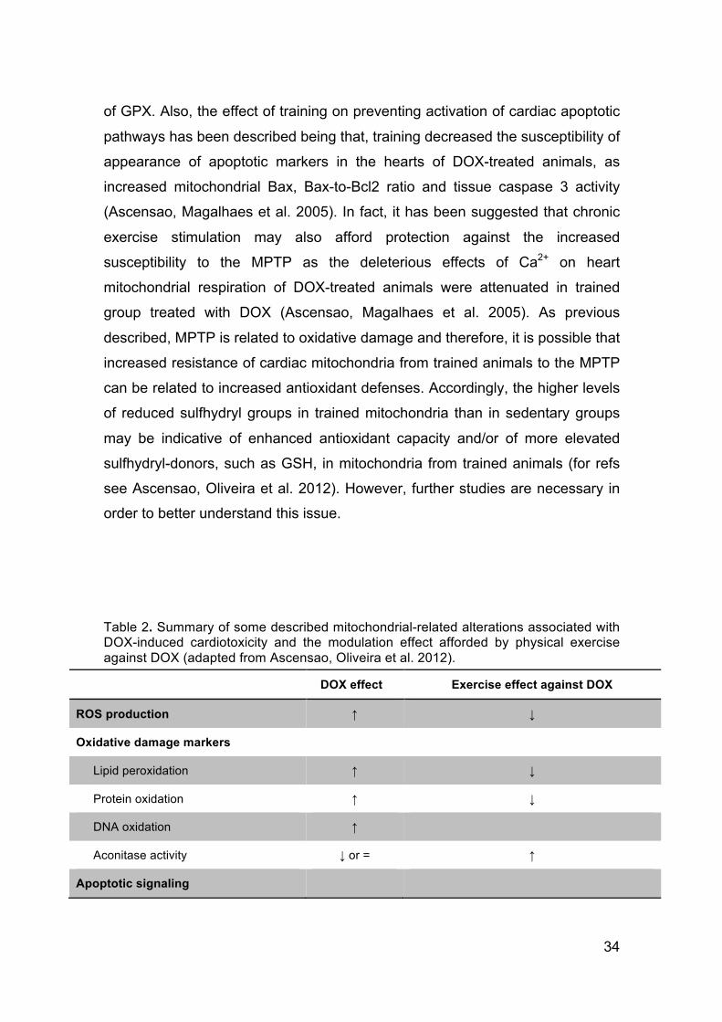
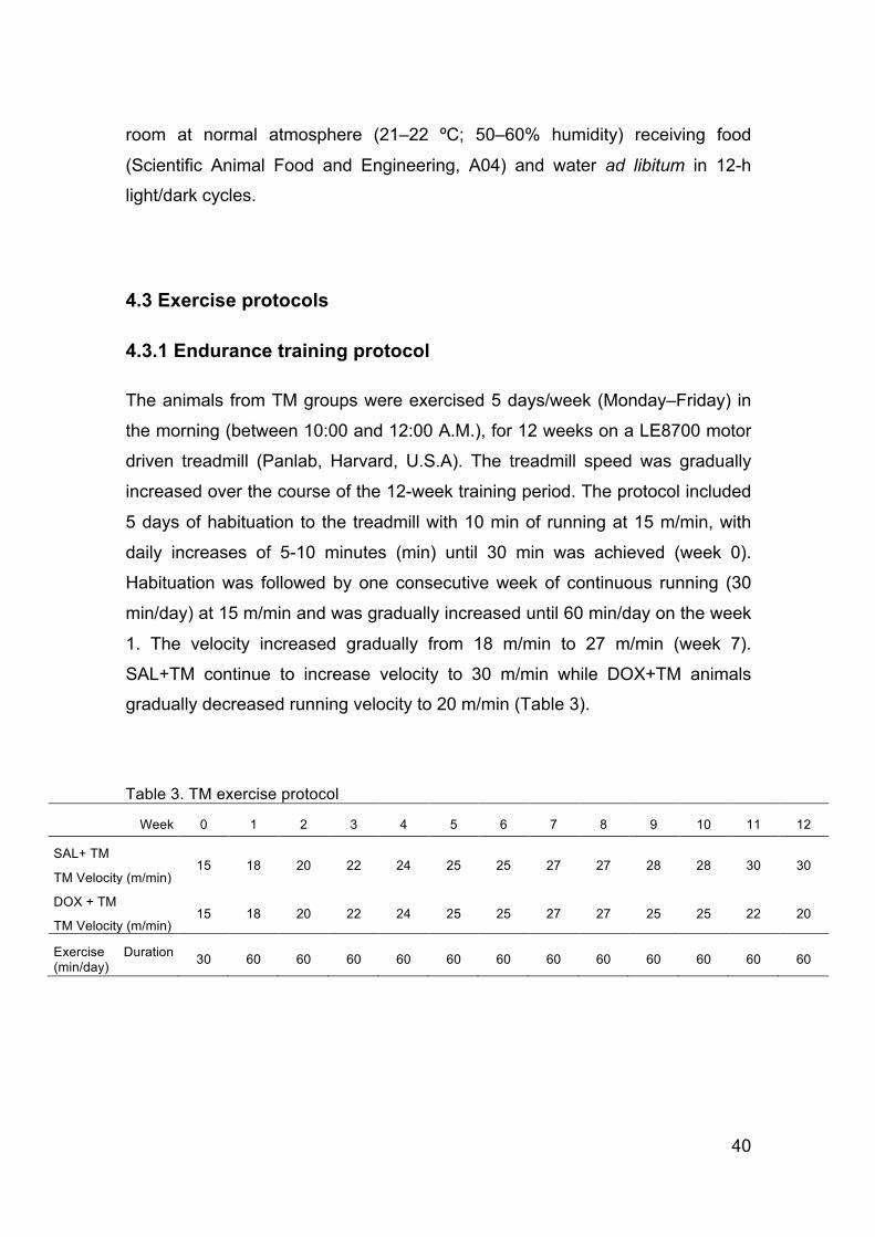