Page 1
www.ejpmr.com
Udokwu et al. European Journal of Pharmaceutical and Medical Research
151
EFFECTS OF XYLOPIA AETHIOPICA EXTRACT AND MELATONIN ON OSMOTIC
FRAGILITY OF CYCLOPHOSPHAMIDE INTOXICATED WISTAR RATS
Udokwu Euphemia Ifeoma, Okoroiwu I. L., Anonde Andrew Chekwube and Okolie N. J. C. and Obeagu
Emmanuel Ifeanyi*
Department of Medical Laboratory Science, Imo State University, Owerri, Nigeria.
Article Received on 12/08/2020 Article Revised on 01/09/2020 Article Accepted on 22/09/2020
The hormone Melatonin is the main neuroendocrine
secretary product of the pineal gland in animals and an
evolutionary ancient derivative of serotonin with
hormonal properties (slominski et al., 2018). It is also
produced in plants where it functions as a first line of
defence against oxidative stress (Tan et al., 2012).
Xylopia aethiopica, a shrub locally referred to as
Ethiopian pepper, Negro pepper, Guinean pepper,
Senegal pepper, Kili pepper and spice tree in the savanna
zone and coastal regions of Africa is amongst these
plants with great therapeutic potential. It is an
angiosperm belonging to the family Annonaceae (Obodo
et al., 2013), and is among the species that thrive in the
evergreen rain forests of tropical and subtropical Africa
which matures into a slim, tall tree of approximately 60
cm in diameter and up to 30m high with a straight stem
having a slightly stripped or smooth bark.
The red blood cell integrity is largely dependent on its
ability to maintain its membrane constituents that are
mostly polyunsaturated free fatty acid, and if this is
compromised will result to erythrocyte fragility which
becomes more fragile with consequent destruction by the
macrophages. This erythrocyte fragility or red cell
osmotic fragility is the ability of red blood cells to
undergo haemolysis when subjected to stress and the
absolute extent of haemolysis can be measured (Rodak,
2007). When this happens the membranes of the cells
undergo lipid peroxidation leading to oxidative
deterioration of polyunsaturated fatty acid accumulation
of reactive oxygen species (ROS) which are associated
with tissue damage by clearing off the sialic acid from
the cells making them more prone to phagocytosis. Also,
the osmotic fragility of red cells can occur from the
increased phosphorylation of P38 and JNK genes which
promotes increased production of ROS (Robin and
Steven, 2000). Factors such as cell’s size, surface area to
volume ratio, membrane composition and integrity can
equally influence the osmotic fragility of the cells
(Fischbach, et al., 2008). Alteration in the integrity of
blood leads to loss of blood and can be life threatening as
blood is a necessary components of animal body. The
body tends to protect itself from this life threatening
exsanguination by converting the blood from its liquid
state to a solid state in a process known as blood clotting
or coagulation. This formation of a clot is often referred
to as secondary haemostasis and it usually involves two
main pathways namely extrinsic and intrinsic pathways
that make use of clotting factors. Estimation of
coagulation tests like prothrombin time ,activated partial
thromboplastin time etc. are developed to diagnose
disorders of coagulation which can lead to an increased
bleeding (haemorrhage) or obstructive clotting
(thrombosis) (Xiangqun, et al., 2014).
For this study, the cyclophosphamide was chosen
because it is one of the most frequently used antitumor
agents in clinical practice and also its association with
rapidly killing of dividing cells in the body.
Considering the above, this present study was designed
to evaluate the effects of Xylopia aethiopica and
melatonin on osmotic fragility in cyclophosphamide
induced wistar rats, with a view of finding a lasting
solution to the life threatening effects reported about this
cytotoxic drug.
AIM
The research work is aimed at evaluating the effects of
Xylopia aethiopica and melatonin on osmotic fragility in
cyclophosphamide intoxicated adult wistar rats.
SJIF Impact Factor 6.222
Research Article
ISSN 2394-3211
EJPMR
EUROPEAN JOURNAL OF PHARMACEUTICAL
AND MEDICAL RESEARCH
www.ejpmr.com
ejpmr, 2020,7(10), 151-190
INTRODUCTION Cyclophosphamide has been in use clinically to treat a wide range of cancers including malignant lymphomas,
myeloma, leukaemia, mycosis, fungoides, neuroblastoma, adenocarcinoma, retinoblastoma, and breast carcinoma
(Mohammed et al., 2017). Other clinical uses for cyclophosphamide can be seen in immunosuppressive therapy
following organ transplants or as a treatment for autoimmune disorders such as rheumatoid arthritis, Wegener’s
granulomatosis, and nephritic syndrome in children (Chabner et al., 2001).
*Corresponding Author: Obeagu, Emmanuel Ifeanyi
Department of Medical Laboratory Science, Imo State University, Owerri, Nigeria.
Page 2
www.ejpmr.com
Udokwu et al. European Journal of Pharmaceutical and Medical Research
152
MATERIALS AND METHODS
COLLECTION OF PLANT MATERIALS AND AU
THENTICATION
Pods of Xylopia aethiopica were purchased from Orie-
Ugba vegetable market, Umuahia North Local
Government Area, Abia State, Nigeria. and were taken to
the Department of Forestry and Environmental
Management, Michael Okpara University of Agriculture,
Umudike where they were identified by a botanist/forest
manager. Voucher number MOUAU/VPP/18/012 was
assigned to a specimen sample of the pods which was
deposited in the herbarium of the Department.
PREPARATION OF PLANT EXTRACTS
Extract of the fruit pods was prepared in accordance with
the Soxhlet method described by Jensen, (2007). The
plant materials were subjected to further drying under
shade for 14 days and were pulverized into powder in a
manual blender powered by a Honda petrol engine. One
hundred grams of the powdered sample was introduced
into the extraction chamber of the soxhlet extractor and
extraction was carried out with ethanol as solvent.
Temperature was maintained at 650C throughout the
extraction period of 48 hours. At the end of the period,
the extract in solution was dried in a hot air oven at 40oC
to obtain a dry dark oily extract. The weight of the
extract was taken and percentage yield was calculated
using the formular:
% yield =X x 100
Q 1
Where X = weight of dried extract and Q = weight of
powdered plant material before extraction (100g)
(Bandiola, 2018).
ANIMALS USED FOR STUDY
One hundred and ninety five matured wistar albino rats
were used for the studies. Of the number, 30 were used
for the acute toxicity evaluation of the extract, 35 for
acute toxicity study of cyclophosphamide and 130 were
used for the main study. The rats were kept in aluminum
cages and allowed to acclimatize for two weeks to allow
for proper adaptation to the environment and living
conditions. They were allowed access to feed (Vital feed,
Nigeria) and water ad libitum but were starved for 12
hours prior to commencement of any experiment. All
animal experiments were carried out in compliance with
NIH guidelines for Care and Use of Laboratory Animals
(OECD, 2001). All experiments were carried out in the
Physiology Laboratory of the Department of Physiology
and Pharmacology, College of Veterinary Medicine,
Michael Okpara University of Agriculture, Umudike,
Nigeria.
EXPERIMENTAL DESIGN The rats (130 in number) were assigned to 13 groups of
10 rats each and were treated according to the order
below:
Group I: 10 mg/kg Cyclophosphamide, Food and water
Group II: 10 mg/kg Cyclophosphamide, 400 mg/kg
Extract, Food and water
Group III: 10 mg/kg Cyclophosphamide, 0.5 mg/kg
Melatonin, Food and water
Group IV: 10 mg/kg Cyclophosphamide, 400mg/kg
Extract, 0.5mg/kg Melatonin, Food and water
Group V: 30 mg/kg Cyclophosphamide, Food and water
Group VI: 30 mg/kg Cyclophosphamide, 400 mg/kg
Extract, Food and water
Group VII: 30mg/kg Cyclophosphamide, 0.5mg/kg
Melatonin, Food and water
Group VIII: 30mg/kg Cyclophosphamide+400mg/kg
Extract, 0.5mg/kg Melatonin, Food and water
Group IX: 50 mg/kg Cyclophosphamide, Food and water
Group X: 50 mg/kg Cyclophosphamide, 400 mg/kg
Extract, Food and water
Group XI: 50mg/kg Cyclophosphamide, 0.5mg/kg
Melatonin, Food and water
Group XII: 50mg/kg Cyclophosphamide, 400mg/kg
Extract, 0.5mg/kg Melatonin, Food and water
Group XIII: Food and water only
Treatments were done daily via the oral route for twenty
one (21) days. Three animals were sacrificed in each
group for blood collection by cardiac puncture into
EDTA and sodium citrate bottles for haematology and
osmotic fragility studies. Liver and kidney samples were
also collected and preserved in 10% formalin for
histological examination.
DETERMINATION OF RED BLOOD CELLS
OSMOTIC FRAGILITY
The method of Adenkole and Olurenmi, (2014) was
adopted. Sodium chloride solution (200 ml) was
prepared for each sample in concentrations ranging from
0.1-0.85% at pH 7.4. A set of test tubes, each containing
5mls of sodium chloride solution of concentration
ranging from 0.1 to 0.85% were serially arranged in a
test tube rack. One set of test tubes was used to analyze
each sample. A drop of the freshly collected blood was
placed into each of the ten test tubes using a dropper
pipette and each was mixed by gently inverting the test
tubes about three times. The test tubes were then allowed
to stand at room temperature for 30 minutes and then
centrifuged at 3000 rpm for 10 minutes before reading
the absorbance of the supernatant in each test tube on a
spectrophotometer at 540nm. The same procedure was
repeated for each sample collected. Percentage
haemolysis was calculated using the expression:
Percentage Haemolysis = Absorbance of test x100
Absorbance of control
STATISTICAL ANALYSIS
Results were expressed as means ± standard error of
mean (SEM). Statistical analysis was done using one-
way analysis of variance (ANOVA). Significant
differences were assessed at 95% level of significance
between control and treated groups using Duncan and
LSD (Post Hoc) tests. P values less than 0.05 were
Page 3
www.ejpmr.com
Udokwu et al. European Journal of Pharmaceutical and Medical Research
153
considered significant. Computer software package,
SPSS version 21 was employed.
RESULTS
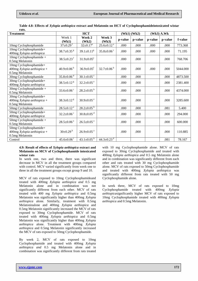
Result of effects of Xylopia aethiopica extract and
Melatonin on osmotic fragility (0) scores of
Cyclophosphamide intoxicated wistar rats In week one and two and three, there was no significant
difference in all the treatment groups compared with the
control group.
In week one, two and three, treatment with 400mg
Xylopia aethiopica and 0.5mg Melatonin alone and in
combination did not significantly osmotic fragility of rats
exposed to 10, 30 and 50mg Cyclophosphamide.
Table 1: Effects of Xylopia aethiopica extract and Melatonin on osmotic fragility (0) scores of Cyclophosphamide
intoxicated wistar rats.
Treatment 0 (Wk1) (Wk2) (Wk3) A.Wk
Week 1
(Wk1)
Week 2
(Wk2)
Week 3
(Wk3)
p-
value
p-
value
p-
value
p-
value
F-
value
10m Cyclophosphamide 100±0 a 100.27±0.32
a 100.6±0.31
a 1.000 1.000 .922 .318 1.394
10mg Cyclophosphamide+
400mg Xylopia aethiopia 100±0
a 100.27±0.32
a 100.3±0.35
a 1.000 1.000 1.000 .711 .361
10mg Cyclophosphamide +
0.5mg Melatonin 100±0
a 100.27±0.32
1.000 1.000 .449 .703
10mg Cyclophosphamide+
400mgXylopia aethiopia +
0.5mg Melatonin
100±0 a 100.27±0.32
a 100.3±0.35
a 1.000 1.000 1.000 .711 .361
30mg Cyclophosphamide 100±0 a 100.27±0.32
a
1.000 1.000 .449 .703
30mg Cyclophosphamide+
400mg Xylopia aethiopica 100±0
a 100.27±0.32
a
1.000 1.000 .449 .703
30mg Cyclophosphamide +
0.5mg Melatonin 100±0
a 100.27±0.32
a
1.000 1.000 .449 .703
30mg Cyclophosphamide+
400mg Xylopia aethiopica+
0.5mgMelatonn
100±0 a 100.27±0.32
a
1.000 1.000 .449 .703
50mg Cyclophosphamide 100±0 a 100.27±0.32
a
1.000 1.000 .449 .703
50mg Cyclophosphamide +
400mg Xylopia aethiopica 100±0
a 100.27±0.32
a
1.000 1.000 .449 .703
50mg Cyclophoshamide +
0.5mg Melatonin 100±0
a 100.27±0.32
a
1.000 1.000 .449 .703
50mg Cyclophosphamide +
400mg Xylopia aethiopia+
0.5mg Melatonin
100±0 a 100.27±0.32
a
1.000 1.000 .449 .703
Control 100±0 a 100.27±0.32
a 100.3±0.35
a .711 .361
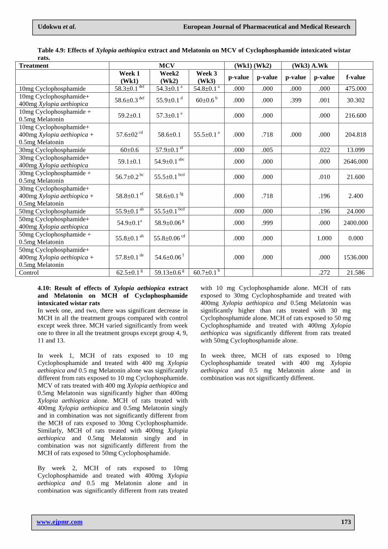
Result of effects of Xylopia aethiopica extract and
Melatonin on osmotic fragility (0.1) scores of
Cyclophosphamide intoxicated wistar rats
In week one, two and three, there was significant
increase in osmotic fragility in all the treatment groups
compared with control except group 4 in week one and
three. Osmotic fragility varied significantly from week
one to three in all the treatment except group 13.
In week one and two, there is a dose dependent increase
in osmotic fragility of rats treated with 10, 30 and 50mg
of Cyclophosphamide. Treatment with 400mg Xylopia
aethiopica alone and 400mg Xylopia aethiopica and
0.5mg Melatonin and 0.5mg Melatonin significantly
decreased the osmotic fragility compared with rats
exposed to 10mg Cyclophosphamide. Similarly,
treatment with 400mg Xylopia aethiopica alone and
400mg Xylopia aethiopica and 0.5mg Melatonin
significantly decreased osmotic fragility of rats exposed
to 30mg Cyclophosphamide. Osmotic fragility of rats
treated with 400mg Xylopia aethiopica and 0.5mg
Melatonin was significantly lower than 400mg Xylopia
aethiopica alone. Treatment with 400mg Xylopia
aethiopica and 0.5mg Melatonin significantly decreased
osmotic fragility of rats exposed to 50mg
Cyclophosphamide.
In week three, treatment with 400mg Xylopia aethiopica
alone and in combination 0.5 mg Melatonin significantly
decreased the osmotic fragility of rats exposed to 10mg
Cyclophosphamide.
Page 4
www.ejpmr.com
Udokwu et al. European Journal of Pharmaceutical and Medical Research
154
Table 2: Effects of Xylopia aethiopica extract and Melatonin on osmotic fragility (0.1) scores of
Cyclophosphamide intoxicated wistar rats.
Treatment 0.1 (Wk1) (Wk2) (Wk3) A.Wk
Week 1
(Wk1)
Week 2
(Wk2)
Week 3
(Wk3) p-value p-value p-value p-value f-value
10mg Cyclophosphamide 92.2±0.12 h 92.05±0.29
e 97.02±0.69
b .000 .000 .000 .000 41.529
10mg Cyclophosphamide+
400mg Xylopia aethiopica 90.1±0.23
g 89.39±0.28
d 83.95±0.29
a .000 .011 .172 .000 154.267
10mg Cyclophosphamide +
0.5mg Melatonin 89.54±0.28
g 89.14±0.28
d .000 .045 .368 1.029
10mg Cyclophosphamide+
400mg Xylopia aethiopica +
0.5mg Melatonin
88.1±0.06 f 88.39±0.28
cd 85.41±0.3
a .036 .811 1.000 .000 47.286
30mg Cyclophosphamide 98.2±0.06 j 97.77±0.31
g .000 .000 .242 1.887
30mg Cyclophosphamide +
400mg Xylopia aethiopica 95.12±0.01
i 94.97±0.3
f .000 .000 .652 .237
30mg Cyclophosphamide +
0.5mg Melatonin 86.2±0.06
d 85.73±0.27
b .000 .004 .168 2.820
30mg Cyclophosphamide +
400mg Xylopia aethiopica +
0.5mg Melatonin
85.21±0.01 c 85.34±0.27
b .000 .000 .662 .223
50mg Cyclophosphamide 98.19±0.01 j 98.28±0.31
g .000 .000 .798 .075
50mg Cyclophosphamide +
400mg Xylopia aethiopica 95.15±0.01
i 94.5±0.3
f .000 .000 .096 4.683
50mg Cyclophosphamide +
0.5mg Melatonin 83.12±0.01
b 89.26±0.28
d .000 .023 .000 470.987
50mg Cyclophosphamide +
400mg Xylopia aethiopica+
0.5mg Melatonin
82.12±0.02 a 83.34±0.26
a .000 .000 .010 21.376
Control 87.51±0.01 e 87.64±0.28
c 85.37±0.3
a .091 29.403
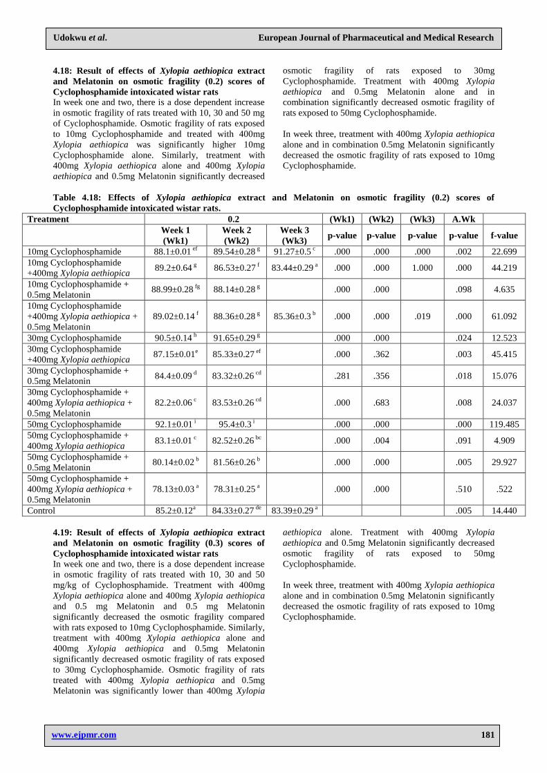
Result of effects of Xylopia aethiopica extract and
Melatonin on osmotic fragility (0.2) scores of
Cyclophosphamide intoxicated wistar rats In week one and two, there is a dose dependent increase
in osmotic fragility of rats treated with 10, 30 and 50 mg
of Cyclophosphamide. Osmotic fragility of rats exposed
to 10mg Cyclophosphamide and treated with 400mg
Xylopia aethiopica was significantly higher 10mg
Cyclophosphamide alone. Similarly, treatment with
400mg Xylopia aethiopica alone and 400mg Xylopia
aethiopica and 0.5mg Melatonin significantly decreased
osmotic fragility of rats exposed to 30mg
Cyclophosphamide. Treatment with 400mg Xylopia
aethiopica and 0.5mg Melatonin alone and in
combination significantly decreased osmotic fragility of
rats exposed to 50mg Cyclophosphamide.
In week three, treatment with 400mg Xylopia aethiopica
alone and in combination 0.5mg Melatonin significantly
decreased the osmotic fragility of rats exposed to 10mg
Cyclophosphamide.
Table 3: Effects of Xylopia aethiopica extract and Melatonin on osmotic fragility (0.2) scores of
Cyclophosphamide intoxicated wistar rats.
Treatment 0.2 (Wk1) (Wk2) (Wk3) A.Wk
Week 1
(Wk1)
Week 2
(Wk2)
Week 3
(Wk3) p-value p-value p-value p-value f-value
10mg Cyclophosphamide 88.1±0.01 ef
89.54±0.28 g 91.27±0.5
c .000 .000 .000 .002 22.699
10mg Cyclophosphamide
+400mg Xylopia aethiopica 89.2±0.64
g 86.53±0.27
f 83.44±0.29
a .000 .000 1.000 .000 44.219
10mg Cyclophosphamide +
0.5mg Melatonin 88.99±0.28
fg 88.14±0.28
g
.000 .000 .098 4.635
10mg Cyclophosphamide
+400mg Xylopia aethiopica +
0.5mg Melatonin
89.02±0.14 f 88.36±0.28
g 85.36±0.3
b .000 .000 .019 .000 61.092
30mg Cyclophosphamide 90.5±0.14 h 91.65±0.29
g
.000 .000 .024 12.523
30mg Cyclophosphamide
+400mg Xylopia aethiopica 87.15±0.01
e 85.33±0.27
ef
.000 .362 .003 45.415
Page 5
www.ejpmr.com
Udokwu et al. European Journal of Pharmaceutical and Medical Research
155
30mg Cyclophosphamide +
0.5mg Melatonin 84.4±0.09
d 83.32±0.26
cd
.281 .356 .018 15.076
30mg Cyclophosphamide +
400mg Xylopia aethiopica +
0.5mg Melatonin
82.2±0.06 c 83.53±0.26
cd
.000 .683 .008 24.037
50mg Cyclophosphamide 92.1±0.01 i 95.4±0.3
i
.000 .000 .000 119.485
50mg Cyclophosphamide +
400mg Xylopia aethiopica 83.1±0.01
c 82.52±0.26
bc
.000 .004 .091 4.909
50mg Cyclophosphamide +
0.5mg Melatonin 80.14±0.02
b 81.56±0.26
b
.000 .000 .005 29.927
50mg Cyclophosphamide +
400mg Xylopia aethiopica +
0.5mg Melatonin
78.13±0.03 a 78.31±0.25
a
.000 .000 .510 .522
Control 85.2±0.12a 84.33±0.27
de 83.39±0.29
a .005 14.440
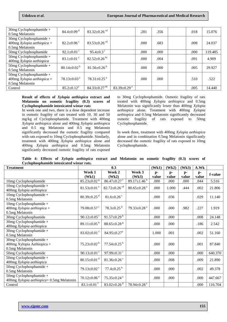
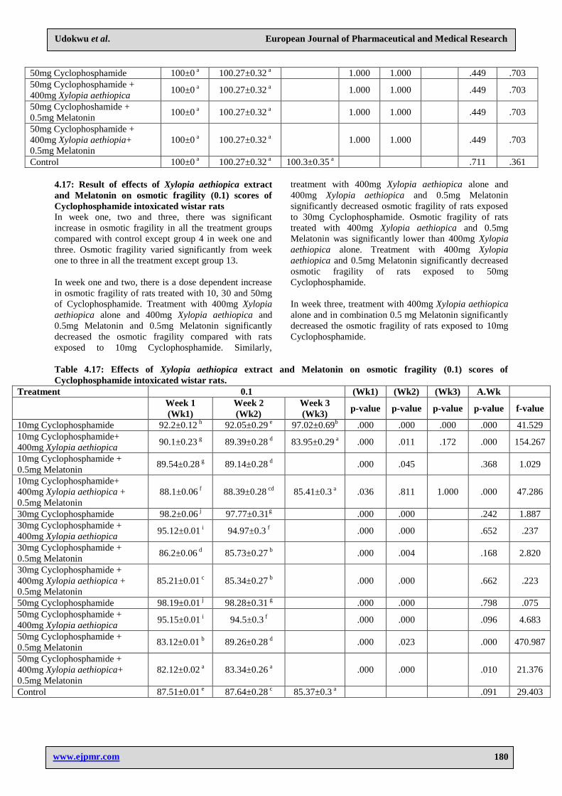
Result of effects of Xylopia aethiopica extract and
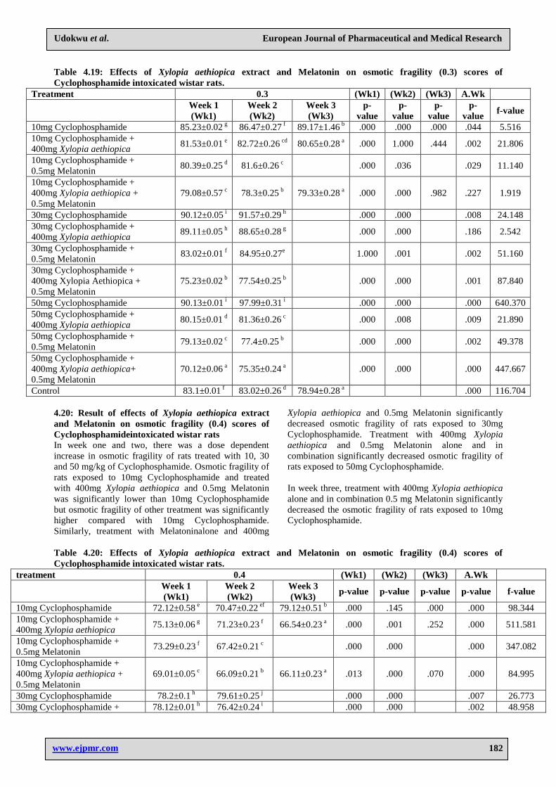
Melatonin on osmotic fragility (0.3) scores of
Cyclophosphamide intoxicated wistar rats
In week one and two, there is a dose dependent increase
in osmotic fragility of rats treated with 10, 30 and 50
mg/kg of Cyclophosphamide. Treatment with 400mg
Xylopia aethiopica alone and 400mg Xylopia aethiopica
and 0.5 mg Melatonin and 0.5 mg Melatonin
significantly decreased the osmotic fragility compared
with rats exposed to 10mg Cyclophosphamide. Similarly,
treatment with 400mg Xylopia aethiopica alone and
400mg Xylopia aethiopica and 0.5mg Melatonin
significantly decreased osmotic fragility of rats exposed
to 30mg Cyclophosphamide. Osmotic fragility of rats
treated with 400mg Xylopia aethiopica and 0.5mg
Melatonin was significantly lower than 400mg Xylopia
aethiopica alone. Treatment with 400mg Xylopia
aethiopica and 0.5mg Melatonin significantly decreased
osmotic fragility of rats exposed to 50mg
Cyclophosphamide.
In week three, treatment with 400mg Xylopia aethiopica
alone and in combination 0.5mg Melatonin significantly
decreased the osmotic fragility of rats exposed to 10mg
Cyclophosphamide.
Table 4: Effects of Xylopia aethiopica extract and Melatonin on osmotic fragility (0.3) scores of
Cyclophosphamide intoxicated wistar rats.
Treatment 0.3 (Wk1) (Wk2) (Wk3) A.Wk
Week 1 (Wk1)
Week 2 (Wk2)
Week 3 (Wk3)
p-
value p-
value p-
value p-
value f-value
10mg Cyclophosphamide 85.23±0.02 g 86.47±0.27
f 89.17±1.46 b .000 .000 .000 .044 5.516
10mg Cyclophosphamide + 400mg Xylopia aethiopica
81.53±0.01 e 82.72±0.26
cd 80.65±0.28 a .000 1.000 .444 .002 21.806
10mg Cyclophosphamide + 0.5mg Melatonin
80.39±0.25 d 81.6±0.26
c
.000 .036
.029 11.140
10mg Cyclophosphamide + 400mg Xylopia aethiopica + 0.5mg Melatonin
79.08±0.57 c 78.3±0.25
b 79.33±0.28 a .000 .000 .982 .227 1.919
30mg Cyclophosphamide 90.12±0.05 i 91.57±0.29
h
.000 .000
.008 24.148 30mg Cyclophosphamide + 400mg Xylopia aethiopica
89.11±0.05 h 88.65±0.28
g
.000 .000
.186 2.542
30mg Cyclophosphamide + 0.5mg Melatonin
83.02±0.01 f 84.95±0.27
e
1.000 .001
.002 51.160
30mg Cyclophosphamide + 400mg Xylopia Aethiopica + 0.5mg Melatonin
75.23±0.02 b 77.54±0.25
b
.000 .000
.001 87.840
50mg Cyclophosphamide 90.13±0.01 i 97.99±0.31
i
.000 .000
.000 640.370 50mg Cyclophosphamide + 400mg Xylopia aethiopica
80.15±0.01 d 81.36±0.26
c
.000 .008
.009 21.890
50mg Cyclophosphamide + 0.5mg Melatonin
79.13±0.02 c 77.4±0.25
b
.000 .000
.002 49.378
50mg Cyclophosphamide + 400mg Xylopia aethiopica+ 0.5mg Melatonin
70.12±0.06 a 75.35±0.24
a
.000 .000
.000 447.667
Control 83.1±0.01 f 83.02±0.26
d 78.94±0.28 a
.000 116.704
Page 6
www.ejpmr.com
Udokwu et al. European Journal of Pharmaceutical and Medical Research
156
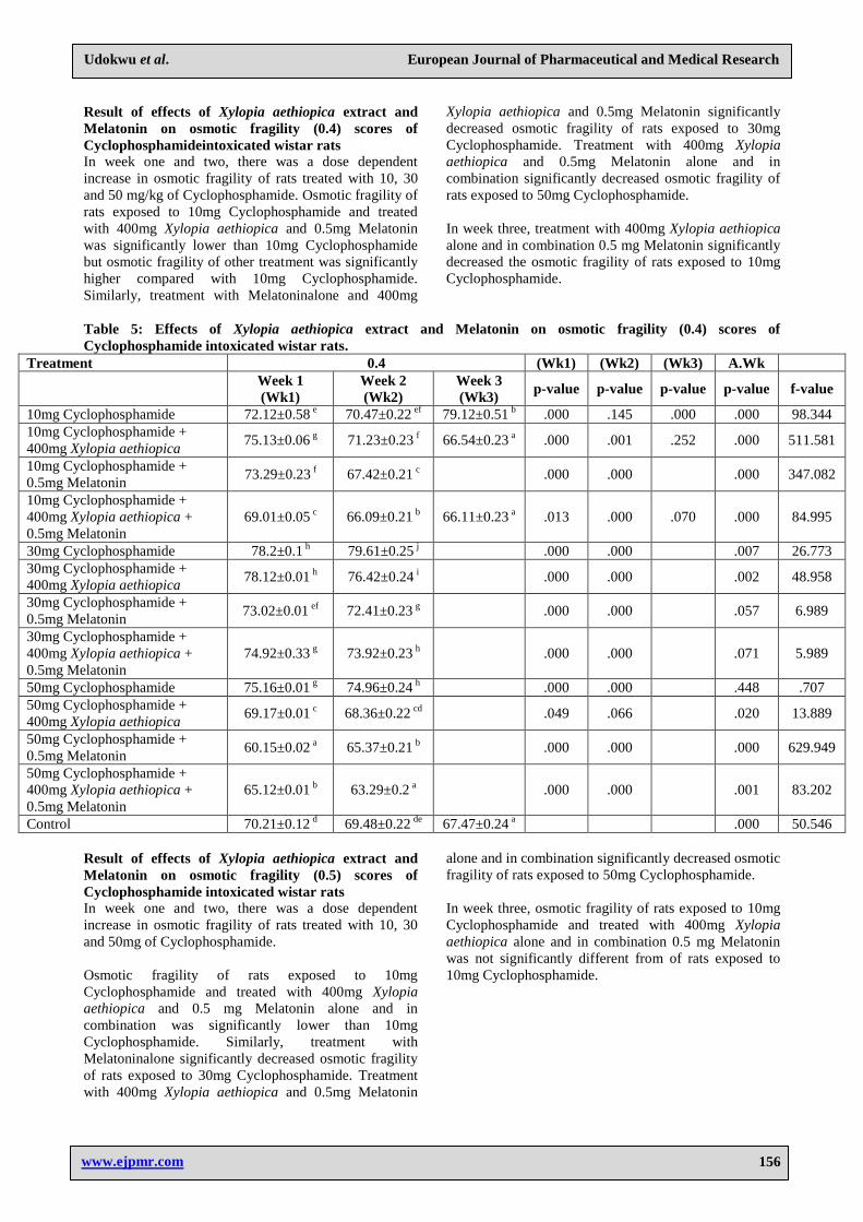
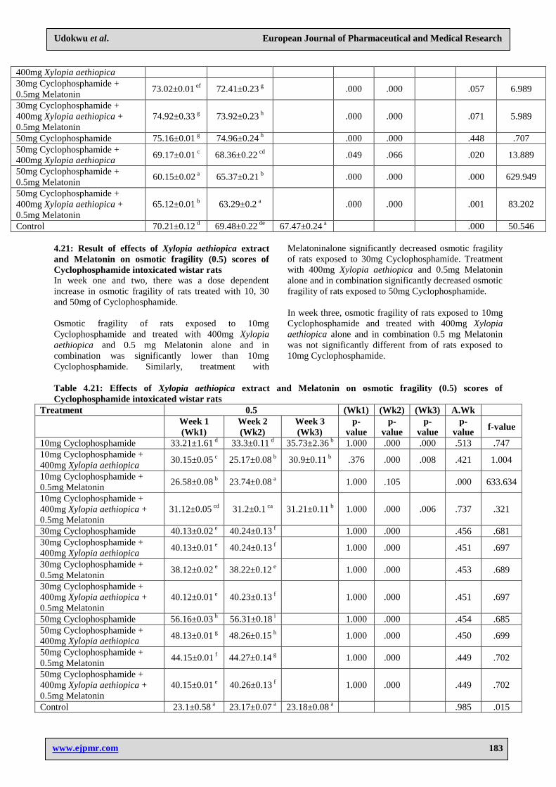
Result of effects of Xylopia aethiopica extract and
Melatonin on osmotic fragility (0.4) scores of
Cyclophosphamideintoxicated wistar rats
In week one and two, there was a dose dependent
increase in osmotic fragility of rats treated with 10, 30
and 50 mg/kg of Cyclophosphamide. Osmotic fragility of
rats exposed to 10mg Cyclophosphamide and treated
with 400mg Xylopia aethiopica and 0.5mg Melatonin
was significantly lower than 10mg Cyclophosphamide
but osmotic fragility of other treatment was significantly
higher compared with 10mg Cyclophosphamide.
Similarly, treatment with Melatoninalone and 400mg
Xylopia aethiopica and 0.5mg Melatonin significantly
decreased osmotic fragility of rats exposed to 30mg
Cyclophosphamide. Treatment with 400mg Xylopia
aethiopica and 0.5mg Melatonin alone and in
combination significantly decreased osmotic fragility of
rats exposed to 50mg Cyclophosphamide.
In week three, treatment with 400mg Xylopia aethiopica
alone and in combination 0.5 mg Melatonin significantly
decreased the osmotic fragility of rats exposed to 10mg
Cyclophosphamide.
Table 5: Effects of Xylopia aethiopica extract and Melatonin on osmotic fragility (0.4) scores of
Cyclophosphamide intoxicated wistar rats.
Treatment 0.4 (Wk1) (Wk2) (Wk3) A.Wk
Week 1
(Wk1)
Week 2
(Wk2)
Week 3
(Wk3) p-value p-value p-value p-value f-value
10mg Cyclophosphamide 72.12±0.58 e 70.47±0.22
ef 79.12±0.51
b .000 .145 .000 .000 98.344
10mg Cyclophosphamide +
400mg Xylopia aethiopica 75.13±0.06
g 71.23±0.23
f 66.54±0.23
a .000 .001 .252 .000 511.581
10mg Cyclophosphamide +
0.5mg Melatonin 73.29±0.23
f 67.42±0.21
c
.000 .000 .000 347.082
10mg Cyclophosphamide +
400mg Xylopia aethiopica +
0.5mg Melatonin
69.01±0.05 c 66.09±0.21
b 66.11±0.23
a .013 .000 .070 .000 84.995
30mg Cyclophosphamide 78.2±0.1 h 79.61±0.25
j
.000 .000 .007 26.773
30mg Cyclophosphamide +
400mg Xylopia aethiopica 78.12±0.01
h 76.42±0.24
i
.000 .000 .002 48.958
30mg Cyclophosphamide +
0.5mg Melatonin 73.02±0.01
ef 72.41±0.23
g
.000 .000 .057 6.989
30mg Cyclophosphamide +
400mg Xylopia aethiopica +
0.5mg Melatonin
74.92±0.33 g 73.92±0.23
h
.000 .000 .071 5.989
50mg Cyclophosphamide 75.16±0.01 g 74.96±0.24
h
.000 .000 .448 .707
50mg Cyclophosphamide +
400mg Xylopia aethiopica 69.17±0.01
c 68.36±0.22
cd
.049 .066 .020 13.889
50mg Cyclophosphamide +
0.5mg Melatonin 60.15±0.02
a 65.37±0.21
b
.000 .000 .000 629.949
50mg Cyclophosphamide +
400mg Xylopia aethiopica +
0.5mg Melatonin
65.12±0.01 b 63.29±0.2
a
.000 .000 .001 83.202
Control 70.21±0.12 d 69.48±0.22
de 67.47±0.24
a .000 50.546
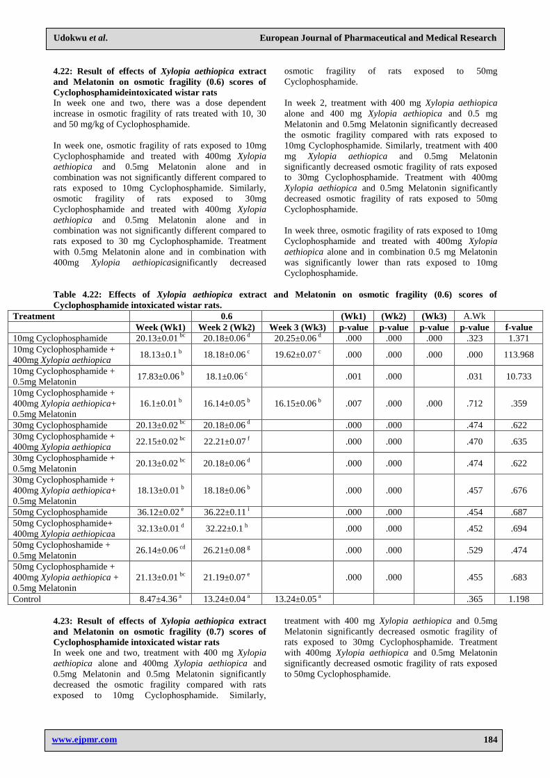
Result of effects of Xylopia aethiopica extract and
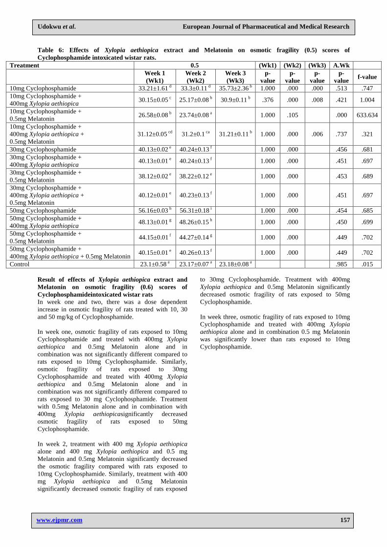
Melatonin on osmotic fragility (0.5) scores of
Cyclophosphamide intoxicated wistar rats
In week one and two, there was a dose dependent
increase in osmotic fragility of rats treated with 10, 30
and 50mg of Cyclophosphamide.
Osmotic fragility of rats exposed to 10mg
Cyclophosphamide and treated with 400mg Xylopia
aethiopica and 0.5 mg Melatonin alone and in
combination was significantly lower than 10mg
Cyclophosphamide. Similarly, treatment with
Melatoninalone significantly decreased osmotic fragility
of rats exposed to 30mg Cyclophosphamide. Treatment
with 400mg Xylopia aethiopica and 0.5mg Melatonin
alone and in combination significantly decreased osmotic
fragility of rats exposed to 50mg Cyclophosphamide.
In week three, osmotic fragility of rats exposed to 10mg
Cyclophosphamide and treated with 400mg Xylopia
aethiopica alone and in combination 0.5 mg Melatonin
was not significantly different from of rats exposed to
10mg Cyclophosphamide.
Page 7
www.ejpmr.com
Udokwu et al. European Journal of Pharmaceutical and Medical Research
157
Table 6: Effects of Xylopia aethiopica extract and Melatonin on osmotic fragility (0.5) scores of
Cyclophosphamide intoxicated wistar rats.
Treatment 0.5 (Wk1) (Wk2) (Wk3) A.Wk
Week 1 (Wk1)
Week 2 (Wk2)
Week 3 (Wk3)
p-
value p-
value p-
value p-
value f-value
10mg Cyclophosphamide 33.21±1.61 d 33.3±0.11
d 35.73±2.36 b 1.000 .000 .000 .513 .747
10mg Cyclophosphamide + 400mg Xylopia aethiopica
30.15±0.05 c 25.17±0.08
b 30.9±0.11 b .376 .000 .008 .421 1.004
10mg Cyclophosphamide + 0.5mg Melatonin
26.58±0.08 b 23.74±0.08
a
1.000 .105
.000 633.634
10mg Cyclophosphamide + 400mg Xylopia aethiopica + 0.5mg Melatonin
31.12±0.05 cd 31.2±0.1
ca 31.21±0.11 b 1.000 .000 .006 .737 .321
30mg Cyclophosphamide 40.13±0.02 e 40.24±0.13
f
1.000 .000
.456 .681 30mg Cyclophosphamide + 400mg Xylopia aethiopica
40.13±0.01 e 40.24±0.13
f
1.000 .000
.451 .697
30mg Cyclophosphamide + 0.5mg Melatonin
38.12±0.02 e 38.22±0.12
e
1.000 .000
.453 .689
30mg Cyclophosphamide + 400mg Xylopia aethiopica + 0.5mg Melatonin
40.12±0.01 e 40.23±0.13
f
1.000 .000
.451 .697
50mg Cyclophosphamide 56.16±0.03 h 56.31±0.18
i
1.000 .000
.454 .685 50mg Cyclophosphamide + 400mg Xylopia aethiopica
48.13±0.01 g 48.26±0.15
h
1.000 .000
.450 .699
50mg Cyclophosphamide + 0.5mg Melatonin
44.15±0.01 f 44.27±0.14
g
1.000 .000
.449 .702
50mg Cyclophosphamide + 400mg Xylopia aethiopica + 0.5mg Melatonin
40.15±0.01 e 40.26±0.13
f
1.000 .000
.449 .702
Control 23.1±0.58 a 23.17±0.07
a 23.18±0.08 a
.985 .015
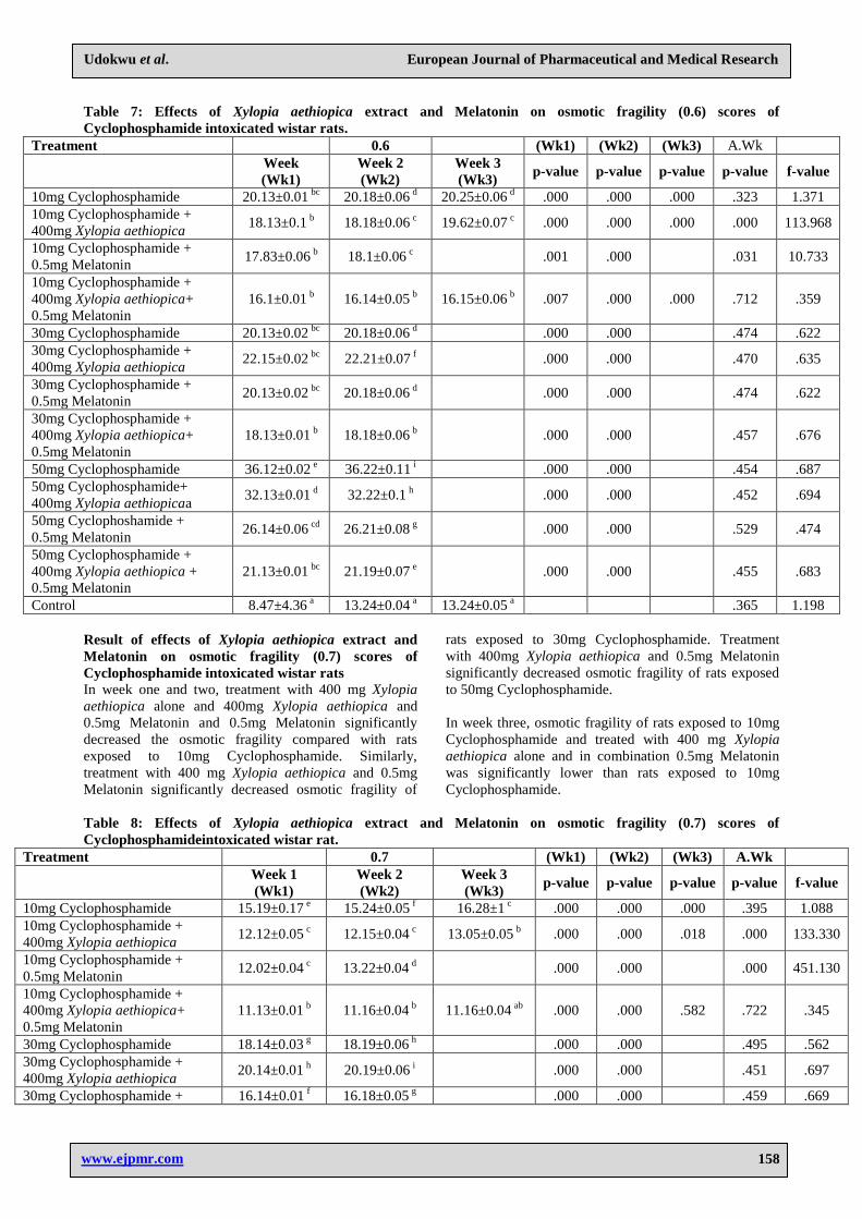
Result of effects of Xylopia aethiopica extract and
Melatonin on osmotic fragility (0.6) scores of
Cyclophosphamideintoxicated wistar rats In week one and two, there was a dose dependent
increase in osmotic fragility of rats treated with 10, 30
and 50 mg/kg of Cyclophosphamide.
In week one, osmotic fragility of rats exposed to 10mg
Cyclophosphamide and treated with 400mg Xylopia
aethiopica and 0.5mg Melatonin alone and in
combination was not significantly different compared to
rats exposed to 10mg Cyclophosphamide. Similarly,
osmotic fragility of rats exposed to 30mg
Cyclophosphamide and treated with 400mg Xylopia
aethiopica and 0.5mg Melatonin alone and in
combination was not significantly different compared to
rats exposed to 30 mg Cyclophosphamide. Treatment
with 0.5mg Melatonin alone and in combination with
400mg Xylopia aethiopicasignificantly decreased
osmotic fragility of rats exposed to 50mg
Cyclophosphamide.
In week 2, treatment with 400 mg Xylopia aethiopica
alone and 400 mg Xylopia aethiopica and 0.5 mg
Melatonin and 0.5mg Melatonin significantly decreased
the osmotic fragility compared with rats exposed to
10mg Cyclophosphamide. Similarly, treatment with 400
mg Xylopia aethiopica and 0.5mg Melatonin
significantly decreased osmotic fragility of rats exposed
to 30mg Cyclophosphamide. Treatment with 400mg
Xylopia aethiopica and 0.5mg Melatonin significantly
decreased osmotic fragility of rats exposed to 50mg
Cyclophosphamide.
In week three, osmotic fragility of rats exposed to 10mg
Cyclophosphamide and treated with 400mg Xylopia
aethiopica alone and in combination 0.5 mg Melatonin
was significantly lower than rats exposed to 10mg
Cyclophosphamide.
Page 8
www.ejpmr.com
Udokwu et al. European Journal of Pharmaceutical and Medical Research
158
Table 7: Effects of Xylopia aethiopica extract and Melatonin on osmotic fragility (0.6) scores of
Cyclophosphamide intoxicated wistar rats.
Treatment
0.6
(Wk1) (Wk2) (Wk3) A.Wk
Week
(Wk1)
Week 2
(Wk2)
Week 3
(Wk3) p-value p-value p-value p-value f-value
10mg Cyclophosphamide 20.13±0.01 bc
20.18±0.06 d 20.25±0.06
d .000 .000 .000 .323 1.371
10mg Cyclophosphamide +
400mg Xylopia aethiopica 18.13±0.1
b 18.18±0.06
c 19.62±0.07
c .000 .000 .000 .000 113.968
10mg Cyclophosphamide +
0.5mg Melatonin 17.83±0.06
b 18.1±0.06
c
.001 .000 .031 10.733
10mg Cyclophosphamide +
400mg Xylopia aethiopica+
0.5mg Melatonin
16.1±0.01 b 16.14±0.05
b 16.15±0.06
b .007 .000 .000 .712 .359
30mg Cyclophosphamide 20.13±0.02 bc
20.18±0.06 d
.000 .000 .474 .622
30mg Cyclophosphamide +
400mg Xylopia aethiopica 22.15±0.02
bc 22.21±0.07
f
.000 .000 .470 .635
30mg Cyclophosphamide +
0.5mg Melatonin 20.13±0.02
bc 20.18±0.06
d
.000 .000 .474 .622
30mg Cyclophosphamide +
400mg Xylopia aethiopica+
0.5mg Melatonin
18.13±0.01 b 18.18±0.06
b
.000 .000 .457 .676
50mg Cyclophosphamide 36.12±0.02 e 36.22±0.11
i
.000 .000 .454 .687
50mg Cyclophosphamide+
400mg Xylopia aethiopicaa 32.13±0.01
d 32.22±0.1
h
.000 .000 .452 .694
50mg Cyclophoshamide +
0.5mg Melatonin 26.14±0.06
cd 26.21±0.08
g
.000 .000 .529 .474
50mg Cyclophosphamide +
400mg Xylopia aethiopica +
0.5mg Melatonin
21.13±0.01 bc
21.19±0.07 e
.000 .000 .455 .683
Control 8.47±4.36 a 13.24±0.04
a 13.24±0.05
a .365 1.198
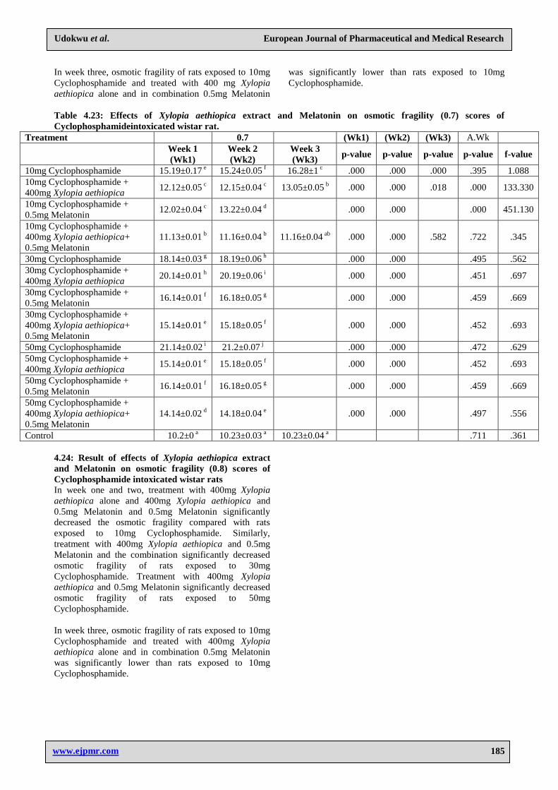
Result of effects of Xylopia aethiopica extract and
Melatonin on osmotic fragility (0.7) scores of
Cyclophosphamide intoxicated wistar rats In week one and two, treatment with 400 mg Xylopia
aethiopica alone and 400mg Xylopia aethiopica and
0.5mg Melatonin and 0.5mg Melatonin significantly
decreased the osmotic fragility compared with rats
exposed to 10mg Cyclophosphamide. Similarly,
treatment with 400 mg Xylopia aethiopica and 0.5mg
Melatonin significantly decreased osmotic fragility of
rats exposed to 30mg Cyclophosphamide. Treatment
with 400mg Xylopia aethiopica and 0.5mg Melatonin
significantly decreased osmotic fragility of rats exposed
to 50mg Cyclophosphamide.
In week three, osmotic fragility of rats exposed to 10mg
Cyclophosphamide and treated with 400 mg Xylopia
aethiopica alone and in combination 0.5mg Melatonin
was significantly lower than rats exposed to 10mg
Cyclophosphamide.
Table 8: Effects of Xylopia aethiopica extract and Melatonin on osmotic fragility (0.7) scores of
Cyclophosphamideintoxicated wistar rat.
Treatment
0.7
(Wk1) (Wk2) (Wk3) A.Wk
Week 1 (Wk1)
Week 2 (Wk2)
Week 3 (Wk3)
p-value p-value p-value p-value f-value
10mg Cyclophosphamide 15.19±0.17 e 15.24±0.05
f 16.28±1 c .000 .000 .000 .395 1.088
10mg Cyclophosphamide + 400mg Xylopia aethiopica
12.12±0.05 c 12.15±0.04
c 13.05±0.05 b .000 .000 .018 .000 133.330
10mg Cyclophosphamide + 0.5mg Melatonin
12.02±0.04 c 13.22±0.04
d
.000 .000
.000 451.130
10mg Cyclophosphamide + 400mg Xylopia aethiopica+ 0.5mg Melatonin
11.13±0.01 b 11.16±0.04
b 11.16±0.04 ab .000 .000 .582 .722 .345
30mg Cyclophosphamide 18.14±0.03 g 18.19±0.06
h
.000 .000
.495 .562 30mg Cyclophosphamide + 400mg Xylopia aethiopica
20.14±0.01 h 20.19±0.06
i
.000 .000
.451 .697
30mg Cyclophosphamide + 16.14±0.01 f 16.18±0.05
g
.000 .000
.459 .669
Page 9
www.ejpmr.com
Udokwu et al. European Journal of Pharmaceutical and Medical Research
159
0.5mg Melatonin 30mg Cyclophosphamide + 400mg Xylopia aethiopica+ 0.5mg Melatonin
15.14±0.01 e 15.18±0.05
f
.000 .000
.452 .693
50mg Cyclophosphamide 21.14±0.02 i 21.2±0.07
j
.000 .000
.472 .629 50mg Cyclophosphamide + 400mg Xylopia aethiopica
15.14±0.01 e 15.18±0.05
f
.000 .000
.452 .693
50mg Cyclophosphamide + 0.5mg Melatonin
16.14±0.01 f 16.18±0.05
g
.000 .000
.459 .669
50mg Cyclophosphamide + 400mg Xylopia aethiopica+ 0.5mg Melatonin
14.14±0.02 d 14.18±0.04
e
.000 .000
.497 .556
Control 10.2±0 a 10.23±0.03
a 10.23±0.04 a
.711 .361
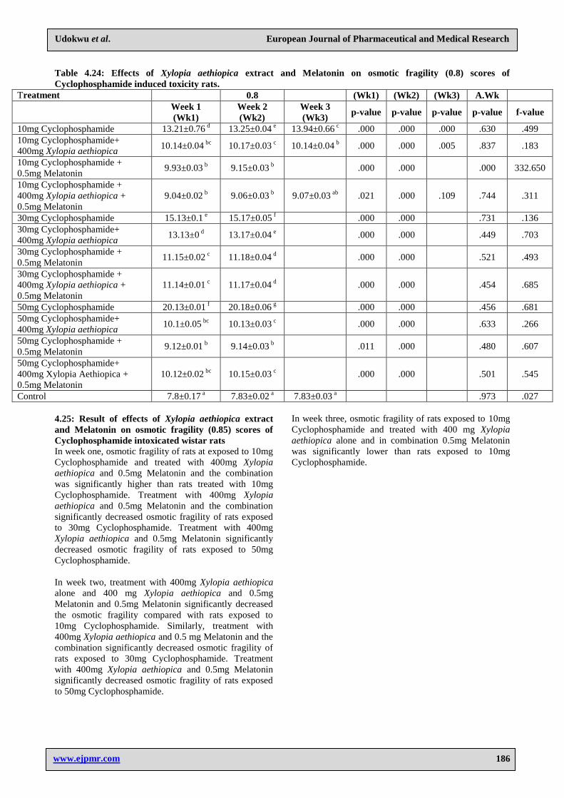
Result of effects of Xylopia aethiopica extract and
Melatonin on osmotic fragility (0.8) scores of
Cyclophosphamide intoxicated wistar rats
In week one and two, treatment with 400mg Xylopia
aethiopica alone and 400mg Xylopia aethiopica and
0.5mg Melatonin and 0.5mg Melatonin significantly
decreased the osmotic fragility compared with rats
exposed to 10mg Cyclophosphamide. Similarly,
treatment with 400mg Xylopia aethiopica and 0.5mg
Melatonin and the combination significantly decreased
osmotic fragility of rats exposed to 30mg
Cyclophosphamide. Treatment with 400mg Xylopia
aethiopica and 0.5mg Melatonin significantly decreased
osmotic fragility of rats exposed to 50mg
Cyclophosphamide.
In week three, osmotic fragility of rats exposed to 10mg
Cyclophosphamide and treated with 400mg Xylopia
aethiopica alone and in combination 0.5mg Melatonin
was significantly lower than rats exposed to 10mg
Cyclophosphamide.
Table 9: Effects of Xylopia aethiopica extract and Melatonin on osmotic fragility (0.8) scores of
Cyclophosphamide induced toxicity rats.
Treatment
0.8
(Wk1) (Wk2) (Wk3) A.Wk
Week 1
(Wk1)
Week 2
(Wk2)
Week 3
(Wk3) p-value p-value p-value p-value f-value
10mg Cyclophosphamide 13.21±0.76 d 13.25±0.04
e 13.94±0.66
c .000 .000 .000 .630 .499
10mg Cyclophosphamide+
400mg Xylopia aethiopica 10.14±0.04
bc 10.17±0.03
c 10.14±0.04
b .000 .000 .005 .837 .183
10mg Cyclophosphamide +
0.5mg Melatonin 9.93±0.03
b 9.15±0.03
b
.000 .000 .000 332.650
10mg Cyclophosphamide +
400mg Xylopia aethiopica +
0.5mg Melatonin
9.04±0.02 b 9.06±0.03
b 9.07±0.03
ab .021 .000 .109 .744 .311
30mg Cyclophosphamide 15.13±0.1 e 15.17±0.05
f
.000 .000 .731 .136
30mg Cyclophosphamide+
400mg Xylopia aethiopica 13.13±0
d 13.17±0.04
e
.000 .000 .449 .703
30mg Cyclophosphamide +
0.5mg Melatonin 11.15±0.02
c 11.18±0.04
d
.000 .000 .521 .493
30mg Cyclophosphamide +
400mg Xylopia aethiopica +
0.5mg Melatonin
11.14±0.01 c 11.17±0.04
d
.000 .000 .454 .685
50mg Cyclophosphamide 20.13±0.01 f 20.18±0.06
g
.000 .000 .456 .681
50mg Cyclophosphamide+
400mg Xylopia aethiopica 10.1±0.05
bc 10.13±0.03
c
.000 .000 .633 .266
50mg Cyclophosphamide +
0.5mg Melatonin 9.12±0.01
b 9.14±0.03
b
.011 .000 .480 .607
50mg Cyclophosphamide+
400mg Xylopia Aethiopica +
0.5mg Melatonin
10.12±0.02 bc
10.15±0.03 c
.000 .000 .501 .545
Control 7.8±0.17 a 7.83±0.02
a 7.83±0.03
a .973 .027
Page 10
www.ejpmr.com
Udokwu et al. European Journal of Pharmaceutical and Medical Research
160
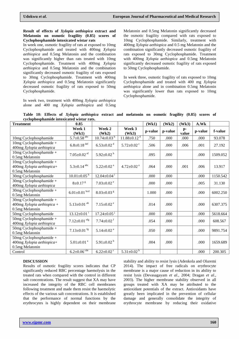
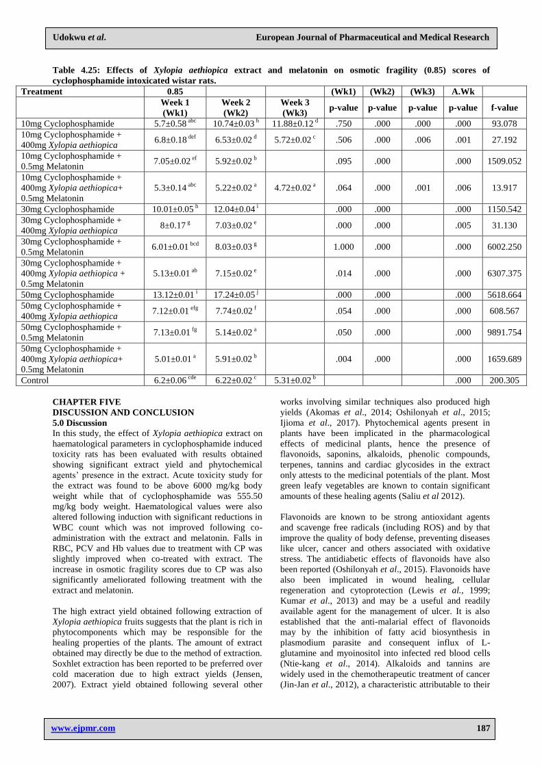
Result of effects of Xylopia aethiopica extract and
Melatonin on osmotic fragility (0.85) scores of
Cyclophosphamide intoxicated wistar rats In week one, osmotic fragility of rats at exposed to 10mg
Cyclophosphamide and treated with 400mg Xylopia
aethiopica and 0.5mg Melatonin and the combination
was significantly higher than rats treated with 10mg
Cyclophosphamide. Treatment with 400mg Xylopia
aethiopica and 0.5mg Melatonin and the combination
significantly decreased osmotic fragility of rats exposed
to 30mg Cyclophosphamide. Treatment with 400mg
Xylopia aethiopica and 0.5mg Melatonin significantly
decreased osmotic fragility of rats exposed to 50mg
Cyclophosphamide.
In week two, treatment with 400mg Xylopia aethiopica
alone and 400 mg Xylopia aethiopica and 0.5mg
Melatonin and 0.5mg Melatonin significantly decreased
the osmotic fragility compared with rats exposed to
10mg Cyclophosphamide. Similarly, treatment with
400mg Xylopia aethiopica and 0.5 mg Melatonin and the
combination significantly decreased osmotic fragility of
rats exposed to 30mg Cyclophosphamide. Treatment
with 400mg Xylopia aethiopica and 0.5mg Melatonin
significantly decreased osmotic fragility of rats exposed
to 50mg Cyclophosphamide.
In week three, osmotic fragility of rats exposed to 10mg
Cyclophosphamide and treated with 400 mg Xylopia
aethiopica alone and in combination 0.5mg Melatonin
was significantly lower than rats exposed to 10mg
Cyclophosphamide.
Table 10: Effects of Xylopia aethiopica extract and melatonin on osmotic fragility (0.85) scores of
cyclophosphamide intoxicated wistar rats.
Treatment 0.85
(Wk1) (Wk2) (Wk3) A.Wk
Week 1 (Wk1)
Week 2 (Wk2)
Week 3 (Wk3)
p-value p-value p-
value p-value f-value
10mg Cyclophosphamide 5.7±0.58 abc 10.74±0.03
h 11.88±0.12 d .750 .000 .000 .000 93.078
10mg Cyclophosphamide + 400mg Xylopia aethiopica
6.8±0.18 def 6.53±0.02
d 5.72±0.02 c .506 .000 .006 .001 27.192
10mg Cyclophosphamide + 0.5mg Melatonin
7.05±0.02 ef 5.92±0.02
b
.095 .000
.000 1509.052
10mg Cyclophosphamide + 400mg Xylopia aethiopica+ 0.5mg Melatonin
5.3±0.14 abc 5.22±0.02
a 4.72±0.02 a .064 .000 .001 .006 13.917
30mg Cyclophosphamide 10.01±0.05 h 12.04±0.04
i
.000 .000
.000 1150.542 30mg Cyclophosphamide + 400mg Xylopia aethiopica
8±0.17 g 7.03±0.02
e
.000 .000
.005 31.130
30mg Cyclophosphamide + 0.5mg Melatonin
6.01±0.01 bcd 8.03±0.03
g
1.000 .000
.000 6002.250
30mg Cyclophosphamide + 400mg Xylopia aethiopica + 0.5mg Melatonin
5.13±0.01 ab 7.15±0.02
e
.014 .000
.000 6307.375
50mg Cyclophosphamide 13.12±0.01 i 17.24±0.05
j
.000 .000
.000 5618.664 50mg Cyclophosphamide + 400mg Xylopia aethiopica
7.12±0.01 efg 7.74±0.02
f
.054 .000
.000 608.567
50mg Cyclophosphamide + 0.5mg Melatonin
7.13±0.01 fg 5.14±0.02
a
.050 .000
.000 9891.754
50mg Cyclophosphamide + 400mg Xylopia aethiopica+
0.5mg Melatonin 5.01±0.01
a 5.91±0.02 b
.004 .000
.000 1659.689
Control 6.2±0.06 cde 6.22±0.02
c 5.31±0.02 b
.000 200.305
DISCUSSION
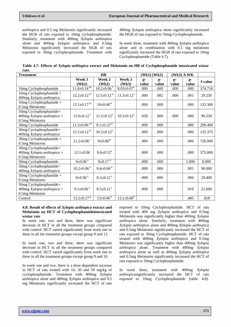
Results of osmotic fragility scores indicates that CP
significantly reduced RBC percentage haemolysis in the
treated rats when compared with the control in different
salt concentrations. The result suggest that XA may have
increased the integrity of the RBC cell membranes
following treatment and made them resist the haemolytic
effects of the various salt concentrations. It is established
that the performance of normal functions by the
erythrocytes is highly dependent on their membrane
stability and ability to resist lysis (Adenkola and Oluremi
2014). The impact of free radicals on erythrocyte
membrane is a major cause of reduction in its ability to
resist lysis (Devasagayam et al., 2004; Dragan et al.,
2003). The higher membrane stability observed in all
groups treated with XA may be attributed to the
antioxidant potentials of the extract. Antioxidants have
greatly been implicated in the prevention of cellular
damage and generally consolidate the integrity of
erythrocyte membrane by reducing their oxidative
Page 11
www.ejpmr.com
Udokwu et al. European Journal of Pharmaceutical and Medical Research
161
damage due the impact of free radicals (Adenkola and
Oluremi 2014).
CONCLUSION
This study provides evidence that Xylopia aethiopica is a
valuable medicinal food for combating
cyclophosphamide induced systemic toxicity. Xylopia
aethiopica may provide protective effects for toxicants
capable of inducing oxidative stress. Also It can be seen
that despite the high potent immunosuppressive effect of
cyclophosphamide on blood cells, melatonin and Xylopia
aethiopica have shown to exert their ameliorative effects
through their antioxidant and antitumour properties.
Therefore, they may be of value in the prevention of
diseases arising from the oxidative effects of consumed
toxicant substances like cyclophosphamide.
REFERENCES
1. Adenkola, A.Y. and Oluremi, O.I.A. (2014).
Erythrocyte Osmotic Fragility Score in Rabbits
fed Hibiscus sabdariffa in Graded Level. Nigerian
Journal Physiological Sciences, 29: 113-117.
2. Afsharian, P., Terelius, Y., Hassan, Z., Nilsson,
C., Lundgren, S. and Hassan, M. (2007) The Effect of
Repeated Administration of Cyclophosphamide on
Cytochrome P450 2B in Rats. Clinical Cancer
Research, 13(14): 4218- 4224.
3. Ajiboye, B.O., Ibukun, E.O., Edobor, G., Ojo, A.O.
and Onikanni, S.A.(2013). Qualitative and
quantitative analysis of phytochemicals in senecio
biafrae leaf. International journal of inventions in
pharmaceutical sciences, 1(5): 428-432.
4. Akomas S.C., Okafor A.I., and Ijioma, S.N (2014).
Hypoglycemic, Hematologic and Hypolipidemic
Activity of Mucuna Pruriensethanol leaf extract in
alloxan induced diabetic rats. Annual Research and
Review in Biology, 4(24): 4284-4292.
5. Anthony, H. T., Mariana, A., Maureen, F. and
Henrik O. (2016).Endocrine regulation of circadian
physiology. Journal of Endocrinology230: R1–R11.
6. Atalay, F., Gulmez, O. and Ugurlu, A.O. (2014)
Cardiotoxicity following cyclophosphamidetherapy:
a case report. Journal of Medical Case Reports, 8:
252.
7. Azevedo, L., Alves de Lima, P. L., Gomes, J. C.,
Stringheta, P. C., Ribeiro, D. A., and Salvadori, D.
M. (2007). Differential response related to
genotoxicity between eggplant
(Solanummelanogena) skin aqueous extract and its
main purified anthrocyanin (delphinidin) in vivo.
Food Chemistry and Toxicology, 45: 852 – 858.
8. Bandiola, T.B. (2018). Extraction and Qualitative
Phytochemical Screening of Medicinal Plants: A
Brief Summary. International Journal of Pharmacy,
8(1): 137-143.
9. Bendich, A. (1993). Physiological role of
antioxidants in the immune system. Journal of Diary
Sciences, 76(9): 2789-2794.
10. Boddy, A.V., and Yule, S.M. (2000). Metabolism
and pharmacokinetics of oxazaphosphorines.
Clinical Pharmacokinetics, 38(4): 291-304.
11. Burkhard, P., Russel J., R., Li‐Dun, C., and Lucien,
C., M.(1993). Melatonin hydroxyl radical‐mediated
oxidative damage and aging. Journal of pineal
research, 14(4): 151-168.
12. Camus, P., Von der Thüsen, J., Hansell, D.M. and
Colby, T.V.(2014) Pleuroparenchymal
fibroelastosis: one more walk on the wild side of
drugs? European Respiratory Journal, 44: 289.
13. Chabner, B. A., Ryand, D. P., Pax-Ares, L., Garcis-
Carbonero, and Calaresi, P. (2001). Antineoplastic
Agents. In Goodman and Gilman’s The
Pharmacological Basis of Therapeutics, 10th ed. J.G.
Harnam and L.E. Libmirde, eds New York, NY:
McGraw Hill, 1389-1459.
14. Chakraborty, S. K., Ugir Hossan, N., Murmu, J. K.,
Das, S. P., and Bhattacharya, S. (2009). Modulation
of cyclophosphamide-induced cellular toxicity by
diphenylmethylselenocyanate in vivo, an enzymatic
study. Journal of Cancer Molecules, 4: 183 – 189.
15. Chernecky CC, Krech RL and Berger BJ. 1993.
Laboratory tests and diagonistic procedures. W.B
Saunders Company, a division of Harcourt Brace &
company, Philadelphia, 252-255, 638-639.
16. Chris-Ozoko, L.E., Ekundina, V., and Winiki, C.
(2015).Histomorphological Effects of
Xylopiaaethiopica on the Liver and Kidney of
Albino Wistar Rats. Scholars Academic Journal of
Biosciences, 3(2A): 150-154.
17. Chung, K.T., Wong, T.Y. and Wei, C.T. (1998).
Tannins and human health: A review. Critical.
Reviews in Food Science, 38(6): 421-434.
18. Deka, M. and Kalita J.C. (2012). Preliminary
Phytochemical Analysis and Acute oral toxicity
study of Mucuna pruriens Linn in Albino Mice.
International Research Journal of Pharmacy, 3(2):
49-55.
19. Emadi, A., Jones, R.J. and Brodsky, R.A, (2009).
"Cyclophosphamide and cancer: golden
anniversary". Nature Reviews. Clinical Oncology,
6(11): 638–647.
20. Ferreira, S.G., Peliciari-Garcia, R.A., Takahashi-
Hyodo, S. A., Rodrigues, A.C., Amaral, F.G., Berra,
C.M., Bordin, S., Curi, R., and Cipolla-Neto,
J.(2013). Effects of melatonin on DNA damage
induced by cyclophosphamide in rats. Brazilian
Journal of Medical and Biological Research,
46(3): 278-286.
21. Fetse, J., Kofie, W., and Adosraku, R., K.(2016).
Ethnopharmacological Importance of
Xylopiaaethiopica (DUNAL) A. RICH
(Annonaceae)- A Review. British Journal of
Pharmaceutical Research, 11(1): 1-21.
Page 12
www.ejpmr.com
Udokwu et al. European Journal of Pharmaceutical and Medical Research
162
22. Fischbach, F. and Dunning, M. (2008). A manual of
laboratory and diagnostic tests (8th ed.). Lippincott
Williams & Wilkins, 116.
23. Fraiser L.H., Kanekal S. and James, P.K. (1991).
Cyclophosphamide Toxicity Characterising and
Avoiding the Problem. Practical Therapeutics
Drugs, 42(5): 781-795.
24. Frinken, M.D and Barnes, H.J. (1988). Effect of
cyclophosphamide on selected haematological
parameters of turkey. Avian Diseases, 32(4):
812-817.
25. Gharib, M.I. and Burnett, A.K. (2002).
Chemotherapy induced cardiotoxicity: current
practice and prospects of prophylaxis. European
Journal of Heart Failure, 4: 235-242.
26. Gitanjah, K., Renu, C., and Mahindra, N. (2017).
Effect of cyclophosphamide on liver in albino rats:
A comparative dose dependent histomorphological
study. International Journal of Biomedical and
Advance Research, 8(03): 102 – 107.
27. Groehler, A.T., Villalta, P.W., Campbell, C. and
Tretyakova, N. (2016).Covalent DNA-Protein
Cross-Linking by Phosphoramide Mustard and
Nornitrogen Mustard in Human Cells. Chemical
Research in Toxicology, 29(2): 190-202.
28. Guyton, A.C and Hall, J.E (1996). Textbook of
Medical Physiology. W.B Saunders Company.
Philadelphia, 771– 778.
29. Hart, G. D. (2001). Descriptions of blood and blood
disorders before the advent of laboratory studies.
British Journal of Haematology, 115(4): 719- 728.
30. Haubitz, M. (2007). Acute and long term toxicity of
cyclophosphamide. Transplantationsmedizin, 19: 26.
31. Hoyt, D.G. and Lazo, J.S.(1994). Acute pneumocyte
injury, poly(ADP-ribose) polymerase activity, and
pyridine nucleotide levels after in vitro exposure of
murine lung slices to cyclophosphamide. Biochem
Pharmacol, 48: 1757.
32. Idowu, A.T., Bola, A.A., Peniel, N.F.(2017).
Haematological properties of aqueous extracts of
Phyllantus amarus and Xylopia aethiopica in albino
rats. Studies on Ethnomedicines, 3: 2-8.
33. Igwe, K.K., Nwankwo, P.O., Otuokere, I.E., Ijioma,
S.N., Amaku, F.M. (2015). Identification of the
Phytocomponents in Loranthus micranthus
methanolic extract by Gas Chromatography Mass
Spectrometry. International Journal of Research
Science and Management, 2(12): 53 – 56.
34. Ijioma, S.N., Nwankudu, O.N., Ede, N.E., Ibeh, R.
(2017). Erythrocyte osmotic fragility and
haematological profile of rats administered graded
doses of Tetrapleura tetraptera fruit and
Gongronema latifolium leaf extracts. Contemporary
Journal of Empirical Research, 2(3): 79-97.
35. Ijioma, S.N., Nwankudu, O.N., and Igwe, K.K.
(2018). Preliminary oral acute toxicity evaluation of
Euphorbia heterophylla whole plant extract. Journal
of Laboratory Science, 5(1): 28-32.
36. Ijioma.S.N., Okafor, A.I., Ndukuba, P.I and
Akomas, S.C (2014). Hypoglycemic, hematologic
and hypolipidemic activity of Jatropha tanjorensis
ethanol leaf extract in alloxan induced diabetic rats.
Annals of Biological Research, 5(9): 15-19.
37. Ikumawoyi, V. O., Awodele, O., Rotimi, K., and
Fashina, A. Y. (2016). Evaluation of effects of the
hydroethanolic root extract of Zanthoxylum
zanthoxyloides on haematological parameters and
oxidative stress in cyclophosphamide treated rats.
African Journal of Traditional Complement and
Alternative Medicine, 13(5): 153–159.
38. Inyang, O.I., Okon, U.A., Ekpeyong, U. (2011).
Reduction of platelets and lymphocytes counts and
elevation of neutrophil counts in rats treated with
aqueous leaf extract of Ocimum gratissimum.
African Journal of Biochemical Research, 5: 22-28.
39. Irma, P., Agnieszka, G. and Danuta, S. (2010).
Saponins as cytotoxic agent: a review. Phytochem.
Rev., 9(3): 425-474.
40. Jensen, W.B. (2007). The Origin of the Soxhlet
Extractor. Journal of Chemical Education, 84(12):
1913-1914.
41. Jin-Jan, L., Jiao-Lin, B., Xia-Ping, C., Min, K. and
Yi-Tao, W. (2012). Alkaloids isolated from natural
herbs as anticancer agents. Evidence-Based
Complementary and Alternative Medicine. Article
I.D: 485042.
42. Jury, J.I., Timur, V.S., and Vladimir, L.R. (2011).
Promotion of the toxic action of cyclosphosphamide
by digestive tract luminal ammonia in rats.
Toxicology, 450: 875.
43. Kumar, M., Manish, K.G., Anit, S. and Goel, R.J
(2013). Healing effects of Musa sapientum var.
Paradisiacal in diabetic rats with co-occuring gastric
ulcer, cytokines and growth factor by PCR
amplification. BML Complementary and Alternative
Medicone, 13: 305.
44. Leung AY (1980). Encyclopedia of Common
Natural Ingredients used in food, drugs and
cosmetics. New York: John Wiley & Sons, 231.
45. Lewis, D., Field, W., Shaw, G. (1999). A natural
flavonoid present in unripe plankain banana pulp
protects the gastric mucosa from aspirin-Induced
erosion. Journal of Ethnorpharmacology, 65(3):
283-6.
46. Li, F., Patterson, A.D., Hofer, C.C., Krausz, K.W.,
Gonzalez, F.J. and Idle, J.R. (2010).Comparative
metabolism of cyclophosphamide and ifosfamide in
the mouse using UPLC-ESI-QTOFMS-based
metabolomics. Biochemical Pharmacology, 80(7):
1063-1074.
47. Lima, M. V., Ferreira, F. V., Macedo, F. Y., de
Castro Brito, G. A. and Ribeiro, R. A.
(2007).'Histological changes in bladders of patients
submitted to ifosfamide chemotherapy even with
mesna prophylaxis', Cancer Chemotherapy
Pharmacology, 59(5): 643-650.
48. Ma, B., Yao, w., Hui, P., Ho, W.M., Johnson, P.J.
(2002). Acute toxicity of adjunct doxorubicin and
cyclophosphamide for early breast cancer: a
retrospective review of Chinese patients and
Page 13
www.ejpmr.com
Udokwu et al. European Journal of Pharmaceutical and Medical Research
163
comparism with an historic western series.
Radiother Oncol., 62(2): 185-189.
49. Madubunyi, I.I, Onoja, S.O., Asuzu, I.U. (2012). In
vitro antioxidant and vivo antidibetic potential of the
methanol extract of Ficus glumosa Del (Moraceae)
stem bark in alloxan-induced diabetic mice.
Comparative Clinical Pathology, 21: 389-394.
50. Mahmoud, A.M. (2014). Hesperidin protects against
cyclophosphamide-induced hepatotoxicity by
upregulation of PPARg and abrogation of oxidative
stress and inflammation. Canadian Journal of
Physiology Pharmacology, 92: 717-724.
51. Malik, S.W., Myers, J.L., Deremes, R.A., Specks, U.
(1996). Lung toxicity associated with
cyclophosphamide use. Two distinct patterns.
American Journal of Respiratory and Critical Care
Medicine, 154: 1851-1856.
52. Małyszko, J., Kozłowska, K., Kozłowski. L. and
Małyszko, J. (2017) Nephrotoxicity of anticancer
treatment. Nephrology Dialysis Transplantation,
32(6): 924–936.
53. Marinello, A.J., Bansal, S.K., Paul, B., Koser, P.L.,
Love, J., Struck, R.F. and Gurtoo, H.L. (1984).
Metabolism and binding of cyclophosphamide and
its metabolite acrolein to rat hepatic microsomal
cytochrome P-450. Cancer Research, 44(10):
4615-4621.
54. Meirow, D. and Nugent, D. (2001). The effects of
radiotherapy and chemotherapy on female
reproduction. Human Reproduction Update, 7(6):
535-543.
55. Moghe, A., Ghare, S., Lamoreau, B., Mohammad,
M., Barve, S., McClain, C. and Joshi-Barve, S.
(2015). Molecular mechanisms of acrolein toxicity:
relevance to human disease. Toxicological sciences,
143(2): 242-255.
56. Mohammed, B. J., Adhraa, B. H., and Alas, S.
(2017). Modifying effect of vit. E on
cyclophosphamide drug – induced, haematological
parameters and cytotoxic effect in male albino rats.
European Journal of Research, 3(3): 77 – 82.
57. Moschella, F., Torelli, G.F., Valentini, M., Urbani,
F., Buccione, C., Petrucci, M. T., Natalino,
F., Belardelli, F., Foà, R., and Proietti, E. (2013).
Cyclophosphamide Induces a Type I Interferon–
Associated Sterile Inflammatory Response Signature
in Cancer Patients' Blood Cells: Implications for
Cancer Chemoimmunotherapy. Clinical Cancer
Research, 19(15): 4249-4261.
58. Mukherjee, N., Carroll, B.L., Spees, J.L. and
Delay, E.R. (2013). Correction: Pre-Treatment with
Amifostine Protects against Cyclophosphamide-
Induced Disruption of Taste in Mice. PLOS ONE,
8(7): 10.
59. Nakahara, T., Uchi, H., Lesokhin, A. M., Avogadri,
F., Rizzuto, G. A., Hirschhorn-Cymerman, D.,
Panageas, K. S., Merghoub, T., Wolchok, J. D. and
Houghton, A.N. (2010). Cyclophosphamide
enhances immunity by modulating the balance of
dendritic cell subsets in lymphoid organs. Blood,
115(22): 4384–4392.
60. Nnodim, J., Emejulu, A., Amaechi, A., Nwosu, N.,
and Emmanuel, C. (2011). Influence of
Xylopiaaethiopica Fruits on Some Hematological
and Biochemical Profile. Al Ameen Journal of
Medical Science, 4(2): 191 - 196.
61. Ntie-Kang F., Amoa, P., Lifonga, L.L., Ndom, J.C.,
Sippi, W., Mbaze, L.M. (2014). The potential of
antimalarial compounds derived from African
medicinal plants. Part 2: A pharmacological
evaluation of non- alkaloid and non terpenoids.
Malaria Journal, 13: 81.
62. Obodo, B. N., Iweka, F. K., Obhakhan, J. O., Dada,
F.L., Festus, O. O., Onoyovwi, A. O., Maduagwuna,
G. N., and Okoye, C. F.(2013). Hepatic potentials of
Xylopia aethiopica leaves in adult Wistar rats.
International Journal of Herbs and
Pharmacological Research, 2(3): 36 – 41.
63. OECD (2001). Guidelines for the Testing of
Chemicals/Section 4: Health Effects Tests no, 423:
Acute oral Toxicity – Acute Toxic Class method.
Organization for Economic Cooperation and
Development, Paris.
64. Ofem, O.E., Ani, E.J. and Eno, A.E. (2012). Effect
of aqueous leaves extract of Ocimum gratissimum
on hematological parameters in rats. International
Journal of Applied Basic Medical Research, 2(1):
38–42E.
65. Ogbonnia, S., Adekunle, A.A., Bosa, M.K. and
Enwuru, V.N. (2008) Evaluation of acute and
subacute toxicity of Alstonia congensis Engler
(Apocynaceae)bark and Xylopia aethiopica (Dunal)
A. Rich (Annonaceae) fruits mixtures used in the
treatment of diabetes. Africa Journal biotechnology,
(6): 701-705.
66. Oladunmoye, S. (2006). Immunomodulatory effects
of ethanolic extract of Tridaxprocumbensa
eruginosa. Trends in Medicinal Research, 1(2):
22- 126.
67. Olson, H., Betton, G., Robinson, D., Thomas. K.,
Monro, A., Kolaja, G. and Lilly, P. (2000). Nitric
oxide and peroxynitrite. In: health and disease.
Physiological Reviews, 87(1): 315-424.
68. Omole, J.G., Ayoka, O.A., Alabi, Q.K., Adefisayo,
M.A., Asafa, M.A., Olubunmi, B.O. and Fadeyi,
B.A.(2018). Protective Effect of Kolaviron on
Cyclophosphamide-Induced Cardiac Toxicity in
Rats. Journal of evidence-based integrative
medicine, 23: 1-11.
69. Orhue, E.S., Idu, M., Ataman, J.E and Ebite, L.E.
(2008). Haematological and histopathological
studies of Jatropha tanjorensis leaves in Rabbits.
Asian Journal of Biological Sciences, 1(2): 84-89.
70. Oshilonya, H.U., Oshilonya, L.U., Ijioma, S.N.
(2017). Phytochemical analysis and preliminary in
vitro non mutagenic activity of Caulis bambusae
stem extract. International Journal of Healthcare
Sciences, 5(1): 349-353.
Page 14
www.ejpmr.com
Udokwu et al. European Journal of Pharmaceutical and Medical Research
164
71. Oyagbemi, A.A., Omobowale, O.T., Asenuga, E.R.,
Akinleye, A.S., Ogunsanwo, R.O and Saba, A.B.
(2016). Cyclophosphamide-induced hepatotoxicity
in wistar rats: The modulatory role of gallic acid as a
hepatoprotective and chemopreventive
phytochemical. international journal of
preventivemedicine, 7(1): 51.
72. Panigrahy, S.K Jatawa, S., Tiwari, A. (2011).
Therapeutic use of cyclophosphamide and its
cytotoxic action: A challenge for researchers
Journal of Pharmacy Research, 4(8): 2755-2757.
73. Parekh, H., Jadeja, and Chanda, S. (2005). Efficacy
of aqueous and methanol extracts of some medicinal
plants for potential antibacterial activity. Turkish
Journal Biology, 29: 205-210.
74. Perazella, M.A. (2012). Chemotherapeutic Agents.
Clinical Journal of the American Society of
Nephrology, 7(10): 1713-1721.
75. Proietti, E., Greco, G., and Garrone, B. (1998).
Importance of cyclophosphamide-induced bystander
effect on T cells for a successful tumor eradication
in response to adoptive immunotherapy in
mice. Journal of Clinical Investigation, 101:
429–41.
76. Raji, Y., Fadare, O. O., Adisa, R. A., and Salami, S.
A. (2006). Comprehensive assessment of the effect
of Sphenocentrum jollyanum root extract on male
reproductive activity in albino rats. Reproductive
Medicine and Biology, 5(4): 283-292.
77. Raquel, J., Gabriel, O. L., Celso, P. and Claudriana,
L. (2012). Evaluation of biochemical and oxidative
parameters in mice exposed to the herbicide
glyphosate-Roundupp. Interdisciplinary Toxicology,
5(3): 133 – 140.
78. Robin, L.S. and Steven, R.G.(2000). Cell and
Molecular Response to Stress.1 Edition Elsevier
Science, 129- 139.
79. Rodak, B.F. (2007). Haematology: Clinical
principles and applications. 3rd
edition Elsevier
Health Sciences, 291.
80. Rodriguez-Antona, C. and Ingelman-Sundberg, M.
(2006).Cytochrome P450 pharmacogenetics and
cancer. Oncogene, 25(11): 1679-1691.
81. Saliu, J.A., Elekofehinti, O.O., Komolafe K., and
Oboh G. (2012), Effects of some green leafy
vegetables on the haematological parameters of
diabetic rats. Journal of Natural product Plant
Resources, 2(4): 482-485.
82. Schmidt, M., and Koelbl, H. (2012).Adjuvant
chemotherapy in early breast cancer. Minerva
Ginecologica, 64: 53–65.
83. Segura, A., Yuste, A., Cercos, A., López-Tendero,
P., Gironés, R., Pérez-Fidalgo, J.A. and Herranz,
C.(2001). Pulmonary fibrosis induced by
cyclophosphamide. Annals of Pharmacotherapy, 35:
894-897.
84. Sembulingam K and Prema S. (2009). Essentials of
Medical Physiology 2nd
Edition, 912-913.
85. Shokrzadeh, M., Naghshvar, F., Ahmadi, A.,
Chabra, A. and Jeivad, F. (2014). The potential
ameliorative effects of melatonin against
cyclophosphamide-induced DNA damage in murine
bone marrow cells. European Review for Medical
and Pharmacological Sciences, 18(5): 605-611.
86. Slater, S. (2001). Non – curative chemotherapy for
cancer: is it worth it? Clinical Medicine, 1: 220–222.
87. Slominski, A. T., Hardeland, R., Zmijewski, M.
A., Slominski, R. M., Reiter, R. J., and Paus, R.
(2018). Melatonin: A Cutaneous Perspective on its
Production, Metabolism, and Functions. Journal of
Investigative Dermatology, 138(3): 490-499.
88. Surya, Y.A., Rosenfeld, J.M. and Hillcoat, B.L.
(1978) Cross-linking of DNA in L1210 cells and
nuclei treated with cyclophosphamide and
phosphoramide mustard. Cancertreatment reports,
62: 23- 29.
89. Tan, D. X., Hardeland, R., Manchester, L. C.,
Korkmaz, A., Ma, S., Rosales-Corral, S., and Reiter,
R. J. (2012). "Functional roles of melatonin in
plants, and perspectives in nutritional and
agricultural science". Journal of Experimental
Botany, 63(2): 577–97.
90. Tatiane, Y., Nakamura, K., Lucimara, A. S., Natália,
A., Maria José, S. S. (2009). Toxic effects of
different doses of cyclophosphamide on the
reproductive parameters of male mice. Brazilian
Journal of Pharmaceutical Sciences, 45(2):
313– 319.
91. Thomas, A. P., Hoang, J., Vongbunyong, K.,
Nguyen, A., Kuntol Rakshit, K. and Matveyenko,
A., V. (2016). Administration of Melatonin and
Metformin Prevents Deleterious Effects of Circadian
Disruption and Obesity in Male
Rats. Endocrinology, 157(12): 4720-4731.
92. Torres-Urrutia, C., Guzman, L., Schmeda-
Hirschman, G., Moore-Carrasco, R., Alavcon, M.,
Astuillo, L., Gutierrez, M., Cassarco, G., Yuri, J. A.,
Aranda, E. and Palomo, I. (2011). Anti-platelet, anti-
coagulant and fibrinolytic activity in vitro of extracts
from selected fruits and vegetables. Blood
coagulation fibrinolysis, 22(3): 197 – 205.
93. Tran, A., Bournerias, F., Beller, C.L., Mir, O., Rey,
E., Pons, G., Delahousse, M. and Tréluyer, J.(2008).
Serious haematological toxicity of
cyclophosphamide in relation to CYP2B6, GSTA1
and GSTP1polymorphisms. British Journal of
Clinical Pharmacology, 65(2): 279–280.
94. Ukpo, G.E., Ehieneta, T.S., Adegoke, A.Y., Salako,
A.O. (2017). Evaluation of the haematological and
biochemical effects of averone, a herbal
formulationagainst cyclophosphamide-induced
immunomodulation in male rats. International
Journal of Pharmaceutical Sciences and Research,
027.
95. Vincent, T., DeVita, Jr. and Edward, C. (2008) A
History of Cancer Chemotherapy. Cancer Research,
68(21): 8643–8653.
96. Wang, D. and Wang, H. (2012). Oxazaphosphorine
bioactivation and detoxification: the role of
Page 15
www.ejpmr.com
Udokwu et al. European Journal of Pharmaceutical and Medical Research
165
xenobiotic receptors. Acta Pharmaceutica Sinica,
B2(2): 107–117.
97. WHO (2015). Model list of essential medicines (19th
list) World Health Organisation
www.who.int/medicines/publications/essentialmedic
ines/EMI.
98. Woo, S., Krzyzanski, W., and Jusko, W. J. (2008).
Pharmacodynamic model. Pharmacology Journal,
62: 123– 133.
99. Woode, E., Chrissie, S., and Abaidoo, Abass, A.
(2011): An evaluation of the effect of ethanolic fruit
extracts of Xylopiaaethiopica on haematological and
biochemical parameters in male rats. Der Pharmacia
Sinica, 2(2): 39-45.
100. Xiangqun, X., Jinhai, G., Gangjun, L. and
Zhongping, C.(2014). Evaluation of Optical
Coherence Tomography for the Measurement of the
Effects of Activators and Anticoagulants on the
Blood Coagulation InVitro. IEEE transactions on
bio-medical engineering, 60(8): 2100–2106.
101. Xie, H.J., Yasar, U., Lundgren, S., Griskevicius, L.,
Terelius, Y., Hassan, M. and Rane, A. (2003) Role
of polymorphic human CYP2B6 in
cyclophosphamide bioactivation.
Pharmacogenomics J., 3: 53-61.
102. Zanger, U.M. and Schwab, M. (2013). Cytochrome
P450 enzymes in drug metabolism: regulation of
gene expression, enzyme activities, and impact of
genetic variation. Pharmacology & Therapeutic,
138(1): 103-141.
103. Yang, M., Zhou, M., Ye, J.Y., Cheung, Y.F., Chan,
S., Ha, S.Y. and Chan, G.C.(2008). The Effect and
Underlying Mechanism of Melatonin on Platelet
Formation and Survival in a Thrombocytopenic
Model. Blood, 112: 1241.
104. Yeh, E.T., Tong, A.T., Lenihan, D.J., Yusuf, S.W.,
Swafford, J., Champion, C., Durand, J.B., Gibbs, H.
and Zafarmand, A.A. (2004) Ewer MS:
Cardiovascular complication of cancertherapy:
diagnosis, pathogenesis, and management.
Circulation, 109: 3122–3131.
105. Zhang, J., Tian, Q. and Zhou, S. (2006). Clinical
Pharmacology of Cyclophosphamide and
Ifosfamide. Current Drug Therapy, 1: 55-84.
106. Zhang, Y., Yao, X.B., Bao, B.L., Zhang, Y. (2006).
Anti-fatique, antilipidaemia activity of a
triterpenoid-rich extract from Chinese bamboo
shavings. Phytotherapy. Research, 20: 872-876.
CHAPTER ONE
1.0 INTRODUCTION The increase in the cases of cancer has been on the
alarming rate in recent years which has given great
concern to health care providers, and there have been
concerted efforts in awareness creation on the menace of
these cancers. Chemotherapy which is a category of
cancer treatment that uses one or more anti–cancer drugs
as part of a standardized regime is one of the major
categories of the medical discipline specifically devoted
to pharmacotherapy for cancer, and as of 1970′s, these
chemotherapeutic agents (cytotoxic agents) have been
recognised as a definitive treatment or as an adjuvant
therapy in asymptomatic patients with the aim of
improving survival (Slater, 2001).
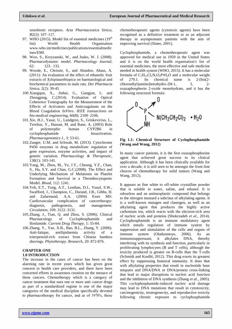
Cyclophosphamide, a chemotherapeutic agent was
approved for medical use in 1959 in the United States
and it is on the world health organisation′s list of
essential medicines, the most effective and safe medicine
needed in health system (WHO, 2015). It has a molecular
formula of C7H15Cl2N2O2P•H2O and a molecular weight
of 279.1. Its chemical name is 2-[bis(2-
chloroethyl)amino]tetrahydro-2H-1, 3, 2-
oxazaphosphorin 2-oxide monohydrate, and it has the
following structural formula:
Fig 1.1: Chemical Structure of Cyclophosphamide
(Wang and Wang, 2012)
In many cancer patients, it is the first oxazaphosphorine
agent that achieved great success in its clinical
application. Although it has been clinically available for
over a decade, it is still seen to be amongst the front-line
choices of chemotherapy for solid tumors (Wang and
Wang, 2012).
It appears as fine white to off-white crystalline powder
that is soluble in water, saline, and ethanol. It is
odourless and an antineoplastic compound that belongs
to the nitrogen mustard a subclass of alkylating agents. It
is a well-known mutagen and clastogen, as well as an
alkylating agent that produces the highly active
carbonium ion, which reacts with the electron-rich area
of nucleic acids and proteins (Shokrzadeh et al., 2014).
Cyclophosphamide is an immune modulatory agent
which entails regulation of immune system by
suppression and stimulation of the cells and organs of
immune system (Oladunmoye, 2006). As an
immunosuppressant, it alkylates DNA, thereby
interfering with its synthesis and function, particularly in
proliferating lymphocytes (B and T cells), although the
toxicity produced is greater on B-cells than the T-cells
(Schmidt and Koelbl, 2012). This drug exerts its greatest
effect by suppressing humoral immunity. It does that
with alkylating properties that result in nucleotide base
mispairs and DNA/DNA or DNA/protein cross-linking
that lead to major disruptions in nucleic acid function
and the inhibition of DNA synthesis (Zhang et al., 2005).
This cyclophosphamide-induced nucleic acid damage
may lead to DNA mutations that result in cytotoxicity,
carcinogenicity, teratogenecity, and reproductive toxicity
following chronic exposure to cyclophosphamide
Page 16
www.ejpmr.com
Udokwu et al. European Journal of Pharmaceutical and Medical Research
166
(Meirow and Nugent, 2001). On the other hand,
cyclophosphamide, although historically regarded as an
immunosuppressant, has been shown to act as a strong
adjuvant for either adoptive or active immunotherapy
when used with carefully defined dosages and
combination modalities. To further explain this
stimulatory effects of cyclophosphamide, Proietti et
al.(1998), using mouse models, identified type I
interferon (IFN-I) as an important mediator of
cyclophosphamide immunomodulation. Subsequent
studies showed that IFN-I was indeed induced in vivo by
cyclophosphamide and that this cytokine was responsible
for the expansion of memory CD4+ and CD8
+ T cells.
More recent data indicated that cyclophosphamide can
affect dendritic cell homeostasis and can restore an
activated polyfunctional helper phenotype in tumor-
specific adoptively transferred CD4+ T cells through
IFN-I–dependent mechanisms (Moschella etal., 2013).
Nakaharaetal.(2010), demonstrated in their work that
cyclophosphamide can improve immune responses by
preferentially depleting CD8+ lymphoid-resident
dendritic cells (DCs), thereby leading to diminished Treg
suppression and enhanced effector T-cell function in
vivo.
Cyclophosphamide has been in use clinically to treat a
wide range of cancers including malignant lymphomas,
myeloma, leukaemia, mycosis, fungoides,
neuroblastoma, adenocarcinoma, retinoblastoma, and
breast carcinoma (Mohammed et al., 2017). Other
clinical uses for cyclophosphamide can be seen in
immunosuppressive therapy following organ transplants
or as a treatment for autoimmune disorders such as
rheumatoid arthritis, Wegener’s granulomatosis, and
nephritic syndrome in children (Chabner et al., 2001).
Cyclophosphamide metabolism takes place in the liver
and is relatively inert until the binding phosphorus-
nitrogen is broken by means of metabolism catalysed by
hepatic enzymes of cytochrome P450, which are
responsible for the reaction of initial drug activation via
4-hydroxylation and N-dechloroethylation. Cytochrome
P4502B6, cytochrome P4503A4, cytochrome P4502C9,
and cytochrome P4502C19 have been shown to be the
major cytochrome P450s responsible for the metabolism
of cyclophosphamide and among these enzymes,
cytochrome P4502B6 was pointed out as the main
cytochrome P450 isozyme for the formation of 4-
hydroxy-cyclophosphamide (4-OH-CPA) (Afsharian et
al., 2007). When the inactive cyclophosphamide is
metabolized by P450 oxidase enzymes, it is transformed
into 4-hydroxy-cyclophosphamide that originates
the aldophosphamide. The latter, by its time, is carried to
other tissues, where it is converted into mustard
phosphoramide (the effectively cytotoxic molecule) and
acrolein, which is responsible for the adverse effects
(Tatiane et al., 2007).
The use of cyclophosphamide is however, limited by its
toxicity. Some of the adverse effects may include
alopecia, nausea, vomiting, thrombocytopenia, mucosal
ulcerations, transverse striations in the nails, brief spells
of dizziness, increased skin pigmentation, pulmonary
fibrosis, leukopenia, facial abrasion, haematuria,
diarrhoea, haemorrhagic cystitis, and petechial
haemorrhage in lungs and small bowel (Gitanjah et al.,
2017), but negative effects on the haematological system
have been observed especially in leucocyte and platelet
levels (Azevedo et al., 2007). It also affects cells that
respond to erythropoietic agents resulting in progressive
anaemia (Woo et al., 2008), and as a pro-oxidant drug it
can elicit generation of oxidative stress after
administration (Ikumawoyi et al., 2016). The
cyclophosphamide as a chemotherapeutic drug kills
dividing cells rapidly in the body, including cancer cells
and normal cells (Chakraborty et al., 2009). This
negative health effects associated with
cyclophosphamide present a significant health and safety
threat to laboratory staff, animal handlers, and other
personnel who may be subject to accidental exposure. As
a result of this health and safety threat the Institutional
Biosafety Committee (IBC) has classified
cyclophosphamide as a reportable hazardous chemical
that must be registered on the Institutional Animal Care
and Use Committee (IACUC) protocol as Chemical
Hazards. Despite its wide spectrum of clinical uses,
cyclophosphamide has been implicated in some adverse
conditions like hepatotoxicity, nephrotoxicity
haematotoxicity etc. and in attempt to reduce high level
of toxicity and check increase in the resistance of this
drug, various studies have been conducted in the past to
test the efficacy of low dose of cyclophosphamide in
treatment of carcinomas with positive results. Therefore,
cellular sensitivity to cyclophosphamide can be seen as
a function of cellular thiol concentration, metabolism
by aldehyde dehydrogenases to form inactive
metabolites, and the ability of DNA to repair alkylated
nucleotides.
The hormone Melatonin is the main neuroendocrine
secretary product of the pineal gland in animals and an
evolutionary ancient derivative of serotonin with
hormonal properties (slominski et al., 2018). It is also
produced in plants where it functions as a first line of
defence against oxidative stress (Tan et al., 2012). This
Melatonin, also known as N-acetyl-5-methoxy
tryptamine exists in organisms as different as algae, rats
and humans, and it is synthesized by a number of cells
and tissues but in mammals, the pineal gland is the main
source of this indolamine. The synthetic one has a
molecular formula of C13H16N2O2 and molecular weight
of 232.283.
It serves as an endocrine signal contributing to
entrainment/ regulation of organisms’ circadian rhythms
(Anthony et al., 2016), with maximum formation of this
melatonin occurring during the night. It′s nocturnal
synthesis is mainly regulated by the norepinephrine
released from sympathetic nerve endings, triggering the
transcription and translation of arylalkylamine-N-
Page 17
www.ejpmr.com
Udokwu et al. European Journal of Pharmaceutical and Medical Research
167
acetyltransferase, the most important enzyme involved in
melatonin synthesis (Ferreira et al., 2013). Melatonin is
involved in important physiological functions like
control of seasonal reproduction as well as influences on
the immune system. It also has anti-ageing effect and can
affect body mass, adipocity and energy metabolism
which vary according to species. This pineal indolamine
is a very potent and efficient endogenous radical
scavenger that reacts with the highly toxic hydroxyl
radical providing on‐site protection against oxidative
damage to biomolecules within every cellular
compartment (Burkhard et al., 1993). It quenches the
peroxyl radical, hypochlorous acid, and singlet oxygen,
all of which cause cell damage. Previous studies showed
that melatonin is preferentially localized inside the
nucleus and can protect nuclear DNA from oxidative
damage by interacting with double-stranded DNA
thereby promoting its stability. More so, it′s powerful
antioxidant action acts either directly on free radical
species or by modulating the gene expression of
antioxidant enzymes such as glutathione peroxidase,
catalase and superoxide dismutase. This antioxidant
effect of melatonin involves DNA repair, and can repair
the oxidation induced by the guanosine (G•) radical
(Ferreira et al., 2013). It can protect tissues from the
oxidative damage caused by glutathione depletion and
ischemia-reperfusion injury. Apart from its antioxidant
property, melatonin is a potential antitumor agent.
Therefore, studying of the effects of melatonin in
chemotherapy seems as an interesting area of
investigation.
Since time immemorial, the use of plants and its extracts
for the treatment and management of diseases has been
in existence all over the globe especially in Africa
particularly in the south Sahara (Woode et al., 2011).
This was assumed to be due to poverty and illiteracy,
which limited their accessibility to conventional medical
services. However, a large number of these tropical
plants and their extracts have beneficial therapeutic
effects such as contraceptive, aphrodisiac, fertility
enhancing capacities, as well as anti-oxidant, anti-
inflammatory, anti-cancer, and anti-microbial potentials
(Raji et al., 2006). Also, evidence abound that plant
derivatives have therapeutic potentials against vast
human, animal and plant diseases (Ogbonnia et al.,
2008), and thus, plants have become indispensable to
human and animal existence. In fact, according to Chris-
Ozoko et al. (2015), several conventional products
derived from plants exist today such as Aspirin (a
chemical copy of the analgesic chemical in the bark of
willow tree), Digoxin (from fox glove), morphine (from
Opium Poppy) etc. and interestingly, many herbal
products are now been approved by health agencies and
organizations.
Xylopiaaethiopica, a shrub locally referred to as
Ethiopian pepper, Negro pepper, Guinean pepper,
Senegal pepper, Kili pepper and spice tree in the savanna
zone and coastal regions of Africa is amongst these
plants with great therapeutic potential. It is an
angiosperm belonging to the family Annonaceae (Obodo
et al., 2013), and is among the species that thrive in the
evergreen rain forests of tropical and subtropical Africa
which matures into a slim, tall tree of approximately 60
cm in diameter and up to 30m high with a straight stem
having a slightly stripped or smooth bark. The seeds of
Xylopiaaethiopica which have musky flavours are used
as pepper substitutes in Nigeria and the fruits are
popularly used as a condiment in many local dishes by
ethnic groups and communities in Africa. In the Igbo
community, the fruits are used as spices and aqueous
decoction is used after child birth probably for its
antiseptic properties and to arrest bleeding (Nnodim et
al., 2011). Xylopiaaethiopica is used in the treatment of
cough, biliousness, bronchitis, rheumatism, dysentery,
malaria, uterine fibroid, amenorrhea, boils, sores,
wounds and cuts among others (fetse et al., 2016).
According to Obodo et al. (2013), The seeds of
Xylopiaaethiopica have been shown to contain bitter
chemical constituents like alkaloids, glycosides,
saponnis, tannins, sterols, carbohydrate, protein, free
fatty acids, mucilage’s and acidic compounds; some of
which might be responsible for its documented medicinal
and pharmacological properties like anti-inflammatory,
cytotoxic, hypoglycaemic and antioxidant effects.
Since antiquity, blood has been seen as the
essentialcomponent of life (Hart, 2001). It is a tissue that
consists of fluid plasma in which are suspended a
number of formed elements which are red blood cells,
white blood cells and platelets. These blood cells exist at
fairly constant levels, suggesting the existence of
feedback regulatory mechanisms (Ofem, et al., 2012).
These blood cells which are haematological
parameterscan be used as indicators of toxicity and have
a broad potential application in environmental and
occupationalmonitoring (Raquel et al., 2012).
The red blood cell integrity is largely dependent on its
ability to maintain its membrane constituents that are
mostly polyunsaturated free fatty acid, and if this is
compromised will result to erythrocyte fragility which
becomes more fragile with consequent destruction by the
macrophages. This erythrocyte fragility or red cell
osmotic fragility is the ability of red blood cells to
undergo haemolysis when subjected to stress and the
absolute extent of haemolysis can be measured (Rodak,
2007). When this happens the membranes of the cells
undergo lipid peroxidation leading to oxidative
deterioration of polyunsaturated fatty acid accumulation
of reactive oxygen species (ROS) which are associated
with tissue damage by clearing off the sialic acid from
the cells making them more prone to phagocytosis. Also,
the osmotic fragility of red cells can occur from the
increased phosphorylation of P38 and JNK genes which
promotes increased production of ROS (Robin and
Steven, 2000). Factors such as cell’s size, surface area to
volume ratio, membrane composition and integrity can
equally influence the osmotic fragility of the cells
Page 18
www.ejpmr.com
Udokwu et al. European Journal of Pharmaceutical and Medical Research
168
(Fischbach, et al., 2008). Alteration in the integrity of
blood leads to loss of blood and can be life threatening as
blood is a necessary components of animal body. The
body tends to protect itself from this life threatening
exsanguination by converting the blood from its liquid
state to a solid state in a process known as blood clotting
or coagulation. This formation of a clot is often referred
to as secondary haemostasis and it usually involves two
main pathways namely extrinsic and intrinsic pathways
that make use of clotting factors. Estimation of
coagulation tests like prothrombin time, activated partial
thromboplastin time etc. are developed to diagnose
disorders of coagulation which can lead to an increased
bleeding (haemorrhage) or obstructive clotting
(thrombosis) (Xiangqun, et al., 2014).
Analysis ofblood parameters is relevant to risk
evaluation of alterations of the haematological system in
humans (Olsonet al., 2000). These haematological
parameters are those parameters that are related to the
blood and blood-forming organs. The evaluation of these
parameters does not only serve diagnostic purpose for
routine clinical management of patients but plays
important purpose of risk assessment in animals and
humans exposed to various risk factors (Saliu et al.,
2012). The reason for the use of haematological
parameters for this purpose is that any form of alteration
or disruption of physiological processes, whether such
disruption is induced chemically, physically or
biologically, will correspondingly alter the
haematological parameters both in humans and animals.
It is also an established fact that chronic diseases affect
the blood cells adversely. Damages mediated by free
radicals can result in the disruption of membrane fluidity,
oxidative DNA, alteration of platelet functions, etc. and
have generally been considered to be linked with many
chronic health problems such as cancers, inflammation,
aging and atherosclerosis. Although organisms possess
antioxidant defence and repair systems that have evolved
to protect them against free radicals but these systems are
insufficient to protect them completely against oxidative
damage.
For this study, the cyclophosphamide was chosen
because it is one of the most frequently used antitumor
agents in clinical practice and also its association with
rapidly killing of dividing cells in the body.
Considering the above, this present study was designed
to evaluate the effects of Xylopia aethiopica and
melatonin on some haematological parameters, in
cyclophosphamide induced wistar rats, with a view of
finding a lasting solution to the life threatening effects
reported about this cytotoxic drug.
AIM
The research work is aimed at evaluating the effects of
Xylopia aethiopica and melatonin on some
haematological parameters in cyclophosphamide
intoxicated adult wistar rats.
MATERIALS AND METHODS
COLLECTION OF PLANT MATERIALS AND AU
THENTICATION
Pods of Xylopia aethiopica were purchased from Orie-
Ugba vegetable market, Umuahia North Local
Government Area, Abia State, Nigeria and were taken to
the Department of Forestry and Environmental
Management, Michael Okpara University of Agriculture,
Umudike where they were identified by a botanist/forest
manager. Voucher number MOUAU/VPP/18/012 was
assigned to a specimen sample of the pods which was
deposited in the herbarium of the Department.
PREPARATION OF PLANT EXTRACTS
Extract of the fruit pods was prepared in accordance with
the Soxhlet method described by Jensen, (2007). The
plant materials were subjected to further drying under
shade for 14 days and were pulverized into powder in a
manual blender powered by a Honda petrol engine. One
hundred grams of the powdered sample was introduced
into the extraction chamber of the soxhlet extractor and
extraction was carried out with ethanol as solvent.
Temperature was maintained at 650C throughout the
extraction period of 48 hours. At the end of the period,
the extract in solution was dried in a hot air oven at 40oC
to obtain a dry dark oily extract. The weight of the
extract was taken and percentage yield was calculated
using the formular:
% yield =X x 100
Q 1
Where X = weight of dried extract and Q = weight of
powdered plant material before extraction (100g)
(Bandiola, 2018).
ANIMALS USED FOR STUDY
One hundred and ninety five matured wistar albino rats
were used for the studies. Of the number, 30 were used
for the acute toxicity evaluation of the extract, 35 for
acute toxicity study of cyclophosphamide and 130 were
used for the main study. The rats were kept in aluminum
cages and allowed to acclimatize for two weeks to allow
for proper adaptation to the environment and living
conditions. They were allowed access to feed (Vital feed,
Nigeria) and water ad libitum but were starved for 12
hours prior to commencement of any experiment. All
animal experiments were carried out in compliance with
NIH guidelines for Care and Use of Laboratory Animals
(OECD, 2001). All experiments were carried out in the
Physiology Laboratory of the Department of Physiology
and Pharmacology, College of Veterinary Medicine,
Michael Okpara University of Agriculture, Umudike,
Nigeria.
EXPERIMENTAL DESIGN The rats (130 in number) were assigned to 13 groups of
10 rats each and were treated according to the order
below:
Group I: 10 mg/kg Cyclophosphamide, Food and water
Page 19
www.ejpmr.com
Udokwu et al. European Journal of Pharmaceutical and Medical Research
169
Group II: 10 mg/kg Cyclophosphamide, 400 mg/kg
Extract, Food and water
Group III: 10 mg/kg Cyclophosphamide, 0.5 mg/kg
Melatonin, Food and water
Group IV: 10 mg/kg Cyclophosphamide, 400mg/kg
Extract, 0.5mg/kg Melatonin, Food and water
Group V: 30 mg/kg Cyclophosphamide, Food and water
Group VI: 30 mg/kg Cyclophosphamide, 400 mg/kg
Extract, Food and water
Group VII: 30mg/kg Cyclophosphamide, 0.5mg/kg
Melatonin, Food and water
Group VIII: 30mg/kg Cyclophosphamide+400mg/kg
Extract, 0.5mg/kg Melatonin, Food and water
Group IX: 50 mg/kg Cyclophosphamide, Food and water
Group X: 50 mg/kg Cyclophosphamide, 400 mg/kg
Extract, Food and water
Group XI: 50mg/kg Cyclophosphamide, 0.5mg/kg
Melatonin, Food and water
Group XII: 50mg/kg Cyclophosphamide, 400mg/kg
Extract, 0.5mg/kg Melatonin, Food and water
Group XIII: Food and water only
Treatments were done daily via the oral route for twenty
one (21) days. Three animals were sacrificed in each
group for blood collection by cardiac puncture into
EDTA and sodium citrate bottles for haematology and
osmotic fragility studies. Liver and kidney samples were
also collected and preserved in 10% formalin for
histological examination.
HEMATOLOGICAL STUDIES Haematological parameters including Red blood cell
count (RBCC), packed cell volume (PCV), haemoglobin
concentration (Hb), mean corpuscular volume (MCV),
mean corpuscular haemoglobin (MCH), mean
corpuscular haemoglobin concentration (MCHC), white
blood cells count (WBCC) and Platelets (PLT) count
were determined. These parameters were obtained at
once for each blood sample in an Automated
Haematology Analyser (Mindray company, China),
following the procedures by the producer.
DETERMINATION OF RED BLOOD CELLS
OSMOTIC FRAGILITY
The method of Adenkule and Olurenmi, (2014) was
adopted. Sodium chloride solution (200 mls) was
prepared for each sample in concentrations ranging from
0.1-0.85% at pH 7.4. A set of test tubes, each containing
5mls of sodium chloride solution of concentration
ranging from 0.1 to 0.85% were serially arranged in a
test tube rack. One set of test tubes was used to analyze
each sample. A drop of the freshly collected blood was
placed into each of the ten test tubes using a dropper
pipette and each was mixed by gently inverting the test
tubes about three times. The test tubes were then allowed
to stand at room temperature for 30 minutes and then
centrifuged at 3000 rpm for 10 minutes before reading
the absorbance of the supernatant in each test tube on a
spectrophotometer at 540nm. The same procedure was
repeated for each sample collected. Percentage
haemolysis was calculated using the expression:
Percentage Haemolysis = Absorbance of test x100
Absorbance of control
DETERMINATION OF ACTIVATED PARTIAL
THROMBOPLASTIN TIME (APTT)
AUTHOMATED KL340 COAGULATION
ANALYZER
Procedure
After gentle swirling of the reagent vials, enough volume
of reagent 1 (CaCl₂) was pre-warmed for immediate use
in a clean and dry plastic tube maintained at 370C. About
100 µl of test plasma was pipette into a test cuvette at
300C. About 100µl of pre-warmed reagent 2 (APTT
Reagent) was added to the cuvette. The mixture was well
mixed and incubated at 370C for 3 minutes before
forcibly pipetting 100µl of pre-warmed reagent 1 into the
test cuvette while starting the stop watch at the same
time to note time in seconds it takes for blood to clot.
This time in seconds is recorded as the APTT.
DETERMINATION OF PROTHROMBIN TIME
(PT) USING AUTHOMATED KL340
COAGULATION ANALYZER
Procedure
Prothrombin reagent was dispensed into a thoroughly
clean and dry plastic tube for immediate use and pre-
warm at 370C for 10 minutes. About 100 µl of test
plasma was introduced into a test cuvette at 370C and
incubated for 3 minutes before. The mixture was well
mixed and incubated at 370C for 3 minutes before adding
forcibly 200µl of the pre-warmed prothrombin reagent
while starting the stop watch at the same time to record
the time in seconds it takes for blood to clot. This time in
seconds is recorded as the PT.
STATISTICAL ANALYSIS
Results were expressed as means ± standard error of
mean (SEM). Statistical analysis was done using one-
way analysis of variance (ANOVA). Significant
differences were assessed at 95% level of significance
between control and treated groups using Duncan and
LSD (Post Hoc) tests. P values less than 0.05 were
considered significant. Computer software package,
SPSS version 21 was employed.
RESULTS
4.1 PERCENTAGE YIELD OF THE EXTRACT
Following extraction and drying of the extract in
solution, 9.23g of the extract was obtained and
represented 9.23% yield (Table 4.1).
Table 4.1: Percentage yield of XA extract.
Weight of plant material Weight of extract Percentage yield
100g 9.23g 9.23%
Page 20
www.ejpmr.com
Udokwu et al. European Journal of Pharmaceutical and Medical Research
170
Result of effects of Xylopia aethiopica extract and
Melatonin on RBC of Cyclophosphamide intoxicated
wistar rats
In week one, and two, there was significant decrease in
RBC in all the treatment groups compared with control
except week three. RBC varied significantly from week
one to three in all the treatment groups except group 1, 9
and 13.
In week one there is a dose dependent decrease in RBC
of rats treated with 10, 30 and 50 mg/kg of
cyclophosphamide. In week one, treatment with 400mg
Xylopia aethiopica and 0.5 mg Melatonin significantly
increased the RBC of rats exposed to 10mg
Cyclophosphamide. RBC of rats treated with 400 mg
Xylopia aethiopica and 0.5mg Melatonin was
significantly higher than 400mg Xylopia aethiopica
alone. Similarly, treatment with 400mg Xylopia
aethiopica alone and 400mg Xylopia aethiopica and
0.5mg Melatonin significantly increased the RBC of rats
exposed to 30mg Cyclophosphamide. RBC of rats
treated with 400mg Xylopia aethiopica and 0.5mg
Melatonin was significantly higher than 400mg Xylopia
aethiopica. Treatment with 400mg Xylopia aethiopica
only significantly increased the RBC of rats exposed to
50mg Cyclophosphamide.
In week two, treatment with 400mg Xylopia aethiopica
alone and 400mg Xylopia aethiopica and 0.5 mg
Melatonin significantly increased the RBC of rats
exposed to 10mg Cyclophosphamide. Treatment with
400mg Xylopia aethiopica alone significantly increased
the RBC of rats exposed to 30mg Cyclophosphamide.
Treatment with 400mg Xylopia aethiopica, 0.5 mg
Melatoninand the combination had no significant effect
of RBC of rats exposed to 50mg Cyclophosphamide.
In week three, treatment with 400mg Xylopia
aethiopicaalone and in combination with 0.5mg
Melatonin had no significantly effect on the RBC of rats
exposed to 10mg Cyclophosphamide (Table 4.6).
Table 4.6: Effects of Xylopia aethiopica extract and Melatonin on RBC of Cyclophosphamideintoxicated wistar
rats.
Treatment
RBC (Wk1) (Wk2) (Wk3) A.Wk
Week 1
(Wk1)
Week 2
(Wk2)
Week 3
(Wk3) p-value p-value p-value p-value f-value
10mg Cyclophosphamide 6.35±0.05 cd
5.89±0.01 e 9±4.32
a .000 .000 .942 .656 20.737
10mg Cyclophosphamide +
400mg Xylopia aethiopica 6.61±0.33
d 7±0.06 5.96±0.02
a .000 .001 .970 .002 64.225
10mg Cyclophosphamide + 0.5mg
Melatonin 6.5±0.2
d 5.57±0.01
.000 .000 .001 42.551
10mg Cyclophosphamide +
400mg Xylopia aethiopica + 0.5mg
Melatonin
7.1±0.2 e 6.3±0.12
f 5.89±0.01
a .838 .000 .965 .000 171.564
30mg Cyclophosphamide 5.96±0.01 bc
5.2±0.06 c
.000 .000 .000 816.750
30mg Cyclophosphamide +400mg
Xylopia aethiopica 6.53±0.03
d 5.87±0.01
c
.000 .000 .000 4335.000
30mg Cyclophosphamide + 0.5mg
Melatonin 5.93±0.02
d 5.08±0.01
bc
.000 .000 .000 5766.000
30mg Cyclophosphamide +
400mg Xylopia aethiopica + 0.5mg
Melatonin
6.51±0.02 c 5.27±0.01
c
.000 .000 .000 .119
50mg Cyclophosphamide 5.1±0.1 a 5.08±0.01
bc
.000 .000 .748 2457.600
50mg Cyclophosphamide +
400mg Xylopia aethiopica 5.87±0.01
c 5.23±0.01
.000 .000 .000 777.600
50mg Cyclophoshamide + 0.5mg
Melatonin 5.11±0.01
a 4.75±0.01
a
.000 .000 .000 405.600
50mg Cyclophosphamide +
400mg Xylopia aethiopica + 0.5mg
Melatonin
5.19±0.01 a 4.93±0.01
ab
.000 .000 .000 .007
Control 7.3±0.2 e 7.29±0.01
h 7.3±0.03
a .993 .453
4.7: Result of effects of Xylopia aethiopica extract and
Melatonin on HB of Cyclophosphamideintoxicated
wistar rats
In week one, two and three, there was significant
decrease in HGB in all the treatment groups compared
with control. HGB varied significantly from week one to
three in all the treatment groups except group 9 and 13.
In week one and two, there is a dose dependent decrease
in HGB of rats treated with 10 and 50 mg/kg of
cyclophosphamide. Treatment with 400mg Xylopia
Page 21
www.ejpmr.com
Udokwu et al. European Journal of Pharmaceutical and Medical Research
171
aethiopica and 0.5 mg Melatonin significantly increased
the HGB of rats exposed to 10mg cyclophosphamide.
Similarly, treatment with 400mg Xylopia aethiopica
alone and 400mg Xylopia aethiopica and 0.5mg
Melatonin significantly increased the HGB of rats
exposed to 30mg cyclophosphamide. Treatment with
400mg Xylopia aethiopica alone significantly increased
the HGB of rats exposed to 50mg Cyclophosphamide.
In week three, treatment with 400mg Xylopia aethiopica
alone and in combination with 0.5 mg melatonin
significantly increased the HGB of rats exposed to 10mg
Cyclophosphamide (Table 4.7).
Table 4.7: Effects of Xylopia aethiopica extract and Melatonin on HB of Cyclophosphamide intoxicated wistar
rats.
Treatment HB (Wk1) (Wk2) (Wk3) A.WK
Week 1 (Wk1)
Week 2 (Wk2)
Week 3 (Wk3)
p-
value p-
value p-
value p-
value f-value
10mg Cyclophosphamide 11.8±0.14 de 10.2±0.06
f 8.03±0.07a .000 .000 .000 .000 374.718
10mg Cyclophosphamide + 400mg Xylopia aethiopica
12.2±0.12 ef 12.5±0.12
h 11.3±0.12c .000 .082 .000 .001 29.250
10mg Cyclophosphamide +
0.5mg Melatonin 12.1±0.17
ef 10±0.06ef
.000 .000
.000 132.300
10mg Cyclophosphamide+ 400mg Xylopia aethiopica +
0.5mg Melatonin 12.6±0.12
f 11.5±0.12f 10.3±0.12
c .020 .000 .000 .000 99.250
30mg Cyclophosphamide 11.3±0.06 cd 9.1±0.12
cd
.000 .000
.000 290.400 30mg Cyclophosphamide+ 400mg Xylopia aethiopica
12.1±0.12 ef 10.2±0.12
e
.000 .000
.000 135.375
30mg Cyclophosphamide +
0.5mg Melatonin 11.2±0.06
c 9±0.06bc
.000 .000
.000 726.000
30mg Cyclophosphamide+ 400mg Xylopia aethiopica +
0.5mg Melatonin 12.1±0.06 9.6±0.12
e
.000 .000
.000 375.000
50mg Cyclophosphamide 9±0.06 a 9±0.17
b
.000 .000
1.000 0.000 50mg Cyclophosphamide+ 400mg Xylopia aethiopica
10.2±0.06 b 9.4±0.06
d
.000 .000
.001 96.000
50mg Cyclophosphamide +
0.5mg Melatonin 9±0.06
a 8.3±0.12 a
.000 .000
.006 29.400
50mg Cyclophosphamide+ 400mg Xylopia aethiopica +
0.5mg Melatonin 9.1±0.06
a 8.5±0.12 a
.000 .000
.010 21.600
Control 13.2±0.17 g 13±0.06
h 13.1±0.06d
.485 .818
4.8: Result of effects of Xylopia aethiopica extract and
Melatonin on HCT of Cyclophosphamideintoxicated
wistar rats
In week one, two and three, there was significant
decrease in HCT in all the treatment groups compared
with control. HCT varied significantly from week one to
three in all the treatment groups except group 9 and 13.
In week one, two and three, there was significant
decrease in HCT in all the treatment groups compared
with control. HCT varied significantly from week one to
three in all the treatment groups except group 9 and 10.
In week one and two, there is a dose dependent increase
in HCT of rats treated with 10, 30 and 50 mg/kg of
cyclophosphamide. Treatment with 400mg Xylopia
aethiopica alone and 400mg Xylopia aethiopica and 0.5
mg Melatonin significantly increased the HCT of rats
exposed to 10mg Cyclophosphamide. HCT of rats
treated with 400 mg Xylopia aethiopica and 0.5mg
Melatonin was significantly higher than 400mg Xylopia
aethiopica alone. Similarly, treatment with 400mg
Xylopia aethiopica alone and 400mg Xylopia aethiopica
and 0.5mg Melatonin significantly increased the HCT of
rats exposed to 30mg Cyclophosphamide. HCT of rats
treated with 400mg Xylopia aethiopica and 0.5mg
Melatonin was significantly higher than 400mg Xylopia
aethiopica alone. Treatment with 400mg Xylopia
aethiopica alone as well as 400mg Xylopia aethiopica
and 0.5mg Melatonin significantly increased the HCT of
rats exposed to 50mg Cyclophosphamide.
In week three, treatment with 400mg Xylopia
aethiopicasignificantly increased the HCT of rats
exposed to 10mg Cyclophosphamide (table 4.8).
Page 22
www.ejpmr.com
Udokwu et al. European Journal of Pharmaceutical and Medical Research
172
Table 4.8: Effects of Xylopia aethiopica extract and Melatonin on HCT of Cyclophosphamideintoxicated wistar
rats.
Treatment HCT (Wk1) (Wk2) (Wk3) A.Wk
Week 1
(Wk1)
Week 2
(Wk2)
Week 3
(Wk3) p-value p-value p-value p-value f-value
10mg Cyclophosphamide 37±0.29 f 32±0.17
e 25.6±0.12
a .000 .000 .000 .000 773.368
10mg Cyclophosphamide+
400mg Xylopia aethiopica 38.7±0.35
g 39.1±0.11
g 35.8±0.06
c .000 .000 .000 .000 71.195
10mg Cyclophosphamide +
0.5mg Melatonin 38.5±0.23
f 31.9±0.05
e
.000 .000 .000 768.706
10mg Cyclophosphamide+
400mg Xylopia aethiopica +
0.5mg Melatonin
40.9±0.06 h 36.9±0.05
f 32.7±0.06
b .000 .000 .000 .000 5044.000
30mg Cyclophosphamide 35.8±0.06 d 30.1±0.05
c
.000 .000 .000 4873.500
30mg Cyclophosphamide+
400mg Xylopia aethiopica 38.5±0.12
g 32.2±0.05
e
.000 .000 .000 2381.400
30mg Cyclophosphamide +
0.5mg Melatonin 33.6±0.06
c 28.2±0.05
b
.000 .000 .000 4374.000
30mg Cyclophosphamide+
400mg Xylopia aethiopica +
0.5mg Melatonin
38.3±0.12 g 30.9±0.05
e
.000 .000 .000 3285.600
50mg Cyclophosphamide 28.5±0.12 a 28.2±0.05
b
.000 .000 .081 5.400
50mg Cyclophosphamide+
400mg Xylopia aethiopica 32.2±0.06
c 30.8±0.05
d
.000 .000 .000 294.000
50mg Cyclophosphamide +
0.5mg Melatonin 28.5±0.06
a 26.5±0.05
a
.000 .000 .000 600.000
50mg Cyclophosphamide+
400mg Xylopia aethiopica +
0.5mg Melatonin
30±0.29 b 26.9±0.05
a
.000 .000 .000 110.885
Control 45.6±0.06 i 43.1±0.05
h 44.3±0.23
d .081 78.167
4.9: Result of effects of Xylopia aethiopica extract and
Melatonin on MCV of Cyclophosphamide intoxicated
wistar rats In week one, two and three, there was significant
decrease in MCV in all the treatment groups compared
with control. MCV varied significantly from week one to
three in all the treatment groups except group 9 and 10.
MCV of rats exposed to 10mg Cyclophosphamideand
treated with 400mg Xylopia aethiopica and 0.5 mg
Melatonin alone and in combination was not
significantly different from each other. MCV of rats
treated with 400 mg Xylopia aethiopica and 0.5mg
Melatonin was significantly higher than 400mg Xylopia
aethiopica alone. Similarly, treatment with 0.5mg
Melatoninalone and 400mg Xylopia aethiopica and
0.5mg Melatonin significantly increased the MCV of rats
exposed to 30mg Cyclophosphamide. MCV of rats
treated with 400mg Xylopia aethiopica and 0.5mg
Melatonin was significantly higher than 400mg Xylopia
aethiopica alone. Treatment with 400mg Xylopia
aethiopica and 0.5mg Melatonin significantly increased
the MCV of rats exposed to 50mg Cyclophosphamide.
By week 2, MCV of rats exposed to 10mg
Cyclophosphamide and treated with 400mg Xylopia
aethiopica and 0.5 mg Melatonin alone and in
combination was significantly different from rats treated
with 10 mg Cyclophosphamide alone. MCV of rats
exposed to 30mg Cyclophosphamide and treated with
400mg Xylopia aethiopica and 0.5 mg Melatonin alone
and in combination was significantly different from each
other and rats treated with 30 mg Cyclophosphamide
alone. MCV of rats exposed to 50mg Cyclophosphamide
and treated with 400mg Xylopia aethiopica was
significantly different from rats treated with 50 mg
Cyclophosphamide alone.
In week three, MCV of rats exposed to 10mg
Cyclophosphamide treated with 400mg Xylopia
aethiopicasignificantly higher MCV of rats exposed to
10mg Cyclophosphamide treated with 400mg Xylopia
aethiopica and 0.5mg Melatonin.
Page 23
www.ejpmr.com
Udokwu et al. European Journal of Pharmaceutical and Medical Research
173
Table 4.9: Effects of Xylopia aethiopica extract and Melatonin on MCV of Cyclophosphamide intoxicated wistar
rats.
Treatment MCV (Wk1) (Wk2) (Wk3) A.Wk
Week 1
(Wk1)
Week2
(Wk2)
Week 3
(Wk3) p-value p-value p-value p-value f-value
10mg Cyclophosphamide 58.3±0.1 def
54.3±0.1 a 54.8±0.1
a .000 .000 .000 .000 475.000
10mg Cyclophosphamide+
400mg Xylopia aethiopica 58.6±0.3
def 55.9±0.1
d 60±0.6
b .000 .000 .399 .001 30.302
10mg Cyclophosphamide +
0.5mg Melatonin 59.2±0.1 57.3±0.1
e
.000 .000 .000 216.600
10mg Cyclophosphamide+
400mg Xylopia aethiopica +
0.5mg Melatonin
57.6±02 cd
58.6±0.1 55.5±0.1 a .000 .718 .000 .000 204.818
30mg Cyclophosphamide 60±0.6 57.9±0.1 ef
.000 .005 .022 13.099
30mg Cyclophosphamide+
400mg Xylopia aethiopica 59.1±0.1 54.9±0.1
abc
.000 .000 .000 2646.000
30mg Cyclophosphamide +
0.5mg Melatonin 56.7±0.2
bc 55.5±0.1
bcd
.000 .000 .010 21.600
30mg Cyclophosphamide+
400mg Xylopia aethiopica +
0.5mg Melatonin
58.8±0.1 ef
58.6±0.1 fg
.000 .718 .196 2.400
50mg Cyclophosphamide 55.9±0.1 ab
55.5±0.1 bcd
.000 .000 .196 24.000
50mg Cyclophosphamide+
400mg Xylopia aethiopica 54.9±0.1
a 58.9±0.06
g
.000 .999 .000 2400.000
50mg Cyclophosphamide +
0.5mg Melatonin 55.8±0.1
ab 55.8±0.06
cd
.000 .000 1.000 0.000
50mg Cyclophosphamide+
400mg Xylopia aethiopica +
0.5mg Melatonin
57.8±0.1 de
54.6±0.06 f
.000 .000 .000 1536.000
Control 62.5±0.1 g 59.13±0.6
g 60.7±0.1
b .272 21.586
4.10: Result of effects of Xylopia aethiopica extract
and Melatonin on MCH of Cyclophosphamide
intoxicated wistar rats
In week one, and two, there was significant decrease in
MCH in all the treatment groups compared with control
except week three. MCH varied significantly from week
one to three in all the treatment groups except group 4, 9,
11 and 13.
In week 1, MCH of rats exposed to 10 mg
Cyclophosphamide and treated with 400 mg Xylopia
aethiopica and 0.5 mg Melatonin alone was significantly
different from rats exposed to 10 mg Cyclophosphamide.
MCV of rats treated with 400 mg Xylopia aethiopica and
0.5mg Melatonin was significantly higher than 400mg
Xylopia aethiopica alone. MCH of rats treated with
400mg Xylopia aethiopica and 0.5mg Melatonin singly
and in combination was not significantly different from
the MCH of rats exposed to 30mg Cyclophosphamide.
Similarly, MCH of rats treated with 400mg Xylopia
aethiopica and 0.5mg Melatonin singly and in
combination was not significantly different from the
MCH of rats exposed to 50mg Cyclophosphamide.
By week 2, MCH of rats exposed to 10mg
Cyclophosphamide and treated with 400mg Xylopia
aethiopica and 0.5 mg Melatonin alone and in
combination was significantly different from rats treated
with 10 mg Cyclophosphamide alone. MCH of rats
exposed to 30mg Cyclophosphamide and treated with
400mg Xylopia aethiopica and 0.5mg Melatonin was
significantly higher than rats treated with 30 mg
Cyclophosphamide alone. MCH of rats exposed to 50 mg
Cyclophosphamide and treated with 400mg Xylopia
aethiopica was significantly different from rats treated
with 50mg Cyclophosphamide alone.
In week three, MCH of rats exposed to 10mg
Cyclophosphamide treated with 400 mg Xylopia
aethiopica and 0.5 mg Melatonin alone and in
combination was not significantly different.
Page 24
www.ejpmr.com
Udokwu et al. European Journal of Pharmaceutical and Medical Research
174
Table 4.10: Effects of Xylopia aethiopica extract and Melatonin on MCH of Cyclophosphamide intoxicated
wistar rats.
Treatment MCH Wk1 Wk2 Wk3 A.Wk
Week 1 (Wk1)
Week 2 (Wk2)
Week 3 (Wk3)
P-
value p-
value p-
value p-
value F-value
10mg Cyclophosphamide 18.6±0.1 de 17.3±0.1
ab 17.6±0.1 a .008 .091 .999 .000 69.500
10mg Cyclophosphamide+ 400mg Xylopia aethiopica
18.5±0.1 cd 17.9±0.1
cde 19±0.3 a .062 1.000 .989 .015 9.100
10mg Cyclophosphamide + 0.5mg Melatonin 18.6±0.1 de 18±0.1
de
.008 .974
.021 13.500 10mg Cyclophosphamide+ 400mg Xylopia aethiopica + 0.5mg Melatonin
17.7±0.1 ab 18.23±0.1
e 22.27±4.6 a .062 .216 .570 .450 .915
30mg Cyclophosphamide 19±0.1 e 17.5±0.01
abcd
.000 .717
.000 337.500 30mg Cyclophosphamide+ 400mg Xylopia aethiopica
18.6±0.1 de 17.4±0.1
abc
.008 .315
.001 86.400
30mg Cyclophosphamide + 0.5mg Melatonin 18.9±0.1 de 17.7±0.2
abcde
.000 1.000
.007 25.412 30mg Cyclophosphamide+ 400mg Xylopia aethiopica + 0.5mg Melatonin
18.6±0.1 de 18.2±0.1
e
.008 .315
.008 24.000
50mg Cyclophosphamide 17.7±0.1 ab 17.7±0.1
abcde
.062 1.000
1.000 0.000 50mg Cyclophosphamide+ 400mg Xylopia aethiopica
17.4±0.06 a 18±0.06
de
.000 .974
.002 54.000
50mg Cyclophosphamide + 0.5mg Melatonin 17.6±0.06 a 17.5±0.12
abcd
.008 .717
.482 .600 50mg Cyclophosphamide+ 400mg Xylopia aethiopica + 0.5mg Melatonin
17.5±0.06 a 17.2±0.06
a
.001 .020
.021 13.500
Control 18.1±0.06 bc 17.8±0.06
bcde 18±0.06 .008 .091 .999 .276 7.000
4.11: Result of effects of Xylopia aethiopica extract
and Melatonin on MCHC of Cyclophosphamide
intoxicated wistar rats
In week one, two and three, there was significant
decrease in MCHC in all the treatment groups compared
with control. MCHC varied significantly from week one
to three in all the treatment groups except group 9 and
13.
In week 1, MCHC of rats exposed to 10 mg
Cyclophosphamide and treated with 400 mg Xylopia
aethiopica and0.5 mg Melatonin alone was significantly
different from rats exposed to 10 mg Cyclophosphamide.
MCHC of rats treated with 400 mg Xylopia aethiopica
and 0.5mg Melatonin was significantly higher than
400mg Xylopia aethiopica alone. MCHC of rats treated
with 400mg Xylopia aethiopica and 0.5mg Melatonin
singly and in combination was not significantly different
from the MCHC of rats exposed to 30mg
Cyclophosphamide. Similarly, MCHC of rats treated
with 400mg Xylopia aethiopica and 0.5 mg Melatonin
was significantly different from the MCHC of rats
exposed to 50mg Cyclophosphamide.
By week 2, MCHC of rats exposed to 10mg
Cyclophosphamide and treated with 400mg Xylopia
aethiopica and 0.5 mg Melatonin alone and in
combination was significantly different from rats treated
with 10 mg Cyclophosphamide alone. MCHC of rats
exposed to 30 mg Cyclophosphamide and treated with
400mg Xylopia aethiopica and 0.5 mg melatonin was
significantly higher than rats treated with 30 mg
Cyclophosphamide alone. MCHC of rats exposed to 50
mg Cyclophosphamide and treated with 400 mg Xylopia
aethiopica was significantly different from rats treated
with 50 mg Cyclophosphamide alone.
In week three, MCHC of rats exposed to 10mg
Cyclophosphamide treated with 400 mg Xylopia
aethiopica and 0.5 mg melatonin alone and in
combination was not significantly different.
Page 25
www.ejpmr.com
Udokwu et al. European Journal of Pharmaceutical and Medical Research
175
Table 4.11: Effects of Xylopia aethiopica extract and Melatonin on MCHC of Cyclophosphamide intoxicated
wistar rats.
Treatment MCHC (Wk1) (Wk2) (Wk3) A.Wk
Week 1
(Wk1)
Week 2
(Wk2)
Week3
(Wk3) p-value p-value p-value p-value F-value
10mg Cyclophosphamide 31.9±0.1 e 31.9±0.2
d 32±0.6
a .000 .000 .999 .972 .029
10mg Cyclophosphamide+
400mg Xylopia aethiopica 31.5±0.1
de 32±0.1
d 31.6±0.1
a .000 .000 .999 .002 21.000
10mg Cyclophosphamide +
0.5mg Melatonin 31.4±0.1
d 31.5±0.15
bcd
.000 .000 .573 .375
10mg Cyclophosphamide+
400mg Xylopia aethiopica +
0.5mg Melatonin
30.8±0.1 c 31.2±0.1
bc 56.07±2.4
a .000 .000 .465 .406 1.050
30mg Cyclophosphamide 31.6±0.1 de
30.2±0.1 cd
.000 1.000 .001 73.500
30mg Cyclophosphamide +
400mg Xylopia aethiopica 31.4±0.1
d 31.7±0.1
a
.000 .000 .140 3.375
30mg Cyclophosphamide +
0.5mg Melatonin 33.3±0.1
f 31.9±0.1
d
.000 .000 .000 117.600
30mg Cyclophosphamide+
400mg Xylopia aethiopica +
0.5mg Melatonin
31.6±0.1 de
31.1±0.1 b
.000 .000 .018 15.000
50mg Cyclophosphamide 31.6±0.1de
31.9±0.1d
.000 .000 .081 5.400
50mg Cyclophosphamide+
400mg Xylopia aethiopica 31.4±0.06
d 30.5±0.11547
a
.000 .739 .002 48.600
50mg Cyclophosphamide +
0.5mg Melatonin 31.6±0.1
de 31.3±0.1
ab
.000 .000 .223 2.077
50mg Cyclophosphamide+
400mg Xylopia aethiopica +
0.5mg Melatonin
30.3±0.1 b 31.6±0.6
bcd
.000 .000 .000 253.500
Control 28.9±0.1 a 30.2±0.06
a 29.6±0.17
a .226 34.636
4.12: Result of effects of Xylopia aethiopica extract
and Melatonin on WBC of Cyclophosphamide
intoxicated wistar rats
In week one, two and three, there was significant
decrease in WBC in all the treatment groups compared
with control. WBC varied significantly from week one to
three in all the treatment.
In week one and two, there is a dose dependent decrease
in WBC of rats treated with 10, 30 and 50 mg/kg of
Cyclophosphamide. Treatment with 400mg Xylopia
aethiopica alone and 400mg Xylopia aethiopica and 0.5
mg Melatonin significantly increased the WBC of rats
exposed to 10mg Cyclophosphamide. WBC of rats
treated with 400 mg Xylopia aethiopica and 0.5mg
Melatonin was significantly higher than 400mg Xylopia
aethiopica alone. Similarly, treatment with 400mg
Xylopia aethiopica alone and 400mg Xylopia aethiopica
and 0.5mg Melatonin significantly increased the WBC of
rats exposed to 30mg Cyclophosphamide. WBC of rats
treated with 400mg Xylopia aethiopica and 0.5mg
Melatonin was significantly higher than 400mg Xylopia
aethiopica alone. Treatment with 400mg Xylopia
aethiopica and 0.5mg Melatonin significantly increased
the WBC of rats exposed to 50mg Cyclophosphamide.
In week three, treatment with 400mg Xylopia
aethiopicasignificantly increased the WBC of rats
exposed to 10mg Cyclophosphamide.
Page 26
www.ejpmr.com
Udokwu et al. European Journal of Pharmaceutical and Medical Research
176
Table 4.12: Effects of Xylopia aethiopica extract and Melatonin on WBC of Cyclophosphamide intoxicated
wistar rats.
WBC (Wk1) (Wk2) (Wk3) A.Wk
Treatment Week 1
(Wk1)
Week 2
(Wk2)
Week 3
(Wk3)
p-
value
p-
value p-value p-value f-value
10mg cyclophosphamide 1.8±0.06 cde
1±0.06 d 0.3±0.06
a .000 .000 .000 .000 148.000
10mg Cyclophosphamide +
400mg Xylopia aethiopica 2.7±0.12 1.4±0.06
e 0.4±0.06
a .000 .000 .000 .000 64.500
10mg Cyclophosphamide +
0.5mg Melatonin 2±0.12
de 1±0.06
d
.000 .000 .001 60.000
10mg Cyclophosphamide +
400mg Xylopia aethiopica +
0.5mg Melatonin
3.5±0.12 g 2.3±0.06
f 1.9±0.06
b .000 .000 .000 .000 392.000
30mg Cyclophosphamide 1.2±0.06 b 0.6±0.06
bc
.000 .000 .002 54.000
30mg Cyclophosphamide +
400mg Xylopia aethiopica 1.9±0.06
cde 1.1±0.12
de
.000 .000 .003 38.400
30mg Cyclophosphamide +
0.5mg Melatonin 1.6±0.12
bc 0.6±0.06
bc
.000 .000 .001 60.000
30mg Cyclophosphamide +
400mg Xylopia aethiopica +
0.5mg Melatonin
2.3±0.12 ef
1±0.06 a .000 .000 .000 363.000
50mg Cyclophosphamide 0.6±0.12 bc
0.3±0.06a
.000 .000 .081 5.400
50mg Cyclophosphamide +
400mg Xylopia aethiopica 0.6±0.06
a 0.5±0.12
b .000 .000 .482 .600
50mg Cyclophosphamide +
0.5mg Melatonin 0.4±0.06
a 0.1±0
a
.000 .000 .004 37.500
50mg Cyclophosphamide +
400mg Xylopia aethiopica +
0.5mg Melatonin
1.4±0.06 bc
0.9±0.06 b
.000 .000 .000 507.000
Control 8.4±0.23 h 9.6±0.12
g
.008 12.103
Values are mean± SEM
4.13: Result of effects of Xylopia aethiopicaextract and
Melatonin on Platelet of
Cyclophosphamideintoxicated wistar rats
In week one, and two, there was significant decrease in
platelet in all the treatment groups compared with control
except week three. Platelet varied significantly from
week one to three in all the treatment groups except
group 2 and 13.
In week one and two, there was a dose dependent
decrease in platelet count of rats treated with 10, 30 and
50 mg/kg of Cyclophosphamide. Treatment with 400mg
Xylopia aethiopica alone and 400mg Xylopia aethiopica
and 0.5mg Melatonin significantly increased the platelet
count of rats exposed to 10mg Cyclophosphamide.
Platelet count of rats treated with 400 mg Xylopia
aethiopica and 0.5mg Melatonin was significantly higher
than 400mg Xylopia aethiopica alone. Similarly,
treatment with 400mg Xylopia aethiopica alone and
400mg Xylopia aethiopica and 0.5mg Melatonin
significantly increased the platelet of rats exposed to
30mg Cyclophosphamide. Platelet count of rats treated
with 400mg Xylopia aethiopica and 0.5mg Melatonin
was significantly higher than 400mg Xylopia aethiopica
alone. Treatment with 400mg Xylopia aethiopica and
0.5mg Melatonin significantly increased the platelet of
rats exposed to 50mg Cyclophosphamide.
In week three, treatment with 400mg Xylopia aethiopica
and 0.5 mg Melatonin alone and in combination did not
significantly increase the platelet of rats exposed to
10mg Cyclophosphamide.
Page 27
www.ejpmr.com
Udokwu et al. European Journal of Pharmaceutical and Medical Research
177
Table 4.13: Effects of Xylopia aethiopica extract and Melatonin on Platelet of Cyclophosphamideintoxicated
wistar rats.
Treatment
Platelet (Wk1) (Wk2) (Wk3) A.Wk
Week 1
(Wk1)
Week 2
(Wk2)
Week 3
(Wk3) p-value
p-
value
p-
value p-value F-value
10mgCyclophosphamide 37±0.29 k 32±0.17
k 25.6±0.11
a .000 .000 .001 .000 20247.621
10mg Cyclophosphamide+
400mg Xylopia aethiopica 38.7±0.35
c 39.1±0.11
d 35.8±0.06
a .000 .000 .002 .999 .001
10mg Cyclophosphamide +
0.5mg Melatonin 38.5±0.23
j 31.9±0.05
h
.000 .000 .000 145.200
10mg Cyclophosphamide+
400mg Xylopia aethiopica +
0.5mg Melatonin
40.9±0.06 i 36.9±0.05
g 32.7±0.06
a .000 .000 .001 .000 5683.568
30mg Cyclophosphamide 35.8±0.06 j 30.1±0.05
e
.000 .000 .000 3307.500
30mg Cyclophosphamide+
400mg Xylopia aethiopica 38.5±0.12
d 32.2±0.05
bc
.000 .000 .000 672.923
30mg Cyclophosphamide +
0.5mg Melatonin 33.6±0.06
g 28.2±0.05
d
.000 .000 .000 1559.294
30mg Cyclophosphamide+
400mg Xylopia aethiopica +
0.5mg Melatonin
38.3±0.12 f 30.9±0.05
c
.000 .000 .000 4455.375
50mg Cyclophosphamide 28.5±0.12 e 28.2±0.05
e
.000 .000 .001 86.400
50mg Cyclophosphamide+
400mg Xylopia aethiopica 32.2±0.06
a 30.8±0.74
a
.000 .000 .000 240.000
50mg Cyclophosphamide +
0.5mg Melatonin 28.5±0.06
c 26.5±0.06
bc
.000 .000 .008 24.870
50mg Cyclophosphamide+
400mg Xylopia aethiopica +
0.5mg Melatonin
30±0.29 b 26.9±0.06
b
.000 .000 .000 3.594
Control 45.6±0.06 l 43.1±0.06
j 44.3±0.23
b .131 90.067
Table 4.14 Result of effects of Xylopia aethiopica
extract and Melatonin on PT (s) of
Cyclophosphamide intoxicated wistar rats
In week one and two and three, there was significant
increase in average PT in all the treatment groups
compared with control except group 1 and 3 in week one.
Average PT varied significantly from week one to three
in all the treatment except group 13.
In week 1, average PT of rats exposed to 10 mg
Cyclophosphamide and treated with 400 mg Xylopia
aethiopica and 0.5 mg Melatonin alone was significantly
different from rats exposed to 10 mg Cyclophosphamide.
Average PT of rats treated with 400mg Xylopia
aethiopica and 0.5mg Melatonin singly and in
combination was not significantly different from the
average PT of rats exposed to 30mg Cyclophosphamide.
Similarly, average PT of rats treated with 400mg Xylopia
aethiopica and 0.5 mg Melatonin was not significantly
different from the average PT of rats exposed to 50mg
Cyclophosphamide.
By week 2, average PT of rats exposed to 10mg
Cyclophosphamide and treated with 400 mg Xylopia
aethiopica alone was significantly different from rats
treated with 10 mg Cyclophosphamide alone. Average
PT of rats exposed to 30 mg Cyclophosphamide and
treated with 400mg Xylopia aethiopica and 0.5mg
Melatonin singly and in combination was significantly
higher than rats treated with 30mg Cyclophosphamide
alone. Similarly, average PT of rats exposed to 50 mg
Cyclophosphamide and treated with 400 mg Xylopia
aethiopica and0.5mg melatonin singly and in
combination was significantly different from rats treated
with 50mg Cyclophosphamide alone.
In week three, average PT of rats exposed to 10mg
Cyclophosphamide treated with 400 mg Xylopia
aethiopica and 0.5 mg Melatonin alone and in
combination was significantly different from rats treated
with 10mg Cyclophosphamide.
Page 28
www.ejpmr.com
Udokwu et al. European Journal of Pharmaceutical and Medical Research
178
Table 4.14: Result of effects of Xylopia aethiopica extract and Melatonin on PT (s) of Cyclophosphamide
intoxicated wistar rats.
Treatment PT (s) (Wk1) (Wk2) (Wk3) A.Wk
Week 1
(Wk1)
Week 2
(Wk2)
Week 3
(Wk3) p-value p-value p-value p-value f-value
10mg Cyclophosphamide 13.9±0.12 ab
18.9±0.06 b 20±0.06
b .434 .000 .000 .000 1585.500
10mg Cyclophosphamide+
400mg Xylopia aethiopica 17±0.06
c 20.1±0.12
de 21±0.17
c .000 .000 .000 .000 283.071
10mg Cyclophosphamide + 0.5mg
Melatonin 14.3±0.35
ab 19.2±0.17
bc
.059 .000 .000 160.067
10mgCyclophosphamide+
400mg Xylopia aethiopica +
0.5mg Melatonin
15.1±0.12 b 19±0.12
b 20.9±0.12
c .000 .000 .000 .000 655.750
30mg Cyclophosphamide 17.4±0.23 cd
19.7±0.06 cd
.000 .000 .001 93.353
30mgCyclophosphamide +
400mg Xylopia aethiopica 17.9±0.17
cde 21±0.17
f
.000 .000 .000 160.167
30mg Cyclophosphamide + 0.5mg
Melatonin 17.1±0.12
c 20.5±0.06
ef
.000 .000 .000 693.600
30mg Cyclophosphamide+ 400mg
Xylopia aethiopica + 0.5mg
Melatonin
17.3±0.12 cd
20.7±0.06 ef
.000 .000 .000 693.600
50mg Cyclophosphamide 18.1±0.69 a 20.8±0.23
f
.000 .000 .021 13.669
50mg Cyclophosphamide+
400mg Xylopia aethiopica
18.9±0.12
cde
21.9±0.12 h
.000 .000 .000 337.500
50mg Cyclophosphamide + 0.5mg
Melatonin 18.5±0.17
de 21.1±0.12
fg
.000 .000 .000 156.000
50mgCyclophosphamide+
400mg Xylopia aethiopica +
0.5mg Melatonin
18±0.17 cde
21.7±0.12 gh
.000 .000 .000 315.923
Control 13±0.23 a 13.8±0.06
a 13.7±0.12
a .090 8.143
4-15: Result of effects of Xylopia aethiopica extract
and Melatonin on APTT (s) of
Cyclophosphamideintoxicated wistar rats
In week one, two and three, there was significant
increase in APTT in all the treatment groups compared
with control except group 1 and 3 in week one. APTT
varied significantly from week one to three in all the
treatment except group 13.
In week one and two, there was a dose dependent
increase in APTT of rats treated with 10, 30 and 50
mg/kg of Cyclophosphamide.
In week 1 and 2, treatment with 400mg Xylopia
aethiopica alone, 0.5mg Melatonin alone and 400mg
Xylopia aethiopica and 0.5mg Melatonin significantly
increased the APTT of rats exposed to 10mg
Cyclophosphamide.
Treatment with 0.5mg of Melatonin and 0.5mg of
Melatonin combined with 400mg Xylopia aethiopica
decreases APTT of exposed rats compared with rats
treated with 30mg Cyclophosphamide. 400mg Xylopia
aethiopica alone significantly increased the APTT of rats
exposed to 30mg Cyclophosphamide. APTT of rats
treated with 400mg Xylopia aethiopica alone and 0.5mg
Melatonin alone was significantly higher compared to
exposed to 50mgCcyclophophamide.
In week three, treatment with 400mg Xylopia aethiopica
and 0.5mg Melatonin significantly increased average
APTT of rats exposed to 10mg Cyclophosphamide.
Page 29
www.ejpmr.com
Udokwu et al. European Journal of Pharmaceutical and Medical Research
179
Table 4.15: Effects of Xylopia aethiopica extract and Melatonin on APTT (s) of Cyclophosphamide intoxicated
wistar rats.
treatment AVERAGE APTT (s) (Wk1) (Wk2) (Wk3) A.W k
Week 1
(Wk1)
Week 2
(Wk2)
Week 3
(Wk3) p-value p-value p-value p-value F-value
10mg Cyclophosphamide 30.3±0.06 a 34.3±0.06
b 39.2±0.17
b .489 .000 .000 .000 1625.727
10mg Cyclophosphamide +400mg
Xylopia aethiopica 35.3±0.17
cd 35±0.06
c 40.93±0.09
c .000 .000 .000 .000 815.216
10mg Cyclophosphamide + 0.5mg
Melatonin 32±0.58
b 35.9±0.17
d .000 .000 .003 41.862
10mg Cyclophosphamide+400mg
Xylopia aethiopica + 0.5mg
Melatonin
34.5±0.12 cd
34.4±0.06 b 39.3±0.06
b .000 .000 .000 .000 1176.500
30mg Cyclophosphamide 37.5±0.23f 38.3±0.06
e .000 .000 .028 11.294
30mg Cyclophosphamide +400mg
Xylopia aethiopica 39.1±0.23
g 40.6±0.06
g .000 .000 .003 39.706
30mg Cyclophosphamide + 0.5mg
Melatonin 34.4±0.12
c 38.4±0.06
e .000 .000 .000 960.000
30mgCyclophoshamide+400mg
Xylopia aethiopica + 0.5mg
Melatonin
35.9±0.06 cde
39.1±0.12 f .000 .000 .000 614.400
50mg Cyclophosphamide 37±0.06 ef
51.3±0.12 a .000 .000 .000 12269.400
50mg Cyclophosphamide +400mg
Xylopia aethiopica 46.8±0.06
i 51.6±0.17
hi .000 .000 .000 691.200
50mg Cyclophosphamide + 0.5mg
Melatonin 42.2±0.64
h 51±0.06
h .000 .000 .000 190.426
50mg Cyclophosphamide +400mg
Xylopia aethiopica + 0.5mg
Melatonin
36.4±0.35 def
51.4±0.17 hi
.000 .000 .000 1500.000
Control 29.3±0.35 a 29±0.12
a 29.07±0.03
a .602 .554
4.16: Result of effects of Xylopia aethiopica extract
and Melatonin on osmotic fragility (0) scores of
Cyclophosphamide intoxicated wistar rats In week one and two and three, there was no significant
difference in all the treatment groups compared with the
control group.
In week one, two and three, treatment with 400mg
Xylopia aethiopica and 0.5mg Melatonin alone and in
combination did not significantly osmotic fragility of rats
exposed to 10, 30 and 50mg Cyclophosphamide.
Table 4.16: Effects of Xylopia aethiopica extract and Melatonin on osmotic fragility (0) scores of
Cyclophosphamide intoxicated wistar rats.
Treatment 0 (Wk1) (Wk2) (Wk3) A.Wk
Week 1
(Wk1)
Week 2
(Wk2)
Week 3
(Wk3) p-value p-value
p-
value
p-
value F-value
10m Cyclophosphamide 100±0 a 100.27±0.32
a 100.6±0.31
a 1.000 1.000 .922 .318 1.394
10mg Cyclophosphamide+
400mg Xylopia aethiopia 100±0
a 100.27±0.32
a 100.3±0.35
a 1.000 1.000 1.000 .711 .361
10mg Cyclophosphamide +
0.5mg Melatonin 100±0
a 100.27±0.32
1.000 1.000 .449 .703
10mg Cyclophosphamide+
400mgXylopia aethiopia +
0.5mg Melatonin
100±0 a 100.27±0.32
a 100.3±0.35
a 1.000 1.000 1.000 .711 .361
30mg Cyclophosphamide 100±0 a 100.27±0.32
a
1.000 1.000 .449 .703
30mg Cyclophosphamide+
400mg Xylopia aethiopica 100±0
a 100.27±0.32
a
1.000 1.000 .449 .703
30mg Cyclophosphamide +
0.5mg Melatonin 100±0
a 100.27±0.32
a
1.000 1.000 .449 .703
30mg Cyclophosphamide+
400mg Xylopia aethiopica+
0.5mgMelatonn
100±0 a 100.27±0.32
a
1.000 1.000 .449 .703
Page 30
www.ejpmr.com
Udokwu et al. European Journal of Pharmaceutical and Medical Research
180
50mg Cyclophosphamide 100±0 a 100.27±0.32
a
1.000 1.000 .449 .703
50mg Cyclophosphamide +
400mg Xylopia aethiopica 100±0
a 100.27±0.32
a
1.000 1.000 .449 .703
50mg Cyclophoshamide +
0.5mg Melatonin 100±0
a 100.27±0.32
a
1.000 1.000 .449 .703
50mg Cyclophosphamide +
400mg Xylopia aethiopia+
0.5mg Melatonin
100±0 a 100.27±0.32
a
1.000 1.000 .449 .703
Control 100±0 a 100.27±0.32
a 100.3±0.35
a .711 .361
4.17: Result of effects of Xylopia aethiopica extract
and Melatonin on osmotic fragility (0.1) scores of
Cyclophosphamide intoxicated wistar rats
In week one, two and three, there was significant
increase in osmotic fragility in all the treatment groups
compared with control except group 4 in week one and
three. Osmotic fragility varied significantly from week
one to three in all the treatment except group 13.
In week one and two, there is a dose dependent increase
in osmotic fragility of rats treated with 10, 30 and 50mg
of Cyclophosphamide. Treatment with 400mg Xylopia
aethiopica alone and 400mg Xylopia aethiopica and
0.5mg Melatonin and 0.5mg Melatonin significantly
decreased the osmotic fragility compared with rats
exposed to 10mg Cyclophosphamide. Similarly,
treatment with 400mg Xylopia aethiopica alone and
400mg Xylopia aethiopica and 0.5mg Melatonin
significantly decreased osmotic fragility of rats exposed
to 30mg Cyclophosphamide. Osmotic fragility of rats
treated with 400mg Xylopia aethiopica and 0.5mg
Melatonin was significantly lower than 400mg Xylopia
aethiopica alone. Treatment with 400mg Xylopia
aethiopica and 0.5mg Melatonin significantly decreased
osmotic fragility of rats exposed to 50mg
Cyclophosphamide.
In week three, treatment with 400mg Xylopia aethiopica
alone and in combination 0.5 mg Melatonin significantly
decreased the osmotic fragility of rats exposed to 10mg
Cyclophosphamide.
Table 4.17: Effects of Xylopia aethiopica extract and Melatonin on osmotic fragility (0.1) scores of
Cyclophosphamide intoxicated wistar rats.
Treatment 0.1 (Wk1) (Wk2) (Wk3) A.Wk
Week 1
(Wk1)
Week 2
(Wk2)
Week 3
(Wk3) p-value p-value p-value p-value f-value
10mg Cyclophosphamide 92.2±0.12 h 92.05±0.29
e 97.02±0.69
b .000 .000 .000 .000 41.529
10mg Cyclophosphamide+
400mg Xylopia aethiopica 90.1±0.23
g 89.39±0.28
d 83.95±0.29
a .000 .011 .172 .000 154.267
10mg Cyclophosphamide +
0.5mg Melatonin 89.54±0.28
g 89.14±0.28
d .000 .045 .368 1.029
10mg Cyclophosphamide+
400mg Xylopia aethiopica +
0.5mg Melatonin
88.1±0.06 f 88.39±0.28
cd 85.41±0.3
a .036 .811 1.000 .000 47.286
30mg Cyclophosphamide 98.2±0.06 j 97.77±0.31
g .000 .000 .242 1.887
30mg Cyclophosphamide +
400mg Xylopia aethiopica 95.12±0.01
i 94.97±0.3
f .000 .000 .652 .237
30mg Cyclophosphamide +
0.5mg Melatonin 86.2±0.06
d 85.73±0.27
b .000 .004 .168 2.820
30mg Cyclophosphamide +
400mg Xylopia aethiopica +
0.5mg Melatonin
85.21±0.01 c 85.34±0.27
b .000 .000 .662 .223
50mg Cyclophosphamide 98.19±0.01 j 98.28±0.31
g .000 .000 .798 .075
50mg Cyclophosphamide +
400mg Xylopia aethiopica 95.15±0.01
i 94.5±0.3
f .000 .000 .096 4.683
50mg Cyclophosphamide +
0.5mg Melatonin 83.12±0.01
b 89.26±0.28
d .000 .023 .000 470.987
50mg Cyclophosphamide +
400mg Xylopia aethiopica+
0.5mg Melatonin
82.12±0.02 a 83.34±0.26
a .000 .000 .010 21.376
Control 87.51±0.01 e 87.64±0.28
c 85.37±0.3
a .091 29.403
Page 31
www.ejpmr.com
Udokwu et al. European Journal of Pharmaceutical and Medical Research
181
4.18: Result of effects of Xylopia aethiopica extract
and Melatonin on osmotic fragility (0.2) scores of
Cyclophosphamide intoxicated wistar rats In week one and two, there is a dose dependent increase
in osmotic fragility of rats treated with 10, 30 and 50 mg
of Cyclophosphamide. Osmotic fragility of rats exposed
to 10mg Cyclophosphamide and treated with 400mg
Xylopia aethiopica was significantly higher 10mg
Cyclophosphamide alone. Similarly, treatment with
400mg Xylopia aethiopica alone and 400mg Xylopia
aethiopica and 0.5mg Melatonin significantly decreased
osmotic fragility of rats exposed to 30mg
Cyclophosphamide. Treatment with 400mg Xylopia
aethiopica and 0.5mg Melatonin alone and in
combination significantly decreased osmotic fragility of
rats exposed to 50mg Cyclophosphamide.
In week three, treatment with 400mg Xylopia aethiopica
alone and in combination 0.5mg Melatonin significantly
decreased the osmotic fragility of rats exposed to 10mg
Cyclophosphamide.
Table 4.18: Effects of Xylopia aethiopica extract and Melatonin on osmotic fragility (0.2) scores of
Cyclophosphamide intoxicated wistar rats.
Treatment 0.2 (Wk1) (Wk2) (Wk3) A.Wk
Week 1
(Wk1)
Week 2
(Wk2)
Week 3
(Wk3) p-value p-value p-value p-value f-value
10mg Cyclophosphamide 88.1±0.01 ef
89.54±0.28 g 91.27±0.5
c .000 .000 .000 .002 22.699
10mg Cyclophosphamide
+400mg Xylopia aethiopica 89.2±0.64
g 86.53±0.27
f 83.44±0.29
a .000 .000 1.000 .000 44.219
10mg Cyclophosphamide +
0.5mg Melatonin 88.99±0.28
fg 88.14±0.28
g
.000 .000 .098 4.635
10mg Cyclophosphamide
+400mg Xylopia aethiopica +
0.5mg Melatonin
89.02±0.14 f 88.36±0.28
g 85.36±0.3
b .000 .000 .019 .000 61.092
30mg Cyclophosphamide 90.5±0.14 h 91.65±0.29
g
.000 .000 .024 12.523
30mg Cyclophosphamide
+400mg Xylopia aethiopica 87.15±0.01
e 85.33±0.27
ef
.000 .362 .003 45.415
30mg Cyclophosphamide +
0.5mg Melatonin 84.4±0.09
d 83.32±0.26
cd
.281 .356 .018 15.076
30mg Cyclophosphamide +
400mg Xylopia aethiopica +
0.5mg Melatonin
82.2±0.06 c 83.53±0.26
cd
.000 .683 .008 24.037
50mg Cyclophosphamide 92.1±0.01 i 95.4±0.3
i
.000 .000 .000 119.485
50mg Cyclophosphamide +
400mg Xylopia aethiopica 83.1±0.01
c 82.52±0.26
bc
.000 .004 .091 4.909
50mg Cyclophosphamide +
0.5mg Melatonin 80.14±0.02
b 81.56±0.26
b
.000 .000 .005 29.927
50mg Cyclophosphamide +
400mg Xylopia aethiopica +
0.5mg Melatonin
78.13±0.03 a 78.31±0.25
a
.000 .000 .510 .522
Control 85.2±0.12a 84.33±0.27
de 83.39±0.29
a .005 14.440
4.19: Result of effects of Xylopia aethiopica extract
and Melatonin on osmotic fragility (0.3) scores of
Cyclophosphamide intoxicated wistar rats
In week one and two, there is a dose dependent increase
in osmotic fragility of rats treated with 10, 30 and 50
mg/kg of Cyclophosphamide. Treatment with 400mg
Xylopia aethiopica alone and 400mg Xylopia aethiopica
and 0.5 mg Melatonin and 0.5 mg Melatonin
significantly decreased the osmotic fragility compared
with rats exposed to 10mg Cyclophosphamide. Similarly,
treatment with 400mg Xylopia aethiopica alone and
400mg Xylopia aethiopica and 0.5mg Melatonin
significantly decreased osmotic fragility of rats exposed
to 30mg Cyclophosphamide. Osmotic fragility of rats
treated with 400mg Xylopia aethiopica and 0.5mg
Melatonin was significantly lower than 400mg Xylopia
aethiopica alone. Treatment with 400mg Xylopia
aethiopica and 0.5mg Melatonin significantly decreased
osmotic fragility of rats exposed to 50mg
Cyclophosphamide.
In week three, treatment with 400mg Xylopia aethiopica
alone and in combination 0.5mg Melatonin significantly
decreased the osmotic fragility of rats exposed to 10mg
Cyclophosphamide.
Page 32
www.ejpmr.com
Udokwu et al. European Journal of Pharmaceutical and Medical Research
182
Table 4.19: Effects of Xylopia aethiopica extract and Melatonin on osmotic fragility (0.3) scores of
Cyclophosphamide intoxicated wistar rats.
Treatment 0.3 (Wk1) (Wk2) (Wk3) A.Wk
Week 1 (Wk1)
Week 2 (Wk2)
Week 3 (Wk3)
p-
value p-
value p-
value p-
value f-value
10mg Cyclophosphamide 85.23±0.02 g 86.47±0.27
f 89.17±1.46 b .000 .000 .000 .044 5.516
10mg Cyclophosphamide + 400mg Xylopia aethiopica
81.53±0.01 e 82.72±0.26
cd 80.65±0.28 a .000 1.000 .444 .002 21.806
10mg Cyclophosphamide + 0.5mg Melatonin
80.39±0.25 d 81.6±0.26
c
.000 .036
.029 11.140
10mg Cyclophosphamide + 400mg Xylopia aethiopica + 0.5mg Melatonin
79.08±0.57 c 78.3±0.25
b 79.33±0.28 a .000 .000 .982 .227 1.919
30mg Cyclophosphamide 90.12±0.05 i 91.57±0.29
h
.000 .000
.008 24.148 30mg Cyclophosphamide + 400mg Xylopia aethiopica
89.11±0.05 h 88.65±0.28
g
.000 .000
.186 2.542
30mg Cyclophosphamide + 0.5mg Melatonin
83.02±0.01 f 84.95±0.27
e
1.000 .001
.002 51.160
30mg Cyclophosphamide + 400mg Xylopia Aethiopica + 0.5mg Melatonin
75.23±0.02 b 77.54±0.25
b
.000 .000
.001 87.840
50mg Cyclophosphamide 90.13±0.01 i 97.99±0.31
i
.000 .000
.000 640.370 50mg Cyclophosphamide + 400mg Xylopia aethiopica
80.15±0.01 d 81.36±0.26
c
.000 .008
.009 21.890
50mg Cyclophosphamide + 0.5mg Melatonin
79.13±0.02 c 77.4±0.25
b
.000 .000
.002 49.378
50mg Cyclophosphamide + 400mg Xylopia aethiopica+
0.5mg Melatonin 70.12±0.06
a 75.35±0.24 a
.000 .000
.000 447.667
Control 83.1±0.01 f 83.02±0.26
d 78.94±0.28 a
.000 116.704
4.20: Result of effects of Xylopia aethiopica extract
and Melatonin on osmotic fragility (0.4) scores of
Cyclophosphamideintoxicated wistar rats
In week one and two, there was a dose dependent
increase in osmotic fragility of rats treated with 10, 30
and 50 mg/kg of Cyclophosphamide. Osmotic fragility of
rats exposed to 10mg Cyclophosphamide and treated
with 400mg Xylopia aethiopica and 0.5mg Melatonin
was significantly lower than 10mg Cyclophosphamide
but osmotic fragility of other treatment was significantly
higher compared with 10mg Cyclophosphamide.
Similarly, treatment with Melatoninalone and 400mg
Xylopia aethiopica and 0.5mg Melatonin significantly
decreased osmotic fragility of rats exposed to 30mg
Cyclophosphamide. Treatment with 400mg Xylopia
aethiopica and 0.5mg Melatonin alone and in
combination significantly decreased osmotic fragility of
rats exposed to 50mg Cyclophosphamide.
In week three, treatment with 400mg Xylopia aethiopica
alone and in combination 0.5 mg Melatonin significantly
decreased the osmotic fragility of rats exposed to 10mg
Cyclophosphamide.
Table 4.20: Effects of Xylopia aethiopica extract and Melatonin on osmotic fragility (0.4) scores of
Cyclophosphamide intoxicated wistar rats.
treatment 0.4 (Wk1) (Wk2) (Wk3) A.Wk
Week 1
(Wk1)
Week 2
(Wk2)
Week 3
(Wk3) p-value p-value p-value p-value f-value
10mg Cyclophosphamide 72.12±0.58 e 70.47±0.22
ef 79.12±0.51
b .000 .145 .000 .000 98.344
10mg Cyclophosphamide +
400mg Xylopia aethiopica 75.13±0.06
g 71.23±0.23
f 66.54±0.23
a .000 .001 .252 .000 511.581
10mg Cyclophosphamide +
0.5mg Melatonin 73.29±0.23
f 67.42±0.21
c
.000 .000 .000 347.082
10mg Cyclophosphamide +
400mg Xylopia aethiopica +
0.5mg Melatonin
69.01±0.05 c 66.09±0.21
b 66.11±0.23
a .013 .000 .070 .000 84.995
30mg Cyclophosphamide 78.2±0.1 h 79.61±0.25
j
.000 .000 .007 26.773
30mg Cyclophosphamide + 78.12±0.01 h 76.42±0.24
i
.000 .000 .002 48.958
Page 33
www.ejpmr.com
Udokwu et al. European Journal of Pharmaceutical and Medical Research
183
400mg Xylopia aethiopica
30mg Cyclophosphamide +
0.5mg Melatonin 73.02±0.01
ef 72.41±0.23
g
.000 .000 .057 6.989
30mg Cyclophosphamide +
400mg Xylopia aethiopica +
0.5mg Melatonin
74.92±0.33 g 73.92±0.23
h
.000 .000 .071 5.989
50mg Cyclophosphamide 75.16±0.01 g 74.96±0.24
h
.000 .000 .448 .707
50mg Cyclophosphamide +
400mg Xylopia aethiopica 69.17±0.01
c 68.36±0.22
cd
.049 .066 .020 13.889
50mg Cyclophosphamide +
0.5mg Melatonin 60.15±0.02
a 65.37±0.21
b
.000 .000 .000 629.949
50mg Cyclophosphamide +
400mg Xylopia aethiopica +
0.5mg Melatonin
65.12±0.01 b 63.29±0.2
a
.000 .000 .001 83.202
Control 70.21±0.12 d 69.48±0.22
de 67.47±0.24
a .000 50.546
4.21: Result of effects of Xylopia aethiopica extract
and Melatonin on osmotic fragility (0.5) scores of
Cyclophosphamide intoxicated wistar rats
In week one and two, there was a dose dependent
increase in osmotic fragility of rats treated with 10, 30
and 50mg of Cyclophosphamide.
Osmotic fragility of rats exposed to 10mg
Cyclophosphamide and treated with 400mg Xylopia
aethiopica and 0.5 mg Melatonin alone and in
combination was significantly lower than 10mg
Cyclophosphamide. Similarly, treatment with
Melatoninalone significantly decreased osmotic fragility
of rats exposed to 30mg Cyclophosphamide. Treatment
with 400mg Xylopia aethiopica and 0.5mg Melatonin
alone and in combination significantly decreased osmotic
fragility of rats exposed to 50mg Cyclophosphamide.
In week three, osmotic fragility of rats exposed to 10mg
Cyclophosphamide and treated with 400mg Xylopia
aethiopica alone and in combination 0.5 mg Melatonin
was not significantly different from of rats exposed to
10mg Cyclophosphamide.
Table 4.21: Effects of Xylopia aethiopica extract and Melatonin on osmotic fragility (0.5) scores of
Cyclophosphamide intoxicated wistar rats
Treatment 0.5 (Wk1) (Wk2) (Wk3) A.Wk
Week 1 (Wk1)
Week 2 (Wk2)
Week 3 (Wk3)
p-
value p-
value p-
value p-
value f-value
10mg Cyclophosphamide 33.21±1.61 d 33.3±0.11
d 35.73±2.36 b 1.000 .000 .000 .513 .747
10mg Cyclophosphamide + 400mg Xylopia aethiopica
30.15±0.05 c 25.17±0.08
b 30.9±0.11 b .376 .000 .008 .421 1.004
10mg Cyclophosphamide + 0.5mg Melatonin
26.58±0.08 b 23.74±0.08
a
1.000 .105
.000 633.634
10mg Cyclophosphamide + 400mg Xylopia aethiopica + 0.5mg Melatonin
31.12±0.05 cd 31.2±0.1
ca 31.21±0.11 b 1.000 .000 .006 .737 .321
30mg Cyclophosphamide 40.13±0.02 e 40.24±0.13
f
1.000 .000
.456 .681 30mg Cyclophosphamide + 400mg Xylopia aethiopica
40.13±0.01 e 40.24±0.13
f
1.000 .000
.451 .697
30mg Cyclophosphamide + 0.5mg Melatonin
38.12±0.02 e 38.22±0.12
e
1.000 .000
.453 .689
30mg Cyclophosphamide + 400mg Xylopia aethiopica + 0.5mg Melatonin
40.12±0.01 e 40.23±0.13
f
1.000 .000
.451 .697
50mg Cyclophosphamide 56.16±0.03 h 56.31±0.18
i
1.000 .000
.454 .685 50mg Cyclophosphamide + 400mg Xylopia aethiopica
48.13±0.01 g 48.26±0.15
h
1.000 .000
.450 .699
50mg Cyclophosphamide + 0.5mg Melatonin
44.15±0.01 f 44.27±0.14
g
1.000 .000
.449 .702
50mg Cyclophosphamide + 400mg Xylopia aethiopica +
0.5mg Melatonin 40.15±0.01
e 40.26±0.13 f
1.000 .000
.449 .702
Control 23.1±0.58 a 23.17±0.07
a 23.18±0.08 a
.985 .015
Page 34
www.ejpmr.com
Udokwu et al. European Journal of Pharmaceutical and Medical Research
184
4.22: Result of effects of Xylopia aethiopica extract
and Melatonin on osmotic fragility (0.6) scores of
Cyclophosphamideintoxicated wistar rats In week one and two, there was a dose dependent
increase in osmotic fragility of rats treated with 10, 30
and 50 mg/kg of Cyclophosphamide.
In week one, osmotic fragility of rats exposed to 10mg
Cyclophosphamide and treated with 400mg Xylopia
aethiopica and 0.5mg Melatonin alone and in
combination was not significantly different compared to
rats exposed to 10mg Cyclophosphamide. Similarly,
osmotic fragility of rats exposed to 30mg
Cyclophosphamide and treated with 400mg Xylopia
aethiopica and 0.5mg Melatonin alone and in
combination was not significantly different compared to
rats exposed to 30 mg Cyclophosphamide. Treatment
with 0.5mg Melatonin alone and in combination with
400mg Xylopia aethiopicasignificantly decreased
osmotic fragility of rats exposed to 50mg
Cyclophosphamide.
In week 2, treatment with 400 mg Xylopia aethiopica
alone and 400 mg Xylopia aethiopica and 0.5 mg
Melatonin and 0.5mg Melatonin significantly decreased
the osmotic fragility compared with rats exposed to
10mg Cyclophosphamide. Similarly, treatment with 400
mg Xylopia aethiopica and 0.5mg Melatonin
significantly decreased osmotic fragility of rats exposed
to 30mg Cyclophosphamide. Treatment with 400mg
Xylopia aethiopica and 0.5mg Melatonin significantly
decreased osmotic fragility of rats exposed to 50mg
Cyclophosphamide.
In week three, osmotic fragility of rats exposed to 10mg
Cyclophosphamide and treated with 400mg Xylopia
aethiopica alone and in combination 0.5 mg Melatonin
was significantly lower than rats exposed to 10mg
Cyclophosphamide.
Table 4.22: Effects of Xylopia aethiopica extract and Melatonin on osmotic fragility (0.6) scores of
Cyclophosphamide intoxicated wistar rats.
Treatment
0.6
(Wk1) (Wk2) (Wk3) A.Wk
Week (Wk1) Week 2 (Wk2) Week 3 (Wk3) p-value p-value p-value p-value f-value
10mg Cyclophosphamide 20.13±0.01 bc
20.18±0.06 d 20.25±0.06
d .000 .000 .000 .323 1.371
10mg Cyclophosphamide +
400mg Xylopia aethiopica 18.13±0.1
b 18.18±0.06
c 19.62±0.07
c .000 .000 .000 .000 113.968
10mg Cyclophosphamide +
0.5mg Melatonin 17.83±0.06
b 18.1±0.06
c
.001 .000 .031 10.733
10mg Cyclophosphamide +
400mg Xylopia aethiopica+
0.5mg Melatonin
16.1±0.01 b 16.14±0.05
b 16.15±0.06
b .007 .000 .000 .712 .359
30mg Cyclophosphamide 20.13±0.02 bc
20.18±0.06 d
.000 .000 .474 .622
30mg Cyclophosphamide +
400mg Xylopia aethiopica 22.15±0.02
bc 22.21±0.07
f
.000 .000 .470 .635
30mg Cyclophosphamide +
0.5mg Melatonin 20.13±0.02
bc 20.18±0.06
d
.000 .000 .474 .622
30mg Cyclophosphamide +
400mg Xylopia aethiopica+
0.5mg Melatonin
18.13±0.01 b 18.18±0.06
b
.000 .000 .457 .676
50mg Cyclophosphamide 36.12±0.02 e 36.22±0.11
i
.000 .000 .454 .687
50mg Cyclophosphamide+
400mg Xylopia aethiopicaa 32.13±0.01
d 32.22±0.1
h
.000 .000 .452 .694
50mg Cyclophoshamide +
0.5mg Melatonin 26.14±0.06
cd 26.21±0.08
g
.000 .000 .529 .474
50mg Cyclophosphamide +
400mg Xylopia aethiopica +
0.5mg Melatonin
21.13±0.01 bc
21.19±0.07 e
.000 .000 .455 .683
Control 8.47±4.36 a 13.24±0.04
a 13.24±0.05
a .365 1.198
4.23: Result of effects of Xylopia aethiopica extract
and Melatonin on osmotic fragility (0.7) scores of
Cyclophosphamide intoxicated wistar rats In week one and two, treatment with 400 mg Xylopia
aethiopica alone and 400mg Xylopia aethiopica and
0.5mg Melatonin and 0.5mg Melatonin significantly
decreased the osmotic fragility compared with rats
exposed to 10mg Cyclophosphamide. Similarly,
treatment with 400 mg Xylopia aethiopica and 0.5mg
Melatonin significantly decreased osmotic fragility of
rats exposed to 30mg Cyclophosphamide. Treatment
with 400mg Xylopia aethiopica and 0.5mg Melatonin
significantly decreased osmotic fragility of rats exposed
to 50mg Cyclophosphamide.
Page 35
www.ejpmr.com
Udokwu et al. European Journal of Pharmaceutical and Medical Research
185
In week three, osmotic fragility of rats exposed to 10mg
Cyclophosphamide and treated with 400 mg Xylopia
aethiopica alone and in combination 0.5mg Melatonin
was significantly lower than rats exposed to 10mg
Cyclophosphamide.
Table 4.23: Effects of Xylopia aethiopica extract and Melatonin on osmotic fragility (0.7) scores of
Cyclophosphamideintoxicated wistar rat.
Treatment
0.7
(Wk1) (Wk2) (Wk3) A.Wk
Week 1 (Wk1)
Week 2 (Wk2)
Week 3 (Wk3)
p-value p-value p-value p-value f-value
10mg Cyclophosphamide 15.19±0.17 e 15.24±0.05
f 16.28±1 c .000 .000 .000 .395 1.088
10mg Cyclophosphamide + 400mg Xylopia aethiopica
12.12±0.05 c 12.15±0.04
c 13.05±0.05 b .000 .000 .018 .000 133.330
10mg Cyclophosphamide + 0.5mg Melatonin
12.02±0.04 c 13.22±0.04
d
.000 .000
.000 451.130
10mg Cyclophosphamide + 400mg Xylopia aethiopica+ 0.5mg Melatonin
11.13±0.01 b 11.16±0.04
b 11.16±0.04 ab .000 .000 .582 .722 .345
30mg Cyclophosphamide 18.14±0.03 g 18.19±0.06
h
.000 .000
.495 .562 30mg Cyclophosphamide + 400mg Xylopia aethiopica
20.14±0.01 h 20.19±0.06
i
.000 .000
.451 .697
30mg Cyclophosphamide + 0.5mg Melatonin
16.14±0.01 f 16.18±0.05
g
.000 .000
.459 .669
30mg Cyclophosphamide + 400mg Xylopia aethiopica+ 0.5mg Melatonin
15.14±0.01 e 15.18±0.05
f
.000 .000
.452 .693
50mg Cyclophosphamide 21.14±0.02 i 21.2±0.07
j
.000 .000
.472 .629 50mg Cyclophosphamide + 400mg Xylopia aethiopica
15.14±0.01 e 15.18±0.05
f
.000 .000
.452 .693
50mg Cyclophosphamide + 0.5mg Melatonin
16.14±0.01 f 16.18±0.05
g
.000 .000
.459 .669
50mg Cyclophosphamide + 400mg Xylopia aethiopica+ 0.5mg Melatonin
14.14±0.02 d 14.18±0.04
e
.000 .000
.497 .556
Control 10.2±0 a 10.23±0.03
a 10.23±0.04 a
.711 .361
4.24: Result of effects of Xylopia aethiopica extract
and Melatonin on osmotic fragility (0.8) scores of
Cyclophosphamide intoxicated wistar rats
In week one and two, treatment with 400mg Xylopia
aethiopica alone and 400mg Xylopia aethiopica and
0.5mg Melatonin and 0.5mg Melatonin significantly
decreased the osmotic fragility compared with rats
exposed to 10mg Cyclophosphamide. Similarly,
treatment with 400mg Xylopia aethiopica and 0.5mg
Melatonin and the combination significantly decreased
osmotic fragility of rats exposed to 30mg
Cyclophosphamide. Treatment with 400mg Xylopia
aethiopica and 0.5mg Melatonin significantly decreased
osmotic fragility of rats exposed to 50mg
Cyclophosphamide.
In week three, osmotic fragility of rats exposed to 10mg
Cyclophosphamide and treated with 400mg Xylopia
aethiopica alone and in combination 0.5mg Melatonin
was significantly lower than rats exposed to 10mg
Cyclophosphamide.
Page 36
www.ejpmr.com
Udokwu et al. European Journal of Pharmaceutical and Medical Research
186
Table 4.24: Effects of Xylopia aethiopica extract and Melatonin on osmotic fragility (0.8) scores of
Cyclophosphamide induced toxicity rats.
Treatment
0.8
(Wk1) (Wk2) (Wk3) A.Wk
Week 1
(Wk1)
Week 2
(Wk2)
Week 3
(Wk3) p-value p-value p-value p-value f-value
10mg Cyclophosphamide 13.21±0.76 d 13.25±0.04
e 13.94±0.66
c .000 .000 .000 .630 .499
10mg Cyclophosphamide+
400mg Xylopia aethiopica 10.14±0.04
bc 10.17±0.03
c 10.14±0.04
b .000 .000 .005 .837 .183
10mg Cyclophosphamide +
0.5mg Melatonin 9.93±0.03
b 9.15±0.03
b
.000 .000 .000 332.650
10mg Cyclophosphamide +
400mg Xylopia aethiopica +
0.5mg Melatonin
9.04±0.02 b 9.06±0.03
b 9.07±0.03
ab .021 .000 .109 .744 .311
30mg Cyclophosphamide 15.13±0.1 e 15.17±0.05
f
.000 .000 .731 .136
30mg Cyclophosphamide+
400mg Xylopia aethiopica 13.13±0
d 13.17±0.04
e
.000 .000 .449 .703
30mg Cyclophosphamide +
0.5mg Melatonin 11.15±0.02
c 11.18±0.04
d
.000 .000 .521 .493
30mg Cyclophosphamide +
400mg Xylopia aethiopica +
0.5mg Melatonin
11.14±0.01 c 11.17±0.04
d
.000 .000 .454 .685
50mg Cyclophosphamide 20.13±0.01 f 20.18±0.06
g
.000 .000 .456 .681
50mg Cyclophosphamide+
400mg Xylopia aethiopica 10.1±0.05
bc 10.13±0.03
c
.000 .000 .633 .266
50mg Cyclophosphamide +
0.5mg Melatonin 9.12±0.01
b 9.14±0.03
b
.011 .000 .480 .607
50mg Cyclophosphamide+
400mg Xylopia Aethiopica +
0.5mg Melatonin
10.12±0.02 bc
10.15±0.03 c
.000 .000 .501 .545
Control 7.8±0.17 a 7.83±0.02
a 7.83±0.03
a .973 .027
4.25: Result of effects of Xylopia aethiopica extract
and Melatonin on osmotic fragility (0.85) scores of
Cyclophosphamide intoxicated wistar rats In week one, osmotic fragility of rats at exposed to 10mg
Cyclophosphamide and treated with 400mg Xylopia
aethiopica and 0.5mg Melatonin and the combination
was significantly higher than rats treated with 10mg
Cyclophosphamide. Treatment with 400mg Xylopia
aethiopica and 0.5mg Melatonin and the combination
significantly decreased osmotic fragility of rats exposed
to 30mg Cyclophosphamide. Treatment with 400mg
Xylopia aethiopica and 0.5mg Melatonin significantly
decreased osmotic fragility of rats exposed to 50mg
Cyclophosphamide.
In week two, treatment with 400mg Xylopia aethiopica
alone and 400 mg Xylopia aethiopica and 0.5mg
Melatonin and 0.5mg Melatonin significantly decreased
the osmotic fragility compared with rats exposed to
10mg Cyclophosphamide. Similarly, treatment with
400mg Xylopia aethiopica and 0.5 mg Melatonin and the
combination significantly decreased osmotic fragility of
rats exposed to 30mg Cyclophosphamide. Treatment
with 400mg Xylopia aethiopica and 0.5mg Melatonin
significantly decreased osmotic fragility of rats exposed
to 50mg Cyclophosphamide.
In week three, osmotic fragility of rats exposed to 10mg
Cyclophosphamide and treated with 400 mg Xylopia
aethiopica alone and in combination 0.5mg Melatonin
was significantly lower than rats exposed to 10mg
Cyclophosphamide.
Page 37
www.ejpmr.com
Udokwu et al. European Journal of Pharmaceutical and Medical Research
187
Table 4.25: Effects of Xylopia aethiopica extract and melatonin on osmotic fragility (0.85) scores of
cyclophosphamide intoxicated wistar rats.
Treatment 0.85
(Wk1) (Wk2) (Wk3) A.Wk
Week 1 (Wk1)
Week 2 (Wk2)
Week 3 (Wk3)
p-value p-value p-value p-value f-value
10mg Cyclophosphamide 5.7±0.58 abc 10.74±0.03
h 11.88±0.12 d .750 .000 .000 .000 93.078
10mg Cyclophosphamide + 400mg Xylopia aethiopica
6.8±0.18 def 6.53±0.02
d 5.72±0.02 c .506 .000 .006 .001 27.192
10mg Cyclophosphamide + 0.5mg Melatonin
7.05±0.02 ef 5.92±0.02
b
.095 .000
.000 1509.052
10mg Cyclophosphamide + 400mg Xylopia aethiopica+ 0.5mg Melatonin
5.3±0.14 abc 5.22±0.02
a 4.72±0.02 a .064 .000 .001 .006 13.917
30mg Cyclophosphamide 10.01±0.05 h 12.04±0.04
i
.000 .000
.000 1150.542 30mg Cyclophosphamide + 400mg Xylopia aethiopica
8±0.17 g 7.03±0.02
e
.000 .000
.005 31.130
30mg Cyclophosphamide + 0.5mg Melatonin
6.01±0.01 bcd 8.03±0.03
g
1.000 .000
.000 6002.250
30mg Cyclophosphamide + 400mg Xylopia aethiopica + 0.5mg Melatonin
5.13±0.01 ab 7.15±0.02
e
.014 .000
.000 6307.375
50mg Cyclophosphamide 13.12±0.01 i 17.24±0.05
j
.000 .000
.000 5618.664 50mg Cyclophosphamide + 400mg Xylopia aethiopica
7.12±0.01 efg 7.74±0.02
f
.054 .000
.000 608.567
50mg Cyclophosphamide + 0.5mg Melatonin
7.13±0.01 fg 5.14±0.02
a
.050 .000
.000 9891.754
50mg Cyclophosphamide + 400mg Xylopia aethiopica+
0.5mg Melatonin 5.01±0.01
a 5.91±0.02 b
.004 .000
.000 1659.689
Control 6.2±0.06 cde 6.22±0.02
c 5.31±0.02 b
.000 200.305
CHAPTER FIVE
DISCUSSION AND CONCLUSION
5.0 Discussion
In this study, the effect of Xylopia aethiopica extract on
haematological parameters in cyclophosphamide induced
toxicity rats has been evaluated with results obtained
showing significant extract yield and phytochemical
agents’ presence in the extract. Acute toxicity study for
the extract was found to be above 6000 mg/kg body
weight while that of cyclophosphamide was 555.50
mg/kg body weight. Haematological values were also
altered following induction with significant reductions in
WBC count which was not improved following co-
administration with the extract and melatonin. Falls in
RBC, PCV and Hb values due to treatment with CP was
slightly improved when co-treated with extract. The
increase in osmotic fragility scores due to CP was also
significantly ameliorated following treatment with the
extract and melatonin.
The high extract yield obtained following extraction of
Xylopia aethiopica fruits suggests that the plant is rich in
phytocomponents which may be responsible for the
healing properties of the plants. The amount of extract
obtained may directly be due to the method of extraction.
Soxhlet extraction has been reported to be preferred over
cold maceration due to high extract yields (Jensen,
2007). Extract yield obtained following several other
works involving similar techniques also produced high
yields (Akomas et al., 2014; Oshilonyah et al., 2015;
Ijioma et al., 2017). Phytochemical agents present in
plants have been implicated in the pharmacological
effects of medicinal plants, hence the presence of
flavonoids, saponins, alkaloids, phenolic compounds,
terpenes, tannins and cardiac glycosides in the extract
only attests to the medicinal potentials of the plant. Most
green leafy vegetables are known to contain significant
amounts of these healing agents (Saliu et al 2012).
Flavonoids are known to be strong antioxidant agents
and scavenge free radicals (including ROS) and by that
improve the quality of body defense, preventing diseases
like ulcer, cancer and others associated with oxidative
stress. The antidiabetic effects of flavonoids have also
been reported (Oshilonyah et al., 2015). Flavonoids have
also been implicated in wound healing, cellular
regeneration and cytoprotection (Lewis et al., 1999;
Kumar et al., 2013) and may be a useful and readily
available agent for the management of ulcer. It is also
established that the anti-malarial effect of flavonoids
may by the inhibition of fatty acid biosynthesis in
plasmodium parasite and consequent influx of L-
glutamine and myoinositol into infected red blood cells
(Ntie-kang et al., 2014). Alkaloids and tannins are
widely used in the chemotherapeutic treatment of cancer
(Jin-Jan et al., 2012), a characteristic attributable to their
Page 38
www.ejpmr.com
Udokwu et al. European Journal of Pharmaceutical and Medical Research
188
antioxidant effects and ability to prevent cellular
oxidative damage and lipid peroxidation. Tannins
specifically inhibit the generation of superoxide radicals
(Chung et al., 1998) and have shown some degree of
inflammatory and anti-ulcer effects (Parekh et al., 2005).
Steroids and terpenes increase protein synthesis, promote
growth of muscles and bones (Huang, 2007). Results
obtained following the evaluation of healing effects of
saponins and glycosides show that these can alleviate
cardiaovascular problems due to their anti-
hypercholesterol effects associated (Ijioma et al., 2018).
Phenolic compounds are known to possess numerous
pharmacological activities including increasing bile
secretion, reducing blood cholesterol and other lipids
levels and antimicrobial activity against some bacteria
(Leung, 1980). Phenolics have also been implicated in
antiulcer effects (Matsuda et al., 2003), anti-
inflammatory activity (Araujo and Leon, 2001),
antioxidant potentials (Ghasemzadeh et al., 2011),
cytotoxic and antitumor effects (Murakami et al., 2004),
and antispasmodic activity (Ammon and Wahl, 1991).
The zero toxicity recorded following the administration
of graded doses of the extract to the experimental rats,
even at a high dose of 6000 mg/kg body weight suggests
that the extract have high margin of safety and as such
may be free of any form of acute oral toxicity. This
conclusion agrees with the OECD guidelines for acute
toxicity studies (OECD. 2001) which stipulates that the
expected outcome for any toxic substance is mortality
and that where no mortality is observed at the end of the
study period, then the subject is adjudged as been safe
for oral administration. Similar conclusions were reached
in other studies involving the acute toxicity study of
other medicinal plants (Akomas et al., 2014; Madubunyi
et al., 2012). Toxicity effects following the consumption
of plants usually are as a result of consuming toxic
amounts of phytochemicals (Oshilonyah, 2016). In this
study however, acute toxicity result of Xylopia
aethiopica suggest that the plant may contain only
moderate amounts of these phytoagents sufficient
enough to produce desired pharmacological effects.
An acute toxicity value of 555.0 mg/kg body weight
obtained for cyclosphamide suggest that at this dose the
use of the agent may be toxic. Result of a similar acute
toxicity study carried on cyclophosphamide reveal that
the later can cause severe acute toxicity effects including
mortality of animals to which the agent was administered
(Haubitz, 2007). Other toxicity effects such as
hepatotoxicity and nephrotoxicity (Ma et al., 2002),
hyperammonaemia, enterotoxicity and neurotoxicity
(Jury et al., 2011) and pulmonary toxicity (Malik et al.,
1996) have been reported following the administration of
cyclophosphamide. Therefore the rats that died during
acute toxicity evaluation of cyclophosphamide may have
died due to multiple systemic toxicity effects of the
agent.
Changes in haematological parameters such as WBC,
RBC, PCV and haemoglobin concentration have been
used to assess the degree of toxicity of substances on
experimental animals. Bone marrow is the site of
continued proliferation and turnover of blood cells and
also a source of cells involved in immune activity. White
blood cells form the first of body’s defense against
infections and are major players in immune responses
(Sembulingam and Prema, 2010). Fall in the values of
white blood cells of rats treated with cyclophosmide is
indicative of leukopenia. Thus cyclophosphamide is a
potential immunosuppressant lowers immune strength of
the body. The immunosuppressive effects of
cyclophosphamide may be linked to its cytotoxic effect
which is fully established (Haubitz, 2007, Ma et al.,
2002; Jury et al., 2011, Malik et al., 1996). Leukopenia
observed in this study is in agreement with the results of
a similar study in which the effects of cyclophosphamide
on haematological values was evaluated showed that the
administration of cyclophosphamide to experimental rats
caused severe leucopenia in the treated rats (Frinken and
Barnes, 1988). Co-treatment with Xylopia aethiopica
extract significantly ameliorated leukopenia, an effect
attributable to the phytochemicals present in the extract.
Phytochemicals possess antioxidant properties which
protect body cells from oxidative stress and dangers due
to attack on the cells by oxidative agents (Ijioma et al.,
2017). Results of a similar study have shown that
antioxidant agents can improve the body’s immune
strength by increasing the WBC count of the
experimental animals (Bendich 1993).
Adverse changes in red blood cells parameters may be
indicators of toxicities affecting the liver and bone
marrow as such can be used to assess anemic conditions
(Chernecky, 2003). In this study, treatment with
cyclophomide significantly lowered red blood cells
parameters including RBC, PCV and haemoglobin
concentrations in the treated rats. These results are in
agreement with previous reports on the haematotoxicity
of cyclophosphamide (Frinken and Barnes, 1988; Ukpo
et al., 2017). Irma et al., 2010 had reported that agents
with cytotoxic effects may cause haemolysis of red blood
cells, causing anaemia due to fall in the number of
circulating red blood cells. Thus anaemic effect of
cyclophosmamide may be linked to its cytotoxic
properties of the drug molecules on the cell with
hemolysis as the possible outcome. Co-treatment with
Xylopia aethiopica extract significantly ameliorated this
fall in RBC parameters, suggesting that the extract may
contain substances with blood building property. Idowu
et al., 2017 reported that Xylopia aethiopica improved
RBC count in an experiment designed to study the effect
of the extract on haemtological values. Xylopia
aethiopica may not be nutritionally different from other
green leafy vegetables/herbs people consume for
nutritional purposes. Green leafy vegetables have being
implicated in the elevation of RBC counts because of
their high iron content (Saliu et al., 2012) and potentials
Page 39
www.ejpmr.com
Udokwu et al. European Journal of Pharmaceutical and Medical Research
189
for improving bone marrow functions (Orhue et al.,
2008) and potentials for improving bone marrow.
The decrease seen in RBC values observed with
melatonin agrees with the work in disagreed. However,
Skwarlo-Sonta (2002) concluded that effects exerted by
melatonin on these parameters are different and can
depend on several factors, such as the dose and the way
of melatonin application, species, sex, age of animal, its
immune system maturation, way of immune system
activation and parameter examined as well as the season,
circadian rhyme of both immunity and pineal gland
function, stressful conditions and accompanying
experimental procedure. Hence, a modulator role can be
attributed to melatonin in the haemapoesis and its
functions from this study.
The fall in platelets counts following CP administration
suggests thrombocytopenia may have occurred due to the
cytotoxic effects of CP (Friken and Barnes, 1988; Ukpo
et al., 2017). CP-induced thrombocytopenia and
leucopenia (Glaser and Kiecolt-Glaser 2005) can
increase the PLT destruction and/or reduce the PLTs
production in bone marrow Hackett 2003. Bone marrow
is the organ most affected by immunosuppressant. Loss
of stem cells and inability of bone marrow to regenerate
new blood cells will result in thrombocytopenia and
leucopoenia. Our results also show a significant increase
mean platelet rats treated with XA and melatonin and a
possible synergistic effect. This in effect agrees with the
countering effect of XA on the toxicity of
cyclophosphamide on the bone marrow. The increase in
platelets observed with melatonin can be as a result of
melatonin being acetylated product of serotonin since
previous studies have demonstrated involvement of
serotonin in megakaryocytopoiesis. Yang et al. (2008)
also hypothesized that therapeutic effects of melatonin
may be involved in directly stimulating
megakaryocytopoiesis and having anti-apoptric effect in
megakaryocytopoiesis via activation of Akt/Erk
signaling. The fact that the extract was able to lower
platelets counts may be why it further increased both
prothrombin time (PT) and activated partial
thromboplastin time (APTT) in the test animals,
suggesting that the extract may have anti-platelets
aggregation and possible fibrinolytic activity. Falls in
platelets counts have directly being linked to increased
bleeding and clotting times and has a little advantage of
reducing the risk of blood clots being developed within
the blood vessels and its consequent cardiovascular
problems (Inyang et al., 2011; Torres-Uruttia et al.,
2011; Akomas and Ijioma, 2014).
Results of osmotic fragility scores indicates that CP
significantly reduced RBC percentage haemolysis in the
treated rats when compared with the control in different
salt concentrations. The result suggest that XA may have
increased the integrity of the RBC cell membranes
following treatment and made them resist the haemolytic
effects of the various salt concentrations. It is established
that the performance of normal functions by the
erythrocytes is highly dependent on their membrane
stability and ability to resist lysis (Adenkola and Oluremi
2014). The impact of free radicals on erythrocyte
membrane is a major cause of reduction in its ability to
resist lysis (Devasagayam et al., 2004; Dragan et al.,
2003). The higher membrane stability observed in all
groups treated with XA may be attributed to the
antioxidant potentials of the extract. Antioxidants have
greatly been implicated in the prevention of cellular
damage and generally consolidate the integrity of
erythrocyte membrane by reducing their oxidative
damage due the impact of free radicals (Adenkola and
Oluremi 2014).
CONCLUSION
This study provides evidence that Xylopia aethiopica is a
valuable medicinal food for combating
cyclophosphamide induced systemic toxicity. The
ameliorative effects of Xylopia aethiopica may be
mediated at least through scavenging reactions of free
radicals. Thus, Xylopia aethiopica may provide
protective effects for toxicants capable of inducing
oxidative stress. Also It can be seen that despite the high
potent immunosuppressive effect of cyclophosphamide
on blood cells, melatonin and Xylopia aethiopica have
shown to exert their ameliorative effects through their
antioxidant and antitumour properties. Therefore, they
may be of value in the prevention of diseases arising
from the oxidative effects of consumed toxicant
substances like cyclophosphamide.
REFERENCES
1. Adenkola, A.Y. and Oluremi, O.I.A. (2014).
Erythrocyte Osmotic Fragility Score in Rabbits
fed Hibiscus sabdariffa in Graded Level. Nigerian
Journal Physiological Sciences, 29: 113-117.
2. Akomas S.C., Okafor A.I., and Ijioma, S.N (2014).
Hypoglycemic, Hematologic and Hypolipidemic
Activity of Mucuna Pruriensethanol leaf extract in
alloxan induced diabetic rats. Annual Research and
Review in Biology, 4(24): 4284-4292.
3. Bandiola, T.B. (2018). Extraction and Qualitative
Phytochemical Screening of Medicinal Plants: A
Brief Summary. International Journal of Pharmacy,
8(1): 137-143.
4. Chabner, B. A., Ryand, D. P., Pax-Ares, L., Garcis-
Carbonero, and Calaresi, P. (2001). Antineoplastic
Agents. In Goodman and Gilman’s The
Pharmacological Basis of Therapeutics, 10th ed. J.G.
Harnam and L.E. Libmirde, eds New York, NY:
McGraw Hill, 1389-1459.
5. Fischbach, F. and Dunning, M. (2008). A manual of
laboratory and diagnostic tests (8th ed.). Lippincott
Williams & Wilkins.p. 116.
6. Jensen, W.B. (2007). The Origin of the Soxhlet
Extractor. Journal of Chemical Education, 84(12):
1913-1914.
7. Mohammed, B. J., Adhraa, B. H., and Alas, S.
(2017). Modifying effect of vit. E on
Page 40
www.ejpmr.com
Udokwu et al. European Journal of Pharmaceutical and Medical Research
190
cyclophosphamide drug – induced, haematological
parameters and cytotoxic effect in male albino rats.
European Journal of Research, 3(3): 77 – 82.
8. Obodo, B. N., Iweka, F. K., Obhakhan, J. O., Dada,
F.L., Festus, O. O., Onoyovwi, A. O., Maduagwuna,
G. N., and Okoye, C. F.(2013). Hepatic potentials of
Xylopia aethiopica leaves in adult Wistar rats.
International Journal of Herbs and
Pharmacological Research, 2(3): 36 – 41.
9. OECD (2001). Guidelines for the Testing of
Chemicals/Section 4: Health Effects Tests no, 423:
Acute oral Toxicity – Acute Toxic Class method.
Organization for Economic Cooperation and
Development, Paris.
10. Robin, L.S. and Steven, R.G.(2000). Cell and
Molecular Response to Stress.1 Edition Elsevier
Science, 129- 139.
11. Rodak, B.F. (2007). Haematology: Clinical
principles and applications. 3rd
edition Elsevier
Health Sciences, 291.
12. Slominski, A. T., Hardeland, R., Zmijewski, M.
A., Slominski, R. M., Reiter, R. J., and Paus, R.
(2018). Melatonin: A Cutaneous Perspective on its
Production, Metabolism, and Functions. Journal of
Investigative Dermatology, 138(3): 490-499.
13. Tan, D. X., Hardeland, R., Manchester, L. C.,
Korkmaz, A., Ma, S., Rosales-Corral, S., and Reiter,
R. J. (2012)." Functional roles of melatonin in
plants, and perspectives in nutritional and
agricultural science". Journal of Experimental
Botany, 63(2): 577–97.
14. Xiangqun, X., Jinhai, G., Gangjun, L. and
Zhongping, C.(2014). Evaluation of Optical
Coherence Tomography for the Measurement of the
Effects of Activators and Anticoagulants on the
Blood Coagulation InVitro. IEEE transactions on
bio-medical engineering, 60(8): 2100–2106.
15. Yang, M., Zhou, M., Ye, J.Y., Cheung, Y.F., Chan,
S., Ha, S.Y. and Chan, G.C.(2008). The Effect and
Underlying Mechanism of Melatonin on Platelet
Formation and Survival in a Thrombocytopenic
Model. Blood, 112: 1241.