32
399 SOME EFFECTS OF DDT ON THE GUPPY AND THE BROWN TROUT SPECIAL SCIENTIFIC REPORT-FISHERIES Na 399 UNITED STATES DEPARTMHIT^FJljEJNTERIOR^ FISH AND WILDLIFE SERVICE

399
SOME EFFECTS OF DDT ON THE GUPPYAND THE BROWN TROUT
SPECIAL SCIENTIFIC REPORT-FISHERIES Na 399
UNITED STATES DEPARTMHIT^FJljEJNTERIOR^
FISH AND WILDLIFE SERVICE
EXPLANATORY NOTE
The series embodies results of investigations, usually of restricted
scope, intended to aid or direct management or utilization practices and as
guides for administrative or legislative action. It is issued in limited quantities
for official use of Federal, State or cooperating agencies and in processed form
for economy and to avoid delay in publication
.

UNITED STATES DEPARTMENT OF THE INTERIOR, STEWART L . UDALL, SECRETARYFish and Wildlife Seirvice, Clarence F . Pautzke, Commissioner
Bureau of Sport Fisheries and Wildlife, Daniel H . Janzen, Director
SOME EFFECTS OF DDT ON THE GUPPY AND THE BROWN TROUT
By
Susan Frances King
United States Fish and Wildlife Service
Special Scientific Report --Fisheries No. 399
Washington, D. C,
March 1962

'
: ^ cTiMU
'UC^rtAi'V

CONTENTS
Page
Introduction 1
Materials and Methods 3
Bioassays with the guppy 3
Bioassays with young brown trout 5
Histological study of trout fry and adult
guppy tissues following exposure to DDT .... 6
Results 6
Bioassays with the guppy 6
Bioassays with young brown trout 8
Hi stopathological condition of trout fry and adult
guppy tissues following exposure to DDT .... 9
Discussion 10
Evaluation of bioassay data 10
Histopathological conditions 12
Summary and Conclusions 14
Literature Cited 15
Explanation of Figures 20

Editor's Note : This paper was prepared under the direction of Assistant
Professor Kathryn M. Eschenberg, Mount Holyoke College and was accepted
by the Faculty in partial fulfillment of the requirements for the degree of
Bachelor of Arts with Honor, 1961. Despite certain limitations, described
below, the paper (somewhat condensed from the original) has been accepted
for publication because (1) it is an unusual and excellent study by an under-
graduate, and (2) so little information is available on the subject treated.
Biometricians may criticize the paper from the standpoint of small numbers
and need for replication. One reviewer says: "One of the most interesting
observations related to the apparent increase in resistance following exposure
to effectively sublethal levels . Part of the increase, but likely only part, was
due to the previous selective killing of weaker individuals. Good experimental
procedure would require that this test be repeated, with parallel series of
previously unexposed fish. . . .There is need for a replicated study of numbers
of young bom to guppies held in DDT. especially because the numbers are so
small." It is the Editor's hope that the limitations of the paper may stimulate
further work in this important area
.

SOME EFFECTS OF DDT ON THE GUPPY AND THE BROWN TROUT
By
Susan Frances King-'1/
Since the discovery of DDT's insecticidal
properties in 1942, pesticide production has
mushroomed to over two hundred basic types
prepared in more than six thousand formulations.
These pesticides have been used extensively in
the control of plant and animal pests and disease
carriers . The chemical agents used to control
insects are, however, deleterious to higher
forms of life as well. As a result, precaution-
ary measures of pesticide application have
evolved along with the development of the pesti -
cides. Nonetheless, after application of the
chemicals reports of destruction of fish and
wildlife are frequently received by the conser-
vation offices
.
DDT production constitutes at least 31
percent of total pesticide production (Annon.,
1960). Its uses at present outnumber the uses
of the other chlorinated hydrocarbons and it
has proven most valuable as a means of con-
trolling forest pests. DDT was one of the first
insecticides to be studied by conservationists
in relation to the effects on fish and wildlife
.
Evidence of the toxicity of DDT, especially to
fishes, determined by field studies and laDora-
tory bioassays, has accumulated in the literature
It has been ranked fifth in toxicity to fish in a
series of nine of the most commonly used
chlorinated hydrocarbons (Henderson et al .
,
1959a). Endrin, toxaphene, dieldrin, and aldrin
outrank it in toxicity and heptachlor, chlordane,
methoxychlor, and lindane are somewhat less
toxic. Much of the early data toxicity, espe-
cially that concerning fish, is incomplete and of
little practical application, since vital inform-
ation such as environmental conditions is often
lacking (Cottam et al.
, 1946; Nelson et al.,1947;
Adams et al. ,1949; Ginsberg et^aL, 1954). Moreextensive studies of effects of insecticides in
the field may be found in the works of Surber
(1951), Ingram and Tarzwell (1954), Kerswill andElson(1955), Shepard(1956, etal., 1959), Leffler
(1958), Tarzwell (1958), and Keenleyside (1959).
Some of the first laboratory bioassays
on fish were conducted by Surber (1947), Linduska
and Surber (1948), and Lawrence (1950). Surber
and Lawrence found a vast difference in toxicity
levels of insecticides in the laboratory and in the
field. The toxicity of DDT to bluegills in the
laboratory was .14 ppm (parts per million) and
.04 ppm in the field. Even the tolerance level
median (TL/m) reported in the literature by
different workers for the same species of fish
is found to vary considerably. Surber designated
.14 ppm and Henderson e^al_. (1959a) .021 ppmDDT as toxic concentrations to bluegills.
Henderson has conducted brief bioassays follow-
ing standardized procedure on fatheads, bluegills,
goldfish, and guppies with ten different chlorin-
ated hydrocarbons, including DDT in hard and
soft water and in various formulations. Each
species varied considerably in sensitivity to
each compound. The TL/m in ppm DDT in
acetone solution for 96 hours for guppies was
.043; for fathead minnows, .032; goldfish, .027;
and bluegill, .016. Tarzwell and Henderson
(1957) have studied effects of dieldrin, a chlorin-
ated hydrocarbon similar to DDT in chemical
structure and toxicity effects, on fatheads, small
bluegill, and green sunfish
.
Until recently, little consideration was
given to the more extensive effects which DDTmight have on growth and reproduction of large
organisms. Allison (Fishery Research Biologist
stationed at the National Fish Hatchery, Jackson,
Wyoming) is beginning long term exposure of cut-
throat trout to DDT in bath form and in the diet,
to study growth and reproduction. In addition,
studies to understand effects of size, sex, physical
conditions, dosage rate, water chemistry, and
environment on toxicity are in progress at the
Denver, Colorado, Fish -Pesticide Research
Laboratory (De Witt ^al_., 1960). Studies of effects
1/ Present address: Duke University, Durham, N.C.
1

of repeated sublethal doses of pesticides on
survival, growth, and reproductive potential,
through at least one reproductive cycle and
with the use of histology and hematology as
tools are also in process there (vonLimbach,
1969^. Some studies of inhibitory effects of
DDT on reproduction of quail, pheasants, anddogs (DeWitt, 1955, 1956; Rudd^LaL, 1956a, h;
and Kitselman, 1953, respectively) have already
been reported, and a possibility of similar
effects on fish and wildlife seems unquestion-
able.
A standardized procedure and meansof interpreting data were found to be essential
for comparing results of different workers, and,
to avoid the discrepancies in toxicity values
reported by the first investigators in this field.
The first effort to standardize bioassay methods
was made by Hart et al. (1945). This procedure,
simplified by Doudoroff^ al_. (1951, in affiliation
with the toxicity subcommittee of the Federation
of Sewage and Industrial Wastes Association), is
followed by most industries which test the toxic-
ity of their wasteson fish (U.S. Public Health
Service, 1956, 1957). The method is simple and
yielas reliable, reproducible results.
The bioassay procedure involves placing
the fish in serial dilutions of a toxicant and
recording the percent survival in each concen-
tration at specified times . The dilutions are
generally in a logarithmic series for ease in
plotting results . To express the effects of a
substance on fish, a value termed the tolerance
level median (TL/m) has been used by various
workers (State Water Pollution Control Board,
1952; Tarzwell, 1957a, b). This value is the con-
centration of the substance under investigation
in which 50 percent of the test animals are able
to survive for a specified period of time under
the conditions of the experiment.
Studies to determine the methods of
action of DDT and other insecticides on fish
and wildlife have also involved both biochemical
and histological investigations . The chlorinated
hydrocarbons such as DDT are readily concen-
trated in fish, especially in the fatty tissues
(Garner, 1957; Cope, 1959), but seem to be of
little harm to the fish in low concentrations. Nocomplete survey of histopathological conditions
occurring in fish exposed to chlorinated hydro-
carbons was found in the literature, though fairly
extensive work has been done by Baxter(1959) onlambs exposed to aldrin, by Kitselman (1953) on
dogs exposed to aldrin and dieldrin, and by Bell
(1961) on goats exposed to DDD. The external
signs of stress in animals exposed to some of
the chlorinated hydrocarbons, and histopatholog-
ical conditions involving particularly the
degeneration of the zona fasiculata of the adrenal
gland and severe liver and intestinal disturbances
have lead various workers to suggest a direct
inhibitory effect of the insecticides on adrenal
tissue (Nelson ^al_., 1949; Gowdey et al.
, 1955;
and Bell, 1961). Modification of the normal enzymesystem and metaboUc pathways by insecticides is
being investigated at present by Hosein (reported
by Weiss, 1960).
The present study was undertaken to
determine the effects of chronic exposure of
DDT on fish, using the guppy (Lebistes retic -
ulatus) as the basic laboratory test animal and
correlating results with those obtained using the
local brown trout fry (Salmo trutta) .
Henderson etal_., (1957) state, "While
they (guppies) are not of economical or recrea-
tional importance in receiving waters and
biological assay results may not be applied to
other fish directly, guppies are among the mostdesirable form from the standpoint of mainten-
ance in the laboratory and uniformity of available
stock. If provision is made for comparison with
important local species under similar conditions,
the guppy may be considered a .desirable test fish
for routine biological assay work."
Guppies (Lebistes reticulatus ) belong to
the Poeciliidae, a family of live bearers. They
2/ vonLimbach, B. 1960 Communication of August 2, 1960, to Chief,
Fish-Pesticide Research Laboratory
are moderately resistant to polluted waters
(Hart^al_. , 1945), and are adaptable to labora-
tory conditions of temperature, food, and
handling. Their small size ana availability are
also important factors . They thrive under
crowded conditions, are inexpensive, and can
tolerate temperatures from 65° to 100" F.
(U.S. Fish and WildlifeService, I960). For the
experiments under consideration, the small
size, and rapid growth and reproductive rates
were primary factors in the choice of the guppy
as a test fish. TTiey have also been used by
other workers in bioassay studies (Warrenet_aL,, 1958; Henderson e^aL, 1959a, b).
In order to determine sublethal dosages
of DDT, 14 day bioassays were conducted under
standard conditions using trout and guppy fry
and adult guppies. Once the sublethal dosage
for the adult guppy was determined, a series
of sublethal dilutions of DDT was set up to ob-
serve the guppies over a more extended period
of exposure to DDT. Less extensive studies
were conducted to observe the possible effects
of the DDT on growth and survival of guppies
born in the toxicant and the effects of gradually
increased concentrations on sensitivity or resist-
ance to the DDT. The brain, liver, kidney, and
intestine of the trout fry and adult guppies wereprepared for histological examination in order
to determine the histopathological conditions
resulting from the presence of DDT in the organ-
ism.
MATERIALS AND METHODS
The bioassay procedures in this study
were conducted in accordance with the standard-
ized methods as far as they were applicable . Astock solution of 250 milligrams technical DDT^/in 25 cc acetone solvent (Fisher certified re-
agent) was used for all the tests (recommendedby Cope^al_., 1947).
The test water for bioassays is of
particular importance. It must be ideal for
the fish in the absence of the toxicant under
investigation. The water used in all the bio-
assays came from a deep well near the
Connecticut River by South Hadley, Massachu-setts. Use of this water source avoided the
fluctuation of water characteristics such as pH,
alkalinity, and hardness which occurs in city
water(Farris, 1950). Well water such as this
which lacks high mineral content is considered
satisfactory for guppies (U.S. Fish and Wildlife
Service, 1960). The pH of the water was 8.4. ApH of 7 is ideal for trout, while a range of 6 . 7 -
8.6 is recommended for guppies (Turner, 1937).
In order that gases in the water might
come to equilibrium with gases in the atmos-phere, the water was allowed to stand in open
glass vessels for a minimum of 7 days before a
test was begun. To offset any possible decrease
in oxygen level which might result from the
addition of acetone (Henderson, et al., 1959a) air
was vigorously pumped through the water for at
least one hour before the tests. Carbon dioxide
level was not measured since this gas remains
at a sufficiently low level if the water contains
sufficient oxygen.
Bioassays with the guppy
In the experiments involving adult
guppies, light^temperature, food, water, oxygen,
and sex ratio were as advocated by Gordon (1955;
also SilUman, 1948; U.S. Fish and Wildlife
Service, 1960; Breder e^aL, 1932; Gibson et ah
,
1955). Fifteen watt candelabra Light bulbs weresuspended 7-1/2" above the water in the aquaria
and turned on approximately 13 hours a day.
Temperature, checked at least twice daily,
averaged between 23° and 25° C, ideal for
guppies, with variations for short periods from21° to 27° C. Though temperature is usually
kept within a variation of + 1° C in controlled
experiments, a constant water bath was not
available and the variation of + 3° C was well
within the temperature range in which guppies
thrive (72° - 82° F). Food consisted of dried
commercial aquarium food daily with white
worms, Enchytraeus, twice a week . A varied
3/ Technical DDT, or C14H9CI5, is a white powder, soluble in mostorganic solvents, but not in water. It is 76 percent p, p'isomer,
25 percent o, p'isomer . The DDT used in the experiments camefrom a commercial supplier, the Diamond Alkali Company.

diet is considered desirable for best growth
and breeding. The fish were fed all they could
eat within a five minute period in the mornings,
and excess food was removed to prevent clouding
of the water. The fish were acclimatized to
laboratory conditions for a minimum of 7 days
before toxicity tests. No guppies were taJcen
from tanks where losses were occurring.
To facilitate exchange of gases such as
loss of carbon dioxide and uptake of oxygen at
the surface, an electric air pump operated con-
stantly, allowing air to enter each tank by means
of glass tubing. The bubbling, though sufficient
to keep the water slowly circulating, was
limited to less than eight bubbles a minute in
order to prevent spraying of water and conse-
quent loss of DDT onto the glass covers and
sides of the tanks. The allotted volume of
water per fish was 700 cc/guppy. To prevent
competition for oxygen, Turner (1937), Farris
(1950), and Silliman and Gutsell(1958) recom-
mended about 9 adult guppies per 6 liters or
660 cc/fish. The frequent removal of wastes
was imperative as the presence of wastes in
the water would increase the carbon dioxide
level and chlorinated hydrocarbons are likely to
adsorb to organic material. Therefore, two
liters of water were siphoned from each bio-
assay test aquarium and filtered weekly.
One to 14 -day TL/m determinations for
adult guppies: -- To determine what concen-
trations of DDT to use in the two week test
series, a wide range of concentrations -- 1.0,
.56, .18, and .10 ppm (milligrams per liter)
DDT were tested for a 24 -hour period by placing
two fish in 1400 cc of each concentration in wide
mouth jars of 3-1/2 liter capacity. As one of the
two survived in the . 10 ppm for 24 hours and none
survived the other concentrations, . 10 ppm was
the highest concentration tested in the 2 -week
tests
.
Ten fish has been considered an adequate
number for bioassays by Hart_et al_., (1945) and
10 guppies, 6 female and 4 male, were used.
EXiplicate samples of 5 fish were placed into
3-1/2 liter duplicate samples of DDT solutions,
in museum jars 5-3/4" high and 8-1/2" in diam-
eter. Concentrations of . 10, .056, .032, .018,
.010, .0056, and .0032 ppm of DDT had been
added from the stock DDT solution within 30
minutes prior to the test. Records of percent
survival were kept and dead fish removed as
soon as possible. To determine the TL/m for
one, 4, 7, and 14 days the recommended method
of straight line graphical interpretation was used,
plotting the percent survival of fish against the
logarithmic concentration (fig. 1, form recom-
mended by Doudoroff^al_., 1951). As Hart
noted, there are generally not enough points for
a sigmoid or s -shaped curve and the straight
line graphic interpolation is sufficient for prac-
tical purposes. The median is not influenced by
extreme variance and 100 percent mortality
criteria would not be a measure of tolerance
.
.OlS ppn
30 40 50 60
PBR CQlTSUHTIVil
Figure 1: -Fourteen day TL/m determination
for adult guppies exposed to DDT(data from table 1)
.
Resistance to .032 ppm DDT of two
different strains: -- Even though the guppies
were acclimatized for at least 7 days before
being tested in DDT, it seemed possible that
strains from varied backgrounds might differ
in tolerance to the toxicant and a duplicate set
of 5 fish from a different strain was exposed
to .032 ppm DDT under constant conditions to
compare results with the 14-day bioassay.
Resistance to DDT of fish exposed to
sublethal dosages: --To determine if a primary
exposure to a sublethal concentration of the
insecticide increased or reduced the resistance
of the fish to DDT, those adult guppies surviv-
ing the 2 -week test series were transferred
immediately after 14 days into tanks of freshly
prepared concentrations of .032 ppm under
constant conditions . Records on survival were
kept for 30 days
.
Stability of the stock solution: - - Toascertain if the toxicity of the stock solution
remained constant, the stock which had been
standing for two months was compared with a
freshly prepared stock solution. Identical test
solutions of .032 ppm in 3-1/2 liters of water
were made from each stock to which duplicate
sets of five normal fish from the same strain
were added and observed for 14 days.
Thirty-day exposures to sublethal dos-
ages of DDT with some observations on growth
and reporduction: -- Conditions for extended
tests varied from the 14-day TL/m determina-
tions only by the use of two gallon "squash"
aquaria which held 7 liters of test solution and
10 fish per tank. No duplicates were conducted.
Concentrations of .0185, found previously to be
the concentration in which at least 50 percent of
the guppies could survive in a 2 -week period,
.010, .0056, .0018, and .001 ppm were tested
and a control was set up, this time adding the
same concentration of acetone to the control as
used in the .0185 ppm tank (.013 cc acetone).
It was hoped that effect of DDT on weight
could be determined and the fish from each tank
were weighed, males and females separately, at
the beginning of the experiment, and at two
intervals of two weeks. The weight was deter-
mined by weighing the fish to the nearest 1/100
of a gram in 150 ml glass beakers with a mini-
mum of water. To avoid excessive handling of
the fish, measurements of length were not taken.
As a result of some disease or of an
increasingly high level of dissolved wastes in
the water, the fish started to die rapidly in each
of the tanks including the control after 30 days,
and the experiment was discontinued. A repeat
of the test using sterilized glassware and filter-
ing the water every 5 instead of 7 days endedalso because of the unexplained deaths of the
fish after 30 days
.
An apparatus was designed to trap and
separate young born in DDT from the adult
guppies and consisted of a glass beaker 4" high
in which was suspended a glass funnel covered
with nylonized cloth. The beaker was set in-
side the larger aquarium under the usual
experimental conditions. Gravid guppies were
placed in the covered funnel, and the newly
born fish settled through the stem of the funnel
into the beaker below . Plastic traps were not
used as the plastic reacts with and fouls the
DDT test solutions, indicating possible chemical
changes affecting the toxicity of the solutions
.
Young guppies, born within a 2 -day
period 23 and 24 days after the experiment wasbegun, were placed separately in 3-1/2 liter
widemouthed jars with 340 cc untreated well
water per fish and kept under constant conditions
of light, temperature, and food (dried aquarium
food only) for 40 days. Measurements of length
were taken at the end of this period.
One to 14 -day TL/m determinations for
14-21 day guppy fry: -- Five 2- to 3-week-
old guppy fry were placed in 1700 cc of water
(340 cc/fish) in each of 9 widemouthed jars.
DDT was added in concentrations of .056, .032,
.018, .010, .0056, .0032, .0018, and .0010
ppm . Guppies of the same age in untreated well
water served as controls. Light, temperature,
and feeding conditions were kept constant. Only
dried aquarium food was used. Daily records
for survival rates were kept and any dead fish
removed immediately
.
Bioassays with young brown trout
In order to have conditions similar to
those used on the adult guppies, 700 cc well
water/fish were allotted in the trout assays.
No mechanical aeration was used during the
tests though the solution was aerated prior to
the addition of DDT. The trout fry used in the
bioassays and histological studies came from a
local Massachusetts State Fish Hatchery and had
been raised in running water of approximately7° C. and a pH of 7. Before the tests, the fry
were kept in tanks in non -circulating well water
(pH 8.4) at approximately 8° C. Extended
acclimatization to laboratory conditions wasavoided before most of the following experiments
as it was feared that the trout might becomeless resistant in the still water
.
One to 14-day TL/m determinations
for 14-day trout fry with yolk sacs: -- Four-
teen-day-old trout fry obtained from the
hatchery two days prior to the experiment
were exposed to serial dilutions of DDT in the
following concentrations: 10, 3.2, 1.0, .56,
.32, and . 18 ppm DDT. Five fry were used
for each concentration and were placed in 3-1/2
liters of the DDT treated water. Museum jars
served as containers. The control consisted
of five fish in untreated well water. Observa-
tions on percent survival and general physical
reactions were recorded and dead fish removeddaily . At the end of two weeks, the surviving
trout fry were fixed in Bouin's fixative and pre-
pared for histological examination.
One to 14 -day TL/m determinations
for 10 -week trout fry: -- Ten-week -old trout
fry, pigmented and without yolk sacs, weretested under conditions identical to those ap-
plied to the 2 -week fry. The concentrations
tested were .0056, .0032, .0018, .0010,
.00056 ppm with a control of untreated well
water . The trout had been kept in aquaria in
the laboratory for 7 weeks before the experi-
ment, had been fed medium sized tropical fish
food since the fourth week, and were fed daily
during the bioassay.
One to 14-day TL/m determinations
for number one fingerlings: - - A bioassay on
11 -week -old fish, averaging approximately
3.3 cm in length, was conducted two days
after the fish arrived from the local hatchery.
Conditions were similar to those in the previous
trout assays except that 10 fish were used for
each concentration instead of five and were kept
in 2 -gallon "squash" type aquaria. The finger
-
lings were regularly fed food pellets obtained
from the hatchery. The concentrations tested
were .032, .010, .0032, .0010, and .00032
ppm.
Histological study of trout fry and adult guppy
tissues following exposure to DDT
Hematoxylin -eosin stained paraffin
sections of Bouin's-fixed tissue cut at 8u were
made of the kidney, liver, intestine, and brain
of four male guppies, two of which were sacri-
ficed after 24 hours and two after 48 hours of
exposure to .032 ppm DDT. This concentra-
tion is toxic to adult guppies within 96 hours.
Similar preparations were made from two
normal guppies for controls . The fish werefed during the exposure to DDT. Cross sec-
tions of trout fry surviving a 14-day bioassay
in 10, 3.2, and .56 ppm DDT, begun when the
fry were two weeks old,were also prepared for
histological study. Three normal trout of the
same age served as controls.
RESULTS
Bioassays w ith the guppy
One to 14 -day TL/m determinations
for adult guppies: -- The TL/m values for
adult guppies over a 2 -week period were deter-
mined by graphical analysis to be .027, .0195, .0195,
and .018 ppm for 2, 4, 7, and 14 days, respec-
tively (table 1; refer to fig. 1 for the 14 day
determination). As .018 was the TL/m for
Table 1: --Survival of adult giqipies In DDT solutions
and TL/m detemixiatiors over a 2-week period.
Concentration

a.
I
M6-1
O
O
.056 r
,032
.027
0195.018
.010
Ejqjeriaenta]
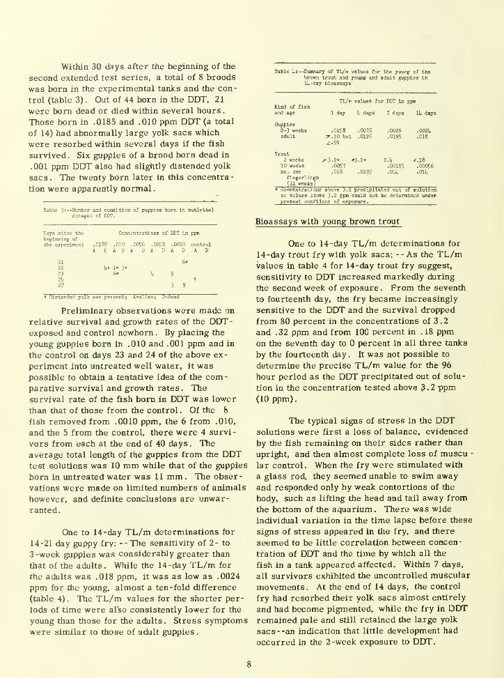
Within 30 days after the beginning of the
second extended test series, a total of 8 broods
was born in the experimental tanks and the con-
trol (table 3). Out of 44 born in the DDT, 21
were bom dead or died within several hours.
Those born in .0185 and .010 ppm DDT (a total
of 14) had abnormally large yolk sacs which
were resorbed within several days if the fish
survived. Six guppies of a brood born dead in
.001 ppm DDT also had slightly distended yolk
sacs. The twenty born later in this concentra-
tion were apparently normal
.
Table 3:—Nranberdosage:

One to 14 -day TL/m determinations for
lO-week trout fry: --After the fry had resorbed
their yolk sacs, they became sensitive to exceed-
ingly small dosages. The 14-day TL/m value
for the 10-week fry was approximately 1/300
as great as the value for the 2 -week fry. The
appearance and duration of stress symptoms
before death appeared later and lasted longer
in the lower concentrations of DDT. A change
in pigmentation was the first indication of
stress. The fry became much lighter except
for the tail tip, which was very dark. Disturb-
ances of nervous and muscular control were
indicated first by quick jerky movements, fol-
lowed by complete loss of balance. Just before
death, the fry were found lying on their backs,
the mouths operated with apparent difficulty, and
there was no response to probing with a glass
rod.
One to 14 -day TL/m determinations for
number one fingerlings: --After the trout had
grown to the fingerling stage, they were moreresistant to DDT than the 10 -week fry which
had recently lost the yolk sacs. None of the
fish in .032 ppm survived more than 2 days,
but of the fish in .010 ppm, 70 percent survived
14 days of exposure and 100 percent of the fish
in the lower concentrations of .0032, .0010, and
.00032 survived the 2 -week test. On the basis
of the data, the 7- and 14-day TL/m was deter-
mined to be .014 ppm DDT (table 4). The 14-
day TL/m for these fish was found to be 25 times
more than for the 10 -week fry.
Histopathological condition of trout fry and adult
guppy tissues following exposure to DDT
In general, histopathological conditions
were similar in the tissues of both the trout and
guppies despite the differences in age, DDTconcentration, and exposure periods. Conse-
quently, the description of tissue changes caused
directly or indirectly by DDT is presented as
that which is typical either of the experimentals
or of the controls.
Brain: --No histopathological condition
was observed in the mid- or hindbrain of the
guppy or trout. A few very small areas of
vacuolation, perhaps indicative of irregularly
swollen myelin sheaths or degenerating and
swelling oUgodendroglia, were present in the
outer portion of the cerebral lobes of one trout
fry.
Intestine: --Marked abnormalities werepresent along the intestinal tract of both fish.
In the guppy both the connective tissue and colum-
nar epithelium were necrotic and highly disorgan-
ized (figs . 3 and 4) . In most villi it was not
possible to distinguish whether the cells of the
epithelial layer had disappeared or dispersed.
The changes in the trout intestine were even moremarked. The connective tissue between the villi
and the smooth muscle was either necrotic or
entirely absent (figs . 5 and 6) . The columnar
cells were irregular as in the gut of the guppy.
There was also severe vacuolation of the
epithelium (fig. 7). This necrotic layer wasoccasionally observed sloughing off into the
lumen
.
Liver: -- Degeneration of varying degrees
was noted in the liver tissue. In the extreme
cases in the guppy Uver, necrosis gave the
entire organ a foamy appearance (figs . 8 and 9)
.
Both the normal and the experimental trout livers
were full of small vacuoles (figs. 10 and 11), per-
haps fat vacuoles resulting from the resorption of
the fatty yolk . Mounts also found fatty vacuolation
in the livers of normal and experimental guppies
in endrin and attributed it to a high content of fat
in the diet. There was some enlargement of the
vacuoles in the livers of the experimental fish,
and swollen nuclei were scattered in the liver of
the DDT-exposed trout.
Kidney: --There was no visible change in
the guppy kidney tubules by the first or second day,
but the tubules of the trout were generally occluded
or congested with debris. There was some slough-
ing of the epithelial cells of the tubules (fig. 12).
4/ Mount, D. Personal communication of October 31, 1960, to Susan King
from Taft Sanitary Engineering Center. (Thesis -1960. A study of the chronic
effects of endrin, an insecticide, on the guppy /Lebistes reticulatus /, and on
the bluntnose minnow /Pimephales notatus/, Ohio State University.

Adrenal tissue: --The adrenal tissue of
the bony fish is characteristically scattered as
epithelial whorls in the kidney tissue Brown,
1957; Andrew, 1959). The solid, epithelial
masses of cells found in the kidney of the trout
and guppies and tentatively identified as adrenal
tissue were markedly affected by exposure to
DDT. Necrosis of this tissue (more severe in
the guppies than in the trout) resulted in a foamyappearance (figs. 14 and 16) in contrast to the
solid structure characteristic of the normalanimals (figs. 13 and 15). In the kidney of the
experimental trout fry, the only region with
adrenal -like epithelial cells was in the middle
portion around the central blood vessel (fig. 16).
The cells, though in a disorganized state, looked
healthy, and their position near the blood vessel
was evidence either of recovery or of incomplete
destruction originally.
Macroscopic observations of the guppy
spleen; --In dissection of adult guppies killed
in DDT, it was noted that the spleens weregenerally much smaller than in the normals, andthe bright red color of stored red blood cells
was present only in spots or absent entirely. Adepletion in the red blood cell content of the
spleen was found by Baxter (1959) in lambs poi-
soned with aldrin and attributed to the withdrawal
of red blood cells from the stock in the spleen to
replace cells lost through congestion in the lungs
and hemorrhagic conditions throughout the body.
DISCUSSION
Evaluation of bioassay data
The bioassay procedure as applied to
long-term experiments of chronic exposure is
not entirely reliable. Few similar tests have
been conducted previously, and a standardized
long range procedure has not been developed.
The concentration of DDT probably does not
remain constant, and it is assumed that the
DDT was gradually removed from the water,
by adsorption onto the glass, concentration in
the fish tissues, and perhaps lost to some extent
in the filtered organic wastes (Garner, 1957).
As the tests were intended to simulate whatmight occur after one spraying of DDT in the
field, it was not desirable to apply a continuous
flow of DDT test solution. The data do not
represent fish under constant exposure to a
specific concentration, but rather fish in water
treated once with a certain concentration of DDT.Doudoroff (1953) also did not renew the test solu-
tions for 10-day bioassays. A comparison of the
laboratory stock solution two months after its
preparation with a freshly made solution on adult
guppies showed no difference in toxicity of the
two solutions
.
There was close correlation of the TL/mvalues determined in the 14-day tolerance tests
on adult guppies with those reported by Henderson
etal., (1959a). The deviation from Henderson's
value of .028 ppm for the 7-day TL/m for guppies
in soft water was -.009 ppm or 13 percent lower
(.0195 ppm). The test with a second and obviously
more resistant strain yielded a value greater than
.032 ppm as the 7-day TL/m. This variation in
values indicates the fallacy of designating any
specific concentration as the TL/m. One mayspecify a certain range of concentrations as having
a known toxicity, however, such as .032 ppm to
.018 ppm for the 7-day TL/m for guppies. Before
designating a safe concentration for specific fishes
in a locale to be treated with an insecticide, it
would be imperative to conduct a preliminary test
on a representative fish sample in the particular
receiving water
.
There seems to be no immediate explana-
tion for the increased resistance to DDT once fish
have been exposed to sublethal doses. As indi-
cated by the histopathological conditions discussed
later, the action of DDT seems to have only a
destructive effect on the tissues and a later in-
crease in dosage would only accelerate tissue
necrosis
.
The 30-day test of chronic exposure to
DDT in dosages sublethal to guppies at 14 days
demonstrated that the TL/m value determined
for 14 days (.018 ppm) applies to even longer
periods of exposure under the same conditions.
The loss of the fish which occurred twice after
a 30 -day period in the original test water might
possibly be avoided by completely replacing the
test solutions with fresh DDT solutions, perhaps
10

as often as every two weeks. Though Mounti'
advised a continual flow of the treated water,
this procedure would not provide data repre-
senting the results of one dosage of a specific'
concentration on the test fish. Although the
cause of the deaths might be a highly contagious
disease, this seems unlikely, particularly since
sterilized equipment was used in the second
experiment. It seems most likely that the deaths
resulted from an increasingly high concentration
of dissolved nitrogenous wastes which were not
removed by filtering.
Though relatively few guppies were born
in the test aquaria, the survival rate was some-
what reduced for tnose fish born in DDT solu-
tions. These DDT-exposed fish were generally
less mature at the time of birth as indicated by
the distended yolk sacs. The correlation of
failure of yolk resorption and hypoactivity of
the adrenal gland will be discussed later. The
sli^t, but perhaps significant, difference in
length between the fry born in DDT water and
those bom in untreated water is attributed to a
failure of the previously DDT-exposed fry to re-
cover completely in a 40 -day period. The only
literature on growth effects is that of Linduska
and Surber (1948) who reported no apparent
inhibition of growth of oysters in DDT treated
beds.
6/Mount— found that guppies generally
failed to have more than one litter while in
endrin. It was not possible in the short period
of my tests to determine if DDT had a similar
effect. Data on quail egg and chick survival
support the assumption that insecticides have
a harmful effect on fish and wildlife reproduc-
tion.
For practical application, one mustcompare the susceptibility of other fishes
relative to that of guppies before deducing con-
centrations safe for other species. The 14-day
bioassay with a local fish, the brown trout fry
and fingerlings, revealed a marked difference
in susceptibility to DDT. Once the young trout
fry were feeding, they became much moresensitive to DDT than were the young guppies
.
While dosages above .0024 ppm were sublethal
to the young guppies, any concentration above
.00056 ppm proved lethal to 50 percent of the
trout fry at 10 weeks of age. It seems probable
that water is not brought into the intestine of
the young fry until they begin to feed, which is
normally at about the fourth or fifth week. Oncethe treated water enters the intestine, the DDTcan enter the body quickly by way of the blood,
thus accounting for the sudden increased
sensitivity. However, after a period of growth
to the first fingerling stage, the trout becamemuch more resistant to DDT than when they
first began feeding. In a short bioassay on
number five fingerlings placed in .10, .0056,
and .0032 ppm that percent, 50 percent, and
50 percent, respectively, survived a 2 -week
exposure. The trout fingerlings thus appeared
to be only slightly more sensitive than the adult
guppies to DDT (.014 ppm for the 14-day TL/mfor trout compared with .018 ppm for guppies),
while the feeding trout fry were exceedingly
sensitive in comparison (.00056 ppm TL/m).
While it was not possible to test the
sensitivity of adult trout under exacting labora-
tory conditions, the trend toward decreased
sensitivity displayed by the flnerlings makes it
seem likely that adult trout, like adult guppies,
are more resistant than the fry to DDT.
It is the ultimate hope that effects of
field applications of insecticides on fish can be
predicted in advance, by the use of standard
laboratory tests. Accumulation of TL/m values
under standardized conditions, such as those
values determined in the present study, is the
first step in realizing this hope. Application
of laboratory findings to field conditions involve
major but not impossible difficulties. Close
approximations of safe insecticide dosages maybe obtained by short bioassays on the fish fromthe water area involved, using the receiving
water for the test solutions to which the DDTconcentrations are added in the same formula-
tion as that to be used in the field. A comparisonof the short bioassay data with results obtained
after longer exposures under standarized con-
ditions, such as the values obtained in the survey
6/
See footnote 4
See footnote 4
11

of Henderson^ al^,,(1959a, b) or in the present
study, would provide a basis for determining a
safe application level. Factors which might af-
fect the toxicity of a field dosage, such as a
possible interaction of DDT with organic and
inorganic materials, any variation in volume of
water per fish, stress situations, rainfall, or
contaminated runoff from insecticide treated
area (Young et^al_., 1951) should be considered
before application of the insecticide
.
The removal of food may actually prove
to be of more imminent danger to fish than the
toxicity of the insecticide, and evaluation of
effects of DDT might better.be based on inverte-
brate food organisms rather than on fish. DDTand other chlorinated hydrocarbons are known
to be toxic to lower organisms in the fish food
chain, though specific TL/m values have not
been determined (Harrington et al., 1958). It
requires only an ounce or two of DDT per acre
to kill crustaceans (Leedy)^. The microfauna,
especially the protozoa, are relatively resistant
to the chlorinated hydrocarbons (DeWitt et al.,
1960) . Even if over 50 percent of the lower
food-chain organisms survived, the dead ones
might be eaten by fish. Though DeWitt and
George (1960) report no harmful effects on fish
from eating insects killed by DDT sprayings,
Hoffman (1959) and Janzen (1960) report fish
affected by insects sprayed with DDT. The
amount of poisoned insects required to kill fish
varied considerably, however.
It is obvious that use of standardized pro-
cedures in the laboratory results in comparable,
reliable, and reproducible data, but cannot
reproduce conditions of a particular natural
situation, which are never constant or identical
with other natural situations
.
Histopathological conditions
Although bioassays with DDT are neces-
sary for estimation of fish survival after field
application, it seems of at least equal importance
to determine the physiological cause of death.
Some research concerning effects of the DDT on
the tissues of exposed animals is now in progress
in several laboratories. Janzen (1960) has re-
ported that pesticides are concentrated in fish,
especially in the fatty tissues. Damage of liver
and kidney tissues, reduction of red blood cell
production, depressed growth rates, and reduced
efficiency of reproduction may result. These
conditions would suggest increased sensitivity to
diseases. Janzen pointed out that under stress,
such as lack of food, the DDT stored in the fat
is likely to be released into the system. The
effects of such a release are not yet known . Cope
(1959) reported storage of chlorinated hydro -
carbons in the kidney, pyloric caecum, and brain,
but none in the liver. In other chemical bioassays
conducted at his laboratory, DDT has been found
in tissues two years after exposure of trout (up
to .94 ppm DDT in the tissues) and whitefish (0.7
ppm DDT and 1.2 ppm DDE)
.
The determination of DDT concentrations
in tissues or even test solutions is difficult, and
no method has yet been established that is both
rapid and accurate. An elaborate paper chro-
matographic analysis for DDT was proposed by
Mitchell (1954) . The analysis is based on sub-
jective color comparisons with a standard and is,
therefore, subject to some error. Cope's results
mentioned above stem from a complex bipchemical
analysis of tissues requiring special equipment
and skilled technique. This procedure is still be-
ing perfected. Amounts of stored insecticide in
fish tissue depend on the compound and species.
Small amounts seem to be of no harm to the fish
.
This is indeed fortunate as every assayed fish,
even from supposedly uncontamlnated waters, at
the Denver Laboratory has had some DDT in the
tissues . Mount?/found high concentrations of
endrin in the liver, intestine, spleen, and kidney
of carp after 2-7 days of exposure . Similar and
more extensive assays have been performed on
quail and pheasant (Anon., 1951; DeWitt, 1955;
DeWitt et al ., L960) and the chlorinated hydro-
carbons have been found to accumulate in the
tissues of birds also, especially in the fat and
muscle.
7/ Leedy, D. L. November 15, 1960 Draft report on (1) wildlife values and
(2) pesticide usage in conservation programs. U.S. Fish and Wildlife
Service, U.S. Department of the Interior
8/ See footnote 4.
12

Techniques to determine interaction of
chlorinated hydrocarbons with a specific enzyme
or metabolic pathway in organisms exposed to
toxicants are being developed by Weiss (1960).
This investigator cites similar work of Hosein
who has found that a shift in a metabolic pathway
was affected, leading to an increased production
of carnitine, which accumulates in the brain and
interferes with nerve function, resulting in con-
vulsive activity.
The histopathological findings for the
fish exposed to DDT show close agreement with
the results of other workers using different test
animals and chlorinated hydrocarbons. Though
brain lesions have been seen in the cerebral
lobes in the dog (Kitselman, 1953) after aldrin
and dieldrin poisoning and sometimes in lamb
and poultry after aldrin poisoning (Baxter, 1959),
there are other cases cited by Baxter, in which
insecticide poisoning has not produced brain
lesions. This is true for sheep exposed to
dieldrin and occasionally for the lamb. The
absence of lesions also in the trout and guppy
forebrains indicates that brain lesions may not
be a significant cause of death in insecticide
poisoning.
No histological studies of the intestine
of animals exposed to insecticides were found
in the literature, though Gowdey and Stravraky
(1955) mentioned that dieldrin and aldrin had
inhibitory effects on intestinal motility. Theseverity of the intestinal lesions found in the
trout and guppies may have been a direct cause
of death in the fish by preventing the normaldigestion and assimilation of food. If the dis-
turbances in the intestine are an indirect cause
of death, through starvation, the insensitivity
of the trout to DDT before the yolk sacs are
resorbed could be explained. However, further
study is needed to determine whether the fish
are resistant because the mouth does not oper-
ate to bring water into the intestine at this
stage in development, thus preventing absorp-
tion of DDT into the blood stream (as suggested
by Mounts', or because a normally functioning
intestine is not needed while the yolk sac is still
present
.
Nelson e^aL., (1949) using DDD, Kitsel-
man (1953) using dieldrin and aldrin on dogs, and
Baxter (1959) studying effects of aldrin on lambs
all found moderate to severe fatty degeneration
in the liver after exposure to the insecticide.
The indications of degeneration in the lamb liver
were necrosis of the hepatic cells, accumulation
of refractile round bodies which looked similar
to vacuoles in hematoxylin -eosin, bleeding, and
general congestion. The liver in the dogs wasfound in the severest cases to be quite foamy in
appearance. A more complete histological study
series of the test fish might reveal recovery
potential of necrotic livers and at what stage in
the necrotic process the swollen cells which
were seen in the trout liver appear. Waud (1952,
as reported by Gowdey^ al_., 1955) found that
the blood sugar of cats fed aldrin doubled and
that this level was even higher when convulsions
set in before death. The lethal dosage of DDTgiven the guppies (.032 ppm) may have had a
similar effect, causing a complete conversion
of the stored glycogen in the liver to glucose
.
Bell (1961), Kitselman (1953), and Baxter
(1959), using different animals and insecticides
reported that chlorinated hydrocarbons caused
degeneration of the renal tubules . The severity
of degeneration varied with the animal and con-
centration and ranged from an increase in tubular
fat and slight hemorrhaging in the surrounding
tissue to eventual occlusion of the tubules with
debris and sloughing of necrotic epithelium into
the lumen. Although there was no noticeable
fatty degeneration in the tubules of the trout or
guppies, marked necrosis of the epithelium had
occurred in many of the trout fry tubules after
two weeks
.
Though both interrenal and chromatin cells
of the adrenal gland are scattered in the bony
fish, both were seen best in the whorls of epithe-
lial cells in the kidney of both the trout and guppy.
Degeneration of this tissue in the fish was indica-
tive that the adrenal tissue was directly affected
by the DDT.
The role of the adrenal gland in the ef-
fects of DDT on an organism is implicated in
9/ See footnote 4.
13

connection with yolk sac retraction and differ-
entiation of the intestine. By various experi-
ments on the chick embryo, Mogg (1953; Mooget al., 1955) has demonstrated that glucocorti-
coids, hormones secreted by the zona fasciculata
of the adrenal gland, accelerate the retraction of
the yolk sac in chicks and the later stages of dif-
ferentiation of the duodenal mucosa . She did not
propose a definite mode of action of the adrenal
cortical hormone, though she suggested an in-
direct effect on phosphatase activity and possibly
other enzymes.
The various kinds of evidence seem to
indicate that DDT has a direct inhibitory and
destructive effect on the adi'enal cortical tissue
and that other effects are secondary. The
destruction of adrenal tissue of the fish exposed
to DDT and the atrophy of the zona fasciculata
found by others in animals exposed to various
chlorinated hydrocarbons might be considered
primary effects leading to the secondary effects
such as those found in the kidney, liver, and
intestine. The hyperexcitability and loss of
muscular control, the failure of trout yolk sac
resorption, the vacuolation in the liver, and the
deterioration of the intestine could be immediate
causes of death under extreme conditions . The
major characteristics for Addison's disease, a
hypoactive condition of the adrenal gland in man,
are similar for animals exposed to the chlorin-
ated hydrocarbons. Pigmentation is affected,
and there are gastro-intestinal disturbances,
weaker muscles, hypoglycemia, and some ef-
fects on reproduction.
SUMMARY AND CONCLUSIONS
Fourteen day bioassays were conducted
on young and adult guppies by exposing tiiem to
serial logarithmic dilutions of DDT under con-
trolled conditions. The values at which 50 per-
cent of the fish survived (TL/m) in 1, 4, 7 and
14 days were determined. On the basis of these
assays, it was determined that young guppies two
to three weeks of age are approximately 10 times
more sensitive to DDT than the adults. The 14-
day TL/m for the young was .0024 ppm and,
though the TL/m varied slightly with different
strains of fish, .0J8 ppm was established as
the sublethal dosage for the adult fish
.
Similar 14-day bioassays were conducted
with young brown trout . The fry, while still
depending on the yolk sac for food (two weeks old)
were found to be exceedingly resistant to DDT.Over 50 percent were able to withstand . 18 ppmDDT in a 14-day period. Once the yolk sac wasresorbed, the fish became very sensitive to the
insecticide. The 14- day TL/m for 10-week-old
fry was .00056 ppm and .014 ppm for the numberone fingerlings
.
The 14-day TL/m for adult guppies(.018
ppm DDT) was sublethal to adult guppies over an
extended exposure of 30 days. It was postulated
that exposed fish would not be harmed seriously
in DDT solutions of a concentration below the 14-
day TL/m
.
Limited observations on growth and repro-
duction indicated that exposure to DDT did not
prevent reproduction in guppies in the first 30
days of chronic exposure, but many of the guppies
born in DDT were dead or died within several
hours. The survival rate was lower for those
born in DDT than for those born in untreated
water. The data, however, were not sufficient
to make definite conclusions. Observations in
the literature indicate that after the first litter,
insecticides will inhibit reproduction. The cause
of the death of guppies in aquaria kept under con-
trolled conditions needs to be ascertained before
the effects of one dosage of DDT on reproduction
can be determined.
Guppies born in DDT, especially in con-
centrations of .010 and .0185, had abnormally
large yolk sacs . The sacs were resorbed in
several days if the fry survived. Guppies born
in DDT and removed to untreated water were
slightly shorter after 40 days than guppies bomin the control and treated similarly (averages
of 10 mm and 11 mm respectively), though the
data are inconclusive.
Exposure of the guppies to sublethal
dosages of DDT for 14 days and then removal
to a concentration toxic to normal fish within
three days -- .032 ppm, demonstrated that the
fish had increased resistance to the toxicant
and the dosage in most cases was no longer
lethal. It was found that the greatest resistance
14

developed in the fish first exposed to .010 and
.0056 ppm DDT.
The physical signs of stress exhibited by
fish in DDT, include hyperexcitability and loss
of muscular control, the presence of yolk sacs
in the guppies bom in DDT, the failure of the
sac resorption in trout fry exposed to DDT, and
various histopathological conditions found in the
tissues. This syndrome suggests that DDT has
a direct effect on adrenal tissue, especially that
portion secreting the glucocorticoids . The mostmarked histopathological conditions in the fish
attributed to the DDT were found in the liver,
the intestine, and the adrenal -like tissue in the
kidney. Modification of the kidney tubules and
the gross appearance of the spleen were occa -
sionally observed.
There is a very serious need for exten-
sive study of the chlorinated hydrocarbons and
their effects on fish survival, reproductive
potential, and physiological condition as ex-
hibited by the tissues. There are many factors
in the field which modify bioassay determina -
tions. It is likely that the safe dosages of
insecticides for fish will be less than that neces-
sary for insect control . If such is found to be
the case, recourse to other means of control of
the insects will be necessary to prevent exten-
sive loss of fish.
LITERATURE CITED
Adams, L., M. G. Haravan, N. W. Hosley,
and D. W. Johnston.
1949. The effects on fish, birds, andman of DDT used in control of forest
insects in Idaho and Wyoming. Journal
of Wildlife Management, vol. 13, p.
245-254.
Andrew, W.1959. Textbook of Comparative
Histology. Oxford University Press,
New York, chap. 13, p. 527-530.
Anonymous.1951. Insecticide storage In adipose
tissue. Journal of the AmericanMedical Association, vol. 145, p.
735-736.
Anonymous.1960. A. I. E. C. holds pesticide
symposium . Agricultural Chemicals,
vol. 15, p. 8.
Baxter, J. T.
1959. Some observations on the histo-
pathology of aldrin poisoning in lambs.
Journal of Comparative Pathology and
Therapeutics, vol. 69, p. 185-191.
Bell, J. T., Jr.
1961 . Histological and histochemical
effects of DDD on goat adrenals,
pituitaries, livers, and kidney.
American Association of Anatomists,
Anatomical Record, vol. 139, p. 207.
Brown, M. E.
1957. The Physiology of Fishes.
Academy Press, Inc., New York, vol. 1,
chap. 6, p. 252 -254.
Breder, C. M., Jr., andC. W. Coates.
1932 . A preliminary study of population
stability and sex ratio of Lebistes .
Copeia, p. 147 - 155.
Clawson, S. G.
1958 . Wild turkey populations on an
area treated with heptachlor anddieldrin. Alabama Birdlife, vol.6,
p. 4-8. (Cited by DeWitt^al_., 1960.)
Cope, O. B.
1959 . The retention of DDT by trout and
whitefish. Biological Problems in WaterPollution, U.S. Department of Health,
Education and Welfare.
Cope, O. B., C. M. GjuUin, and A. Storm.1947. Effects of some insecticides on
trout and salmon in Alaska with refer-
ence to blackfly control. Transactions
American Fisheries Society, vol. 77,
p. 160- 177.
Cottam, C. and E. Higgins.
1946. DDT: Its effects on fish and wild-
life. U. S. Department of the
Interior, Fish and Wildlife Service,
Circular 11, 14 p.
15

Davidow, B. and F.J. Sabatlno.
1954. Biological survey test for
chlorinated insecticides. Journal of
Association of Official Agricultural
Chemists, vol. 37, p. 902 - 905.
DeWitt, J. B.
1955. Effects of chlorinated hydrocarbon
insecticides upon quail and pheasants
.
Journal of Agricultural and FoodChemistry, vol. 3, p. 672-676.
1956. Chronic toxicity to quail and
pheasants of some chlorinated
insecticides. Journal of Agricultural
and Food Chemistry, vol. 4, p. 863 -
866.
1957. H -bomb in the pea patch . Wild-
life in North Carolina, vol. 21, p. 1-4.
DeWitt, J. B. and J. L. George1960. Bureau of Sport Fisheries and
Wildlife- -Pesticide Wildlife Review:
1959. U. S. Department of the
Interior, Fish and Wildlife Service,
Circular 84, 36 p.
Doudoroff, P., B. G. Anderson, G. E. Burdick,
P. S. Galtsoff, W. G. Hart, R. Patrick,
E. K. Strong, E. W. Surber, and
W. M. Van Horn.
1951. Bioassay methods for the evalua-
tion of acute toxicity of industrial
wastes to fish. Sewage and Industrial
Wastes, vol. 23, p. 1380-1397.
Doudoroff, P., M. Katz, andC. M. Tarzwell.
1953. Toxicity of some organic insecti-
cides to fish. Sewage and Industrial
Wastes, vol. 25, p. 840-844.
Fischer, C. D.
1956. Pesticides: past, present, andprospect. Chemical Week, October 27,
p. 59-90; November 3, p. 52-88.
Foster, A. C.
1951. Some plant responses to certain
insecticides in the soil. U.S. Depart-
ment of Agriculture, Circular 862, p.
1-41. (Cited by Surber, 1951).
Garner, R.J.1957. Veterinary Toxicology. Bailliere,
Tindall and Cox, London, chap. 4,
p. 205-231.
George, J. L.
1959. Effects on fish and wildlife of
chemical treatments of large areas
.
Journal of Forestry, vol. 57, p. 250-
254.
George, J. L., R. F. Darsie, Jr., and
P. F. Springer.
1957. Effects on wildlife of aerial
applications of strobane, DDT, and
BHC to tidal marshes in Delaware.
Journal of Wildlife Management, vol.
21, p. 42-53.
Gibson, M. B. andB. Hurst.
1955. The effect of salinity and temper-ature on the preadult growth of guppies.
Copeia, p. 241-243.
Ginsberg, J. M. and D. M. Jobbins.
1954. Research activities on chemical
control of mosquitos in New Jersey in
1953. Proceedings of the New Jersey
Mosquito Extermination Association,
vol. 42, p. 270-291.
ElUs, M. M.1937. Detection and measurement of
stream pollution. U.S. Bureau of
Fisheries Bulletin 48, p. 365 - 437.
Farris, E. J.
1950. The Care and Breeding of Labora-
tory Animals . John Wiley and Sons,
Inc., New York, chap. 4, p. 345-427.
Gordon, M.1955. Guppies as pets. Aquarium,
New York Zoology Society, 32 p.
Gowdey, D. W. and G. W. Stravraky.
1955. A study of the autonomic mani-
festations seen in acute aldrin and
dieldrin poisoning. Canadian Journal
of Biochemistry and Physiology, vol.
33, p. 272 -282.
16
Harrington, R. W., Jr. and W. L. Eidlingmayer
.
1958. Effects of dieldrin on fishes and
invertebrates of a salt marsh. Journal
of Wildlife Management, vol .22, p
.
76 - 82
.
Hart, W. B., P. Doudoroff, and J. Greenbank.
1945. The Evaluation of the Toxicity of
Industrial Wastes, Chemicals, and
other Substances to Fresh-water
Fishes. The Atlantic Refining Company,
Philadelphia
.
Henderson, C, Q. H. Pickering, and
CM. Tarzwell.
1959a. Relative toxicity of ten chlorin-
ated hydrocarbon insecticides to four
species of fish. Transactions AmericanFisheries Society, vol. 88, p. 23-32.
Janzen, D. H.
1960 . Problems with fish and pesticides
in the United States. U.S. Depart-
ment of the Interior, Fish and Wildlife
Service. (International Union for
the Protection of Nature, M'arsaw,
Poland. June.)
Keenleyside, M. H. A.
1959. Insecticides and wildlife. Canadian
Audubon, vol. 21, p. 1-7.
Kerswill, C. J. and P. F. Elson.
1955. Preliminary observations on effects
of 1954 DDT spraying on Mlramichi
salmon stocks . Progress Report of the
Atlantic Coast Station of the Fishery
Reserve Board of Canada, vol. 62, p.
17-23.
1959b. The toxicity of organic phosphorus Kitselman, C. H.
and chlorinated hydrocarbon insecti -
cides to fish. Transactions 2nd
Seminar on Biological Problems in
Water Pollution, U.S. Department of
Health, Education, and Welfare,
April, 13 p.
1953 . Long term studies on dogs fed aldrin
and dieldrin in sublethal dosages, with
reference to the histopathologjcal find'-
ings and reproduction. Journal of
American Veterinary Medical Associa-
tion, vol. 123, p. 28-30.
Henderson, C . and CM. Tarzwell
.
1957. Bioassays for control of industrial
effluents . Sewage and Industrial
Wastes, vol. 29, p. 1002-1017.
Hoffman, C H.
1959 . Are the insecticides required for
insect control hazardous to aquatic life?
Biological Problems in Water Pollution,
U.S. Department of Health, Education,
and Welfare.
Ingram, W . M . and CM. Tarzwell
.
1954. Selected bibliography of publica-
tions relating to undesirable effects
upon aquatic life by algicides, insecti-
cides, and weekicides . U. S. Depart-
ment of Health, Education, and Welfare.
Public Health Service, Publication
No. 400, 28 p.
Lawrence, J. M.1950. Toxicity of some new insecticides
to several species of pondfish. The
Progressive Fish-Culturist, vol. 13,
p. 141 -146.
Leffler, R. L.
1958. Letter of May 1, 1957, to the
Honorable H . C . Bonner, Chairman on
Merchant Marine and Fisheries. Houseof Representatives, 85th Congress, 2nd
Session, Report No. 2181.
Unduska, J. P. and E. W. Surber.
1948. Effects of DDT and other insecti-
cides on fish and wildlife- -Summary of
investigations during 1947. U. S.
Department of the Interior, Fish and
Wildlife Service. Circular 15, 19 p.
Long, C N. H., B. Katzin. and E. Fry.
1940. The adrenal cortex and carbo -
hydrate metabolism. Endocrinology,
vol. 26, p. 309-344.
17

Mitchell, C.
1954. Separation and identification of
chlorinated organic pesticides by
paper chromatography VI. Tech.
BHG, lindane, DDT, rhothane. Journal
of Association of Official Agricultural
Chemists, vol. 37, p. 996-1001.
Moog, F
.
1953. The functional differentiation of
the small intestine. III. The influence
of the pituitary adrenal system on the
differentiation of phosphatase in the
duodenum of the suckling mouse.
Journal of Experimental Zoology, vol.
124, p. 329-346.
Moog, F. and D. Richardson.
1955. The functional differentiation of
the small intestine. IV. The influence
of adrenocortical hormones on dif-
ferentiation and phosphatase synthesis
in the duodenum of the chick embryo
.
Journal of Experimental Zoology, vol
.
130. p. 29-56.
Nelson, A. L., and E. W. Surber.
1947. DDT investigations by the Fish
and Wildlife Service in 1946. U.S.Department of the Interior, Fish
and Wildlife Service, Special Scientific
Report --Fisheries No. 41, 8 p.
Nelson, A. A. and G. Woodard.
1949. A. M. A. Archives of Pathology,
vol. 48, p. 387-294.
Rudd, R. L. and R. E. Genelly.
1956a. Chronic toxicity of DDT, toxa-
phene, and dieldrin to ring-necked
pheasants. California Department of
Fish and Game, Game Bulletin, vol.
42, p. 5- 14.
1956b. Effects of DDT, toxaphene and
dieldrin on pheasant reproduction.
Auk, vol. 73, p. 529-539.
Shepard, H. H.
1956. The pesticide situation for 1955 -
56. Commodity Stabilization Service,
U. S. Department of Agriculmre, 17 p.
Shepard, H. H., J. N. Mahan, and
C. A. Graham.1959. The pesticide situation for 1958-
59. Commodity Stabilization Service,
U. S. Department of Agriculture, 24 p.
Silliman, R. P.
1948. Factors affecting population
levels in Lebistes reticulatus. Copeia,
40P- 47.
Silliman, R. P., J. S. Gutsell, C. E. Dunbar,
and S. B. Friddle.
1958. Experimental exploitations of fish
populations. U. S. Department of the
Interior. Fish and Wildlife Service
Fishery Bulletin 133, vol. 58, p. 215-252.
State Water Pollution Control Board.
1952 . Propagation of fish and aquatic
life. Water Quality Criteria, p. 156-
158.
Surber, E. W.1947. Annual report for the fiscal year
1947. U.S. Fish and Wildlife Service,
Branch of Fishery Biology, 114 p.
(mimeo.)
1951 . Toxicities of some chemical
substances to fish. Pollution Abate-
ment Conference , Manufacturing
Chem. Association, Inc., vol o, p.
35-45.
Tarzwell, CM.1950. Effects of DDT mosquito
larviciding on wildlife. V. Effects
on fishes of the routine manual and
airplane application of DDT and other
mosquito larvicides . U. S. Depart-
ment of Health, Education, and
Welfare, Public Health Service, vol
.
65, p. 231 -255.
1957a . The use of bioassays in relation
to the disposal of toxic wastes. Pro-
ceedings 3rd Ontario Industrial Waste
Conference for 1956 - 1957.
18

Tarzwell, CM.1957b. Water quality criteria for
aquatic life. Transactions Seminar
on Biological Problems in Water
Pollution, U. S. Department of Health,
Education, and Welfare, April, 1956,
1957, p. 246-272.
1958. The toxicity of some organic
insecticides to fishes . Proceedings
of the 12th Annual Conference of the
Southeastern Association of Game and
Fish Commissioners, p. 233-239.
U.S. Public Health Service
.
1956, 1957. Bioassay Investigations for
International Joint Commission. U.S.Department of Health, Education, and
Welfare
.
Warren, C. E. and P. Doudoroff.
1958. The development of methods for
using bioassays in the control of pulp
mill waste disposal. Technical
Association of the Pulp and Paper
Industry, vol. 41, no. 8, p. 211A-216A.
Tarzwell, C. M. andC. Henderson.
1957. Toxicity of dieldrin to fish
.
Transactions American Fisheries
Society, vol. 86, p. 245-257.
Waud, R. A.
1952 . Journal of Pharmacology and
Experimental Therapeutics, vol. 106,
p. 423. (Cited by Gowdey et al. , 1955).
Turner, C. L.
1937. Reproductive cycles and superfeta-
tion in poeciliid fishes. Biological
Bulletin, vol. 72, p. 145-164.
U.S. Fish and Wildlife Service.
1960. Care of tropical aquarium fishes.
U.S. Department of the Interior,
Fish and Wildlife Service, Fishery
Leaflet 411,
Weiss, CM.1960. Personal communication from
Acting Head of Department of Sanitary
Engineering, University of North
Carolina, October 6
.
Young, L. A. and H. P. Nicholson.
1951. Stream pollution resulting from
the use of organic insecticides. TheProgressive Fish-Culturist , vol. 13,
p. 193- 198.
19

<•»>•>
Fij^ire 3 Figiire k
• I* » ^«-,» V -
»>. .*>} ' ;,
FigTire 7
Explanation of Figui-es
Photomicrographs of the intestine of li-week trout fry (yolk sac
still present) and adult guppies; fixed in Bouin's fluid, embed-ded in paraffin, sectioned at 8 ji and stained in Hematoxylin-Eosin. X li35
Figure 3—Intestine of normal guppy showing well-defined columnarepithelium and connective tissue.
Figure h—Intestine of guppy in .032 ppm DDT for two days showingdisorganization and necrosis of columnar epithelium andconnective tissue. Nuclei of the epithelial layer ap-ppear indistinguishable from nuclei of the connectivetissue.
Figure S—Intestine of normal trout fry showing organized columnarepithelium, a few goblet cells, and normal connectivetissue.
Figure 6—Intestine of trout fry in 10 ppm DDT for lii days showingabsence of connective tissue below columnar epitheliumin parts. Necrosis of remaining connective tissue invillus is marked.
Figure 7—Intestine of trout fry in 3.2 ppm DDT for Ih days showingsevere vacuolation and absence of organization of colum-nar epithelium. Connective tissue did not appear to beaffected in this trout.
20

t:M ' -i
Fij'tire 8 Figure 9
•« i'^
-^^py^
Fisiire 10 Figure 11
Explanation of FigTores
Photomicrographs of the liver of U-week trout fry (yolk sac still
present) and adult guppies; fixed in Bouin's fluid, embedded in
paraffin, sectioned at 8 >i and stained in Hematoxylin-Eosin.
Figure 8—Liver of normal guppy in which liver cells are solidly
packed . X lU?
Figure 9—Liver of guppy in .032 ppm DDT for one day showing foamy
appearance due to severe vacuolation and necrosis of en-
tire liver. X li;?
Figure 10—Liver of normal trout fry showing even distribution of
small vacuoles, perhaps fat vacuoles resulting from re-
sorption of the fatty yolk. Nuclei all appear to be
about the same size. X 14.35
Figure 11—Liver of trout fry in 3-2 ppm DDT for one day shewing
vacuoles as in the normal though some are possibly
larger. Large irregularly swollen nuclei are scattered
throughout the liver. X k3^
21

Figuro 12
Figure 13 FiLTire lU
Fi,":ure 1$ ?i"iu'e 16
Explanation of Figures
Photomicrographs of the kidney tissue including tubules and adre-
nal tissue of U-week trout fry (yolk sac still present) and adult
guppies; fixed in Bouin's fluid, embedded in paraffin, sectioned
at 8 )i, and stained in Hematoxylin-Eosin. X U25
Figure 12—Kidney of trout fry in 3.2 ppm DDT for lU days showing
congestion of tubules and necrosis of tubular epithe-
lium which is sloughing off into the lumen.
Figure 13—Kidney of normal guppy showing solid whorl of epithelial
cells. This adrenal tissue was found scattered through-
out the kidney.
Figure lU—Kidney tissue of guppy in .032 ppm DDT for two days show-
ing foamy appearance of whorls of epithelial cells whichare in a state of necrosis and vacuolation.
Figure 15—Kidney tissue of normal trout fry showing solidly packed
whorl of epithelial cells of adrenal tissue adjacent to
a kidney tubule.
Figure 16--Kidney tissue of trout fry in 10 ppm DDT for lU days
showing apparently healthy or normal epithelial cells,
identified as adrenal tissue, around the central blood
vessel. The disorganized appearance of these cells and
their position near the blood vessel indicate either
recovery of adrenal tissue or incomplete destruction
originally.
22 6 «57

A

H \

MBL WHOI Library - Serials
5 Wh SE 01526


