Efficacy and safety of convex probe EBUS-TBNA in sarcoidosis: A systematic review and meta-analysis Ritesh Agarwal, Arjun Srinivasan, Ashutosh N. Aggarwal, Dheeraj Gupta* Department of Pulmonary Medicine, Postgraduate Institute of Medical Education and Research, Chandigarh, India Received 13 January 2012; accepted 22 February 2012 Available online 13 March 2012 KEYWORDS Sarcoidosis; Endobronchial ultrasound; EBUS; Transbronchial needle aspiration; TBNA; Meta-analysis Summary Background and aim: Real-time endobronchial ultrasound-guided transbronchial needle aspi- ration (EBUS-TBNA) is a minimally invasive technique for diagnosis of mediastinal lymphade- nopathy. Although most studies have reported the utility of EBUS-TBNA in malignancy, its use has been extended to benign conditions including sarcoidosis. Herein, we perform a systematic review and meta-analysis of studies reporting the diagnostic yield and safety of EBUS-TBNA in sarcoidosis. Methods: We searched the PubMed and EmBase databases for relevant studies published from 2004 to 2011, and included studies that have reported the diagnostic yield of EBUS-TBNA in sarcoidosis. The quality of studies was assessed using the QualSyst tool. We calculated the proportions with 95% confidence interval (CI) to assess the diagnostic yield of EBUS-TBNA in individual studies and then pooled the results using a random effects model. Heterogeneity was assessed using the I 2 and Cochran-Q tests while publication bias was assessed using both graphical and statistical methods. Results: Our search yielded 15 studies (553 patients of sarcoidosis). The diagnostic yield of EBUS-TBNA ranged from 54 to 93% with the pooled diagnostic accuracy being 79% (95% CI, 71e86%) by the random effects model. The yield was not statistically different in studies employing on-site cytological evaluation (80.1%) vs. those without (81.3%). However, the diagnostic yield was significantly higher in prospective studies (83.9%) vs. the retrospective studies (74.3%). Only five minor complications were reported in 553 patients. There was evidence of heterogeneity and publication bias. Conclusions: EBUS-TBNA is a safe and efficacious procedure in the diagnosis of sarcoidosis, and should be routinely employed wherever available. ª 2012 Elsevier Ltd. All rights reserved. * Corresponding author. Department of Pulmonary Medicine, Postgraduate Institute of Medical Education and Research, Sector-12, Chandigarh 160012, India. Tel.: þ91 172 2756823; fax: þ91 172 2748215. E-mail address: [email protected](D. Gupta). 0954-6111/$ - see front matter ª 2012 Elsevier Ltd. All rights reserved. doi:10.1016/j.rmed.2012.02.014 Available online at www.sciencedirect.com journal homepage: www.elsevier.com/locate/rmed Respiratory Medicine (2012) 106, 883e892
Transcript
Respiratory Medicine (2012) 106, 883e892
Available online at www.sciencedirect.com
journal homepage: www.elsevier .com/locate/rmed
Efficacy and safety of convex probe EBUS-TBNA insarcoidosis: A systematic review and meta-analysis
Ritesh Agarwal, Arjun Srinivasan, Ashutosh N. Aggarwal, Dheeraj Gupta*
Department of Pulmonary Medicine, Postgraduate Institute of Medical Education and Research, Chandigarh, India
Received 13 January 2012; accepted 22 February 2012Available online 13 March 2012
* Corresponding author. DepartmenChandigarh 160012, India. Tel.: þ91 1
E-mail address: dheeraj1910@gma
0954-6111/$ - see front matter ª 201doi:10.1016/j.rmed.2012.02.014
Summary
Background and aim: Real-time endobronchial ultrasound-guided transbronchial needle aspi-ration (EBUS-TBNA) is a minimally invasive technique for diagnosis of mediastinal lymphade-nopathy. Although most studies have reported the utility of EBUS-TBNA in malignancy, itsuse has been extended to benign conditions including sarcoidosis. Herein, we performa systematic review and meta-analysis of studies reporting the diagnostic yield and safety ofEBUS-TBNA in sarcoidosis.Methods: We searched the PubMed and EmBase databases for relevant studies published from2004 to 2011, and included studies that have reported the diagnostic yield of EBUS-TBNA insarcoidosis. The quality of studies was assessed using the QualSyst tool. We calculated theproportions with 95% confidence interval (CI) to assess the diagnostic yield of EBUS-TBNA inindividual studies and then pooled the results using a random effects model. Heterogeneitywas assessed using the I2 and Cochran-Q tests while publication bias was assessed using bothgraphical and statistical methods.Results: Our search yielded 15 studies (553 patients of sarcoidosis). The diagnostic yieldof EBUS-TBNA ranged from 54 to 93% with the pooled diagnostic accuracy being 79% (95% CI,71e86%) by the random effects model. The yield was not statistically different in studiesemploying on-site cytological evaluation (80.1%) vs. those without (81.3%). However, thediagnostic yield was significantly higher in prospective studies (83.9%) vs. the retrospectivestudies (74.3%). Only five minor complications were reported in 553 patients. There wasevidence of heterogeneity and publication bias.Conclusions: EBUS-TBNA is a safe and efficacious procedure in the diagnosis of sarcoidosis, andshould be routinely employed wherever available.ª 2012 Elsevier Ltd. All rights reserved.
t of Pulmonary Medicine, Postgraduate Institute of Medical Education and Research, Sector-12,72 2756823; fax: þ91 172 2748215.il.com (D. Gupta).
Sarcoidosis is a multisystem granulomatous disorder ofunknown etiology that commonly presents with bilateralhilar adenopathy, pulmonary infiltrates, ocular and skinlesions. The diagnosis is established in presence ofcompatible clinicoradiographic findings and histologicevidence of noncaseating epithelioid cell granulomas afterexclusion of other known causes for granulomatousinflammation.1 As the lung and mediastinal lymph nodes aremost often affected in sarcoidosis, bronchoscopic tech-niques are often employed for demonstration of non-caseating granulomas. Bronchoscopic lung biopsy (BLB),endobronchial biopsy (EBB) and transbronchial needleaspiration (TBNA) are currently the most commonly usedmethods for demonstration of granuloma in sarcoidosis.Endobronchial ultrasound-guided TBNA (EBUS-TBNA) isa minimally invasive technique for sampling the hilar/mediastinal lymph nodes, and can improve the diagnosticyield by direct visualization of lymph node beyond thetracheobronchial wall thereby allowing real-time samplingof the lymph nodes.2
Krasnik et al. first reported the utility of convex probeEBUS-TBNA in sampling mediastinal nodes in 2003.3 Withtime, the diagnostic yield of EBUS-TBNA has been furtherenhanced by rapid on-site cytological evaluation (ROSE),increasing the number of lymph nodes sampled, increase inthe number of aspirates taken per node, and use of a largerbore 21G needle. Although EBUS-TBNA was primarilyintended for minimally invasive staging of bronchogeniccarcinoma, its use has been extended in diagnosis oflymphoma and benign conditions like tuberculosis andsarcoidosis.4e6 Several systematic reviews and meta-analyses have reported the diagnostic performance ofEBUS-TBNA but most of these reviews have primarilyfocused on patients with malignancy.7e11
Sarcoidosis is a common pulmonary disorder worldwide.Demonstration of noncaseating granulomas and exclusion ofother causes of granulomatous inflammation is essentialparticularly in countries with high prevalence of tubercu-losis. We had previously reported the diagnostic yield ofBLB and the additive yield of EBB in patients with sarcoid-osis.12 We have also recently reported the diagnostic yieldof TBNA in patients with mediastinal lymphadenopathy ofdiverse etiologies including sarcoidosis.13 In this study, weperform a systematic review and meta-analysis to definethe diagnostic efficacy and safety of convex probe EBUS-TBNA in patients with sarcoidosis.
Material and methods
Search strategy
We first searched the literature for available systematicreview that had reported the diagnostic efficacy of EBUS-TBNA in sarcoidosis. No systematic reviews were found. Allthe authors independently searched two computer data-bases PubMed and EmBase for relevant studies publishedfrom 2004 to 2011 describing the diagnostic value of EBUS-TBNA in patients with sarcoidosis using the following searchterms: (“ebus”OR“ebus tbna”OR“tbna”OR“endobronchial
ultrasound” OR “endobronchial ultrasonography” OR“endobronchial ultrasound-guided” OR “endoscopic ultra-sound” OR “transbronchial needle aspiration”) ANDsarcoidosis; and, (“ebus” OR “endobronchial ultrasound” OR“endobronchial ultrasonography” OR “endobronchial ultra-sound-guided” OR “endoscopic ultrasound”) AND (“tbna” OR“transbronchial needle aspiration”). We reviewed thereference lists of primary studies, reviews, and editorials. Inaddition, we reviewed our personal files. We excluded thefollowing studies: (a) abstracts, editorials, reviews and casereports; (b) studies describing diagnostic accuracyof TBNAorendoscopic ultrasound-guided fine needle aspiration (EUS-FNA) or radial probe EBUS-TBNA in sarcoidosis; (c) studiesdescribing EBUS-TBNA in �10 patients with sarcoidosis; (d)studies in which the denominator number i.e. number ofpatients with final diagnosis of sarcoidosis (granulomas onEBUS-TBNA or demonstration of granulomas from any site byany methodology AND a clinical picture deemed by theinvestigator to be compatible with sarcoidosis) was notreported. The criteria for conclusive diagnosis by EBUS-TBNAin sarcoidosis was lymph node aspirates showing epithelioid,noncaseating granulomas without necrosis OR epithelioidand giant cells AND absence of identifiable malignancy,lymphoma, or infection (i.e. tuberculosis or fungal disease).
Initial review of studies
The initial database created from the electronic searcheswas compiled and all duplicate citations were eliminated.Two reviewers (RA and AS) screened these citations,without blinding, by title and abstract review to capturethe relevant studies. Any disagreement was resolved bydiscussion between the authors. This database was thenscreened again to include only primary articles, and the fulltext of each citation was obtained and reviewed. Studieswere eligible for inclusion if they reported the diagnosticyield of convex probe EBUS-TBNA in patients with clinicalsuspicion of sarcoidosis.
Data abstraction
Data was recorded on a standard data extraction form. Thefollowing items were extracted: (a) publication details(title; authors; and other citation details) including thegeographic location of the study; (b) type of study(prospective or retrospective); (c) stage of sarcoidosis andlymph node size on CT chest; (d) type of sedation used,diameter of EBUS-TBNA needle, stations sampled, size oflymph node on EBUS, number of lymph node aspirated and/or passes made through EBUS, availability of on-sitecytology; (e) diagnostic yield of EBUS-TBNA in sarcoidosiswherein the numerator was the diagnosis of sarcoidosiswith EBUS-TBNA, and the denominator was number ofpatients with confirmed sarcoidosis; and, (f) complicationsassociated with the procedure.
Assessment of study quality
The quality and validity of each article included in thismeta-analysis was assessed using the QualSyst tool forqualitative studies.14 This tool consists of 10 questions with
Systematic review & meta-analysis 885
score from 0 to 2 with the maximum total score being 20.Each study was independently evaluated by two authors(RA, AS) for the stated criteria. Weighted Cohen’s kappa (k)co-efficient was used to determine the inter-observeragreement for selection of studies.
Statistical analysis
The statistical software package (StatsDirect, version 2.7.8for MS Windows; StatsDirect Ltd; Cheshire, UK [http://www.statsdirect.com]) was used to perform all the statis-tical analysis.
Determination of the pooled effect
We calculated the diagnostic yield of EBUS-TBNA by calcu-lating proportion with 95% confidence intervals (CI) for eachstudy and then pooled the data to derive a pooled proportionwith 95%CI. For the purpose of proportionmeta-analysis, theproportions were first turned into a quantity (the Freeman-Tukey variant of the arcsine square root transformedproportion) suitable for the random effects summary.15,16
The pooled proportion was calculated as the back-transform of the weighted mean of the transformedproportions, using DerSimonian weights for the randomeffects model17 in the presence of significant heterogeneity.
Assessment of heterogeneity
The impact of heterogeneity on the pooled estimates of theoutcome was assessed using the Cochran Q statistic and I2
test (measures the extent of inconsistency among theresults of the studies). An I2 value �50% indicates signifi-cant heterogeneity.18 As the Cochran Q test has a lowsensitivity for detecting heterogeneity, a p value <0.1 wasconsidered to be significant for the presence of statisticalheterogeneity.19
Sensitivity/subgroup analysis
We planned sensitivity analysis a priori by using subgroupanalysis of prospective vs. retrospective studies due to thelimitations associated with retrospective studies, and theoccurrence of errors associated with the retrieval ofinformation retrospectively from databases. A subgroupanalysis was also planned by partitioning the studies basedon the utilization of ROSE for histological diagnosis.
Assessment of publication bias
The presence of publication bias was evaluated using theBegg’s funnel plot,20 which is a measure of the proportion(in the X-axis) against the standard error of the proportion(in the Y-axis). Each open circle represents an individualstudy in the meta-analysis. The line in the center indicatesthe pooled proportion and the other two lines indicate the95% CI. The proportion estimates from smaller studies areexpected to be scattered above and below the summaryestimate, producing a triangular or funnel shape, if there isno publication bias.
Publication bias was also investigated using threestatistical tests: (a.) Egger test: detects asymmetry of thefunnel plot21; (b.) Harbord’s test: similar to Egger’s test butuses a modified linear regression method22; and, (c.) Beggand Mazumdar’s test: tests the interdependence of vari-ance and effect size using a rank correlation method.23
An Institutional review board clearance was not requiredfor this study as this was a meta-analysis of publishedstudies.
Results
Our initial database search retrieved a total of 504 citations(Fig. 1) of which 15 studies finally met our inclusioncriteria.24e38 Of these, nine studies were pro-spective24e26,29,31e34,37 and six were retrospective (Table 1).These 15 studies were published from across the globe andincluded 553 confirmed patients of sarcoidosis. Eight studiesincluded stage I and II patients of sarcoidosis,25,26,28e31,37,38
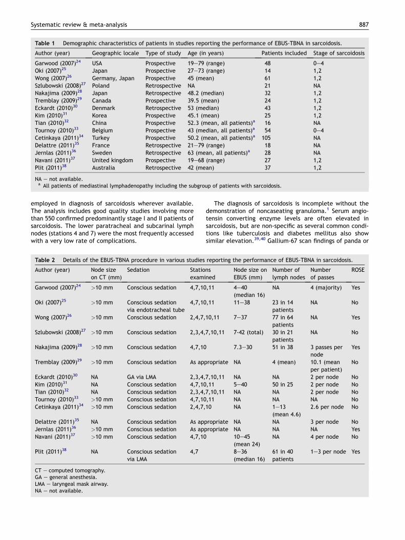
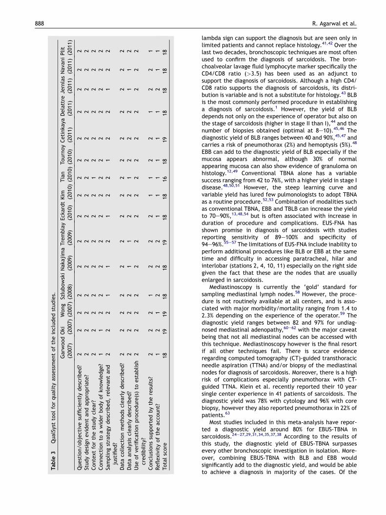
two studies24,33 included all stages of sarcoidosis while thestage was not reported in five studies (Table 1). The proce-dure was performed under conscious sedation without anyartificial airway in 12 studies (Table 2). Two studies usedconscious sedation with either endotracheal tube or laryn-geal mask25,38 while one study used general anesthesia withlaryngeal mask.30 Majority of the studies had sampled theparatracheal, subcarinal, hilar and interlobar nodes, and allthe studies had used the 22G dedicated EBUS-TBNA needle.The lymph node size on CT and EBUS, the number of lymphnodes aspirated and the number of aspirates per patient isshown in Table 2. Five studies employed additional rapid on-site cytology,24,26,28,36,38 and two studies employed liquid-based cytological technique for diagnosis.30,35 The qualityof studies was generally good (Table 3) with themedian (IQR)score being 18 (18e19). The inter-observer agreement forscoring the quality of studies was good. (Cohen’s kZ 0.78).
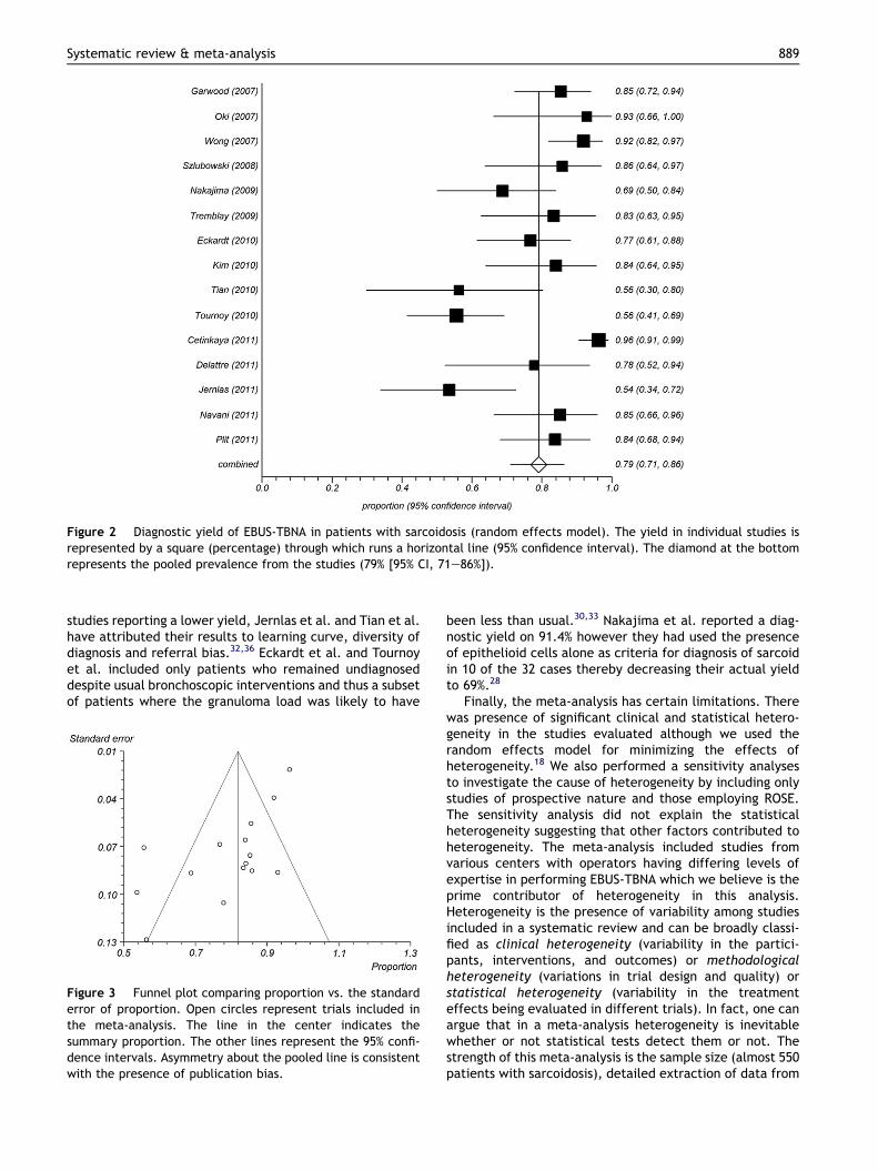
The diagnostic yield of EBUS-TBNA in sarcoidosis rangedfrom 54 to 93% with the pooled accuracy being 79% (95% CI,71e86%) by the random effects model (Fig. 2). The yieldwas not statistically different (p Z 0.66) in studiesemploying on-site cytological evaluation (165/206; 80.1%)vs. those without (282/347; 81.3%). The diagnostic yieldwas higher in prospective studies (314/374; 83.9%)compared to retrospective studies (133/179; 74.3%), andthis difference was statistically significant (p Z 0.006).Only five minor complications (minimal pneumothorax,minor bleeding, airway edema/hypoxemia [n Z 2], pro-longed cough) were reported in 532 patients.24,34,36,37
There was clinical heterogeneity reflected in the natureof the study (prospective vs. retrospective), inclusion ofpatients with various stages, variation in the number ofpasses or lymph nodes sampled, utilization of on-sitecytology and use of different cytological techniques(Tables 1and 2). There was also significant statisticalheterogeneity (I2 79.7; [95% CI, 65.9e86.3%]; Cochran Qstatistic 69.03, p < 0.0001). There was evidence of publi-cation bias on visual examination of the funnel plot (Fig. 3).There was also evidence of publication bias on some (Egger:biasZ �3.22, p < 0.0008) but not all statistical tests (Begg-Mazumdar: Kendall’s tau Z �0.371, p Z 0.05; Harbord-Egger: bias Z �2.89, p Z 0.19).
Figure 1 Citation selection process for the systematic review.
886 R. Agarwal et al.
Sensitivity analysis
A subgroup analysis was performed and only prospectivestudies were included after which there was no significantchange in heterogeneity (I2 84%; Cochran Q statistic 50.1[p < 0.0001]) or publication bias (Egger: bias Z �3.01,p < 0.02; Begg-Mazumdar: Kendall’s tau Z �0.33,p Z 0.18; Harbord-Egger: bias Z �2.81, p Z 0.38). Simi-larly there was no change in heterogeneity (I2 79.4%;Cochran Q statistic 19.4 [p Z 0.0007]) and publication bias
(Egger: bias Z �5.46, p Z 0.01; Begg-Mazumdar: Kendall’stau Z �1, p < 0.0001; Harbord-Egger: bias Z �13.56,p Z 0.03) after inclusion of studies employing ROSE.
Discussion
The result of this meta-analysis suggests an excellentoverall diagnostic yield (79%) of EBUS-TBNA in sarcoidosissuggesting that this technique should be routinely
Table 1 Demographic characteristics of patients in studies reporting the performance of EBUS-TBNA in sarcoidosis.
Author (year) Geographic locale Type of study Age (in years) Patients included Stage of sarcoidosis
Garwood (2007)24 USA Prospective 19e79 (range) 48 0e4Oki (2007)25 Japan Prospective 27e73 (range) 14 1,2Wong (2007)26 Germany, Japan Prospective 45 (mean) 61 1,2Szlubowski (2008)27 Poland Retrospective NA 21 NANakajima (2009)28 Japan Retrospective 48.2 (median) 32 1,2Tremblay (2009)29 Canada Prospective 39.5 (mean) 24 1,2Eckardt (2010)30 Denmark Retrospective 53 (median) 43 1,2Kim (2010)31 Korea Prospective 45.1 (mean) 25 1,2Tian (2010)32 China Prospective 52.3 (mean, all patients)a 16 NATournoy (2010)33 Belgium Prospective 43 (median, all patients)a 54 0e4Cetinkaya (2011)34 Turkey Prospective 50.2 (mean, all patients)a 105 NADelattre (2011)35 France Retrospective 21e79 (range) 18 NAJernlas (2011)36 Sweden Retrospective 63 (mean, all patients)a 28 NANavani (2011)37 United kingdom Prospective 19e68 (range) 27 1,2Plit (2011)38 Australia Retrospective 42 (mean) 37 1,2
NA e not available.a All patients of mediastinal lymphadenopathy including the subgroup of patients with sarcoidosis.
Systematic review & meta-analysis 887
employed in diagnosis of sarcoidosis wherever available.The analysis includes good quality studies involving morethan 550 confirmed predominantly stage I and II patients ofsarcoidosis. The lower paratracheal and subcarinal lymphnodes (stations 4 and 7) were the most frequently accessedwith a very low rate of complications.
Table 2 Details of the EBUS-TBNA procedure in various studies
Author (year) Node sizeon CT (mm)
Sedation Statioexami
Garwood (2007)24 >10 mm Conscious sedation 4,7,10
Oki (2007)25 >10 mm Conscious sedationvia endotracheal tube
4,7,10
Wong (2007)26 >10 mm Conscious sedation 2,4,7,
Szlubowski (2008)27 >10 mm Conscious sedation 2,3,4,
Nakajima (2009)28 >10 mm Conscious sedation 4,7,10
Tremblay (2009)29 >10 mm Conscious sedation As app
Eckardt (2010)30 NA GA via LMA 2,3,4,Kim (2010)31 NA Conscious sedation 4,7,10Tian (2010)32 NA Conscious sedation 2,3,4,Tournoy (2010)33 >10 mm Conscious sedation 4,7,10Cetinkaya (2011)34 >10 mm Conscious sedation 2,4,7,
Delattre (2011)35 NA Conscious sedation As appJernlas (2011)36 >10 mm Conscious sedation As appNavani (2011)37 >10 mm Conscious sedation 4,7,10
Plit (2011)38 NA Conscious sedationvia LMA
4,7
CT e computed tomography.GA e general anesthesia.LMA e laryngeal mask airway.NA e not available.
The diagnosis of sarcoidosis is incomplete without thedemonstration of noncaseating granuloma.1 Serum angio-tensin converting enzyme levels are often elevated insarcoidosis, but are non-specific as several common condi-tions like tuberculosis and diabetes mellitus also showsimilar elevation.39,40 Gallium-67 scan findings of panda or
reporting the performance of EBUS-TBNA in sarcoidosis.
nsned
Node size onEBUS (mm)
Number oflymph nodes
Numberof passes
ROSE
,11 4e40(median 16)
NA 4 (majority) Yes
,11 11e38 23 in 14patients
NA No
10,11 7e37 77 in 64patients
NA Yes
7,10,11 7-42 (total) 30 in 21patients
NA No
7.3e30 51 in 38 3 passes pernode
Yes
ropriate NA 4 (mean) 10.1 (meanper patient)
No
7,10,11 NA NA 2 per node No,11 5e40 50 in 25 2 per node No7,10,11 NA NA 2 per node No,11 NA NA NA No10 NA 1e13
(mean 4.6)2.6 per node No
ropriate NA NA 3 per node Noropriate NA NA NA Yes
10e45(mean 24)
NA 4 per node No
8e36(median 16)
61 in 40patients
1e3 per node Yes
Table
3QualSysttoolforquality
assessmentoftheincludedstudies.
Garw
ood
(200
7)Oki
(200
7)Wong
(200
7)Szlubowski
(200
8)Naka
jima
(200
9)Tremblay
(200
9)Eck
ardt
(201
0)Kim
(201
0)Tian
(201
0)Tournoy
(201
0)Cetinka
ya(201
1)Delattre
(201
1)Je
rnlas
(201
1)Nava
ni
(201
1)Plit
(201
1)
Question/o
bjectivesufficientlydescribed?
22
22
22
22
22
22
22
2Studydesign
evidentandappropriate?
22
22
22
22
22
22
22
2Context
forthestudyclear?
22
22
22
22
12
22
22
2Connectionto
awiderbodyofkn
owledge
?1
12
22
22
22
22
22
22
Samplingstrategy
described,releva
ntand
justified?
22
21
12
12
21
22
12
2
Data
collectionmethodsclearlydescribed?
22
22
22
22
22
22
22
2Data
analysisclearlydescribed?
22
22
11
22
12
22
11
2Use
ofve
rifica
tionproce
dure(s)to
establish
credibility?
22
22
22
22
22
22
22
2
Conclusionssupportedbytheresults?
22
21
22
21
12
21
22
1Reflexivity
oftheacc
ount?
12
12
22
11
11
11
21
1Totalscore
1819
1918
1819
1818
1618
1918
1818
18
888 R. Agarwal et al.
lambda sign can support the diagnosis but are seen only inlimited patients and cannot replace histology.41,42 Over thelast two decades, bronchoscopic techniques are most oftenused to confirm the diagnosis of sarcoidosis. The bron-choalveolar lavage fluid lymphocyte marker specifically theCD4/CD8 ratio (>3.5) has been used as an adjunct tosupport the diagnosis of sarcoidosis. Although a high CD4/CD8 ratio supports the diagnosis of sarcoidosis, its distri-bution is variable and is not a substitute for histology.43 BLBis the most commonly performed procedure in establishinga diagnosis of sarcoidosis.1 However, the yield of BLBdepends not only on the experience of operator but also onthe stage of sarcoidosis (higher in stage II than I),44 and thenumber of biopsies obtained (optimal at 8e10).45,46 Thediagnostic yield of BLB ranges between 40 and 90%,45,47 andcarries a risk of pneumothorax (2%) and hemoptysis (5%).48
EBB can add to the diagnostic yield of BLB especially if themucosa appears abnormal, although 30% of normalappearing mucosa can also show evidence of granuloma onhistology.12,49 Conventional TBNA alone has a variablesuccess ranging from 42 to 76%, with a higher yield in stage Idisease.48,50,51 However, the steep learning curve andvariable yield has lured few pulmonologists to adopt TBNAas a routine procedure.52,53 Combination of modalities suchas conventional TBNA, EBB and TBLB can increase the yieldto 70e90%,13,48,54 but is often associated with increase induration of procedure and complications. EUS-FNA hasshown promise in diagnosis of sarcoidosis with studiesreporting sensitivity of 89e100% and specificity of94e96%.55e57 The limitations of EUS-FNA include inability toperform additional procedures like BLB or EBB at the sametime and difficulty in accessing paratracheal, hilar andinterlobar (stations 2, 4, 10, 11) especially on the right sidegiven the fact that these are the nodes that are usuallyenlarged in sarcoidosis.
Mediastinoscopy is currently the ‘gold’ standard forsampling mediastinal lymph nodes.58 However, the proce-dure is not routinely available at all centers, and is asso-ciated with major morbidity/mortality ranging from 1.4 to2.3% depending on the experience of the operator.59 Thediagnostic yield ranges between 82 and 97% for undiag-nosed mediastinal adenopathy,60e62 with the major caveatbeing that not all mediastinal nodes can be accessed withthis technique. Mediastinoscopy however is the final resortif all other techniques fail. There is scarce evidenceregarding computed tomography (CT)-guided transthoracicneedle aspiration (TTNA) and/or biopsy of the mediastinalnodes for diagnosis of sarcoidosis. Moreover, there is a highrisk of complications especially pneumothorax with CT-guided TTNA. Klein et al. recently reported their 10 yearsingle center experience in 41 patients of sarcoidosis. Thediagnostic yield was 78% with cytology and 96% with corebiopsy, however they also reported pneumothorax in 22% ofpatients.63
Most studies included in this meta-analysis have repor-ted a diagnostic yield around 80% for EBUS-TBNA insarcoidosis.24e27,29,31,34,35,37,38 According to the results ofthis study, the diagnostic yield of EBUS-TBNA surpassesevery other bronchoscopic investigation in isolation. More-over, combining EBUS-TBNA with BLB and EBB wouldsignificantly add to the diagnostic yield, and would be ableto achieve a diagnosis in majority of the cases. Of the
Figure 2 Diagnostic yield of EBUS-TBNA in patients with sarcoidosis (random effects model). The yield in individual studies isrepresented by a square (percentage) through which runs a horizontal line (95% confidence interval). The diamond at the bottomrepresents the pooled prevalence from the studies (79% [95% CI, 71e86%]).
Systematic review & meta-analysis 889
studies reporting a lower yield, Jernlas et al. and Tian et al.have attributed their results to learning curve, diversity ofdiagnosis and referral bias.32,36 Eckardt et al. and Tournoyet al. included only patients who remained undiagnoseddespite usual bronchoscopic interventions and thus a subsetof patients where the granuloma load was likely to have
Figure 3 Funnel plot comparing proportion vs. the standarderror of proportion. Open circles represent trials included inthe meta-analysis. The line in the center indicates thesummary proportion. The other lines represent the 95% confi-dence intervals. Asymmetry about the pooled line is consistentwith the presence of publication bias.
been less than usual.30,33 Nakajima et al. reported a diag-nostic yield on 91.4% however they had used the presenceof epithelioid cells alone as criteria for diagnosis of sarcoidin 10 of the 32 cases thereby decreasing their actual yieldto 69%.28
Finally, the meta-analysis has certain limitations. Therewas presence of significant clinical and statistical hetero-geneity in the studies evaluated although we used therandom effects model for minimizing the effects ofheterogeneity.18 We also performed a sensitivity analysesto investigate the cause of heterogeneity by including onlystudies of prospective nature and those employing ROSE.The sensitivity analysis did not explain the statisticalheterogeneity suggesting that other factors contributed toheterogeneity. The meta-analysis included studies fromvarious centers with operators having differing levels ofexpertise in performing EBUS-TBNA which we believe is theprime contributor of heterogeneity in this analysis.Heterogeneity is the presence of variability among studiesincluded in a systematic review and can be broadly classi-fied as clinical heterogeneity (variability in the partici-pants, interventions, and outcomes) or methodologicalheterogeneity (variations in trial design and quality) orstatistical heterogeneity (variability in the treatmenteffects being evaluated in different trials). In fact, one canargue that in a meta-analysis heterogeneity is inevitablewhether or not statistical tests detect them or not. Thestrength of this meta-analysis is the sample size (almost 550patients with sarcoidosis), detailed extraction of data from
890 R. Agarwal et al.
individual studies and the robust statistical methodsapplied in the analysis. Future studies on EBUS-TBNA insarcoidosis should employ a uniform methodology withregards to the number of lymph nodes aspirated (at leasttwo lymph node stations), the number of passes per lymphnode (at least two passes per lymph node) and the use ofconsistent liquid-based cytology protocol.
Conclusions
In conclusion, the result of this study suggests a highdiagnostic yield and safety of EBUS-TBNA in sarcoidosis,indicating that EBUS-TBNA should be routinely employed inthe diagnosis of sarcoidosis wherever available.
Author contributions
RA e systematic review, meta-analysis, drafted and revisedthe manuscript
AS e systematic review, drafted and revised themanuscript
ANA e systematic review, drafted and revised themanuscript
DG e conceived the article, systematic review, draftedand revised the manuscript
DG e guarantor of the paper, taking responsibility forthe integrity of the work as a whole, from inception topublished article
Conflict of interest statement
All authors do not have any conflict of interest.
Financial disclosure
None.
References
1. Statement on sarcoidosis. Joint Statement of the AmericanThoracic Society (ATS), the European Respiratory Society (ERS)and the World Association of Sarcoidosis and Other Granulo-matous Disorders (WASOG) adopted by the ATS Board ofDirectors and by the ERS Executive Committee, February 1999.American Journal of Respiratory and Critical Care medicine1999;160:736e55.
2. Chin Jr R, McCain TW, Lucia MA, Cappellari JO, Adair NE,Lovato JF, et al. Transbronchial needle aspiration in diagnosingand staging lung cancer: how many aspirates are needed?American Journal of Respiratory and Critical Care Medicine2002;166:377e81.
3. Krasnik M, Vilmann P, Larsen SS, Jacobsen GK. Preliminaryexperience with a new method of endoscopic transbronchialreal time ultrasound guided biopsy for diagnosis of mediastinaland hilar lesions. Thorax 2003;58:1083e6.
4. Navani N, Molyneaux PL, Breen RA, Connell DW, Jepson A,Nankivell M, et al. Utility of endobronchial ultrasound-guidedtransbronchial needle aspiration in patients with tuberculousintrathoracic lymphadenopathy: a multicentre study. Thorax2011;66:889e93.
5. Ko HM, da Cunha Santos G, Darling G, Pierre A, Yasufuku K,Boerner SL, et al. Diagnosis and subclassification of lymphomasand non-neoplastic lesions involving mediastinal lymph nodesusing endobronchial ultrasound-guided transbronchial needleaspiration. Diagnostic Cytopathology 2011.
6. Kennedy MP, Jimenez CA, Bruzzi JF, Mhatre AD, Lei X, Giles FJ,et al. Endobronchial ultrasound-guided transbronchial needleaspiration in the diagnosis of lymphoma. Thorax 2008;63:360e5.
7. Yasufuku K, Pierre A, Darling G, de Perrot M, Waddell T,Johnston M, et al. A prospective controlled trial of endobron-chial ultrasound-guided transbronchial needle aspirationcompared with mediastinoscopy for mediastinal lymph nodestaging of lung cancer. The Journal of Thoracic and Cardio-vascular Surgery 2011;142(6):1393e400 e1.
8. Yasufuku K, Nakajima T, Motoori K, Sekine Y, Shibuya K,Hiroshima K, et al. Comparison of endobronchial ultrasound,positron emission tomography, and CT for lymph node stagingof lung cancer. Chest 2006;130:710e8.
9. Holty JE, Kuschner WG, Gould MK. Accuracy of transbronchialneedle aspiration for mediastinal staging of non-small cell lungcancer: a meta-analysis. Thorax 2005;60:949e55.
10. Gu P, Zhao YZ, Jiang LY, Zhang W, Xin Y, Han BH. Endobronchialultrasound-guided transbronchial needleaspiration for stagingoflung cancer: a systematic review and meta-analysis. EuropeanJournal of Cancer (Oxford, England: 1990) 2009;45:1389e96.
11. Varela-Lema L, Fernandez-Villar A, Ruano-Ravina A. Effec-tiveness and safety of endobronchial ultrasound-transbronchialneedle aspiration: a systematic review. The European Respi-ratory Journal: Official Journal of the European Society forClinical Respiratory Physiology 2009;33:1156e64.
12. Gupta D, Mahendran C, Aggarwal AN, Joshi K, Jindal SK.Endobronchial vis a vis transbronchial involvement on fiber-optic bronchoscopy in sarcoidosis. Sarcoidosis, Vasculitis, andDiffuse Lung Diseases: Official Journal of WASOG / WorldAssociation of Sarcoidosis and Other Granulomatous Disorders2001;18:91e2.
13. Khan A, Agarwal R, Aggarwal AN, Gupta N, Bal A, Singh N, et al.Blind transbronchial needle aspiration without an on-sitecytopathologist: experience of 473 procedures. The NationalMedical Journal of India 2011;24:136e9.
14. Kmet LM, Lee RC, Cook LS. Standard quality assessmentcriteria for evaluating primary research papers from a varietyof fields. Edmonton: AHFMR, www.ahfmr.ab.ca; 2004.
15. Freeman MF, Tukey JW. Transformations Related to the Angularand the square root. The Annals of Mathematical Statistics1950;21:607e11.
16. Miller JJ. The Inverse of the Freeman-Tukey double arcsinetransformation. American Statistician 1978;32:138.
17. DerSimonian R, Laird N. Meta-analysis in clinical trials.Controlled Clinical Trials 1986;7:177e88.
18. Deeks JJ, Higgins JPT, Altman DG. Analysing and presentingresults [updated March 2004]. In: Alderson P, Green S,Higgins JPT, editors. Cochrane reviewers’ handbook, vol. 422.Chichester, UK: John Wiley & Sons, Ltd; 2004. p. 68e139.
19. Fleiss JL. Analysis of data from multiclinic trials. ControlledClinical Trials 1986;7:267e75.
20. Dear K, BeggC. An approach to assessing publicationbias prior toperforming a meta-analysis. Statistical Science 1992;7:237e45.
21. Egger M, Smith GD, Schneider M, Minder C. Bias in meta-analysis detected by a simple, graphical test. British MedicalJournal 1997;315:629e34.
22. Harbord RM, Egger M, Sterne JA. A modified test for small-study effects in meta-analyses of controlled trials with binaryendpoints. Statistics in Medicine 2006;25:3443e57.
23. Begg CB, Mazumdar M. Operating characteristics of a rankcorrelation test for publication bias. Biometrics 1994;50:1088e101.
24. Garwood S, Judson MA, Silvestri G, Hoda R, Fraig M, Doelken P.Endobronchial ultrasound for the diagnosis of pulmonarysarcoidosis. Chest 2007;132:1298e304.
25. Oki M, Saka H, Kitagawa C, Tanaka S, Shimokata T, Kawata Y,et al. Real-time endobronchial ultrasound-guided trans-bronchial needle aspiration is useful for diagnosing sarcoidosis.Respirology (Carlton, Vic) 2007;12:863e8.
26. Wong M, Yasufuku K, Nakajima T, Herth FJ, Sekine Y,Shibuya K, et al. Endobronchial ultrasound: new insight for thediagnosis of sarcoidosis. The European Respiratory Journal:Official Journal of the European Society for Clinical Respira-tory Physiology 2007;29:1182e6.
27. Szlubowski A, Kuzdzal J, Pankowski J, Obrochta A, Soja J,Hauer J, et al. Ultrasound guided transbronchial needle aspi-ration as a diagnostic tool for lung cancer and sarcoidosis].Pneumonologia i alergologia polska: organ Polskiego Towar-zystwa Ftyzjopneumonologicznego, vol. 76. Polskiego Towar-zystwa Alergologicznego, i Instytutu Gruzlicy i Chorob Pluc;2008. p. 229e36.
28. Nakajima T, Yasufuku K, Kurosu K, Takiguchi Y, Fujiwara T,Chiyo M, et al. The role of EBUS-TBNA for the diagnosis ofsarcoidosis e comparisons with other bronchoscopic diagnosticmodalities. Respiratory Medicine 2009;103:1796e800.
29. Tremblay A, Stather DR, Maceachern P, Khalil M, Field SK. Arandomized controlled trial of standard vs endobronchialultrasonography-guided transbronchial needle aspiration inpatients with suspected sarcoidosis. Chest 2009;136:340e6.
30. Eckardt J, Olsen KE, Jorgensen OD, Licht RB. Minimally invasivediagnosis of sarcoidosis by EBUS when conventional diagnosticsfail. Sarcoidosis, Vasculitis, and Diffuse Lung Diseases: OfficialJournal of WASOG/World Association of Sarcoidosis and OtherGranulomatous Disorders 2010;27:43e8.
31. Kim WY, Chang YJ, Lyu JW, Park YS, Jang SJ, Song JW, et al.Pulmonary sarcoidosis diagnosed by endobronchial ultrasoundfine needle aspiration. [Korean]. Tuberculosis RespiratoryDiseases 2010;68:267e72.
32. Tian Q, Chen LA, Wang HS, Zhu BH, Tian L, Yang Z, et al.Endobronchial ultrasound-guided transbronchial needle aspi-ration of undiagnosed mediastinal lymphadenopathy. ChineseMedical Journal 2010;123:2211e4.
33. Tournoy KG, Bolly A, Aerts JG, Pierard P, De Pauw R, Leduc D,et al. The value of endoscopic ultrasound after bronchoscopyto diagnose thoracic sarcoidosis. The European RespiratoryJournal: Official Journal of the European Society for ClinicalRespiratory Physiology 2010;35:1329e35.
34. Cetinkaya E, Gunluoglu G, Ozgul A, Gunluoglu MZ, Ozgul G,Seyhan EC, et al. Value of real-time endobronchial ultrasound-guided transbronchial needle aspiration. Annals of ThoracicMedicine 2011;6:77e81.
35. Delattre C, Fournier C, Bouchindhomme B, Renaud F,Escande F, Ramon P, et al. Endoscopic ultrasound guidedtransbronchial fine needle aspiration: a French Department ofPathology’s 4-year experience. Journal of Clinical Pathology2011.
36. Jernlas B, Nyberger H, Ek L, Ohman R, Jonsson P, Nozohoor S.Diagnostic yield and efficacy of endobronchial ultrasound-guided transbronchial needle aspiration in mediastinal lymph-adenopathy. The Clinical Respiratory Journal 2011.
37. Navani N, Booth HL, Kocjan G, Falzon M, Capitanio A, Brown JM,et al. Combination of endobronchial ultrasound-guided trans-bronchial needle aspiration with standard bronchoscopic tech-niques for the diagnosis of stage I and stage II pulmonarysarcoidosis. Respirology (Carlton, Vic) 2011;16:467e72.
38. Plit M, Pearson R, Da Costa J, Glanville AR. The diagnosticutility of endobronchial ultrasound-guided transbronchialneedle aspiration compared to transbronchial and endobron-chial biopsy for suspected sarcoidosis. Internal MedicineJournal 2011.
39. Lieberman J. Elevation of serum angiotensin-converting-enzyme (ACE) level in sarcoidosis. The American Journal ofMedicine 1975;59:365e72.
40. Allen RK. A review of angiotensin converting enzyme in healthand disease. Sarcoidosis 1991;8:95e100.
41. Israel HL, Albertine KH, Park CH, Patrick H. Whole-bodygallium 67 scans. Role in diagnosis of sarcoidosis. The AmericanReview of Respiratory Disease 1991;144:1182e6.
42. Sulavik SB, Spencer RP, Weed DA, Shapiro HR, Shiue ST,Castriotta RJ. Recognition of distinctive patterns of gallium-67distribution in sarcoidosis. Journal of Nuclear Medicine: Offi-cial Publication, Society of Nuclear Medicine 1990;31:1909e14.
43. Nagai S, Izumi T. Bronchoalveolar lavage. Still useful in diag-nosing sarcoidosis? Clinics in Chest Medicine 1997;18:787e97.
44. British thoracic society guidelines on diagnostic flexible bron-choscopy. Thorax 2001;56(Suppl. 1):i1e21.
45. Gilman MJ, Wang KP. Transbronchial lung biopsy in sarcoidosis.An approach to determine the optimal number of biopsies. TheAmerican Review of Respiratory Disease 1980;122:721e4.
46. Roethe RA, Fuller PB, Byrd RB, Hafermann DR. Trans-bronchoscopic lung biopsy in sarcoidosis. Optimal number andsites for diagnosis. Chest 1980;77:400e2.
47. Koonitz CH, Joyner LR, Nelson RA. Transbronchial lung biopsyvia the fiberoptic bronchoscope in sarcoidosis. Annals ofInternal Medicine 1976;85:64e6.
48. Bilaceroglu S, Perim K, Gunel O, Cagirici U, Buyuksirin M.Combining transbronchial aspiration with endobronchial andtransbronchial biopsy in sarcoidosis. In: Monaldi archives forchest disease Z Archivio Monaldi per le malattie del tor-ace/Fondazione clinica del lavoro, vol. 54. IRCCS [and] Istitutodi clinica tisiologica e malattie apparato respiratorio, Uni-versita di Napoli, Secondo ateneo; 1999. p. 217e23.
49. Shorr AF, Torrington KG, Hnatiuk OW. Endobronchial biopsy forsarcoidosis: a prospective study. Chest 2001;120:109e14.
50. Trisolini R, Lazzari Agli L, Cancellieri A, Poletti V, Tinelli C,Baruzzi G, et al. The value of flexible transbronchial needleaspiration in the diagnosis of stage I sarcoidosis. Chest 2003;124:2126e30.
51. Cetinkaya E, Yildiz P, Altin S, Yilmaz V. Diagnostic value oftransbronchial needle aspiration by Wang 22-gauge cytologyneedle in intrathoracic lymphadenopathy. Chest 2004;125:527e31.
52. Haponik EF, Shure D. Underutilization of transbronchial needleaspiration: experiences of current pulmonary fellows. Chest1997;112:251e3.
53. Dasgupta A, Mehta AC. Transbronchial needle aspiration. Anunderused diagnostic technique. Clinics in Chest Medicine1999;20:39e51.
54. Trisolini R, Lazzari Agli L, Cancellieri A, Poletti V, Candoli P,Paioli D, et al. Transbronchial needle aspiration improves thediagnostic yield of bronchoscopy in sarcoidosis. Sarcoidosis,Vasculitis, and Diffuse Lung Diseases: Official Journal ofWASOG / World Association of Sarcoidosis and Other Granu-lomatous Disorders 2004;21:147e51.
55. Annema JT, Veselic M, Rabe KF. Endoscopic ultrasound-guidedfine-needle aspiration for the diagnosis of sarcoidosis. TheEuropean Respiratory Journal: Official Journal of the Euro-pean Society for Clinical Respiratory Physiology 2005;25:405e9.
56. Wildi SM, Judson MA, Fraig M, Fickling WE, Schmulewitz N,Varadarajulu S, et al. Is endosonography guided fine needleaspiration (EUS-FNA) for sarcoidosis as good as we think?Thorax 2004;59:794e9.
57. Fritscher-Ravens A, Sriram PV, Topalidis T, Hauber HP, Meyer A,Soehendra N, et al. Diagnosing sarcoidosis usingendosonography-guided fine-needle aspiration. Chest 2000;118:928e35.
892 R. Agarwal et al.
58. Lerut T, De Leyn P, Coosemans W, Decaluwe H, Decker G,Nafteux P, et al. Cervical videomediastinoscopy. ThoracicSurgery Clinics 2010;20:195e206.
59. Reich JM, Brouns MC, O’Connor EA, Edwards MJ. Media-stinoscopy in patients with presumptive stage I sarcoidosis:a risk/benefit, cost/benefit analysis. Chest 1998;113:147e53.
60. Gossot D, Toledo L, Fritsch S, Celerier M. Mediastinoscopy vsthoracoscopy for mediastinal biopsy. Results of a prospectivenonrandomized study. Chest 1996;110:1328e31.
61. Porte H, Roumilhac D, Eraldi L, Cordonnier C, Puech P,Wurtz A. The role of mediastinoscopy in the diagnosis ofmediastinal lymphadenopathy. European Journal of Cardio-thoracic Surgery: Official Journal of the European Associa-tion for Cardio-thoracic Surgery 1998;13:196e9.
62. Mikhail JR, Shepherd M, Mitchell DN. Mediastinal lymph nodebiopsy in sarcoidosis. Endoscopy 1979;11:5e8.
63. Klein JS, Johnson A, Watson E, Mount S. CT-guided trans-thoracic needle biopsy in the diagnosis of sarcoidosis. Journalof Thoracic Imaging 2009;24:23e30.