Efficacy Comparison of 16 Interventions for Myopia Control in Children A Network Meta-analysis Jinhai Huang, MD, 1,2, * Daizong Wen, MD, 1,3, * Qinmei Wang, MD, 1,2, * Colm McAlinden, MB BCh, PhD, 1,4,5, * Ian Flitcroft, FRCOphth, DPhil, 6, * Haisi Chen, MD, 1,2 Seang Mei Saw, PhD, 7 Hao Chen, MD, 1 Fangjun Bao, MD, 1,2 Yune Zhao, MD, 1,2 Liang Hu, MD, 1,2 Xuexi Li, MD, 3 Rongrong Gao, MD, 1,2 Weicong Lu, MD, 1,2 Yaoqiang Du, MD, 1 Zhengxuan Jinag, PhD, 8 Ayong Yu, PhD, 1,2 Hengli Lian, MS, 9 Qiuruo Jiang, MD, 1,2 Ye Yu, MD, 1,2 Jia Qu, MD, PhD 1,2 Purpose: To determine the effectiveness of different interventions to slow down the progression of myopia in children. Methods: We searched MEDLINE, EMBASE, Cochrane Central Register of Controlled Trials, World Health Organization International Clinical Trials Registry Platform, and ClinicalTrials.gov from inception to August 2014. We selected randomized controlled trials (RCTs) involving interventions for controlling the progression of myopia in children with a treatment duration of at least 1 year for analysis. Main Outcome Measures: The primary outcomes were mean annual change in refraction (diopters/year) and mean annual change in axial length (millimeters/year). Results: Thirty RCTs (involving 5422 eyes) were identified. Network meta-analysis showed that in compar- ison with placebo or single vision spectacle lenses, high-dose atropine (refraction change: 0.68 [0.52e0.84]; axial length change: 0.21 [0.28 to 0.16]), moderate-dose atropine (refraction change: 0.53 [0.28e0.77]; axial length change: 0.21 [0.32 to 0.12]), and low-dose atropine (refraction change: 0.53 [0.21e0.85]; axial length change: 0.15 [0.25 to 0.05]) markedly slowed myopia progression. Pirenzepine (refraction change: 0.29 [0.05e0.52]; axial length change: 0.09 [0.17 to 0.01]), orthokeratology (axial length change: 0.15 [0.22 to 0.08]), and peripheral defocus modifying contact lenses (axial length change: 0.11 [0.20 to 0.03]) showed moderate effects. Progressive addition spectacle lenses (refraction change: 0.14 [0.02e0.26]; axial length change: 0.04 [0.09 to 0.01]) showed slight effects. Conclusions: This network analysis indicates that a range of interventions can significantly reduce myopia progression when compared with single vision spectacle lenses or placebo. In terms of refraction, atropine, pirenzepine, and progressive addition spectacle lenses were effective. In terms of axial length, atropine, ortho- keratology, peripheral defocus modifying contact lenses, pirenzepine, and progressive addition spectacle lenses were effective. The most effective interventions were pharmacologic, that is, muscarinic antagonists such as atropine and pirenzepine. Certain specially designed contact lenses, including orthokeratology and peripheral defocus modifying contact lenses, had moderate effects, whereas specially designed spectacle lenses showed minimal effect. Ophthalmology 2016;123:697-708 ª 2016 by the American Academy of Ophthalmology. This is an open access article under the CC BY-NC-ND license (http://creativecommons.org/licenses/by-nc-nd/4.0/). Supplemental material is available at www.aaojournal.org. Myopia has emerged as a worldwide public health issue and is 1 of the 5 ocular conditions identified as immediate pri- orities by the World Health Organization’s Global Initiative for the Elimination of Avoidable Blindness. 1 In developed countries, myopia is the most common medical condition requiring treatment, with an adult prevalence varying from 15% to 49%. 2 Although myopia is often highlighted as an Asian problem, the UK 1958 birth cohort study 3 and Gutenberg Health Study 4 showed a high prevalence of myopia in Western countries. A study of university students in the United Kingdom showed no significant difference in myopia prevalence between Asian (53.4%) and white (50%) students. 4 Furthermore, the prevalence of myopia is increasing in both Asia and the West: in Singapore doubling between 1987 and 1992 and 2009 and 2010 5 and in the United States increasing from 25% to 41.6% over a 30-year period. 6 In addition to the optical impact of myopia on vision and the associated costs of correction, myopia is a major risk factor for ocular disease. 7 Myopia increases the risk of eye 697 Ó 2016 by the American Academy of Ophthalmology This is an open access article under the CC BY-NC-ND license (http://creativecommons.org/licenses/by-nc-nd/4.0/). Published by Elsevier Inc. http://dx.doi.org/10.1016/j.ophtha.2015.11.010 ISSN 0161-6420/15
Transcript
Efficacy Comparison of 16 Interventionsfor Myopia Control in Children
Qiuruo Jiang, MD,1,2 Ye Yu, MD,1,2 Jia Qu, MD, PhD1,2
Purpose: To determine the effectiveness of different interventions to slow down the progression of myopia inchildren.
Methods: We searched MEDLINE, EMBASE, Cochrane Central Register of Controlled Trials, World HealthOrganization International Clinical Trials Registry Platform, and ClinicalTrials.gov from inception to August 2014.We selected randomized controlled trials (RCTs) involving interventions for controlling the progression of myopiain children with a treatment duration of at least 1 year for analysis.
Main OutcomeMeasures: The primary outcomes were mean annual change in refraction (diopters/year) andmean annual change in axial length (millimeters/year).
Results: Thirty RCTs (involving 5422 eyes) were identified. Network meta-analysis showed that in compar-ison with placebo or single vision spectacle lenses, high-dose atropine (refraction change: 0.68 [0.52e0.84]; axiallength change: �0.21 [�0.28 to �0.16]), moderate-dose atropine (refraction change: 0.53 [0.28e0.77]; axiallength change: �0.21 [�0.32 to �0.12]), and low-dose atropine (refraction change: 0.53 [0.21e0.85]; axial lengthchange: �0.15 [�0.25 to �0.05]) markedly slowed myopia progression. Pirenzepine (refraction change: 0.29[0.05e0.52]; axial length change: �0.09 [�0.17 to �0.01]), orthokeratology (axial length change: �0.15 [�0.22to �0.08]), and peripheral defocus modifying contact lenses (axial length change: �0.11 [�0.20 to �0.03])showed moderate effects. Progressive addition spectacle lenses (refraction change: 0.14 [0.02e0.26]; axial lengthchange: �0.04 [�0.09 to �0.01]) showed slight effects.
Conclusions: This network analysis indicates that a range of interventions can significantly reduce myopiaprogression when compared with single vision spectacle lenses or placebo. In terms of refraction, atropine,pirenzepine, and progressive addition spectacle lenses were effective. In terms of axial length, atropine, ortho-keratology, peripheral defocus modifying contact lenses, pirenzepine, and progressive addition spectacle lenseswere effective. The most effective interventions were pharmacologic, that is, muscarinic antagonists such asatropine and pirenzepine. Certain specially designed contact lenses, including orthokeratology and peripheraldefocus modifying contact lenses, had moderate effects, whereas specially designed spectacle lenses showedminimal effect. Ophthalmology 2016;123:697-708 ª 2016 by the American Academy of Ophthalmology. This is anopen access article under the CC BY-NC-ND license (http://creativecommons.org/licenses/by-nc-nd/4.0/).
Supplemental material is available at www.aaojournal.org.
Myopia has emerged as a worldwide public health issue andis 1 of the 5 ocular conditions identified as immediate pri-orities by the World Health Organization’s Global Initiativefor the Elimination of Avoidable Blindness.1 In developedcountries, myopia is the most common medical conditionrequiring treatment, with an adult prevalence varyingfrom 15% to 49%.2 Although myopia is often highlightedas an Asian problem, the UK 1958 birth cohort study3 andGutenberg Health Study4 showed a high prevalence ofmyopia in Western countries. A study of university
� 2016 by the American Academy of OphthalmologyThis is an open access article under the CC BY-NC-ND license(http://creativecommons.org/licenses/by-nc-nd/4.0/). Published by Elsevier Inc.
students in the United Kingdom showed no significantdifference in myopia prevalence between Asian (53.4%)and white (50%) students.4 Furthermore, the prevalence ofmyopia is increasing in both Asia and the West: inSingapore doubling between 1987 and 1992 and 2009 and20105 and in the United States increasing from 25% to41.6% over a 30-year period.6
In addition to the optical impact of myopia on vision andthe associated costs of correction, myopia is a major riskfactor for ocular disease.7 Myopia increases the risk of eye
diseases, including glaucoma, cataract, and retinaldetachment.8,9 The risks associated with myopia are signifi-cant even in low myopes (<�3 diopters [D]) and comparableto the risks of smoking and hypertension to cardiovascularhealth.9 There is also a clear dose-response relationship withincreased risks at higher levels of myopia. Myopia is theprimary risk factor for myopic maculopathy,10 which is nowthe second most common cause of low vision in Beijing.11
Outside Asia, myopic maculopathy is 1 of the top 5 causesof blindness among working-age people in the UnitedKingdom,12 Ireland,12 and Israel.13
Standard clinical care currently treats only the optical andmedical consequences of myopia rather than limiting itsprogression. Despite the lack of consensus on the causes ofmyopia, a range of potential interventions to reduce itsprogression have been tested. These have been based onclinical observations, animal models of myopia develop-ment, or both.14e20 Trials of such treatments have provideda substantial evidence base, but most studies are of a singleintervention versus control, lacking direct head-to-headcomparison. Furthermore, there are inconsistencies amongtrials examining the same intervention. Three meta-analyseshave shown the efficacy of multifocal spectacle lenses,21
atropine,22 and increasing time outdoors23 on myopiacontrol. Another composite meta-analysis24 has assessedthe effects of several interventions, including eye drops,spectacles, and contact lenses among children.
This article provides a network meta-analysis of in-terventions proposed to reduce myopia progression. Thisnetwork approach is an extension of a traditional meta-analysis that allows for both direct and indirect compari-sons, even when 2 strategies have not been directlycompared.25 A network meta-analysis integrates relevantdata without losing the strength of randomization in indi-vidual randomized controlled trials (RCTs).26 We conductedthis network meta-analysis with the aim of derivingevidence-based clinical guidelines for myopia control inchildren.
Methods
Eligibility Criteria
Trials were eligible for our network meta-analysis if they (1)compared interventions for slowing the progression of myopia tocontrol patients or other therapeutic interventions in children and (2)had a treatment duration of at least 1 year. We excluded trials if they(1) included patients agedmore than 18 years when enrolled in trials,(2) included patients with less than 0.25 D of spherical equivalentmyopia at baseline, (3) were a nonrandomized or noncomparativestudy, (4) did not have the required outcomemeasures, or (5) failed toprovide data suitable for meta-analysis. We used mean annualchange in refraction (diopters/year) and mean annual change in axiallength (millimeters/year) as our primary outcomes. We specifiedtropicamide as a placebo at the outset, because a previous studyby Shih et al27 found that 0.5% tropicamide had a similar effectto placebo on myopia progression. Likewise, single visionspectacle lenses were prespecified as a control along with placebo.Furthermore, the concentration of atropine was classed into 3groups: high-dose atropine (1% and 0.5%), moderate-dose atro-pine (0.1%), and low-dose atropine (0.01%).
698
Search Methods
We searched MEDLINE, EMBASE, Cochrane Central Register ofControlled Trials, World Health Organization International ClinicalTrials Registry Platform, and ClinicalTrials.gov databases (frominception to August 2014) for RCTs in any language. The searchstrategy is shown in the Appendix (available at www.aaojournal.org).We also examined reference lists from reports on clinical trials, meta-analyses, and systematic reviews to identify relevant studies.
Study Selection and Data Collection
Two investigators (D.Z.W., J.H.H.) independently reviewed thetitles, abstracts, and full text articles for inclusion using standard-ized data extraction forms. They conducted a focused discussion toresolve any disagreements. When the same population wasinvolved in multiple articles, we included only the primary reportin the meta-analysis. We extracted the following information fromeach trial: (1) first author, (2) year of publication, (3) follow-upduration, (4) type of intervention, (5) sample size, (6) baselinecharacteristics (age, refraction, axial length, dropouts from totalnumber), and (7) end points (mean change in refraction and axiallength). For any missing data, we contacted the authors of trialreports or used GetData GraphDigitizer 2.24 (http://getdata-graph-digitizer.com) to read data from figures.
Risk of Bias Assessment
Study quality was assessed by Cochrane Collaboration’s risk-of-bias method.28 The methodology examined the following aspectsof each trial: random sequence generation and allocationconcealment (both items related to selection bias), blinding ofparticipants and personnel (detection bias), incomplete outcomedata (attrition bias), selective reporting (reporting bias), and otherbias. We graded each of the item domains at “low,” “high,” or“unclear” risk of bias.
Statistical Analysis
We conducted direct head-to-head comparisons using a random-effects model to estimate weighted mean differences and 95%confidence intervals (CIs),29 and assessed heterogeneity with the I2
statistic,30 with I2 values greater than 50% indicating substantialheterogeneity. We performed direct comparisons using STATAversion 10.0 (StataCorp LP, College Station, TX). For allcomparisons, the stated values represent the differences in finalrefraction or axial elongation between the first intervention andthe second intervention. In terms of refractive error, a positivemean difference therefore indicates that the first intervention isbetter (less myopia progression). In terms of axial length, anegative mean difference indicates the first intervention is better(less axial elongation).
We performed aBayesian random-effects networkmeta-analysesusing WinBUGS version 1.4 (MRC Biostatistics Unit, Cambridge,UK) to estimate pooledweightedmean differences and 95% credibleintervals (CrIs).We estimated the posterior densities for all unknownparameters using the Markov chain Monte Carlo method for eachmodel. Each chain used 50 000 iterations with a burn-in number of20 000, thin interval of 1, and updates varying between 80 and 110.The choice of burn-in was made according to the GelmaneRubinapproach.31 The code is available from the authors on request. Weranked treatments on the basis of the relative treatment effectscompared with placebo or single vision spectacle lenses and theanalysis of ranking probabilities. We defined refraction change�0.50 D/year or axial length change �0.18 mm/year as a “strong”effect, refraction change from 0.25 D/year to 0.50 D/year oraxial length change from 0.09 mm/year to 0.18 mm/year as a
Huang et al � Network Meta-analysis: Interventions for Myopia
“moderate” effect, and refraction change from 0 to 0.25 D/yearor axial length change range from 0 to 0.09 mm/year as a “weak”effect. Inconsistency between direct and indirect evidence wasassessed by “node-splitting.”32 Further sensitivity analyses wereundertaken by removal of trials that caused high heterogeneity indirect comparisons. We also performed an additional networkmeta-analyses in 4 subgroups of studies: subgroups 1 (16 studies)and 2 (11 studies) examining trialswith different ethnicity (Asian andwhite subjects), and subgroups 3 (20 studies) and 4 (17 studies)examining trials with different treatment durations (1 year frombaseline and 2 years from baseline).
Results
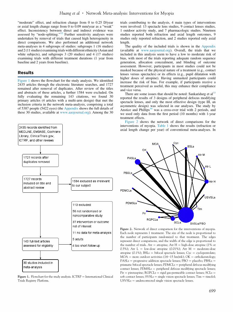
Figure 1 shows the flowchart for the study analysis. We identified2435 articles through the electronic literature searches, and 1727remained after removal of duplicates. After review of the titlesand abstracts of these articles, a further 1584 were excluded. Onfully evaluating the remaining 143 citations, we found 30primary articles (4 articles with a multi-arm design) that met theinclusion criteria in the network meta-analysis, comprising a totalof 5387 people (5422 eyes) (the Appendix shows the full details ofthese 30 studies, available at www.aaojournal.org). Among the 30
Figure 1. Flowchart for the study analysis. ICTRP¼ International ClinicalTrials Registry Platform.
trials contributing to the analysis, 4 main types of interventionswere involved: 13 spectacle lens studies, 9 contact lenses studies,1 outdoor activity study, and 7 pharmacologic studies. Nineteenstudies reported both refraction and axial length outcomes, 9studies only reported refraction, and 2 studies reported only axiallength.
The quality of the included trials is shown in the Appendix(available at www.aaojournal.org). Overall, the trials that weincluded in this analysis seem to have a low to moderate risk ofbias, with most of the trials reporting adequate random sequencegeneration, allocation concealment, and blinding of outcomeassessment. However, participants in most studies could not bemasked because of the physical nature of a treatment (e.g., contactlenses versus spectacles) or its effects (e.g., pupil dilatation withhigher doses of atropine). Having unmasked participants couldincrease the risk of bias. For example, if participants receive atreatment perceived as useful, this may enhance their complianceand vice versa.
There are some issues that should be noted: Sankaridurg et al33
reported the results of 3 designs of peripheral defocus modifyingspectacle lenses, and only the most effective design (type III, anasymmetric design) was selected in our analyses. The study byAnstice and Phillips34 was a cross-over trial with 2 periods, andwe used only data from the first period (10 months) with 1-yeartreatment effects.
Figure 2 shows the network of direct comparisons for theinterventions of myopia. Table 1 shows the results (refraction oraxial length change per year) of conventional meta-analyses. In
Figure 2. Network of direct comparison for the interventions of myopia.Each node represents 1 treatment. The size of the node is proportional tothe number of participants randomized to that treatment. The edgesrepresent direct comparisons, and the width of the edge is proportional tothe number of trials. Atr ¼ atropine; Atr H ¼ high-dose atropine (1% or0.5%); Atr L ¼ low-dose atropine (0.01%); Atr M ¼ moderate-doseatropine (0.1%); BSLs ¼ bifocal spectacle lenses; Cyc ¼ cyclopentolate;MOA ¼ more outdoor activities (14e15 hrs/wk); OK ¼ orthokeratology;PASLs ¼ progressive addition spectacle lenses; PBO ¼ placebo; PBSLs ¼prismatic bifocal spectacle lenses; PDMCLs ¼ peripheral defocus modifyingcontact lenses; PDMSLs ¼ peripheral defocus modifying spectacle lenses;Pir ¼ pirenzepine; RGPCLs ¼ rigid gas-permeable contact lenses; SCLs ¼soft contact lenses; SVSLs ¼ single vision spectacle lenses; Tim ¼ timolol;USVSLs ¼ undercorrected single vision spectacle lenses.
No. of Studies Mean Difference (95% CI) I2 No. of Studies Mean Difference (95% CI) I2
Atr H 4 0.70 (0.42e0.99) 93.9% 2 �0.21 (�0.25 to �0.18) 5.8%Atr M 1 0.59 (0.43e0.75)BSLs 4 0.09 (�0.05 to 0.24) 85.6% 2 �0.06 (�0.10 to �0.02) 0%Cyc 1 0.33 (0.07e0.59)MOA 1 0.14 (0.06e0.22)OK 2 �0.14 (�0.19 to �0.10) 0%PASLs 7 0.12 (0.07e0.18) 51.1% 5 �0.04 (�0.07 to �0.01) 51.5%PBSLs 1 0.34 (0.22e0.46) 1 �0.09 (�0.14 to �0.04)PDMSLs 1 0.12 (�0.06 to 0.30) 1 �0.05 (�0.12 to 0.02)Pir 2 0.29 (0.13e0.44) 47.6% 2 �0.09 (�0.15 to �0.02) 0.0%RGPCLs 1 �0.03 (�0.13 to 0.07) 1 0.02 (�0.04 to 0.08)SCLs 2 �0.06 (�0.10 to �0.02) 0.0% 1 0.01 (�0.01 to 0.03)Tim 1 �0.02 (�0.15 to 0.11)USVSLs 2 �0.11 (�0.22 to 0.00) 0.0% 1 0.03 (�0.02 to 0.08)vs. SVSLs/PBOAtr M 2 �0.23 (�0.61 to 0.15) 94.7% 1 0.00 (�0.03 to 0.03)Atr L 1 �0.10 (�0.19 to �0.01) 1 0.07 (0.03e0.11)Cyc 1 �0.36 (�0.61 to �0.11)PASLs 1 �0.51 (�0.64 to �0.38) 1 0.18 (0.13e0.23)vs. Atr HAtr M 1 0.06 (�0.03 to 0.15) 1 �0.07 (�0.11 to �0.03)vs. Atr LPBSLs 1 0.08 (�0.03 to 0.19) 1 �0.01 (�0.07 to 0.05)Tim 1 �0.11 (�0.23 to 0.01)vs. BSLsSCLs 3 �0.31 (�0.60 to �0.02) 90.6% 3 0.12 (0.05e0.19) 82.3%vs. PDMCLsSCLs 1 �0.21 (�0.34 to �0.08) 1 �0.02 (�0.09 to 0.05)vs. RGPCLs
Atr ¼ atropine; Atr H ¼ high-dose atropine (1% or 0.5%); Atr L ¼ low-dose atropine (0.01%); Atr M ¼ moderate-dose atropine (0.1%); BSLs ¼ bifocalspectacle lenses; CI ¼ confidence interval; Cyc ¼ cyclopentolate; D ¼ diopter; MOA ¼ more outdoor activities (14e15 hrs/wk); OK ¼ orthokeratology;PASLs ¼ progressive addition spectacle lenses; PBO ¼ placebo; PBSLs ¼ prismatic bifocal spectacle lenses; PDMCLs ¼ peripheral defocus modifyingcontact lenses; PDMSLs ¼ peripheral defocus modifying spectacle lenses; Pir ¼ pirenzepine; RGPCLs ¼ rigid gas-permeable contact lenses; SCLs ¼ softcontact lenses; SVSLs ¼ single vision spectacle lenses; Tim ¼ timolol; USVSLs ¼ undercorrected single vision spectacle lenses.
Ophthalmology Volume 123, Number 4, April 2016
direct comparison with single vision spectacle lenses/placebo, thefollowing interventions were all found to be effective with statis-tically significant effect (P < 0.05): high-dose atropine (refractionchange: 0.70 D, 95% CI, 0.42e0.99; axial length change: �0.21mm, 95% CI, �0.25 to �0.18), moderate-dose atropine (refractionchange: 0.59 D, 95% CI, 0.43e0.75), cyclopentolate (refractionchange: 0.33 D, 95% CI, 0.07e0.59), more outdoor activities(refraction change: 0.14 D, 95% CI, 0.06e0.22), orthokeratology(axial length change: �0.14 mm, 95% CI, �0.19 to �0.10), pro-gressive addition spectacle lenses (refraction change: 0.12 D, 95%CI, 0.07e0.18; axial length change: �0.04 mm, 95% CI, �0.07to �0.01), prismatic bifocal spectacle lenses (refraction change:0.34 D, 95% CI, 0.22e0.46; axial length change: �0.09 mm, 95%CI, �0.14 to �0.04), and pirenzepine (refraction change: 0.29 D,95% CI, 0.13e0.44; axial length change: �0.09 mm, 95%CI, �0.15 to �0.02). On direct comparison, high-dose atropinewas superior (P < 0.05) to low-dose atropine (refraction change:0.10 D, 95% CI, 0.01e0.19; axial length change: �0.07 mm, 95%CI, �0.11 to �0.03), cyclopentolate (refraction change: 0.36 D,95% CI, 0.11e0.61), and progressive addition spectacle lenses(refraction change: 0.51 D, 95% CI, 0.38e0.64; axial lengthchange: �0.18 mm, 95% CI, �0.23 to �0.13). Direct comparisonof peripheral defocus modifying contact lenses (refraction change:0.31 D, 95% CI, 0.02e0.60; axial length change: �0.12 mm, 95%CI, �0.19 to �0.05) and rigid gas-permeable contact lenses
700
(refraction change: 0.21 D, 95% CI, �0.08 to �0.34) showedsuperiority (P < 0.05) to soft contact lenses.
There was heterogeneity among some within-trial comparisons(I2 > 50%), for example, high-dose atropine (1% and 0.5%)versus placebo (refraction change: 0.70 D, 95% CI, 0.42e0.99,I2 ¼ 93.9%), bifocal spectacle lenses versus single visionspectacle lenses (refraction change: 0.09 D, 95% CI, �0.05 to 0.24,I2 ¼ 85.6%), progressive addition spectacle lenses versus singlevision spectacle lenses (refraction change: 0.12 D, 95% CI, �0.07to �0.18, I2 ¼ 51.1%; axial length change: �0.04 mm, 95%CI, �0.07 to �0.01, I2 ¼ 51.5%), high-dose atropine (1% and0.5%) versus moderate-dose atropine (0.1%) (refraction change:0.23 D, 95% CI, �0.15 to 0.61, I2 ¼ 94.7%), and peripheraldefocus modifying contact lenses versus soft contact lenses(refraction change: 0.31 D, 95% CI, 0.02e0.6, I2 ¼ 90.6%; axiallength change: �0.12 mm, 95% CI, �0.019 to �0.05, I2 ¼ 82.3%).The forest plots demonstrating this heterogeneity are shown in theAppendix (available at www.aaojournal.org).
We also performed a random effects network meta-analysiscombining the direct and indirect evidence to compare differentinterventions with single vision spectacle lenses/placebo (Fig 3)and with each other (Fig 4). As shown in Figure 3 and Table 2,in comparison with placebo or single vision spectacle lenses,high-dose atropine (refraction change: 0.68 D, 95% CrI,0.52e0.84; axial length change: �0.21 mm, 95% CrI, �0.28
Figure 3. Results of network meta-analysis using single vision spectacle lenses/placebo as referent intervention. Atr ¼ atropine; Atr H ¼ high-dose atropine(1% or 0.5%); Atr L ¼ low-dose atropine (0.01%); Atr M ¼ moderate-dose atropine (0.1%); BSLs ¼ bifocal spectacle lenses; CrI ¼ credible interval; Cyc ¼cyclopentolate; MOA ¼ more outdoor activities (14e15 hrs/wk); OK ¼ orthokeratology; PASLs ¼ progressive addition spectacle lenses; PBO ¼ placebo;PBSLs ¼ prismatic bifocal spectacle lenses; PDMCLs ¼ peripheral defocus modifying contact lenses; PDMSLs ¼ peripheral defocus modifying spectaclelenses; Pir ¼ pirenzepine; RGPCLs ¼ rigid gas-permeable contact lenses; SCLs ¼ soft contact lenses; SVSLs ¼ single vision spectacle lenses; Tim ¼ timolol;USVSLs ¼ undercorrected single vision spectacle lenses.
Figure 4. Network meta-analysis comparing all interventions of myopia. Atr ¼ atropine; Atr H ¼ high-dose atropine (1% or 0.5%); Atr L ¼ low-doseatropine (0.01%); Atr M ¼ moderate-dose atropine (0.1%); BSLs ¼ bifocal spectacle lenses; CrI ¼ credible interval; Cyc ¼ cyclopentolate;MOA ¼ more outdoor activities (14e15 hrs/wk); OK ¼ orthokeratology; PASLs ¼ progressive addition spectacle lenses; PBO ¼ placebo;PBSLs ¼ prismatic bifocal spectacle lenses; PDMCLs ¼ peripheral defocus modifying contact lenses; PDMSLs ¼ peripheral defocus modifying spectaclelenses; Pir ¼ pirenzepine; RGPCLs ¼ rigid gas-permeable contact lenses; SCLs ¼ soft contact lenses; SVSLs ¼ single vision spectacle lenses; Tim ¼ timolol;USVSLs ¼ undercorrected single vision spectacle lenses.
Huang et al � Network Meta-analysis: Interventions for Myopia
701
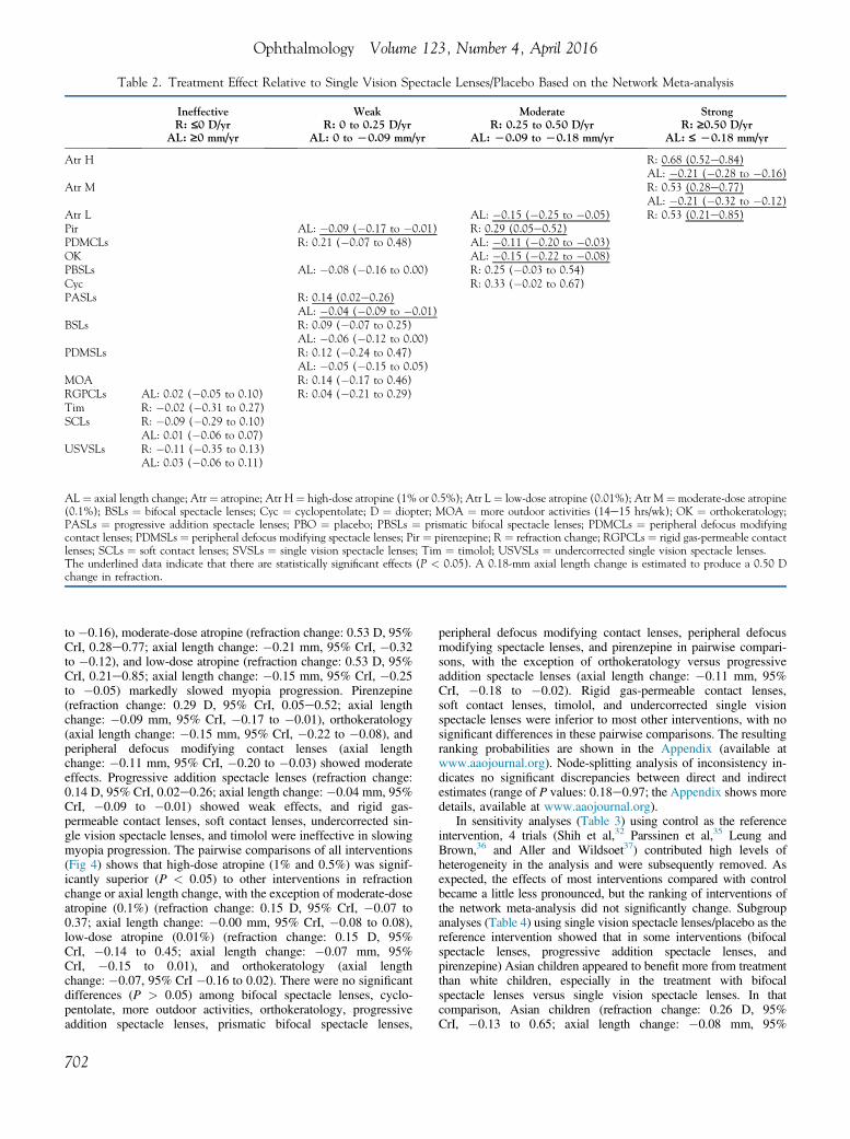
Table 2. Treatment Effect Relative to Single Vision Spectacle Lenses/Placebo Based on the Network Meta-analysis
IneffectiveR: £0 D/yr
AL: ‡0 mm/yr
WeakR: 0 to 0.25 D/yr
AL: 0 to L0.09 mm/yr
ModerateR: 0.25 to 0.50 D/yr
AL: L0.09 to L0.18 mm/yr
StrongR: ‡0.50 D/yr
AL: £ L0.18 mm/yr
Atr H R: 0.68 (0.52e0.84)AL: �0.21 (�0.28 to �0.16)
Atr M R: 0.53 (0.28e0.77)AL: �0.21 (�0.32 to �0.12)
Atr L AL: �0.15 (�0.25 to �0.05) R: 0.53 (0.21e0.85)Pir AL: �0.09 (�0.17 to �0.01) R: 0.29 (0.05e0.52)PDMCLs R: 0.21 (�0.07 to 0.48) AL: �0.11 (�0.20 to �0.03)OK AL: �0.15 (�0.22 to �0.08)PBSLs AL: �0.08 (�0.16 to 0.00) R: 0.25 (�0.03 to 0.54)Cyc R: 0.33 (�0.02 to 0.67)PASLs R: 0.14 (0.02e0.26)
AL: �0.04 (�0.09 to �0.01)BSLs R: 0.09 (�0.07 to 0.25)
AL: �0.06 (�0.12 to 0.00)PDMSLs R: 0.12 (�0.24 to 0.47)
AL: �0.05 (�0.15 to 0.05)MOA R: 0.14 (�0.17 to 0.46)RGPCLs AL: 0.02 (�0.05 to 0.10) R: 0.04 (�0.21 to 0.29)Tim R: �0.02 (�0.31 to 0.27)SCLs R: �0.09 (�0.29 to 0.10)
AL: 0.01 (�0.06 to 0.07)USVSLs R: �0.11 (�0.35 to 0.13)
AL: 0.03 (�0.06 to 0.11)
AL¼ axial length change; Atr ¼ atropine; Atr H¼ high-dose atropine (1% or 0.5%); Atr L ¼ low-dose atropine (0.01%); Atr M ¼ moderate-dose atropine(0.1%); BSLs ¼ bifocal spectacle lenses; Cyc ¼ cyclopentolate; D ¼ diopter; MOA ¼ more outdoor activities (14e15 hrs/wk); OK ¼ orthokeratology;PASLs ¼ progressive addition spectacle lenses; PBO ¼ placebo; PBSLs ¼ prismatic bifocal spectacle lenses; PDMCLs ¼ peripheral defocus modifyingcontact lenses; PDMSLs ¼ peripheral defocus modifying spectacle lenses; Pir ¼ pirenzepine; R ¼ refraction change; RGPCLs ¼ rigid gas-permeable contactlenses; SCLs ¼ soft contact lenses; SVSLs ¼ single vision spectacle lenses; Tim ¼ timolol; USVSLs ¼ undercorrected single vision spectacle lenses.The underlined data indicate that there are statistically significant effects (P < 0.05). A 0.18-mm axial length change is estimated to produce a 0.50 Dchange in refraction.
Ophthalmology Volume 123, Number 4, April 2016
to �0.16), moderate-dose atropine (refraction change: 0.53 D, 95%CrI, 0.28e0.77; axial length change: �0.21 mm, 95% CrI, �0.32to �0.12), and low-dose atropine (refraction change: 0.53 D, 95%CrI, 0.21e0.85; axial length change: �0.15 mm, 95% CrI, �0.25to �0.05) markedly slowed myopia progression. Pirenzepine(refraction change: 0.29 D, 95% CrI, 0.05e0.52; axial lengthchange: �0.09 mm, 95% CrI, �0.17 to �0.01), orthokeratology(axial length change: �0.15 mm, 95% CrI, �0.22 to �0.08), andperipheral defocus modifying contact lenses (axial lengthchange: �0.11 mm, 95% CrI, �0.20 to �0.03) showed moderateeffects. Progressive addition spectacle lenses (refraction change:0.14 D, 95% CrI, 0.02e0.26; axial length change: �0.04 mm, 95%CrI, �0.09 to �0.01) showed weak effects, and rigid gas-permeable contact lenses, soft contact lenses, undercorrected sin-gle vision spectacle lenses, and timolol were ineffective in slowingmyopia progression. The pairwise comparisons of all interventions(Fig 4) shows that high-dose atropine (1% and 0.5%) was signif-icantly superior (P < 0.05) to other interventions in refractionchange or axial length change, with the exception of moderate-doseatropine (0.1%) (refraction change: 0.15 D, 95% CrI, �0.07 to0.37; axial length change: �0.00 mm, 95% CrI, �0.08 to 0.08),low-dose atropine (0.01%) (refraction change: 0.15 D, 95%CrI, �0.14 to 0.45; axial length change: �0.07 mm, 95%CrI, �0.15 to 0.01), and orthokeratology (axial lengthchange: �0.07, 95% CrI �0.16 to 0.02). There were no significantdifferences (P > 0.05) among bifocal spectacle lenses, cyclo-pentolate, more outdoor activities, orthokeratology, progressiveaddition spectacle lenses, prismatic bifocal spectacle lenses,
702
peripheral defocus modifying contact lenses, peripheral defocusmodifying spectacle lenses, and pirenzepine in pairwise compari-sons, with the exception of orthokeratology versus progressiveaddition spectacle lenses (axial length change: �0.11 mm, 95%CrI, �0.18 to �0.02). Rigid gas-permeable contact lenses,soft contact lenses, timolol, and undercorrected single visionspectacle lenses were inferior to most other interventions, with nosignificant differences in these pairwise comparisons. The resultingranking probabilities are shown in the Appendix (available atwww.aaojournal.org). Node-splitting analysis of inconsistency in-dicates no significant discrepancies between direct and indirectestimates (range of P values: 0.18e0.97; the Appendix shows moredetails, available at www.aaojournal.org).
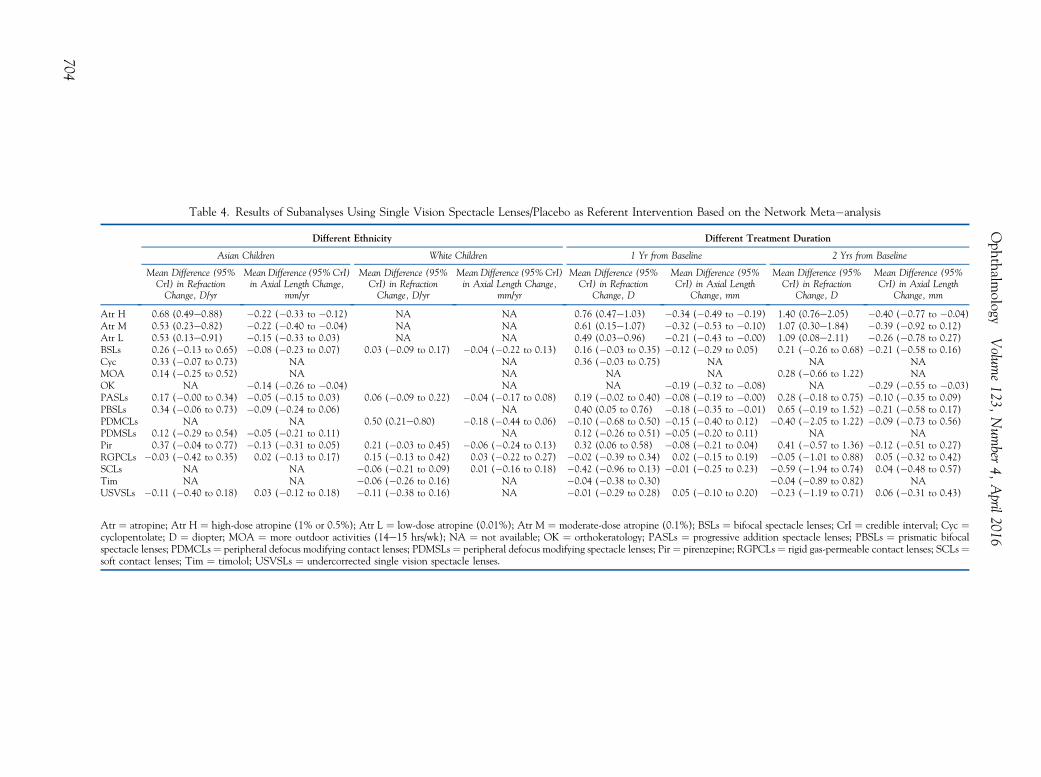
In sensitivity analyses (Table 3) using control as the referenceintervention, 4 trials (Shih et al,32 Parssinen et al,35 Leung andBrown,36 and Aller and Wildsoet37) contributed high levels ofheterogeneity in the analysis and were subsequently removed. Asexpected, the effects of most interventions compared with controlbecame a little less pronounced, but the ranking of interventions ofthe network meta-analysis did not significantly change. Subgroupanalyses (Table 4) using single vision spectacle lenses/placebo as thereference intervention showed that in some interventions (bifocalspectacle lenses, progressive addition spectacle lenses, andpirenzepine) Asian children appeared to benefit more from treatmentthan white children, especially in the treatment with bifocalspectacle lenses versus single vision spectacle lenses. In thatcomparison, Asian children (refraction change: 0.26 D, 95%CrI, �0.13 to 0.65; axial length change: �0.08 mm, 95%
Table 3. Results of Sensitivity Analyses Performed by Removal of Trials That Caused High Heterogeneity Across Studies Based on theNetwork Meta-analysis
Original Data Sensitivity Analyses
Mean Difference (95% CrI)in Refraction, D/yr
Mean Difference (95% CrI)in Axial Length, mm/yr
Mean Difference (95% CrI)in Refraction, D/yr
Mean Difference (95% CrI)in Axial Length, mm/yr
Atr H 0.68 (0.52e0.84) �0.21 (�0.28 to �0.16) 0.55 (0.45e0.68) �0.21 (�0.26 to �0.17)Atr M 0.53 (0.28e0.77) �0.21 (�0.32 to �0.12) 0.51 (0.33e0.71) �0.21 (�0.28 to �0.14)Atr L 0.53 (0.21e0.85) �0.15 (�0.25 to �0.05) 0.45 (0.27e0.66) �0.14 (�0.22 to �0.07)BSLs 0.09 (�0.07 to 0.25) �0.06 (�0.12 to 0.00) 0.16 (0.05e0.26) �0.06 (�0.11 to �0.01)Cyc 0.33 (�0.02 to 0.67) NA 0.26 (0.00e0.52) NAMOA 0.14 (�0.17 to 0.46) NA 0.14 (�0.02 to 0.30) NAOK NA �0.15 (�0.22 to �0.08) NA �0.14 (�0.20 to �0.08)PASLs 0.14 (0.02e0.26) �0.04 (�0.09 to �0.01) 0.10 (0.03e0.17) �0.03 (�0.06 to �0.00)PBSLs 0.25 (�0.03 to 0.54) �0.08 (�0.16 to 0.00) 0.28 (0.12e0.45) �0.08 (�0.14 to �0.02)PDMCLs 0.21 (�0.07 to 0.48) �0.11 (�0.20 to �0.03) 0.07 (�0.10 to 0.25) �0.08 (�0.15 to �0.02)PDMSLs 0.12 (�0.24 to 0.47) �0.05 (�0.15 to 0.05) 0.12 (�0.11 to 0.35) �0.05 (�0.13 to 0.03)Pir 0.29 (0.05e0.52) �0.09 (�0.17 to �0.01) 0.28 (0.13e0.43) �0.09 (�0.16 to �0.01)RGPCLs 0.04 (�0.21 to 0.29) 0.02 (�0.05 to 0.10) 0.04 (�0.10 to 0.17) 0.02 (�0.03 to 0.08)SCLs �0.09 (�0.29 to 0.10) 0.01 (�0.06 to 0.07) �0.08 (�0.19 to 0.01) 0.01 (�0.04 to 0.05)Tim �0.02 (�0.31 to 0.27) NA 0.02 (�0.15 to 0.19) NAUSVSLs �0.11 (�0.35 to 0.13) 0.03 (�0.06 to 0.11) �0.11 (�0.26 to 0.04) 0.03 (�0.04 to 0.10)
Atr ¼ atropine; Atr H ¼ high-dose atropine (1% or 0.5%); Atr L ¼ low-dose atropine (0.01%); Atr M ¼ moderate-dose atropine (0.1%); BSLs ¼ bifocalspectacle lenses; CrI ¼ credible interval; Cyc ¼ cyclopentolate; D ¼ diopter; MOA ¼ more outdoor activities (14e15 hrs/wk); NA ¼ not available; OK ¼orthokeratology; PASLs ¼ progressive addition spectacle lenses; PBO ¼ placebo; PBSLs ¼ prismatic bifocal spectacle lenses; PDMCLs ¼ peripheral defocusmodifying contact lenses; PDMSLs ¼ peripheral defocus modifying spectacle lenses; Pir ¼ pirenzepine; RGPCLs ¼ rigid gas-permeable contact lenses;SCLs ¼ soft contact lenses; SVSLs ¼ single vision spectacle lenses; Tim ¼ timolol; USVSLs ¼ undercorrected single vision spectacle lenses.All mean difference use SVSLs/PBO as the referent intervention.
Huang et al � Network Meta-analysis: Interventions for Myopia
CrI,�0.23 to0.07) andwhite children (refraction change: 0.03D,95%CrI,�0.09 to 0.17; axial length change:�0.04mm,95%CrI,�0.22 to0.13) differed by 0.23 D in refraction change and 0.05 mm in axiallength change. These differences did not reach statisticalsignificance, and additional trial data are required to adequatelyaddress the question of whether race has an impact on the efficacyof myopia control treatments. Further subgroup analyses stratifiedby different treatment durations showed that most interventions losetheir early effect in the second year, especially in the protection ofaxial length change.
Discussion
Our study is a network meta-analysis aimed specifically atinvestigating the efficacy or comparative effectiveness ofdifferent interventions to slowmyopia progression. In addition,the present study updates previous evidence-based re-views.24,38,39 A previous review by Saw et al39 and a morerecent Cochrane review29 both concluded that the evidencefrom randomized clinical trials of that time does not providesufficient information to support interventions to slow downthe progression of myopia. The increased availability ofhigh-quality clinical trials combined with the network meta-analysis techniques used in this article can now provide someguidance regarding the management of myopic progression.
The main findings of our analysis are as follows:
1. High-dose atropine (1% and 0.5%), moderate-doseatropine (0.1%), and low-dose atropine (0.01%)showed clear effects in myopia control (all with
statistically significant effect); pirenzepine, ortho-keratology, peripheral defocus modifying contactlenses, cyclopentolate, and prismatic bifocal spec-tacle lenses showed moderate effects (all with sta-tistically significant effect except for cyclopentolateand prismatic bifocal spectacle lenses); progressiveaddition spectacle lenses, bifocal spectacle lenses,peripheral defocus modifying spectacle lenses, andmore outdoor activities showed weak effects (onlyprogressive addition spectacle lenses with statisti-cally significant effect); rigid gas-permeable contactlenses, soft contact lenses, undercorrected singlevision spectacle lenses, and timolol were ineffective(all with no statistically significant effect).
2. High-dose atropine (1% and 0.5%) was significantlysuperior to other interventions except moderate-doseatropine (0.1%) and low-dose atropine (0.01%).Among bifocal spectacle lenses, cyclopentolate,more outdoor activities, orthokeratology, progres-sive addition spectacle lenses, prismatic bifocalspectacle lenses, peripheral defocus modifyingcontact lenses, peripheral defocus modifying spec-tacle lenses, and pirenzepine, pairwise comparisonsshowed no significant differences apart from abenefit of orthokeratology over progressive additionspectacle lenses. Rigid gas-permeable contact len-ses, soft contact lenses, timolol, and undercorrectedsingle vision spectacle lenses were inferior to mostother interventions, with no significant differenceswithin this group.
703
Table 4. Results of Subanalyses Using Single Vision Spectacle Lenses/Placebo as Referent Intervention Based on the Network Meta�analysis
Different Ethnicity Different Treatment Duration
Asian Children White Children 1 Yr from Baseline 2 Yrs from Baseline
Mean Difference (95%CrI) in RefractionChange, D/yr
Mean Difference (95% CrI)in Axial Length Change,
mm/yr
Mean Difference (95%CrI) in RefractionChange, D/yr
Mean Difference (95% CrI)in Axial Length Change,
mm/yr
Mean Difference (95%CrI) in Refraction
Change, D
Mean Difference (95%CrI) in Axial Length
Change, mm
Mean Difference (95%CrI) in Refraction
Change, D
Mean Difference (95%CrI) in Axial Length
Change, mm
Atr H 0.68 (0.49e0.88) �0.22 (�0.33 to �0.12) NA NA 0.76 (0.47e1.03) �0.34 (�0.49 to �0.19) 1.40 (0.76e2.05) �0.40 (�0.77 to �0.04)Atr M 0.53 (0.23e0.82) �0.22 (�0.40 to �0.04) NA NA 0.61 (0.15e1.07) �0.32 (�0.53 to �0.10) 1.07 (0.30e1.84) �0.39 (�0.92 to 0.12)Atr L 0.53 (0.13e0.91) �0.15 (�0.33 to 0.03) NA NA 0.49 (0.03e0.96) �0.21 (�0.43 to �0.00) 1.09 (0.08e2.11) �0.26 (�0.78 to 0.27)BSLs 0.26 (�0.13 to 0.65) �0.08 (�0.23 to 0.07) 0.03 (�0.09 to 0.17) �0.04 (�0.22 to 0.13) 0.16 (�0.03 to 0.35) �0.12 (�0.29 to 0.05) 0.21 (�0.26 to 0.68) �0.21 (�0.58 to 0.16)Cyc 0.33 (�0.07 to 0.73) NA NA 0.36 (�0.03 to 0.75) NA NA NAMOA 0.14 (�0.25 to 0.52) NA NA NA NA 0.28 (�0.66 to 1.22) NAOK NA �0.14 (�0.26 to �0.04) NA NA �0.19 (�0.32 to �0.08) NA �0.29 (�0.55 to �0.03)PASLs 0.17 (�0.00 to 0.34) �0.05 (�0.15 to 0.03) 0.06 (�0.09 to 0.22) �0.04 (�0.17 to 0.08) 0.19 (�0.02 to 0.40) �0.08 (�0.19 to �0.00) 0.28 (�0.18 to 0.75) �0.10 (�0.35 to 0.09)PBSLs 0.34 (�0.06 to 0.73) �0.09 (�0.24 to 0.06) NA 0.40 (0.05 to 0.76) �0.18 (�0.35 to �0.01) 0.65 (�0.19 to 1.52) �0.21 (�0.58 to 0.17)PDMCLs NA NA 0.50 (0.21e0.80) �0.18 (�0.44 to 0.06) �0.10 (�0.68 to 0.50) �0.15 (�0.40 to 0.12) �0.40 (�2.05 to 1.22) �0.09 (�0.73 to 0.56)PDMSLs 0.12 (�0.29 to 0.54) �0.05 (�0.21 to 0.11) NA 0.12 (�0.26 to 0.51) �0.05 (�0.20 to 0.11) NA NAPir 0.37 (�0.04 to 0.77) �0.13 (�0.31 to 0.05) 0.21 (�0.03 to 0.45) �0.06 (�0.24 to 0.13) 0.32 (0.06 to 0.58) �0.08 (�0.21 to 0.04) 0.41 (�0.57 to 1.36) �0.12 (�0.51 to 0.27)RGPCLs �0.03 (�0.42 to 0.35) 0.02 (�0.13 to 0.17) 0.15 (�0.13 to 0.42) 0.03 (�0.22 to 0.27) �0.02 (�0.39 to 0.34) 0.02 (�0.15 to 0.19) �0.05 (�1.01 to 0.88) 0.05 (�0.32 to 0.42)SCLs NA NA �0.06 (�0.21 to 0.09) 0.01 (�0.16 to 0.18) �0.42 (�0.96 to 0.13) �0.01 (�0.25 to 0.23) �0.59 (�1.94 to 0.74) 0.04 (�0.48 to 0.57)Tim NA NA �0.06 (�0.26 to 0.16) NA �0.04 (�0.38 to 0.30) �0.04 (�0.89 to 0.82) NAUSVSLs �0.11 (�0.40 to 0.18) 0.03 (�0.12 to 0.18) �0.11 (�0.38 to 0.16) NA �0.01 (�0.29 to 0.28) 0.05 (�0.10 to 0.20) �0.23 (�1.19 to 0.71) 0.06 (�0.31 to 0.43)
Atr ¼ atropine; Atr H ¼ high-dose atropine (1% or 0.5%); Atr L ¼ low-dose atropine (0.01%); Atr M ¼ moderate-dose atropine (0.1%); BSLs ¼ bifocal spectacle lenses; CrI ¼ credible interval; Cyc ¼cyclopentolate; D ¼ diopter; MOA ¼ more outdoor activities (14e15 hrs/wk); NA ¼ not available; OK ¼ orthokeratology; PASLs ¼ progressive addition spectacle lenses; PBSLs ¼ prismatic bifocalspectacle lenses; PDMCLs ¼ peripheral defocus modifying contact lenses; PDMSLs ¼ peripheral defocus modifying spectacle lenses; Pir ¼ pirenzepine; RGPCLs ¼ rigid gas-permeable contact lenses; SCLs ¼soft contact lenses; Tim ¼ timolol; USVSLs ¼ undercorrected single vision spectacle lenses.
Ophthalm
ologyVolum
e123,
Num
ber4,
April2016
704
Huang et al � Network Meta-analysis: Interventions for Myopia
3. Asian children appeared to benefit more fromtreatment than white children, and most in-terventions lose their early effect in the second year.
Certain trials caused high heterogeneity across studies,but removal of them only introduced less pronounced effectsof most interventions, without a significant change in theresults, and we did not find any statistically significant in-consistencies in the network. This implies that the results arerelatively reliable.
The major advantage of our current meta-analyticapproach over individual trials is the larger sample sizethat results from incorporating both direct and indirect evi-dence. This approach also differs from traditional meta-analyses in that traditional meta-analyses are characterizedby a series of smaller meta-analyses of different activecomparisons and thus provides less robust information.Although comparisons between specific classes of in-terventions for myopia control have been investigated inmultiple studies, others have been performed only in asingle trial or have never been performed. Thus, a networkmeta-analysis makes it possible to both validate previousempirical evidence of direct comparisons and provide evi-dence regarding comparisons for which no direct empiricalevidence exists.40
Previous trials suggested that, with the exception oftimolol, drug treatments (especially atropine) showed thehighest efficacy, which is consistent with our results.41 Itremains unclear how atropine slows down myopiaprogression. Earlier studies have suggested that this maybe due to the effects of atropine on lens accommodation,whereas subsequent studies have shown that atropine’seffects on myopia is via a nonaccommodative pathway inthe retina or sclera.18,19 However, the inevitable side ef-fects of higher doses of atropine (i.e., glare, photophobia,and near vision blur) and the rebound phenomenon afterstopping treatment have restricted its widespread clinicaluse.42,43 There appears to be a differential dose-dependentsensitivity to atropine’s impact on myopia progression, pu-pil size, and accommodation. Low-dose atropine (0.01%) isstill one of the most effective interventions identified in thisanalysis and has been found to induce minimal clinicalsymptoms.44 Furthermore, this lower dose does not displaythe same rebound effect that has been seen in higher doses.This makes low-dose atropine a definite candidate treatmentfor myopia progression, although this result needs to bereplicated in other populations.
Alternatively, pirenzepine, a selective antimuscarinicagent, represents a viable alternative to atropine for thecontrol of myopia progression. Pirenzepine is less likely toproduce pupillary dilatation and cycloplegia with moderateeffects in myopia control.45,46 Of note, the analysis of pir-enzepine was limited by involvement of only 2 articles;thus, further trials with larger sample sizes are required toconfirm its effect.
Multifocal spectacle lenses have been tested in control-ling the progression of myopia for several years, but theirefficacy is controversial.47,48 A previous meta-analysis21
indicated that multifocal spectacle lenses slowed myopiaprogression by a mean of 0.25 D in school-aged children
compared with single vision spectacle lenses. In the currentstudy, our results suggest only modest effects of bifocalspectacle lenses and progressive addition spectacle lenses.Furthermore, there was no significant difference betweenbifocal spectacle lenses and progressive addition spectaclelenses in pairwise comparison. As for specifically designedmultifocal spectacle lenses (prismatic bifocal spectacle len-ses), our meta-analysis showed that they have a moderateeffect in myopia control, but this was not statistically sig-nificant with wide CrIs. This is partly because only 1 rele-vant RCT was included, so further trials are warranted.Overall, multifocal spectacle lenses do not seem to be aviable option for controlling progression of myopia.
In terms of contact lenses, orthokeratology has beenshown to be an effective treatment in controlling progres-sion of myopia.49,50 Orthokeratology flattens the centralcornea while steeping the midperipheral cornea to reducerelative peripheral hyperopia, which may slow the elonga-tion of the axial length.51,52 However, orthokeratology is notin widespread use because of a variety of possible issues,such as the additional skills required by practitioners forfitting these lenses, the discomfort during overnight wear,the cost, and the risks of infective keratitis.53e55 In recentyears, soft contact lenses with myopia control features thatcreate additional myopic defocus on the retina have gener-ated great interest in myopia control.33 Our results showedthat peripheral defocus modifying contact lenses weresuperior to peripheral defocus modifying spectacle lenses.Similar to other interventions, the limited relevant RCTsincluded in this meta-analysis showed wide CrIs thus,more RCTs are required to demonstrate its efficacy. Incomparison, other contact lenses such as standard rigid gas-permeable contact lenses and conventional soft contactlenses showed no effect on myopia control in our study.
A previous review has indicated that increasing outdooractivities may be a simple strategy to reduce the risk ofmyopia progression.23 However, in the current study, only 1RCT of outdoor activities contributed to the analysis, andthe effect was modest. Further trials are required toelucidate the value of this intervention.
Some epidemiologic studies have reported racial differ-ences between childhood myopia prevalence in Asians andwhite subjects within the same country, highlighting the po-tential role of ethnicity.56,57 In accordance with previousstudies,21 we found that Asians appeared to benefit morefrom treatment than white patients. This finding may beexplicable on the basis of an increased geneticsusceptibility of Asians to myopia or a faster rate ofprogression in Asians. Also similar to previous studies,58,59
our study found that most interventions lose their early ef-fect in the second year, which may be due to increased age.
Study Limitations
There are some inherent limitations in this analysis thatshould be highlighted. Optical interventions vary for eachindividual patient. For example, multifocal spectacle lenseshave different refractive powers for each patient, and the off-axis effects of orthokeratology vary with refractive correction.Both placebo and single vision spectacle lenses are used as
705
Ophthalmology Volume 123, Number 4, April 2016
controls. The quality of trials conducted and reporting varied(some studies were not double-blind). There was a widevariation in subject age (mean age range, 8.3e14.0 years),but because studies reported only the age range or mean, datawere insufficient to determine how treatment varies with age.Our study provides information on the efficacy but not thesafety of different treatment options because of lack of datawithin the included articles. Clinical decisions on any inter-vention require information on efficacy, short-term/long-termbenefits, and the risks of side effects, so additional exami-nation of the safety of these interventions is important. Inaddition, high heterogeneity was found in some combina-tions, and most interventions are based on indirect compari-sons (113 pairs). More trials are required to confirm theresults from these indirect comparisons.
The fundamental challenge in this analysis is the lack ofsufficient data on some treatments, which results in wideCrIs. Future trials with larger sample sizes are required toprovide better-quality data to help establish the effect ofvarious interventions in controlling myopia. In addition, thepossible additive or even synergistic effects of differentcombinations (e.g., combined atropine and contact lenstreatments) have not, to date, been adequately addressed.This is certainly a worthy question for future studies andmay help to provide treatments for myopic progression thatare both effective and easily tolerated by the patient.
Notwithstanding these limitations, it is unlikely that thenumber of head-to-head trials necessary to address all theseclinical questions will be conducted. At least 136 trials areneeded to compare all interventions of myopia control, andin their absence, our network meta-analysis provides avaluable approach to the issue.
In conclusion, on the basis of evidence from the availableRCTs used in this analysis, the following evidence-basedguidelines might be proposed. (1) Rigid gas-permeablecontact lenses, conventional soft contact lenses, timolol,and undercorrected single vision spectacle lenses are inef-fective in slowing the progression of myopia in children. (2)Atropine, pirenzepine, orthokeratology, soft contact lenseswith myopia control features (peripheral defocus modifyingdesigns), and progressive addition spectacle lenses areeffective and produce a statistically significant reduction ofmyopia progression in terms of refraction or axial length. (3)The introduction of myopia treatments into clinical practicemay be limited by side effects (e.g., atropine 1%), cost andcomplexity (e.g., orthokeratology), and limited effectiveness(e.g., progressive add spectacle lenses). This leaves low-dose atropine (0.01%), pirenzepine, and soft contact lenseswith myopia control features (e.g., peripheral defocusmodifying designs) as viable options for the active man-agement of myopia progression.
References
1. Pararajasegaram R. VISION 2020dthe right to sight: fromstrategies to action. Am J Ophthalmol 1999;128:359–60.
2. Pan CW, Ramamurthy D, Saw SM. Worldwide prevalence andrisk factors for myopia. Ophthalmic Physiol Opt 2012;32:3–16.
706
3. Rahi JS, Cumberland PM, Peckham CS. Myopia over thelifecourse: prevalence and early life influences in the 1958British birth cohort. Ophthalmology 2011;118:797–804.
4. Wolfram C, Hohn R, Kottler U, et al. Prevalence of refractiveerrors in the European adult population: the Gutenberg HealthStudy (GHS). Br J Ophthalmol 2014;98:857–61.
5. Tay MT, Au Eong KG, Ng CY, Lim MK. Myopia andeducational attainment in 421,116 young Singaporean males.Ann Acad Med Singapore 1992;21:785–91.
6. Vitale S, Sperduto RD, Ferris FL 3rd. Increased prevalenceof myopia in the United States between 1971e1972 and1999e2004. Arch Ophthalmol 2009;127:1632–9.
7. Vitale S, Cotch MF, Sperduto R, Ellwein L. Costs of refractivecorrection of distance vision impairment in the United States,1999-2002. Ophthalmology 2006;113:2163–70.
8. Marcus MW, de Vries MM, Junoy Montolio FG,Jansonius NM. Myopia as a risk factor for open-angle glau-coma: a systematic review and meta-analysis. Ophthalmology2011;118:1989–1994 e2.
9. Flitcroft DI. The complex interactions of retinal, optical andenvironmental factors in myopia aetiology. Prog Retin EyeRes 2012;31:622–60.
10. Hayashi K, Ohno-Matsui K, Shimada N, et al. Long-termpattern of progression of myopic maculopathy: a natural his-tory study. Ophthalmology 2010;117:1595–611. 611 e1e4.
11. Xu L, Wang Y, Li Y, et al. Causes of blindness and visualimpairment in urban and rural areas in Beijing: the Beijing EyeStudy. Ophthalmology 2006;113:1134.e1–e11.
12. Evans JR, Fletcher AE, Wormald RP. Causes of visualimpairment in people aged 75 years and older in Britain: anadd-on study to the MRC Trial of Assessment and Manage-ment of Older People in the Community. Br J Ophthalmol2004;88:365–70.
13. Avisar R, Friling R, Snir M, et al. Estimation of prevalenceand incidence rates and causes of blindness in Israel, 1998-2003. Isr Med Assoc J 2006;8:880–1.
14. Goss DA. Nearwork and myopia. Lancet 2000;356:1456–7.15. Saw SM, Chua WH, Hong CY, et al. Nearwork in early-onset
myopia. Invest Ophthalmol Vis Sci 2002;43:332–9.16. Gwiazda J, Thorn F, Bauer J, Held R. Myopic children show
insufficient accommodative response to blur. Invest Oph-thalmol Vis Sci 1993;34:690–4.
17. Smith EL 3rd, Kee CS, Ramamirtham R, et al. Peripheralvision can influence eye growth and refractive development ininfant monkeys. Invest Ophthalmol Vis Sci 2005;46:3965–72.
18. McBrien NA, Moghaddam HO, Reeder AP. Atropine reducesexperimental myopia and eye enlargement via a non-accommodative mechanism. Invest Ophthalmol Vis Sci1993;34:205–15.
19. Stone RA, Lin T, Laties AM. Muscarinic antagonist effects onexperimental chick myopia. Exp Eye Res 1991;52:755–8.
20. Rose KA, Morgan IG, Ip J, et al. Outdoor activity reduces theprevalence of myopia in children. Ophthalmology 2008;115:1279–85.
21. Li SM, Ji YZ, Wu SS, et al. Multifocal versus single visionlenses intervention to slow progression of myopia in school-agechildren: a meta-analysis. Surv Ophthalmol 2011;56:451–60.
22. Song YY, Wang H, Wang BS, et al. Atropine in amelioratingthe progression of myopia in children with mild to moderatemyopia: a meta-analysis of controlled clinical trials. J OculPharmacol Ther 2011;27:361–8.
23. Sherwin JC, Reacher MH, Keogh RH, et al. The associationbetween time spent outdoors and myopia in children and ad-olescents: a systematic review and meta-analysis. Ophthal-mology 2012;119:2141–51.
Huang et al � Network Meta-analysis: Interventions for Myopia
24. Walline JJ, Lindsley K, Vedula SS, et al. Interventions to slowprogression of myopia in children. Cochrane Database SystRev 2011;CD004916.
25. Lumley T. Network meta-analysis for indirect treatmentcomparisons. Stat Med 2002;21:2313–24.
26. Ades AE, Sculpher M, Sutton A, et al. Bayesian methods forevidence synthesis in cost-effectiveness analysis. Pharmacoe-conomics 2006;24:1–19.
27. Shih YF, Chen CH, Chou AC, et al. Effects of different con-centrations of atropine on controlling myopia in myopic chil-dren. J Ocul Pharmacol Ther 1999;15:85–90.
28. Higgins JPT, Green S; Cochrane Collaboration. CochraneHandbook for Systematic Reviews of Interventions. xxi.Chichester, England; Hoboken, NJ: Wiley-Blackwell; 2008:649.
29. DerSimonian R, Laird N. Meta-analysis in clinical trials.Control Clin Trials 1986;7:177–88.
31. Marshall DC. Cryptic failure of partitioned Bayesian phylo-genetic analyses: lost in the land of long trees. Syst Biol2010;59:108–17.
32. Dias S, Welton NJ, Caldwell DM, Ades AE. Checking con-sistency in mixed treatment comparison meta-analysis. StatMed 2010;29:932–44.
33. Sankaridurg P, Donovan L, Varnas S, et al. Spectacle lensesdesigned to reduce progression of myopia: 12-month results.Optom Vis Sci 2010;87:631–41.
34. Anstice NS, Phillips JR. Effect of dual-focus soft contact lenswear on axial myopia progression in children. Ophthalmology2011;118:1152–61.
35. Parssinen O, Hemminki E, Klemetti A. Effect of spectacle useand accommodation on myopic progression: final results of athree-year randomised clinical trial among schoolchildren. Br JOphthalmol 1989;73:547–51.
36. Leung JT, Brown B. Progression of myopia in Hong KongChinese schoolchildren is slowed by wearing progressivelenses. Optom Vis Sci 1999;76:346–54.
37. Aller TA, Wildsoet C. Results of a one-year prospectiveclinical trial (CONTROL) of the use of bifocal soft contactlenses to control myopia progression. Ophthalmic and Physi-ological Optics 2006;26:8–9.
38. Leo SW, Young TL. An evidence-based update on myopia andinterventions to retard its progression. J AAPOS 2011;15:181–9.
39. Saw SM, Shih-Yen EC, Koh A, Tan D. Interventions to retardmyopia progression in children: an evidence-based update.Ophthalmology 2002;109:415–21. discussion 22e4; quiz25e6, 43.
40. Bucher HC, Guyatt GH, Griffith LE, Walter SD. The results ofdirect and indirect treatment comparisons in meta-analysis ofrandomized controlled trials. J Clin Epidemiol 1997;50:683–91.
41. Jensen H. Myopia progression in young school children. Aprospective study of myopia progression and the effect of atrial with bifocal lenses and beta blocker eye drops. ActaOphthalmol Suppl 1991;1–79.
42. Kennedy RH, Dyer JA, Kennedy MA, et al. Reducing theprogression of myopia with atropine: a long term cohort study
of Olmsted County students. Binocul Vis Strabismus Q2000;15:281–304.
43. Tong L, Huang XL, Koh AL, et al. Atropine for the treatmentof childhood myopia: effect on myopia progression aftercessation of atropine. Ophthalmology 2009;116:572–9.
44. Chia A, Chua WH, Cheung YB, et al. Atropine for the treat-ment of childhood myopia: safety and efficacy of 0.5%, 0.1%,and 0.01% doses (Atropine for the Treatment of Myopia 2).Ophthalmology 2012;119:347–54.
45. Dorje F, Wess J, Lambrecht G, et al. Antagonist bindingprofiles of five cloned human muscarinic receptor subtypes.J Pharmacol Exp Ther 1991;256:727–33.
46. Buckley NJ, Bonner TI, Buckley CM, Brann MR. Antagonistbinding properties of five cloned muscarinic receptorsexpressed in CHO-K1 cells. Mol Pharmacol 1989;35:469–76.
47. Cheng D, Schmid KL, Woo GC, Drobe B. Randomized trial ofeffect of bifocal and prismatic bifocal spectacles on myopicprogression: two-year results. Arch Ophthalmol 2010;128:12–9.
48. Edwards MH, Li RW, Lam CS, et al. The Hong Kong pro-gressive lens myopia control study: study design and mainfindings. Invest Ophthalmol Vis Sci 2002;43:2852–8.
49. Cho P, Cheung SW, Edwards M. The longitudinal orthoker-atology research in children (LORIC) in Hong Kong: a pilotstudy on refractive changes and myopic control. Curr Eye Res2005;30:71–80.
50. Hiraoka T, Kakita T, Okamoto F, et al. Long-term effect ofovernight orthokeratology on axial length elongation inchildhood myopia: a 5-year follow-up study. Invest Oph-thalmol Vis Sci 2012;53:3913–9.
51. Queiros A, Gonzalez-Meijome JM, Jorge J, et al. Peripheralrefraction in myopic patients after orthokeratology. Optom VisSci 2010;87:323–9.
52. Sankaridurg P, Holden B, Smith E 3rd, et al. Decrease in rateof myopia progression with a contact lens designed to reducerelative peripheral hyperopia: one-year results. Invest Oph-thalmol Vis Sci 2011;52:9362–7.
53. Cho P, Cheung SW, Edwards MH, Fung J. An assessment ofconsecutively presenting orthokeratology patients in a HongKong based private practice. Clin Exp Optom 2003;86:331–8.
54. Santolaria E, Cervino A, Queiros A, et al. Subjective satis-faction in long-term orthokeratology patients. Eye ContactLens 2013;39:388–93.
55. Chan TC, Li EY, Wong VW, Jhanji V. Orthokeratology-associated Infectious Keratitis in a Tertiary Care Eye Hospitalin Hong Kong. Am J Ophthalmol 2014;158:1130–1135.e2.
56. He M, Zheng Y, Xiang F. Prevalence of myopia in urban andrural children in mainland China. Optom Vis Sci 2009;86:40–4.
57. Ip JM, Huynh SC, Robaei D, et al. Ethnic differences in theimpact of parental myopia: findings from a population-basedstudy of 12-year-old Australian children. Invest OphthalmolVis Sci 2007;48:2520–8.
58. Kakita T, Hiraoka T, Oshika T. Influence of overnight ortho-keratology on axial elongation in childhood myopia. InvestOphthalmol Vis Sci 2011;52:2170–4.
59. Chua WH, Balakrishnan V, Chan YH, et al. Atropine for thetreatment of childhood myopia. Ophthalmology 2006;113:2285–91.
Originally received: June 15, 2015.Final revision: November 1, 2015.Accepted: November 6, 2015.Available online: January 26, 2016. Manuscript no. 2015-981.1 School of Ophthalmology and Optometry, Wenzhou Medical University,Wenzhou, Zhejiang, China.2 Key Laboratory of Vision Science, Ministry of Health P.R. China,Wenzhou, Zhejiang, China.3 Department of Ophthalmology, No.180 Hospital of Chinese PLA,Quanzhou, Fujian.4 ABM University Health Board, Swansea, United Kingdom.5 Flinders University, Adelaide, South Australia, Australia.6 Department of Ophthalmology, Children’s University Hospital, Dublin,Ireland.7 Department of Epidemiology and Public Health, Yong Loo Lin School ofMedicine, National University of Singapore, Singapore, Singapore.8 Department of Ophthalmology, The Second Affiliated Hospital of AnhuiMedical University, Hefei, Anhui, China.9 Department of Biological Statistics, Eye hospital of Wenzhou MedicalUniversity, Wenzhou, Zhejiang, China.
*J.H., D.W., Q.W., C.M., and I.F. contributed equally as first authors.
Financial Disclosure(s):The author(s) have no proprietary or commercial interest in any materialsdiscussed in this article.
Supported in part by the National Natural Science Foundation of China(81300807), Foundation of Wenzhou City Science & Technology Bureau(J20140014,Y20120176), Zhejiang Provincial & Ministry of Health
708
Research Fund For Medical Sciences (WKJ-ZJ-1530), Health Bureau ofZhejiang Province (2016RCB013), and National Science and TechnologyMajor Project (2014ZX09303301). The funding source had no role in thedesign and conduct of the study; collection, management, analysis, andinterpretation of the data; preparation, review, or approval of the manu-script; and decision to submit the manuscript for publication.
Author Contributions:
Conception and design: Huang, Wen, Wang, McAlinden, Flitcroft, HaisiChen, Hao Chen, Bao, Zhao, Li, Gao, Jinag, Ye Yu, Qu
Data Collection: Huang, Wen, Wang, Saw, Hu, Gao, Du, Ayong Yu, Lian,Jiang