+ All Categories
Home > Documents > Efficacy of Helicteres isora L. against free radicals, lipid peroxidation, protein oxidation and DNA...

Efficacy of Helicteres isora L. against free radicals, lipid peroxidation, protein oxidation and DNA...

Date post: 21-Dec-2016
Category:
Upload: varsha
View: 213 times
Download: 1 times
Share this document with a friend
6
Original Article Efficacy of Helicteres isora L. against free radicals, lipid peroxidation, protein oxidation and DNA damage Vinay Kumar a, *, Mansi Sharma a , Melissa Lemos a , Varsha Shriram b a Department of Biotechnology, Modern College, Ganeshkhind, Pune 411053, Maharashtra, India b Department of Botany, Prof. Ramkrishna More College, Akurdi, Pune 411044, Maharashtra, India article info Article history: Received 24 April 2013 Accepted 21 May 2013 Available online 27 June 2013 Keywords: DPPH Fenton’s reaction Free radicals Lipid peroxidation abstract Background: Free radicals are implicated with cellular disorders through their detrimental actions on proteins, lipids and DNA and are causative factors for large number of degen- erative diseases and aging process. Antioxidants of plant origin hold great significance and have therefore gained utmost importance in recent past. Present investigation was a step in this direction, with an objective to comprehensively evaluate the protective effects of Helicteres isora fruits against free radicals, protein oxidation and DNA damage. Methods: Aqueous (AqE), aqueous-methanol (AqME), methanol (ME) and acetone (AE) ex- tracts of mature pods (fruits) were obtained and concentrated in vacuo. Total phenols, flavonoids, ascorbic acid and carotenoids were estimated from extracts using standard methods. Antioxidant activities of extracts with varying concentrations (200e1000 mg/ml) were determined by total antioxidant activity (TAA), ferric reducing antioxidant power, DPPH, and OH radical scavenging assays besides lipid peroxidation inhibition. Extracts were assessed for protection against AAPH (2,2 0 -Azobis(2-methylpropionamidine) dihy- drochloride) induced-protein oxidation using SDS-PAGE and Fenton’s reagent induced- DNA damage using DNA nicking assay. Results: The results postulated that the plant is a rich source of total phenols, flavonoids and ascorbic acid. Extracts showed concentration-dependent free radical scavenging ac- tivities and lipid peroxidation inhibition. Amongst all four extracts, AqME showed highest antioxidant potential in terms of reducing power (360 5.9 gallic acid equivalent: GAE), TAA (150 5.6 GAE), scavenging of free radicals including DPPH (75.6%) and OH (100%) besides maximal (97.4%) lipid peroxidation inhibition at 1000 mg/ml concentration. All the extracts barring ME effectively protected the DNA from hydroxyl radical-induced damage. Similarly, fruit extracts effectively protected the AAPH-induced-protein oxidation. Conclusions: H. isora fruits exhibited broad-spectrum antioxidant potencies against free radicals and significantly ameliorated various impairments associated with free radical formation including lipid peroxidation, protein oxidation and DNA damage. Copyright ª 2013, JPR Solutions; Published by Reed Elsevier India Pvt. Ltd. All rights reserved. * Corresponding author. Tel.: þ91 9767839708; fax: þ91 20 25650931. E-mail address: [email protected] (V. Kumar). Available online at www.sciencedirect.com journal homepage: www.elsevier.com/locate/jopr journal of pharmacy research 6 (2013) 620 e625 0974-6943/$ e see front matter Copyright ª 2013, JPR Solutions; Published by Reed Elsevier India Pvt. Ltd. All rights reserved. http://dx.doi.org/10.1016/j.jopr.2013.05.017
Transcript

ww.sciencedirect.com

j o u rn a l o f p h a rma c y r e s e a r c h 6 ( 2 0 1 3 ) 6 2 0e6 2 5

Available online at w

journal homepage: www.elsevier .com/locate/ jopr

Original Article

Efficacy of Helicteres isora L. against free radicals, lipidperoxidation, protein oxidation and DNA damage

Vinay Kumar a,*, Mansi Sharma a, Melissa Lemos a, Varsha Shriram b

aDepartment of Biotechnology, Modern College, Ganeshkhind, Pune 411053, Maharashtra, IndiabDepartment of Botany, Prof. Ramkrishna More College, Akurdi, Pune 411044, Maharashtra, India

a r t i c l e i n f o

Article history:

Received 24 April 2013

Accepted 21 May 2013

Available online 27 June 2013

Keywords:

DPPH

Fenton’s reaction

Free radicals

Lipid peroxidation

* Corresponding author. Tel.: þ91 9767839708E-mail address: [email protected]

0974-6943/$ e see front matter Copyright ªhttp://dx.doi.org/10.1016/j.jopr.2013.05.017

a b s t r a c t

Background: Free radicals are implicated with cellular disorders through their detrimental

actions on proteins, lipids and DNA and are causative factors for large number of degen-

erative diseases and aging process. Antioxidants of plant origin hold great significance and

have therefore gained utmost importance in recent past. Present investigation was a step

in this direction, with an objective to comprehensively evaluate the protective effects of

Helicteres isora fruits against free radicals, protein oxidation and DNA damage.

Methods: Aqueous (AqE), aqueous-methanol (AqME), methanol (ME) and acetone (AE) ex-

tracts of mature pods (fruits) were obtained and concentrated in vacuo. Total phenols,

flavonoids, ascorbic acid and carotenoids were estimated from extracts using standard

methods. Antioxidant activities of extracts with varying concentrations (200e1000 mg/ml)

were determined by total antioxidant activity (TAA), ferric reducing antioxidant power,

DPPH, and OH radical scavenging assays besides lipid peroxidation inhibition. Extracts

were assessed for protection against AAPH (2,20-Azobis(2-methylpropionamidine) dihy-

drochloride) induced-protein oxidation using SDS-PAGE and Fenton’s reagent induced-

DNA damage using DNA nicking assay.

Results: The results postulated that the plant is a rich source of total phenols, flavonoids

and ascorbic acid. Extracts showed concentration-dependent free radical scavenging ac-

tivities and lipid peroxidation inhibition. Amongst all four extracts, AqME showed highest

antioxidant potential in terms of reducing power (360 � 5.9 gallic acid equivalent: GAE),

TAA (150 � 5.6 GAE), scavenging of free radicals including DPPH (75.6%) and OH (100%)

besides maximal (97.4%) lipid peroxidation inhibition at 1000 mg/ml concentration. All the

extracts barring ME effectively protected the DNA from hydroxyl radical-induced damage.

Similarly, fruit extracts effectively protected the AAPH-induced-protein oxidation.

Conclusions: H. isora fruits exhibited broad-spectrum antioxidant potencies against free

radicals and significantly ameliorated various impairments associated with free radical

formation including lipid peroxidation, protein oxidation and DNA damage.

Copyright ª 2013, JPR Solutions; Published by Reed Elsevier India Pvt. Ltd. All rights

reserved.

; fax: þ91 20 25650931.(V. Kumar).

2013, JPR Solutions; Published by Reed Elsevier India Pvt. Ltd. All rights reserved.

j o u r n a l o f p h a rm a c y r e s e a r c h 6 ( 2 0 1 3 ) 6 2 0e6 2 5 621

1. Introduction aqueous-methanol (AqME), and aqueous extracts (AqE), with

Free radicals, the molecules or molecular fragments contain-

ing one or more unpaired electrons in atomic or molecular

orbital are generated naturally in living organisms as

byproducts of endogenousmetabolism and are even known to

play significant roles in cell signaling. However, when gener-

ated in excess, they are known to be associated with cellular

disorders through their actions on proteins, lipids and DNA.1

Free radicals cause DNA damage-induced mutation and

chromosomal damage, causes biomolecular oxidation besides

oxidizing the cellular thiols, which eventually affects key en-

zymes and lipid peroxidation2,3 and as a result, are thought to

underline the process of ageing and causes over 100 diseases

including cataractogenesis, cardiovascular problems, inflam-

matory disorders, neurodegenerative diseases, immune sys-

tem decline and carcinogenesis.1e4 Antioxidants play an

imperative role in scavenging free radicals and providing

protection against oxidative stress and associated diseases,

and hence received a great deal of attention in recent past.

In contemporary times, a noticeable upsurge of interest

has been evidenced in evaluating the antioxidant potentials of

medicinal plants for scavenging free radicals and therefore

reducing the oxidative stress-induced tissue injuries. The

possible detrimental effects of synthetic antioxidants have

further enhanced the interest in searching for potential anti-

oxidants of plant origin.5,6 Consequently, the antioxidants of

phyto-origin have seen an unprecedented demand in bio-

pharmaceuticals, nutraceuticals besides their use as food

additives.

Helicteres isora L. (Sterculiaceae) commonly known as East

Indian screw tree, is medicinally important sub-deciduous

small tree. Various parts of the plant have traditional usage

against colic, cough, asthma and diabetes.7e9 The fruits are

astringent, stomachic, vermifugal, and useful in flatulence10

besides antispasmodic.11 Roots and barks possess hypolipi-

demic, hypoglycemic and antinociceptive activities,9,12e14 Our

group has reported plasmid-curing activities from fruits.15

The present study was aimed to evaluate H. isora fruit ex-

tracts for total phenols and flavonoid contents, efficacy in free

radical scavenging, lipid peroxidation inhibition, and protein

oxidation and DNA damage protection activities using in vitro

models.

2. Materials and methods

Mature pods of H. isora were collected from Satara region of

Western Ghats, India. Samples were authenticated by Dr. Rani

Bhagat, at Anantrao Pawar College, Pune (Ref. No. APCP/21/

2012-13).

2.1. Plant extracts

One Kilogram powder of shade dried pods was soaked in 3 L

acetone/methanol/aqueous-methanol (1:1) or distilled water.

The extract was prepared by cold percolation for 24 h at room

temperature (RT: 26�2 �C). The filtrate was concentrated in

vacuo at 40, 40, 56 and 60 �C to get acetone (AE), methanol (ME),

2.74%, 3.10%, 4.20% and 4.9% yield, respectively.

2.2. Phytochemical investigation

Total phenols were estimated using FolineCiocalteu method16

and expressed as mg gallic acid equivalents (GAE) g�1 extract.

TotalflavonoidswereestimatedusingmodifiedMarinovaetal17

and expressed as mg quercetin equivalents/g extract. Total

ascorbic acid was estimated by 2,4-dinitrophenylhydrazine

method.18 Carotenoids were estimated by following Jensen19

and concentration was expressed as mg b-carotene equiva-

lents/g extract.

2.3. Determination of antioxidant activities

2.3.1. Total antioxidant activity (TAA)The assay is based on the reduction of Mo(VI) to Mo(V) by

sample compound and formation of green colored phosphate/

Mo(V) complex at acidic pH (4.0).20 0.1 ml of extract from

varying concentrations (200e1000 mg/ml) was added to 1 ml

reagent solution (0.6 M H2SO4, 28 mM sodium phosphate and

4 mM ammonium molybdate). The mixture was incubated at

95 �C for 90 min and the absorbance was measured at 695 nm

after cooling the samples and TAA was expressed as GAE.

2.3.2. FRAP (ferric reducing antioxidant power)The spectrophotometric method is based on reduction of

Fe3þ-tetra(2-pyridyl)pyrazine (TPTZ) complex to Fe2þ-tripyr-

idyltriazine at low pH.21 FRAP reagent contained 300 mM ac-

etate buffer, 10 ml TPTZ dissolved in 40 mM HCl and 20 mM

FeCl3.6H2O in 10:1:1 ratio. Five hundred ml standard was added

to 1 ml reaction mixture and incubated at 37 �C for 30 min.

Absorbance was taken at 593 nm against blank and FRAP

values were expressed as GAE.

2.3.3. DPPH radical scavenging activityThe antioxidant activity of the plant extract was examined on

the basis of the scavenging effect on the stable 1,1-diphenyl-2-

picrylhydrazyl (DPPH) free radical activity as described by

Braca et al.22 Ethanolic solution of DPPH 0.05 mM (300 ml) was

added to 40 ml extract with 200e1000 mg/ml concentrations.

After 5 min, absorbance was measured at 517 nm. The radical

scavenging activity of the plant extract was expressed as %

inhibition against control.

2.3.4. Hydroxyl radical scavenging activityHydroxyl radical scavenging activity was measured by

studying the competition between deoxyribose and test

extract for hydroxyl radical generated by Fenton’s reaction.23

The reaction mixture contained deoxyribose (2.8 mM in

KH2PO4eKOH buffer, pH 7.4), FeCl3 (0.1 mM), EDTA (0.1 mM),

H2O2 (1 mM), ascorbate (0.1 mM), with 200e1000 mg/ml con-

centrations of extracts in a final volume of 1.0ml. The reaction

mixture was incubated for 1 h at 37 �C. Deoxyribose degra-

dation was measured as thiobarbituric acid (TBA) assay. One

ml of TBA (1%) and 1 ml of TCA (2.8%) were added to above

mixture and incubated at 100 �C for 20 min. The development

Table 1 e Total phenols, flavonoids, ascorbic acid and carotenoid contents of H. isora extracts.

Extract Total phenols*(mean � SE)

Flavonoids^

(mean � SE)Ascorbic acid#

(mean � SE)Carotenoids$

(mean � SE)

AE 17.3 � 0.9a 24.0 � 0.8c 25.6 � 1.3a 1.3 � 0.2c

ME 29.5 � 1.1b 31.5 � 1.1d 35.1 � 1.9b 1.6 � 0.2b,c

AqME 40.1 � 1.7d 29.9 � 0.7b 105.3 � 2.9d 1.8 � 0.3a,b

AqE 34.2 � 1.2c 26.2 � 0.8a 40.0 � 1.7c 1.4 � 0.1a

*Gallic acid;^quercetin; #ascorbic acid and; $b-carotene equivalents mg g�1 extract.

Means within a column followed by different letters are significantly different at P � 0.05 according to DMRT.

j o u rn a l o f p h a rma c y r e s e a r c h 6 ( 2 0 1 3 ) 6 2 0e6 2 5622

of pink color was measured at 532 nm and % inhibition was

calculated.

2.3.5. Determination of inhibition of lipid peroxidationLipid peroxidation inhibition was evaluated using modified

Halliwell and Gutteridge24 method. Freshly excised goat liver

was minced using glass Teflon homogenizer in cold phos-

phate buffered saline (pH 7.4). 10% homogenate was prepared

and filtered to obtain a clear homogenate and this processwas

carried on ice. Varying concentrations (200e1000 mg/ml) of the

extracts were added to the liver homogenate and lipid per-

oxidation was initiated by adding 100 ml ferrous sulfate

(15 mM) to 3 ml of the tissue homogenate. After 30 min, 100 ml

aliquotwas taken in a tube containing 1.5ml of 10% TCA. After

10 min, tubes were centrifuged and supernatant was mixed

with 1.5 ml of 0.67% TBA in 50% acetic acid. The mixture was

heated for 30 min in a boiling water bath. The intensity of the

pink colored complex was measured at 535 nm. The degree of

lipid peroxidation was assayed by estimating the TBARS (TBA-

reactive species) content and results were expressed as per-

centage inhibition.

2.4. DNA damage protection activity

The ability of different extracts to protect DNA (pBR322,

Merck, India) from damaging effects of hydroxyl radicals

generated by Fenton’s reagent (FR) was assessed by modified

DNA nicking assay.25 The reaction mixture contained 2.5 ml of

DNA (0.25 mg) and 10 ml FR (30 mM H2O2, 500 mM ascorbic acid

and 800 mM FeCl3) followed by the addition of 5 ml of extracts

and the final volume was made 20 ml with DW. The reaction

mixture was then incubated for 45 min at 37 �C and followed

by addition of 2.5 ml loading buffer (0.25% bromophenol blue,

Table 2 e FRAP and total antioxidant activities (TAA) of H. isor

Conc. of extracts(mg/ml)

TAA of various extracts (GAE) (mean � S

AE ME AqME A

200 12 � 1.2a 29 � 2.0a 42 � 2.3a 21

400 27 � 2.4b 40 � 3.2b 54 � 2.9b 32

600 38 � 2.9c 57 � 4.5c 88 � 3.3c 36

800 51 � 2.8d 78 � 4.8d 99 � 3.8d 42

1000 62 � 3.7e 80 � 4.9d 150 � 5.6e 45

GAE: Gallic acid equivalents.

Means within a column followed by different letters are significantly diff

50% glycerol). The results were analyzed on 0.8% agarose gel

electrophoresis using EtBr-staining.

2.5. Protein oxidation inhibition activity

Oxidation of BSA (5 mg) in phosphate buffer was initiated by

25 mM AAPH26 and inhibited by different H. isora extracts

(50 mg/ml). After incubation of 2 h at 37 �C, 0.02% BHT was

added to prevent the formation of further peroxyl radical. The

samples were then electrophoresed using 12% SDS-PAGE

using the Protean� II System (Bio-Rad, USA) and the gel was

stained with 0.25% CBB R-250.

2.6. Statistical analyses

The results are presented as means of 3 replicates � standard

error (SE). Means were compared through Duncan’s Multiple

Range Test (DMRT) at P � 0.05, using MSTAT-C software. The

graphs were plotted using Microcal Origin 6.0.

3. Results and discussion

Results depicted in Table 1 revealed that the plant is a rich

source of phenols, flavonoids and ascorbic acid; and their

quantities showed solvent-type-dependent variations.

Several reports have shown a correlation between higher

amounts of polyphenols in plants and correspondingly their

higher antioxidant potential16,25e27 as they inhibit free radical

formation and/or interrupt propagation of autoxidation.28 Our

results supported these hypotheses. Phenolic contents were

found in the range of 17.3e40.1 GAE mg/g extract. Overall,

AqME showedmaximumamounts of polyphenols followed by

a extracts.

E) FRAP of various extracts (GAE) (mean � SE)

qE AE ME AqME AqE

� 1.5a 50 � 1.7a 198 � 3.1a 260 � 3.7a 200 � 4.1a

� 2.4b 140 � 3.9b 229 � 3.4b 310 � 4.9b 220 � 5.5b

� 2.1b 170 � 3.7c 240 � 4.0c 330 � 5.2c 222 � 5.9b

� 2.5c 190 � 2.8d 250 � 4.1d 350 � 4.4d 230 � 5.6c

� 2.8c 200 � 3.1e 270 � 3.9e 360 � 5.9e 239 � 4.9d

erent at P � 0.05 according to DMRT.

200 400 600 800 1000

0

10

20

30

40

50

60

70

80

90

100

110

ed

c

b

a

e

d

c

b

a

dccba

dccb

a

% s

caven

gin

g o

f O

H rad

icals

Concentration of extract (µgml-1

)

AA AqE AE AqME

Fig. 2 e OH scavenging activities of H. isora. Each value

represents mean ± SE. The bars with different letters are

significantly different at P £ 0.05 according to DMRT.

j o u r n a l o f p h a rm a c y r e s e a r c h 6 ( 2 0 1 3 ) 6 2 0e6 2 5 623

ME, AqE and AE, respectively. Likewise, AqME showed signif-

icantly higher amount of ascorbic acid.

Antioxidant potential of plants is generally attributed to

phytochemicals present and the synergies between them and

therefore, should not to be evaluated by a single method.

Hence, in order to explore and understand possible mecha-

nisms, array of antioxidant assays including TAA, FRAP, DPPH

and OH radical scavenging assays were performed for evalu-

ating antioxidant activities of H. isora. These results validated

the traditional usage of this plant against aging and diabetes

and shown a broad-range of antioxidant properties.

The results of TAA and FRAP scavenging activity are

summarized in Table 2. Extracts showed concentration-

dependent TAA. AqME showed highest TAA whereas AE

showed lowest TAA among all the extracts. The results pre-

sented in Table 2 showed notable antioxidant potential of

extracts of H. isora in terms of FRAP in a dose-dependent

manner. AqME showed highest ferric reducing power with

360 � 5.9 GAE followed by ME (270 � 3.9 GAE), AqE (239 � 4.9

GAE) and AE (200 � 3.1 GAE) at 1000 mg/ml extract concentra-

tion. Since antioxidant capacity is directly correlated with the

reducing capacity of plants and their products, the FRAP assay

is considered as a reliable method for evaluation of antioxi-

dant potentials of plant extracts and compounds21 and our

results are in conformity of these hypotheses.

The reduction capacity of stable DPPH radicals was deter-

mined by decrease in its absorbance at 517 nm induced by the

antioxidants present in extracts and the results are illustrated

in Fig. 1. All extracts showed tendency to quench the DPPH

radicals in a concentration-dependent fashion. AqME

proved a potent free radical scavenger and showed DPPH

inhibition followed by AqE, ME and AE, respectively, at

1 mg/ml concentration. Many authors have attributed higher

free radical scavenging ability of plants to their phenol con-

tents and their ability to donate hydrogen atom.2,6,16 Likewise,

200 400 600 800 1000

0

5

10

15

20

25

30

35

40

45

50

55

60

65

70

75

80

85

a ab

cd

ed

c

ba

e

d

c

b

a

e

d

cb

a

e

d

cb

a

% in

hib

itio

n o

f D

PP

H r

ad

ical

Concentration of extract (µgml-1

)

AA AE AqE ME AqME

Fig. 1 e DPPH radical scavenging activities of H. isora. Each

value represents mean ± SE. The lines with the same color

and symbol with different letters are significantly different

at P £ 0.05 according to DMRT.

OH radical scavenging activitywas also observedmaximum in

AqME (Fig. 2).

One of the major consequences of free radical formation is

the oxidative damage to cellular components including lipid

membranes, and is believed to be associatedwith pathology of

many diseases and conditions.16 Therefore, inhibition of lipid

peroxidation is considered as most important index of anti-

oxidant potential. Fig. 3 illustrates that this plant has

tremendous potential in terms of lipid peroxidation inhibi-

tion. AqME offered a good degree of protection against the

200 400 600 800 1000

0

20

40

60

80

100e

eed

d

cdd

c

cccb

bbba

aaa

e

d

c

ba

% in

hib

itio

n o

f lip

id p

ero

xid

atio

n

Concentration of extract (µgml-1

)

AA AE AqE ME AqME

Fig. 3 e Inhibition of lipid peroxidation in goat liver by

H. isora. Each value represents mean ± SE. The bars with

different letters are significantly different at P £ 0.05

according to DMRT.

Fig. 4 e DNAdamageprotecting activities ofH. isora. Lane 1:

pBR322DNA; Lane 2: DNAD FRD quercetin (50 mg/ml); Lane

3: DNAD Fenton’s reagent (FR); Lane 4: DNAD FRD AqME;

Lane 5: DNA D FR D ME; Lane 6: DNA D FR D AE; Lane 7:

DNA D FR D AqE.

j o u rn a l o f p h a rma c y r e s e a r c h 6 ( 2 0 1 3 ) 6 2 0e6 2 5624

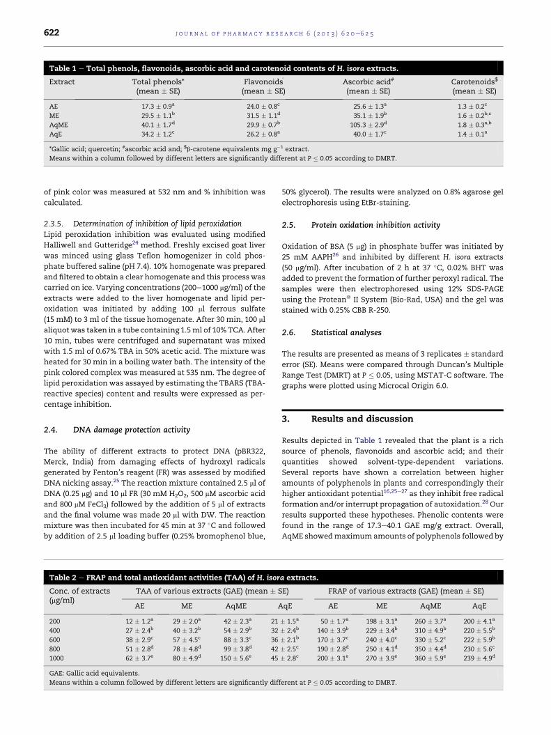
biological end-point of oxidative damage and showed 97%

lipid peroxidation inhibition at 1 mg/ml concentration.

Extracts were evaluated for their oxidative damage pro-

tective activity against a model DNA pBR322 and the results

are illustrated in Fig. 4. Hydroxyl radicals generated by Fen-

ton’s reaction are known to cause DNA damage as in the

present investigation, Fig. 4 (lane 3) showed absence of DNA

band and only a smear of degraded DNAwas observed. All the

extracts except methanol showed observable protection of

DNA intactness. Free radicals are known for DNA strand

breaking and damage which eventually contributes to carci-

nogenesis, mutagenesis and cytotoxicity.16 Various re-

searchers have reported the similar results and used plant

extracts and fractions for DNA protection against oxidative

damage.16,28 One of the interesting finding of present study

was that ME did not show significant DNA protection activity

which can be attributed to its inability to scavenge OH radicals

(Fig. 2).

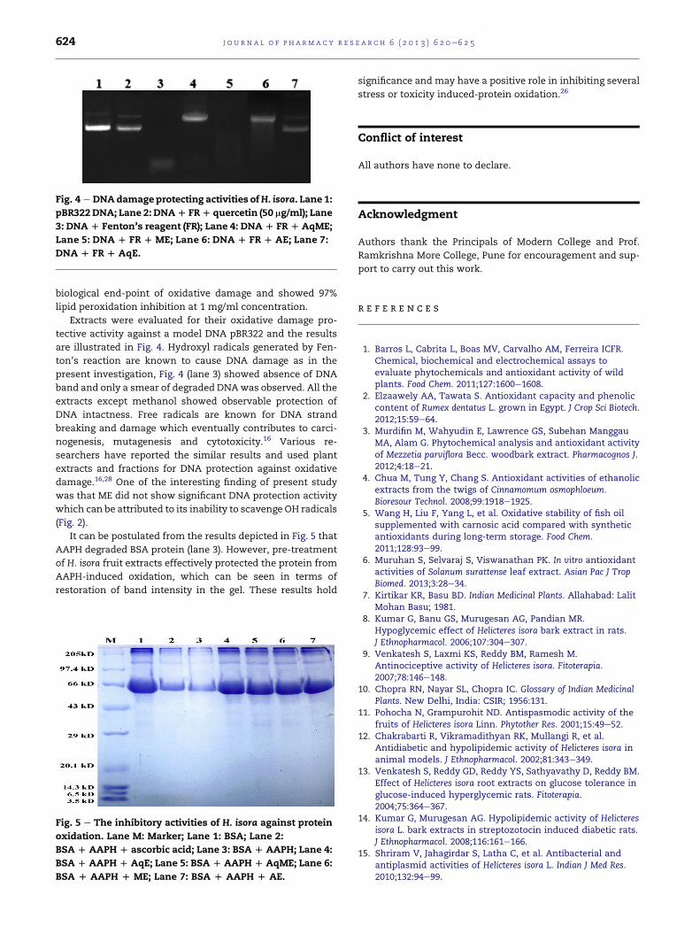
It can be postulated from the results depicted in Fig. 5 that

AAPH degraded BSA protein (lane 3). However, pre-treatment

of H. isora fruit extracts effectively protected the protein from

AAPH-induced oxidation, which can be seen in terms of

restoration of band intensity in the gel. These results hold

Fig. 5 e The inhibitory activities of H. isora against protein

oxidation. Lane M: Marker; Lane 1: BSA; Lane 2:

BSA D AAPH D ascorbic acid; Lane 3: BSA D AAPH; Lane 4:

BSA D AAPH D AqE; Lane 5: BSA D AAPH D AqME; Lane 6:

BSA D AAPH D ME; Lane 7: BSA D AAPH D AE.

significance and may have a positive role in inhibiting several

stress or toxicity induced-protein oxidation.26

Conflict of interest

All authors have none to declare.

Acknowledgment

Authors thank the Principals of Modern College and Prof.

Ramkrishna More College, Pune for encouragement and sup-

port to carry out this work.

r e f e r e n c e s

1. Barros L, Cabrita L, Boas MV, Carvalho AM, Ferreira ICFR.Chemical, biochemical and electrochemical assays toevaluate phytochemicals and antioxidant activity of wildplants. Food Chem. 2011;127:1600e1608.

2. Elzaawely AA, Tawata S. Antioxidant capacity and phenoliccontent of Rumex dentatus L. grown in Egypt. J Crop Sci Biotech.2012;15:59e64.

3. Murdifin M, Wahyudin E, Lawrence GS, Subehan ManggauMA, Alam G. Phytochemical analysis and antioxidant activityof Mezzetia parviflora Becc. woodbark extract. Pharmacognos J.2012;4:18e21.

4. Chua M, Tung Y, Chang S. Antioxidant activities of ethanolicextracts from the twigs of Cinnamomum osmophloeum.Bioresour Technol. 2008;99:1918e1925.

5. Wang H, Liu F, Yang L, et al. Oxidative stability of fish oilsupplemented with carnosic acid compared with syntheticantioxidants during long-term storage. Food Chem.2011;128:93e99.

6. Muruhan S, Selvaraj S, Viswanathan PK. In vitro antioxidantactivities of Solanum surattense leaf extract. Asian Pac J TropBiomed. 2013;3:28e34.

7. Kirtikar KR, Basu BD. Indian Medicinal Plants. Allahabad: LalitMohan Basu; 1981.

8. Kumar G, Banu GS, Murugesan AG, Pandian MR.Hypoglycemic effect of Helicteres isora bark extract in rats.J Ethnopharmacol. 2006;107:304e307.

9. Venkatesh S, Laxmi KS, Reddy BM, Ramesh M.Antinociceptive activity of Helicteres isora. Fitoterapia.2007;78:146e148.

10. Chopra RN, Nayar SL, Chopra IC. Glossary of Indian MedicinalPlants. New Delhi, India: CSIR; 1956:131.

11. Pohocha N, Grampurohit ND. Antispasmodic activity of thefruits of Helicteres isora Linn. Phytother Res. 2001;15:49e52.

12. Chakrabarti R, Vikramadithyan RK, Mullangi R, et al.Antidiabetic and hypolipidemic activity of Helicteres isora inanimal models. J Ethnopharmacol. 2002;81:343e349.

13. Venkatesh S, Reddy GD, Reddy YS, Sathyavathy D, Reddy BM.Effect of Helicteres isora root extracts on glucose tolerance inglucose-induced hyperglycemic rats. Fitoterapia.2004;75:364e367.

14. Kumar G, Murugesan AG. Hypolipidemic activity of Helicteresisora L. bark extracts in streptozotocin induced diabetic rats.J Ethnopharmacol. 2008;116:161e166.

15. Shriram V, Jahagirdar S, Latha C, et al. Antibacterial andantiplasmid activities of Helicteres isora L. Indian J Med Res.2010;132:94e99.

j o u r n a l o f p h a rm a c y r e s e a r c h 6 ( 2 0 1 3 ) 6 2 0e6 2 5 625

16. Gul M, Bhakshu L, Ahmed F, Anand K, Qureshi I, Ghazi I.Evaluation of Abelmoschus moschatus extracts for antioxidant,free radical scavenging, antimicrobial and antiproliferativeactivities using in vitro assays. BMC Complement Altern Med.2011;11:64.

17. Marinova D, Ribarova F, Atanassova M. Total phenolics andtotal flavonoids in Bulgarian fruits and vegetables. J Univ ChemTechnol Metall. 2005;40:255e260.

18. Kapur A, Haskovic A, Copra-Janicijevic A, et al.Spectrophotometric analysis of total ascorbic acid content invarious fruits and vegetables. Bull Chem Technol BosniaHerzegovina. 2012;38:40e43.

19. Jensen A. Chlorophyll and carotenoids. In: Hallebust JA,Craigie JS, eds. Handbook of Physiochemical and BiochemicalMethods. Cambridge: Cambridge University Press; 1978:5e70.

20. Prieto P, Pineda M, Aguilar M. Spectrophotometricquantitation of antioxidant capacity through the formation ofphosphomolybdenum complex: specific application todetermination of vitamin. Anal Biochem. 1999;269:337e341.

21. Benzie IF, Strain JJ. Ferric reducing antioxidant power assay:direct measure of total antioxidant activity of total biologicalfluids and modified version for simultaneous measurementof total antioxidant power and ascorbic acid concentration.Meth Enzymol. 1999;299:15e27.

22. Braca A, Sortino C, Politi M, Morelli I, Mendez J. Antioxidantactivity of flavonoids from Licania licaniaeflora. JEthnopharmacol. 2002;79:379e381.

23. Kunchandy E, Rao MNA. Oxygen radical scavenging activity ofcurcuminoid. Int J Pharm. 1990;58:235e242.

24. Halliwell B, Gutteridge JMC. Protection against lipidperoxidation. In: Free Radicals in Biology and Medicine. Tokyo:Japan Scientific Societies Press; 1989:2.

25. Lee JC, Kim HR, Kim J, Jang YS. Antioxidant property of anethanol extract of the stem of Opuntia ficus-indica var. SabotenJ Agric Food Chem. 2002;50:6490e6496.

26. Kumar PG, Navya K, Ramya EM, Venkataramana M, Anand T,Anilakumar CR. DNA damage protecting and free radicalscavenging properties of Terminalia arjuna bark in PC-12 cellsand plasmid DNA. Free Rad Antiox. 2013. http://dx.doi.org/10.1016/j.fra.2013.04.001.

27. Senthilkumar R, Parimelazhagan T, Chaurasia OP,Srivastava RB. Free radical scavenging property andantiproliferative activity of Rhodiola imbricata Edgew extractsin HT-29 human colon cancer cells. Asian Pac J Trop Med;2013:11e19.

28. Chaudhary A, Rampal G, Sharma U, et al. Anticancer,antioxidant activities and GC-MS analysis of glucosinolates intwo cultivars of broccoli. Med Chem Drug Dis. 2012;2:30e37.


Recommended