1Department of Biology, Faculty of Basic Sciences, Shahed University, Tehran, Iran2Department of Clinical Biochemistry, Faculty of Medicine, Shahid Beheshti University of Medical Sciences, Tehran, Iran
3Department of Biological Sciences, Faculty of Science, Imam Hossein University, Tehran, Iran
1. IntroductionHelicobacter pylori is a major human pathogen. It has been estimated that half of the world’s population is infected with this bacterium (1,2). H. pylori colonizes human gastric mucosa and causes gastritis, duodenal ulcers, and even gastric cancer (1–3). Treatment of H. pylori infection with antibiotics leads to increased risk of antibiotic resistance. The high cost of available treatment measures and the increased number of reported relapses generate the need for new alternative therapeutic approaches (4). Urease enzyme is an important virulence factor since it allows for survival under acidic conditions and the possibility of H. pylori colonization (5,6). The UreC subunit of urease enzyme exists in catalytic sites, showing great vaccine potential and antibody development for treatment of H. pylori infection (4,7). Classic antibodies have functional limitations such as interaction with the immune system and inadequate pharmacokinetics or tissue accessibility. Camels and sharks produce heavy-chain antibodies (8,9). The variable domain of heavy-chain antibodies (VHH),
also called nanobodies, is the smallest (~15 kDa) available intact antigen-binding fragment. The heavy-chain antibodies are less antigenic as compared to conventional antibodies (4,9–11). High solubility and low aggregation propensity, easy cloning, suitability for display systems, and resistance to temperature and pH are other advantages of nanobodies (9,10,12). Nanobodies’ efficiency for the treatment of intestinal infections such as retroviral intestinal infections has been investigated (13). The VHH against the UreC subunit of urease was previously produced and expressed in Escherichia coli TOP10 (4).
Yeast expression systems have emerged as heterologous expression hosts for several reasons. As eukaryotic systems, they are capable of performing many eukaryote-specific posttranslational modifications such as folding, glycosylation, and disulfide bond formation (14,15). Among yeasts, Pichia pastoris has become increasingly popular in recent years for protein expression (14). This methylotrophic yeast has a promoter derived from the alcohol oxidase 1 (AOX1) gene, which is one of strongest
Background/aim: Helicobacter pylori is a major health problem. One of the therapeutic approaches is administration of antibody against H. pylori. The methylotrophic Pichia pastoris is a suitable host for expression of recombinant antibody fragments. The aims of this study were the expression and the evaluation of camelid nanobody in the yeast Pichia pastoris.
Materials and methods: The camelid-derived heavy-chain antibody (nanobody) against the UreC subunit of urease from H. pylori was subcloned in the pPink-HC shuttle vector and transferred into Escherichia coli TOP10. After digestion and purification, the shuttle vector was transformed in the PichiaPink expression system. The expression was evaluated in an in vitro system.
Results: The yield of the nanobody expressed in P. pastoris was estimated to be 5 mg/L as compared to 2 mg/L expressed by E. coli. The nanobody was purified and binding affinity to the UreC antigen was evaluated using ELISA. Neutralization abilities of the two nanobodies expressed in yeast and E. coli were compared. The yeast-expressed nanobody specifically detected recombinant UreC and inhibited urease activity with high efficiency.
Conclusion: The results suggest attribution of the enhanced quality and quantity of the nanobody produced in P. pastoris to better posttranslational modification and folding in the yeast cell.
Received: 26.09.2015 Accepted/Published Online: 31.08.2016 Final Version: 18.04.2017
Research Article
696
POURASADI et al. / Turk J Med Sci
and most tightly regulated promoters (16,17). The length of the oligosaccharide chains added posttranslationally to protein in P. pastoris is much shorter than those in S. cerevisiae; thus, glycoproteins generated in P. pastoris are more suitable for therapeutic use (17). P. pastoris yeast has the ability of large-scale production of heterologous proteins in fermenters (18).
In order to increase the production and inhibitory effects of VHH antibodies, in this study a nanobody against the UreC subunit of urease was produced in a P. pastoris strain by the PichiaPink expression system. The yields of VHH antibody expressed in the PichiaPink expression system and E. coli were compared. In vitro neutralization of the nanobody in both the yeast and the bacterium was also investigated.
2. Materials and methodsThis experimental study was done at Shahed University’s biotechnology laboratory.2.1. Expression and purification of recombinant UreC antigenpET28a harboring the ureC gene was obtained from our previous study (7). The recombinant antigen induced with isopropyl β-D-1-thiogalactopyranoside (IPTG) was expressed in modified E. coli strain BL21(DE3) (from Novagen) at 37 °C. The UreC protein containing the His-tag was purified by Ni-NTA affinity agarose chromatography under both native and denatured conditions.2.2. VHH amplification and subcloningThe VHH gene fragment was amplified from the pET28a vector by PCR method using the EcoRI restriction site and Kozak sequence in the forward primer (5’-CTAGAATTCGAAACGATGGAGGTGCAG CTGSWGSAKTCKG-3’) and the KpnI restriction sites in the reverse primer (5’-CTAGGTACCTGA CACCACC ACCACCACCACGGAGACGGTGACCWGGG-3’). The PCR product was purified using a PCR product purification kit (Bioneer, South Korea). VHH fragments and the pPink-HC vector were digested with EcoRI and KpnI and purified before proceeding for ligation reaction. Ligation was performed with T4 DNA ligase overnight at 12 °C and the mixture was transformed into freshly prepared competent E. coli TOP10 cells. Positive clones were confirmed with colony PCR and restriction digestion analysis.2.3. Transformation and screening of P. pastorisIn order to promote integration into the P. pastoris genome, the pPink-HC vectors carrying the VHH gene were linearized with Vha4641 enzyme (isoschizomer of AflII) and then purified by ethanol precipitation. The PichiaPink Expression strain was grown at 30 °C and 250 rpm in YPD medium until an A600 nm of 1.5 was reached. The cells were
then harvested and washed two times with sterile ice-cold water and resuspended in 2 mL of 1 M sterile ice-cold sorbitol. The cells were centrifuged and resuspended in 60 µL of ice-cold, sterile 1 M sorbitol. Approximately 10 µg of linearized construct was added to the cell suspension in an electroporation cuvette and incubated on ice for 5 min. The cells were pulsed in the electroporator according to the manufacturer’s instructions, and then 1 mL of ice-cold YPDS medium was added to the cuvette and incubated at 28 °C. Next, 300 µL of the cell mixture was taken from the cuvette and spread on minimal dextrose agar and incubated at 30 °C until distinct colonies were formed. White colonies were picked up and plasmid integration into the yeast genome was confirmed by PCR.2.4. Nanobody expression and analysis For expression of the nanobody, 10 mL of BMGY medium (buffered glycerol-complex medium: 1% yeast extract; 2% peptone; 100 mM potassium phosphate, pH 6.0; 1.34% yeast nitrogen base (YNB); 0.0004% biotin; 1% glycerol) was inoculated with isolated clones and grown at 30 °C with vigorous shaking at 250 rpm for 48 h. The cells were then transferred into 100 mL of BMGY medium and grown at 30 °C and 250 rpm until OD600 0.6 was reached. The cultures were centrifuged at 1500 × g for 5 min at room temperature. The cells were resuspended in 40 mL of BMMY medium (buffered methanol-complex medium: 1% yeast extract; 2% peptone; 100 mM potassium phosphate, pH 6.0; 1.34% YNB; 0.0004% biotin; 0.5% methanol) in order to induce the expression of the nanobody. Cultures were grown for 96 h at 30 °C and 250 rpm with the addition of methanol to a final concentration of 0.5% v/v every 24 h. The cells were harvested by centrifugation at 2000 × g for 10 min. The pellet was resuspended in breaking buffer (50 mM sodium phosphate, pH 7.4; 1 mM PMSF; 1 mM EDTA; 5% glycerol) and homogenized with acid-washed glass beads. The mixture was centrifuged at 13,000 × g for 20 min and clear supernatant was collected.2.5. SDS-PAGE analysis and dot blot techniqueThe expression of the nanobody was studied on 15% SDS-PAGE under reducing conditions. For dot blotting, 1 µg of nanobody expressed in yeast was transferred to nitrocellulose paper. Recombinant UreC antigen containing His-tag fusion at the N-terminal was used as the positive control. The membranes were dried and blocked with 3% BSA in PBST [10 mM phosphate buffer, pH 7.4; 150 mM NaCl; 0.05% (v/v) Tween-20]. After 16 h the membranes were washed and the membrane containing yeast-produced nanobody was incubated with 10 µg/mL of UreC antigen solution for 2 h at 37 °C with mild agitation. Antibody–antigen reaction was detected with a 1/5000 dilution of HRP conjugated with anti-His Tag antibody (Roche, Germany) and DAB (Bangalore GeNei, India) as a substrate.
697
POURASADI et al. / Turk J Med Sci
2.6. Affinity measurement of nanobody against UreC antigenVHH nanobody binding affinity to the UreC antigen containing His-tag was evaluated using ELISA testing as described by Beatty et al. (19). Various concentrations (5, 10, 15, and 20 µg/mL) of nanobody produced in yeast were coated on a 96-well microplate and incubated at 4 °C overnight. After washing six times with PBST, the wells were blocked with 5% skim milk in PBST and incubated for 1 h at 37 °C, and the UreC antigen was added at 5, 10, 15, and 20 µg/mL concentrations and incubated at 4 °C overnight. After washing with PBST, 100 µL of a 1/10,000 dilution of HRP-conjugated anti-His antibody was added and incubated for 1 h at 37 °C. The immune reactivity was developed with 100 µL of 3,3’,5,5’-tetramethylbenzidine chromogenic substrate. The reaction was stopped with 3 N H2SO4 and optical density was measured at 450 nm using an auto microplate reader. Each experiment was performed in triplicate.2.7. Comparison of VHH expression in PichiaPink and E. coliVHH nanobody expression in E. coli strain BL21(DE3) was induced with 1 mM IPTG for 16 h at 28 °C at an OD600 of 0.5 as described previously. In order to compare the yield of VHH nanobody expressed in PichiaPink and E. coli, a test was designed using an equal amount of yeast and bacterial cells. After determining the number of cells in a specific volume of the medium, an equal number of bacterial and yeast cells were broken and their expression levels were analyzed using SDS-PAGE.2.8. In vitro neutralization of nanobody expressed in yeast and bacteriaThe H. pylori reference strain (Sydney strain: SS1) was cultured in Brucella agar medium (Difco). Colonies were transferred into brain-heart infusion broth and kept under microaerophilic conditions for 24 h at 37 °C. From those colonies, 109 cfu was mixed with 0–20 µg concentrations of each UreC VHH in PBS and incubated in microplate wells for 16 h at 4 °C. BSA was used as a negative control, and 100 µL of PBS buffer containing 10% urea and 15 g/L phenol red (pH 7.0) was added to each well and wells were incubated at room temperature for 30 min. Color development was measured at OD550 nm and measurements were repeated every 30 min for 3 h. Inhibition percentage was calculated by the following equation:
% inhibition = [(enzymatic activity without nanobody – enzymatic activity with nanobody) / (enzymatic activity without nanobody)] × 100.
Each experiment was performed in triplicate.2.9. Statistical analysisData were reported as mean ± SD and statistical analysis was performed using one-way ANOVA (SPSS 16.0). The
significance (P < 0.01) of differences were assessed by post hoc comparison of means using the lowest significant differences (Duncan).
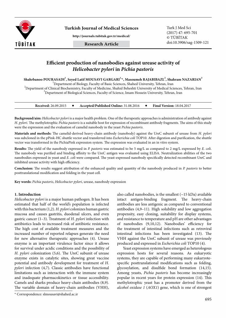
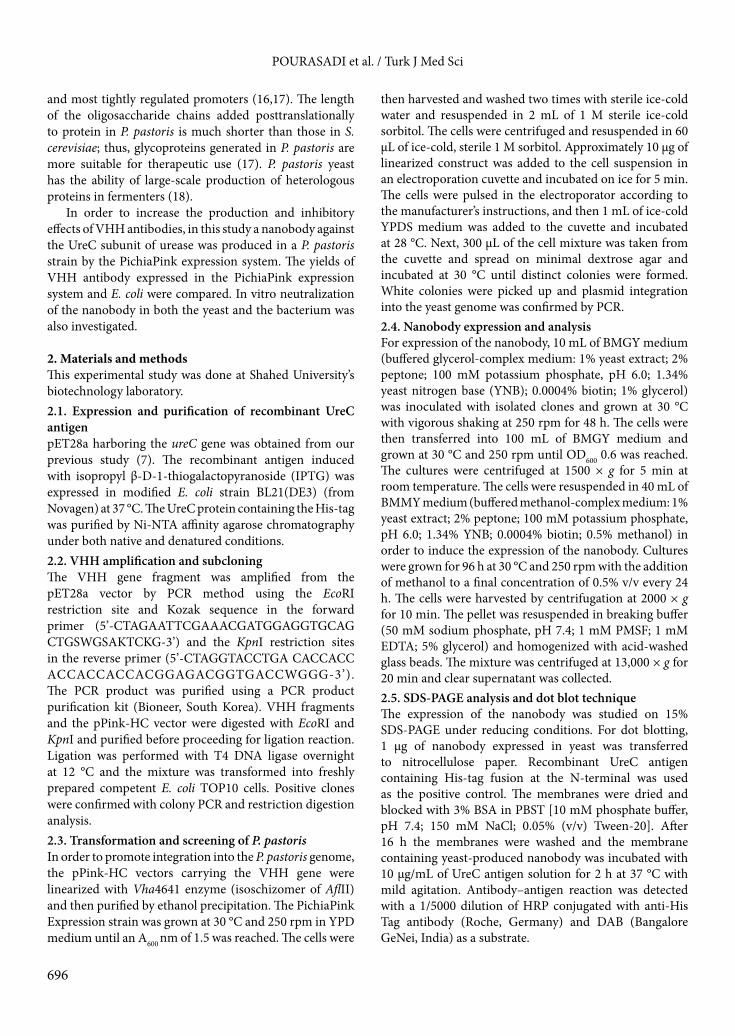
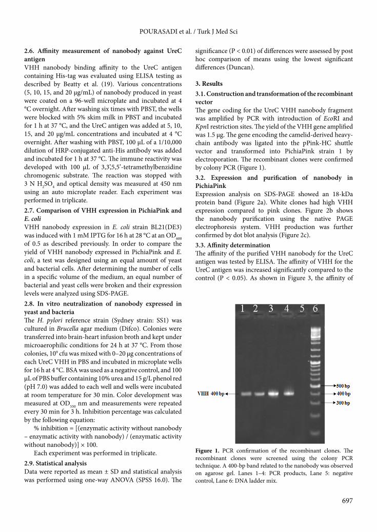
3. Results3.1. Construction and transformation of the recombinant vectorThe gene coding for the UreC VHH nanobody fragment was amplified by PCR with introduction of EcoRI and KpnI restriction sites. The yield of the VHH gene amplified was 1.5 µg. The gene encoding the camelid-derived heavy-chain antibody was ligated into the pPink-HC shuttle vector and transformed into PichiaPink strain 1 by electroporation. The recombinant clones were confirmed by colony PCR (Figure 1).3.2. Expression and purification of nanobody in PichiaPinkExpression analysis on SDS-PAGE showed an 18-kDa protein band (Figure 2a). White clones had high VHH expression compared to pink clones. Figure 2b shows the nanobody purification using the native PAGE electrophoresis system. VHH production was further confirmed by dot blot analysis (Figure 2c).3.3. Affinity determinationThe affinity of the purified VHH nanobody for the UreC antigen was tested by ELISA. The affinity of VHH for the UreC antigen was increased significantly compared to the control (P < 0.05). As shown in Figure 3, the affinity of
Figure 1. PCR confirmation of the recombinant clones. The recombinant clones were screened using the colony PCR technique. A 400-bp band related to the nanobody was observed on agarose gel. Lanes 1–4: PCR products, Lane 5: negative control, Lane 6: DNA ladder mix.
698
POURASADI et al. / Turk J Med Sci
Figure 2. Production, purification, and dot blot analysis of VHH produced in PichiaPink. a) SDS-PAGE gel stained with Coomassie blue, Lane 1: negative control (cell lysate before induction), Lane 2: VHH expressed in PichiaPink, Lane 3: molecular weight marker. b) Purification of recombinant VHH, Lane 1: molecular weight marker, Lane 2: the purified VHH protein. c) Dot blot analysis of VHH, Lane 1: positive control (the specific reaction of the anti-His tag antibody with bacterial nanobody), Lane 2: the specific reaction of the anti-His tag antibody with yeast nanobody, Lane 3: negative control.
Figure 3. Binding assay of nanobody with recombinant UreC antigen.
699
POURASADI et al. / Turk J Med Sci
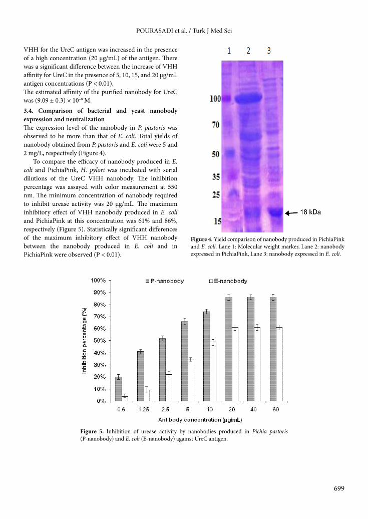
VHH for the UreC antigen was increased in the presence of a high concentration (20 µg/mL) of the antigen. There was a significant difference between the increase of VHH affinity for UreC in the presence of 5, 10, 15, and 20 µg/mL antigen concentrations (P < 0.01). The estimated affinity of the purified nanobody for UreC was (9.09 ± 0.3) × 10–8 M.3.4. Comparison of bacterial and yeast nanobody expression and neutralizationThe expression level of the nanobody in P. pastoris was observed to be more than that of E. coli. Total yields of nanobody obtained from P. pastoris and E. coli were 5 and 2 mg/L, respectively (Figure 4).
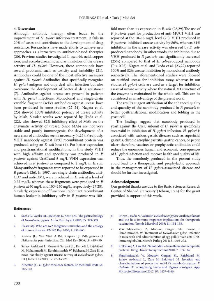
To compare the efficacy of nanobody produced in E. coli and PichiaPink, H. pylori was incubated with serial dilutions of the UreC VHH nanobody. The inhibition percentage was assayed with color measurement at 550 nm. The minimum concentration of nanobody required to inhibit urease activity was 20 µg/mL. The maximum inhibitory effect of VHH nanobody produced in E. coli and PichiaPink at this concentration was 61% and 86%, respectively (Figure 5). Statistically significant differences of the maximum inhibitory effect of VHH nanobody between the nanobody produced in E. coli and in PichiaPink were observed (P < 0.01).
Figure 4. Yield comparison of nanobody produced in PichiaPink and E. coli. Lane 1: Molecular weight marker, Lane 2: nanobody expressed in PichiaPink, Lane 3: nanobody expressed in E. coli.
Figure 5. Inhibition of urease activity by nanobodies produced in Pichia pastoris (P-nanobody) and E. coli (E-nanobody) against UreC antigen.
700
POURASADI et al. / Turk J Med Sci
4. DiscussionAlthough antibiotic therapy often leads to the improvement of H. pylori infection treatment, it fails in 20% of cases and contributes to the development of drug resistance. Researchers have made efforts to achieve new approaches as alternatives to antibiotic-based therapies (20). Previous studies investigated L-ascorbic acid, copper ions, and acetohydroxamic acid as inhibitors of the urease activity of H. pylori. However, these compounds have several problems, such as toxicity and instability (21). Antibodies could be one of the most effective measures against H. pylori. Antibodies that specifically recognize H. pylori antigens not only deal with infection but also overcome the development of bacterial drug resistance (7). Antibodies against urease are present in patients with H. pylori infections. Monoclonal and single-chain variable fragment (scFv) antibodies against urease have been produced in some studies (22–24). Nagata et al. (23) showed 100% inhibition potency of urease activity by MAb. Similar results were reported by Ikeda et al. (22), who showed 82% inhibitory effect of MAb on the enzymatic activity of urease. Since MAbs are not very stable and poorly immunogenic, the development of a new class of antibodies seems necessary (4,25). Previously, VHH nanobody against UreC recombinant protein was produced using an E. coli host (4). For better expression and posttranslational modifications, in this study VHH with high affinity and specificity was produced in P. pastoris against UreC and 5 mg/L VHH expression was achieved in P. pastoris as compared to 2 mg/L in E. coli. Many antibody fragments were reported to be expressed in P. pastoris (26). In 1997, two single-chain antibodies, anti-CD7 and anti-DMI, were produced in E. coli at a level of 0.25 mg/L, whereas these fragments were produced in P. pastoris at 60 mg/L and 100–250 mg/L, respectively (27,28). Similarly, expression of functional rabbit antirecombinant human leukemia inhibitory scFv in P. pastoris was 100-
fold more than its expression in E. coli (28,29).The use of P. pastoris yeast for production of anti-MUC1 VHH was reported at the 10–15 mg/L level (25). VHH produced in P. pastoris inhibited urease activity by 86%, whereas 61% inhibition in the urease activity was observed by E. coli-produced nanobody. In other words, the inhibition due to VHH produced in P. pastoris was significantly increased (25%) compared to that of E. coli-produced nanobody (P < 0.05). Nagata et al. and Ikeda et al. (23,22) reported 100% and 82% urease inhibition by monoclonal antibody, respectively. The aforementioned studies were focused on purified urease for inhibition assay, whereas in our studies H. pylori cells are used as a target for inhibition assay of urease activity where the natural 3D structure of the enzyme is maintained in the whole cell. This can be considered as an advantage of the present work.
The results suggest attribution of the enhanced quality and quantity of the nanobody produced in P. pastoris to better posttranslational modification and folding in the yeast cell.
The findings suggest that nanobody produced in yeast against the UreC subunit of H. pylori is specifically successful in inhibition of H. pylori infection. H. pylori is associated with various gastric diseases such as superficial gastritis, chronic atrophic gastritis, gastric cancer, or peptic ulcer; therefore, vaccines or prophylactic antibodies could reduce the enormous human and economic consequences of H. pylori infection and improve health and quality of life.
Thus, the nanobody produced in the present study could lead to a therapeutic and prophylactic approach in the management of H. pylori-associated disease and should be further investigated.
Acknowledgment Our grateful thanks are due to the Basic Sciences Research Center of Shahed University (Tehran, Iran) for the grant provided in support of this work.
References
1. Sachs G, Weeks DL, Melchers K, Scott DR. The gastric biology of Helicobacter pylori. Annu Rev Physiol 2003; 65: 349-369.
2. Blaser MJ. Who are we? Indigenous microbes and the ecology of human diseases. EMBO Rep 2006; 7: 956-960.
3. Kusters JG, Van Vliet AHM, Kuipers EJ. Pathogenesis of Helicobacter pylori infection. Clin Med Rev 2006; 19: 449-490.
4. Safaee Ardekani L, Mousavi Gargari SL, Rasooli I, Rajabibazl M, Mohammadi M, Ebrahimizadeh W, Bakherad H, Zare H. A novel nanobody against urease activity of Helicobacter pylori. Int J Infect Dis 2013; 17: e723-e728.
5. Atherton JC. H. pylori virulence factors. Br Med Bull 1998; 54: 105-120.
6. Prinz C, Hafsi N, Voland P. Helicobacter pylori virulence factors and the host immune response: implications for therapeutic vaccination. Trends Microbiol 2003; 11: 134-138.
7. Veis Malekshahi Z, Mousavi Gargari SL, Rasooli I, Ebrahimizadeh W. Treatment of Helicobacter pylori infection in mice with oral administration of egg yolk-driven anti-UreC immunoglobulin. Microb Pathog 2011; 51: 366-372.
8. Kolkman JA, Law DA. Nanobodies – from llamas to therapeutic proteins. Drug Discov Today Technol 2010; 7: 139-146.
9. Ebrahimizadeh W, Mousavi Gargari SL, Rajabibazl M, Safaee Ardekani L, Zare H, Bakherad H. Isolation and characterization of protective anti-LPS nanobody against V. cholerae O1 recognizing Inaba and Ogawa serotypes. Appl Microbiol Biotechnol 2012; 97: 4457-4466.
10. Rajabibazl M, Rasaee MJ, Foruzandeh M, Rahimpour A, Kiani J, Rahbarizadeh F, Alirezapour B, Mohammadi M. Production of chimeric recombinant single domain antibody-green fluorescent fusion protein in Chinese hamster ovary cells. Hybridoma 2007; 26: 1-9.
11. Eyer L, Hruska K. Single-domain antibody fragments derived from heavy-chain antibodies: a review. Vet Med 2012; 57: 439-513.
12. Deffar K, Shi H, Li L, Wang X, Zhu X. Nanobodies - the new concept in antibody engineering. Afr J Biotechnol 2009; 8: 2645-2652.
13. Van der Vaart J, Pant N, Wolvers D, Bezemer S, Hermans P, Bellamy K. Reduction in morbidity of rotavirus induced diarrhoea in mice by yeast produced monovalent llama-derived antibody fragments. Vaccine 2006; 24: 4130-4137.
14. Cereghino GPL, Cregg JM. Applications of yeast in biotechnology: protein production and genetic analysis. Curr Opin Biotechnol 1999; 10: 422-427.
15. Daly R, Hearn MTW. Expression of heterologous proteins in Pichia pastoris: a useful experimental tool in protein engineering and production. J Mol Recognit 2004; 18: 119-138.
16. Cregg JM, Cereghino JL, Shi J, Higgins DR. Recombinant protein expression in Pichia pastoris. Mol Biotechnol 2000; 16: 23-52.
17. Balamurugan V, Reddy GR, Suryanaryana VVS. Pichia pastoris: a notable heterologous expression system for the production of foreign proteins—vaccines. Indian J Microbiol 2007; 6: 175-186.
18. Cereghino GPL, Cereghino JL, Ilgen C, Cregg JM. Production of recombinant proteins in fermenter cultures of the yeast Pichia pastoris. Curr Opin Biotechnol 2002; 13: 329-332.
20. Malfertheiner P, Megraud F, O’Morain C, Bazzoli F, El-Omar E, Graham D. Current concepts in the management of Helicobacter pylori infection: the Maastricht III consensus report. Gut 2007; 56: 772-781.
21. Houimel M, Mach JP, Corthesy-Theulaz I, Corthesy B, Fisch I. New inhibitors of Helicobacter pylori urease holoenzyme selected from phage-displayed peptide libraries. Eur J Biochem 1999; 262: 774-780.
22. Ikeda Y, Fujii R, Ogino K, Fukushima K, Hifumi E, Uda T. Immunological features and inhibitory effects on enzymatic activity of monoclonal antibodies against Helicobacter pylori urease. J Ferment Bioeng 1998; 86: 271-276.
23. Nagata K, Mizuta T, Tonokatu Y, Fukuda Y, Okamura H, Hayashi T, Shimoyama T, TamuraT. Monoclonal antibodies against the native urease of Helicobacter pylori: synergistic inhibition of urease activity by monoclonal antibody combinations. Infect Immun 1992; 60: 4826-4831.
24. Yanan W, Quanming Z, Ping L, Gang G, Xiaoqin W. Preparation of anti-Helicobacter pylori UreB monoclonal antibody and the detection of the ability of inhibition the urease activity. J Immunol 2005; 21: 393-396.
25. Rahbarizadeh F, Rasaee MJ, Forouzandeh M, Allameh AA. Over expression of anti-MUC1 single-domain antibody fragments in the yeast Pichia pastoris. Mol Immunol 2006; 43: 426-435.
26. Emberson LM, Trivett AJ, Blower PJ, Nicholls PJ. Expression of an anti-CD33 single-chain antibody by Pichia pastoris. J Immunol Methods 2005; 305: 135-151.
27. Eldin P, Pauza ME, Hieda Y, Lin G, Murtaugh MP, Pentel PR, Pennell AC. High-level secretion of two antibody single chain Fv fragments by Pichia pastoris. J Immunol Methods 1997; 201: 67-75.
28. Verma R, Boleti E, George AJT. Antibody engineering: comparison of bacterial, yeast, insect and mammalian expression systems. J Immunol Methods 1998; 216: 165-181.
29. Ridder R, Schmitz R, Legay F, Gram H. Generation of rabbit monoclonal antibody fragments from a combinatorial phage display library and their production in the yeast Pichia pastoris. Nat Biotechnol 1995; 13: 255-260.


![Nanobodies as Affinity Capture Reagents2019... · different nanobodies [Nb1, Nb1.1, Nb2, Nb3] B. Affinity capture experiment testing five different nanobodies [Nb 1, Nb1.1, Nb2, Nb3,](https://static.documents.pub/doc/80x56/5f4a16886e8dc261cd363d06/nanobodies-as-affinity-capture-reagents-2019-different-nanobodies-nb1-nb11.jpg)


