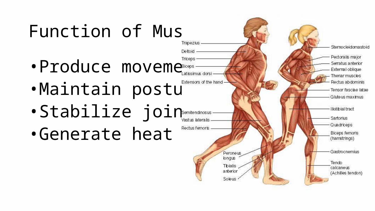
• Muscles are responsible for all types of body movement• Three basic muscle types are found in the body• Skeletal muscle• Cardiac muscle• Smooth muscle
Characteristics of Muscles
•Muscle cells are elongated (muscle cell = muscle fiber)• Contraction of muscles is due to the movement of
microfilaments• All muscles share some terminology• Prefix myo refers to muscle• Prefix mys refers to muscle• Prefix sarco refers to flesh
Skeletal Muscle Characteristics
• Most are attached by tendons to bones• Cells are multinucleate• Striated – have visible banding• Voluntary – subject to conscious control• Cells are surrounded and bundled by connective tissue
Connective Tissue Wrappings of Skeletal Muscle• Endomysium – around
single muscle fiber• Perimysium – around a
fascicle (bundle) of fibers
Figure 6.1
Connective Tissue Wrappings of Skeletal Muscle• Epimysium – covers the
entire skeletal muscle• Fascia – on the outside of
the epimysium
Figure 6.1
Skeletal Muscle Attachments
• Epimysium blends into a connective tissue attachment• Tendon – cord-like structure• Aponeuroses – sheet-like structure
• Sites of muscle attachment• Bones• Cartilages• Connective tissue coverings
Smooth Muscle Characteristics
• Has no striations• Spindle-shaped cells• Single nucleus• Involuntary – no
conscious control• Found mainly in the
walls of hollow organs
Figure 6.2a
Cardiac Muscle Characteristics• Has striations• Usually has a single
nucleus• Joined to another muscle
cell at an intercalated disc• Involuntary• Found only in the heart
Figure 6.2b
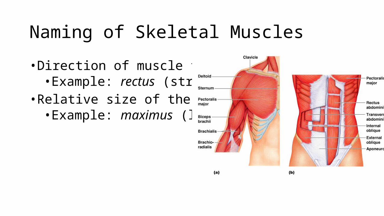
Naming of Skeletal Muscles
• Direction of muscle fibers• Example: rectus (straight)
• Relative size of the muscle• Example: maximus (largest)
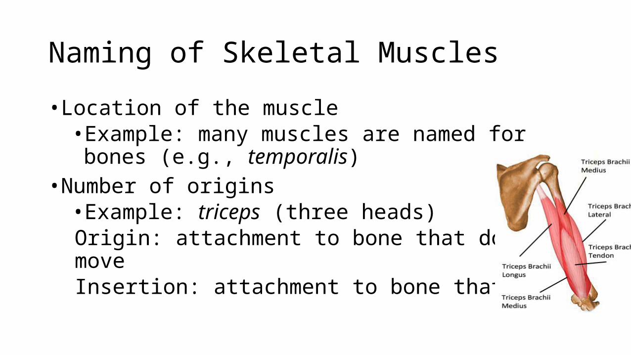
Naming of Skeletal Muscles
• Location of the muscle• Example: many muscles are named for bones (e.g., temporalis)
• Number of origins• Example: triceps (three heads)Origin: attachment to bone that does NOT moveInsertion: attachment to bone that MOVES
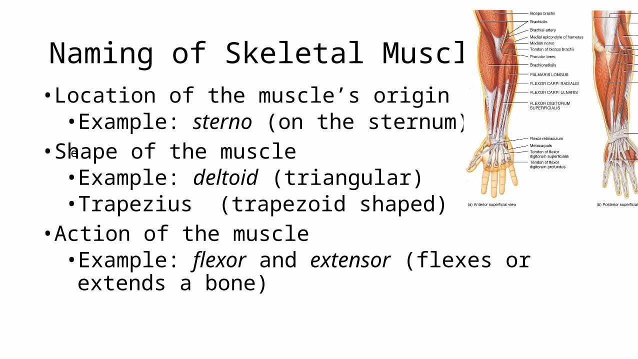
Naming of Skeletal Muscles• Location of the muscle’s origin and insertion• Example: sterno (on the sternum)
• Shape of the muscle• Example: deltoid (triangular)• Trapezius (trapezoid shaped)
• Action of the muscle• Example: flexor and extensor (flexes or extends a bone)
e
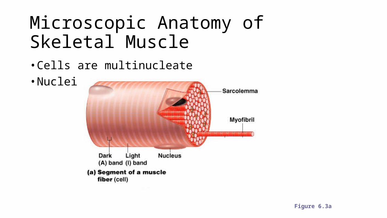
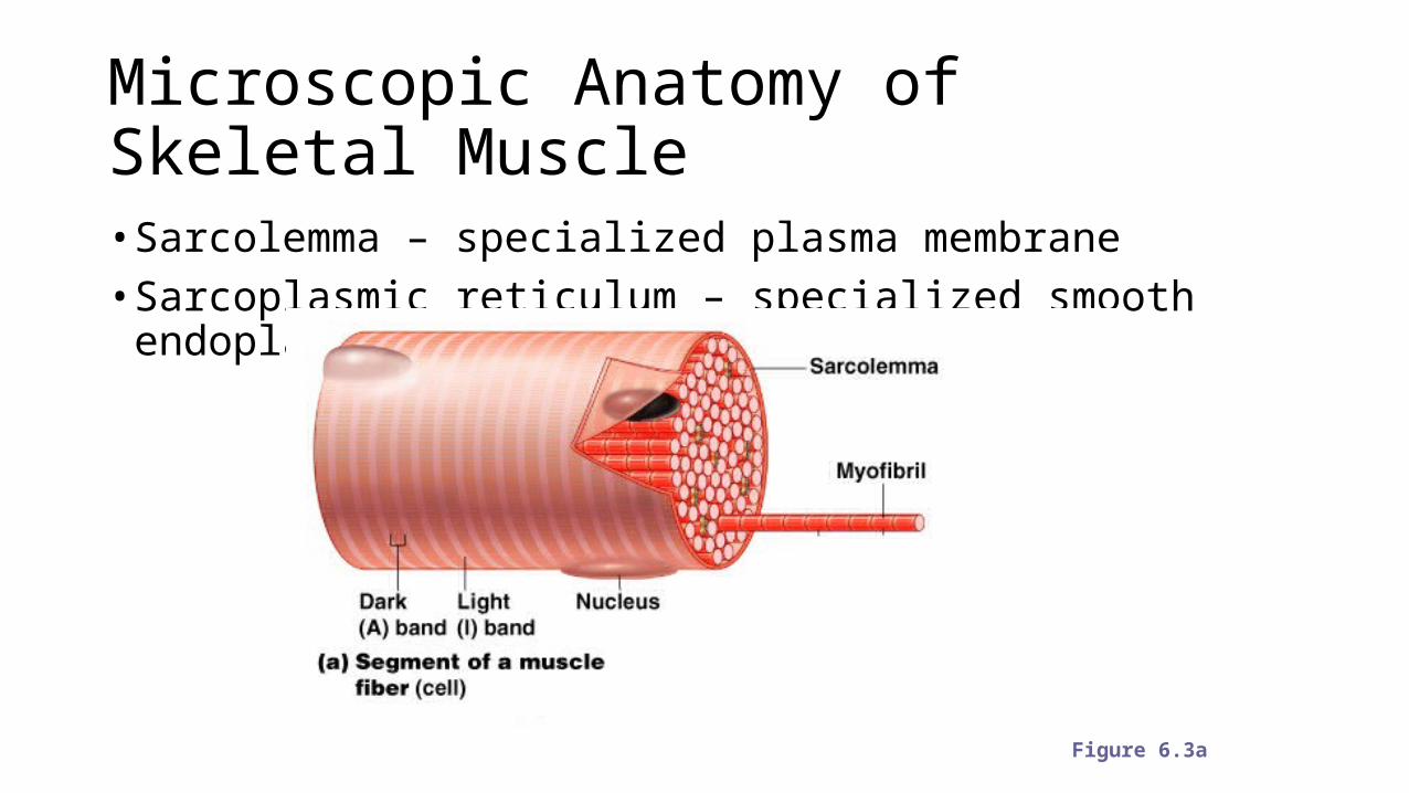
Microscopic Anatomy of Skeletal Muscle• Cells are multinucleate• Nuclei are just beneath the sarcolemma
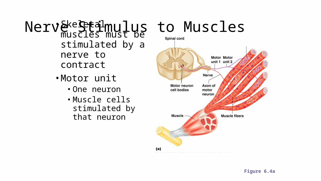
Nerve Stimulus to Muscles• Skeletal muscles must be stimulated by a nerve to contract• Motor unit• One neuron• Muscle cells
stimulated by that neuron
Figure 6.4a
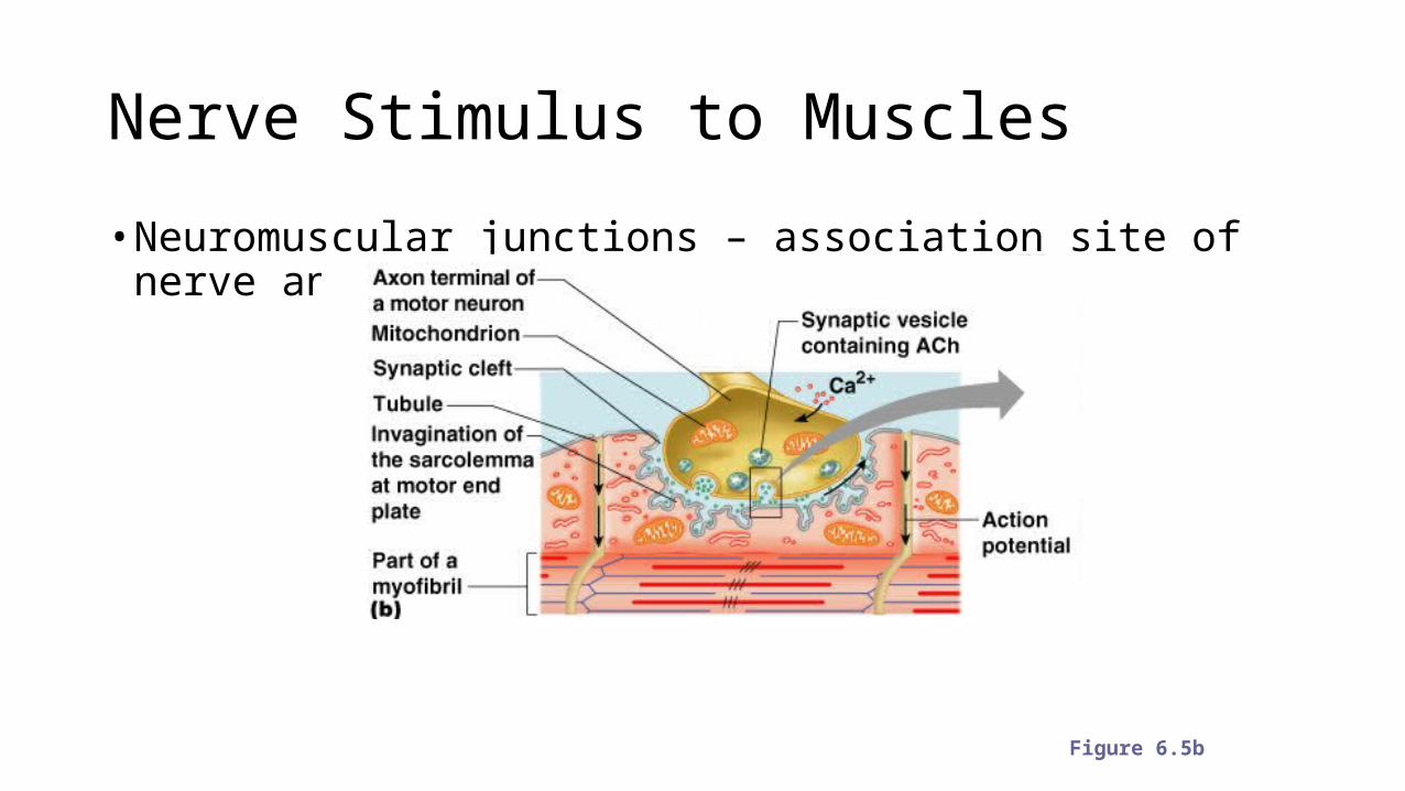
Nerve Stimulus to Muscles
• Neuromuscular junctions – association site of nerve and muscle
Figure 6.5b
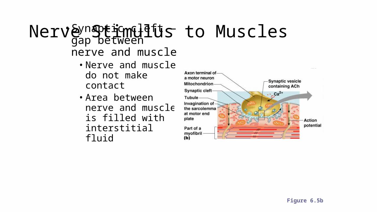
Nerve Stimulus to Muscles• Synaptic cleft – gap between nerve and muscle• Nerve and muscle do
not make contact• Area between nerve
and muscle is filled with interstitial fluid
Figure 6.5b
Transmission of Nerve Impulse to Muscle• Neurotransmitter – chemical released by nerve upon arrival of nerve
impulse• The neurotransmitter for skeletal muscle is acetylcholine
• Neurotransmitter attaches to receptors on the sarcolemma• Sarcolemma becomes permeable to sodium (Na+)
Transmission of Nerve Impulse to Muscle• Sodium rushing into the cell generates an action potential• Once started, muscle contraction cannot be stopped