Electroactive 3D Materials for Cardiac Tissue Engineering Amy Gelmi 1, Jiabin Zhang 1, Artur Cieslar-Pobuda 2, Monika K. Ljunngren2, Marek Jan Los 2, Mehrdad Rafat 3, Edwin W.H. Jager 1 1 Biosensors and Bioelectronics Centre, Dept. of Physics, Chemistry and Biology (IFM), Linköping University, Linköping, Sweden 2 Division of Cell Biology, and Integrative Regenerative Medicine Center (IGEN), Department of Clinical and Experimental Medicine (IKE), Linköping University, Sweden 3Dept. of Biomedical Engineering, Linköping University, Linköping, Sweden
1. Abstract By-pass surgery and heart transplantation are traditionally used to restore the heart’s functionality after a myocardial Infarction (MI or heart attack) that results in scar tissue formation and impaired cardiac function. However, both procedures are associated with serious post-surgical complications. Therefore, new strategies to help re-establish heart functionality are necessary. Tissue engineering and stem cell therapy are the promising approaches that are being explored for the treatment of MI. The stem cell niche is extremely important for the proliferation and differentiation of stem cells and tissue regeneration. For the introduction of stem cells into the host tissue an artificial carrier such as a scaffold is preferred as direct injection of stem cells has resulted in fast stem cell death. Such scaffold will provide the proper microenvironment that can be altered electronically to provide temporal stimulation to the cells. We have developed an electroactive polymer (EAP) scaffold for cardiac tissue engineering. The EAP scaffold mimics the extracellular matrix and provides a 3D microenvironment that can be easily tuned during fabrication, such as controllable fibre dimensions, alignment, and coating. In addition, the scaffold can provide electrical and electromechanical stimulation to the stem cells which are important external stimuli to stem cell differentiation. We tested the initial biocompatibility of these scaffolds using cardiac progenitor cells (CPCs), and continued onto more sensitive induced pluripotent stem cells (iPS). We present the fabrication and characterisation of these electroactive fibres as well as the response of increasingly sensitive cell types to the scaffolds.
2. Introduction Myocardial infarction (MI), commonly referred to as a heart attack, is a leading cause of death worldwide with a high associated health care cost for survivors. After an MI the cardiac tissue damaged due to lack of oxygen causes a wound healing response that replaces the damaged cardiac tissue with non-contractile scar tissue. This results in reduced cardiac function, leading to reduced quality of life and further complications for the patient. Cardiac stem cell therapy is an approach that aims to replace and regenerate new functional cardiac tissue, through the introduction of targeted stem cells that will differentiate into cardiomyocytes around the affected areas within the heart. Stem cell therapy has advantages over current therapies, such as by-pass graft surgery or complete organ transplantation, as it does not require donor organs or complicated open heart surgeries. However, current clinical trials of cardiac stem cell therapy have been unsuccessful; high stem cell mortality within the first few days after injection and low retention are major contributors to this lack of clinical efficacy.[1] The cardiac environment is also quite difficult for injected cells to survive due to immune responses, inadequate vascularization, fibrosis and inadequate access to nutrients.[2,3] For these reasons, the direct injection of stem cells into cardiac tissue is not a beneficial approach; instead, delivering the stem cells on an implantable platform, or cardiac patch, would provide a more stable environment and allow the stem cells time to develop into effective tissue[4]. Grafting stem cells onto bio-engineered tissue scaffolds can address the majority of the issues that currently limit the efficacy of cardiac stem cell therapy. The use of electrospun fibres for the support of cardiac cells has an advantage due to similarity to extracellular matrix morphology, as well as the ability to tailor fibres in dimension, composition, and functionality. Different types of fibre materials have been used for stem cell graft materials, such as nano- and micro-sized fibres with different polymer compositions [5] and bio-functionalised fibres [6]. The 3-dimensional morphology provided by the fibres makes for a good basis for a cardiac patch. The introduction of an electroactive material to the fibres provides another aspect to the influence and control of the stem cells on the fibres[7]. Electroactive polymers (EAP), such as polypyrrole (PPy), are conductive and when reduced/oxidized they will mechanically actuate [8]. This provides the possibility to stimulate stem cells both electrically and mechanically while growing on the cardiac patch. Electrical and mechanical stimuli have demonstrated in the past to
Downloaded From: http://proceedings.spiedigitallibrary.org/ on 04/24/2015 Terms of Use: http://spiedl.org/terms
stimulate stem cells and to influence differentiation into cardiac type cells[9-11]. EAP coated fibres have been demonstrated to work as support for many types of cells, including neural, myogenic, and cardiac cells [12-15]. Following on from this foundation, we will produce EAP coated fibres using PPy to investigate the response from primary and stem cells. Our previous work has demonstrated the efficacy of using PPy materials prepared with dodecylbenzenesulfonate (DBS), as the polymer showed to be biocompatible with endothelial progenitor cells and cardiac progenitor cells (CPCs)[16]. CPCs are resident cardiac stem cells with the ability to generate cardiomyocytes, smooth muscle, and endothelial cells and have the potential to generate new functional cardiac tissue [17,18]. Hence, we begin this study observing PPy(DBS) coated fibre materials and the response from CPCs to observe how primary cells respond. We will then move onto iPS cells, which are generally more sensitive and difficult to culture successfully[19] but offer true pluripotency compared to the CPCs. Comparing the behavior of the two cell types will help elucidate the suitability of EAP coated fibres for cardiac tissue engineering.
3. Materials & methods
3.1. Scaffold fabrication The electroactive scaffolds are prepared in a step-by-step process as shown in the scheme in Figure 1. 50:50 poly(lactic-co-glycolic acid) was prepared as a 17.5% wt/wt solution in chloroform. The PLGA solution was electrospun at a voltage of 20 kV with a flow rate of 0.5 mL/hour with a throw distance of 120 mm(Fig.1A). The electrospun PLGA fibres were then collected and dried over night to evaporate any remaining solvent. The fibres were then coated with a solution of 5% wt/wt iron (III) chloride in methanol using a spincoater (WS-400B-6NPP/LITE, Laurell Tech. Corp., USA) with an initial step of 1000 RPM for 120 seconds, followed by 2500 RPM for 30 seconds (Fig. 1B). The FeCl3 coated fibres were then dried over night to evaporate any remaining solvent. The fibres are then exposed to pyrrole (Py) vapour in a sealed vessel at 50°C for 60 seconds (Fig. 1C). An aqueous monomer solution of 0.1M Py and 0.1 M dodecylbenzenesulfonic acid (TCI) was prepared. For electropolymerisation the aqueous pyrrole solutions were prepared with 0.1 M concentration of dopant (DBSA) and 0.1 M pyrrole. The VPP coated mesh was then placed into the aqueous pyrrole/dopant solution in a 3 point electrochemical cell (Fig. 1D) The counter electrode was a gold coated silicon wafer, and the reference a Ag/AgCl reference electrode. A constant potential of 0.67 V was applied to the electrochemical cell for 600 or 1800 sec. The ECP coated mesh was then lightly rinsed three times with DI water, dried gently with N2 gas, and stored in a Petri dish. All chemicals are supplied from Sigma Aldrich unless indicated otherwise.
Proc. of SPIE Vol. 9430 94301T-2
Downloaded From: http://proceedings.spiedigitallibrary.org/ on 04/24/2015 Terms of Use: http://spiedl.org/terms
Figure 1: EAPphase coating P
3.2. Charac
3.2.1. SEM The fibres we1550 scanninprepared for Swith MilliQ wfor SEM imag
3.2.2. ElectrThe input parvoltammetry become fully
3.3. Cell cul
3.3.1. CPC CPCs were ismedium usesupplemented(Invitrogen), All samples wwashing withto check the cell culture tecell maintenawell.
P fibre fabricatioPPy onto FeCl3
cterisation
ere sputter coang electron mSEM after beiwater (18.1Ω)ging.
rochemistry rameters were(CV) VPP ohydrated. Th
lture
solated from td was Dulbd with 10 % 0.5 % DMSOwere firstly inh sterile PBS aefficacy of thesting. All decance medium w
on schematic; (A3 coated fibres,
ated with goldmicroscope (Ze
ing fixed to th), then dried v
e set at applier ECP samplee CVs were p
the hearts of ecco's Modif FCS, 1 % p
O (Sigma-Aldrncubated overaqueous solut
he bacterial decontaminated was added. C
A) electrospinnand (D) electro
d (30Å) to imeiss, Germanyhe fibre samplvia gradual eth
ed voltage ranes were soakeerformed in 7
adult mice usfied Eagle Mpenicillin – srich) and 20 ngrnight in 5 x cion. The mate
econtaminationsamples were
CPCs were col
ning PLGA fibropolymerising P
mprove conducy) with an elles with formahanol dehydra
nge of -1V toed in the PBS
7.4 pH PBS (S
sing a cardiacMedium: Nutstreptomycin g/ml Epidermconcentrated perials were then. If no microe place on thellected by tryp
es, (B) spin-coaPPy onto the VP
ctivity for SEMlectron beam aldehyde. The
ation and final
0.4V, 3 cyclS solution for
Sigma Aldrich
stem cells istrient Mixture(Invitrogen),
mal Growth Fapenicillin-strepen incubated fobial growth w bottom of a
psinization and
ating FeCl3 ontoPP coated fibres
M. The fibres energy of 5.0
e fibre and iPSlly sputter coa
les, and scan r at least 30 mtablet).
olation kit (Me F-12 (DM1X Insulin-T
actor (EGF) (Inptomycin solufor 24 h in stewas observed,12-well cell cd seeded at a
o PLGA fibres,s where anion A
were examine02 kV. The iS were then caated with a go
rate 50mV/s. minutes to all
Millipore). TheMEM/F12) (S
Transferrin-Senvitrogen). ution followederile antibiotic, the samples ulture plate andensity of 5 x
, (C) vapour A- is DBS.
ed in the LEOPS cells werearefully rinsedld layer (30Å
Before cycliclow the fibres
e maintenanceigma-Aldrichelenium (ITS
d by thoroughc-free mediumwere used fornd 1 ml of thex 10⁴ cells per
O e d )
c s
e ) )
h m r e r
Proc. of SPIE Vol. 9430 94301T-3
Downloaded From: http://proceedings.spiedigitallibrary.org/ on 04/24/2015 Terms of Use: http://spiedl.org/terms
After 3 dayViability/Cytdensity and aquantified usi
3.3.2. IPS Human inducmediated tranmaintained o(ReproCELLTechnologiesmg/ml collagsuspension in2-mercaptoetdifferentiationPDMS ring o
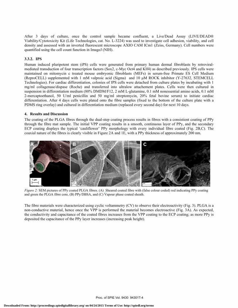
4. Results aThe coating othrough the fECP coating coaxial nature
Figure 2: SEMand green the P
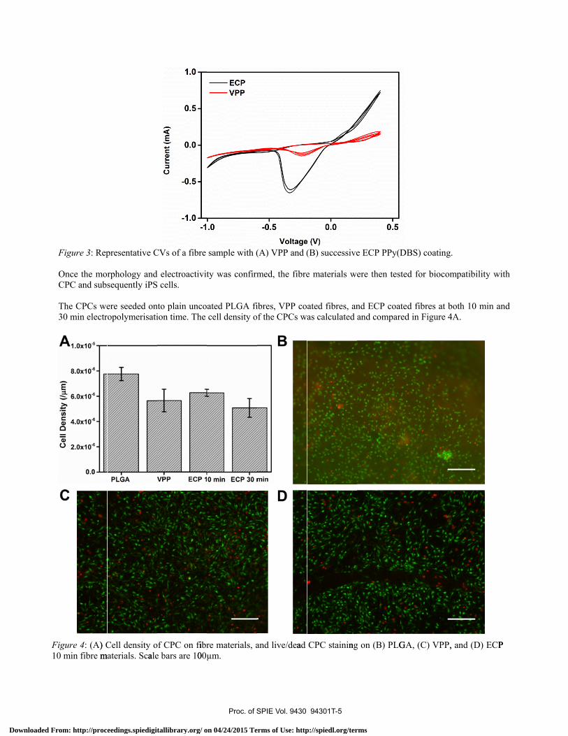
The fibre matnon-conductithe conductivdeposited the
ys of culturtotoxicity Kit assessed with ing the cell co
ced pluripotensduction of fon mitomycin) supplement
s). For cardiacgenase/dispasen differentiatiothanol, 50 U/n. After 4 da
overlay) and cu
and Discussionof the PLGA fibre mat samp
displays the e of the fibres
M pictures of PPPLGA fibre cor
terials were chve material, h
vity and capace capacitance o
re, once the(Life Technoan inverted fl
ount function i
nt stem (iPS)four transcriptn c treated med with 1 mMc differentiatioe (Roche) anon medium (8/ml penicillin
ays cells wereultured in diff
n fibres through
mple. The initiatypical ‘caul
s is clearly vis
Py coated PLGAre, (B) PPy/DBS
haracterized uhence once thcitance of the of the PPy lay
e control samlogies, cat. N
fluorescent miin ImageJ (NI
) cells were tion factors (S
mouse embryoM valproic acon, colonies ond transferred0% DMDM/F
n and 50 mge plated onto ferentiation m
h the dual-stepal VPP coatinliflower’ PPy ible in Figure
A fibres. (A) ShSA, and (C) Va
using cyclic vohe VPP is perfcoated fibres
yer increases (
mple becameo. L-3224) wacroscope AXI
IH).
generated froSox2, c-Myc Oonic fibroblascid (Sigma) of IPS cells wd into ultraloF12, 2 mM L-g/ml streptomthe fibre sam
medium (replac
p coating procng results in a
morphology 2A and 1E, w
heared coated fapour phase coa
oltammetry (Cformed the mincreases fromincreasing pea
e confluent, as used to invIO CAM ICm
om primary hOct4 and Klf4sts (MEFs) inand 10 μM R
were detached ow attacheme-glutamine, 0.mycin, 20% fmples (fixed toced every seco
cess results ina smooth, con
with every inwith a PPy thic
fibre with (falseated sheath.
CV) to observematerial becomm the VPP coak height).
a Live/Deavestigate cell am1 (Zeiss, Ger
human derma4) as describedn serum-free ROCK inhibitfrom culture
ent plates. Ce1 mM nonessfetal bovine o the bottom ond day) for n
n fibres with antinuous layer ndividual fibrckness of appr
e colour coded)
e their electromes electroacti
ating to the E
ad Assay (Ladhesion, viabrmany). Cell
al fibroblasts d previously. IPrimate ES
tor (Y-27632,plates by incuells were thesential amino aserum) to inof the culture
next 10 days.
a consistient cr of PPy, and re coated (Figroximately 20
red indicating P
oactivity (Fig. ive (Fig. 3A).
ECP coating; a
LIVE/DEAD®bility, and celnumbers were
by retroviralIPS cells wereCell Medium, STEMCELLubating with 1en cultured inacids, 0.1 mMnitiate cardiace plate with a
coating of PPythe secondaryg. 2B,C). The00 nm.
PPy coating
3). PLGA is a. As expectedas more PPy is
® ll e
-e
m L 1 n
M c a
y y e
a d, s
Proc. of SPIE Vol. 9430 94301T-4
Downloaded From: http://proceedings.spiedigitallibrary.org/ on 04/24/2015 Terms of Use: http://spiedl.org/terms
0.5
1.0
0.5
0.0
-0.5
-1.0
1
ECPVPP
.0 -0.5
Voltage
0.0
(V)
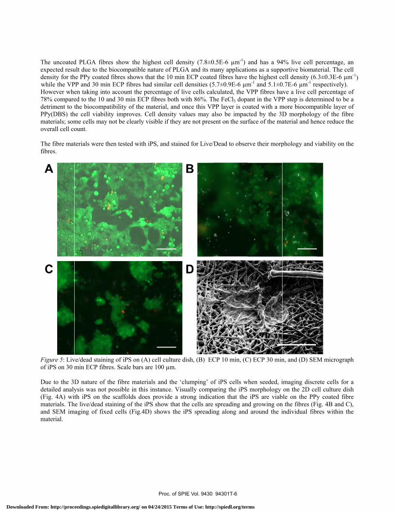
Figure 3: Rep Once the moCPC and sub The CPCs we30 min electr
presentative C
rphology andsequently iPS
ere seeded onropolymerisati
CVs of a fibre
d electroactivit cells.
nto plain uncoion time. The
sample with (
ty was confirm
ated PLGA ficell density o
(A) VPP and (
med, the fibre
ibres, VPP coof the CPCs w
(B) successive
e materials we
ated fibres, anas calculated
e ECP PPy(DB
ere then teste
nd ECP coateand compared
BS) coating.
d for biocomp
d fibres at bod in Figure 4A
patibility with
th 10 min andA.
h
d
Figure 4: (A)) Cell density of CPC on fiibre materials, and live/deaad CPC staininng on (B) PLGGA, (C) VPP,, and (D) ECPP 10 min fibre mmaterials. Scaale bars are 1000µm.
Proc. of SPIE Vol. 9430 94301T-5
Downloaded From: http://proceedings.spiedigitallibrary.org/ on 04/24/2015 Terms of Use: http://spiedl.org/terms
The uncoatedexpected resudensity for th 1) while the VHowever whe78% comparedetriment to tPPy(DBS) thmaterials; somoverall cell co The fibre matfibres.
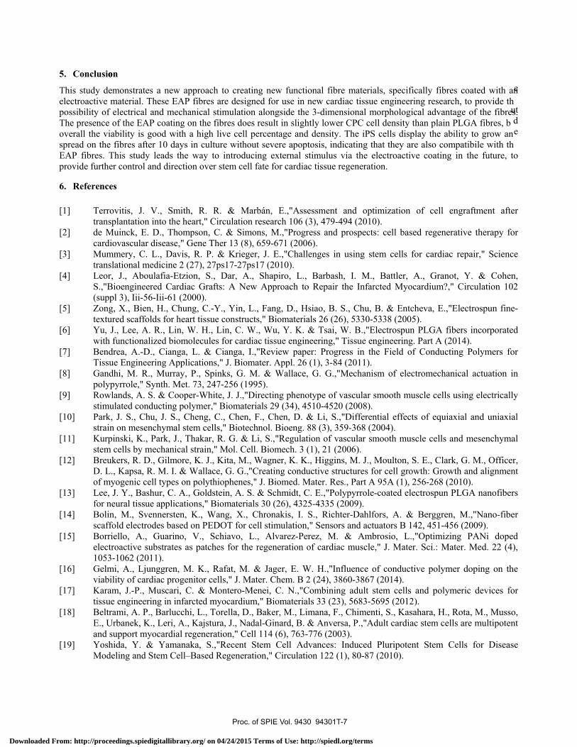
Figure 5: Livof iPS on 30 m Due to the 3Ddetailed analy(Fig. 4A) witmaterials. Thand SEM immaterial.
d PLGA fibreult due to the bhe PPy coated
VPP and 30 men taking intoed to the 10 anthe biocompa
he cell viabilitme cells may nount.
terials were th
ve/dead staininmin ECP fibre
D nature of thysis was not pth iPS on the
he live/dead stmaging of fixe
es show the hbiocompatiblefibres shows
min ECP fibreo account the nd 30 min EC
atibility of thety improves. not be clearly
hen tested with
ng of iPS on (Aes. Scale bars
he fibre matepossible in the scaffolds dotaining of the ed cells (Fig.4
highest cell de nature of PLthat the 10 m
es had similar percentage of
CP fibres both material, andCell density
y visible if they
h iPS, and stai
A) cell cultureare 100 µm.
erials and the is instance. V
oes provide a iPS show that
4D) shows the
density (7.8±0LGA and its mmin ECP coatecell densities
f live cells calh with 86%. Td once this VPvalues may ay are not pres
ined for Live/
e dish, (B) EC
‘clumping’ oVisually comp
strong indicat the cells aree iPS spreadi
0.5E-6 µm-1) many applicatied fibres have
s (5.7±0.9E-6 lculated, the Vhe FeCl3 dopaPP layer is coalso be impacent on the sur
/Dead to obser
CP 10 min, (C
of iPS cells waring the iPS ation that the spreading anng along and
and has a 94ions as a suppthe highest ce
µm-1 and 5.1±VPP fibres haant in the VPP
oated with a mcted by the 3Drface of the ma
rve their morp
C) ECP 30 min
when seeded, imorphology iPS are viab
nd growing ond around the i
4% live cell pportive biomatell density (6.
±0.7E-6 µm-1 ave a live cell P step is deter
more biocompD morphologaterial and hen
phology and v
n, and (D) SEM
imaging discron the 2D ce
ble on the PPyn the fibres (Fiindividual fib
percentage, anterial. The cel.3±0.3E-6 µm
respectively)percentage o
rmined to be aatible layer o
gy of the fibrence reduce the
viability on the
M micrograph
rete cells for aell culture dishy coated fibreig. 4B and C)res within the
n ll
m-
). f a f e e
e
h
a h e ), e
Proc. of SPIE Vol. 9430 94301T-6
Downloaded From: http://proceedings.spiedigitallibrary.org/ on 04/24/2015 Terms of Use: http://spiedl.org/terms
This study demonstrates a new approach to creating new functional fibre materials, specifically fibres coated with an electroactive material. These EAP fibres are designed for use in new cardiac tissue engineering research, to provide th
e
possibility of electrical and mechanical stimulation alongside the 3-dimensional morphological advantage of the fibres. The presence of the EAP coating on the fibres does result in slightly lower CPC cell density than plain PLGA fibres, b
ut
overall the viability is good with a high live cell percentage and density. The iPS cells display the ability to grow and
spread on the fibres after 10 days in culture without severe apoptosis, indicating that they are also compatibile with the
EAP fibres. This study leads the way to introducing external stimulus via the electroactive coating in the future, to provide further control and direction over stem cell fate for cardiac tissue regeneration. 6. References [1] Terrovitis, J. V., Smith, R. R. & Marbán, E.,"Assessment and optimization of cell engraftment after
transplantation into the heart," Circulation research 106 (3), 479-494 (2010). [2] de Muinck, E. D., Thompson, C. & Simons, M.,"Progress and prospects: cell based regenerative therapy for
cardiovascular disease," Gene Ther 13 (8), 659-671 (2006). [3] Mummery, C. L., Davis, R. P. & Krieger, J. E.,"Challenges in using stem cells for cardiac repair," Science
translational medicine 2 (27), 27ps17-27ps17 (2010). [4] Leor, J., Aboulafia-Etzion, S., Dar, A., Shapiro, L., Barbash, I. M., Battler, A., Granot, Y. & Cohen,
S.,"Bioengineered Cardiac Grafts: A New Approach to Repair the Infarcted Myocardium?," Circulation 102 (suppl 3), Iii-56-Iii-61 (2000).
[5] Zong, X., Bien, H., Chung, C.-Y., Yin, L., Fang, D., Hsiao, B. S., Chu, B. & Entcheva, E.,"Electrospun fine-textured scaffolds for heart tissue constructs," Biomaterials 26 (26), 5330-5338 (2005).
[6] Yu, J., Lee, A. R., Lin, W. H., Lin, C. W., Wu, Y. K. & Tsai, W. B.,"Electrospun PLGA fibers incorporated with functionalized biomolecules for cardiac tissue engineering," Tissue engineering. Part A (2014).
[7] Bendrea, A.-D., Cianga, L. & Cianga, I.,"Review paper: Progress in the Field of Conducting Polymers for Tissue Engineering Applications," J. Biomater. Appl. 26 (1), 3-84 (2011).
[8] Gandhi, M. R., Murray, P., Spinks, G. M. & Wallace, G. G.,"Mechanism of electromechanical actuation in polypyrrole," Synth. Met. 73, 247-256 (1995).
[9] Rowlands, A. S. & Cooper-White, J. J.,"Directing phenotype of vascular smooth muscle cells using electrically stimulated conducting polymer," Biomaterials 29 (34), 4510-4520 (2008).
[10] Park, J. S., Chu, J. S., Cheng, C., Chen, F., Chen, D. & Li, S.,"Differential effects of equiaxial and uniaxial strain on mesenchymal stem cells," Biotechnol. Bioeng. 88 (3), 359-368 (2004).
[11] Kurpinski, K., Park, J., Thakar, R. G. & Li, S.,"Regulation of vascular smooth muscle cells and mesenchymal stem cells by mechanical strain," Mol. Cell. Biomech. 3 (1), 21 (2006).
[12] Breukers, R. D., Gilmore, K. J., Kita, M., Wagner, K. K., Higgins, M. J., Moulton, S. E., Clark, G. M., Officer, D. L., Kapsa, R. M. I. & Wallace, G. G.,"Creating conductive structures for cell growth: Growth and alignment of myogenic cell types on polythiophenes," J. Biomed. Mater. Res., Part A 95A (1), 256-268 (2010).
[13] Lee, J. Y., Bashur, C. A., Goldstein, A. S. & Schmidt, C. E.,"Polypyrrole-coated electrospun PLGA nanofibers for neural tissue applications," Biomaterials 30 (26), 4325-4335 (2009).
[14] Bolin, M., Svennersten, K., Wang, X., Chronakis, I. S., Richter-Dahlfors, A. & Berggren, M.,"Nano-fiber scaffold electrodes based on PEDOT for cell stimulation," Sensors and actuators B 142, 451-456 (2009).
[15] Borriello, A., Guarino, V., Schiavo, L., Alvarez-Perez, M. & Ambrosio, L.,"Optimizing PANi doped electroactive substrates as patches for the regeneration of cardiac muscle," J. Mater. Sci.: Mater. Med. 22 (4), 1053-1062 (2011).
[16] Gelmi, A., Ljunggren, M. K., Rafat, M. & Jager, E. W. H.,"Influence of conductive polymer doping on the viability of cardiac progenitor cells," J. Mater. Chem. B 2 (24), 3860-3867 (2014).
[17] Karam, J.-P., Muscari, C. & Montero-Menei, C. N.,"Combining adult stem cells and polymeric devices for tissue engineering in infarcted myocardium," Biomaterials 33 (23), 5683-5695 (2012).
[18] Beltrami, A. P., Barlucchi, L., Torella, D., Baker, M., Limana, F., Chimenti, S., Kasahara, H., Rota, M., Musso, E., Urbanek, K., Leri, A., Kajstura, J., Nadal-Ginard, B. & Anversa, P.,"Adult cardiac stem cells are multipotent and support myocardial regeneration," Cell 114 (6), 763-776 (2003).
[19] Yoshida, Y. & Yamanaka, S.,"Recent Stem Cell Advances: Induced Pluripotent Stem Cells for Disease Modeling and Stem Cell–Based Regeneration," Circulation 122 (1), 80-87 (2010).
5. Conclusioon
Proc. of SPIE Vol. 9430 94301T-7
Downloaded From: http://proceedings.spiedigitallibrary.org/ on 04/24/2015 Terms of Use: http://spiedl.org/terms