Electroencephalographic recordings in dogs suffering from idiopathic and symptomatic epilepsy: Diagnostic value of interictal short time EEG protocols supplemented by two activation techniques Christina Brauer a,b,⇑ , Sabine B.R. Kästner a,b , Karl Rohn c , Henning C. Schenk a , Julia Tünsmeyer a , Andrea Tipold a,b a Department of Small Animal Medicine and Surgery, University of Veterinary Medicine Hannover, D-30559 Hannover, Germany b Center for Systems Neuroscience, University of Veterinary Medicine Hannover, D-30559 Hannover, Germany c Department of Biometry, Epidemiology, and Information Processing, University of Veterinary Medicine Hannover, D-30559 Hannover, Germany article info Article history: Accepted 11 October 2011 Keywords: Hyperventilation Photic stimulation Propofol qEEG Rocuronium bromide Seizures abstract The diagnostic value of interictal short time electroencephalographic (EEG) recordings in epileptic dogs under general anaesthesia with propofol and the muscle relaxant rocuronium bromide was investigated. Two activation techniques, namely photic stimulation and hyperventilation, were evaluated for their potential to enhance the diagnostic validity of these recordings. Sixty-one dogs suffering from idiopathic epilepsy and 28 dogs suffering from symptomatic epilepsy were included. Electroencephalograms were recorded using five subdermal EEG electrodes (F3, F4, Cz, O1 and O2). All 89 EEGs were analysed visually and 61 were also evaluated quantitatively with fast Fourier transformation. Interictal paroxysmal epileptiform activity was found in 25% of idiopathic and in 29% of symptomatic epileptic dogs. Quantitative analysis of the EEGs (qEEGs) detected significant differences of frequency analysis in single reading points without any continuous changes of frequency bands. A comparison between healthy and affected brain hemispheres in seven dogs with focal lesions of one hemisphere did not show any significant differences in qEEG analysis. qEEG was not more sensitive than visual eval- uation. Despite the use of activation techniques, the results showed that short time EEG recordings in epi- leptic dogs can detect interictal epileptic activity in less than one third of all seizuring dogs and is not a useful screening method. Ó 2011 Elsevier Ltd. All rights reserved. Introduction In veterinary medicine, diagnosis, prognosis and treatment op- tions for epileptic patients have been based on physical and neuro- logical examinations, blood examination, cerebrospinal fluid (CSF) and electroencephalogram (EEG) evaluation (Knecht et al., 1983). With the introduction of newer imaging techniques, such as com- puted tomography (CT) and magnetic resonance imaging (MRI), the diagnosis of symptomatic epilepsy of an epileptic patient im- proved as they allowed morphological changes to be visualised (Podell, 1996). In patients with presumptive idiopathic or crypto- genic epilepsy, MRI and CT show that no underlying macroscopic structural aetiology can be detected (Thomas, 2010). MRI and CT of the brain do not however visualise actual brain function. EEG can still be valuable in confirming the diagnosis of canine epilepsy (Berendt et al., 1999), which is of special interest when the seizure description by the owners is not conclusive or complex focal seizures cannot be distinguished from a movement disorder (Kube et al., 2006) or syncope (Penning et al., 2009). EEG can also be used to localise the seizure focus as described by Jaggy and Bernardini (1998), who were able to localise the seizure focus in 35% (13/37) of epileptic dogs. About 20–50% of human epilepsy patients show interictal epi- leptiform discharges on their first routine EEG (Glick, 2002), whereas interictal EEG changes can be found in 20–86% of dogs with seizures (Holliday et al., 1970; Jaggy and Bernardini, 1998; Berendt et al., 1999; Jeserevics et al., 2007). The aim of the current study was to evaluate the diagnostic value of a short time EEG pro- tocol (approximately 15–20 min recording time) using two differ- ent activation techniques in epileptic dogs in order to provide a protocol for routine use in veterinary practice. 1090-0233/$ - see front matter Ó 2011 Elsevier Ltd. All rights reserved. doi:10.1016/j.tvjl.2011.10.006 ⇑ Corresponding author. Tierklinik Hofheim, Im Langgewann 9, D-65719 Hofheim am Taunus, Germany. Tel.: +49 6192 290290. E-mail address: [email protected](C. Brauer). The Veterinary Journal 193 (2012) 185–192 Contents lists available at SciVerse ScienceDirect The Veterinary Journal journal homepage: www.elsevier.com/locate/tvjl
Transcript
The Veterinary Journal 193 (2012) 185–192
Contents lists available at SciVerse ScienceDirect
The Veterinary Journal
journal homepage: www.elsevier .com/ locate/ tv j l
Electroencephalographic recordings in dogs suffering from idiopathicand symptomatic epilepsy: Diagnostic value of interictal short time EEG protocolssupplemented by two activation techniques
Christina Brauer a,b,⇑, Sabine B.R. Kästner a,b, Karl Rohn c, Henning C. Schenk a, Julia Tünsmeyer a,Andrea Tipold a,b
a Department of Small Animal Medicine and Surgery, University of Veterinary Medicine Hannover, D-30559 Hannover, Germanyb Center for Systems Neuroscience, University of Veterinary Medicine Hannover, D-30559 Hannover, Germanyc Department of Biometry, Epidemiology, and Information Processing, University of Veterinary Medicine Hannover, D-30559 Hannover, Germany
The diagnostic value of interictal short time electroencephalographic (EEG) recordings in epileptic dogsunder general anaesthesia with propofol and the muscle relaxant rocuronium bromide was investigated.Two activation techniques, namely photic stimulation and hyperventilation, were evaluated for theirpotential to enhance the diagnostic validity of these recordings. Sixty-one dogs suffering from idiopathicepilepsy and 28 dogs suffering from symptomatic epilepsy were included. Electroencephalograms wererecorded using five subdermal EEG electrodes (F3, F4, Cz, O1 and O2). All 89 EEGs were analysed visuallyand 61 were also evaluated quantitatively with fast Fourier transformation.
Interictal paroxysmal epileptiform activity was found in 25% of idiopathic and in 29% of symptomaticepileptic dogs. Quantitative analysis of the EEGs (qEEGs) detected significant differences of frequencyanalysis in single reading points without any continuous changes of frequency bands. A comparisonbetween healthy and affected brain hemispheres in seven dogs with focal lesions of one hemispheredid not show any significant differences in qEEG analysis. qEEG was not more sensitive than visual eval-uation. Despite the use of activation techniques, the results showed that short time EEG recordings in epi-leptic dogs can detect interictal epileptic activity in less than one third of all seizuring dogs and is not auseful screening method.
� 2011 Elsevier Ltd. All rights reserved.
Introduction
In veterinary medicine, diagnosis, prognosis and treatment op-tions for epileptic patients have been based on physical and neuro-logical examinations, blood examination, cerebrospinal fluid (CSF)and electroencephalogram (EEG) evaluation (Knecht et al., 1983).With the introduction of newer imaging techniques, such as com-puted tomography (CT) and magnetic resonance imaging (MRI),the diagnosis of symptomatic epilepsy of an epileptic patient im-proved as they allowed morphological changes to be visualised(Podell, 1996). In patients with presumptive idiopathic or crypto-genic epilepsy, MRI and CT show that no underlying macroscopicstructural aetiology can be detected (Thomas, 2010).
ll rights reserved.
gewann 9, D-65719 Hofheim
(C. Brauer).
MRI and CT of the brain do not however visualise actual brainfunction. EEG can still be valuable in confirming the diagnosis ofcanine epilepsy (Berendt et al., 1999), which is of special interestwhen the seizure description by the owners is not conclusive orcomplex focal seizures cannot be distinguished from a movementdisorder (Kube et al., 2006) or syncope (Penning et al., 2009).EEG can also be used to localise the seizure focus as described byJaggy and Bernardini (1998), who were able to localise the seizurefocus in 35% (13/37) of epileptic dogs.
About 20–50% of human epilepsy patients show interictal epi-leptiform discharges on their first routine EEG (Glick, 2002),whereas interictal EEG changes can be found in 20–86% of dogswith seizures (Holliday et al., 1970; Jaggy and Bernardini, 1998;Berendt et al., 1999; Jeserevics et al., 2007). The aim of the currentstudy was to evaluate the diagnostic value of a short time EEG pro-tocol (approximately 15–20 min recording time) using two differ-ent activation techniques in epileptic dogs in order to provide aprotocol for routine use in veterinary practice.
186 C. Brauer et al. / The Veterinary Journal 193 (2012) 185–192
Materials and methods
Dogs
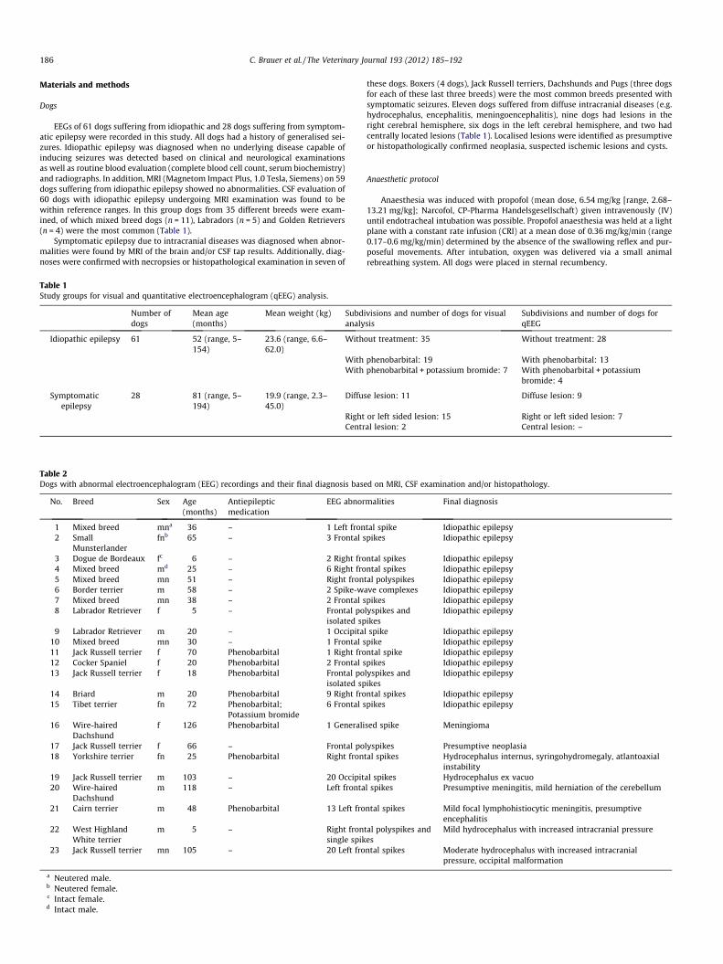
EEGs of 61 dogs suffering from idiopathic and 28 dogs suffering from symptom-atic epilepsy were recorded in this study. All dogs had a history of generalised sei-zures. Idiopathic epilepsy was diagnosed when no underlying disease capable ofinducing seizures was detected based on clinical and neurological examinationsas well as routine blood evaluation (complete blood cell count, serum biochemistry)and radiographs. In addition, MRI (Magnetom Impact Plus, 1.0 Tesla, Siemens) on 59dogs suffering from idiopathic epilepsy showed no abnormalities. CSF evaluation of60 dogs with idiopathic epilepsy undergoing MRI examination was found to bewithin reference ranges. In this group dogs from 35 different breeds were exam-ined, of which mixed breed dogs (n = 11), Labradors (n = 5) and Golden Retrievers(n = 4) were the most common (Table 1).
Symptomatic epilepsy due to intracranial diseases was diagnosed when abnor-malities were found by MRI of the brain and/or CSF tap results. Additionally, diag-noses were confirmed with necropsies or histopathological examination in seven of
Table 1Study groups for visual and quantitative electroencephalogram (qEEG) analysis.
Number ofdogs
Mean age(months)
Mean weight (kg) Subdanaly
Idiopathic epilepsy 61 52 (range, 5–154)
23.6 (range, 6.6–62.0)
With
WithWith
Symptomaticepilepsy
28 81 (range, 5–194)
19.9 (range, 2.3–45.0)
Diffus
RightCentr
Table 2Dogs with abnormal electroencephalogram (EEG) recordings and their final diagnosis base
No. Breed Sex Age(months)
Antiepilepticmedication
EEG abnor
1 Mixed breed mna 36 – 1 Left fron2 Small
Munsterlanderfnb 65 – 3 Frontal s
3 Dogue de Bordeaux fc 6 – 2 Right fro4 Mixed breed md 25 – 6 Right fro5 Mixed breed mn 51 – Right fron6 Border terrier m 58 – 2 Spike-w7 Mixed breed mn 38 – 2 Frontal s8 Labrador Retriever f 5 – Frontal po
isolated sp9 Labrador Retriever m 20 – 1 Occipita
10 Mixed breed mn 30 – 1 Frontal s11 Jack Russell terrier f 70 Phenobarbital 1 Right fro12 Cocker Spaniel f 20 Phenobarbital 2 Frontal s13 Jack Russell terrier f 18 Phenobarbital Frontal po
isolated sp14 Briard m 20 Phenobarbital 9 Right fro15 Tibet terrier fn 72 Phenobarbital;
Potassium bromide6 Frontal s
16 Wire-hairedDachshund
f 126 Phenobarbital 1 Generali
17 Jack Russell terrier f 66 – Frontal po18 Yorkshire terrier fn 25 Phenobarbital Right fron
19 Jack Russell terrier m 103 – 20 Occipit20 Wire-haired
Dachshundm 118 – Left fronta
21 Cairn terrier m 48 Phenobarbital 13 Left fro
22 West HighlandWhite terrier
m 5 – Right fronsingle spik
23 Jack Russell terrier mn 105 – 20 Left fro
a Neutered male.b Neutered female.c Intact female.d Intact male.
these dogs. Boxers (4 dogs), Jack Russell terriers, Dachshunds and Pugs (three dogsfor each of these last three breeds) were the most common breeds presented withsymptomatic seizures. Eleven dogs suffered from diffuse intracranial diseases (e.g.hydrocephalus, encephalitis, meningoencephalitis), nine dogs had lesions in theright cerebral hemisphere, six dogs in the left cerebral hemisphere, and two hadcentrally located lesions (Table 1). Localised lesions were identified as presumptiveor histopathologically confirmed neoplasia, suspected ischemic lesions and cysts.
Anaesthetic protocol
Anaesthesia was induced with propofol (mean dose, 6.54 mg/kg [range, 2.68–13.21 mg/kg]; Narcofol, CP-Pharma Handelsgesellschaft) given intravenously (IV)until endotracheal intubation was possible. Propofol anaesthesia was held at a lightplane with a constant rate infusion (CRI) at a mean dose of 0.36 mg/kg/min (range0.17–0.6 mg/kg/min) determined by the absence of the swallowing reflex and pur-poseful movements. After intubation, oxygen was delivered via a small animalrebreathing system. All dogs were placed in sternal recumbency.
ivisions and number of dogs for visualsis
Subdivisions and number of dogs forqEEG
out treatment: 35 Without treatment: 28
phenobarbital: 19 With phenobarbital: 13phenobarbital + potassium bromide: 7 With phenobarbital + potassium
bromide: 4
e lesion: 11 Diffuse lesion: 9
or left sided lesion: 15 Right or left sided lesion: 7al lesion: 2 Central lesion: –
d on MRI, CSF examination and/or histopathology.
malities Final diagnosis
tal spike Idiopathic epilepsypikes Idiopathic epilepsy
Mild hydrocephalus with increased intracranial pressure
ntal spikes Moderate hydrocephalus with increased intracranialpressure, occipital malformation
C. Brauer et al. / The Veterinary Journal 193 (2012) 185–192 187
The end-tidal carbon dioxide (EtCO2) tension, the peripheral oxygen saturationof haemoglobin (SPO2) and the pulse rate were constantly measured with a side-stream capnograph and a pulse oximeter clip attached to the tongue and observedon a patient monitor (OxiMax NPB75, Nellcor Puritan Bennett). After achieving astable plane of anaesthesia, all but one dog received the peripheral muscle relaxantrocuronium bromide (Esmeron 10 mg/mL, Organon) at a mean dose of 0.4 mg/kg IV(range, 0.2–0.8 mg/kg) to prevent electromyographic artefacts in the EEG readings(Brauer et al., 2011).
EEG recordings
EEGs were obtained via five subdermal needle electrodes (F3, F4, Cz, O1 and O2;disposable subdermal stainless steel EEG needle, Viasys Healthcare) (Redding,1978). The reference electrode was placed on the bridge of the nose and the groundelectrode immediately caudal to the occipital protuberance. Two subdermal needleelectrodes were used to record a lead II electrocardiogram. The EEG (NicoletOnenEEG, Viasys Healthcare) was recorded with sensitivity = 70 lV/cm; time con-stant = 0.3 s; Hf = 70 Hz; notch filter inserted; impedance of all electrodes <10 kX.
Two activation techniques were used during EEG recording. After obtainingbaseline data without any activation, a photic stimulation with a photic stimulator(Photic stimulator, Viasys Healthcare) placed approximately 20 cm in front of theclosed eyes was carried out. The flash frequency was gradually increased in stepsof 5 Hz from 5 to 50 Hz and then decreased in the same way. Each flash intervalwas applied for 8 s and followed by a pause of 5 s until the next flash intervalstarted. After the end of the photic stimulation study and an interval of 3 min
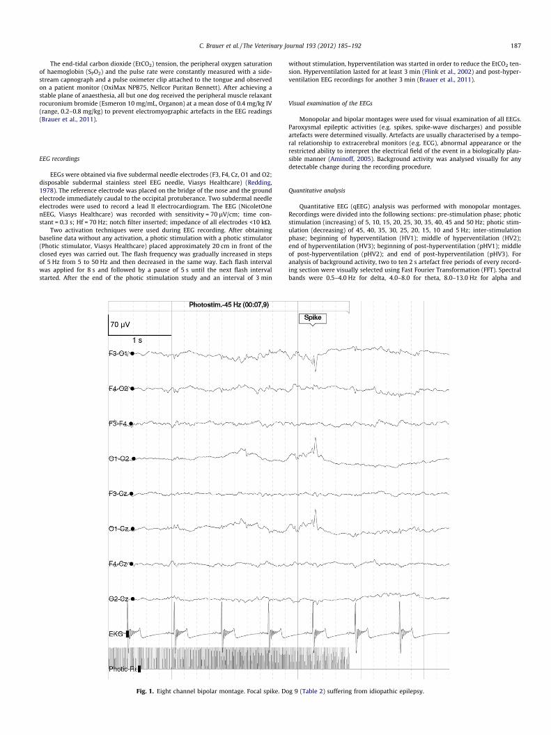
Fig. 1. Eight channel bipolar montage. Focal spike. Do
without stimulation, hyperventilation was started in order to reduce the EtCO2 ten-sion. Hyperventilation lasted for at least 3 min (Flink et al., 2002) and post-hyper-ventilation EEG recordings for another 3 min (Brauer et al., 2011).
Visual examination of the EEGs
Monopolar and bipolar montages were used for visual examination of all EEGs.Paroxysmal epileptic activities (e.g. spikes, spike-wave discharges) and possibleartefacts were determined visually. Artefacts are usually characterised by a tempo-ral relationship to extracerebral monitors (e.g. ECG), abnormal appearance or therestricted ability to interpret the electrical field of the event in a biologically plau-sible manner (Aminoff, 2005). Background activity was analysed visually for anydetectable change during the recording procedure.
Quantitative analysis
Quantitative EEG (qEEG) analysis was performed with monopolar montages.Recordings were divided into the following sections: pre-stimulation phase; photicstimulation (increasing) of 5, 10, 15, 20, 25, 30, 35, 40, 45 and 50 Hz; photic stim-ulation (decreasing) of 45, 40, 35, 30, 25, 20, 15, 10 and 5 Hz; inter-stimulationphase; beginning of hyperventilation (HV1); middle of hyperventilation (HV2);end of hyperventilation (HV3); beginning of post-hyperventilation (pHV1); middleof post-hyperventilation (pHV2); and end of post-hyperventilation (pHV3). Foranalysis of background activity, two to ten 2 s artefact free periods of every record-ing section were visually selected using Fast Fourier Transformation (FFT). Spectralbands were 0.5–4.0 Hz for delta, 4.0–8.0 for theta, 8.0–13.0 Hz for alpha and
g 9 (Table 2) suffering from idiopathic epilepsy.
188 C. Brauer et al. / The Veterinary Journal 193 (2012) 185–192
13.0–30.0 Hz for beta activity. In order to minimize errors through different skullsizes, forms and thicknesses, the relative power of the spectral bands was calculatedfor every lead and averaged for different study groups.
Statistical analysis
Data of background analysis were compared to prior obtained healthy referencevalues from our EEG laboratory (Brauer et al., 2011). For statistical analysis of qEEGdata, dogs were divided into the following groups: (1) dogs suffering from idio-pathic epilepsy without antiepileptic treatment (n = 28); (2) dogs suffering from idi-opathic epilepsy under phenobarbital treatment (n = 13); (3) dogs suffering fromidiopathic epilepsy under treatment with phenobarbital and potassium bromide(n = 4); (4) dogs suffering from symptomatic epilepsy with diffuse intracranial le-sions (n = 9); and (5) dogs sufferings from symptomatic epilepsy suffering from fo-cal lesions of either the left or right hemisphere (n = 7).
Data were not normally or log-normally distributed and goodness of fit fornormal distribution of model residuals of all parameters was rejected by visualassessment of normal probability plots and the Kolmogorov–Smirnov test. Inde-pendent samples of relative power values of the spectral bands of the first fourgroups were compared to healthy reference values by the Wilcoxon 2-sample teststratified by lead and event. In the group with repeated measurements of focallesions of the right or left brain hemisphere, data of the healthy hemisphereswere compared to the affected hemispheres with Friedman’s Chi-Square test(Cochran–Mantel–Haenszel Statistics). Results were considered significant ifP < 0.05. Analyses were carried out with the statistical software SAS, version 9.1(SAS Institute).
Theta and delta activity dominated the background activity ofthe EEG and was superimposed by faster alpha and beta activity.There was no visible change in background activity between peri-ods with either photic stimulation or hyperventilation and periodswithout any stimulation. During hyperventilation a mean CO2 ten-sion of 24 mm Hg (range, 18–31 mm Hg) in a mean of 231 s (range,180–512 s) was reached.
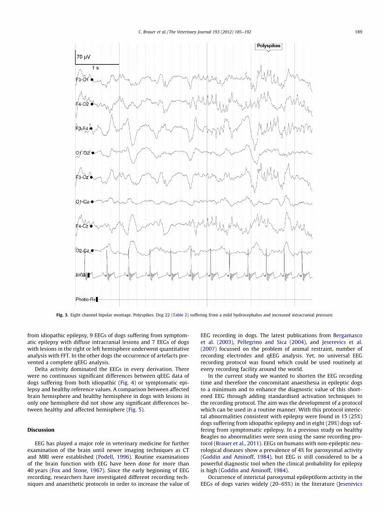
Interictal paroxysmal activity occurred in 15 dogs (25%) suffer-ing from idiopathic epilepsy (Table 2) and consisted of singlespikes, polyspikes and spike slow wave-complexes (Figs. 1 and2). Eight dogs (29%) suffering from symptomatic epilepsy had vis-ible abnormalities in their EEGs (Table 1; Fig. 3). No sudden onsetof paroxysmal activity during the application of both activationtechniques occurred in any of these dogs.
Quantitative analysis
EEGs of 45 dogs (28 without treatment, 13 receiving phenobar-bital, 4 receiving phenobarbital and potassium bromide) suffering
x. Dog 6 (Table 2) suffering from idiopathic epilepsy.
Fig. 3. Eight channel bipolar montage. Polyspikes. Dog 22 (Table 2) suffering from a mild hydrocephalus and increased intracranial pressure.
C. Brauer et al. / The Veterinary Journal 193 (2012) 185–192 189
from idiopathic epilepsy, 9 EEGs of dogs suffering from symptom-atic epilepsy with diffuse intracranial lesions and 7 EEGs of dogswith lesions in the right or left hemisphere underwent quantitativeanalysis with FFT. In the other dogs the occurrence of artefacts pre-vented a complete qEEG analysis.
Delta activity dominated the EEGs in every derivation. Therewere no continuous significant differences between qEEG data ofdogs suffering from both idiopathic (Fig. 4) or symptomatic epi-lepsy and healthy reference values. A comparison between affectedbrain hemisphere and healthy hemisphere in dogs with lesions inonly one hemisphere did not show any significant differences be-tween healthy and affected hemisphere (Fig. 5).
Discussion
EEG has played a major role in veterinary medicine for furtherexamination of the brain until newer imaging techniques as CTand MRI were established (Podell, 1996). Routine examinationsof the brain function with EEG have been done for more than40 years (Fox and Stone, 1967). Since the early beginning of EEGrecording, researchers have investigated different recording tech-niques and anaesthetic protocols in order to increase the value of
EEG recording in dogs. The latest publications from Bergamascoet al. (2003), Pellegrino and Sica (2004), and Jeserevics et al.(2007) focussed on the problem of animal restraint, number ofrecording electrodes and qEEG analysis. Yet, no universal EEGrecording protocol was found which could be used routinely atevery recording facility around the world.
In the current study we wanted to shorten the EEG recordingtime and therefore the concomitant anaesthesia in epileptic dogsto a minimum and to enhance the diagnostic value of this short-ened EEG through adding standardised activation techniques tothe recording protocol. The aim was the development of a protocolwhich can be used in a routine manner. With this protocol interic-tal abnormalities consistent with epilepsy were found in 15 (25%)dogs suffering from idiopathic epilepsy and in eight (29%) dogs suf-fering from symptomatic epilepsy. In a previous study on healthyBeagles no abnormalities were seen using the same recording pro-tocol (Brauer et al., 2011). EEGs on humans with non-epileptic neu-rological diseases show a prevalence of 4% for paroxysmal activity(Goddin and Aminoff, 1984), but EEG is still considered to be apowerful diagnostic tool when the clinical probability for epilepsyis high (Goddin and Aminoff, 1984).
Occurrence of interictal paroxysmal epileptiform activity in theEEGs of dogs varies widely (20–65%) in the literature (Jeserevics
Fig. 4. Relative power values of the Cz electrode (Median) of healthy controls compared to 28 dogs suffering from idiopathic epilepsy (IE) without any anti-epileptictreatment (IE without treatment), 13 dogs suffering from IE under treatment with phenobarbital (IE + pheno) and 4 dogs suffering from IE under treatment withphenobarbital and potassium bromide (KBr) (IE + pheno + KBr). Significant differences were detected at single reading points but without showing any tendency for acontinuous change: delta activity: IE without treatment: pause (P = 0.035); theta activity: IE without treatment: a35 Hz (P = 0.045), IE + pheno + KBr: b40 Hz (P = 0.02); alphaactivity: IE without treatment: b45 Hz (P = 0.035), IE + pheno: a45 Hz (P = 0.017), IE + pheno + KBr: b10 Hz (P = 0.04); beta activity: IE + pheno: a45 Hz (P = 0.032), IE + pheno:a50 Hz (P = 0.044), IE + pheno + KBr: b15 Hz (P = 0.04).
190 C. Brauer et al. / The Veterinary Journal 193 (2012) 185–192
et al., 2007; Berendt et al., 1999). Holliday et al. (1970) foundabnormalities in the EEGs of 71% of all investigated dogs withrecurrent seizures, whereas Jaggy and Bernardini (1998) obtainedconsistent and characteristic findings in 86% of the interictal EEGsin dogs with idiopathic epilepsy. In dogs suffering from intracranialmass lesions, paroxysmal discharges were not detected in the EEGsof six dogs with observed seizures (Steiss et al., 1990).
In the present study interictal paroxysmal discharges occurredin a similar range as described above. In human studies only upto 50% of human epilepsy patients have interictal paroxysmal dis-charges on their first routine EEG (Glick, 2002). Anatomical differ-ences between humans and dogs can explain relatively lowernumbers of these discharges in dogs. Whereas the human skull isnot covered by thick muscle layers, the skull of dogs is nearly com-pletely coated by masticatory muscles leading to a larger distancebetween recording electrode and epileptic focus resulting in loweramplitudes and possible masking of paroxysmal discharges. Usingmore recording electrodes as described by Pellegrino and Sica(2004) could have enhanced the detection of interictal changes.These authors published a standardised recording protocol using12 electrodes including two temporalis electrodes going throughthe temporalis musculature with direct contact to the skull. Suchelectrodes were not used in the current study with client ownedpatients due to the possible damage of blood vessels and nervesby inserting the electrodes (Pellegrino and Sica, 2004).
In human medicine, activation techniques are widely used(Mendez and Brenner, 2006). It has been shown that they are ableto make a diagnostic contribution in 11% of patients with normalroutine EEGs (Angus-Leppan, 2007). However, the described
activation techniques were not able to numerically enhance theinterictal occurrence of paroxysmal activity within the short timeprotocol described in the current study with dogs.
In human medicine, responses to photic stimulation are de-scribed as either normal (no change of EEG rhythms, photic driv-ing) or abnormal (photo-paroxysmal response; Angus-Leppan,2007). A photo-paroxysmal response is characterised by the occur-rence of spike-slow wave and polyspike-slow wave complexes incoherence with the photic stimulation (Mendez and Brenner,2006). In veterinary medicine, photic stimulation has been usedbefore by Holliday et al. (1970) and Goiz-Marquez et al. (2009)to improve the diagnostic value of the EEG. Goiz-Marquez et al.(2009) do not mention if there were more paroxysmal dischargesunder photic stimulation >30 s and an unspecified flash frequency.Holliday et al. (1970) added photic stimulation at flash rates of 5–25 Hz each for 30–40 s to the recording protocols of 13 dogs. In se-ven of these dogs paroxysmal activity was detected under this acti-vation technique.
These different results may be due to different methods of ani-mal restraint. Holliday et al. (1970) just fixed the dogs on a tablewith tape, whereas Goiz-Marquez et al. (2009) used general anaes-thesia with propofol in combination with xylazine. In the currentstudy propofol in combination with the muscle relaxant rocuroni-um (Brauer et al., 2011) was applied. Pharmacological depressionof the visual cortex may be an explanation for not attaining moreparoxysmal discharges through photic stimulation in epilepticdogs under propofol anaesthesia. Although, it has been shown thatrecording of visual evoked potentials in dogs was hindered by pro-pofol (Krause, 2003), propofol was chosen in the current study
Fig. 5. Relative power values of affected and unaffected brain hemispheres (Median) of dogs with focal lesion restricted to one hemisphere (n = 7). No significant differencescould be detected between affected and unaffected hemispheres.
C. Brauer et al. / The Veterinary Journal 193 (2012) 185–192 191
because it is widely used in veterinary studies (Accatino et al.,1997) and has proconvulsive activity (Löscher, 2009). In addition,the best yield of interictal discharges was found using propofol(Jaggy and Bernardini, 1998).
Hyperventilation has not been described before as a routinelyused activation technique in veterinary medicine. In our protocoldogs were hyperventilated for at least 3 min as recommended bythe International League Against Epilepsy (Flink et al., 2002).Hyperventilation can cause slowing of the background activity inhumans which is considered abnormal when slowing persists aftercessation of the procedure (Aminoff, 2005). Hyperventilation ismore effective in patients with generalised seizures and may leadto the occurrence of interictal discharges and even to seizures(Mendez and Brenner, 2006). Although only dogs with generalisedseizures were investigated in our study, we could not provoke anysudden onset of paroxysmal discharges with the described proce-dure. This may be due to the fact that the dogs were under generalanaesthesia, although the level of anaesthesia was kept as light aspossible. A slowing of the background activity (i.e. a change in theqEEG analysis) could have been masked by the overall slowing ofthe EEG activity through anaesthesia. However, controlled non-pharmacologic hyperventilation of a dog is not possible withoutgeneral anaesthesia.
Interictal EEG recording is an essential part of the pre-operativeassessment of human epilepsy surgery candidates (Kuzniecky andDevinsky, 2007). Epilepsy surgery has been shown to have a benefi-cial effect in cases of medically uncontrolled epilepsy (Kuznieckyand Devinsky, 2007; Schmidt and Stavem, 2009). Epilepsy surgeryhas not been used for treatment of drug-resistant epileptic dogs,but may be an option if the pre-operative assessment could beimproved and epileptic foci identified (Bagley et al., 1996; Berendt,2004). Our results show that short time interictal EEG recording un-der general anaesthesia with propofol is only capable of providing
information concerning the seizure focus in a limited number ofdogs. Future research in this field should concentrate on modifica-tions of the mode of restraint, use of other activation techniques(e.g. pharmacological activation with ketamine or chlorpromazine)during EEG recording and particularly on other methods to deter-mine the seizure focus (e.g. intracranial electrodes combined withvideo EEG monitoring, positron emission tomography, single pho-ton emission CT, functional MRI and magnetoencephalography).
All dogs investigated in the current study underwent furtherdiagnostic procedures such as MRI and/or CSF examination. Finaldiagnoses or presumptive diagnoses in cases of symptomatic epi-lepsy without pathological examinations were based on these fur-ther procedures and/or pathological examinations. In all of thesecases EEG recordings did not have an effect on any decision con-cerning prognosis and treatment.
Conclusions
The diagnostic value of short time EEG recording using two acti-vation techniques in epileptic dogs was evaluated. In about 25% ofthe dogs interictal discharges occurred. This number could not beincreased by the systematic use of photic stimulation or hyperven-tilation. qEEG analysis was not more sensitive than visual evalua-tion. We do only recommend the described EEG recording fordogs suffering from atypical seizure like phenomena and not forthe systematic search of a seizure focus.
Conflict of interest statement
None of the authors of this paper has a financial or personalrelationship with other people or organisations that could inappro-priately influence or bias the content of the paper.
192 C. Brauer et al. / The Veterinary Journal 193 (2012) 185–192
Acknowledgements
The authors want to thank Professor Wolfgang Löscher from theDepartment of Pharmacology, Toxicology and Pharmacy, Universityof Veterinary Medicine Hannover, Germany and Dr. Alois Ebnerfrom the Epilepsie-Zentrum Bethel, Mara Krankenhaus, Bielefeld,Germany for their kind advice regarding unclear forms of EEG wavesand for carefully reading the manuscript. Christina Brauer receiveda Doctoral Scholarship from the Ministry for Science and Culture ofLower Saxony, Germany.
References
Accatino, A., Jaggy, A., Gaillard, C., Aeschbacher, G., 1997. Electroencephalographicfindings of encephalitis in beagle dogs experimentally infected with caninedistemper virus (CDV). Zentralblatt für Veterinärmedizin B 44, 39–48.
Aminoff, M.J., 2005. Electroencephalography: General principles and clinicalapplications. In: Aminoff, M.J. (Ed.), Electrodiagnosis in Clinical Neurology.Elsevier, Philadelphia, pp. 37–84.
Angus-Leppan, H., 2007. Seizures and adverse events during routine scalpelectroencephalography: A clinical and EEG analysis of 1000 records. ClinicalNeurophysiology 118, 22–30.
Bagley, R.S., Harrington, M.L., Moore, M.P., 1996. Surgical treatments for seizure.Adaptability for dogs. Veterinary Clinics of North America: Small AnimalPractice 26, 827–842.
Berendt, M., 2004. Epilepsy. In: Vite, C.H. (Ed.), Braund’s Clinical Neurology in SmallAnimals: Localization, Diagnosis and Treatment. International VeterinaryInformation Service, Ithaca.
Berendt, M., Hogenhaven, H., Flagstad, A., Dam, M., 1999. Electroencephalography indogs with epilepsy: Similarities between human and canine findings. ActaNeurologica Scandinavia 99, 276–283.
Bergamasco, L., Accatino, A., Priano, L., Neiger-Aeschbacher, G., Cizinauskas, S.,Jaggy, A., 2003. Quantitative electroencephalographic findings in beaglesanaesthetized with propofol. The Veterinary Journal 166, 58–66.
Brauer, C., Kästner, S.B.R., Schenk, H.C., Tünsmeyer, J., Tipold, A., 2011.Electroencephalographic recordings in dogs: Prevention of muscle artefactsand evaluation of two activation techniques in healthy individuals. Research inVeterinary Science 90, 306–311.
Flink, R., Pedersen, B., Guekht, A.B., Malmgren, K., Michelucci, R., Neville, B., Pinto, F.,Stephani, U., Ozkara, C., 2002. Guidelines for the use of EEG methodology in thediagnosis of epilepsy. International league against epilepsy: Commission report.Commission on European Affairs: Subcommission on European Guidelines. ActaNeurologica Scandinavia 106, 1–7.
Fox, M.W., Stone, A.B., 1967. An electroencephalographic study of epilepsy in thedog. Journal of Small Animal Practice 8, 703–708.
Glick, T.H., 2002. The sleep-deprived electroencephalogram: Evidence and practice.Archives of Neurology 59, 1235–1239.
Goddin, D.S., Aminoff, M.J., 1984. Does the interictal EEG have a role in the diagnosisof epilepsy? Lancet 324, 837–839.
Goiz-Marquez, G., Caballero, S., Solis, H., Rodriguez, C., Sumano, H., 2009.Electroencephalographic evaluation of gold wire implants inserted inacupuncture points in dogs with epileptic seizures. Research in VeterinaryScience 86, 152–161.
Jaggy, A., Bernardini, M., 1998. Idiopathic epilepsy in 125 dogs: A long-term study.Clinical and electroencephalographic findings. Journal of Small Animal Practice39, 23–29.
Jeserevics, J., Viitmaa, R., Cizinauskas, S., Sainio, K., Jokinen, T.S., Snellman, M.,Bellino, C., Bergamasco, L., 2007. Electroencephalography findings in healthyand Finnish Spitz dogs with epilepsy: Visual and background quantitativeanalysis. Journal of Veterinary Internal Medicine 21, 1299–1306.
Knecht, C.E., Sorjonen, D.C., Simpson, S.T., 1983. Ancillary tests in the diagnosis ofseizures. Journal of the American Animal Hospital Association 20, 455–458.
Krause, A., 2003. Standardization and clinical use of visual evoked potentials (VEP)in the dog. Doctoral Thesis. Small Animal Clinic. University of VeterinaryMedicine Hannover, Germany.
Kube, S.A., Vernau, K.M., LeCouteur, R.A., 2006. Dyskinesia associated with oralphenobarbital administration in a dog. Journal of Veterinary Internal Medicine20, 1238–1240.
Kuzniecky, R., Devinsky, O., 2007. Surgery insight: Surgical management of epilepsy.Nature Clinical Practice Neurology 3, 673–681.
Löscher, W., 2009. Preclinical assessment of proconvulsant drug activity and itsrelevance for predicting adverse events in humans. European Journal ofPharmacology 610, 1–11.
Mendez, O.E., Brenner, R.P., 2006. Increasing the yield of EEG. Journal of ClinicalNeurophysiology 23, 282–293.
Pellegrino, F.C., Sica, R.E., 2004. Canine electroencephalographic recordingtechnique: Findings in normal and epileptic dogs. Clinical Neurophysiology115, 477–487.
Penning, V.A., Connolly, D.J., Gajanayake, I., McMahon, L.A., Luis Fuentes, V.,Chandler, K.E., Volk, H.A., 2009. Seizure-like episodes in 3 cats with intermittenthigh-grade atrioventricular dysfunction. Journal of Veterinary InternalMedicine 23, 200–205.
Podell, M., 1996. Seizures in dogs. Veterinary Clinics of North America: SmallAnimal Practice 26, 779–809.
Schmidt, D., Stavem, K., 2009. Long-term seizure outcome of surgery versus nosurgery for drug-resistant partial epilepsy: A review of controlled studies.Epilepsia 50, 1301–1309.
Steiss, J.E., Cox, N.R., Knecht, C.D., 1990. Electroencephalographic andhistopathologic correlations in eight dogs with intracranial mass lesions.American Journal of Veterinary Research 51, 1286–1291.
Thomas, W.B., 2010. Idiopathic epilepsy in dogs and cats. Veterinary Clinics of NorthAmerica: Small Animal Practice 40, 161–179.