Page 1
ELECTROENCEPHALOGRAPHY REFERRALS AND OUTCOMES IN
A TERTIARY PSYCHIATRIC HOSPITAL
Molokashe Meriam Molokomme
A research report submitted to the Faculty of Health Sciences, University of the
Witwatersrand, Johannesburg, in partial fulfilment of the requirements for the degree of
Master of Medicine in the branch of Psychiatry
Johannesburg, 2012
Page 3
iii
DEDICATION
For my loving parents, siblings and my dearest son Thabiso.
Page 4
iv
PUBLICATIONS AND PRESENTATIONS ARISING FROM THIS STUDY
Preliminary findings of this research report were presented during the Psychiatry Research
day 2010, held at the University of the Witwatersrand.
Page 5
v
ABSTRACT
INTRODUCTION
The electroencephalography (EEG), since its inception in the 1930s, has become one of the
most used investigative tools in psychiatry. Its uses include exclusion of seizure disorders
and encephalopathic conditions. In psychiatry distinguishing between a primary psychiatric
disorder and psychiatric manifestations of an underlying medical condition is of vital
importance. This determines which course of management the psychiatrist should follow,
and most importantly, determines the prognosis.
However, EEG studies done in psychiatry have yielded unfavourable results. The yield of
positive (abnormal) EEG results is very low. Despite this, it is still widely requested by most
psychiatrists.
There is a dearth of literature assessing the usefulness of EEG in psychiatry in our South
African setting. The current study looked at which users are referred for EEG and the
outcomes thereof.
METHODS
The study was conducted at Sterkfontein psychiatric hospital. A retrospective review of
clinical records, and EEG reports, of inpatients 18yrs and older that underwent EEG
between January 2008 to June 2009 was done. A data sheet was used as a recording tool.
Data was analysed using the Statistica 9.0 system.
Page 6
vi
RESULTS
The total sample was 85. Seventy four (87%) records were normal, 7(8,2%) were abnormal,
2(2,4%) were inconclusive and two EEG reports were unavailable. Only one user’s diagnosis
changed based on abnormal EEG results. There was no statistically significant correlation
between abnormal EEG results and demographic variables, symptoms, admission diagnosis
and medications.
CONCLUSION
The positive yield of EEG results remains very low in psychiatry. EEG results do not appear
to influence the treating psychiatrist’s decision regarding management.
Page 7
vii
ACKNOWLEDGEMENTS
Prof Ugasvaree Subramaney: supervisor
Thank you for your endless support and dedication to this project, and most importantly, your
patience.
Dr Elena Libhaber: statistician
Thank you for opening your door and passing on the knowledge.
Mrs Trudie Botes: EEG technician
Thank you for your contribution.
Page 8
viii
TABLE OF CONTENTS
DECLARATION....................................................................................................................ii
DEDICATION........................................................................................................................iii
PUBLICATIONS AND PRESENTATIONS..........................................................................iv
ABSTRACT..........................................................................................................................v
ACKNOWLEDGEMENTS....................................................................................................vii
TABLE OF CONTENTS.......................................................................................................viii
LIST OF FIGURES...............................................................................................................xii
LIST OF TABLES................................................................................................................xiii
CHAPTER 1: INTRODUCTION..........................................................................................1
1.1 BACKGROUND.........................................................................................................1
1.1.1 USES OF EEG...............................................................................................1
1.1.2 EEG RECORDING........................................................................................2
1.1.3 ABNORMAL EEG PATTERNS.....................................................................3
1.1.4 EEG LIMITATIONS........................................................................................4
1.2 HYPOTHESIS...........................................................................................................5
1.3 STUDY AIM..............................................................................................................5
1.4 STUDY OBJECTIVES..............................................................................................5
CHAPTER 2: LITERATURE REVIEW................................................................................6
Page 9
ix
2.1 AFRICAN LITERATURE REVIEW............................................................................6
2.2 INTERNATIONAL LITERATURE REVIEW...............................................................8
CHAPTER 3: METHODOLOGY...........................................................................................12
3.1 SITE OF THE STUDY...............................................................................................12
3.2 STUDY DESIGN.......................................................................................................12
3.2.1 INCLUSION CRITERIA....................................................................................13
3.2.2 EXCLUSION CRITERIA..................................................................................13
3.2.3 DATA SHEET..................................................................................................13
3.2.4 EEG PROCEDURE.........................................................................................13
3.2.5 ETHICS...........................................................................................................14
3.2.6 STATISTICAL ANALYSIS...............................................................................14
CHAPTER 4: RESULTS.......................................................................................................15
4.1 SOCIODEMOGRAPHIC PROFILE...........................................................................15
4.2 REASON FOR REFERRAL TO EEG........................................................................19
4.3 SYMPTOMS REPORTED PRIOR TO EEG REFERRAL.........................................20
4.4 ADMISSION DIAGNOSIS.........................................................................................21
4.5 MEDICATIONS PRIOR TO EEG REFERRAL..........................................................22
4.6 EEG RESULTS.........................................................................................................23
4.7 EEG RESULTS MATCHED WITH VARIABLES.......................................................23
4.7.1 EEG RESULTS MATCHED WITH DEMOGRAPHICS..................................23
Page 10
x
4.7.2 EEG RESULTS MATCHED WITH SYMPTOMS............................................25
4.7.3 EEG RESULTS MATCHED WITH ADMISSION DIAGNOSIS.......................26
4.7.4 EEG RESULTS MATCHED WITH MEDICATIONS.......................................27
4.8 CHANGES IN MANAGEMENT POST EEG.............................................................27
4.8.1 ABNORMAL EEG RESULTS..........................................................................27
4.8.2 NORMAL EEG RESULTS...............................................................................27
CHAPTER 5: DISCUSSION.................................................................................................29
5.1 SOCIODEMOGRAPHIC PROFILE................................................................................29
5.2 REASON FOR REFERRAL TO EEG.............................................................................29
5.3 SYMPTOMS PRIOR TO EEG REFERRAL ...................................................................30
5.4 ADMISSION DIAGNOSIS...............................................................................................31
5.5 MEDICATIONS PRIOR TO EEG REFERRAL...............................................................31
5.6 EEG RESULTS...............................................................................................................32
5.7 CHANGES IN MANAGEMENT......................................................................................33
5.7.1 ABNORMAL EEG RESULTS..............................................................................33
5.7.2 NORMAL EEG RESULTS...................................................................................34
5.8 LIMITATIONS.................................................................................................................34
CHAPTER 6: CONCLUSIONS AND RECOMMENDATIONS............................................35
REFERENCES.....................................................................................................................36
Page 11
xi
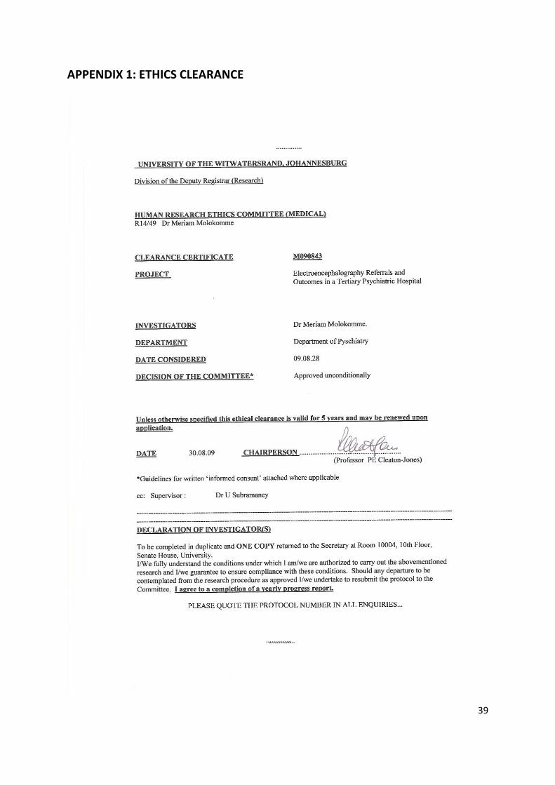
APPENDIX 1: ETHICS CLEARANCE................................................................................39
APPENDIX 2: POSTGRADUATE COMMITTEE CLEARANCE........................................40
APPENDIX 3: CONSENT FROM HOSPITAL CEO...........................................................41
APPENDIX 4: INFORMATION SHEET..............................................................................42
APPENDIX 5: INFORMED CONSENT..............................................................................43
APPENDIX 6: DATA SHEET..............................................................................................44
Page 12
xii
LIST OF FIGURES
FIGURE 1: SAMPLE POPULATION AGE GROUPS..........................................................16
FIGURE 2: GENDER DISTRIBUTION OF THE SAMPLE POPULATION..........................16
FIGURE 3: MARITAL STATUS OF THE SAMPLE POPULATION.....................................17
FIGURE 4: HIGHEST LEVEL OF EDUCATION OF THE SAMPLE POPULATION...........17
FIGURE 5: EMPLOYMENT STATUS OF THE SAMPLE POPULATION...........................18
FIGURE 6: REASON FOR REFERRAL TO EEG...............................................................19
FIGURE 7: SYMPTOMS REPORTED PRIOR TO EEG REFERRAL................................20
FIGURE 8: MEDICATIONS THE SAMPLE POPULATION WERE ON PRIOR TO EEG
REFERRAL..........................................................................................................................22
Page 13
xiii
LIST OF TABLES
TABLE 1: DEMOGRAPHICS OF THE SAMPLE POPULATION...................................15
TABLE 2: ADMISSION DIAGNOSIS..............................................................................21
TABLE 3: EEG RESULTS..............................................................................................23
TABLE 4: EEG RESULTS MATCHED WITH DEMOGRAPHICS.................................24
TABLE 5: EEG RESULTS MATCHED WITH SYMPTOMS..........................................25
TABLE 6: EEG RESULTS MATCHED WITH ADMISSION DIAGNOSIS.....................26
TABLE 7: SUMMARY OF USERS WITH ABNORMAL EEG RESULTS......................28
Page 14
1
CHAPTER 1: INTRODUCTION
1.1 BACKGROUND
The EEG (electroencephalography) has become established as one of the principal
investigative tools of cerebral function based on the work by Hans Berger in the 1930s.1
Hans Berger was the discoverer of brain waves. Through his mentor, Karl Langenstrass,
M.D., who had trained at the University of Jena under Hans Berger, Robert Cohn
established an EEG laboratory and built his own EEG machine at St. Elizabeth’s Hospital in
Washington, D.C.2 The EEG has been in use worldwide since then.
The EEG is a recording of the electrical potential activity of the brain. It is a non-invasive, low
cost, neurodiagnostic technique widely available in general and psychiatric hospitals in
South Africa.3
1.1.1 Uses of EEG
EEG is mainly used to detect seizure activity in the brain. It is also used for detection of
possible organic aetiologies such as metabolic encephalopathies and tumors. EEG also
plays a major role in polysomnography studies. The wave patterns seen on EEG during
polysomnography indicate the sleep stage. Other uses include monitoring the success of the
stimulus in producing seizure activity during electroconvulsive therapy (ECT).4
The EEG has a role in detecting presence of complex partial seizures (TLE- temporal lobe
epilepsy). TLE has several symptoms which may be considered ‘psychiatric’. These include
Page 15
2
perceptual disturbances such as visual, olfactory and tactile hallucinations in the pre-ictal
phase; episodes of brief disorganized behaviour in the ictal phase; confusion and amnesia of
the ictal phase in the post-ictal phase and psychotic symptoms, mood symptoms, episodic
violence and personality disturbances in the interictal phase.4
A patient with an acute episode of schizophrenia may present with hallucinations, as well as
disorganised and violent behaviour. Episodic mood changes such as irritability and
depression that interfere with functioning may indicate the presence of a bipolar disorder.4
A significant association exists between the presence of an organic factor in the history,
mental status examination, or physical examination and the yield of abnormal EEG’s.5
1.1.2 EEG recording
The electrodes normally used to record the EEG are attached to the scalp with a conductive
paste. The EEG must be recorded with the patient as motionless as possible, to eliminate
the introduction of muscle artefact. Hyperventilation, photic stimulation and sleep deprivation
increase the likelihood of picking up a positive result. A normal EEG consists of a mixture of
frequencies, which are divided into four bandwidths. Delta waves oscillate below 4Hz, theta
waves from 4-8Hz; activity below 8Hz is also called slow wave activity. Alpha waves, the
frequency of the posterior dominant rhythm, are from 8-13Hz, beta waves are over 13Hz
(fast activity). Normal activity consists of posterior alpha rhythm with the eyes closed; more
anterior regions have random admixtures of theta, alpha, or beta activity.4
Page 16
3
1.1.3 Abnormal EEG patterns
Several wave patterns as seen on an EEG recording can suggest pathologies such as the
three per second spike-and-wave considered a characteristic of petit mal epilepsy. Dementia
is associated with excessive slow wave activity on EEG, whereas depression tends to show
a normal recording, thus EEG can be useful in differentiating between dementia and
pseudodementia seen in depressed patients. In delirium, the EEG characteristically shows a
generalized slowing of activity. Low voltage fast activity is the pattern more likely to be seen
in agitated disorganized patients who are more likely to be identified as ‘psychiatric’.6
In comparison to non-schizophrenic patients, schizophrenic patients tend to show more beta
and theta activity on the EEG which are not treatment related. First episode and chronic
schizophrenia patients show similar EEG changes which suggest that this is characteristic of
the condition rather than duration of the illness and exposure to medications.7
Psychotropic medications can result in changes in the normal EEG pattern. Low voltage fast
activity can be induced by benzodiazepines. EEG abnormalities are also seen in patients
who are on antipsychotic treatment, with risk being particularly high with clozapine and
olanzapine, moderate with risperidone and typical neuroleptics, and low with quetiapine.
Severe EEG abnormalities i.e. spike discharges or spike-and-wave abnormality, have been
associated with olanzapine, chlorpromazine and clozapine.8
Page 17
4
1.1.4 EEG limitations
A normal recording does not necessarily exclude pathology. Serial EEGs may have to be
done to increase the possibility of a positive finding, which increases the cost of a patient’s
workup.6 Only 20%-50% of epileptic patients show interictal epileptiform discharges, which
are associated with a clinical seizure disorder, on their first routine EEG.9
EEG abnormalities are largely non-specific. An EEG has a low spatial resolution but a high
temporal resolution. This implies that it can detect changes in neuronal function in
milliseconds time frame in comparison to other brain function tests such as functional
magnetic resonance imaging which reflects changes in seconds’ time frame.10 The electrical
potential of multiple neurons with similar spatial orientation is recorded on EEG. However,
since the current falls off, scalp EEG may not reflect changes in deeper lying structures.11
Slow growing lesions may not cause many EEG changes, particularly if the lesions are small
and not located near the cortex.6
Deep or medial temporal lobe epileptiform discharges may require use of sphenoidal or
nasopharyngeal electrodes which are invasive and bothersome to patients.6
There is also a concern that some clinicians order EEGs as part of practising ‘defensive
medicine’ to avoid litigations. This may contribute in part to increased frequency of negative
EEGs.1
It is in view of the above limitations that this study was conducted.
Page 18
5
1.2 HYPOTHESIS
It is hypothesised that, in the majority of cases, there will be no changes in the clinical
management based on the EEG results.
1.3 STUDY AIM
To determine whether EEG findings impact on clinical management.
1.4 STUDY OBJECTIVES
• To investigate the reasons why psychiatric patients are referred for an EEG.
• To determine what abnormalities are reported on the sample population.
• To determine which psychiatric signs and symptoms are correlated with a positive
EEG.
Page 19
6
CHAPTER 2: LITERATURE REVIEW
Only a few studies have been done in South Africa and other African countries to assess the
usefulness of EEG in psychiatry; while several studies have been done in other continents.
2.1 AFRICAN LITERATURE REVIEW
Rascher et al searched for relevant articles on MEDLINE, which were published between
1966 and 2003.3 The amount of South African data examining either prevalence or
usefulness of EEG abnormalities among adult psychiatric patients was limited.
The two South African studies of relevance that were found were done by Stein at Hillbrow
hospital in 1991 and Szabo on adolescents at TARA hospital in 1999.3, 12
Stein, cited by Rascher et al, analysed all departmental referrals for EEG over a one year
period. The inclusion criteria were direct referral by the Department of Psychiatry, overt
psychiatric symptomatology and an absence of clinical neurological findings. A total of 145
patients met the inclusion criteria. Seventy one patients were shown to have clearly
demonstrable abnormalities on EEG. Thirty five (50%) of these exhibited definite epileptiform
activity. Forty eight patients (67%) had localised EEG dysfunction, with twenty-three (47%)
of abnormalities being found in the temporal lobe areas. The percentage of abnormal EEG’s
leading to a change in diagnosis or management was not determined.3
Szabo et al, reviewed all admissions to the adolescent inpatient unit at Tara Hospital
between 1990 and 1995. Thirty six patients underwent EEG during this period .Close to half
Page 20
7
(44%) of the patients received a definite diagnosis of complex partial seizures, based on
both clinical features and EEG findings. In the remainder, 34% had nonspecific abnormal
EEG’s and 22% were normal. Aggression and hallucinations increased the likelihood of a
diagnostic EEG, with positive results in 60% of those with aggression and 53% of those with
hallucinations. The other clinical features predictive of a diagnostic EEG were mood
instability in 33%, dissociative states in 33% and a premorbid organic insult in 26%. The
clinical features predictive of an EEG abnormality; but not a change in diagnosis or
management; included aggression in 36%, hallucinations in 45% and a premorbid organic
insult in 36%.12
Rascher et al highlighted that discordance still exists when examining prevalence and
usefulness of EEG abnormalities in adult psychiatric patients. It was suggested that a patient
be carefully assessed for any clinical evidence of organic disease prior to being referred for
EEG, preferably by more than one psychiatrist.3
To our knowledge, there have not been any such published South African studies since this
review article to date. Two studies of relevance, closer to home, found published on
MEDLINE between 2004 and 2009 March were done in Lagos, Nigeria by Aina O F et al.13, 14
One study which was prospective in nature was done over twelve months of setting up an
EEG unit at a psychiatric hospital in Yaba, Lagos. The inclusion criteria included all patients
that had an EEG recording in the unit during the study period. Awake EEG was done on
each subject. Seizure disorder constituted the largest clinical reason for EEG request. The
EEG findings were normal in close to 44% of the sample and abnormal in 56% with
“epileptiform activities” reported as the most common abnormality. The researchers
Page 21
8
concluded that EEG continues to be of importance in the management of neuropathological
disorders.13
The other study was done in children. It looked at neuropsychiatric correlates and EEG
findings among children with developmental disorders. A clinical evaluation was made to
reach a diagnosis, and a waking EEG was performed. The recordings were done by an
EEG technologist blinded to the clinical diagnosis of the subjects. EEG interpretation was
done independently by two psychiatrists trained on EEG interpretation. In a sample of 111
individuals; EEG abnormalities were picked up in 85(76,6%) individuals. In this study there
was no significant correlation between the EEG abnormalities and the developmental
disorder diagnosed. However a number of the subjects also suffered from seizure disorders
and hyperactivity related to the developmental disorder.14
2.2 INTERNATIONAL LITERATURE REVIEW
Warner et al reviewed 190 EEG recordings and charts of psychiatric inpatients at the
University of Texas Harris County Psychiatric Center. Usefulness of screening EEGs, which
was defined as EEG results leading to a change in diagnosis and treatment, was assessed.
Of the 190 charts reviewed, 115 patients (61%) had routine screening EEGs. Thirty six
(31%) of these screens led to an abnormal recording, however only two (1, 7%) led to a
change in diagnosis. Also of significance was that abnormal EEGs were noted more in
patients on psychotropic medications (p<0.006). The usefulness of EEG as a screening test
could not be clearly established based on their results due to the limitations of the
retrospective nature of the study.15
Page 22
9
Boutros et al examined journal articles through MEDLINE search for a 45-year period in
1992 and book chapters that relate EEG to the practice of clinical psychiatry. The authors
concluded that the value of the routine clinical EEG in psychiatry lies in its noninvasive
nature, low cost, and easy availability. The authors also suggested that EEG should be used
in conjunction with other neuroimaging techniques such as CT scan and MRI.6
A total of 91 EEG records were reviewed by Fenton et al. These were newly referred
patients over a 12month period. Forty (44%) of the records were normal, 17(19%)
anomalous and 34(37%) were abnormal. “Anomalous” referred to records with an excess of
slower background features or minor paroxysmal phenomena of non-specific significance
while abnormal ratings were given to those with an unequivocal evidence of organic brain
dysfunction or epileptiform activity. EEG changes due to psychotropic medications were
noted in 12 records (13%). Of note was that analysis of the case records revealed that no
patient manifested clinically convincing complex partial seizures, although suspected TLE
was reason for referral in a third (32%) of all patients. The authors concluded that the EEG
abnormality yield is greatest when the clinical history, physical and psychiatric examinations
reveal a definite organic mental state phenomena and/or abnormal CNS signs.16
In another study by Bowie et al, 76 patients were subjected to a total of 89 EEGs. The
abnormal recordings were divided into those which were definitely abnormal and those
which were equivocal. Of the 76 patients, 11(14,5%) had definitely abnormal EEGs and
11(14,5%) had equivocal abnormal recordings. The study also showed that the referring
psychiatrist could predict whether or not the recording would be abnormal (p<0,05). EEG
results led to a change in management in 10 cases (13,2%). The authors as in other studies
mentioned above also found that there is a reliable association between abnormal EEG
Page 23
10
recordings and organic factors in the history, mental state and neurological examination. The
authors also highlighted that the clinical value of the EEG is reflected not by the proportion of
abnormal recordings but by the influence of the recordings on the patient’s management,
diagnosis and prognosis.17
Lam et al reviewed EEGs of 150 patients. In this study EEG changes due to medications
were considered normal. Abnormal recordings were found in 11,3% of the cases. The
presence of an organic factor on history, mental status examination and physical
examination were more likely to yield positive results. The routine use of EEG for psychiatric
patients (as well as other patients) without any presenting organic factors was discouraged
by the authors because of the low yield, and negligible clinical significance of normal and
abnormal results.5
A study by Puri et al in a mental handicap population revealed 37 abnormal EEG recordings
in 80 of the patients. The abnormal recording did not lead to a change in management in
more than half (23) of the abnormal EEG cases, while in nine cases a change in
management followed a report of a normal EEG.18
Stone et al reported that “many doctors like to routinely order an EEG as part of a diagnostic
work-up for patients with psychiatric disorders”. The authors studied 187 EEGs. In 71%, the
request was to look for evidence of epilepsy, and in 22% it was to determine the presence of
organic brain dysfunction. Only one patient was found to have an unequivocal evidence of
an epileptic focus. A ‘liability to epilepsy’ was found in 11 patients, 10 of whom were being
investigated for epilepsy. Of note is that 68 EEG records which showed any of the three
patterns; diffuse slowing, diffuse excess of fast activity and temporal dysfunction; were
Page 24
11
classified as non-diagnostic .19 Whether or not a repeat EEG in these 68 patients would have
revealed unequivocal evidence of epileptic focus is debatable. The result of only one clear
case of an epileptogenic focus was similar to the 0,5%-2% prevalence rate in the normal
population as found by Gregory et al.20
A more recent study on EEG in psychiatry was done by O’ Sullivan et al. A retrospective
review of 1470 EEGs was done. The referrals were from several departments including
neurology (n=622), psychiatry (n=91) and general medicine (n=490). The reasons for referral
from psychiatry and outcomes post EEG were analysed. The proportion of abnormal EEGs
detected from psychiatric sources was less than the combined non-psychiatric referred
patients, but not to a statistical significance (p<0.08). There was no evidence of a significant
change in clinical management. The underlying diagnosis remained unchanged in all cases.1
The above studies all indicate the limitations of EEG use as a routine screening test. In
addition, South Africa’s health service is under-resourced due to the country’s budgetary
constraints. It then becomes important to assess the use of EEG in our own setting.
Page 25
12
CHAPTER 3: METHODOLOGY
3.1 SITE OF THE STUDY
The study was conducted at Sterkfontein hospital (SFH). SFH is a tertiary psychiatric
hospital with 612 usable beds, and caters for involuntary patients under the Mental Health
Care Act no.17 of 2002 (MHCA). It also has a section for forensic patients. Clients are sent
by the courts for observation in terms of the Criminal Procedures Act no.51 of 1977 (CPA) to
ascertain fitness to stand trial and criminal responsibility. Those who are not fit and/or not
criminally responsible are sent back by the courts as state patients in terms of section 42 of
the MHCA. Mentally ill prisoners may also be admitted to SFH for treatment, care and
rehabilitation as provided for in section 49 of the MHCA.
The hospital was ideal for the study as it is a referral institution. Patients from several other
hospitals are referred to SFH for treatment, meaning the sample was representative of the
psychiatric population in general. At the time of the study the hospital had 12 psychiatrists
and 14 registrars/medical officers.
3.2 STUDY DESIGN
This was a retrospective record review of the patients who were referred for EEG from
January 2008 to the end of June 2009. Source data included both EEG reports and clinical
records.
Page 26
13
3.2.1 Inclusion criteria
Only the records of those admitted to SFH were studied. The sample included adult
(age>18) males and females.
.
3.2.2 Exclusion criteria
Patients admitted to Leratong hospital, a general hospital nearby, and those from community
clinics are referred for EEG study at SFH. Patients who are referred from these facilities for
EEG are not all from the psychiatry department. Some are referred from the medical
outpatients department. For practical reasons, these patients were excluded from the study.
3.2.3 Data sheet (Appendix 6)
Patient’s demographics, multiaxial diagnosis on admission, medications prior to and after
EEG, provocation methods used prior and during EEG, EEG findings and any change in
diagnosis after EEG, were recorded on a data sheet.
3.2.4 EEG procedure
All users, except for one who had a sleep deprived EEG, underwent a routine non-sleep
deprived EEG. Photic stimulation and hyperventilation methods were used during the EEG
recordings. All EEGs were done by one technician with the same EEG machine (Neurofax
EEG 1000/9000 Version 05-11). All records were reported by one neurologist.
Page 27
14
3.2.5 Ethics
The study was approved by both the ethics (Appendix 1) and postgraduate committees
(Appendix 2) of the Witwatersrand University, and the hospital’s chief executive officer
(Appendix 3). An information sheet (Appendix 4) was handed to those who were still
inpatients. The researcher also explained in detail the contents of the information sheet. The
patients then signed the informed consent document (Appendix 5) provided. All the
participants were reassured that their names would not be mentioned anywhere in the study,
and the researcher was the only person who approached them regarding the current study.
All data sheets were stored in a locked cupboard. A password known only to the researcher
was created to access the Microsoft excel sheet where data was captured.
3.2.6 Statistical analysis
Data were analyzed using the Statistica 9.0 statistical program. Results are expressed as
frequencies and percentages for categorical variables and as mean±SD for age for the
whole group. Age was categorized as: 18-30, 31-49, >50. To assess differences between
abnormal results and demographics, symptoms, admission diagnosis and psychotropics, a
Chi square test or Fischer’s exact test was used. Significance was assumed at a both-sided
value of p < 0.05.
Page 28
15
CHAPTER 4: RESULTS
4.1 SOCIODEMOGRAPHIC PROFILE
The total sample was 85. The majority of the users were male (62,4%), unmarried (83,5%),
unemployed(78,8%), and had an education level below grade 12 (72,9%)(Table 1). The
mean age was 33.6(SD11.5). Figures 1-5 depict the demographic profiles in graphical
representations.
Table 1: Demographics of the sample population
VARIABLE n (%)
Age: 18-30
31-49
>50
43 (50.6)
30 (35.3)
12 (14.1)
Gender: male
female
53 (62.4)
32 (37.6)
Marital status: married
single
14 (16.5)
71 (83.5)
HLOE: <grade 12
>grade 12
62 (72.9)
22 (25.9) 1 (1.2) unknown
Employment: employed
unemployed
18 (21.2)
67 (78.8)
Page 29
16
Figure 1: Sample population age groups
50.6
35.3
14.1
0
10
20
30
40
50
60
18-30 31-49 >50
Age groups (%)
Age groups (%)
Figure 2: Gender distribution of the sample population
62.4
37.6
0
10
20
30
40
50
60
70
male female
Gender (%)
Gender (%)
Page 30
17
Figure 3: Marital status of the sample population
Figure 4: Highest level of education (HLOE) of the sample population
72.9
25.9
1.20
10
20
30
40
50
60
70
80
grade<12 grade>12 unknown
HLOE(%)
HLOE(%)
Page 31
18
Figure 5: Employment status of the sample population
Page 32
19
4.2 REASON FOR REFERRAL TO EEG
As depicted in figure 6, most users (69%) were referred for seizure disorder exclusion and
the rest (31%) for other reasons that included treatment non-response and organic workup.
Figure 6: Reason for referral to EEG (sample population)
69%
31%
seizure disorder exclusion
other:treatment nonresponse, workup
-
Page 33
20
4.3 SYMPTOMS REPORTED PRIOR TO EEG REFERRAL
Auditory hallucinations, aggression and visual hallucinations were the most common
documented symptoms in the sample population (Figure 7). None of the users reported
gustatory hallucinations. Jamais vu/déjà vu were noted in only a few users. However, it is
important to note that not all clinical records had documented whether certain symptoms
were present. Symptoms classically considered ‘ictal’ such as olfactory, gustatory, tactile
hallucinations; dysmegalopsia and jamais vu/déjà vu were each not documented in three
records.
Figure 7: Symptoms reported prior to EEG referral
94.1
43.5
84.7
71.8
96.5
84.7
86
50.6
36.5
84.7
0 20 40 60 80 100 120
J/D
Aggr
Dys
Seiz
GH
OH
TH
VH
AH
Dis
not documented
no
yes
Dis: disorientation, AH: auditory hallucination, VH: visual hallucination, TH: tactile
hallucination, OH: olfactory hallucination, GH: gustatory hallucination, Seiz: seizure, Dys:
dysmegalopsia, Aggr: aggression, J/D: jamais vu/déjà vu
Page 34
21
4.4 ADMISSION DIAGNOSIS
Primary mood disorders (bipolar disorder, major depressive disorder) and psychotic
disorders (schizophrenia, schizoaffective disorder) were present in over 50% of the
population. Of note was the large percentage (27%) of users with underlying medical
pathologies manifesting with psychiatric symptoms. This latter group included those with
HIV, previous head injuries and possible temporal lobe epilepsy.
Table 2: Admission diagnosis
ADMISSION DIAGNOSIS FREQUENCY PERCENT
Mood disorder 21 24.7
Psychotic disorder 23 27
Medical/organic pathology 23 27
Substance related disorder 12 14.1
Other 1 1.2
*There were five users without an admission diagnosis.
Page 35
22
4.5 MEDICATIONS PRIOR TO EEG REFERRAL
The most common medication regimen users were on prior to EEG referral was a
combination of an antipsychotic, benzodiazepine and a mood stabiliser. Medications were
continued during EEG recording. Some users were not on any medication (Figure 8).
Figure 8: Medications the sample population were on prior to EEG referral
3 25
23
3
34
3 2 1
9
0
5
10
15
20
25
30
35
40
AP: antipsychotic BZD: benzodiazepine MS: mood stabiliser AD: antidepressant
AC: anticonvulsant
Four users in the AP+BZD+MS/AC group were on anticonvulsants to manage reported seizures, whereas one was on this combination to control both seizures and his mood. One user in the AP+MS/AC was on the anticonvulsant to manage reported seizures.
Page 36
23
4.6 EEG RESULTS
EEG recording was normal in the majority (87%) of the sample population. Abnormalities
were noted in 8,2% of the sample population. The abnormalities reported were slowing and
dysrhythmia. A repeat recording was recommended in two recordings (2,4%) due to
artefacts (table 3).
Table 3: EEG results of the sample population
Results n (%)
Normal, does not exclude epilepsy
74 (87)
‘abnormality’
7 (8.2)
Inconclusive (artefacts)
2 (2.4)
No results available
2 (2.4)
4.7 EEG RESULTS MATCHED WITH VARIABLES
The four users with inconclusive results and unavailable results were excluded from
statistical analysis.
4.7.1 EEG RESULTS MATCHED WITH DEMOGRAPHICS
None of the demographic variables showed a statistically significant correlation with
abnormal EEG results.
Page 37
24
Table 4: EEG results matched with demographics
Demographics
n
Normal EEG
% n
Abnormal EEG
% p-value
Age: 18-30
31-49
>50
37
25
12
45.7
30.9
14.8
3
4
0
3.7
4.9
0.5
Gender:
Male
female
47
27
58
33.3
3
4
3.7
4.9
0.4
Marital status:
married
single
14
60
17.3
74
0
7
8.6
0.34
HLOE:
<G12
>G12
53
20
66.2
25
7
0
8.8
0.2
Employment:
unemployed
employed
60
14
74.1
17.3
4
3
4.9
3.7
0.15
Page 38
25
4.7.2 EEG RESULTS MATCHED WITH SYMPTOMS
None of the symptoms reported prior to EEG referral was associated with an abnormal EEG
result. Gustatory hallucinations were not included in the analysis as no user reported
experiencing them. However, in three clinical records this information was missing.
Table 5: EEG results matched with symptoms
Symptom
n
Normal EEG
% n
Abnormal EEG
% p-value
Disorientation 12 14.8 1 1.2 1
AH 45 56.2 4 5 1
VH 35 43.7 4 5 0.7
TH 8 10.3 1 1.3 1
OH 8 10.3 1 1.3 1
Seizure 22 27.2 2 2.5 1
Dysmegalopsia 8 10.3 2 2.56 0.17
Aggression 44 54.3 2 2.5 0.2
Jamais vu/déjà vu
2 2.6 0 1
AH: auditory hallucinations, VH: visual hallucinations, TH: tactile hallucinations, OH: olfactory hallucinations
Page 39
26
4.7.3 EEG RESULTS MATCHED WITH ADMISSION DIAGNOSIS
There was no positive correlation between the admission diagnosis and abnormal EEG
results (p=0.6).
Table 6: EEG results matched with admission diagnosis
Admission
diagnosis
n
normal EEG
% n
Abnormal EEG
%
Mood disorder 19 25 2 2.6
Psychotic disorder
18 23.7 3 3.9
Medical/organic pathology
21 27.6 1 1.3
Substance related disorder
11 14.5 0 0
other 1 1.3 0 0
Page 40
27
4.7.4 EEG RESULTS MATCHED WITH MEDICATIONS
Although in one of the abnormal recordings it was mentioned that the abnormality could be
due to medication or encephalopathy, there was no statistically significant correlation
between antipsychotics (p=0.6), antidepressants (p=1), benzodiazepines (p=0.6) and mood
stabilisers (p=0.7) and abnormal EEG results.
4.8 CHANGES IN MANAGEMENT POST EEG
4.8.1 ABNORMAL EEG RESULTS
Of the seven users with abnormal EEG results, four had changes in pharmacological
treatment. These changes were based on clinical response rather than EEG results.
However, one had a change in diagnosis based on the EEG results (from schizophrenia to
Psychosis due to TLE).
4.8.2 NORMAL EEG RESULTS
Twenty nine users (39,1%) with normal results had changes made to their treatment.
Page 41
28
Table 7: Summary of users with abnormal EEG results
DEMOGRAPHICS
(users) u1 u2 u3 u4 u5 u6 u7 Age 34 45 22 24 18 32 39 Gender F F M F M M F Marital status S S S S S S S HLOE <G12 <G12 <G12 <G12 <G12 <G12 <G12 Employment U/E U/E E E U/E U/E E Handedness R R R R R R R
REASON FOR REFERRAL
*seizure exclusion # other
* # * * * * *
SYMPTOMS PRIOR REFERRAL TO EEG
Disorientation _ _ + _ _ _ _ AH _ + + + _ _ + VH + _ + _ + _ + TH + _ _ _ _ _ _ OH _ _ _ _ _ _ + GH _ _ _ _ _ _ _ Seizure _ _ + _ _ + _ Dysmegalopsia + _ _ _ _ _ + Aggression + _ + _ _ _ _ Jamais vu/déjà vu
_ _ _ _ _ _ _
MEDICATION PRIOR REFERRAL TO EEG
Antipsychotic + + + + _ _ +
Antidepressant
_
_
_
_
_
_
_
ms/ac
ms
_
ac
ms
_
_
_
benzodiazepine
+
+
+
+
_
_
+
u: user +:yes _ :no M:male F:female Md:married S:single Emp:employed U/E:unemployed G:grade R:right ms: mood stabiliser ac: anticonvulsant
Page 42
29
CHAPTER 5: DISCUSSION
5.1 SOCIODEMOGRAPHIC PROFILE
The demographic profile of the sample population was representative of the usual profile of
users seen at SFH. Users at SFH are mostly young, unemployed, single males. SFH on
average admits one thousand patients per year (hospital statistics for 2008, 2009 and 2010).
A total of 85 EEG recordings amongst adults over a year and a half appears small when one
considers the number of admissions. This may be an indication that the psychiatrists at SFH
refer a patient to EEG when they believe it is necessary rather than routine.
Users aged more than 40 tend to be referred for several investigations to exclude organic
pathology. In this study, only one user aged above 40 showed an abnormality on EEG
recording.
5.2 REASON FOR REFERRAL TO EEG
The fact that only 85 adult MHCUs were referred for an EEG suggests that clinicians may be
referring patients based on clinical grounds rather than as part of a “routine” organic workup.
This is in keeping with the suggestions of Lam, 5 and contrary to the opinion of Stone.19 It
may be that psychiatrists in this particular South African setting are not prone to practising
defensive medicine which in turn maybe due to a minimal number of litigations experienced.
Page 43
30
5.3 SYMPTOMS PRIOR TO EEG REFERRAL
Auditory hallucinations and aggression were the most common symptoms reported (Figure
2). This was not an unexpected finding. Patients referred from other institutions for
involuntary treatment are usually uncontained, aggressive and refusing to take medications.
Some patients may respond to the auditory hallucinations and act on them and are thus a
danger to themselves and others. Symptoms classically referred to as ‘ictal’; e.g. tactile and
olfactory hallucinations, dysmegalopsia; failed to show an association with an abnormal
recording, which, while deemed surprising, may be due the fact that psychiatric patients are
notoriously unreliable historians, and may either over report (if they are suggestible) or
underreport symptoms.
None of the symptoms showed a statistically significant association with abnormal EEG
results, suggesting that neuropsychiatric symptoms may be limited in terms of predicting
abnormal EEG’s/epilepsy particularly if routine, scalp EEGs are done. This is contrary to the
findings in Szabo’s study where aggression and hallucinations increased the likelihood of a
diagnostic EEG.12 Adolescents comprised the sample in Szabo’s study, whereas the current
study focused on adults. The sample size was much smaller in comparison to the current
study, and the data was not subjected to statistical analysis. Adolescents may present to
psychiatric services differently in comparison to adults, adolescents tend to have much
higher incidence than adults of psychotic features when manic.4 It is possible that these
symptoms were overrepresented in the sample.
Two patients with abnormal results had a history of seizures, one had reported generalised
tonic clonic seizures in the past and the other one was suspected to have TLE based on
Page 44
31
clinical grounds (episodes of bizarre behaviour of a short duration with amnesia of the
episode).
5.4 ADMISSION DIAGNOSIS
Mood and psychotic disorders are the most common diagnoses amongst SFH users. In the
previous decades these were diagnosed in the context of primary psychiatric illnesses e.g.
bipolar I disorder, schizophrenia. The HIV epidemic has had a major impact in psychiatry.
Some patients present with mood and psychotic disorders which are secondary to HIV.
Various investigations including EEG have to be done to exclude underlying organic
pathology masquerading as mood or psychotic disorder.
There was no association between the diagnosis and abnormal EEG results.
5.5 MEDICATIONS PRIOR TO EEG REFERRAL
As highlighted above, a diagnosis of a mood disorder is quite common amongst users at
SFH. Most have associated psychotic symptoms and tend to be agitated and aggressive. A
combination of a mood stabiliser, mostly sodium valproate, an antipsychotic and a
benzodiazepine was seen on the majority of prescriptions. Sodium valproate is preferred
over other mood stabilisers because loading dosages can be administered, and it has good
antimanic and anti-aggression properties
There was no association between the various medications and abnormal EEG results.
Psychotropics have been associated with changes on EEG recording.8
Page 45
32
Any drug that has a sedative effect on the CNS may produce slowing on EEG.21 This could
account for the slowing on EEG noted in one of the users in this study who was on sodium
valproate. Sodium valproate has a sedative effect.
Oller-Daurella L, as cited by Montagu JD et al, reported that carbamazepine use can result
in abnormalities on EEG that correlate with clinical improvement.21 One of the users who was
on carbamazepine for suspicion of TLE during EEG recording had nonspecific abnormal
slowing on his record. He did not have a baseline EEG prior to commencing carbamazepine.
This could have assisted in interpreting the EEG results after medication was started.
5.6 EEG RESULTS
Of the seven patients with abnormal recordings, six were referred for seizure exclusion. The
EEG recording could not exclude a seizure disorder conclusively. Thus the clinician had to
rely on the patient’s history, clinical examination, mental state examination and response to
medication to make a final diagnosis, and provide definitive care.
The abnormal results without a clear epileptiform activity in my study could be classified as
‘anomalous’ as defined by Fenton et al. ‘Anomalous’ referred to records with an excess of
slower background features or minor paroxysmal phenomena of non-specific significance
while abnormal ratings were given to those with an unequivocal evidence of organic brain
dysfunction or epileptiform activity.16
In the study conducted by Stone et al only one patient was found to have an unequivocal
evidence of an epileptic focus. Diffuse slowing, diffuse excess of fast activity and temporal
dysfunction; were classified as non-diagnostic.19 It is possible in the current study that a
Page 46
33
repeat EEG or enhancing techniques such as sleep deprivation, prolonged recordings
(Holter EEGs) could have shown unequivocal evidence of an epileptic focus.
5.7 CHANGES IN MANAGEMENT
5.7.1 Abnormal EEG results
Only one (1,2%) patient’s diagnosis changed as a result of an abnormal EEG recording. He
was already on carbamazepine (antiepileptic drug) prior to EEG as he was suspected to
have temporal lobe epilepsy (TLE). His diagnosis was changed from schizophrenia to
psychosis due to TLE.
This is similar to the findings of the study conducted by Warner et al whereby a change in
diagnosis only occurred in two cases (1.7%).15 In the current study, the patient was already
suspected to have TLE by his clinician and was already on an epileptic drug. This highlights
that clinicians in some cases are able to predict outcome of an EEG as noted in the study by
Bowie et al whereby the referring psychiatrist could predict whether or not the recording
would be abnormal (p<0,05).17 It also highlights that clinicians do “treat symptoms rather
than reports” and may use EEG only to confirm what is suspected.
Another user with abnormal results had reported a history of seizures prior to the EEG being
recorded. He had a generalised tonic-clonic seizure in the ward a few days post EEG. He
was commenced on sodium valproate CR (antiepileptic drug) based on the witnessed
seizure (EEG results were not available at the time).
Page 47
34
5.7.2 Normal EEG results
Twenty nine users (39,1%) with normal results had changes made to their treatment based
on clinical response rather than EEG results. These changes were in keeping with their
psychiatric profiles rather than a suspected epileptic focus.
The low positive yield of scalp EEG and the influence on management was notable. This has
been highlighted in previous studies as well. In the study done by Lam et al the routine use
of EEG for psychiatric (as well as other patients) without any presenting organic factors was
discouraged because of the low yield and negligible clinical significance of normal and
abnormal results.5 Of note in the current study is that more than 60% of the sample already
had a suspected or confirmed primary psychiatric disorder or a substance related disorder
without any clear, convincing organic factors on mental status and neurological
examinations.
5.8 LIMITATIONS
The current study is limited by the retrospective design and the small sample size. The
retrospective design of the study has implications for reliability. As the hospital is a teaching
facility, different levels of medical personnel rotate through the hospital every six months.
Therefore, both junior medical officers as well as senior registrars may have been involved in
patient care, and record keeping may be different across levels. In addition, patient reliability
and the manner in which neuropsychiatric symptoms are elicited may be limiting factors as
well as these would have guided referrals.
Page 48
35
CHAPTER 6: CONCLUSIONS AND RECOMMENDATIONS
EEG recording, although inexpensive and non-invasive in comparison to other neuroimaging
studies, clearly has its limitations. The current study demonstrates that scalp EEG recording
had no impact on the clinicians’ decisions regarding patient management, except in only one
case.
It is recommended that clinicians look carefully at the user’s history, mental status
examination and neurological examination prior to referral for an EEG. Focusing on only one
‘ictal’ symptom is discouraged. However, one needs to consider that in an acute psychiatric
setting where patients are quite unstable, reliability remains questionable. The clinician is
sometimes left with the dilemma of whether or not to act on just one symptom. Delirium, if
missed, can be fatal. Thus the risk/benefit ratio must come into play. To reach a definitive
diagnosis in psychiatry usually takes more than one session with the patient. Symptoms are
managed as they present rather than ‘waiting’ to have all symptoms to fit particular criteria.
Page 49
36
REFERENCES
1.O’Sullivan SS, Mullins GM, et al. The role of the standard EEG in clinical psychiatry.
Human Psychopharmacology: Clinical and Experimental 2006;21:265-271.
2.Burnham DL. Robert Cohn, M.D., EEG Pioneer And Complete Neurologist. American
Journal of Psychiatry: Psychiatric News October 20,2006;41:22.
3.Rascher C, Connor M, Jeena Y. The prevalence of electroencephalographic abnormalities
and usefulness of electroencephalography in psychiatry. South African Journal of Psychiatry
2004;7:23-26.
4.Sadock BJ, Sadock VA. Kaplan and Sadock’s Synopsis of Psychiatry, Behavioural
Sciences/Clinical Psychiatry.9th ed. Lippincott Williams and Wilkins. 2003.
5.Lam RW, Hurwitz TA, et al. The clinical use of EEG in a general psychiatric setting.
Hospital and Community Psychiatry 1998 May;39:533-536.
6.Boutros N. A review of indications for routine EEG in clinical psychiatry. Hospital and
Community Psychiatry 1992 July;43:716-719.
7.Sponheim SR, Clementz BA, et al. Resting EEG in first-episode and chronic
schizophrenia. Psychophysiology 1994;31:37-43.
8.Centorrino F, Price BH, et al. EEG abnormalities during treatment with typical and atypical
antipsychotics. American Journal of Psychiatry 2002 January;159(1):109-115.
9.Glick TH. The sleep-deprived electroencephalogram. Archives of neurology 2002;59:1235-
1239.
Page 50
37
10.Anderson J. Cognitive Psychology and It’s Implications.6th ed. NY:New York:Worth
Publishers.p17,2005. Retrieved on June 05,2010. Available
at:http://en.wikipedia.org/wiki/Electroencephalography.
11.Klein S, Thorne BM. Biological Psychology. NY:New York:Worth Publishers,2007.
Retrieved on June 05,2010. Available at:http://en.wikipedia.org/wiki/Electroencephalography.
12.Szabo CP, Magnus C. Complex partial seizures in an adolescent psychiatric inpatient
setting. Journal of Academic Child and Adolescent Psychiatry 1999;38:477-479.
13.Aina OF, Malomo IO, et al. One year EEG unit at psychiatric hospital Yaba, Lagos.
Nigerian Postgraduate Medical Journal 2004 September,11(3):212-4.
14. Aina OF, Ogun OC, et al. Clinical neuropsychiatric correlates and EEG findings among
children with developmental disorders in Lagos, Nigeria. African Journal of Psychiatry
2008;11:123-127.
15.Warner M, Boutros N, et al. Usefulness of screening EEGs in a psychiatry inpatient
population. Journal of Clinical Psychiatry 1990 September;51(9):363-364.
16.Fenton G, Standage K. The EEG in psychiatry. Psychiatric Bulletin 1993;17:601-603.
17.Bowie PCW, Beaini AY, et al. The use of the EEG in clinical psychiatry. Bulletin of the
Royal College of Psychiatrists 1988;12:328-330.
18.Puri BK, Tamrazian S, et al. The use of electroencephalography in an in-patient mental
handicap population. Psychiatric Bulletin 1994;18:277-278.
19.Stone J, Moran G. The utility of EEG in psychiatry and aggression. Psychiatric Bulletin
2003;27:171-172.
Page 51
38
20.Gregory RP, Oates T, et al. Electroencephalogram epileptiform abnormalities in
candidates for aircrew training. Electroencephalography and clinical Neurophysiology
1993;86:75-77.
21.Montagu JD, Rudolf M. Effects of anticonvulsants on the electroencephalogram. Archives
of Disease in Childhood 1983;58:241-243.
Page 52
39
APPENDIX 1: ETHICS CLEARANCE
Page 53
40
APPENDIX 2: POSTGRADUATE COMMITTEE APPROVAL
Page 54
41
APPENDIX 3: CONSENT FROM HOSPITAL CEO
Page 55
42
APPENDIX 4: INFORMATION SHEET
TITLE OF STUDY: EEG REFERRALS AND OUTCOMES IN A TERTIARY PSYCHIATRIC HOSPITAL
SUPERVISOR: DR U SUBRAMANEY (PRINCIPAL PSYCHIATRIST, STERKFONTEIN HOSPITAL)
STUDENT: DR M M MOLOKOMME (REGISTRAR) Hello,
My name is DR MERIAM MOLOKOMME. I am training to become a psychiatrist, and am registered with Witwatersrand University. As part of my training, I am conducting a study of the EEG records done in Sterkfontein hospital from January 2008 to June 2009. The aim of the study is to determine whether EEG results impact on the patient’s diagnosis and treatment.
To conduct the study I will need consent from the patients who had this test during the study period and are still admitted to Sterkfontein hospital. You/your relative have been/has been identified as one of those patients who had an EEG test during the abovementioned period and I would like your consent to look at your/your relative's records. Giving consent is not going to impact in any way on your/your relative's treatment in hospital. The consent only allows me access to your/your relative's clinical and EEG records. I am the only one who will have access to the files for this purpose, thus confidentiality will be respected at all times. Your/your relative's name WILL NOT be used in the study; instead numbers will be allocated to each file.
Your help will be appreciated.
Regards
DR M M MOLOKOMME
MP 0592188
011 933 9239
Page 56
43
APPENDIX 5: INFORMED CONSENT
CONSENT FORM
I confirm that I have read and understood the information sheet and agree to give consent for the study.
DATE:
XPATIENT
I (CEO OF SFH/NEXT-OF-KIN) confirm that I have read and understood the information sheet and agree to give consent for the study on behalf of
DATE:
XCEO/NEX OF KIN
Page 57
44
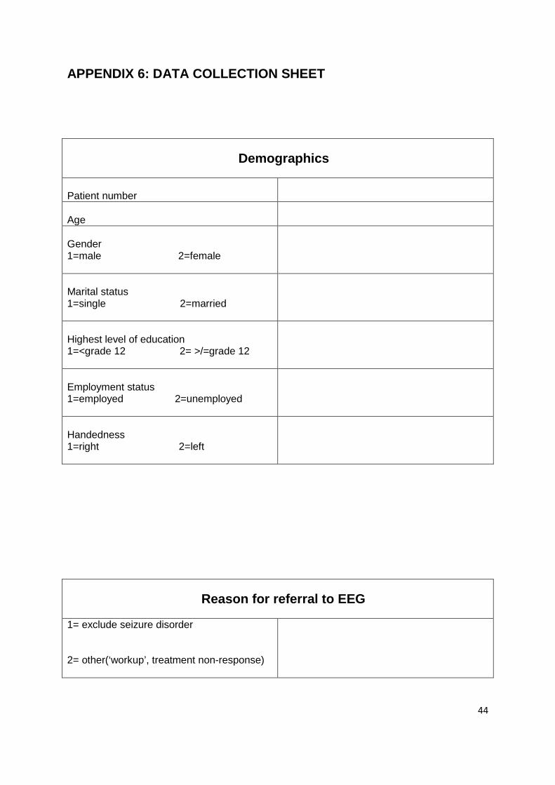
APPENDIX 6: DATA COLLECTION SHEET
Demographics Patient number
Age
Gender 1=male 2=female
Marital status 1=single 2=married
Highest level of education 1=<grade 12 2= >/=grade 12
Employment status 1=employed 2=unemployed
Handedness 1=right 2=left
Reason for referral to EEG 1= exclude seizure disorder 2= other(‘workup’, treatment non-response)
Page 58
45
Symptoms prior EEG request Disorientation 1=yes 2=no
Auditory hallucination 1=yes 2=no
Visual hallucination 1=yes 2=no
Tactile hallucination 1=yes 2=no
Olfactory hallucination 1=yes 2=no
Gustatory hallucination 1=yes 2=no
Seizure 1=yes 2=no
Dysmegalopsia 1=yes 2=no
Aggression 1=yes 2=no
Jamais vu/déjà vu 1=yes 2=no
Admission diagnosis
Diagnosis on discharge
Page 59
46
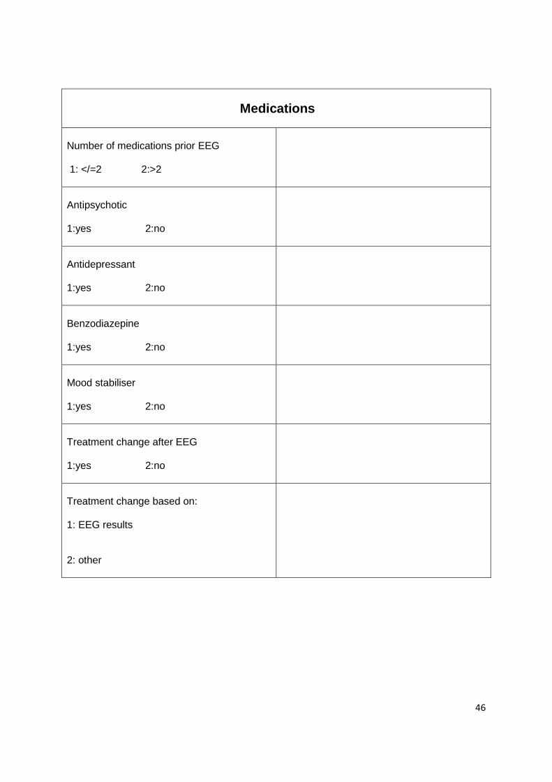
Medications Number of medications prior EEG 1: </=2 2:>2
Antipsychotic 1:yes 2:no
Antidepressant 1:yes 2:no
Benzodiazepine 1:yes 2:no
Mood stabiliser 1:yes 2:no
Treatment change after EEG 1:yes 2:no
Treatment change based on: 1: EEG results 2: other
Page 60
47
EEG
Provocation method prior EEG
1:yes 2:no
Specify:
Provocation method during EEG
1:yes 2:no
Specify:
EEG abnormality
1:yes
2:no
3:non-conclusive
EEG abnormality detected: