January 2012 NASA/CR–2012-217330 Electromagnetic Nondestructive Evaluation of Wire Insulation and Models of Insulation Material Properties Nicola Bowler, Michael R. Kessler, Li Li, Peter R. Hondred, and Tianming Chen Iowa State University, Ames, Iowa
Transcript
January 2012
NASA/CR–2012-217330
Electromagnetic Nondestructive Evaluation of Wire Insulation and Models of Insulation Material Properties
Nicola Bowler, Michael R. Kessler, Li Li, Peter R. Hondred, and Tianming Chen Iowa State University, Ames, Iowa
NASA STI Program . . . in Profile
Since its founding, NASA has been dedicated to the advancement of aeronautics and space science. The NASA scientific and technical information (STI) program plays a key part in helping NASA maintain this important role.
The NASA STI program operates under the auspices of the Agency Chief Information Officer. It collects, organizes, provides for archiving, and disseminates NASA’s STI. The NASA STI program provides access to the NASA Aeronautics and Space Database and its public interface, the NASA Technical Report Server, thus providing one of the largest collections of aeronautical and space science STI in the world. Results are published in both non-NASA channels and by NASA in the NASA STI Report Series, which includes the following report types:
TECHNICAL PUBLICATION. Reports of completed research or a major significant phase of research that present the results of NASA programs and include extensive data or theoretical analysis. Includes compilations of significant scientific and technical data and information deemed to be of continuing reference value. NASA counterpart of peer-reviewed formal professional papers, but having less stringent limitations on manuscript length and extent of graphic presentations.
TECHNICAL MEMORANDUM. Scientific and technical findings that are preliminary or of specialized interest, e.g., quick release reports, working papers, and bibliographies that contain minimal annotation. Does not contain extensive analysis.
CONTRACTOR REPORT. Scientific and technical findings by NASA-sponsored contractors and grantees.
CONFERENCE PUBLICATION. Collected papers from scientific and technical conferences, symposia, seminars, or other meetings sponsored or co-sponsored by NASA.
SPECIAL PUBLICATION. Scientific, technical, or historical information from NASA programs, projects, and missions, often concerned with subjects having substantial public interest.
TECHNICAL TRANSLATION. English-language translations of foreign scientific and technical material pertinent to NASA’s mission.
Specialized services also include creating custom thesauri, building customized databases, and organizing and publishing research results.
For more information about the NASA STI program, see the following:
Access the NASA STI program home page at http://www.sti.nasa.gov
Fax your question to the NASA STI Help Desk at 443-757-5803
Phone the NASA STI Help Desk at 443-757-5802
Write to: NASA STI Help Desk NASA Center for AeroSpace Information 7115 Standard Drive Hanover, MD 21076-1320
National Aeronautics and Space Administration Langley Research Center Prepared for Langley Research Center Hampton, Virginia 23681-2199 under Cooperative Agreement NNX07AU54A
January 2012
NASA/CR–2012-217330
Electromagnetic Nondestructive Evaluation of Wire Insulation and Models of Insulation Material Properties
Nicola Bowler, Michael R. Kessler, Li Li, Peter R. Hondred, and Tianming Chen Iowa State University, Ames, Iowa
Available from:
NASA Center for AeroSpace Information 7115 Standard Drive
Hanover, MD 21076-1320 443-757-5802
The use of trademarks or names of manufacturers in this report is for accurate reporting and does not constitute an official endorsement, either expressed or implied, of such products or manufacturers by the National Aeronautics and Space Administration.
i
Table of Contents
LIST OF FIGURES .................................................................................................................. v
LIST OF TABLES .................................................................................................................... x
LIST OF PUBLICATIONS AND PRESENTATIONS RESULTING FROM THIS WORK xi
Chapter I. Introduction ......................................................................................................... 1
2. History of wiring insulation....................................................................................................................... 2
Breakdown voltage ........................................................................................................................................ 2
Water and saline exposure ............................................................................................................................. 3
Material characterization ............................................................................................................................... 4
4. NDE of wire insulation.............................................................................................................................. 4
Results and discussion ................................................................................................................................. 69
Summary of the physical model ................................................................................................................126
Probe and measurement system.................................................................................................................127
iii
Measurement system and uncertainty analysis ..........................................................................................129
Parameters of the wire under test...............................................................................................................130
Case study: evaluation of polyimide-coated wires after thermal and hydrolytic exposure........................132
Results and discussion ...............................................................................................................................133
Figure 1 Chemical Structure of Kapton® Polyimide. ............................................................................ 8 Figure 2 Results of dynamic mechanical analysis on dried PI. ............................................................ 10 Figure 3 Percentage weight loss of PI as a function of temperature measured at 30 °C/min heating rate .............................................................................................................................................................. 11 Figure 4 The real permittivity (a) and loss factor (b) of dry PI over frequency range 1 Hz to 1 MHz ������������������ ���������� °C. ................................................................................................ 12 Figure 5 Friedman plot for a single step (A) normal reaction, (B) accelerated reaction, and (C) retarded reaction. .................................................................................................................................. 17 Figure 6 TG curves broaden as the rate increases from 2 to 30 Kmin-1. .............................................. 18 Figure 7 DTG curves for the data shown in Figure 6. .......................................................................... 18 Figure 8 Friedman plot from the data shown in Figure 6. .................................................................... 19 Figure 9 Activation energy plot for air atmosphere from (a) Friedman Analysis and (b) Ozawa-Flynn-Wall Analysis. ...................................................................................................................................... 22 Figure 10 Schematic representation of the multistep reaction. ............................................................ 22 Figure 11 Best fit model of the TG data for the four-step reaction models in Fig. 6, with parameters given in Table 3. The curves represent the model and the symbols represent the experimental data. . 23 Figure 12 TG isothermal curves of experimental data and model prediction. ..................................... 23 Figure 13 3-dimensional FT�����������������������������������-1 ramp rate TG on degradation onset. .............................................................................................................................................................. 24 Figure 14 FTIR data for exit gases of a 30 Kmin-1 ramp rate TG on degradation onset for four different spectral ranges. Peak intensities are only proportional within each range and should not be compared from range to range. ............................................................................................................. 25 Figure 15 MS data for exit gases of a 30 Kmin-1 ramp rate TG. ......................................................... 25 Figure 16 The loss factor of PI degraded at 475 °C for 3 hr, measured over frequency from 1 Hz to 1 ��������������������� ���������� °C. ........................................................................................ 27 Figure 17 The loss factor of dry PI and PI degraded at 475 °C for 3 hr as a function of frequency at room temperature. ................................................................................................................................ 27 Figure 18 Effect ��������!���������������������������������"#����$�������%��&�*��+�������;����the standard deviation in measurements on three nominally-identical samples. .................................. 28
<������=�>�������!������?- and ��- relaxations of dry PI and PI degraded at 475 °C for 3 hr. ....... 29 Figure 20 Pyrolysis process of imide groups of PI during heating [29]. .............................................. 30 Figure 21 FTIR spectra of Kapton polyimide at 30, 400, 450 and 480 °C. .......................................... 30 Figure 22 The cumulative distribution function of the measured dielectric strength of PI samples heated at 475 °C for up to 4 hours. Symbols represent experimental data and lines are obtained by least-squares fitting to the data. ............................................................................................................ 35 Figure 23 As for Figure 41 but for PI samples heated for 4 hours at various temperatures from 450 to 480 °C. .................................................................................................................................................. 36 Figure 24 The Weibull-��������;�!��;�!���������@JX���������������������@?X�������;���������heating time. ......................................................................................................................................... 36
vi
Figure 25 The Weibull-��������;�!��;�!���������@JX���������������������@?X�������;���������heating temperature for 4 hr heating time. ........................................................................................... 37 Figure 26 Measured voltage breakdown of degraded Kapton Film (symbols) with best linear fit (solid line). ...................................................................................................................................................... 41 Figure 27 Predicted time to failure at 12 and 14.7 kV and for isothermal temperatures ranging from 250 to 400 °C. .................................................................................................................................................. 42 Figure 28 The real permittivity (a) and loss factor (b) of PI immersed in water and saline solutions, measured at 1 kHz. Error bars indicate the standard deviation in measurements on three nominally-identical samples. ................................................................................................................................. 45 Figure 29 The real permittivity (a) and loss factor (b) of PI following immersion in distilled water. . 46 Figure 30 The real permittivity (a) and loss factor (b) of PI following immersion in 80 g/l saline. .... 47 Figure 31 Effect of dissolved sodium chloride on the real permittivity (a) and loss factor (b) of PI, measured at 1 kHz. ............................................................................................................................... 48 Figure 32 Chain scission mechanism of PI hydrolysis through interaction of H2O with the carbonyl groups [54]. ......................................................................................................................................... 49 Figure 33 The cumulative distribution function of the measured dielectric strength of PI samples immersed in water for 0, 4, 8, 16 and 24 hours. ................................................................................... 51 Figure 34 The Weibull-��������;�!��;�!���������@JX���������������������@?X�������;��������������of PI immersion in distilled water. ....................................................................................................... 51 Figure 35 The cumulative distribution function of the measured dielectric strength of PI samples immersed in water for 24, 48, 72 and 96 hours. ................................................................................... 52 Figure 36 Temperature-pressure phase diagram of crystalline PTFE with the inter- and intra-polymer chain crystalline structures. .................................................................................................................. 55 Figure 37 PTFE (a) below the melting temperature 327 °C; and (b) above the melting temperature. 56 Figure 38 Results of dynamic mechanical analysis on as-received PTFE. ......................................... 56 Figure 39 Real permittivity of as-received PTFE as function of frequency at room temperature. ...... 58 Figure 40 Real permittivity of as-received PTFE as a function of frequency and temperature. .......... 58 Figure 41 Real permittivity of as-received PTFE as a function of temperature at 1.15 kHz. (a): -150to 300 °C; (b): -10 to 50 °C. ................................................................................................................. 59 Figure 42 TG curves for PTFE. ............................................................................................................ 62 Figure 43 DTG curves for PTFE. ......................................................................................................... 62 Figure 44 Friedman Analysis for PTFE. .............................................................................................. 63 Figure 45 PTFE activation energy from (a) Friedman Analysis and (b) Ozawa-Flynn-Wall Analysis. .............................................................................................................................................................. 63 Figure 46 Model of the best fit TG data for a single-step reaction model in air for PTFE. In the plot, the curves represent the model and the shapes represent the modeled experimental data. .................. 64 Figure 47 Real permittivity of PTFE as a function of thermal exposure time at 340 °C in air. ........... 66 Figure 48 Experimental arrangement for permittivity measurement while the sample is under tensile strain, using an Agilent E4980A LCR meter and Test Resources, Inc. tensile load frame Model 150Q250. .............................................................................................................................................. 68 Figure 49 Engineering stress-strain curve of PTFE. ............................................................................. 69
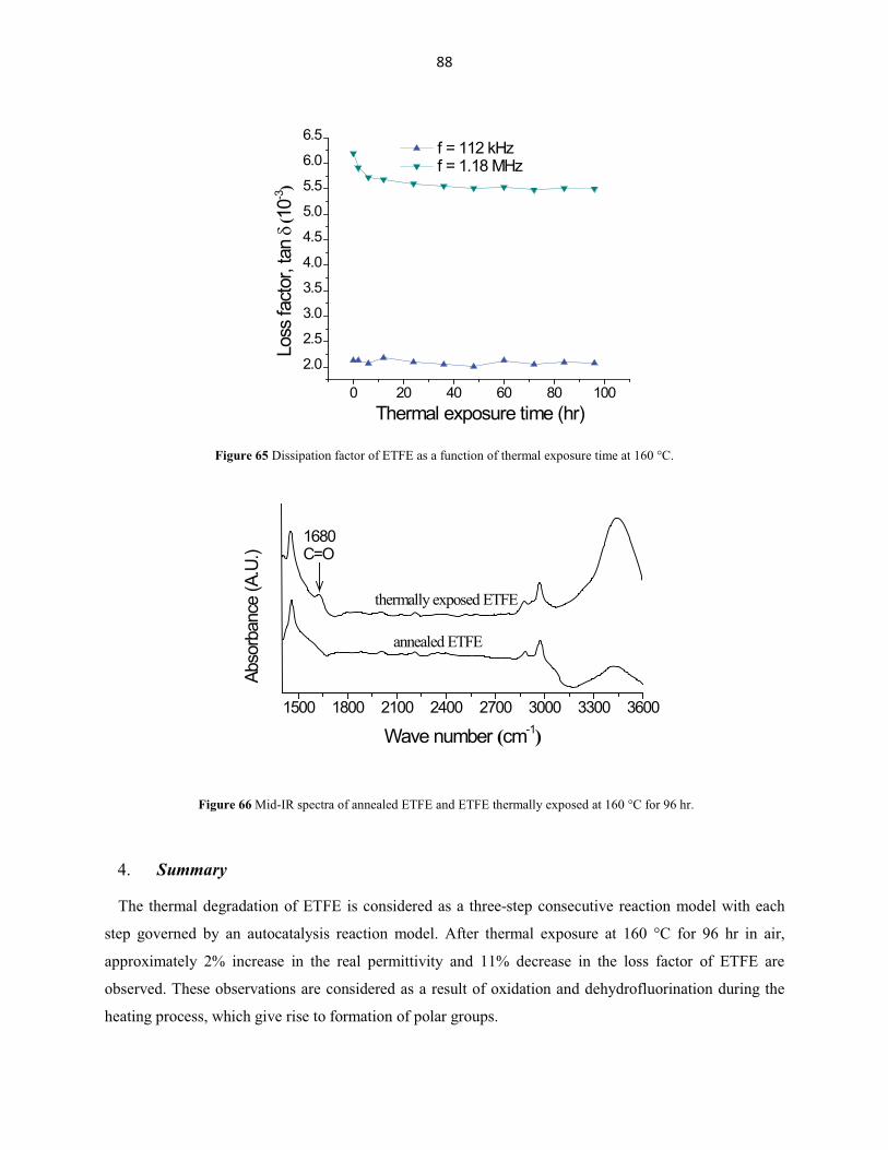
= 2.076 at 0% strain. ............................................................................................................................. 71 Figure 51 The difference between the real relative permittivity of PTFE under strain (solid symbol) and of released PTFE (open symbol), compared with untreated PTFE. .............................................. 71 Figure 52 Homogeneity of extruded ETFE by (a) DSC; (b) DMA. ..................................................... 75 Figure 53 Results of dynamic mechanical analysis on extruded ETFE. .............................................. 76 Figure 54 Real permittivity (a) and loss factor (b) of extruded ETFE as a function of frequency and temperature. .......................................................................................................................................... 77 Figure 55 Real permittivity and dissipation factor of extruded ETFE as a function of temperature at 1.15 kHz. .............................................................................................................................................. 78 Figure 56 TG curves for ETFE............................................................................................................. 81Figure 57 DTG curves for ETFE.......................................................................................................... 81Figure 58 Friedman Analysis for ETFE. .............................................................................................. 82Figure 59 Activation energy from (a) Friedman Analysis and (b) Ozawa-Flynn-Wall Analysis. ....... 82Figure 60 Two-step consecutive model fits to (a) the two slowest heating rates and (b) the two fastest heating rates.......................................................................................................................................... 83Figure 61 DTG model fit of the two slowest heating rates for ETFE. ................................................. 84Figure 62 Model of the best fit TG data for the three-step reaction models air atmospheres. In the plots, the curves represent the model, the data point shapes represent the modeled experimental data,and the + represents the un-modeled experimental data....................................................................... 84Figure 63 Real permittivity and dissipation factor of extruded and annealed ETFE as a function of frequency at room temperature............................................................................................................. 87Figure 64 Real permittivity of ETFE as a function of thermal exposure time at 160 °C. .................... 87Figure 65 Dissipation factor of ETFE as a function of thermal exposure time at 160 °C.................... 88Figure 66 Mid-IR spectra of annealed ETFE and ETFE thermally exposed at 160 °C for 96 hr......... 88Figure 67 Arc-electrode capacitive sensor. The radii of the sensor electrodes and the cylindrical
dielectric rod are denoted 0� and a, respectively. The arc-angle of each sensor electrode is 0� (rad). The length of each electrode in the vertical direction is l and the width in the horizontal direction is
Figure 68 Point source outside of a dielectric rod, assumed infinitely long. ........................................ 93 Figure 69 Discretization of the arc-electrode surfaces into M N� elements of assumed constant surface charge density. ......................................................................................................................... 98
Figure 70 Calculated sensor output capacitance as a function of electrode length l and arc-angle 0� .
The dielectric rod is in free space, with a relative permittivity of 2.5 and a radius of 9.525 mm. ....... 99 Figure 71 Calculated sensor output capacitance C as a function of the ratio of dielectric rod radius a
to electrode radius 0� . The electrode radius, arc-angle, and length are 0 9.525� � mm, 0� = 174.44o,
and l = 4 mm, respectively. ................................................................................................................ 100
viii
Figure 72 Calculated sensor output capacitance as a function of dielectric rod relative permittivity.
0/ 1a � � , l = 4 cm and 0� = 174.44o except where indicated. All the sensor electrodes have fixed
radius 0� =9.525 mm. ........................................................................................................................ 101
Figure 73 Agilent E4980A precision LCR meter and Agilent probe test fixture 16095A used for sensor capacitance measurements. Subfigure: photography of the flexible rectangular planar electrodes fabricated using photolithography. .................................................................................... 102 Figure 74 Measured and calculated C for various sensor configurations (see Table 18) in contact with different dielectric test-pieces. Measurement results and error bars are denoted by the black symbol. ............................................................................................................................................................ 104 Figure 75 Curved patch capacitive sensor. The radii of the sensor electrodes, the conductor, and the
cylindrical test-piece are denoted 0� , a, and b, respectively. The arc-angle of each sensor electrode is
0� (rad). The length of each electrode in the vertical direction is l and the width in the horizontal
direction is 0 0w � �� � . .................................................................................................................... 109
Figure 76 Point source outside of an infinitely long dielectric-coated conductor. ............................. 110 Figure 77 Curved patch capacitive sensor is divided into M N� elements on each electrode, each with assumed constant surface charge density. .......................................................................................... 113
Figure 78 Calculated sensor output capacitance as a function of electrode length l and arc-angle 0� .
The rod is in free space with conductor radius 8a � mm, dielectric radius 0 9b �� � mm and
���!�;��;�������^��_�"r2’=2.5. ........................................................................................................... 114 Figure 79 Calculated sensor output capacitance C as a function of the ratio of cylindrical test-piece
outer radius b to electrode radius 0� and the ratio of conductive core a to cylindrical test-piece outer
radius b. The electrode radius, arc-angle and length are 9 mm, 174o and 4 cm, respectively. ........... 115 Figure 80 Calculated sensor capacitance and dissipation factor as a function of the dielectric coating ��!�������^��_�"r2~�����������_�������^��_�"r2’’. Sensor configuration: 0 9� � mm, / 0.8a b � ,
0/ 1b � � , 4l � cm and 0 170� �� ��;������������;�������������!����&�"r2’’ and the material
�������������;�����������������+����������X&�"r2’=2 in b). .............................................................. 116 Figure 81 Agilent E4980A precision LCR meter and Agilent probe test fixture 16095A used for sensor capacitance measurements. Subfigure: photography of the flexible rectangular planar electrodes fabricated using photolithography. .................................................................................... 118 Figure 82 Photograph of fabricated capacitive probe with curved patch electrodes. Subfigure: capacitive probe holding a wire sample under test. ............................................................................ 128 Figure 83 Algorithm for determination of the effective electrode arc-angle 0� . ................................ 129
Figure 84 Schematic diagram of fluorinated ethylene propylene (FEP)-coated polyimide 150FWN019 film. Nominal thicknesses of each layer of polyimide 150FWN019 film, FEP fluoropolymer film, and the liquid H lacquer film are 25 �m, 13 �m, and 129 �m, respectively. The FEP provides adhesion between the layers of polyimide. Dc=2.09 mm, Dw=2.50 mm (nominal). ........................................ 131 Figure 85 Algorithm for determination of the imaginary permittivity ����������^���������& .......... 132
ix
Figure 86 Left: muffle furnace used for thermal exposure. Upper right: wire samples after heat exposure (brown) and a control wire (yellow). The samples are 4 cm long. Lower right: For hydrolytic exposure, both ends of the sample are sealed with wax. ................................................... 133 Figure 87 a) measured capacitance and b) dissipation factor for heat exposed wires. Uncertainties derive from the standard deviation of measurements on three separate samples. Physical degradation of the sample heated beyond 2 hours at 475 oC prevented accurate capacitance measurement for those conditions. .......................................................................................................................................... 134 Figure 88 Inferred real permittivity ���������������!!_����������������;�mparison with that of polyimide HN film (Chapter II). a) 400 and 425 oC; b) 450 and 475 oC. ........................................... 135 Figure 89 Inferred imaginary permittivity ����������������!!_������������& ................................. 136 Figure 90 As for Figure 87 but for hydrolytically exposed wires. ..................................................... 137 Figure 91 Inferred complex permittivity, a) real part and b) imaginary part, of the hydrolytically exposed wires in comparison with that of polyimide HN film (Chapter II). ...................................... 138
x
LIST OF TABLES
Table 1 History of wiring insulation used in commercial aircraft. ......................................................... 3 \�+!������!�;������������������������;%��������`���!�& ............................................................ 9 Table 3 Parameters used in the kinetic model. ..................................................................................... 24 Table 4 Correlated coefficient of 2-parameter and 3-parameter Weibull distribution of dielectric strength of PI heated at 475 °C ............................................................................................................. 35 Table 5 Weight loss of PI samples heated at 475 °C for up to 4 hours. ............................................... 37 Table 6 Weight loss of PI samples heated for 4 hours at various temperatures from 450 to 480 °C. .. 37 Table 7 Parameters values used to obtain the calculated lifelines shown in Figure 27. ....................... 42 Table 8 Weight gain of PI samples immersed in distilled water for up to 96 hours............................. 52 Table 9 The Weibull-��������;�!��;�!���������@JX���������������������@?X�����_�$�& ............... 52 Table 10 Properties of PTFE and PE. ................................................................................................... 55 Table 11 Kinetic Parameters for the single step autocatalytic model for PTFE. .................................. 64 Table 12 Kinetic Models in comparison for PTFE*. ............................................................................ 64 Table 13 Crystallinity of PTFE for various values of tensile strain, measured by X-ray diffraction immediately upon removing the sample from the tensile tester. .......................................................... 70 Table 14 Properties of ETFE. ............................................................................................................... 73 Table 15 Reaction parameters for two-step consecutive model fits for ETFE. .................................... 83 Table 16 Reaction parameters for the three-step consecutive model fit. .............................................. 85 Table 17 F-Test statistical analysis of the model fits for three-step reactions for ETFE*. .................. 85 Table 18 Parameters of the dielectric test-pieces and the arc-electrode sensors used in benchmark experiments. The areas of the two sets of sensor electrodes are 29 � 20 mm2 and 29 � 40 mm2,respectively. ........................................................................................................................................ 103 Table 19 Comparison of test-piece permittivity values between independently measured ones and inversely determined ones from measured capacitance using the arc-electrode sensors. .................. 104 Table 20 Measured complex permittivity values of the dielectric coating materials. ........................ 120 Table 21 Parameters of the test-pieces and the capacitive sensors used in benchmark experiments. The three copper rods used as the conductive cores in the cylindrical test-pieces had a uniform diameter of 15.90 ± 0.01 mm. ................................................................................................................................ 120 Table 22 Measured and calculated capacitance for various sensor configurations in contact with different cylindrical test-pieces. ......................................................................................................... 121 Table 23 Probe parameters, Figure 82. ............................................................................................... 129
xi
LIST OF PUBLICATIONS AND PRESENTATIONS RESULTING FROM THIS WORK
Articles in Journals (in print or accepted)
1 L. Li, N. Bowler, M. R. Kessler and S.-H. Yoon, Dielectric Response of PTFE and ETFE Wiring Insulation to Thermal Exposure, IEEE Trans. Dielectr. Electr. Insul., 17, 1234-1241, 2010.
2 L. Li, N. Bowler, P. R. Hondred, and M.R. Kessler, Influence of Thermal Degradation and Saline Exposure on Dielectric Permittivity of Polyimide, J. Phys. Chem. Solids. 72, 875-881, 2011.
3 P. R. Hondred, S. Yoon, N. Bowler, E. Moukhina and M. R. Kessler, Degradation Kinetics of Polyimide Film, High Performance Polymers, 23, 335-342, 2011.
4 T. Chen, N. Bowler and J. R. Bowler, Analysis of Arc-electrode Capacitive Sensors for Characterization of Dielectric Cylindrical Rods, IEEE Trans. Instrumentation Meas., DOI: 10.1109/TIM.2011.2157573.
5 L. Li, N. Bowler, P. R. Hondred and M. R. Kessler, Statistical Analysis of Electrical Breakdown Behavior of Polyimide Following Degrading Processes, IEEE Trans. Dielectr. Electr. Insul., 18, 1955-1962, 2011.
Bulletins, Reports, or Conference Proceedings That Have Undergone Stringent Editorial Review by Peers (in print or accepted)
1 S.-H. Yoon, N. Bowler and M. R. Kessler, Thermal Analysis Properties of PTFE Electrical Wiring Insulation Material, Proceedings of the North American Thermal Analysis Society Annual Conference (NATAS 2008). Aug. 18-20, 2008. Atlanta, GA. CD-ROM. Pages: 9.
2 L. Li, N. Bowler, S.-H. Yoon and M. R. Kessler, Dielectric Properties of PTFE Wiring Insulation Materials as a Function of Thermal Exposure, 2008 Annual Report: Conference on Electrical Insulation and Dielectric Phenomena, IEEE Dielectrics and Electrical Insulation Society, 95-98.
3 L. Li, N. Bowler, S.-H. Yoon and M. R. Kessler, Dielectric Properties of ETFE Wiring Insulation as a Function of Thermal Exposure, 2009 Annual Report: Conference on Electrical Insulation and Dielectric Phenomena, IEEE Dielectrics and Electrical Insulation Society. CD-ROM. Pages: 4.
4 P. R. Hondred, S.-H. Yoon, N. Bowler and M. R. Kessler, Onset Degradation Kinetics of Poly(ethylene-alt-tetrafluoroethylene), Proceedings of the 37th North American Thermal Analysis Society Annual Conference (NATAS 2009). Sep. 20-23, 2009. Lubbock, TX. CD-ROM. Pages: 9.
5 P. R. Hondred, S.-H. Yoon, N. Bowler and M. R. Kessler, A Comparison of Degradation Kinetics for Aerospace Wire Insulation Materials, Proceedings of the 38th North American Thermal Analysis Society Annual Conference (NATAS 2010). Aug. 15-18, 2010. University of Pennsylvania, PA. CD-ROM. Pages: 7.
6 Li Li, N. Bowler, P. R. Hondred and M.R. Kessler, Dielectric Response of Polyimide to Thermal and Saline Degradation, 2010 Annual Report: Conference on Electrical Insulation and Dielectric Phenomena, IEEE Dielectrics and Electrical Insulation Society. CD-ROM. Pages: 4.
xii
Bulletins, Reports, or Conference Proceedings That Have Not Undergone Stringent Editorial Review by Peers (in print or accepted)
1 T. Chen and N. Bowler, Cylindrical Capacitive Sensor for the Evaluation of Wire Insulation and Cable Degradation, Aircraft Airworthiness and Sustainment Conference, Austin, TX, 10-13 May 2010. http://www.airworthiness2010.com. Pages: 7.
2 L. Li, N. Bowler, S.-H. Yoon and M. R. Kessler, Dielectric Response of PTFE Wiring Insulation to Thermal Exposure, Aircraft Airworthiness and Sustainment Conference, Austin, TX, 10-13 May 2010. http://www.airworthiness2010.com. Pages: 11.
3 P. R. Hondred, S.-H. Yoon, N. Bowler and M. R. Kessler, Degradation Kinetics of Polyimide Insulation Material, Aircraft Airworthiness and Sustainment Conference, Austin, TX, 10-13 May 2010. http://www.airworthiness2010.com. Pages: 5.
Abstracts (in print or accepted) and Technical Presentations
1 N. Bowler, L. Li, S.-H. Yoon and M. R. Kessler, Dielectric and Thermal Analysis Properties of PTFE Wiring Insulation for Nondestructive Evaluation and Lifetime Prediction, NASA Aviation Safety Technical Conference, Denver, CO, October 21-23, 2008
2 N. Bowler, L. Li, P. R. Hondred, T. Chen and M. R. Kessler, Investigation of Dielectric and Thermal Properties of Wire Insulating Polymers for Development of Capacitive Nondestructive Evaluation, NASA Aviation Safety Technical Conference, McLean, VA, November 17-19, 2009.
3 P. R. Hondred, L. Li, T. Chen, S.-H. Yoon, N. Bowler and M. R. Kessler, Modeling and Nondestructive Evaluation of Wire Insulation, Interagency Wiring Group Meeting, Kennedy Space Center, December 8-10, 2009.
4 P. R. Hondred, S.-H. Yoon, N. Bowler and M. R. Kessler, Degradation Kinetics of Aerospace Wire Insulation Material, Society of Engineering Science 47th Annual Technical Meeting, Iowa State University, October 4-6, 2010.
5 N. Bowler and T. Chen, A Capacitive Sensor for Inspecting Wiring Insulation, The 2011 Aircraft Airworthiness and Sustainment Conference, San Diego, CA, April 18-21, 2011.
6 N. Bowler and T. Chen, Capacitive Sensors for Measuring Complex Permittivity of Planar and Cylindrical Test Pieces, 12th International Symposium on Nondestructive Characterization of Materials, Blacksburg, VA, June 19-24, 2011.
7 N. Bowler, L. Li, P. R. Hondred, T. Chen and M. R. Kessler, Dielectric Properties of Wiring Insulation Polymers in Response to Thermal, Hydrolytic and Mechanical Aging, and a Capacitive Sensor for Inspecting Wiring Insulation, NASA Langley Research Center Nondestructive Evaluation Sciences Branch, Hampton, VA, June 2011.
Publications and Creative Works Submitted but Not Accepted
1. P. Hondred, N. Bowler and M. R. Kessler, Electrothermal Lifetime Prediction of Polyimide Wire Insulation with Application to Aircraft, IEEE Trans. Dielectr. Electr. Insul., submitted September 2011.
2. T. Chen and N. Bowler, Analysis of a Capacitive Sensor for the Evaluation of Circular Cylinders with a Conductive Core, Meas. Sci. Technol., submitted September 2011.
1
Chapter I. Introduction
Polymers have been widely used as wiring electrical insulation materials in space/air-craft. The
dielectric properties of insulation polymers can change over time, however, due to various aging
processes such as exposure to heat, humidity and mechanical stress. Therefore, the study of polymers
used in electrical insulation of wiring is important to the aerospace industry due to potential loss of life
and aircraft in the event of an electrical fire caused by breakdown of wiring insulation.
Part of this research is focused on studying the mechanisms of various environmental aging process of
the polymers used in electrical wiring insulation and the ways in which their dielectric properties change
as the material is subject to the aging processes. The other part of the project is to determine the feasibility
of a new capacitive nondestructive testing method to indicate degradation in the wiring insulation, by
measuring its permittivity.
1. Motivation
Dielectric wiring insulation is used to separate electrical conductors by preventing the flow of charge
between wires. Insulation materials function to maintain a continuous and specified value of permittivity
over a specified range of electromagnetic field frequency and strength. Another essential property of
wiring insulation is the dielectric strength, a field at which the material fails to resist the flow of current
and arcing occurs. The dielectric properties of potential wiring insulation materials are always carefully
considered to guarantee that the selected materials satisfy requirements of the operating environment.
Both the dielectric permittivity and dielectric strength of wire insulation may change over time,
however, due to various degradation processes such as thermal aging, moisture exposure and mechanical
degradation. For example, wiring may be improperly installed and maintained, increasing the risk of
damage due to heat, moisture and chafing [1]. Such damage mechanisms may act acutely, or act to ‘age’
the insulation material over many cycles of aircraft operation. These mechanisms by which wire systems
insulation may be degraded produce what are known as a ‘soft’ faults, which act to modify the impedance
of the affected region of the coated wire structure, when viewed as a transmission line, rather than a ‘hard’
fault such as an open or short in the conductor itself. It has been reported [2] that aircraft suffer from
undiagnosed wiring degradation which may cause short-circuiting, fire and loss of control function.
According to Captain Jim Shaw, manager of the in-flight fire project for the United States Air Line Pilots
Association (ALPA), there are on average three fire and smoke events in jet transport aircraft each day in
USA and Canada alone, and the vast majority are electrical. It was presented in Air Safety Week, 2001,
that aircraft were making emergency landings, suffering fire damage to the point of being written off etc,
2
at the rate of more than one a month based on the experience of the previous few months. These issues
remain a concern for new aircraft.
Motivated by these concerns, the contribution of this work is to explore and record change in dielectric
properties of wire insulation due to various degradation processes.
2. History of wiring insulation
Table 1 shows wiring insulation materials applied in commercial aircraft since the 1960s [1]. PVC
(polyvinyl chloride) and Nylon were the main insulation materials from the 1960s to the 1980s. However,
in the next decades, PI (polyimide) was almost the only wiring insulation polymer used in the listed
airplanes. After the 1900s, another two materials, TKT 1 (Teflon -Kapton -Teflon) and Tefzel® ETFE
(ethylene-tetrafluoroethylene), have been widely used. This work focuses on three polymers: PI, PTFE
and ETFE. More detailed information about these polymers will be introduced in Chapters II, section 3.
3. Technical approach
Permittivity
The permittivity is a parameter that indicates the relative charge storage capability of dielectrics in the
presence of an electric field. In general, permittivity is complex, denoted �� = �’ � ��’’ . Complex
permittivity measurements have been made on the sample materials investigated here, before and after
degradation, to explore the changes in permittivity and dielectric relaxations in response to degradation.
Two instruments were employed to measure complex permittivity of the polymers. The first one is a
Novocontrol Spectrometer, which is capable of measurement over freq���;_������������������������&�
A temperature-controlled sample cell also permits measurements at temperatures from -200 °C to 400 °C.
The other one is an Agilent E4980A LCR meter coupled with a 16451 dielectric test fixture, which is
available from 20 Hz to 2 MHz at room temperature.
Breakdown voltage
Another essential property of dielectric insulators is the dielectric breakdown voltage, the point at
which the applied voltage causes current flow in a device (transistor, capacitor etc) to increase
uncontrollably. Breakdown in a capacitor results in the replacement of a reactive insulating component by
either a low-resistance short circuit or open circuit, usually with disastrous consequences as far as the
overall circuit function is concerned. The probability of its occurrence must therefore be kept to an
1 Teflon is a trade name for PTFE (polytetrafluoroethylene) and Kapton is a trade name for polyimide.
3
absolute minimum. Dielectric breakdown of insulation polymers before and after degradation processes
have been measured by a DIELECTRIC RIGIDITY 6135 which can supply voltage up to 60 kV..
Thermal exposure
Thermal exposure can significantly influence properties of polymers by changing microstructure, phase
morphology, chemical composition, etc. The effect of thermal exposure in air on the permittivity of PI,
PTFE and PI has been explored, which will be discussed in Chapters II, III, and IV, respectively.
Table 1 History of wiring insulation used in commercial aircraft.
Wiring insulation material
Applied years Applied aircraft
PVC/ Nylon 1960-1980 707, 727, 737, and DC-8
Slash 6 1965-1985 DC-9
Poly-X and Stilan 1970-1980 747, DC-10/MD-11
PI 1970 later 727, 737, 757, 767, MD-80/-90, DC-10/MD-11, Lockheed L-1011, Airbus
Tefzel 1975 later 727, 737, 757, 767, MD-80/-90, DC-10/MD-11
Tefzel/PI 1980-1990c 747
TKT 1990 later 737, 757, MD-80/-90, DC-10/MD-11
Water and saline exposure
There are several physical consequences of water absorption to wire insulation material including
plasticization, swelling, and changes in dielectric properties. Even though polyimide has very good
electrical and physical properties, it is very susceptible to humidity, which can give rise to cracks in the
insulation and cause electrical malfunctions. In response to the concern that aircraft which serve in navy
are exposed to sea water, the effect of water and saline exposure on dielectric properties of polyimide has
been studied. Effect of saline exposure on insulating properties of PI will be presented in Chapter II.
Mechanical stress
During cycles of aircraft operation and due to improper installation, wiring insulation materials may be
exposed to mechanical stress, which can result in structural changes and consequently influence the
dielectric properties of the insulation. Given this concern, the influence of mechanical strain on the
4
permittivity of PTFE is investigated in Chapter III. A system capable of measuring dielectric permittivity
while a polymer sample is simultaneously under tensile strain is designed and applied.
Material characterization
Thermal analysis instruments, such as a TGA (thermogravimetric analyzer) Q50 instrument, a DMA
(dynamic mechanical analyzer) Q800 instrument and a DSC (differential scanning calorimeter) Q20
instrument, are used to investigate thermal properties of the polymers. TGA uses heat to induce chemical
and physical changes in materials and performs a corresponding measurement of mass change as a
function of temperature or time. In some advanced instruments, residual gases released from materials can
be analyzed using TGA-tandem instruments, such as TGA-FTIR or TGA-Mass Spectrometry, to
determine the identity of the released gas and give insight into the weight loss mechanism. DMA
measures the mechanical properties of polymer material as function of temperature and frequency, which
reveals molecular relaxations in polymers. DSC is used to measure temperatures and heat flow during
thermal transitions (glass transition, crystallization and melting) in polymeric materials. The degree of
crystallinity of semi-crystalline polymers can also be obtained from the crystallization exotherm. Those
methods have been applied to investigate thermal properties of the three polymers, which will be
presented in Chapters II, III and IV.
X-ray diffraction (XRD) and Infrared (IR) spectroscopy are also utilized. Both of these analysis
methods are widely used to determine properties of polymers. XRD turns out to be a convenient and
reliable method to investigate crystalline structure. The degree of crystallinity of polymers, which plays
an important part in determining their dielectric properties, has been measured by XRD. IR spectroscopy
is one of the most common spectroscopic methods applied to analyze organic compositions. It utilizes a
Michelson interferometer and is based on IR absorption by dipolar molecules as they undergo vibrational
and rotational transitions. Each peak in an IR spectrum indicates characteristic absorption regions for
some commonly observed bond strength and bending deformations. It has been used to detect signs of
oxidation due to thermal exposure of PI and ETFE.
4. NDE of wire insulation
The theme of this research is focused on evaluating wiring insulation status through capacitive methods.
Insulation status can be characterized by its dielectric properties. Model-based capacitive methods
developed in this research relate quantitatively the measurable capacitance to the dielectric properties of
wires under test, and therefore allow for effective determination of wire insulation status. Experimental
studies on realistic aircraft wires showed that dielectric property changes in wiring insulation due to
5
thermal and hydrolytic exposures can be successfully detected using the capacitive methods developed in
this research, for wire type MIL-W-81381/12.
Motivation
This work is motivated by the effective evaluation of degradation status of air- and space-craft wiring
insulation. Degradation in electrical wiring insulation has the potential to cause aviation catastrophe due
to consequent short-circuiting or loss of control function [88]. Different wire inspection techniques have
been developed over the past decade, for the purpose of replacing the traditional visual inspection method.
Causes of failure and aging in aircraft wiring
In [89], causes and modes of failure in legacy aircraft wiring have been categorized. These causes
include chemical degradation such as corrosion of current carriers and hydrolytic scission of polymer
chains, electrical degradation of wiring insulation that may be due to concentrated electric fields at sites
of electrical stress and different kinds of arcing, and mechanical degradation due to vibration, over
bending and other kinds of mechanical stress.
Inspection techniques
Visual inspection is probably the most widely used method for aircraft wire inspection. It is highly
laborious while giving little quantitative information about the condition of the wires. Different physics-
based wire inspection techniques have been developed over the past decade to replace this traditional
inspection method, of which a summary is given here.
Methods that can be applied for wiring conductor inspectionThese wire inspections methods can qualitatively determine if the wiring is faulty but are not suitable
for inspection of aging aircraft wiring. Resistance measurement methods differentiate broken wires from
good ones by measuring the end-to-end cable resistance. High resistance indicates broken wires (open
circuit) while low resistance means the wiring is healthy (short circuit). The low-voltage resistance tests
and dielectric-withstand-voltage tests can detect faults but are not suitable for miniaturization or
pinpointing the fault [90].
One of the most commonly used physics-based techniques for aircraft wiring testing is reflectometry, in
which a high frequency electrical signal is sent down the wire and any impedance discontinuities in the
testing wire results in reflected signals. The location of the fault can be determined from the time or phase
delay between the incident and reflected signals whereas the impedance of the discontinuity is obtained
from the magnitude of the reflection coefficient. An excellent review paper that compares different
reflectometry methods is [91]. Reflectometry, however, is not capable of inspecting the insulation
conditions. Reflectometry methods are distinguished by the types of incident voltage used. Time domain
6
reflectometry (TDR) uses a short rise time voltage step as the incident voltage. This method is susceptible
to noises and is not optimal for live wire testing [92] [93] [94]. Frequency domain reflectometry (FDR)
uses a set of stepped-frequency sine waves as the incident voltage. A conceptual design of a "smart wiring
system" based on FDR methods that can be used for on-board testing of aging aircraft wiring has been
described in [90]. Phase-detection frequency-domain reflectometer (PD-FDR) has also been applied for
locating open and short circuits in a Navy F-18 flight control harness [95]. Sequence time domain
reflectometry (STDR) and spread spectrum time domain reflectometry (SSTDR) use pseudo noise
sequence and sine wave modulated pseudo noise code as the incident voltage, respectively. Testing
systems based on these two techniques are capable of testing live wires and therefore have the potential to
be used on energized aircraft to locate intermittent faults. The parameters that control the accuracy,
latency, and signal to noise ratio for SSTDR in locating defects on live cables has been examined in [96],
and the feasibility of spread spectrum sensors for locating arcs on realistic aircraft cables and live wire
networks has been demonstrated in [97] and [98].
In [99], linear relationships between the capacitance/inductance of open-/short- circuited wires (parallel
insulated round wires, twisted-pair wires, and coaxial cables) and their length have been demonstrated
and enables the determination of cable length from measured capacitance/inductance values.
Methods that can be applied for wiring insulation inspectionInfrared thermography systems and pulsed X-radiography systems have been developed as
nondestructive testing methods of aircraft wiring [100]. Infrared thermography has the benefits of rapidly
examining large areas of wiring and can serve as a global testing method, whereas a portable pulsed X-ray
system can be used to obtain a radiographic image of the portion of the wire or cable.
Ultrasonic methods have also been developed to obtain quantitative information about aircraft wire
insulation [101]. These methods, by modeling insulated wires as cylindrical waveguides, have been able
to relate extensional wave phase velocity to heat damage or exposure in wire insulation and thus provide
quantitative information about the insulation condition.
Acoustic and impedance testing methods aiming at locating intermittent faults in aircraft wires and the
widely used Mil-Std-1553 data bus system have been reported in [102]. Micro-fabricated current sensors
that could be located in key areas of the electrical wiring and interconnects systems have been reported in
[103]. Partial discharge (PD) analysis methods for diagnosing aircraft wiring faults are explored in [104],
where a simulation of PD signal based on high-voltage insulation testing standard [105] has been detailed,
followed by wavelet based analysis to de-noise the PD signals.
7
Capacitance methods developed in this research
Deficiencies of the above methods suitable for wiring insulation inspection include the need of complex
instruments in the measurement and not being able to provide quantitative information about the
insulation condition at specific locations. A favorable solution to these deficiencies is capacitive methods,
from which quantitative information about the permittivity of wiring insulation at specific locations can
be obtained using not so complicated equipment.
A curved patch capacitive sensor, with electrodes that conform to cylindrical test-piece surfaces, has
been developed for wiring insulation evaluation. Numerical models have been developed and verified for
both the homogeneous dielectric cylinder structure and the cylindrical structure of dielectric-coated
conductors. Experimental studies on realistic aircraft wires showed that dielectric property changes in
wiring insulation due to thermal and hydrolytic exposures can be successfully detected using the curved
patch capacitive sensors, for wire type MIL-W-81381/12.
8
Chapter II. Polyimide
Polyimide (PI) is widely used as an insulation material for machines and wiring, and is effective at
temperatures up to 400 °C. Given the fact that polyimide may be exposed to extreme temperatures during
unusual events in service, its thermal degradation kinetics and the effect of thermal degradation on its
permittivity and electrical breakdown behavior have been studied. The lifetime of polyimide under
electrothermal multi-stress is predicted by using a short term technique. As polyimide is commonly
immersed in salt water while serving in navy aircraft, effect of water/saline exposure on its permittivity
and electrical breakdown behavior is also investigated in this chapter.
1. Introduction
Kapton HN is a polyimide film developed by DuPont which has been successfully used as electrical
insulation in a wide range of temperatures, from -269 °C to +400 °C (4 K - 673 K) [3]. The chemical
name for Kapton HN is poly (4,4'-oxydiphenylene-pyromellitimide), and its chemical structure is shown
in Figure 1. Kapton® Polyimide is produced from the condensation of pyromellitic dianhydride and 4,4'-
oxydiphenylamine. In addition to its very light weight and advanced mechanical properties compared to
other insulator types, Kapton HN polyimide has good dielectric properties, such as high breakdown field,
low dielectric constant and low loss factor. Selected properties of 125 ������;%�Kapton HN film are listed
in Table 2 [3]. However, polyimide is very susceptible to hydrolytic degradation, which can give rise to
cracks in the insulation and cause electrical malfunctions [4].
Figure 1 Chemical Structure of Kapton® Polyimide.
Melcher et al [5] explored the effect of moisture on the complex permittivity of polyimide film in a
temperature range from 80 to 325 K. It is presented that the imaginary part "##����������������lm which
was dried for two days shows only one maximum in the temperature range (the lowest curve). Absorption
of water alters this behavior for different water contents at fixed frequency 10 kHz. The peak height
increases with water content and an additional smaller loss peak appears at its lower temperature shoulder.
The shape of the larger peak, which is notified as the high-temperature peak, is the same for all film.
The influence of the high-temperature peak can be subtracted because its shape is essentially independent
OONN NN
OO
OO
OO
OOnn
9
of the water content. But the height of the peak increases with higher water content. The second peak, the
low-temperature peak, is considered to be strongly overlapped by the high-temperature peak. According
to a statement in [6], since the high-temperature peak is present even at low humidity levels, it is proposed
to be associated with water absorbed at the carbonyl groups. And the low-temperature peak is only visible
at higher humidity, it is likely caused by water absorbed at the ether linkage. As the two loss peaks can be
removed by drying the film, it is concluded that the water dipole causes this relaxation process and not an
intrinsic dipole of the polyimide chain.
It is also presented in reference [5] that the increase of the real part "# near room temperature correlates
to the peaks in "##.
Thermal exposure of polyimide has also been explored [7] [8] [9] [10]. No significant changes in the
dielectric properties of polyimide were observed after thermal exposure in air or N2 from 200 to 350 °C
Property ValueDielectric strength 154 kV/mm at 60 Hz, 23 °C and 50% RHDielectric constant 3.5 at 1kHz, 23 °C and 50% RHDissipation factor 0.0026 at 1kHz, 23 °C and 50% RHMelting point none
Glass transition 360 to 410 ºC?-transition 60 to 127 °C�-transition -118 to -28 °C
Ultimate tensile strength 231 MPa at 23 °CImpact strength 78 N cm at 23 °C
Yield point at 3% 69 MPa at 23 °C
2. Sample material
All the PI samples under investigation in this chapter were cut from large sheets of 125-��-thick
Kapton® HN PI film obtained from Dupont.
The storage modulus E�j�!��������!���*����������������PI film were measured from -150 to 180 °C at 1
Hz by DMA and analyzed by the software ‘TA Universal Analysis’ which can estimate values of peaks
and shoulders in the curve to a tenth of a degree, as shown in Figure 2&� �\��� ������ ;�^�� �^��!�� two
molecular relaxations at approximately 60°C and 350 °C. The relaxation at 350 °C is attributed to the
glass transition that occurs in the amorphous phase [3], while the relaxation at approximately 60 °C is
associated with the ?-transition, which is a sub-Tg relaxation that takes place at temperatures between 60
and 127 °C [11]. It is considered to be a result of torsional oscillations of the phenylene ring, involving
10
imide groups of PI [12]. In ��������j� $�� ����+���� �-transition in temperature range between -118 and
-28 °C [11] due to increase in the vibration of aromatic groups as intra- and intermolecular interactions
decrease in the presence of absorbed moisture [12].
Figure 2 Results of dynamic mechanical analysis on dried PI.
Since the permittivity and electrical breakdown strength of Kapton PI film changes significantly with
moisture content [13], dry samples are needed in order to obtain baseline (control) values of dielectric
strength for comparing with those obtained following thermal exposure and immersion in water. In order
to determine a heating temperature to effectively remove water from PI, weight loss of a PI sample was
monitored by thermogravimetric analysis (TGA) while it was heated from 30 to 900 °C in air at
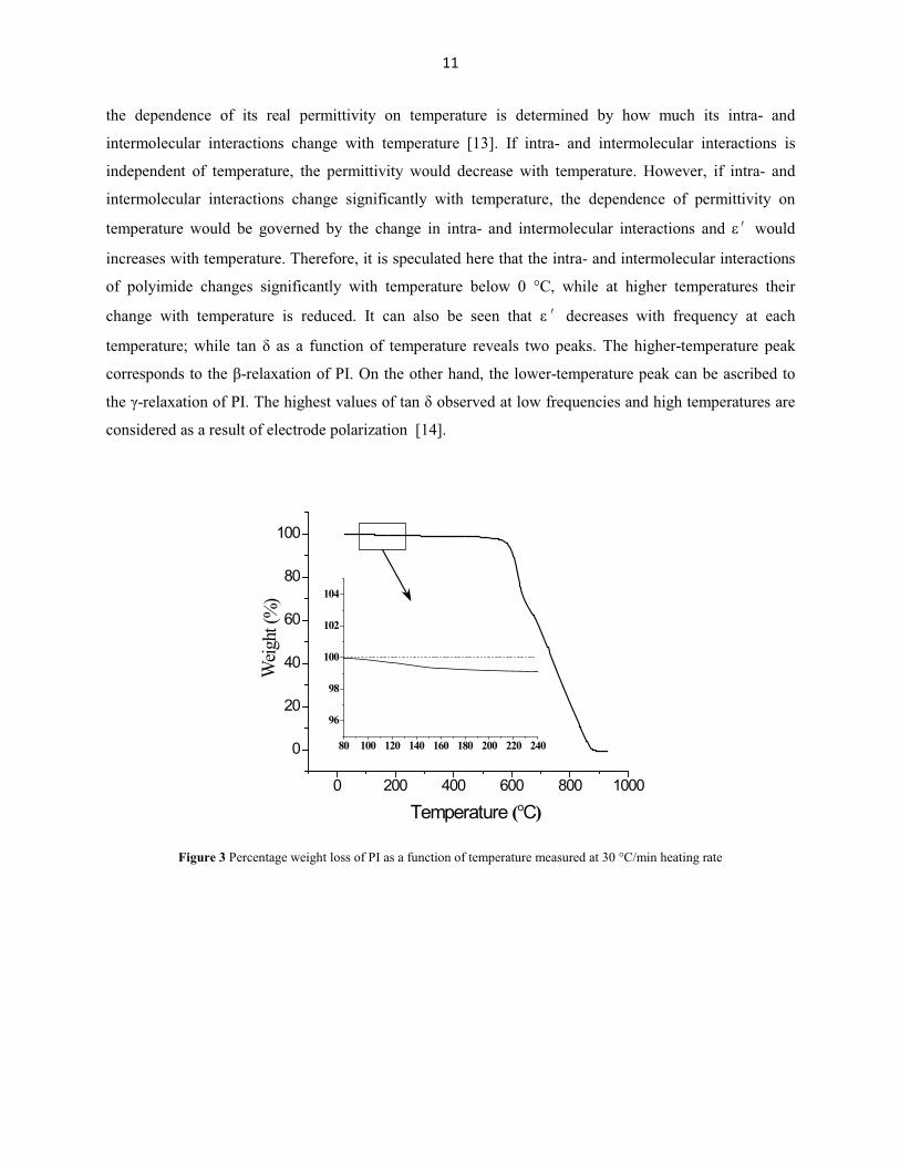
30 °C/min. As shown in Figure 3, an initial weight loss of 1% was observed at approximately 200 °C,
which is attributed to loss of water from the sample during the heating process. Therefore, PI samples for
baseline breakdown measurement were dried by heating at 200 °C for 1 hr, which, moreover, cannot give
rise to degradation of PI.
One dry PI control sample was coated with gold paint immediately upon removal from the isothermal
furnace. Its complex permittivity was measured at frequencies from 1 Hz to 1 MHz over temperatures
��;������� ���� � ���� ��� to 180 °C in increments of 10 °C, by using a Novocontrol Dielectric
Spectrometer with temperature-;����!!��������������!��;�!!&�\�����!�������^��_j�" , and loss factor,
�����j���������_�$��;����!����!���^���������!���������������������;_��nges are plotted as surface
plots in Figures 4a and 4b, respectively. ��!�����������!_�����j�" increases with temperature; while
it decreases with increasing temperature at higher temperatures. Given that polyimide is a polar polymer,
0.016
0.018
0.020
0.022
0.024
Tan
Delta
30
40
50
60
70
80
Loss
Mod
ulus
(MPa
)
1500
2000
2500
3000
3500
4000
4500St
orag
e M
odul
us (M
Pa)
0 100 200 300 400 500
Temperature (°C)
Instrument: DMA Q800 V7.4 Build 126
Universal V4.3A TA Instruments
11
the dependence of its real permittivity on temperature is determined by how much its intra- and
intermolecular interactions change with temperature [13]. If intra- and intermolecular interactions is
independent of temperature, the permittivity would decrease with temperature. However, if intra- and
intermolecular interactions change significantly with temperature, the dependence of permittivity on
temperature would be governed by the change in intra- ���� ������!�;�!�� �����;������ ���� " would
increases with temperature. Therefore, it is speculated here that the intra- and intermolecular interactions
of polyimide changes significantly with temperature below 0 °C, while at higher temperatures their
change with temperature is reduced. It can �!��� +�� ����� ����� " decreases with frequency at each
considered as a result of electrode polarization [14].
0 200 400 600 800 1000
0
20
40
60
80
100
80 100 120 140 160 180 200 220 240
96
98
100
102
104
Weig
ht (%
)
Temperature (oC)
Figure 3 Percentage weight loss of PI as a function of temperature measured at 30 °C/min heating rate
12
Figure 4 \�����!�������^��_�@�X�����!������;���@+X�����_�$���^��������;_�������������������������������������� ����to 180 °C.
3. Thermal degradation
Thermal degradation kinetics
Method and experimentThe most common tool for analyzing polymer degradation is thermogravimetry (TG). TG measures the
degree of degradation (as measured by mass loss) with respect to time (�) and temperature (�) [15]. The
degree of degradation (�) for the case of total decomposition with zero rest mass can be defined as:
� = (�, �) = 1 � �%�
(1)
where ��% is the relative mass obtained directly from the TG experiment.
TG experiments capturing the polymer degradation at different heating rates provide data that can be
used to obtain degradation kinetic parameters, such as activation energy, for various reaction models. In
this work, Kapton is analyzed by TG in an air environment to investigate the degradation in oxidative
environments. Through the use of isoconversional kinetics, the advanced model mechanisms are
identified. A mathematical model representing degradation is developed with an excellent statistical fit to
the experimental TG data and is used to compare isothermal data. Finally, Fourier Transformed Infrared
(FTIR) analysis and Mass Spectroscopy (MS) analysis of the exit gases identifies the breakdown
components of Kapton to verify the complex degradation of Kapton.
(a) (b)
� �
13
A thermogravimetric (TG) analyzer, model Q50 from TA Instruments (New Castle, DE), was used for
all of the TG experiments. The experiments were conducted from room temperature to 900 °C at five
separate ramp rates: 2, 5, 10, 20, and 30 Kmin-1. Under the controlled environment of the TG instrument,
the samples were degraded in an air atmosphere using a balanced purge gas flow rate of 40 mL/min and a
sample purge gas flow rate of 60 mL/min. Samples were placed on a platinum pan during the degradation
process. Kinetic analysis was performed with the Netzsch Thermokinetics 2 program (version 2004.05)
and standard statistical and plotting programs. Further study was conducted through evolved gas analysis,
a technique utilizing MS and FTIR on exit gases from the TG experiments, to verify the degradation
breakdown components and paths. Each test sample was punched out of the film using a circular punch,
5 mm in diameter, ensuring reproducible sample weight and shape. The sample masses were 3.6 ± 0.5 mg.
Kinetic modelingIn degradation kinetics, the degree of degradation (Eqn. 1) varies from 0 (no mass loss) to 1 (complete
mass loss). When modeling, two separate functions are assumed; �(�) and (�), such that the governing
differential equation has the following form:
�J��
= �(�)(J) (2)
where �J ��� is the rate of degradation, �(�) is the temperature-dependent rate constant, and (J)
corresponds to the reaction model [16]. The temperature-dependent rate constant is commonly described
by the Arrhenius equation:
�(�) = � ��� ��� (3)
where � is the universal gas constant, � is the activation energy, and � is a pre-exponential factor [17].
When heating at a constant rate, Eqn. 2 can be redefined to eliminate the time-dependence by dividing
through by the heating rate:
�J��
= ��
(J) ��� ��� (4)
where � = �� ��� is the heating rate.
Through linear transformation, the kinetic parameters ( � and � ) can be obtained by the time-
independent rate equation:
�! "�#
��$&(#) ' = �! *�
�+ � �
��(5)
14
Eqn. 5 follows the linear form - = . + .�0 (with 0 = 1 �� ) and optimal fit of the kinetic parameters is
determined using linear regression. By calculating these parameters through linear regression at several
different mass losses, the variation in the kinetic parameters as a function mass loss is determined.
In one approach for kinetic degradation modeling, constant activation energy and pre-exponential
factors are assumed [17]. The model-free isoconversional method allows for varying kinetic parameters
by assuming both the activation energy and pre-exponential factor are a function of the degree of
degradation [18]. Freidman’s method, a well-known technique, obtains the activation energy by plotting
the logarithmic form of the rate equation for each heating rate:
�! 2�3 *�#��
+#,3
4 = �!5�# (�)6 � �7��7,8
(6)
where the subscripts � and 9 represent the value at a particular degree of degradation and the data from
the given heating rate experiment, respectively [17]. The activation energy at each degree of degradation
is calculated with linear regression from a plot of �!:�3 (�� ��� )#,3; versus 1 �#,3� across all of the
heating rates tested. The Friedman plot not only provides confirmation of the multi-step processes during
the reaction but also provides insight into the type of reaction steps. The type of reaction can be
determined by comparing the slope of the constant fractional mass loss trend line to the slope of the
constant heating rate data at each peak. The peak slope specifically refers to the slope of the linear portion
to the right side of each peak. Comparing the relative magnitude of each negative slope, three types of
reactions are defined: normal, accelerated, and retarded. A normal reaction corresponds to slopes of equal
magnitude in both the fractional mass loss trend line and the peak slope of the constant heating rate
data—Figure 5A. An accelerated reaction corresponds to a steeper peak slope in the constant heating rate
data compared to the fractional mass loss trend line—Figure 5B. A retarded reaction corresponds to a
steeper fractional mass loss trend line compared to the peak slope in the constant heating rate data—
Figure 5C. Similar to the Friedman method, kinetic parameters can also be calculated by the Ozawa and
Flynn-Wall integral isoconversional method [19] [20].
Expanding the kinetic analysis from a single-step reaction to a multistep reaction, the differential
equations are separated based on each step of the reaction. The overall degree of degradation is
constructed as follows:
� = 1 � < �>a>> (7)
where � is the total fractional mass loss, a> is fractional mass loss of a specific reaction step, �> is the
contribution of a specific reaction step into total mass loss, and � represents the given reaction step [21].
The sum of the contributions of all steps is equal to 1:
15
< �>> = 1 (8)
Each fractional mass loss of a specific reaction step can be written as an individual differential equation
modeling the degradation of the reaction step such as [22]:
�5?@A?@BC6
��= �> ���@ ��� 5a>, a>D�6 (9)
The rate of reaction for a degradation from A � B (step 1) is given by �(a� A aE) ��� . The rate of
reaction for the degradation from B � C (step 2) is given by �(aE A aF) ��� . The rate of reaction for the
degradation from C � D (step 3) is given by �(aF A aG) ��� . In this format of differential equations the
values a�, aE, aF and aG are the formal concentrations of the formal substances A, B, C, and D. A is the
educt, B is the product of the first step and educt for the second step, C is the product of the second step
and the educt for the third step, and D is the product of the third step which is the final product of the
whole process. Each value of ai changes from 0 to 1. The initial state corresponds to a�=1, aE=0, aF=0
and aG=0, and final state D corresponds to a�=aE=aF=0, and aG=1. If the reaction steps are completely
separated, then the intermediate state after the first step corresponds to a�=0, aE=1 and aF=aG=0 and the
intermediate state right after the second step corresponds to a�=aE=0, aF=1 and aG=0. The degradation
continues to follow the analogy of chemical kinetics, where step 2 follows step 1, step 3 follows step 2,
but may occur before complete conversion of A to B.
Results and discussionThe TG scans for five different heating rates began at room temperature and the data can be seen in
Figure 6. Like most polyimides, Kapton is extremely stable at intermediate temperatures [23]. The onset
of degradation increases with increasing heating rate and involves a rapid and complete degradation. The
derivative of the weight with respect to temperature provides better insight into the mechanism of
degradation. For a specific heating rate, the number of peaks in the derivative thermograms (DTG)
represents the minimum number of reaction steps involved. By varying the heating rates, the degradation
steps can be separated and isolated. At higher heating rates, for Kapton, the reaction mechanisms can be
separated for better kinetic model understanding. Figure 7 shows the DTG curves. The peaks of the DTG
help ������������������;���������&���������������\��;�^���+�!��j��������������������-1, a minimum
of three reaction steps, or three peaks, can be seen.
The Friedman plot for Kapton can be found in Figure 8. A multi-step reaction is again evident from the
curvature of the plot. For each heating rate, there are separate reaction peaks. This indicates the
probability of a multiple step reaction. Model-free analysis shows a complex process with three peaks for
curves 30 Kmin-1 and 20 Kmin-1 and only two peaks for 2, 5, and 10 Kmin-1. The fluctuation in the
number of peaks indicates that the mechanism of the decomposition changes with heating rate.
16
Furthermore, the type of reaction can be determined by comparing the fractional mass loss trend lines
discussed previously with Figure 8. The fractional mass loss trend lines are the solid linear curves in
Figure 8, and are found from linear regression at specific values of � ranging from 0.2 to 0.8. In all cases
for Kapton, the peak slope is steeper than the fractional mass loss trend line indicating an accelerated
reaction, probably autocatalysis. For autocatalysis, the generic governing differential equation, presented
in Eqn. 2, defines the reaction model, f5aH6, such that:
5.>6 = 51 � .>6I51 + �JK�.>6 (10)
where n represents the reaction order and KM?N represents the autocatalysis constant.
The Friedman analysis is used to calculate the activation energy (EP) and the pre-exponential factor (AP)
from the slope and the y-intercept of the fractional mass loss trend lines, respectively [18] [19] [20]. The
activation energy and pre-exponential factor are shown in Figure 9 and presents activation energies from
20 kJ/mol to 190 kJ/mol. The plot of the activation energy with respect to the amount of degradation
again confirms the multistep reaction by presenting non-constant activation energy throughout the entire
degradation process. The fluctuating activation energy indicates an overlap of multiple reactions. As the
reaction begins, the activation energy is about 190 kJ/mol and then shifts to 40 kJ/mol for a fractional
mass loss of about 0.35. The activation energy increases to 60 kJ/mol for a fractional mass loss of 0.45,
and then decreases to 20 kJ/mol for mass loss 0.8, and finishes by trending upward in the final moments
of decomposition. The error bars show that the activation energy for the beginning of the reaction can be
well-defined. For the last steps at the fractional mass loss 0.7 the error bar of activation energy is much
higher and the lower value can reach almost zero kJ/mol. The error bars are calculated using standard
error from the linear regressions defined by the Friedman Analysis.
A physical meaning for the mass loss dependent activation energy from the Friedman Analysis is
difficult to identify with confidence because of the independence of overlapping degradation mechanisms.
Rather, the Friedman Analysis is useful in identifying multistep reactions. Given the complexity of
backbone structure in polyimide, the chemical structure can rearrange in tandem with the degradation
through aroyl migration or hydrolysis of the imido group. Dine-Hart et al. have proposed possible
degradation pathways in their studies of polyimide film [24] [25].
An integral isoconversional method called Ozawa-Flynn-Wall Analysis was also used to calculate the
activation energy as a function of fractional mass loss [19] [20]. Similar to the differential method used
in the Friedman Analysis, the activation energy can be extracted using isoconversional trend lines. The
benefit of comparing these two methods for activation energy provides insight into the type of reaction
step to best model the degradation. Since the integral method for calculating activation energy cannot
17
utilize separation of variables, degradation kinetics involving competitive reactions show variations
between the activation energies between the Friedman and Ozawa-Flynn-Wall Analysis. In conjunction
with DTG peaks, the experimental data suggests a minimum of three steps with a combination of
competitive and consecutive steps.
Figure 5 Friedman plot for a single step (A) normal reaction, (B) accelerated reaction, and (C) retarded reaction.
-6
-5
-4
-3
-2
-1
1 1.5 2 2.5
Log
d/d
t
1000 K/T
A)
-6
-5
-4
-3
-2
-1
1 1.5 2 2.5Lo
g d
/dt
1000 K/T
C)
-6
-5
-4
-3
-2
-1
1 1.5 2 2.5
Log
d/d
t
1000 K/T
B)
18
0
20
40
60
80
100
300 400 500 600 700 800 900
Wei
ght (
%)
Temperature (°C)
increasingheating rate
(K/min)25
2010
30
Figure 6 TG curves broaden as the rate increases from 2 to 30 Kmin-1.
Figure 7 DTG curves for the data shown in Figure 6.
0
0.5
1
1.5
2
2.5
3
300 400 500 600 700 800 900
Der
iv. W
eigh
t (%
/°C)
Temperature (°C)
increasingheating rate
(K/min)2
5
20
10
30
(K/min)
(K/min)
(K/min)
(K/min)
19
Figure 8 Friedman plot from the data shown in Figure 6.
For the simulation, a model of three parts was used. The schematic representation of the mechanisms
can be seen in Figure 10. The first part is the process from reactant A to reactant B, which proceeds along
two different paths 1 and 2. The second part is the one elementary reaction from reactant B to reactant C,
and the third part is the process from C to D, which also follows two parallel paths. Two different paths
for the third part of the model are necessary because the experimental data, Figures 5 and 6 show that the
decomposition mechanism for the last 60% mass loss depends on the heating rate. A multivariate version
of the Borchardt and Daniels method was used to determine to optimal fit of the kinetic parameters by
multiple linear regression [26]. The results of the model fit can be seen in Figure 11, with parameters
given in Table 3.
These parameters come from the combination of Eqn. 2 with the autocatalytic reaction model found in
Eqn. 10 for each step of the reaction diagramed in Figure 10. The Arrhenius parameters, E and A, are
related to the temperature sensitivity of the reaction [27]. The reaction order and autocatalytic constant
provide additional description of the chemical and physical reactions. The autocatalytic constant describes
the extent in which the degradation reaction itself acts as a catalyst for that reaction. In thermodynamics
of gases and liquids, the reaction order is an integer of stoichiometric equivalence. However, if the
reaction takes place in the solid-solid, solid-liquid, and solid-gaseous phases, physical processes influence
the reaction rate such as diffusion, phase-boundary reactions, or nucleation. Therefore, the direct
evaluation of experimental data with unknown reaction order gives non-integer values. With respect to
the first 3 steps, the effects are minimal and an approximate reaction order of n = 1 can be used without
effecting the model drastically. Yet, in the final two steps, there are significant variations to the reaction
-2
-1.8
-1.6
-1.4
-1.2
-1
-0.8
-0.6
-0.4
0.9 0.95 1 1.05 1.1 1.15 1.2 1.25
DDDDD
GGGGG
GGGG
Log
d/d
t
1000 K/T
0.2
0.4
0.60.8
2 K/min5 K/min10 K/min20 K/min30 K/min
20
order that cannot be approximated away. Therefore, the physical processes influencing the degradation of
the final competitive reaction steps differ from the stoichiometric coefficients and play a significant role
in the degradation.
From a statistical perspective, the model follows the data with an r2 value of 0.99991. The large
activation energies of the initial steps and very small values of activation energies for the last steps are in
agreement with results of Friedman analysis, if the error bars are taken into account.
The kinetic parameters obtained for the multi-step model were then used to develop an isothermal
model that would represent degradation across a 4-hour isothermal exposure for an air atmosphere, also
obtained by TG analysis. We modeled a temperature spread of 425 to 475°C in 25°C increments. Figure
12 shows a comparison of the isothermal experimental data and the mathematical models. Isothermal tests
intrinsically have significant uncertainty because of the variability to achieve the set temperature. While
the isothermal data and the model prediction differ by up to ~16%, the model captures the general trend
and magnitude of weight loss shown in the experimental data. This error is within the bounds of a typical
isothermal test, but could be brought down with improvements to isothermal experimental environment
such as tighter temperature control and gradients. However, the isothermal model was able to accurately
capture the general trend of the mass loss.
To further explore the degradation process, evolved gas analysis was conducted during the TG
experiments. This coupled FTIR and MS analysis involved monitoring the exit gases as a function of time
during the TG experiments at different heating ramp rates. The 3-dimensional FTIR data can be seen in
Figure 13 and the 2-dimensional FTIR data constructed for analysis is shown in Figure 14. The MS data
is shown in Figure 15.
There are four peak groups of interest as seen in the four sections in the FTIR figure. The first major
group of peaks revolves around the 3500 to 3800 cm-1 wavenumber range. These peaks indicate the bond
stretching of functional groups with removable hydrogen (bonds such as NH2, NH, COOH, and OH) [28].
The second major group of peaks occurs in the wavenumber range 2100 to 2400 cm-1. These peaks carry
the largest intensity and indicate bonds such as CO and CO2 [28]. This is to be expected since carbon
dioxide and carbon monoxide are dominant products of degradation. The third set of peaks appears at
wavenumbers around 1000 to 2000 cm-1. Since these fall close to the “fingerprint region” it is difficult to
claim specifically what these peaks indicate. However, this peak area signifies bonds such as N=O, N=C,
NO2, and fragmented aromatic rings [28]. The final set of peaks are found at around 720 cm-1 and
indicates nitro and nitroso compounds (NO bonds) [28]. These conclusions correspond well to typical
breakdown mechanisms proposed for Kapton. Dine-Hart showed that under oxidative degradation,
Kapton initially evolves CO2 through hydrolysis of the imido group followed by the decarboxylation of
the resulting acid group, evolves CO2 through an aroyl migration, and CO through extrusion of the imido
21
group [24] [29]. Blumenfeld also shows the evolution of CO2, CO, and H2O through the initial oxidative
degradation of the aryl ether groups while leaving behind the diimide group [30]. Further degradation at
elevated temperatures cleaves the diimide residual into its nitrogen compounds [30].
The mass spectroscopy data corresponds closely to the FTIR data as well. The first major peak
identified is mass number 44. This peak, which spreads across the whole TG degradation curve,
corresponds to carbon dioxide, which was shown in the FTIR data to be a major exit gas contributor [28].
Peaks 17 and 18 correspond to water and more generally to the removable hydrogen of the functional
groups also found in the FTIR data above [28]. Peak 22 is the double ionized peak for carbon dioxide and
therefore is overshadowed by the first carbon dioxide peak [28]. Peaks 30, 45, and 46 are indicative of
the nitro and nitroso compounds correlating to the FTIR peak in the 720 cm-1 range [28]. One peak not
shown, mass number 28, is the mass number for carbon monoxide [28]. While the FTIR data indicates
evolution of carbon monoxide, the mass number also corresponds to nitrogen in its diatomic form and
consequently the nitrogen in the air environment overshadows the carbon dioxide emission.
Both the MS and 3-dimensional FTIR data validate the multistep reaction by presenting multiple peaks
in the exit gas analysis. The 3-dimensional FTIR data, shown in Figure 13, clearly shows three peaks
along the time axis, at around 2400 cm-1 wavenumber. In addition, the MS data indicates overlapping
peaks, shown in Figure 15, as the reaction progresses providing support for the multistep reaction chosen
in Figure 10.
22
Figure 9 Activation energy plot for air atmosphere from (a) Friedman Analysis and (b) Ozawa-Flynn-Wall Analysis.
Figure 10 Schematic representation of the multistep reaction.
0
50
100
150
200
-2
0
2
4
6
8
10
0 0.2 0.4 0.6 0.8 1
Act
ivat
ion
Ener
gy (k
J/m
ol)
Log(A/s
-1)
Fract. Mass Loss
0
50
100
150
200
-2
0
2
4
6
8
10
0 0.2 0.4 0.6 0.8 1
Act
ivat
ion
Ener
gy (k
J/m
ol)
log(A/s^-1)
Fract. Mass Loss
A B B C D
D
1
2 3 4
5
23
0
20
40
60
80
100
400 500 600 700 800 900
Wei
ght (
%)
Temperature (°C)
increasingheating rate
(K/min)25
2010
30
Figure 11 Best fit model of the TG data for the four-step reaction models in Fig. 6, with parameters given in Table 3. The curves represent the model and the symbols represent the experimental data.
Figure 12 TG isothermal curves of experimental data and model prediction.
Figure 13 3-����������!�<\�����������������������������������-1 ramp rate TG on degradation onset.
25
Wave Number (cm-1)
Figure 14 FTIR data for exit gases of a 30 Kmin-1 ramp rate TG on degradation onset for four different spectral ranges. Peak intensities are only proportional within each range and should not be compared from range to range.
Figure 15 MS data for exit gases of a 30 Kmin-1 ramp rate TG.
Effect of thermal degradation on permittivity
It has been presented previously in the literature that dielectric properties of PI are not changed
significantly by heating at temperatures up to 350 °C for as long as 5000 hours either in air or N2 [7] [8]
[9] [10]. However, given the fact that electrical wiring insulation still has to work under extreme
temperatures, such as may occur in the vicinity of extreme events during service, it is also valuable to
explore the effect of thermal degradation at higher temperatures in air on the dielectric permittivity of PI.
3450
3500
3550
3600
3650
3700
3750
3800
Abs
orba
nce
2100
2150
2200
2250
2300
2350
2400
1000
1200
1400
1600
1800
2000
700
710
720
730
740
750
0 min
2.5 min
5 min
7.5 min
10 min
12.5 min
26
The Kapton PI film was sectioned into 3 cm by 10 cm rectangular samples, which were heated at 400,
425, 450 and 475 °C for 1, 2, 3, 4 and 5 hours in an isothermal muffle furnace. In order to eliminate
possible distortion of the samples during the heating process, the two shorter edges of each PI sample
were stabilized by a pair of mirror-finish stainless steel plates. Consequently, the majority of each sample
was exposed to oxygen and eligible for permittivity measurement. After heating, the PI samples were
removed from the furnace and cooled in air. With higher temperatures or longer heating times, the
samples became more brittle, darker and thicker, which can be associated with the formation of
oxidized layers on the sample surfaces during the initial degradation of PI [24] [29]. Thermal degradation
at temperatures higher than 475 °C or for exposure times longer than 5 hr was not conducted because the
PI became so badly deformed that its permittivity could not be measured successfully.
The thickness of all treated samples was measured by using a micrometer with uncertainty of 1 ��������
cooling. Directly following thickness measurement, the samples were coated with silver paint, serving
both as film electrodes for permittivity measurement and to prevent significant moisture exchange
between the samples and air. The silver paint dries quickly at room temperature, but requires curing for
16 hr at room temperature to achieve a volume resistivity as low as 5×10⁻⁶ to 1.25×10⁻⁵ ���&����������
silver paint was totally cured, each sample was cut into three smaller samples for permittivity
measurement. The permittivity of all treated samples was measured from 1 kHz to 2 MHz at room
temperature, using the Agilent E4980A LCR meter coupled with a 16451 test fixture. The three
strain, e.g. by approximately 19% for 150% strain. As stated in Chapter II, a dielectric material exhibits
some or all of the four polarization mechanisms: atomic, ionic, dipolar and interfacial polarization [74].
Given that PTFE is a non-polar semicrystalline polymer, its real permittivity is associated with only two
polarization mechanisms: atomic polarization due to a shift of the electron clouds in each atom under the
influence of the applied electric field and interfacial polarization due to accumulation of free charges at
interfaces between the amorphous and crystalline polymer phases [74]. The observed decreases in the
real permittivity of PTFE therefore may in principal be as a result of decrease in either atomic
polarization or interfacial polarization, or both. Atomic polarization as defined above, however, cannot be
influenced by mechanical strain. On the other hand, according to the Fringed Micelle Model [76], the
chains of a polymer under mechanical load increase their alignment along the direction in which the load
is applied, hence increasing the degree of alignment in the amorphous phase and producing a greater
degree of crystallinity. Thus, it is hypothesized here that the interfacial polarization of PTFE declines
under mechanical strain due to decreased mobility of free charges, as a result of increased ordering of the
polymer chains.
70
To investigate this hypothesis, the degree of crystallinity of samples with tensile strains of 50, 100 and
150% was determined by XRD, immediately upon removal of the polymer samples from the load frame.
It was observed that the degree of crystallinity increases, overall, with mechanical strain, as listed in
Table 13. The XRD spectra also revealed that the crystal structure of strained PTFE at room temperature
is hexagonal, the same as that of un-strained PTFE [64].
After the samples were released from the tensile loading frame, their permittivity was immediately
measured and observed to recover somewhat, increasing by up to 8% as shown in Figure 51, suggesting
partial recovery of the polymer chains. It is reasonable to conclude that the decrease in the real
permittivity of PTFE is associated with increased ordering of the polymer chains in the presence of
mechanical strain.
The potential effect of stress relief was investigated by measuring permittivity on samples strained to 50,
100 and 150% for 24 hours. Permittivity was measured at times t = 1, 2, 4, 6, and 24 hours. No
significant change in measured permittivity was observed as a function of time up to 24 hours. Finally,
the permittivity of PTFE stretched with strain rate of 25.4 mm/min was observed to be quantitatively
similar to that of PTFE stretched to the same strain with half the strain rate, 12.7 mm/min. Thus, within
the range of stresses and strains studied here, it is concluded that there is no obvious influence of stress
relief and strain rate on the permittivity of strained PTFE.
Table 13 Crystallinity of PTFE for various values of tensile strain, measured by X-ray diffraction immediately upon removing
the sample from the tensile tester.
Tensile strain (%) Degree of crystallinity (%)
0 36 ± 350 42 ± 3100 42 ± 3150 47 ± 3
71
Figure 50 The change in the real relative permittivity of PTFE as a function of tensile strain. Note, "r = 2.076 at 0% strain.
50 60 70 80 90 100110120130140150
-0.40
-0.35
-0.30
-0.25
-0.20
-0.15
-0.10
'r
Strain (%)
strained PTFE released PTFE
Figure 51 The difference between the real relative permittivity of PTFE under strain (solid symbol) and of released PTFE (open symbol), compared with untreated PTFE.
f = 1.00 kHz f = 10.6 kHz f = 112 kHz f = 1.18 MHz
88
0 20 40 60 80 100
2.0
2.5
3.0
3.5
4.0
4.5
5.0
5.5
6.0
6.5
Loss
fact
or, t
an�
(10-3
)
Thermal exposure time (hr)
f = 112 kHz f = 1.18 MHz
Figure 65 Dissipation factor of ETFE as a function of thermal exposure time at 160 °C.
1500 1800 2100 2400 2700 3000 3300 3600
annealed ETFE
Wave number (cm-1)
Abso
rban
ce (A
.U.)
thermally exposed ETFE
1680C=O
Figure 66 Mid-IR spectra of annealed ETFE and ETFE thermally exposed at 160 °C for 96 hr.
4. Summary
The thermal degradation of ETFE is considered as a three-step consecutive reaction model with each
step governed by an autocatalysis reaction model. After thermal exposure at 160 °C for 96 hr in air,
approximately 2% increase in the real permittivity and 11% decrease in the loss factor of ETFE are
observed. These observations are considered as a result of oxidation and dehydrofluorination during the
heating process, which give rise to formation of polar groups.
89
Chapter V. Development of a Capacitive Sensor for Nondestructive Evaluation of Wiring Insulation
Curved patch capacitive sensors have been developed to evaluate the material dielectric properties of
cylindrical structures. These sensors consist of two curved patch electrodes that are coaxial with and
exterior to the cylindrical test-piece. Description on the development of capacitive NDE sensor in this
chapter is divided into three parts. In part 1, a numerical model that relates the permittivity of a
homogeneous dielectric rod to the capacitance of a curved patch sensor is described. Experimental results
that verify the validity of the numerical model are provided. In part 2, a numerical model that relates the
permittivity of a cylindrical dielectric-coated conductor to the capacitance of a curved patch sensor is
described, together with benchmark experiment results that verify the theory. In the last part of this
chapter, a prototype capacitive sensor has been fabricated and applied for dielectric property
measurements of aircraft wires (wire type MIL-W-81381/12). Groups of wires have been exposed to
different thermal and hydrolytic environments. Experimental studies using the prototype capacitive sensor
showed that, thermal and hydrolytic exposure induced dielectric property changes in this type of wire
insulation have been successfully detected.
1. Analysis of a capacitive sensor for the evaluation of circular dielectric cylinders
An arc-electrode capacitive sensor has been developed for the quantitative characterization of
permittivity of cylindrical dielectric rods. The material property of the cylindrical test-piece can be
inversely determined from the sensor output capacitance based on a theoretical model. For the modeling
process, the electrostatic Green's function due to a point source exterior to a dielectric rod is derived. The
sensor output capacitance is calculated numerically using the method of moments (MoM), in which the
integral equation is set up based on the electrostatic Green's function. Numerical calculations on sensor
configuration optimization are performed. Calculations also demonstrate the quantitative relationship
between the sensor output capacitance and the test-piece dielectric and structural properties. Capacitance
measurements on different dielectric rods with different sensor configurations have been performed to
verify the validity of the numerical model. Very good agreement (to within 3%) between theoretical
calculations and measurement results is observed.
Introduction
Increasing demands for dielectric measurements have been observed over the past decade, with new
applications of advanced composites in modern aircraft, automobiles, and shipbuilding. Specifically,
90
dielectric measurements are important for the characterization of thin films, substrates, circuit boards,
printed-wiring boards, bulk materials, powders, liquids and semisolids.
Capacitance methods, because of their simplicity, relatively low cost, and high accuracy, have been
applied to characterize the dielectric properties of many different materials. Over the past 100 years,
closed-form solutions for capacitances due to various canonically shaped electrodes have been found, by
mapping out the electrostatic field in the vicinity of the conductors. Canonical electrode shapes are those
formed from surfaces easy to describe in standard coordinates, including strips, circular discs, annular
rings, cylindrical arcs, spherical caps, etc.
It is convenient to solve capacitance problems associated with electrified strips using Cartesian
coordinates. The capacitances for two parallel and coplanar infinite strips [106], as well as charged thin-
strip quadrupoles [107], have been solved using the triple integral equations. In addition, the potential
associated with a physically more realistic strip of finite length, the potential due to polygonal plates, and
the potential due to a charged elliptical plate have been derived using dual integral equations in Cartesian
coordinates [107].
Using cylindrical coordinates, the solution to dual integral equations has been applied to obtain the
surface charge distribution of a charged disc in free space, and also to obtain the potential due to a circular
disc placed between two parallel earthed planes [106]. The solution of the Fredholm equation has been
applied to solve for the capacitance of an electrified disc situated inside an earthed coaxial infinitely long
hollow cylinder. Also, the field due to two equal coplanar electrified discs has been solved by the method
of Kobayashi potentials, while the capacitance between two identical, parallel and coaxial discs has been
obtained by solving Love's integral equation. These solutions are available in the classic book written by
Sneddon [106]. Furthermore, an axisymmetric problem of several charged coaxial discs has been
considered by the dual integral equation method, and the solution has been obtained for the case when the
distance between neighboring discs is large compared to their radii [108]. The potential of a system of N
charged, arbitrarily located, circular discs has also been considered in [108]. Aside from classic problems
associated with discs, Cooke's solution of a set of triple integral equations has been applied to solve for
the field due to a charged annular ring of finite width in free space [106], while the solution to the integro-
series equations has been applied to obtain the total charge for a capacitor that consists of a coupled disc
and spherical cap [107].
Another set of canonical capacitance problems discussed in the literature are infinitely long axially
slotted open cylinders. In [107], the capacitance generated by a pair of charged symmetrically or
asymmetrically placed circular arcs has been calculated in circular cylindrical coordinates, whereas the
capacitance due to a pair of charged symmetrically placed elliptic arcs has been solved in elliptic cylinder
coordinates [107].
91
The canonical capacitance problems, mentioned above, are all discussed in free space situations, and
need further modifications to be applicable for characterization of material dielectric properties. Other
semi-analytical and numerical capacitive solutions have been developed over the past decade to keep pace
with new applications of dielectric materials. For example, interdigital dielectrometry has been applied
for material dielectric property characterization as one of the most commonly used capacitance sensors.
An excellent review paper on interdigital sensors is [109], in which sensor modeling, fabrication,
measurement setup, and applications are discussed in detail. In addition to a widely-used effective semi-
analytical approach, called the continuum model [110], conformal mapping techniques have also been
applied to obtain closed-form solutions for the capacitance of interdigital sensors in surface contact with
multi-layered dielectric structures [111]. Examples of practical applications of interdigital sensors include
estimation of meat fat content [112] and insulation damage detection in power system cables [113].
Multichannel fringing electric field sensors, designed by finite-element (FE) method calculations for
sensor modeling, optimization and performance evaluation, have been used for material property
measurements [114]. Cylindrical geometry electroquasistatic dielectrometry sensors have been developed
using semi-analytical models to quantitatively relate the dielectric properties of multi-layered test-pieces
to sensor output transcapacitance [115]. Concentric coplanar capacitive sensors for nondestructive
evaluation of multi-layered dielectric structures have been developed in [116], and can be applied to
detect water ingression and inhomogeneities in aircraft radome structures. In addition, rectangular
coplanar capacitance sensors have been developed to detect water intrusion in composite materials [117]
and for damage detection in laminated composite plates [118]. Rectangular capacitive array sensors that
detect surface and subsurface features of dielectric materials have been developed in [119]. In [99],
approaches of determining the length of open-circuited aircraft wires through capacitance measurements
have been presented.
In this research, a model-based capacitive method is developed for the quantitative dielectric property
characterization of circular cylindrical dielectric rods. The work is motivated by testing of cylindrical
components such as wiring insulation or polymeric tubing, and will be developed to deal with those cases
in future. The capacitance sensor consists of two arc-shaped patch electrodes that are located exterior to
and coaxially with the cylindrical test-piece. These two sensor electrodes exhibit a measurable
capacitance whose value depends on both the dielectric and geometrical properties of the dielectric rod.
The arc-electrode configuration offers a nondestructive and convenient way of determining the dielectric
constant of cylindrical test-pieces, compared to cutting a slice from the test-piece for a conventional
parallel-plate capacitance measurement. A numerical method, the method of moments (MoM), is
employed in the numerical calculations. First, the Green's function for a point source over the surface of a
dielectric rod is derived in cylindrical coordinates, in the form of modified Bessel functions of the first
92
and second kinds of integer order n: ( )nI z and ( )nK z . This Green's function then serves as the
integration kernel in MoM calculations, from which the sensor surface charge distribution is obtained.
Once the sensor surface charge distribution is known, one can easily calculate the sensor output
capacitance C through /C Q V� , where Q is the total charge on one electrode and V is the potential
difference between the two sensor electrodes. Validation of numerical results by benchmark experiments
has been performed, and very good agreement (to within 3%) between theoretical calculations and
measurements is observed. The advantage of the arc-electrode capacitive sensor reported in this study,
compared to existing planar capacitive sensors, is that the arc electrodes conform to the surface of a
cylindrical test-piece and concentrate the electric field in the material under test. In addition, the physics-
based model developed in this report allows inverse determination of test-piece permittivity from
measured arc-electrode capacitance.
Modeling
Figure 67 shows the configuration of the arc-electrode capacitive sensor. The capacitive sensor consists
of two identical arc-electrodes coaxial with a cylindrical dielectric rod, and exhibits a measurable
capacitance C that is quantitatively related to the permittivity and diameter of the material under test. In
the theoretical modeling, the cylindrical dielectric rod is assumed to be infinitely long while the arc-
electrodes are infinitesimally thin. The more general case in which the electrodes and the test-piece have
different radii, as shown in Figure 67, is considered theoretically. However, in order to achieve maximum
output capacitance, it is more desirable to have the sensor electrodes in tight contact with the cylindrical
test-piece in measurements. One practical approach to achieving tight surface contact between the
electrode and the test-piece is to deposit the sensor electrodes on a compressible dielectric material used
as the sensor substrate, and press the substrate against the test-piece to conform the arc electrodes to the
test-piece surface. This approach will be attempted in a future version of the sensor.
93
Figure 67 Arc-electrode capacitive sensor. The radii of the sensor electrodes and the cylindrical dielectric rod are denoted 0�and a, respectively. The arc-angle of each sensor electrode is 0� (rad). The length of each electrode in the vertical direction is l
and the width in the horizontal direction is 0 0.w � �� �
Figure 68 Point source outside of a dielectric rod, assumed infinitely long.
Derivation of Green's function in cylindrical coordinates
The electrostatic Green's function due to a point charge outside of an infinitely long dielectric rod is
derived in cylindrical coordinates, to form the integral equations later used in MoM calculations. Figure
68 shows a point charge placed at ( , , )z� �( ( ( exterior to a cylindrical dielectric rod of radius a and
dielectric constant "2. Without loss of generality, the dielectric constant for the medium exterior to the
dielectric rod is assumed to be "1. The resulting potential ( , , )z� �) due to such a point charge satisfies
The derived Green's function, dependent on the permittivity and radius of the dielectric rod under test, is
used later to calculate the capacitance of the arc-electrode sensor.
96
Note on the choice of the Bessel function kernelInstead of using the identity in (55), one can also express 1/ r in terms of the of Bessel function of the
first kind of order zero 0 ( )J -� [121]:
00
1 ( ) ,zJ e dr
--� -. �� / (65)
and express the Green's function (1) ( | )JG (r r in the form of Bessel functions of the first and the second
kind
(1) (1) | | (1) | |00 0
1
1( | ) ( , , ) 2 cos[ ( )] ( , , ) ,4
z z z zJ t
tG K e d t K e d- -� � - - � � � � - -
,
.. .( (� � � �
�
1( ( ( (� � % � 23
$/ /r r � � (66)
where(1) ( , , ) ( ) ( ) ( ) ( ) ( ) 0,1, ,t t t J t tK J J A Y J t� � - -� -� - -� -�( ( (� % �� � (67)
" # " # " # " #" # " # " # " # ,
12
12
aYaJaYaJaJaJA
tttt
ttJ ----
---(�(
(��� (68)
( ) ( ) / ( ) |t t aJ a dJ d �- -� -�(4� and ( ) ( ) / ( ) |t t aY a dY d �- -� -�(
4� . However, the denominator in (68)
contains an infinite number of zeros for - from 0 to . , and increases the complexity in MoM numerical
implementations. Therefore, the Green's function in the form of modified Bessel functions, (64), is a
better choice here for calculating the sensor output capacitance.
Numerical implementation
The capacitance C between the two arc-electrodes is calculated numerically as follows. The Green's
function derived above is used to set up the integral equation in MoM calculations, which leads to the
solution for the surface charge density on each electrode. The two electrodes are oppositely charged in the
numerical calculations. Because of the axisymmetry of the problem, it is only necessary to calculate the
surface charge density on one of the electrodes. The output capacitance C is then calculated from
,QCV
� (69)
where the total charge Q on each electrode is obtained by integrating the surface charge density over the
electrode surface and V is the potential difference between the electrodes.
Calculation methodFigure 69 shows the discretization of the arc-electrode surfaces into M N� elements of assumed
constant surface charge density. Each electrode is discretized into M elements in the � direction and N
97
elements in the z direction. Denote the surface charge density on the left electrode as ( , )s z5 �( ( and that
on the right electrode as ( , )s z5 � ,( (% . The potential at the observation point 0( , , )z� ��r on the
electrode surface due to the charged arc-electrodes can be expressed by integrating (64) over the electrode
surfaces:
" # " #" # " #
" #" # " # zddzG
zddzGz
s
s
(((%((�
(((((�)
//
//
6�,�5
6��5
�
0electrodeRight
1
0
0electrodeLeft
1
0
,|1
,|1,
rr
rr
(70)
In the MoM calculations, the following expansion is used to approximate the continuous function
( , )s z5 �( ( :
1( , ) ( , ),
MN
s j jj
z b z5 � 5 ��
( ( ( (� $ (71)
where j5 is the unknown constant surface charge density on element j and ( , )jb z�( ( is the pulse basis
function
1 on element ( , )
0 elsewhere.j
jb z�
7( ( � 89
(72)
To solve for the MN unknown coefficients j5 , weighting (or testing) functions ( , )iw z� are
introduced to force that the boundary condition for the potential in (70) is satisfied for each element on
the sensor surface. The point-matching method is used, in which the weighting functions are Dirac delta
functions:
( , ) ( ) ( ) on element ,i i iw z z z i� � � � �� � � (73)
where 1,2,...,i MN� . Discretizing the integral equation using weighting functions in each of the MN
elements, (70) is expressed as the following matrix equation:
98
Figure 69 Discretization of the arc-electrode surfaces into M N� elements of assumed constant surface charge density.
All the elements in V share the same potential v that is the potential applied to one of the electrodes. The
other electrode has potential -v. From (75) the surface charge density ( , )s z5 �( ( on one of the electrodes
is solved, and that for the other electrode is simply ( , )s z5 � ,( (� % . The total charge Q on each electrode
can be found by integrating ( , )s z5 �( ( over the electrode surface. The sensor output capacitance C is
ultimately calculated through (69).
Example calculationsWhen numerically calculating the matrix element given in (75), the zero to infinity summation and
integral in (1) ( | )G (r r (see (64)) need to be truncated. The convergence of the Green's function depends
on values o��"2«"1, /a � and 0� . When these values are large, large truncation ranges for the summation
and integral in (64) are needed. It is found that, for the case "2«"1=5, / 1a � � , 0 177� �� and 4l � cm, if
one truncates the summation in (1) ( | )G (r r with 40 terms and the integral with the range from 0 to 2000
for the off-diagonal components in (75), and the summation with 300 terms and the integral with the
range from 0 to 2000 for the diagonal components, accuracy to three significant figures can be achieved
in the final calculated sensor output capacitance C. The cases calculated in Figure 70 to Figure 72 and
Figure 74 have smaller "2«"1, /a � and 0� values than those in the case calculated above. The truncation
standard used here is adopted in all numerical calculations of sensor capacitance value in this section. It
guarantees achieving convergence with accuracy to the third significant digit in all the cases discussed
below.
The dependence of sensor output capacitance on the electrode configuration is investigated as follows.
In Figure 70, sensor output capacitance C is plotted as a function of the electrode length l and the arc-
99
angle 0� . In this example calculation, the infinitely long dielectric rod is assumed to be in free space, with
relative permittivity "r=2.5 and radius 9.525a � mm (chosen to be similar to the radii of the rods used for
experiments described later.) The arc-electrodes share the same radius as the cylindrical rod. It is seen
from Figure 70 that for any fixed electrode arc-angle 0� , there exists a linear relationship between the
sensor capacitance C and the electrode length l. On the other hand, for any given electrode length l, the
sensor output capacitance C increases as the electrode arc-angle 0� increases, and tends to infinity as 0�
tends to 180o. This is explained by the fact that the output capacitance C results from interaction between
the sensor electrodes. The charge density on the electrodes is highest at the electrode edges, and increases
as the electrode edges come closer together. As 0� tends to 180o, the gaps between the edges of the two
electrodes become infinitesimally small and therefore the resulting capacitance tends to infinity, in
accordance with the singular behavior of the charge density at the electrode edges. Figure 70 shows that
in order to achieve maximum sensor output signal, the ideal sensor electrodes would be as long as
practically possible and with large arc-angle 0� .
Figure 70 Calculated sensor output capacitance as a function of electrode length l and arc-angle 0� . The dielectric rod is in free
space, with a relative permittivity of 2.5 and a radius of 9.525 mm.
Figure 71 shows an example of the sensor output capacitance C as a function of the ratio 0/a � (see
Figure 67). Rod parameters are as for Figure 70. The arc-electrodes each have fixed radius 0� = 9.525
mm, arc-angle 0� = 174.44o and length l = 4 cm. In other words, Figure 71 shows the dependence of
sensor capacitance on the cylindrical test-piece diameter, for a fixed arc-electrode sensor configuration. It
is seen from Figure 71 that as the ratio 0/a � increases, sensor output capacitance increases dramatically,
100
especially when this ratio tends to 1. This is because as 0/a � increases, the average permittivity interior
to the arc-electrodes increases and therefore C increases. On the other hand, the sensor's most sensitive
area lies in the region close to the gaps between the two electrodes. As 0/a � tends to 1, the arc-
electrodes are more likely to detect increases in the average permittivity surrounding the sensor. This is
why the sensor output capacitance C changes more rapidly as the ratio 0/a � approaches 1. The
theoretical calculation in Figure 71 demonstrates that, during measurements, unidentified small air gaps
existing between the arc electrodes and the dielectric rod under test can introduce relatively large
uncertainty in the measured C, especially as 0a �4 . Therefore, in order to achieve the strongest sensor
output signal and the smallest uncertainty due to possible air gaps between the electrodes and test-piece, it
is desirable to have the arc-electrodes in tight surface contact with the test-piece.
Figure 71 Calculated sensor output capacitance C as a function of the ratio of dielectric rod radius a to electrode radius 0� . The
electrode radius, arc-angle, and length are 0 9.525� � mm, 0� = 174.44o, and l = 4 mm, respectively.
The sensor output capacitance C as a function of dielectric rod relative permittivity "r2 is plotted in
Figure 72, in which different sensor configurations are considered. A linear relationship between the
sensor output capacitance and the test-piece permittivity is observed and has been verified numerically,
by computation of a sufficient number of data points (seven in this case). It is seen that the slope of sensor
output capacitance versus rod permittivity depends on both the sensor configuration and the ratio 0/a � .
For a given 0/a � , the value of the slope increases as the electrode length l and arc-angle 0� increase.
This is because the value of the slope represents changes in the absolute values of the capacitance for any
101
rod permittivity increment. These absolute value changes in capacitance are most obvious for sensors
with large electrode length l and arc-angle 0� values. This also explains why the value of the slope, for
fixed l and 0� values, increases as 0/a � increases. However, it is worth pointing out that for fixed
electrode radius � , arc-angle 0� , and 0/a � values, although increasing electrode length l increases the
value of the slope, relative changes in capacitance as "r2 changes stay the same, because of the linear
relationship between the sensor output capacitance C and electrode length l (see Figure 70).
Figure 72 Calculated sensor output capacitance as a function of dielectric rod relative permittivity. 0/ 1a � � , l = 4 cm and 0�= 174.44o except where indicated. All the sensor electrodes have fixed radius 0� =9.525 mm.
Experimental verification
Capacitance experiments were performed to verify the validity of the developed theory. Two sets of
rectangular planar electrodes (shown in Figure 73) were fabricated using photolithography by American
Standard Circuits Inc.. The sensor shape was achieved by selectively etching a 18 ; m thick copper
cladding (14 mL standard) off a flat 25.4 ; m thick Kapton® type 100 CR polyimide film. These flexible
electrodes were fixed onto different cylindrical dielectric test-pieces later to form the arc-electrode
capacitance sensors. The sensor dimensions are w=29 mm and l=20 mm for one set and w=29 mm and
l=40 mm for the other (see Figure 67). A Nikon EPIPHOT 200 microscope was used to independently
measure the fabricated sensor dimensions, for the purpose of checking the difference between the
fabricated dimensions and the specified ones, and therefore the accuracy of the fabrication process. The
“traveling microscope” measurement method, with accuracy of 0.01 mm, was used to measure the
relatively large sensor electrode dimensions. It was found that the measured dimensions of the fabricated
electrodes are identical with the nominal values under such measurement accuracy.
102
Three 304.8-mm-long dielectric rods are used in the measurements to simulate the infinitely long
cylindrical dielectric rod. The dielectric rods are long compared with the electrode lengths (factors of
approximately 8 and 15 longer), and the edge effect due to finite rod length can be neglected if the sensor
electrodes are placed at the center of the rods. The rod materials are Acetal Copolymer (TecaformTM),
Cast Acrylic, and Virgin Electrical Grade Teflon® PTFE. A digital caliper, with accuracy of ± 0.01 mm,
was used to independently measure the diameter of each rod. The permittivity of each rod was
independently determined by cutting a slice from the end of each rod, and then measuring the permittivity
of each slice using a Novocontrol Alpha Dielectric Spectrometer at 1 MHz. In the Novocontrol
measurements, both sides of each slice were brushed with silver paint to form the measuring electrodes.
Figure 73 Agilent E4980A precision LCR meter and Agilent probe test fixture 16095A used for sensor capacitance
measurements. Subfigure: photography of the flexible rectangular planar electrodes fabricated using photolithography.
The rectangular planar electrodes were attached to each dielectric rod by taping the thin Kapton® sensor
substrate tightly against the rod material, as shown in Figure 73. The electrodes were aligned carefully so
that the upper and lower edges of the two electrodes were at the same height, the vertical edges of both
electrodes were in parallel, and the two vertical gaps between the two electrodes were of the same size, as
assumed in the theoretical model. Another layer of 25.4-�m-thick Kapton® film was wrapped tightly onto
the outsides of the electrodes in order to minimize the air gap between the electrodes and the dielectric
rod. Because the Kapton® films used were quite thin, influences from their permittivity on the
measurement signal were negligible.
For each dielectric test-piece used in the benchmark experiments, the test-piece material, test-piece
( ) ( ) / ( ),p p pI a K a� - - -� ( ) ( ) / ( ) |p p bI b dI d �- -� -�(4� and similarly for ( )pK b-( . The Green's
function in the form of modified Bessel functions, (85), is used in the following calculations of the sensor
output capacitance. The sensor capacitance is computed later using the derived test-piece geometry and
permittivity dependent Green's function.
Note that (85) can be simplified to the case of a homogeneous dielectric rod, described in [125], by
assigning 0a � . The Green's function (85) can also be expressed in the form of Bessel functions of the
first and second kind. However, the denominator of the integrand in the Green's function contains an
infinite number of zeros and increases the complexity in the numerical implementations.
112
Numerical implementation
Calculation methodThe sensor output capacitance C is calculated numerically using the method of moments (MoM).
Calculation procedures used here are similar to those in [125]. In summary, the following steps are taken
to compute the sensor capacitance.
First, the Green's function (85) is used to set up the integral equation that relates the unknown sensor
surface charge density ( , )s z5 �( ( to the imposed potential ( , )z�) on the sensor electrodes
0Left electrode
0Right electrode
( , ) ( , , | , , ) ( , )
( , , | , , ) ( , ) .
s
s
z G z z z d dz
G z z z d dz
� � � � � 5 � � �
� � � � 5 � , � �
( ( ( ( ( ( () �
( ( ( ( ( ( (� %
/ // /
(88)
In order to solve for the sensor surface charge density numerically, i.e., to use discrete functions
approximating the continuous function ( , )s z5 �( ( , each electrode in Figure 77 is discretized into M N�
rectangular elements. The charge density on each element is assumed to be constant and can be different
from others. Mathematically, this approximation is expressed as
1( , ) ( , ),
MN
s j jj
z b z5 � 5 ��
( ( ( (@ $ (89)
where ( , )jb z�( ( is the selected pulse basis function and j5 is the unknown coefficient to be determined.
Note the axisymmetry of the problem, it is only necessary to calculate the surface charge density on one
of the electrodes.
To solve for the MN unknown coefficients j5 , weighting functions ( , )iw z� are introduced to force
the integral equation (88) to be satisfied for each element on the sensor surface. The point-matching
method is used in this process, in which the weighting functions are Dirac delta functions. Expressions for
( , )jb z�( ( and ( , )iw z� can be found in [125]. Discretizing the integral equation using weighting
functions in each of the MN elements, (88) is expressed as the following matrix equation:
11 12 1 1
21 22 2 2
1 2
,
L
L
L L LL L
G G GG G G
G G G
55
5
A� � � �� � � �A� � � �� �� � � �� � � �
A� � � �
V� � � � �
(90)
where
0element ( , , | , , ) ( , ) ,ij i i i j j j jj
G G z z b z d dz� � � � � � �( ( ( ( ( ( (� / / (91)
113
L MN� and all the elements in V share the same potential that is applied to the electrode. The
unknown coefficients j5 are obtained by solving the matrix equation, and the total charge Q on each
electrode is calculated by integrating the surface charge density over the electrode surface. The
capacitance C between the two electrodes is obtained using the relationship C=Q/V.
Figure 77 Curved patch capacitive sensor is divided into M N� elements on each electrode, each with assumed constant surface
charge density.
Example calculationsThe dielectric-coated conductor is assumed to be in free space in the following calculations, i.e.j�"r1
*=1.
When numerically evaluating elements in the G matrix, the zero to infinity summation and integral in
( , , | , , )G z z� � � �( ( ( (see (85)) need to be truncated. The convergence of the Green's function depends on
values o��"2, ��� ¥�"2«"1, /a b , /b � and 0� . When these values are large, large truncation ranges for the
summation and integral in (85) are needed. It is found that, for the case "r2’=5, 0.02tan� � , a/b = 0.9,
/ 1b � � , 0 178� �� and l = 4 cm, if one truncates the summation in ( , , | , , )G z z� � � �( ( ( to 80 terms and
the integral with the range from 0 to 6000 for the off-diagonal components in G matrix, and the
summation to 400 terms and the integral with the range from 0 to 6000 for the diagonal components,
accuracy to three significant figures can be achieved in the final calculated complex sensor output
capacitance C, for both real and imaginary parts. The cases calculated in Figure 78 to Figure 80 and
Section 2.4 have smaller "r2’, tan� , a/b, /b � and 0� values than those in the case calculated above. The
truncation standard used here is adopted in all of the following numerical calculations, which guarantees
convergence to three significant figure accuracy is achieved for all the numerically calculated capacitance
values in this section.
The dependence of sensor capacitance on the electrode configuration is shown in Figure 78. The sensor
output capacitance is plotted as a function of the electrode length l and the arc-angle 0� . The dielectric
coating has a relative permittivity "r2’=2.5. The radius of the core conductor a = 8 mm and the outer
114
radius of the dielectric-coated conductor b = 9 mm. The sensor electrodes are assumed to be right on the
cylindrical test-piece: 0 b� � . In Figure 78, a linear relationship between the sensor capacitance C and the
electrode length l is observed for any fixed electrode arc-angle 0� . On the other hand, the sensor output
capacitance C increases as the electrode arc-angle 0� increases for any given electrode length l, and tends
to infinity as 0� tends to 180o. This is explained by the fact that as 0� tends to 180o, the gaps between the
two electrodes become infinitesimally small and the resulting capacitance tends to infinity. Figure 78
shows that the sensor output capacitance changes dramatically when 0� and l have large values. When
performing dielectric measurements in practice, it is usually helpful to have large sensor output signal and
therefore to have large 0� and l values. However, when l and 0� are large, C changes rapidly, and it is
important to have accurate sensor configuration information to infer accurately material dielectric
properties from measured C.
Figure 78 Calculated sensor output capacitance as a function of electrode length l and arc-angle 0� . The rod is in free space
with conductor radius 8a � mm, dielectric radius 0 9b �� � mm and dielectric permittivity "r2’=2.5.
The dependence of sensor capacitance on the test-piece geometry is shown in Figure 79. The sensor
output capacitance C is plotted as a function of the ratio 0/b � and the ratio a/b (see Figure 75). The
dielectric coating permittivity "r2’ is as for Figure 78. The sensor electrodes have fixed radius 0� = 9 mm,
arc-angle 0� = 174o and length l = 4 cm. It is observed that for fixed 0/b � values, the sensor capacitance
increases as the ratio a/b increases. Such a trend is more obvious when the ratio 0/b � tends to 1. This is
because the calculated capacitance C is actually the series capacitance of the capacitance between one
electrode and the core conductor and the capacitance between the core conductor and the other electrode.
When the ratio a/b increases, the distance between the sensor electrodes and the core conductor decreases
115
and the resulting total capacitance increases. In particular, when 0/ 1b � � , the output capacitance tends
to infinity as a/b tends to 1, in which case the gaps between the sensor electrodes and the core conductor
approaches zero. This also explains why the sensor capacitance increases as the ratio 0/b � increases for
given a/b values, and why such changes in capacitance are more rapid for large a/b values. The fact that
the overall permittivity of the region between the electrodes and the conductive core increases as 0/b �
increases also contribute to increases in the sensor output signal. In summary, the existence of the
conductive core in the test-piece increases the output capacitance for given sensor configurations, and as
the conductive core radius a approaches zero, the test-piece reduces to a homogeneous dielectric rod.
Figure 79 Calculated sensor output capacitance C as a function of the ratio of cylindrical test-piece outer radius b to electrode
radius 0� and the ratio of conductive core a to cylindrical test-piece outer radius b. The electrode radius, arc-angle and length
are 9 mm, 174o and 4 cm, respectively.
Figure 80 shows the sensor capacitance C and dissipation factor D as functions of the dielectric coating
real permittivity "r2’ and imaginary permittivity "r2’’, respectively. Different sensor configurations are
considered. In Figure 80a), a linear relationship between C and "r2’ is observed for all sensor
configurations. It is seen that the slope of sensor capacitance versus dielectric coating real permittivity,
i.e., the sensor sensitivity, depends on both the sensor configuration and the geometry of the cylindrical
test-piece. The largest slope in Figure 80 occurs when a/b, 0/b � , electrode length l and electrode arc-
angle 0� are the largest of the values considered. However, it is worth pointing out that although
increasing electrode length l increases the value of the slope, relative changes in capacitance stay the
same, because of the linear relationship between the sensor output capacitance C and electrode length l
(see Figure 78. For practical inspection of wires, a/b is fixed, and it is therefore important to keep 0/b �
close to 1 to achieve the highest sensitivity. In the selection of 0� , a trade-off exists. Larger 0� gives rise
116
to larger sensitivity as well as capacitance. On the other hand, larger 0� means that the inter-electrode
gap decreases and the penetration of the field into the insulation decreases as a consequence.
Similar relationships between D and "r2’’ are observed in Figure 80b). The major difference between the
response of C and D to the investigated factors is that D is less sensitive than C to changes in l, 0� and
a/b.
Figure 80 Calculated sensor capacitance and dissipation factor as a function of the dielectric coating real permittivity "r2’and
imaginary permittivity "r2’’. Sensor configuration: 0 9� � mm, / 0.8a b � , 0/ 1b � � , 4l � cm and 0 170� �� except
where indicated in other lines. "r2’’ and the material dissipation factor are assumed to be zero in a). "r2’=2 in b).
Dependence of capacitance on test-piece permittivity and sensor configurationIf l 4. , 0a 4 and 0/ 1b � � , the case of Figure 75 becomes a two-dimensional problem. An
analytical expression for the capacitance per unit length between the two curved patches has been derived
in [127] and takes the following general form
" #,21 %� sFC (92)
where sF is a shape factor that depends solely on the capacitor geometry. Considering the linear plots of
Figure 80a), the following relationship is found to hold in general for the problem discussed in this
section (Figure 77):
" #,21 � %� sFC (93)
where � is a constant showing the dependence of C on "1 and "2. 1� + means C is more dependent on "2
than "1, and vice versa. 1� � means C depends equally on "1 and "2. The factors � and sF in (93) may
be determined by selecting two data points on any of the lines in Figure 80a). It is found that � and sF
are constant for any given sensor configuration, independent of the particular data points selected for the
calculation.
117
As an example shown in Figure 80a), C obtained based on (93) fits nicely on the dashed line for
2.61� � and 1.01sF � m. Similar results are observed for all the other sensor configurations as well. It
is found that � increases as 0/b � , /a b and l increase, and as 0� decreases. The latter relationship can
be explained by the fact that as 0� decreases, more electric field penetrates the dielectric coating, and "2
will therefore have larger impact on C . The product sF� is the slope in the C versus "2 plot and shows
the sensor sensitivity, whose dependency on the sensor configuration has been discussed earlier.
Experiment
Experimental setup and measurement procedures
Benchmark experiments comparing the measured sensor capacitance with numerically-predicted values
were performed to verify the validity of the developed theory. Experiments were conducted at frequency
1 MHz. Note that although the numerical model is developed in the electroquasistatic regime, i.e., the
wave length < (approximately 300 m at 1 MHz) is much greater than the dimension of the test-pieces in
the experiment, the dielectric coatings still have complex permittivities. This is due to losses arising in the
materials due to the polarization response of the polymers lagging behind the switching of the applied
electric field at 1 MHz. For this reason, complex permittivities are considered in the following benchmark
experiments.
Two sets of rectangular planar electrodes (shown in Figure 81) were fabricated using photolithography
by selectively etching a 18-�m-thick copper cladding (14 mL standard) off a flat 25.4-�m-thick Kapton®
type 100 CR polyimide film. These flexible electrodes were attached to different cylindrical test-pieces
later to form the capacitance sensors. The sensor dimensions are w = 29 mm and l = 20 mm for one set
and w = 29 mm and l = 40 mm for the other (see Figure 75). A Nikon EPIPHOT 200 microscope was
used to measure the fabricated sensor dimensions, for the purpose of checking the difference between the
fabricated dimensions and the nominal ones, and therefore the accuracy of the fabrication process. The
“traveling microscope” measurement method, with accuracy of ± 0.01 mm, was used to measure the
dimensions of the relatively large sensor electrodes. It was found that the measured dimensions of the
fabricated electrodes are the same as the nominal ones under such measurement accuracy.
Three cylindrical test-pieces, each being a dielectric tube coated copper rod, were used in the
measurements to simulated the infinitely long dielectric-coated conductors in theory (see Figure 75). The
dielectric tube materials were Acetal Copolymer (TecaformTM), Acrylic, and Virgin Electrical Grade
Teflon® PTFE, respectively. The dielectric tubes were hollowed from homogeneous rods so that the inner
diameter of the tube matched the diameter of the copper rod as closely as possible. All the three dielectric
tubes were in tight surface contact with the central copper rods. The cylindrical test-pieces used are 152.4
118
mm in length. These test-pieces are long compared with the electrode lengths (factors of approximately 4
and 7 longer). The edge effect due to finite rod length can be neglected if the sensor electrodes are placed
sufficiently far from the ends of the test-piece. Prior to being hollowed out, the permittivity of each
dielectric tube was measured, by cutting a disc from the end of each rod and measuring its permittivity
using a Novocontrol Alpha Dielectric Spectrometer at f = 1 MHz and room temperature. The measured
test-piece permittivity values, together with uncertainty, are provided in Table 20. In the Novocontrol
measurements, both sides of each disc (around 19 mm in diameter) were brushed with silver paint to form
the measuring electrodes. The capacitance values resulting from these discs were between 2 and 5 pF. The
impedance measurement accuracy of the Novocontrol at 1 MHz and room temperature for such
capacitance values is 0.1% for the magnitude and 0.06o for the absolute phase accuracy. Note since
Teflon® are low loss materials (loss tangent below 10-4 in the frequency range 20 kHz to 1 MHz at room
temperature [128]), accurate measurements of their imaginary permittivities were not achieved using
either the Novocontrol or the Agilent E4980A LCR meter with the Agilent 16451B dielectric test fixture.
The diameter of the copper rods, the outer diameter of the dielectric-coated conductors and the
thickness of the dielectric coatings were measured using a digital caliper with accuracy of ± 0.01 mm.
Each of these values was measured at ten different locations on the test-piece. An average value and the
corresponding deviation were obtained, in which the deviation is defined as the maximum absolute
difference between the average value and the ten measured values.
Figure 81 Agilent E4980A precision LCR meter and Agilent probe test fixture 16095A used for sensor capacitance
measurements. Subfigure: photography of the flexible rectangular planar electrodes fabricated using photolithography.
The experimental arrangement for the capacitance measurements is shown in Figure 81. The
rectangular patch electrodes were conformed to each cylindrical test-piece by taping the thin Kapton®
sensor substrate tightly against the dielectric material. The thickness of the film (25.4 �m) is accounted
for in the numerical modeling while effects of its permittivity are neglected. This is because the sensor
capacitance is much more sensitive to small variations in 0/b � , when this ratio is very close to 1, than
119
those in the test-piece permittivity. The permittivity of the substrate was hence assumed to be that of the
test-piece, an assumption that introduces negligible uncertainty. The electrodes were aligned carefully in
order to achieve the sensor configuration in the theoretical model as closely as possible. The goal in the
alignment was to keep the upper and lower edges of the two electrodes at the same height, the vertical
edges of both electrodes in parallel, and the two vertical gaps between the two electrodes of the same size.
Another layer of 25.4-�m-thick Kapton® film was wrapped tightly onto the outsides of the electrodes to
further minimize the air gap between the electrodes and the cylindrical test-piece, leaving part of each
electrode exposed to make electrical contact with the probe test fixture later.
In the experimental verifications, two groups of capacitance measurements were performed for each
cylindrical test-piece: one group using the 20-mm-long electrodes and the other using the 40-mm-long
electrodes. For each cylindrical test-piece, the tube material, brass rod diameter, outer diameter of the
dielectric-coated brass, variation in the dielectric tube thickness, electrode radius 0� and electrode arc-
angle 0� are provided in Table 21, with uncertainties. Because the two types of electrodes were attached
at different locations on each test-piece, the outer diameters of the test-pieces in Table 21 were measured
at those individual locations and vary slightly. The electrode radius 0� for each cylindrical test-piece is
obtained by summing the outer radius of the dielectric-coated brass rod and the Kapton® substrate
thickness. The electrode arc-angle 0� and its uncertainty in Table 21 are calculated from the electrode
width w, electrode radius 0� and its uncertainty. Each test-piece has its own electrode arc-angle 0�
because the fabricated electrodes share a fixed width w while the cylindrical test-pieces have different
radii. The parameters shown in Table 20 and Table 21 were used as the inputs in the numerical
calculations.
An Agilent E4980A precision LCR meter was used to measure the sensor output capacitance at room
temperature. The LCR meter operating frequency was set at 1 MHz to approximate the electroquasistatic
assumption in the numerical model. Under these conditions, the measurement accuracy of the LCR meter
for a 4 pF capacitance is 0.15% and the absolute accuracy for the dissipation factor is 0.0015, whereas
those for a 13 pF capacitance are 0.13% and 0.0013, respectively. The measured capacitance values in
this section are all within 4 and 13 pF. If a lower operating frequency is desired in capacitance
measurements, an impedance measurement instrument with higher accuracy when measuring large
impedance values should be used. (According to the relationship 1/ 2Z j fC,� , the impedance Z
resulting from measuring a given capacitance C under a lower frequency f will be larger). The sensor
capacitance C was measured by placing an Agilent probe test fixture 16095A across the two sensor
electrodes, as shown in Figure 81. This probe test fixture was connected to the LCR meter and the
120
measured capacitance was read from the LCR meter screen. Note that in the modeling the two electrodes
are assumed oppositely charged and the conductive core of the test-piece is kept at ground potential. The
calculated capacitance is the series capacitance of the capacitance between one electrode and the
conductive core and the capacitance between the conductive core and the other electrode. When
performing capacitance measurements, however, one needs only to place the probe test fixture across the
two sensor electrodes, as shown in Figure 81. The potential on the conductive core is the average of the
potentials on the two sensor electrodes, due to the symmetry of the problem, and the capacitance picked
up by the probe is the series capacitance calculated in the numerical model.
Table 20 Measured complex permittivity values of the dielectric coating materials.
Dielectric tube material Measured dielectric tube real permittivity r2’
and coaxial cables) and their conductor length have been demonstrated. This relationship enables the
inverse determination of wire length from measured capacitance/inductance values. These techniques all
inspect for so-called ‘hard' faults in the metal wire conductor itself and are not capable of inspecting the
insulation conditions.
Techniques have also been developed for the evaluation of wiring insulation condition. Infrared
thermography and pulsed X-ray systems have been developed for nondestructive testing of wiring
insulation [100]. Infrared thermography offers the advantage of rapidly examining large areas of wiring
and can serve as a global testing method, whereas a portable pulsed X-ray system can be used to obtain a
radiographic image of a portion of the wire or cable. Ultrasonic methods have also been developed for
quantitative assessment of degradation in wiring insulation condition caused by heat damage, by
modeling insulated wires as cylindrical waveguides [101]. Moreover, acoustic and impedance testing
methods aimed at locating intermittent faults in aircraft wires have been reported in [102]. Partial
discharge (PD) analysis methods for diagnosing aircraft wiring faults are explored in [104], in which a
simulation of PD signal based on a high-voltage insulation testing standard [105] has been detailed,
followed by wavelet-based analysis to de-noise the PD signals. Deficiencies of the above methods include
the need of complex instruments in the measurement and not being able to provide quantitative
information about the insulation condition at specific locations. A favorable solution to these deficiencies
is the capacitive method, from which quantitative measurements of the permittivity of wiring insulation at
specific locations can be made, from which its condition can be inferred, using relatively simple
equipment.
126
This section describes a prototype capacitive probe that has been fabricated and applied for the
inspection of wiring insulation condition of type MIL-W-81381/12 but in principle can be extended to
any single-conductor wire. The probe is designed based on a previously developed physical model, in
which a curved patch capacitor is exterior to and coaxial with a cylindrical dielectric that coats a
conductive core cylinder (Chapter V Section 2). In the model, a quantitative relationship between the
complex permittivity of the dielectric coating material and the capacitance and dissipation factor of the
capacitor is described. This relationship is utilized here for quantitative assessment of wiring insulation
permittivity, based on measured probe capacitance. To demonstrate the feasibility of this proposed
technique, groups of wire samples have been thermally and hydrolytically exposed, under different
conditions, to induce dielectric property changes in the insulation. Capacitance measurements were
performed on the samples, and complex permittivity values of their insulation determined inversely by
means of the numerical model. Comparisons made between the complex permittivity of the damaged
wires and the control wires show that both the real and imaginary parts of the insulation permittivity of
the damaged wires increase as the thermal exposure temperature/time and hydrolytic exposure time
increase, and are higher than those of the control wires. Especially, changes in the imaginary permittivity
are more significant than those in the real part. For example, following thermal exposure, the imaginary
permittivity was observed to increase by up to 39% while the real part by up to 17%, for exposures at
temperatures between 400 and 475 oC for various times up to 5 hours. In the hydrolytic exposure
experiment, the imaginary permittivity was observed to increase by up to 60% and the real part by up to
12%, for exposure in water for various times up to 4 days. Permittivity changes in the wire insulation
detected by the capacitive probe are in accordance with results of independent measurements conducted
previously on planar thermally and hydrolytically exposed Kapton® film (Chapter II). These proof-of-
concept experiments have demonstrated the excellent capability of the capacitive probe for quantitative
evaluation of insulation condition for wires of type MIL-W-81381/12, and in principle can be applied to
any single-conductor wire.
Apart from the capacitive probe discussed in this research, many other capacitive techniques have been
developed and applied for NDE of dielectric materials [130]. For example, capacitive arrays have been
developed for robotic sensing using ‘scanning' and ‘staring' modes, [131]. Detailed literature surveys of
capacitive NDE methods can be found in [116]and [125].
Summary of the physical model
The capacitive probe is designed based on the sensor model described in (Chapter V Section 2). The
curved patch capacitive sensor consists of two identical and symmetric electrodes, Figure 75. The wire
under test is modeled as an infinitely long dielectric-coated perfect conductor. A relationship between the
127
complex permittivity "¬� of the dielectric coating, the sensor capacitance C and dissipation factor D is
established in the electroquasistatic regime, in which �* = ��-���; �� being the real permittivity, ���being the
imaginary permittivity, and 1j � � . It is worth pointing out that, by adopting this model, the multilayer
wire insulation Figure 84 is modeled as a one-layer structure. This means the insulation status assessed by
the capacitive probe will be the overall condition of all the insulation layers.
In the numerical model described in Section 2.2, the insulation real permittivity ��� ��� ��� !�����
relationship with the sensor capacitance Cj�������������!������!�����������������¥���« ������!����!_��!�����
to the sensor dissipation factor D. In this section, complex permittivity values of wire samples are inferred
from measured probe capacitance and dissipation factor values, based on this numerical model.
In the model, the conductor is kept at ground potential and potentials on the two electrodes are kept at 1
V and -1 V respectively. The calculated capacitance is the series capacitance of the capacitance between
one electrode and the conductive core, with the capacitance between the conductive core and the other
electrode. In the measurements, however, the conductive core does not have to be grounded. This is
because the potential on the conductive core will be the average of that on the two electrodes, and the
measured capacitance under this circumstance is the series capacitance calculated through the model.
The validity of this physics-based model has been verified by good agreement between numerical
calculations and results of benchmark experiments (Chapter V Section 2). A detailed description of
numerical modeling and experimental verification can be found in (Chapter V Section 2).
Probe and measurement system
Probe fabricationFigure 82 shows the capacitive probe. The probe consists of two 2 x 4 cm2 acrylic plates and an acrylic
rod on which the two plates are mounted so that their surfaces remain parallel. The lower plate is attached
to the acrylic rod using a plastic screw, whereas the upper plate can glide up and down by adjusting
another plastic screw perpendicular to the two plates. The curved sensor electrodes are formed by
brushing a layer of silver paint onto the symmetric grooves in the two plates. The two electrodes are
connected to two pins, which are then connected to an LCR meter for capacitance measurements. In order
to ensure that the two plates are in parallel, two acrylic dowels are attached to the upper plate using epoxy
glue, and inserted into the lower plate. These two acrylic dowels, together with the plastic screw, assure
that the two acrylic plates remain parallel during measurements. The subfigure in Figure 82 shows the
probe holding a wire under test. The probe and the wire are in tight surface contact with each other.
128
Figure 82 Photograph of fabricated capacitive probe with curved patch electrodes. Subfigure: capacitive probe holding a wire
sample under test.
ParametersParameters of the probe are (see Figure 75): electrode radius 0 2.50� � mm and electrode length l = 20
mm. The electrode radius is taken to be the same as the specified wire sample outer radius. The electrode
length is measured directly from the probe.
The region exterior to the curved electrodes in Figure 75 is assumed in the model to be homogeneous,
e.g., air. This assumption is not satisfied, however, for the probe depicted in Figure 82 due to the
existence of the acrylic plates exterior to the electrodes. To account for this effect, along with possible air
gaps between the electrodes and the testing wire due to surface roughness, an effective electrode arc-angle
0� is introduced for the capacitive probe. Steps taken to determine 0� are shown in Figure 83. First, an
assumed electrode arc-angle 0� , the wire sample geometry information, and the assumed wire insulation
real permittivity (�r��¥��&�) are input into the numerical model, from which a computed probe capacitance
Cmodel is obtained. Second, capacitance measurements are performed on the virgin wire samples. Third,
Cmodel is compared to the measured probe capacitance Cmeas. If Cmodel and Cmeas agree to three significant
figures, 0� is considered as the effective electrode arc-angle. Otherwise, a different value is assumed for
the effective electrode arc-angle and the above steps are repeated until the stop criterion is satisfied. For
testing wires of type MIL-W-81381/12, the effective arc-angle is determined as 0� = 80.1 ± 0.5o. This
inferred 0� is quite close to the physical electrode arc-angle, which is in the range 80 to 90 o. Uncertainty
in 0� is due to variations in the measured capacitance (2.11 ± 0.01 pF). Probe parameters are tabulated in
Table 23.
129
Table 23 Probe parameters, Figure 82.
l (mm) 20.0 ± 0.06
0� (mm) 2.50 ± 0.13
0� (degrees) 80.1 ± 0.5
Figure 83 Algorithm for determination of the effective electrode arc-angle 0� .
Measurement system and uncertainty analysis
For capacitance measurements, the probe was connected to an Agilent LCR meter 4980A by an Agilent
probe test fixture 16095A, whose probe pins were connected to the curved patch electrodes of the
prototype probe. Capacitance measurements in this section were performed at 1 MHz and room
temperature. Detailed discussion on the selection of operating frequency is provided in (Chapter V
Section 2).
Uncertainties in the measurement procedure are attributed to achievable precision in the measurement
equipment, uncertainty in 0� , and variation in the geometry of the wires under test. These uncertainties
contribute to uncertainty in the insulation complex permittivity determined inversely utilizing the
numerical model.
The achievable precision of the LCR meter depends on the magnitude of the capacitance measured. In
the LCR meter operation manual, relative uncertainty is provided for the measured capacitance whereas
absolute uncertainty is provided for the measured dissipation factor. Measured capacitance values in this
paper are within the range 2 to 3 pF and for these values the corresponding uncertainty is 0.18% for the
130
capacitance and 0.0018 for the dissipation factor. This means that, in order to detect insulation
degradation by capacitive measurements, the degradation-induced changes in the measured probe
capacitance and dissipation factor must be that greater than these values.
As seen in Table 23, the uncertainty in 0� and l are approximately 5%. When the arc-angle of the
curved patch electrodes is far from 180o, i.e., the distance between the edges of the oppositely-charged
electrodes is relatively large, the capacitance and dissipation factor change slowly as 0� varies (Chapter V
Section 2). This is because the interaction between the electrodes is not intense under this circumstance.
For the degradation cases studied in this paper, the uncertainty in the inferred insulation real permittivity
�r��resulting from the uncertainty in 0� is within 0.01, i.e., less than 0.5%, whereas that in �r� is 0.0002,
i.e., less than 0.2%.
Real wires exhibit variations in their dimensions, surface roughness, roundness and curvature even
when they may appear macroscopically similar. For this reason, in the experiments that follow,
capacitance measurements were performed on three samples, for each degradation condition. Error bars
are included in all the following measurement results. It is worth pointing out that the outer diameters of
the thermally and hydrolytically exposed wire samples still lie in the range between 2.41 and 2.63 mm. As
will be seen in Section 3.6, the standard deviation in the measurements deriving from these sources is the
dominant source of uncertainty in the measurements, larger than that due to the other sources discussed
above. Nonetheless, changes in the insulation complex permittivity, due to thermal and hydrolytic
exposure, are clearly observed even when uncertainty is taken into account.
Parameters of the wire under test
Capacitance measurements throughout this paper are performed on aircraft wire samples of type MIL-
W-81381/12. The wire is composed of a nickel-coated copper conductor, wrapped with two layers of
polyimide 150FW-N019 film and one layer of aromatic polyimide coating. Each layer of the polyimide
150FWN019 film is constructed by attaching a 13- ; m-thick fluorinated ethylene propylene (FEP)
fluoropolymer film to a 25- ; m-thick polyimide FN film, Figure 84. Nominal conductor and outer
diameters of the wire are 2.09 and 2.50 mm, respectively. Actual measured wire outer diameters for wires
examined in this study range between 2.41 and 2.63 mm.
131
Figure 84 Schematic diagram of fluorinated ethylene propylene (FEP)-coated polyimide 150FWN019 film. Nominal thicknesses
of each layer of polyimide 150FWN019 film, FEP fluoropolymer film, and the liquid H lacquer film are 25 �m, 13 �m, and 129
�m, respectively. The FEP provides adhesion between the layers of polyimide. Dc=2.09 mm, Dw=2.50 mm (nominal).
Real permittivityIn order to compare the inferred permittivity of the thermally/hydrolytically exposed wires to that of the
control wires and therefore provide quantitative assessment of the condition of the insulation, an initial
value for the permittivity of the control wires must be assigned.
The real part of the undamaged wire insulation permittivity is assumed to be 2.7 at 1 MHz and room
temperature, the condition under which capacitance measurements were performed. Reasons for this
assumption are as follows. Considering the schematic shown in Figure 84, we note that there are two
main components: polyimide 150FWN019 and an aromatic polyimide outer layer. The manufacturer,
DuPont, does not provide a specific permittivity value for polyimide 150FWN019 films, mentioning only
that it is less than 3 at 1 kHz and room temperature. According to DuPont, the processing conditions of
polyimide 150FWN019 film is, however, very similar to those of the standard polyimide FN films, whose
real permittivity is 2.7 at 1 kHz and room temperature. Therefore, we assume the real permittivity of
polyimide 150FWN019 to be 2.7 at 1 MHz and room temperature, taking into account the fact that the
real permittivity of polyimide does not change significantly over the frequency range from 1 kHz to 1
MHz. Regarding the aromatic polyimide portion of the insulation, the dielectric behavior of aromatic
polyimide, types a through f, is presented in [132]. As will be seen in Section 3.6, degradation of the wire
insulation initiates between 400 oC and 450 oC. Only type f of the aromatic polyimides studied in [132]
presents a thermal degradation initiation temperature (Ti = 430 oC) within this range, whereas Ti of the
other types are greater than 450 oC. The aromatic polyimide coating used in the wire insulation is
therefore inferred to be type f, whose measured real permittivity is 2.7 at 1 MHz and room temperature
[132], the same as that assumed for the polyimide 150FWN019 films. Finally, since the FEP adhesive
layer is a relatively minor constituent of the insulation, accounting for approximately 13% of the
thickness, and exhibits no unusual dielectric properties (�� =2.01 at 1 MHz and room temperature
132
according to DuPont), it is reasonable to assume that overall real permittivity of the undamaged insulation
is 2.7 at 1 MHz and room temperature.
Imaginary permittivityIn order to compare the complex permittivity of thermally and hydrolytically exposed wires to the
control/virgin wires, imaginary permittivity ���for the control wires has to be determined. Figure 85 shows
the steps taken to determine the imaginary permittivity ���for the virgin wires. These steps are similar to
those described in Figure 85, except that the quantity compared here is the probe dissipation factor D,
instead of the capacitance C. The inversely determined �r�� for the control wires is 0.016 ± 0.002.
Uncertainty in �r� is due to variations in the measured dissipation factor of the control wires (0.0055 ±
0.0006) and 0� . The inferred �r��is in accordance with the results for type f aromatic polyimide given in
[132] (�r��£��&���at 1 kHz and room temperature), with the value for polyimide 150FWN019 given by
DuPont (�r�� £� �&��¤� at 1 kHz and room temperature), and with the value for FEP adhesive given by
DuPont (�r��£��&��� at 1 MHz and room temperature).
Figure 85 Algorithm for determination of the imaginary permittivity �� for the virgin wires.
Case study: evaluation of polyimide-coated wires after thermal and hydrolytic exposure
The influence of thermal degradation and saline exposure on the complex permittivity of polyimide HN
films has been studied in (Chapter II). The samples studied were 125-�m-thick. Explanations of how
thermal degradation and saline exposure affect the complex permittivity of polyimide are provided. The
work of (Chapter II) guided the choice of experimental parameters for this study.
For the thermal exposure experiment, the exposure temperatures were selected as 400, 425, 450 and
475 oC. For each exposure temperature, five groups of wires, each with three 4-cm-long samples, were
isothermally heated in a muffle furnace, Figure 86, for 1, 2, 3, 4 and 5 hours.
133
Note, the temperature distribution in the furnace is not uniform. To ensure that all the samples are
heated at the selected temperature in each experiment, a small ceramic bowl was used to accommodate
the wires, and the temperature in the ceramic bowl was measured independently using a thermometer.
The upper right figure in Figure 86 shows some of the heat-damaged wire samples in comparison with a
control wire. Thermally exposed wires were sealed in plastic bags immediately after being taken out of
the furnace to avoid moisture absorption. These samples were then cooled down to room temperature.
The lower right figure in Figure 86 shows a wire sample used in the hydrolytic experiment. Both ends
of the wire were sealed with wax to prevent water from migrating into the samples from its ends and via
the conductor insulator interface. Five groups of wires, each having three 4-cm-long samples, were
immersed in water at room temperature for 0.5, 1, 2, 3, and 4 days, respectively. Capacitance
measurements were performed immediately after the samples were removed from the water. Sample
surfaces were wiped dry with a soft cloth before capacitance measurements.
Figure 86 Left: muffle furnace used for thermal exposure. Upper right: wire samples after heat exposure (brown) and a control
wire (yellow). The samples are 4 cm long. Lower right: For hydrolytic exposure, both ends of the sample are sealed with wax.
Results and discussion
Thermal exposureFigure 87 shows probe capacitance and dissipation factor measured on the thermally exposed wires, as
a function of exposure temperature and time. Measurements were performed on the three normally
identical wire samples in each group and the mean value and standard deviation obtained.
As can be seen in Figure 87, measured capacitance increases as exposure time and temperature increase.
A similar behavior was observed in the measured dissipation factor. In Chapter II, it was found that the
dissipation factor of polyimide HN films did not change significantly unless exposed at 475 oC for more
than 3 hours. This suggests that increases in the dissipation factor of the thermally degraded wires
134
observed here results largely from degradation of the aromatic polyimide coating, rather than the
polyimide 150FWN019 layers.
Figure 87 a) measured capacitance and b) dissipation factor for heat exposed wires. Uncertainties derive from the standard
deviation of measurements on three separate samples. Physical degradation of the sample heated beyond 2 hours at 475 oC
prevented accurate capacitance measurement for those conditions.
Complex permittivity of the wire samples was inferred from the measured probe capacitance and
dissipation factor in the following way. First, an initial guess of the sample complex permittivity �* is
input into the numerical model, from which particular values of probe capacitance Ccalc and dissipation
factor Dcalc are obtained. These values are compared with the measured values Cmeas and Dmeas and �*
adjusted until Ccalc (Dcalc) and Cmeas (Dmeas) agree to within three significant figures. Then, �* is considered
as the inferred sample complex permittivity.
135
Figure 88 shows the inferred insulation real permittivity values in comparison with results presented in
Chapter II. The way in which �� increases with time and temperature of heat exposure, Figure 88, is in
very good agreement with results presented in Chapter II. The inferred imaginary permittivity values of
the wires are presented in Figure 89, in which the existence of aromatic polyimide coating contributes
predominately to the observed increases in the insulation imaginary permittivity. As can be seen from
these two figures, both the real and imaginary parts of the insulation increase as heat exposure
temperature and time increase. Especially, the relative change in the insulation imaginary permittivity is
greater than that in the real permittivity; the imaginary part increases by up to 39% and the real part by up
to 17% compared to the control wires.
Figure 88 Inferred real permittivity �� of the thermally exposed wires in comparison with that of polyimide HN film (Chapter II).
a) 400 and 425 oC; b) 450 and 475 oC.
136
Figure 89 Inferred imaginary permittivity ��� of the thermally exposed wires.
Hydrolytic exposureThe capacitance and dissipation factor for hydrolytically exposed wires are shown in Figure 90. In
accordance with results of previous studies [53] [133], it is observed that both the measured capacitance
and dissipation factor increase as water immersion time increase in the first three days, and do not change
significantly afterwards. Inferred wire insulation complex permittivity is shown in Figure 91 along with
measurement results presented in Chapter II for 125-�m-thick polyimide HN film. The real part of the
wire insulation permittivity increased by up to 12% and the imaginary part by up to 60% compared to the
control wires. As can be seen from Figure 91 permittivity change in wire insulation as a function of
hydrolytic exposure time is in accordance with the results of independent measurements conducted
previously on hydrolytically exposed planar polymer insulation samples.
137
Figure 90 As for Figure 87 but for hydrolytically exposed wires.
138
Figure 91 Inferred complex permittivity, a) real part and b) imaginary part, of the hydrolytically exposed wires in comparison
with that of polyimide HN film (Chapter II).
4. Summary
Complex permittivity is an effective indicator of wiring insulation condition. Changes in wiring
insulation permittivity, induced by thermal degradation and hydrolytic exposure, have been successfully
detected using the capacitive probe presented here, and observations are in accordance with results of
previous research (Chapter II). Furthermore, the one-to-one correspondence that exists between the
insulation permittivity and measured capacitance justifies the monitoring of changes in insulation status
by detecting changes in insulation permittivity, even though the absolute value of the control wire
permittivity is assumed from the literature rather than directly measured.
This section focuses on quantitative evaluation of the insulation permittivity after degradation. In
practice, it may be more convenient under some circumstances to make comparative measurements of the
139
capacitance measured on a reference wire and that on the wire under test. The probe presented here is
capable of both tasks. Additionally, prior knowledge of the locations at which wiring insulation
degradation is most common in any particular aircraft is usually available. The capacitive approach
described in this section can therefore be applied for accurate characterization of localized insulation
degradation.
For practical measurements, a capacitive device similar to this one can be mounted on a mechanical clip
which can be simply applied to an exposed wire. This is the subject of future work.
140
Chapter VI. Conclusion
The research presented in this report is motivated by current concerns for aging of electrical wiring
insulation materials used in space/air-craft. The influence of various environmental aging processes on
insulation capabilities of three insulation polymers has been investigated. Also, a capacitive
nondestructive sensor is developed to detect flaws in wiring insulation.
Chapters II, III and IV of the report present the investigation of the deterioration of insulating function
of three insulation polymers in association with changes in their material characteristics during various
environmental aging processes. The findings of these investigations have successfully answered questions
related to how electrical signatures of insulation material change over time with respect to environmental
aging, and can be used to develop a library containing relationships between material characteristics and
insulation performances. Thermal degradation process of the three polymers has been verified by kinetic
reaction models.
Thermal exposure in air and immersion in water/saline of PI has been found to lead to significant
deterioration of insulating performance of PI, Chapter II. After either the heating process or immersion in
water/saline, the real permittivity and loss factor of PI are increased substantially, there is a significant
decrease in dielectric breakdown strength and a higher density of electrically weak points is observed.
These observations are explained in terms of chemical degradation due to pyrolysis of imide groups
during thermal exposure and formation of ionic side groups during the hydrolysis process of PI while
immersed in water. However, dissolved sodium chloride shows no significant influence on dielectric
properties of PI immersed in saline solutions.
Results of a study on the influence of thermal exposure and tensile strain on permittivity of PTFE are
presented in Chapter III. An increase of approximately 2% in the real permittivity of PTFE is observed
following isothermal heating at 340 °C for 96 hr, due to associated increase in crystallinity that enhances
the interfacial polarization between the amorphous and crystal phases of the polymer. On the other hand,
the real permittivity of PTFE was observed to decrease by approximately 19% as a consequence of 150%
mechanical strain. The observation is attributed to the more ordered structure which arises due to
alignment of the polymer chains along the direction of loading, which limits motion of polar groups and
weakens the interfacial polarization. An increase in the degree of crystallinity was also observed in this
case, but it should be noted that value of crystallinity is not the primary factor in determining interfacial
polarization relaxation intensity (and therefore permittivity). Rather, the nature and degree of perfection
in the crystallized regions play a more dominant role in determining the dielectric properties of the
polymer. The degradation mechanisms studied here (thermal exposure and mechanical stress) give rise to
crystallizations that are significantly different from each other in nature and degree of perfection. Thus,
141
the trends in the measured values of crystallinity and in the permittivity for each of the degradation
mechanisms described above are not in conflict but rather explained by significant differences in the
crystal nature of those samples.
Results of a study on the effect of thermal exposure at 160 °C for 96 hr in air on permittivity of ETFE
are presented in Chapter IV. The 2% increase in the real permittivity and 11% decrease in the loss factor
of ETFE are associated with oxidation and dehydrofluorination of ETFE during heating, which give rise
to formation of polar groups and enhance polarization in the polymer in the presence of an electric field.
Chapter V described the development of a prototype capacitive probe for quantitative NDE of wiring
insulation. A numerical model, based on the electrostatic Green's function due to a point source exterior
to an infinitely long cylindrical dielectric, and exterior to a dielectric-coated cylindrical conductor, was
developed to evaluate quantitatively the permittivity of the dielectric in these cases. The capacitance of a
capacitive sensor with two opposing curved patch electrodes was calculated numerically using the method
of moments based on the Green's function. The quantitative dependence of the sensor output capacitance
on the test-piece permittivity and radius was demonstrated numerically and verified experimentally. The
permittivity of various cylindrical test-pieces was inferred from measured capacitance to within 1%
accuracy for pure dielectric cylinders. For dielectric-coated conductors, numerical calculations of
capacitance and measured agreed to within 7%, on average.
A prototype capacitive probe for quantitative NDE of wiring insulation was designed and fabricated,
based on the numerical model, and the insulation complex permittivity was determined inversely from
measured probe response. Thermal and hydrolytic exposures were used to induce changes in insulation
status for groups of wire samples. Experimental studies on both the damaged and undamaged wire
samples demonstrate that insulation status changes for wire type MIL-W-81381/12 can be successfully
detected and quantified using the capacitive probe described in this research. In principle, the same
technique can be applied to evaluate other wires composed of dielectric-coated conductors.
142
Bibliography
[1] J. H. Collins, “The challenges facing U.S. navy aircraft electrical wiring systems,” 2006, 9th Joint FAA/DoD/NASA Aging Aircraft Conference.
[2] A. Hammoud, M. Staven, J. Suthar and J. Laghari, “Effects of thermal and electric stressing on the breakdown behavior of space wiring,” in IEEE Conf. Electr. Insul. Dielectr. Phenomena (CEIDP),1995.
[4] A. K. Brewer, R. J. Orr and T. A. Janicke, “Hydrolytic deterioration of polyimide insulation on naval aircraft wiring,” in Conference on Electrical Insulation and Dielectric Phenomena, 1988.
[5] J. Melcher, Y. Daben and G. Arlt, “Dielectric effects of moisture in polyimide,” IEEE Transactions on Electrical Insulation, vol. 24, pp. 31-38, 1989.
[6] D. W. van Krevelen, Properties of Polymers, Oxford: Elsevier, 1976.
[7] M. Tsukiji and W. Bitoh, “Thermal degradation and endurance of polyimide films,” in IEEE International Sumposium on Electric Insulation, 1990.
[8] J. P. Bellomo, T. Lebey, J. M. Oraison and F. Peltier, “Study of the aging of the different dielectrics materials under specific electrical constaints,” in Proceeding of the 4th international conference on properties and applications of dielectric material, 1994.
[9] S. Diaham, M. L. Locattelli and T. Lebey, “Improvement of polyimide electrical properties during short-term of thermal aging,” in Conference on Electrical Insulation and Dielectric Phenomena,2008.
[10] H. M. Banford, W. Yufen, D. J. Tedford and M. J. Given, “The dielectric response of polyimide to combined radiation, vacuum and thermal aging,” in Conference on Electrical Insulation and Dielectric Phenomena, 1995.
[11] J. E. Mark, Physical Properties of Polymers, Washington, DC: American Chemical Society, 1993.
[12] H. Ohya, V. V. Kudryavsev and S. I. Semenova, Polymer Membranes: Applications, Fbrications and Poperties, Kodansha, 1996.
[13] C. C. Ku and R. Liepins, Electrical Properties of Polymers, New York: Hanser, 1987.
[14] V. Adamec, “Electrode polarizaiton in electrode dielectrics,” IEEE Transactions on Electrical
143
Insulation, vol. 24, pp. 205-214, 1989.
[15] D. W. Levi, L. Reich and H. T. Lee, "Degradation of polymers by thermal gravimetric techniques," Polymer Engineering and Science, vol. 5, pp. 135-141, 1965.
[16] M. E. Brown, D. Dollimore and A. K. Galewey, "Reactions in the solid state," Chemical Kinetics, vol. 22.
[17] S. Vyazovkin, "Kinetic concepts of thermally stimulated reactions in solids: a view from a historical perspective," International Review in Physical Chemistry, vol. 19, pp. 45-60, 2000.
[18] H. Friedman, "Kinetics of thermal degradation of char-forming plastics from thermo-gravimetry. Applications to a phenol plastic," Journal of Polymer Science, vol. 6c, pp. 183-195, 1963.
[19] T. Ozawa, "A new method of analyzing thermogravimetric data," Bull Chem Soc Jpn, vol. 38, pp. 1881-1886, 1965.
[20] J. H. Flynn and L. A. Wall, "General treatment of the thermogravimetry of polymers," J Res Natl Bur Stand, vol. 70a, pp. 487-453, 1966.
[21] P. Hondred, S. Yoon, N. Bowler, E. Moukhina and M. R. Kessler, "Degradation Kinetics of Polyimide Film," High Performance Polymers, pp. 335-342, 2011.
[22] J. Opfermann, "Kinetic analysis using multivariate non-linear regression. I. Basic concepts," JTherm Anal Calorim, vol. 60, pp. 641-658, 2000.
[23] D. Farrelly, "DuPont employee, personal communications," March 2010.
[24] R. A. Dine-Hart, D. B. V. Parker and W. W. Wright, “Oxidative degradation of a polyimide film I initial studies,” Br. Polym. J., vol. 3, pp. 222-225, 1971.
[25] H. Z. Ding and B. R. Varlow, “A new model for propagation of electrical tree structures in polymeric insulation,” in Conference on Electrical Insulation and Dielectric Phenomena, 2002.
[26] H. J. Borchardt and F. Daniels, "The application of differential thermal analysis to the study of reaction kinetics," J Am Chem Soc, vol. 79, pp. 41-46, 1957.
[27] S. Vyazovkin and C. A. Wight, "Isothermal and non-isothermal kinetics of thermally stimulated reactions of solids," International Reviews in Physical Chemistry, vol. 17, pp. 407-433, 1998.
[28] E. Pretsch, P. Buhlmann and C. Affolter, Structure Determination of Organic Compounds, 4th ed. ed., Springer Berlin Heidelberg, 2000.
[29] R. A. Dine-Hart, D. B. V. Parker and W. W. Wright, “Oxidative degradation of a polyimid film II studies using hydrazine hydrate,” Br. Polym. J., vol. 3, pp. 226-236, 1971.
144
[30] A. B. Blumenfeld and A. L. Wdovina, "Thermal behavior and heat stabilization of polyimides and related polymers," in Polyimides and other High-Temperature Polymers, 1991, pp. 183-188.
[31] M. Gupta, Sangita, M. Mathur and J. Shukla, “Relaxation intensity in some polyacrylates,” Pramana-Journal of Physics, vol. 36, pp. 179-183, 1991.
[32] E. Butta, S. D. Petris and M. Pasquini, “Youngs modulus and secondary mechanical dispersions in polypyromellitimide,” Journal of applied polymer science, vol. 13, p. 1073, 1969.
[33] T. Lim, V. Frosini and V. Zaleckas, “Mechanical relaxation phenomena in polyimide and poly(2,6-dimethyl-p-phenylene oxide),” Polymer Engineering and Science, vol. 13, pp. 51-58, 1973.
[34] A. N. Hammound, E. D. Baumann and E. Overton, “High temperature dielectric properties of Apical, Kapton, PEEK, Teflon and Upilex polymers,” in IEEE Conference on Electrical Insulation and Dielectric Phenomena (CEIDP), 1992.
[35] S. Diaham, S. Zelmat, M. L. Locatelli, S. Dinculescu, M. Decup and T. Lebey, “Dielectric breakdown of polyimide films: area, thickness and temperature dependence,” IEEE Trans. Dieletr. Electr. Insul., vol. 17, pp. 18-27, 2010.
[36] M. Nagao, G. Sawa, M. Fukui and M. Ieda, “Dielectric breakdown of polyimide film in high temperature region,” Japanese Journal of Applyed Physics, vol. 15, pp. 1813-1814, 1976.
[37] S. Zelmat, S. Diaham, M. Decup, M. Locatelli and T. Lebey, “Weibull statistical dielectric breakdown in polyimide up to 400 ºC,” in IEEE Conference on Electrical Insulation and Dielectric Phenomena (CEIDP), 2008.
[38] “IEEE guide for the statistical analysis of electrical insulation breakdown data,” New York, 2005.
[39] E. Tuncer, D. R. James, I. Sauers and A. R. Ellis, “On dielectric breakdown statistics,” J. Phys. D: Appl. Phys., vol. 39, pp. 4257-4268, 2006.
[40] R. B. Abernethy, The New Weibull Handbook, 5th Edition ed., North Palm Beach : Abernethy, R. B.;, 2006.
[41] W. Weibull, “A statistical distribution of wide applicability,” Journal of Applied Mechnics, vol. 18, pp. 293-297, 1951.
[42] “Standard Test Method for Dielectric Breakdown Voltage and Dielectric Strength of Solid Electrical Insulating Materials at Commercial Power Frequencies,” West Conshohocken, PA, Oct 1, 2009.
[43] Y. Muramoto, M. Nagao, F. Mizuno and M. Kosaki, “Self-healing breakdown and electrical conduction of polyimide thin films,” in Proceeding of 30th International Symposium on Electrical Insulating Materials, 1998.
145
[44] K. Iida, S. Nakamura and G. Sawa, “Dielectric breakdown and molecular orientation of Poly(4,4'-Oxydiphenylene Pyromellitimide),” Japanese Journal of Applied Physics, vol. 33, pp. 6235-6239,1994.
[45] L. A. Dissado and J. C. Fothergill, Electrical degradation and breakdown in polymers, The Redwood Press, 1988.
[46] L. Reich and S. A. Stivala, Elements of polymer degradation, New York: McGraw-Hill, 1971.
[47] D. Toop, "Theory of life testing and use of thermogravimetric analysis to predict the thermal life of wire enamels," IEEE Transactions on Electrical Insulation, vol. 6, pp. 2-14, 1971.
[48] T. W. Dakin, "Electrical insulation deterioration treated as a chemical rate phenomenon," AIEE Transactions, Vols. 113-118, p. 67, 1948.
[49] G. Montanari, G. Mazzanti and L. Simoni, "Progress in electrothermal life modeling of electrical insulation during the last decades," IEEE Transactions on Dielectrics and Electrical Insulation, vol. 9, pp. 730-745, 2002.
[50] G. Montanari, "Thermal degradation of electrical insulating materials and the thermokinetic background," IEEE Transactions on Electrical Insulation, vol. 25, pp. 1029-1036, 1990.
[51] G. Montanari and L. Simoni, "Aging phenomenology and modeling," IEEE Transactions on Electrical Insulation, vol. 28, pp. 755-776, 1993.
[52] IEEE Standard Techniques for High Voltage Testing, 1978.
[53] R. Deiasi and J. Russell, "Aqueous degradation of polyimides," J. Appl. Polym. Sci., vol. 15, pp. 2965-2974, 1971.
[54] F. J. Campbell, A. K. Brewer, R. J. Orr and T. A. Janicke, “Hydrolytic deterioration of polyimide insulation on naval,” in Conference on Electrical Insulation and Dielectric Phenomena, 1988.
[55] M. K. V. K. L. L. M.I. Bessonov, Polymides: Thermally Stable Polymers, New York: Plenum Press, 1987.
[56] S. Wu and D. Denton, “Dielectric modeling of polyimide exposed to enviromental stress,” IEEE Transactions on Electrical Insulation, vol. 26, pp. 362-373, 1992.
[57] S. Ebnesajjad, Fluoroplastics, melt processible fluoro-polymers, volume 2: melt processible fluoropolymers, New York: Norwich, 2003.
[58] R. Hosemann, “Crystallinity in high polymers, especially fibers,” Polymer, vol. 3, pp. 349-392,1966.
146
[59] E. N. Brown, C. P. Trujillo, G. T. Gray III and P. J. Rae, “Soft recovery of polytetrafluoroethylene shocked through the crystallinephase II-III transition,” Journal of Applied Physics, vol. 101, pp. 024916-1-10, 2007.
[60] E. N. Brown, P. J. Rae, D. M. Dattelbaum, B. Clausen and D. W. Brown, “In-situ measurement of crystalline lattice strains in polytetrafluoroethylene,” Experimental Mechanics, vol. 48, pp. 119-131, 2008.
[61] C. W. Bunn and E. R. Howells, “Structure of molecules and crystals of fluoro-carbons,” Nature, vol. 174, pp. 549-551, 1954.
[62] C. A. Sperati and H. W. Starkweather, “Fluorine-containing polymers II: polytetrafluoroethylene,” Fortschritte Der Hochpolymeren-Forschung, pp. 465-495, 1961.
[63] C. A. Sperati, "Fluorine-containing polymers II: polytetrafluoroethylene," Fortschr Hochpolym-Forsch Bd, vol. 2, pp. 465-495, 1961.
[64] C. W. Bunn, A. J. Cobbold and R. P. Palmer, “The fine structure of polytetrafluoroethylene,” J. Polym. Sci, vol. 28, pp. 365-370, 1958.
[65] E. S. Clark, “Molecular conformations of polytetrafluoroethylene: form II and IV,” Polymer, vol. 40, pp. 4659-4665, 1999.
[66] J. J. Weeks, I. C. Sanchez and R. K. Eby, “Order-disorder transitions in polytetrafluoroethylene,” Polymer, vol. 21, pp. 325-331, 1980.
[67] J. M. Cox and B. A. Wright, “Thermal degradation of fluorine-containing polymers-1,2,” J. Appl. PolymSci, Vols. 82935-2961, 1964.
[68] H. D. Flack, “High-pressure phase of polytetrafluoroethylene,” J. Polym. Sci, vol. 10, pp. 1799-1809, 1972.
[69] M. Bauccio, ASM Engineering Mateiral Reference, Materials Park, Ohio: ASM International, 1994.
[70] C. T. Lynch, Handbook of Materials Science, Vol III: Nonmetallic Materials and Applications, Cleveland, Ohio: CRC Press, 1975.
[71] B. Riddle and J. Baker-Jarvis, “Complex permittivity measurements of common plastic over variable temperatures,” IEEE Transactions on Microwave Theory and Techniques, vol. 51, pp. 727-733, 2003.
[72] R. Schwodiauer, J. Heitz, E. Arenholz, S. Bauer-Gogonea, S. Bauer and W. Wirges, “Pulsed-laser-deposited and plasma-polymerized polytetrafluoroethylene (PTFE)-like thin film: a comparative study on PTFE-specific properties,” J. Polym. Sci. B: Polym. Phys, vol. 37, pp. 2115-2125, 1999.
147
[73] H. J. Cho, D. J. Byun and K. Y. Choi, “Investigation into the thermal stability of fluoropolymer coating for heat-resistant application,” Polymer-Korea, vol. 29, pp. 96-101, 2005.
[74] S. O. Kasap, Principles of electrical engineering materials and devices, McGraw-Hill, 1997.
[75] I. Alig, S. M. Dudkin, W. Jenninger and M. Marzantowicz, “AC conductivity and dielectric permittivity of poly (ethylene glycol) during crystallization: Percolation picture,” Polymer, vol. 47, pp. 1722-1731, 2006.
[76] S. L. Rosen, Foundamental Principles of Polymeric Materials, 2nd Edition ed., New York: Wiley, 1993.
[77] M. Arous, I. Ben Amor, A. Kallel, Z. Fakhfakh and G. Perrier, "Crystallinity and dielectric relaxations in semi-crystalline poly(ether ether ketone)," J. Phys. Chem. Solids, pp. 1405-1414,2007.
[78] S. V. Gangal, “Tetrafluoroethylene-ethylene copolymer,” in Kirk-Othmer Encyclopedia of Chemical Technoloty, New York, John Wiley & Sons, 1994, pp. 657-671.
[79] D. L. Kerbow, “Ethylene/tetrafluoroethylene copolymer resins,” in Modern fluoropolymers science,New York, John Wiley & Sons, 1994, pp. 480-491.
[80] Z. Dang, D. Xie, Y. Yu, H. Xu and Y. Hou, “Enhancement of dielectric permittivity in the ZnO/PZT ceramic matrix nanocomposites,” Materials Chemistry and Physics, vol. 109, pp. 1-4,2008.
[81] P. Avakian and H. W. Starkweather, “Dielectric properties of fluoropolymers,” in Modern Fluoropolymer Science, New York, John Wiley & Sons, 1997, pp. 91-102.
[82] J. Opfermann and E. Kaisersberer, "An advantageous variant of the Ozawa-Flynn-Wall analysis," Thermochim. Acta., vol. 203, pp. 167-175, 1992.
[83] J. Flynn, Thermal Methods in Polymer Analysis, p. 163, 1978.
[84] G. Guerra, C. Rosa, M. Iuliano, V. Petraccone and P. Corradini, “Structual variations as a function of temperature and dynamic mechanical relaxations for ethylene-tetrafluoroethylene and ethylene-chlorotrifluoroethylene alternating copolymers,” Makromol. Chem, vol. 194, pp. 389-396, 1993.
[85] J. J. Morelli, C. G. Fry, M. A. Grayson, A. C. Lind and C. J. Wolf, “The thermal oxidative degradation of an ethylene-tetrafluoroethylene-copolymer-based electrical wiring insulation,” J. Appl. Polym. Sci, vol. 43, pp. 601-611, 1991.
[86] Y. Mecheri, L. Boukezzi, A. Boubakeur and M. Lallouani, “Dielectric and mechanical behavior of cross-linked polyethylen under thermal aging,” in IEEE Conference on Electrical Insulation and Dielectric Phenomena, 2000.
148
[87] M. Ehsani, H. Borsi, E. Gockenbach, J. Morshedian, G. R. Bakhshandeh and A. A. Shayegani, “Effect of aging on dielectric behavior of outdoor polymeric insulators,” in Conference on Solid Dielectrics, 2004.
[88] C. Furse and R. Haupt, "Down to the wire: The hidden hazard of aging aircraft wiring," IEEE Spectrum, pp. 35-39, 2001.
[89] B. G. Moffat, E. Abraham, M. Desmulliez, D. Koltsov and A. Richardson, "Failure mechanisms of legacy aircraft wiring and interconnects," IEEE Trans. Dielectr. Electr. Insul., pp. 808-822, 2008.
[90] C. Furse, Y. C. Chung, R. Dangol, M. Nielsen, G. Mabey and R. Woodward, "Frequency domain reflectometry for on board testing of aging aircraft wiring," IEEE Trans. Electromagn. Compat., vol. 45, pp. 306-315, 2003.
[91] C. Furse, Y. C. Chung, C. Lo and P. Pendayala, "A critical comparison of reflectometry methods for location of wiring faults," Smart Struct. Syst., vol. 2, pp. 25-46, 2006.
[92] B. Waddoups, "Analysis of reflectometry for detection of chafed aircraft wiring insulation," Logan, UT, 2001.
[93] M. Schmidt, "Use of TDR for cable testing," Logan, UT, 2002.
[94] A. Jani, "Location of small frays using TDR," Logan, UT, 2003.
[95] Y. C. Chung, C. Furse and J. Pruitt, "Application of phase detection frequency domain reflectometry for locating faults in an F-18 flight control harness," IEEE Trans. Electromagn. Compat., vol. 47, pp. 327-334, 2005.
[96] P. Smith, C. Furse and J. Gunther, "Analysis of spread spectrum time domain reflectometry for wire fault location," IEEE Sensors J., vol. 5, pp. 1469-1478, 2005.
[97] C. Furse, P. Smith and M. Safavi, "Feasibility of spread spectrum reflectometry for location of arcs on live wires," IEEE Sensors J., vol. 5, pp. 1445-1450, 2005.
[98] C. Furse, P. Smith, C. Lo, Y. C. Chung and P. Pendayala, "Spread spectrum sensors for critical fault location on live wire networks," Struct. Control Health Monit., vol. 12, pp. 257-267, 2005.
[99] Y. C. Chung, N. N. Amarnath and C. Furse, "Capacitance and inductance sensor circuits for detecting the lengths of open- and short-circuited wires," IEEE Trans. Instrum. Meas., vol. 58, pp. 2495-2502, 2009.
[100] J. White, E. J. Tucholski and R. E. Green, "Nondestructive testing of aircraft and spacecraft wiring," Materials Evaluation, vol. 61, pp. 1315-1320, 2003.
[101] R. F. Anastasi and E. I. Madaras, "Application of ultrasonic guided waves for aging wire insulation
149
assessment," Materials Evaluation, vol. 63, pp. 143-147, 2005.
[102] Z. Xu, S. Saha, D. Koltsov, A. Richardson, B. Honary, J. Hannu, A. Sutherland, B. G. Moffat and M. Desmulliez, "Embedded health monitoring strategies for aircraft wiring systems," in 2ndElectronics system-integration technology conference , Greenwich, England, 2008.
[103] B. G. Moffat, M. Desmulliez, K. Brown, C. Desai, D. Flynn and A. Sutherland, "A micro-fabricated current sensor for arc fault detection of aircraft wiring," in 2nd Electronics system-integration technology conference, Greenwich, England, 2008.
[104] C. Desai, K. Brown, M. Desmulliez and A. Sutherland, "Selection of wavelet for de-noising PD waveforms for prognostics and diagnostics of aircraft wiring," in Annual report conference on electrical insulation dielectric phenomena, Quebec, Canada, 2008.
[105] "High-voltage test techniques - partial discharge measurement," in British standard guide-IEC60270, 2001.
[106] I. N. Sneddon, Mixed boundary value problems in potential theory, Amsterdam: North- Holland Publishing Company, 1966.
[107] S. S. Vinogradov, P. D. Smith and E. D. Vinogradova, Canonical problems in xcattering and potential theory part I: canonical structures in potential theory, Boca Raton Florida: Chapman and Hall/CRC Press, 2001.
[108] V. I. Fabrikant, Mixed boundary value problems of potential theory and their applications in engineering (mathematics and its applications), Kluwer Academic Publishers, 1991.
[109] A. V. Mamishev, K. Sundara-Rajan, F. Yang, Y. Du and M. Zahn, "Interdigital sensors and transducers," Proc. IEEE, vol. 92, pp. 808-845, 2004.
[110] M. Zaretsky, "Parameter estimation using microdielectrometry with application to transformer monitoring," Cambridge, MA, 1987.
[111] R. Igreja and C. J. Dias, "Analytical evaluation of the interdigital electrodes capacitance for a multi-layered structure," Sensor Actuat. A-Phys., vol. 12, pp. 291-301, 2004.
[112] S. C. Mukhopadhyay and C. P. Gooneratne, "A novel planar-type biosensor for noninvasive meat inspection," IEEE Sensors J., vol. 7, pp. 1340-1346, 2007.
[113] R. H. Bhuiyan, R. A. Dougal and M. Ali, "Proximity coupled interdigitated sensors to detect insulation damage in power system cables," IEEE Sensors J., vol. 7, pp. 1589-1596, 2008.
[114] X. B. Li, S. D. Larson, A. S. Zyuzin and A. V. Mamishev, "Design principles for multichannel fringing electric field sensors," IEEE Sensors J., vol. 6, pp. 434-440, 2006.
150
[115] I. C. Shay and M. Zahn, "Cylindrical geometry electroquasistatic dielectrometry sensors," IEEE Trans. Dielectr. Electr. Insul., vol. 12, pp. 41-49, 2005.
[116] T. Chen and N. Bowler, "Analysis of a concentric coplanar capacitive sensor for nondestructive evaluation of multi-layered dielectric structures," IEEE Trans. Dielectr. Electr. Insul., vol. 17, pp. 1307-1318, 2010.
[117] A. A. Nassr, W. H. Ahmed and W. W. El-Dakhakhni, "Coplanar capacitance sensors for detecting water intrusion in composite structures," Meas. Sci. Technol., vol. 19, p. 075702(7pp), 2008.
[118] A. A. Nassr and W. W. El-Dakhakhni, "Non-destructive evaluation of laminated composite plates using dielectrometry sensors," Smart Mater. Struct., vol. 18, p. 055014 (8pp), 2009.
[119] P. J. Shull, A. V. Clark, P. R. Heyliger, J. C. Moulder and B. A. Auld, "Characterization of capacitive array for NDE applications," Res. Nondestr. Eval., vol. 2, pp. 11-27, 1990.
[120] J. D. Jackson, Classical electrodynamics,, John Wiley & Sons, Inc., 1999.
[121] A. Gray and G. B. Mathews, A treatise on Bessel funcitons and their applications to physics,, Dover, New York, 1966.
[122] X. Derobert, J. Iaquinta, G. Klysz and J. Balayssac, "Use of capacitive and GPR techniques for the non-destructive evaluation of cover concrete," NDT&E International, vol. 41, pp. 44-52, 2008.
[123] I. Bord, P. Tardy and F. Menil, "Infuence of the electrodes configuration on a differential capacitive rain sensor performances," Sens. Actuators B, vol. 114, pp. 640-645, 2005.
[124] P. M. Gammell and M. A. Adams, "Detection of strength limiting defects in cellular glasses by dielectric measurements," Journal of Nondestructive Evaluation, vol. 2, pp. 113-118, 1981.
[125] T. Chen and N. Bowler, "Analysis of arc-electrode capacitive sensors for characterization of dielectric cylindrical rods," IEEE Trans. Instrum. Meas., DOI:10.1109/TIM.2011.2157573, 2011.
[126] T. Chen and N. Bowler, "A capacitive probe for quantitative nondestructive evaluation of wiring insulation," NDT&E International, to be submitted, 2011.
[127] T. Chen and N. Bowler, "Analytical solution for capacitance calculation of a curved patch capacitor that conforms to the curvature of a homogeneous dielectric rod," App. Phys. Lett., to be submitted, 2012.
[128] E. S, C. Stochmil and J. L. Bessede, "Dielectric properties of polyer-based," J. Alloys Compd., vol. 310, pp. 368-373, 2000.
[129] L. Li, J. Klavon and N. Bowler, "Reduction in permittivity of polytetrafluoroethylene due to tensile strain," Mater. Lett., to be submitted, 2011.
151
[130] L. K. Baxter, Capacitive sensors: design and applications, Piscataway, NJ: IEEE Press, 1997.
[131] M. Gimple and B. A. Auld, "Capacitive arrays for robotic sensing," Review of progress in quantitative nondestructive evaluation, vol. 6A, pp. 737-743, 1987.
[132] S. Chisca, V. E. Musteata, I. Sava and M. Bruma, "Dielectric behavior of some aromatic polyimide films," Eur. Polym. J., vol. 47, pp. 1186-1197, 2011.
[133] M. M. Koton, V. V. Kudryavtsev, L. A. Laius and M. I. Bessonov, Polymides: thermally stable polymers, New York: Plenum Press, 1987.
REPORT DOCUMENTATION PAGE Form ApprovedOMB No. 0704-0188
2. REPORT TYPE Contractor Report
4. TITLE AND SUBTITLE
Electromagnetic Nondestructive Evaluation of Wire Insulation and Models of Insulation Material Properties
5a. CONTRACT NUMBER
NNX07AU54A
6. AUTHOR(S)
Bowler, Nicola; Kessler, Michael R.; Li, Li; Hondred, Peter R.; Chen, Tianming
7. PERFORMING ORGANIZATION NAME(S) AND ADDRESS(ES) NASA Langley Research CenterHampton, Virginia 23681-2199
9. SPONSORING/MONITORING AGENCY NAME(S) AND ADDRESS(ES)National Aeronautics and Space AdministrationWashington, DC 20546-0001
8. PERFORMING ORGANIZATION REPORT NUMBER
10. SPONSOR/MONITOR'S ACRONYM(S)
NASA
13. SUPPLEMENTARY NOTES
Langley Technical Monitor: K. Elliott Cramer
12. DISTRIBUTION/AVAILABILITY STATEMENTUnclassified - UnlimitedSubject Category 23Availability: NASA CASI (443) 757-5802
Polymers have been widely used as wiring electrical insulation materials in space/air-craft. The dielectric properties of insulation polymers can change over time, however, due to various aging processes such as exposure to heat, humidity and mechanical stress. Therefore, the study of polymers used in electrical insulation of wiring is important to the aerospace industry due to potential loss of life and aircraft in the event of an electrical fire caused by breakdown of wiring insulation.Part of this research is focused on studying the mechanisms of various environmental aging process of the polymers used in electrical wiring insulation and the ways in which their dielectric properties change as the material is subject to the aging processes. The other part of the project is to determine the feasibility of a new capacitive nondestructive testing method to indicate degradation in the wiring insulation, by measuring its permittivity.
15. SUBJECT TERMS
Degradation; Dielectric; Electrical insulation; Electromagnetic field frequency; Permittivity; Wiring
18. NUMBER OF PAGES
16819b. TELEPHONE NUMBER (Include area code)
(443) 757-5802
a. REPORT
U
c. THIS PAGE
U
b. ABSTRACT
U
17. LIMITATION OF ABSTRACT
UU
Prescribed by ANSI Std. Z39.18Standard Form 298 (Rev. 8-98)
3. DATES COVERED (From - To)
5b. GRANT NUMBER
5c. PROGRAM ELEMENT NUMBER
5d. PROJECT NUMBER
5e. TASK NUMBER
5f. WORK UNIT NUMBER
284848.02.02.07.02.03
11. SPONSOR/MONITOR'S REPORT NUMBER(S)
NASA/CR-2012-217330
16. SECURITY CLASSIFICATION OF:
The public reporting burden for this collection of information is estimated to average 1 hour per response, including the time for reviewing instructions, searching existing data sources, gathering and maintaining the data needed, and completing and reviewing the collection of information. Send comments regarding this burden estimate or any other aspect of this collection of information, including suggestions for reducing this burden, to Department of Defense, Washington Headquarters Services, Directorate for Information Operations and Reports (0704-0188), 1215 Jefferson Davis Highway, Suite 1204, Arlington, VA 22202-4302. Respondents should be aware that notwithstanding any other provision of law, no person shall be subject to any penalty for failing to comply with a collection of information if it does not display a currently valid OMB control number.PLEASE DO NOT RETURN YOUR FORM TO THE ABOVE ADDRESS.