General methods.Commercially available reagents were purchased as high purity from Fisher Scientific or Sigma Aldrich and used without further purification. Solvents were purified according to standard methods and stored in the presence of molecular sieve. Thermogravimetric analysis (TGA) was performed under nitrogen on a TA Instrument Q50 from 30°C-600°C at the speed of 10°C/min. Elemental analysis were test on elementar vario EL cube. X-ray powder diffraction (XPRD) data were recorded on a Panalytical Empyrean diffractometer at 40 kV, 40 mA for CukR(λ=1.5418 Å), with a scan speed of 0.5s/step (6°/min) and a step size of 0.05° in 2θ at room temperature. The calculated PXRD patterns were produced using Powder Cell for Windows Version 2.4 (programmed by W. Kraus and G. Nolze, BAM Berlin, 2000).
Synthetic Details
Synthesis of DMA_hydoughnut-1A mixture of 1,3-bdc(10.0mg), VCl3(20.0mg) and 5 mL mixture solvent(DMF:H2O =10:1) were put in a 20mL scintillation vials and heated to 85 °C for 3 days until dry, The resulting dark green plate-like crystals were washed with DMF to give pure DMA_hydoughnut-1 of 12.2mg (yield of 40% based on 1,3-bdc). Elemental analysis Calculated: C 28.68%; H 2.83%; N 3.72%; Experimental: C 27.32%; H 2.95%; N 3.64%;
Synthesis of DEA_hydoughnut-1A mixture of 1,3-bdc(10.0mg), VCl3(20.0mg) and 5 mL mixture solvent(DEF:H2O =10:5) were put in a 20mL scintillation vials and heated to 105 °C for 3 days until dry, The resulting dark green needle crystals were washed with MeOH to give pure DEA_hydoughnut-1 of 15.8mg (yield of 60% based on 1,3-bdc). Elemental analysis Calculated: C 28.61%; H 2.40%; N 1.67%; Experimental: C 28.77%; H 2.58%; N 1.74%;
Synthesis of DMA_hydoughnut-2A mixture of 5-bromo-1,3-bdc (12.0mg), VCl3(20.0mg) and 5 mL mixture solvent(DMF:H2O =10:1) were put in a 20mL scintillation vials and heated to 105 °C for 3 days until dry, The resulting residue were dissolved in DMF and dark green block crystals were obtained by slow diffusion of diethyl ether. Crystals were washed with ether to give pure DMA_hydoughnut-2 of 18.1mg (yield of 51% based on the ligand). Elemental analysis Calculated: C 27.38%; H 2.87%; N 4.47%; Experimental: C 26.64%; H 3.06%; N 4.69%;
Synthesis of DMA_hydoughnut-3A mixture of 5-Methoxy-1,3-bdc(12.0mg), VCl3(20.0mg) and 5 mL mixture solvent(DMF:H2O =10:1) were put in a 20mL scintillation vials and heated to 105 °C for 3 days until dry, The resulting dark green block crystals were washed with DMF/MeOH(1:1) to give pure DMA_hydoughnut-3 of 10.9mg (yield of 29.6% based on the ligand ) Elemental analysis Calculated: C 29.27%; H 3.21%; N 3.38%; Experimental: C 29.59%; H 3.33%; N 3.31%;
2
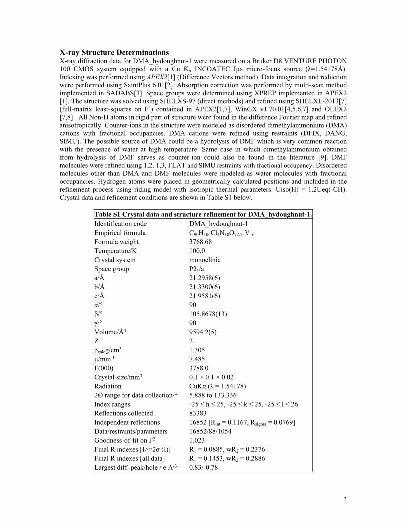
X-ray Structure DeterminationsX-ray diffraction data for DMA_hydoughnut-1 were measured on a Bruker D8 VENTURE PHOTON 100 CMOS system equipped with a Cu Kα INCOATEC Iµs micro-focus source (λ=1.54178Å). Indexing was performed using APEX2[1] (Difference Vectors method). Data integration and reduction were performed using SaintPlus 6.01[2]. Absorption correction was performed by multi-scan method implemented in SADABS[3]. Space groups were determined using XPREP implemented in APEX2 [1]. The structure was solved using SHELXS-97 (direct methods) and refined using SHELXL-2013[7] (full-matrix least-squares on F2) contained in APEX2[1,7], WinGX v1.70.01[4,5,6,7] and OLEX2 [7,8]. All Non-H atoms in rigid part of structure were found in the difference Fourier map and refined anisotropically. Counter-ions in the structure were modeled as disordered dimethylammonium (DMA) cations with fractional occupancies. DMA cations were refined using restraints (DFIX, DANG, SIMU). The possible source of DMA could be a hydrolysis of DMF which is very common reaction with the presence of water at high temperature. Same case in which dimethylammonium obtained from hydrolysis of DMF serves as counter-ion could also be found in the literature [9]. DMF molecules were refined using 1,2, 1,3, FLAT and SIMU restraints with fractional occupancy. Disordered molecules other than DMA and DMF molecules were modeled as water molecules with fractional occupancies. Hydrogen atoms were placed in geometrically calculated positions and included in the refinement process using riding model with isotropic thermal parameters: Uiso(H) = 1.2Ueq(-CH). Crystal data and refinement conditions are shown in Table S1 below.
Table S1 Crystal data and structure refinement for DMA_hydoughnut-1.Identification code DMA_hydoughnut-1Empirical formula C90H106Cl4N10O92.75V16
Formula weight 3768.68Temperature/K 100.0Crystal system monoclinicSpace group P21/aa/Å 21.2958(6)b/Å 21.3300(6)c/Å 21.9581(6)α/° 90β/° 105.8678(13)γ/° 90Volume/Å3 9594.2(5)Z 2ρcalcg/cm3 1.305μ/mm-1 7.485F(000) 3788.0Crystal size/mm3 0.1 × 0.1 × 0.02Radiation CuKα (λ = 1.54178)2Θ range for data collection/° 5.888 to 133.336Index ranges -25 ≤ h ≤ 25, -25 ≤ k ≤ 25, -25 ≤ l ≤ 26Reflections collected 83383Independent reflections 16852 [Rint = 0.1167, Rsigma = 0.0769]Data/restraints/parameters 16852/88/1054Goodness-of-fit on F2 1.023Final R indexes [I>=2σ (I)] R1 = 0.0885, wR2 = 0.2376Final R indexes [all data] R1 = 0.1453, wR2 = 0.2886Largest diff. peak/hole / e Å-3 0.83/-0.78
3
X-ray diffraction data for DEA_hydoughnut-1 were collected using synchrotron radiation (λ = 0.51800 Å, T = 100(2) K) at Advanced Photon Source Beamline 15-ID-B of ChemMatCARS in Argonne National Lab, Argonne, IL. Indexing was performed using APEX2[1] (Difference Vectors method). Data integration and reduction were performed using SaintPlus 6.01[2]. Absorption correction was performed by multi-scan method implemented in SADABS[3]. Space groups were determined using XPREP implemented in APEX2[1]. The structure was solved using SHELXS-97 (direct methods) and refined using SHELXL-2013[7] (full-matrix least-squares on F2) contained in APEX2[1,7], WinGX v1.70.01 [4,5,6,7] and OLEX2[7,8]. All Non-H atoms in rigid part of structure were found in the difference Fourier map and refined anisotropically. Site occupancies of carboxylate moieties (C11, O3, O4), bridging oxygen atom (O11) and terminal oxygen atoms (O40, O12) from the V-based cluster were determined through occupancy refinement and were equal to 0.5 - which confirms the presence of anticipated ligand (1,3-BDC) in the structure. Counter-ions in the structure were modeled as disordered diethylammonium (DEA) cations with fractional occupancies. DEA cations were refined using restraints (DFIX, DANG, SIMU). The possible source of DEA could be a hydrolysis of DEF which is very common reaction with the presence of water at high temperature. Similar case in which dimethylammonium obtained from hydrolysis of DMF serves as counter-ion could also be found in the literature [9]. Disordered molecules other than DEA molecules were modeled as water molecules with fractional occupancies. The crystal did not diffract past approximately 0.95Å resolution limit. Hydrogen atoms were placed in geometrically calculated positions and included in the refinement process using riding model with isotropic thermal parameters: Uiso(H) = 1.2Ueq(-CH). Crystal data and refinement conditions are shown in Table S2 below.
Table S2 Crystal data and structure refinement for DEA_hydoughnut-1Identification code DEA_hydoughnut-1Empirical formula C80H80Cl4N4O81.5V16
Formula weight 3358.32Temperature/K 296.15Crystal system tetragonalSpace group I4/mmma/Å 14.9098(9)b/Å 14.9098(9)c/Å 38.069(2)α/° 90β/° 90γ/° 90Volume/Å3 8462.8(12)Z 2ρcalcg/cm3 1.318μ/mm-1 0.407F(000) 3352.0Crystal size/mm3 0.02 × 0.02 × 0.01Radiation synchrotron (λ = 0.518)2Θ range for data collection/° 2.138 to 31.642Index ranges -14 ≤ h ≤ 15, -15 ≤ k ≤ 13, -40 ≤ l ≤ 40Reflections collected 55527Independent reflections 1527 [Rint = 0.1153, Rsigma = 0.0332]Data/restraints/parameters 1527/15/203Goodness-of-fit on F2 1.149
4
Final R indexes [I>=2σ (I)] R1 = 0.0753, wR2 = 0.2072Final R indexes [all data] R1 = 0.1013, wR2 = 0.2364Largest diff. peak/hole / e Å-3 0.39/-0.47
The X-ray diffraction data for DMA_hydoughnut-2 were measured on a Bruker D8 QUEST PHOTON 100 CMOS system equipped with a Cu Kα INCOATEC Iµs micro-focus source (λ=1.54178Å). Indexing was performed using APEX2[1] (Difference Vectors method). Data integration and reduction were performed using SaintPlus 6.01[2]. Absorption correction was performed by multi-scan method implemented in SADABS[3]. Space groups were determined using XPREP implemented in APEX2 [1]. The structure was solved using SHELXS-97 (direct methods) and refined using SHELXL-2013 [7] (full-matrix least-squares on F2) contained in APEX2 [1,7], WinGX v1.70.01 [4,5,6,7] and OLEX2 [7,8]. All Non-H atoms in rigid part of structure were found in the difference Fourier map and refined anisotropically. Counter-ions in the structure were modeled as disordered dimethylammonium (DMA) cations with fractional occupancies. DMA cations were refined using restraints (DFIX, DANG, SIMU). The possible source of DMA could be a hydrolysis of DMF which is very common reaction with the presence of water at high temperature. Same case in which dimethylammonium obtained from hydrolysis of DMF serves as counter-ion could also be found in the literature [9]. DMF molecules were refined using 1,2, 1,3, FLAT and SIMU restraints with fractional occupancy. Disordered molecules other than DMA and DMF molecules were modeled as water molecules with fractional occupancies. Hydrogen atoms were placed in geometrically calculated positions and included in the refinement process using riding model with isotropic thermal parameters: Uiso(H) = 1.2Ueq(-CH). Crystal data and refinement conditions are shown in Table S3 below.
Table S3 Crystal data and structure refinement for DMA_hydoughnut-2Identification code DMA_hydoughnut-2Empirical formula C51.75H64.75Br4Cl2N7.25O42.75V8Formula weight 2270.41Temperature/K 100.23Crystal system triclinicSpace group P-1a/Å 15.1975(5)b/Å 19.1532(6)c/Å 19.3115(6)α/° 103.8173(18)β/° 95.3294(16)γ/° 98.3478(17)Volume/Å3 5352.9(3)Z 2ρcalcg/cm3 1.409μ/mm-1 8.467F(000) 2252.0Crystal size/mm3 0.1 × 0.1 × 0.08Radiation CuKα (λ = 1.54178)2Θ range for data collection/° 5.842 to 118.21Index ranges -16 ≤ h ≤ 16, -21 ≤ k ≤ 21, -21 ≤ l ≤ 21Reflections collected 106978Independent reflections 15233 [Rint = 0.0878, Rsigma = 0.0679]Data/restraints/parameters 15233/200/1073Goodness-of-fit on F2 1.041Final R indexes [I>=2σ (I)] R1 = 0.0982, wR2 = 0.2603Final R indexes [all data] R1 = 0.1491, wR2 = 0.2992
5
Largest diff. peak/hole / e Å-3 2.51/-1.63
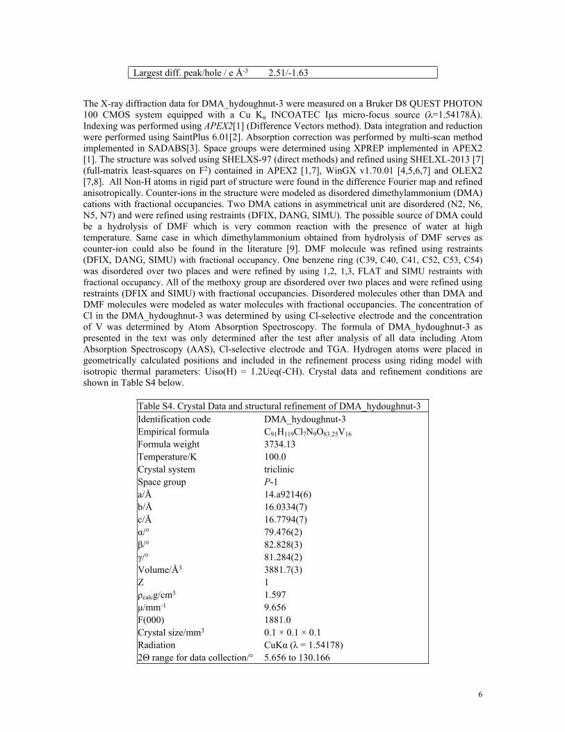
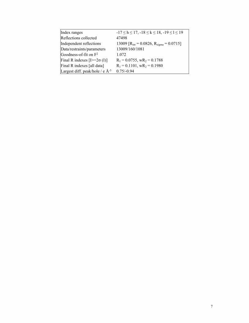
The X-ray diffraction data for DMA_hydoughnut-3 were measured on a Bruker D8 QUEST PHOTON 100 CMOS system equipped with a Cu Kα INCOATEC Iµs micro-focus source (λ=1.54178Å). Indexing was performed using APEX2[1] (Difference Vectors method). Data integration and reduction were performed using SaintPlus 6.01[2]. Absorption correction was performed by multi-scan method implemented in SADABS[3]. Space groups were determined using XPREP implemented in APEX2 [1]. The structure was solved using SHELXS-97 (direct methods) and refined using SHELXL-2013 [7] (full-matrix least-squares on F2) contained in APEX2 [1,7], WinGX v1.70.01 [4,5,6,7] and OLEX2 [7,8]. All Non-H atoms in rigid part of structure were found in the difference Fourier map and refined anisotropically. Counter-ions in the structure were modeled as disordered dimethylammonium (DMA) cations with fractional occupancies. Two DMA cations in asymmetrical unit are disordered (N2, N6, N5, N7) and were refined using restraints (DFIX, DANG, SIMU). The possible source of DMA could be a hydrolysis of DMF which is very common reaction with the presence of water at high temperature. Same case in which dimethylammonium obtained from hydrolysis of DMF serves as counter-ion could also be found in the literature [9]. DMF molecule was refined using restraints (DFIX, DANG, SIMU) with fractional occupancy. One benzene ring (C39, C40, C41, C52, C53, C54) was disordered over two places and were refined by using 1,2, 1,3, FLAT and SIMU restraints with fractional occupancy. All of the methoxy group are disordered over two places and were refined using restraints (DFIX and SIMU) with fractional occupancies. Disordered molecules other than DMA and DMF molecules were modeled as water molecules with fractional occupancies. The concentration of Cl in the DMA_hydoughnut-3 was determined by using Cl-selective electrode and the concentration of V was determined by Atom Absorption Spectroscopy. The formula of DMA_hydoughnut-3 as presented in the text was only determined after the test after analysis of all data including Atom Absorption Spectroscopy (AAS), Cl-selective electrode and TGA. Hydrogen atoms were placed in geometrically calculated positions and included in the refinement process using riding model with isotropic thermal parameters: Uiso(H) = 1.2Ueq(-CH). Crystal data and refinement conditions are shown in Table S4 below.
Table S4. Crystal Data and structural refinement of DMA_hydoughnut-3Identification code DMA_hydoughnut-3Empirical formula C91H119Cl7N9O83.25V16
Formula weight 3734.13Temperature/K 100.0Crystal system triclinicSpace group P-1a/Å 14.a9214(6)b/Å 16.0334(7)c/Å 16.7794(7)α/° 79.476(2)β/° 82.828(3)γ/° 81.284(2)Volume/Å3 3881.7(3)Z 1ρcalcg/cm3 1.597μ/mm-1 9.656F(000) 1881.0Crystal size/mm3 0.1 × 0.1 × 0.1Radiation CuKα (λ = 1.54178)2Θ range for data collection/° 5.656 to 130.166
6
Index ranges -17 ≤ h ≤ 17, -18 ≤ k ≤ 18, -19 ≤ l ≤ 19Reflections collected 47498Independent reflections 13009 [Rint = 0.0826, Rsigma = 0.0715]Data/restraints/parameters 13009/160/1081Goodness-of-fit on F2 1.072Final R indexes [I>=2σ (I)] R1 = 0.0755, wR2 = 0.1788Final R indexes [all data] R1 = 0.1101, wR2 = 0.1980Largest diff. peak/hole / e Å-3 0.75/-0.94
7
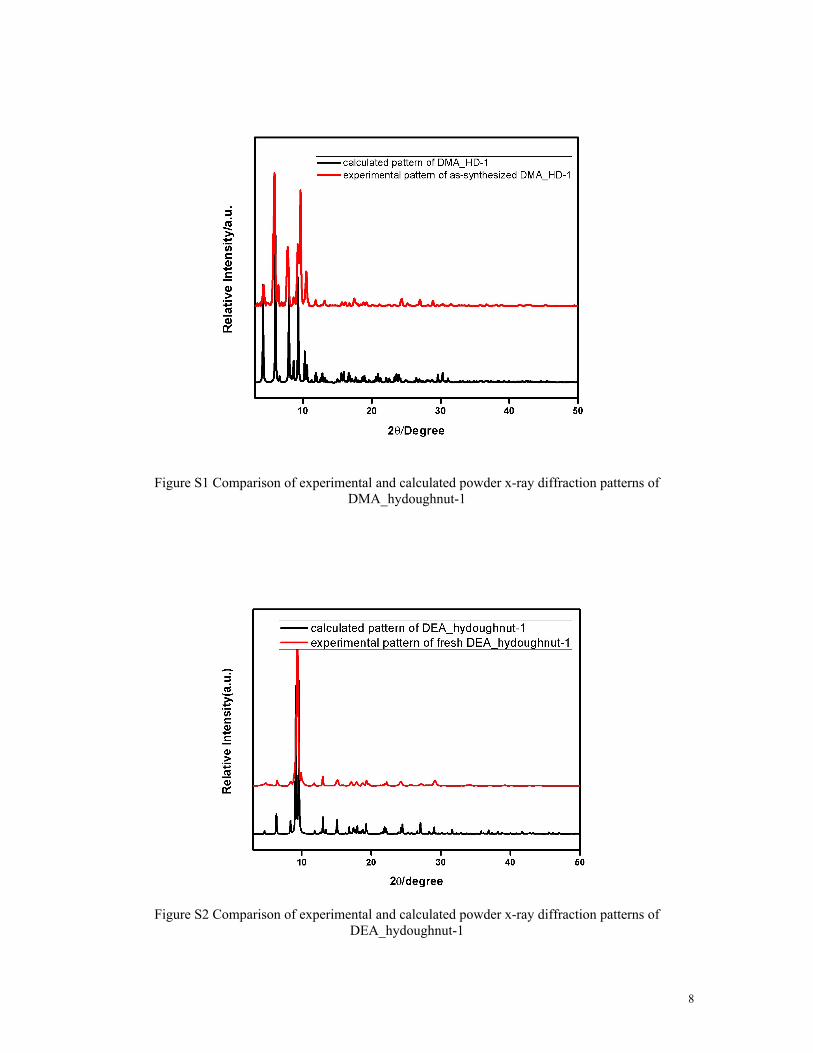
Figure S1 Comparison of experimental and calculated powder x-ray diffraction patterns of DMA_hydoughnut-1
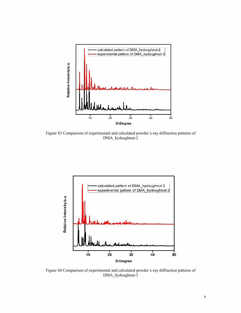
Figure S2 Comparison of experimental and calculated powder x-ray diffraction patterns of DEA_hydoughnut-1
8
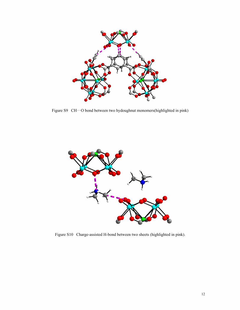
Figure S3 Comparison of experimental and calculated powder x-ray diffraction patterns of DMA_hydoughnut-2
Figure S4 Comparison of experimental and calculated powder x-ray diffraction patterns of DMA_hydoughnut-3
9
Figure S5 Thermogravimetric analysis of DMA_hydoughnut-1
Figure S6 Thermogravimetric analysis of DEA_hydoughnut-1
10
Figure S7 Thermogravimetric analysis of DMA_hydoughnut-2
Figure S8. Thermogravimetric analysis of DMA_hydoughnut-3
11
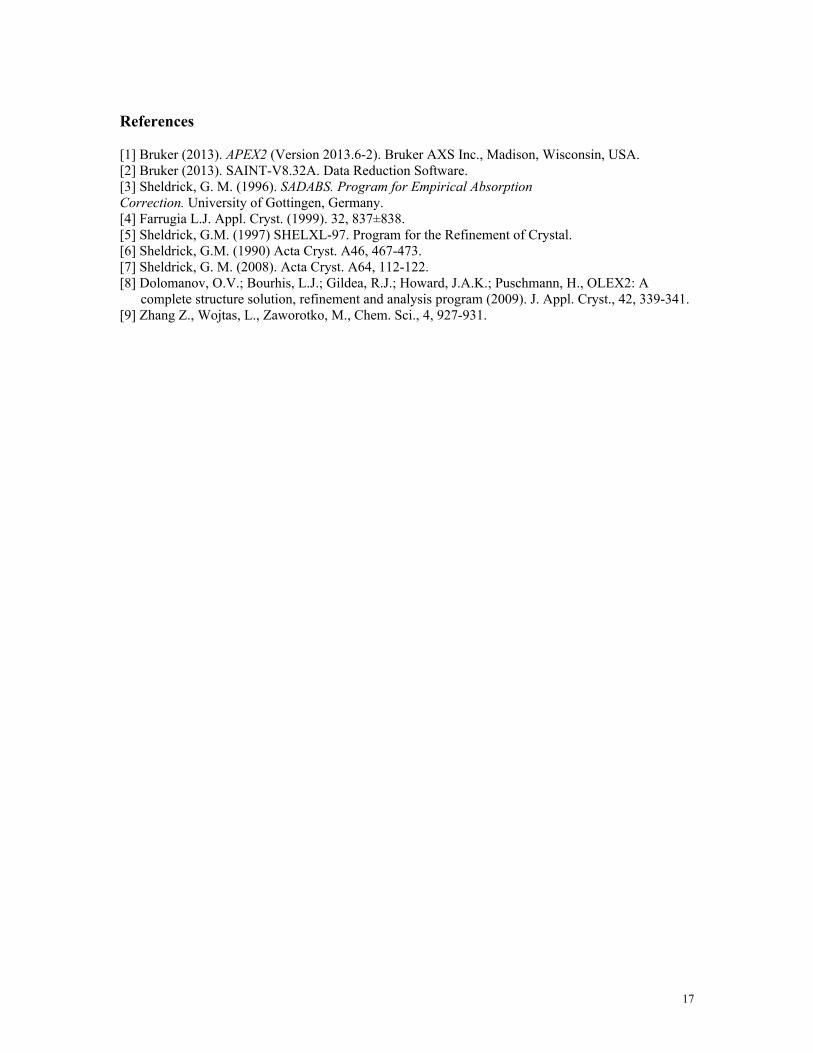
Figure S9 CH···O bond between two hydoughnut monomers(highlighted in pink)
Figure S10 Charge-assisted H-bond between two sheets (highlighted in pink).
12
Figure S11 Two possible ways of connectivity between 1,3-BDC and POVs along c-axis: H-bond(left) and coordination bond(right)
Figure S12 Disordered hydoughnut-1 anions packs with each other to form 2-D sheet in ab plane
13
Figure S13 Interactions between DEA cations and POV moieties. Molecules in ball and stick mode: diethylammonium; Molecules in stick mode: hydoughnut-1 anions.
Figure S14 Br···O interaction along a-axis between POV clusters and Br atom
14
Figure S15 Packing of hydoughnut-2 anions along b-axis(left) and c-axis(right). Two DMF molecules are located between two ligands in a parallel fashion.
Figure S16 CH···O interactions between DMF molecules and POVs in the diagonal direction of bc-plane(highlighted in pink)
15
Figure S17 (a) the hydoughnut-3 anion; (b) crystal packing of hydoughnut-3 anions
[1] Bruker (2013). APEX2 (Version 2013.6-2). Bruker AXS Inc., Madison, Wisconsin, USA.[2] Bruker (2013). SAINT-V8.32A. Data Reduction Software.[3] Sheldrick, G. M. (1996). SADABS. Program for Empirical AbsorptionCorrection. University of Gottingen, Germany.[4] Farrugia L.J. Appl. Cryst. (1999). 32, 837±838.[5] Sheldrick, G.M. (1997) SHELXL-97. Program for the Refinement of Crystal.[6] Sheldrick, G.M. (1990) Acta Cryst. A46, 467-473.[7] Sheldrick, G. M. (2008). Acta Cryst. A64, 112-122.[8] Dolomanov, O.V.; Bourhis, L.J.; Gildea, R.J.; Howard, J.A.K.; Puschmann, H., OLEX2: A complete structure solution, refinement and analysis program (2009). J. Appl. Cryst., 42, 339-341.[9] Zhang Z., Wojtas, L., Zaworotko, M., Chem. Sci., 4, 927-931.