Electronic Supplementary Information for A firmly hybridizable, DNA-like architecture with DAD/ADA- and ADD/DAA-type nonnatural base pairs as an extracellular genetic candidate
Wataru Shirato, Junya Chiba* and Masahiko Inouye*
Graduate School of Pharmaceutical Sciences, University of Toyama, 2630 Sugitani, Toyama 930-0194, Japan
Contents
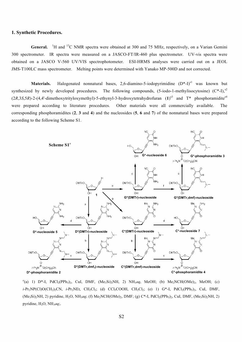
1. Synthetic Procedures. (Scheme S1) p S2
2. Measurements. p S7
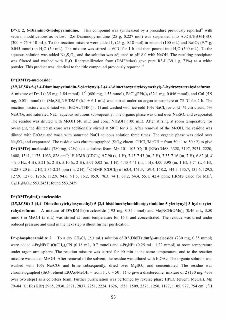
3. DFT Calculations. (Chart S1) p S9
4. Triplex Formation of d(A*)24 vs. d(T*)24. (Figure S1) p S10
5. MALDI-TOF Mass Spectra. (Figure S2) p S11
6. Additional Data for Figure 1C (Figure S3) p S12
7. UV and CD Data for Single-Stranded d(D*)16. (Figure S4) p S13
8. UV and CD Data for Hetero-Duplexes and -Triplexes. (Figure S5) p S14
9. UV and CD Data for Single-Stranded Oligomers. (Figure S6) p S15
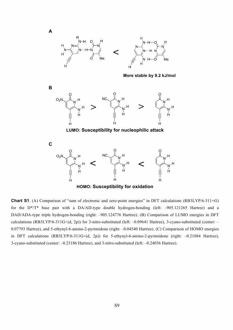
10. UV and CD Data for Various Duplexes. (Figure S7) p S16
D*-I: 2, 6-Diamino-5-iodopyrimidine. This compound was synthesized by a procedure previously reporteds1 with several modifications as below. 2,6-Diaminopyrimidine (25 g, 0.227 mol) was suspended into AcOH/H2O/H2SO4 (300 + 75 + 10 mL). To the reaction mixture were added I2 (23 g, 0.18 mol) in ethanol (100 mL) and NaIO4 (9.71g, 0.045 mmol) in H2O (50 mL). The mixture was stirred at 60˚C for 1 h and then poured into H2O (500 mL). To the aqueous solution was added Na2S2O3, and the solution was adjusted to pH 8.0 with NaOH. The resulting precipitate was filtered and washed with H2O. Recrystallization from (DMF/ether) gave pure D*-I (39.1 g, 73%) as a white powder. This product was identical to the title compound previously reported.s1
calcd for MH+, C12H16N3O4: 266.1141; found 266.1142; UV (H2O, 25 ˚C) ε260 = 5090 Lmol-1cm-1.
S7
Synthesis of artificial DNA olgomers. The artificial DNA oligomers were synthesized by use of phosphoramidites 2,
3, 4, and the previously reported T*-phosphoroamidites4 on an Applied Biosystems 392 synthesizer using standard
β-cyanoethylphosphoramidite chemistry with the coupling reaction time of 15 min. The solid support (Universal Support II® or III®), which allows for 3' placement of nonnatural nucleosides, was purchased from Glen Research.
After automated synthesis, the oligomers were removed from the solid support with 2 M ammonia methanol solution at
30 ºC for 30 min and deprotected with concentrated NH4OH at 40 ºC for 8 h. The oligomers were then purified by
reverse-phase HPLC using a 5C18-AR-II column (4.6 x 150 mm) with an eluent of 5 mM ammonium formate and the
following CH3CN percentages of linear gradient (0–60 min, 3–18%) at a flow rate of 1.0 mL/min.
2. Measurements. MALDI-TOF mass measurements. MALDI-TOF mass spectra were recorded on a Bruker-Daltonics-Autoflex mass spectrometer operating in the negative ion mode with 3-hydroxypicolinic acid as a matrix (see Figure S2). 5'-d(T*D*D*T*D*T*D*T*T*D*): calcd for [M-H]-, C115H130N30O58P9: 3138.58; found 3138.20, d(D*)16 : calcd for [M-H]-, C176H208N64O78P15: 4932.04; found 4930.07, 5'-d(T*D*D*T*G*C*D*T*T*D*): calcd for [M-H]-, C117H130N30O58P9: 3162.58; found 3162.88, 5'-d(T*G*D*T*G*C*D*T*C*D*): calcd for [M-H]-, C119H130N30O58P9: 3186.58; found 3185.74, 5'-d(T*D*T*T*G*C*T*D*T*T*): calcd for [M-H]-, C119H130N26O62P9: 3194.55; found 3194.02, 3'-d(D*T*D*D*C*G*D*T*D*D*): calcd for [M-H]-, C115H130N34O54P9: 3130.61; found 3129.37, 5'-d(D*T*D*D*C*G*D*T*D*D*): calcd for [M-H]-, C115H130N34O54P9: 3130.61; found 3129.60, 5'-d(T*D*T*T*C*C*T*D*T*T*): calcd for [M-H]-, C118H132N26O62P9: 3184.56; found 3183.31. UV and Tm Measurements. UV-vis spectra and Tm melting curves (1.0 ºC/1.0 min) were obtained by JASCO V-560 UV/vis spectrophotometer with a peltier and a temperature controller in a temperature range from 10 to 70 °C. The Tm values were determined from the maxima of the first derivatives of the melting curves measured in a buffer solution: 10 mM Hepes (pH 7.0), 10 mM MgCl2, 100 mM NaCl. Errors were estimated at ± 1.0 ºC. Concentrations of the solutions containing artificial DNAs were determined based on the molar extinction coefficients at 260 nm (e260) of the artificial nucleoside monomers 5, 6, 7, and the previously reported T*-nucleosides4 (see SI Text). CD Measurements. CD spectra were recorded using a JASCO-J-720WI spectropolarimeter with a temperature controller at 10, 20, 30, 40, 50, 60, and 70 °C in a buffer solution: 10 mM Hepes (pH 7.0), 10 mM MgCl2, 100 mM NaCl. Titration Experiments. Titration curves for artificial DNAs were obtained by monitoring a specified wavelength of CD. In entry 5, for example, 3.0 mL of a d(D*)16 solution (4.0 µM with 10 mM Hepes (pH 7.0), 10 mM MgCl2, and 100 mM NaCl) was prepared, and CD measurement of the solution was carried out at 5 ºC using a quartz cell of 1 cm pathlength. Separately, 200 µL of a d(T*)16 solution (200 µM in the same buffer) was then prepared, and 12.0 µL of the d(T*)16 solution (0.2 equivalent against the d(D*)16) was added to the d(D*)16 solution in the quartz cell. The mixed solution was heated to 70 ºC, annealed to 5 ºC for 1 h, and then CD measurement was performed at 5 ºC. A series of the operations were repeated for all the ratios of [d(T*)16]/[d(D*)16] = 0, 0.2, 0.4, 0.6, 0.7, 0.8, 0.9, 1.0, 1.1, 1.2,
S8
1.3, 1.4, 1.6, 1.8, 1.9, 2.0, 2.1, 2.2, 2.3, and 2.4. The normalized CD changes at 308 nm were plotted against [d(T*)16]/[ d(D*)16] (Figure 2C). Job’s Plots. Job’s plots for artificial DNAs were obtained by monitoring a specified wavelength of CD or UV. In entry 11, for example, 10 mL of a 5'-d(T*D*T*T*G*C*T*D*T*T*) solution (1.0 µM with 10 mM Hepes (pH 7.0), 10 mM MgCl2, and 100 mM NaCl) and 10 mL of a 3'-d(D*T*D*D*C*G*D*T*D*D*) solution (1.0 µM in the same buffer) were prepared. The 5'-d(T*D*T*T*G*C*T*D*T*T*) solutions of 0, 100, 200, 300, 400, 500, 600, 700, 800, 900, and 1000 µL were mixed in micro test tubes with 1000, 900, 800, 700, 600, 500, 400, 300, 200, 100, and 0 µL of the 3'-d(D*T*D*D*C*G*D*T*D*D*) solutions, respectively. All the mixed solutions were heated to 70 ºC, followed by slow cooling to 25 ºC over 30 min. UV-vis measurements of all of the solutions were carried out at 25 ºC using a quartz cell of 1 cm pathlength. Against [5'-d(T*D*T*T*G*C*T*D*T*T*)]/ ([5'-d(T*D*T*T*G*C*T*D*T*T*)] + [3'-d(D*T*D*D*C*G*D*T*D*D*)]) were plotted the normalized UV changes for the mixtures of a 5'-d(T*D*T*T*G*C*T*D*T*T*) and 3'-d(D*T*D*D*C*G*D*T*D*D*) at 305 nm. The changes were corrected for by subtracting sum of the intensities for each strand at the same concentrations (Figure 3A). References for ESI S1 P. M. Pelphrey, V. M. Popov, T. M. Joska, J. M. Beierlein, E. S. D. Bolstad, Y. A. Fillingham, D. L. Wright and
A. C. Anderson, J. Med. Chem., 2007, 50, 940.
S2 K. W. Wellington, H. C. Ooi and S. A. Benner, Nucleosides, Nucleotides Nucleic Acids, 2009, 28, 275.
S3 J. Chiba, S. Takeshima, K. Mishima, H. Maeda, Y. Nanai, K. Mizuno and M. Inouye, Chem. Eur. J., 2007, 13,
8124; M. Takase, T. Morikawa, H. Abe and M. Inouye, Org. Lett., 2003, 5, 625.
S4 Y. Doi, J. Chiba, T. Morikawa and M. Inouye, J. Am. Chem. Soc., 2008, 130, 8762.
S5 M. Inouye, K. Kim and T. Kitao, J. Am. Chem. Soc., 1992, 114, 778.
S9
A
B
LUMO: Susceptibility for nucleophilic attack
C
HOMO: Susceptibility for oxidation
Chart S1. (A) Comparison of “sum of electronic and zero-point energies” in DFT calculations (RB3LYP/6-311+G)
for the D*/T* base pair with a DA/AD-type double hydrogen-bonding (left: –905.121265 Hartree) and a
DAD/ADA-type triple hydrogen-bonding (right: –905.124776 Hartree). (B) Comparison of LUMO energies in DFT
5'-d(D*T*D*D*C*G*D*T*D*D*), and (H) 5'-d(T*D*T*T*C*C*T*D*T*T*). See also MALDI-TOF mass measurements (page S7).
A B C
D E F
G H
S12
Figure S3. Absorbance and CD data for 5'-d(T*D*D*T*D*T*D*T*T*D*) (2.0 µM) in 10 mM Hepes (pH 7.0), 10 mM MgCl2, 100 mM NaCl. (A) UV melting (red) and
annealing (black) curves monitored at 303 nm. Melting temperatures were obtained at
39 ± 1.0 ºC from the curves. (B) CD melting curves monitored at 258 (red), 286
(blue), and 313 nm (black) in Figure 1B. Melting temperatures were obtained at 38.5 ±
1.0 ºC from the curves.
S13
Figure S4. UV-vis and CD data for homooligomer d(D*)16 (2.0 µM) in 10 mM Hepes (pH 7.0), 10 mM MgCl2, 100 mM NaCl. (A) UV spectra at 0 (blue) and 70 ºC (red). (B) CD spectra at 0 (blue), 10, 20, 30, 40,
50, 60, and 70 ºC (red). (C) Absorbance monitored at 303 nm.
A
B
C
S14
Figure S5. UV-vis and CD data of hetero-duplex (d(D*)16/dT16, [duplex] = 1.0 µM) and hetero-triplex
(d(T*)16/dA16/ d(T*)16, [triplex] = 1.0 µM) in 10 mM Hepes (pH 7.0), 10 mM MgCl2, 100 mM NaCl. (A) UV spectra at 0 (blue) and 70 ºC (red) for d(D*)16/dT16. (B) UV melting curve monitored at 266 nm for
d(D*)16/dT16. (C) CD spectra at 0 (blue), 10, 20, 30, 40, 50, 60, and 70 ºC (red) for d(D*)16/dT16. (D) The
titration plot of d(D*)16 and dT16 monitored by CD at 248 nm. [d(D*)16] = 1.0 µM, [dT16]/ [d(D*)16] = 0, 0.2, 0.4, 0.6, 0.7, 0.8, 0.9, 1.0, 1.1, 1.2, 1.3, 1.4, 1.6, 1.7, 1.8, 1.9, 2.0, 2.1, 2.2, 2.3, 2.4, 2.6, 2.8 and 3.0. (E) UV spectra
at 0 (blue) and 70 ºC (red) for d(T*)16/dA16/ d(T*)16. (F) UV melting curve monitored at 295 nm for
d(T*)16/dA16/ d(T*)16. (G) CD spectra at 0 (blue), 10, 20, 30, 40, 50, 60, and 70 ºC (red) for d(T*)16/dA16/
d(T*)16. (H) The titration plot of d(T*)16 and dA16 monitored by CD at 263 nm. [d(T*)16] = 1.0 µM, [dA16]/ [d(T*)16] = 0, 0.2, 0.4, 0.6, 0.7, 0.8, 0.9, 1.0, 1.1, 1.2, 1.3, 1.4, 1.6, 1.8, 1.9, and 2.0.
C B A
F E D
H G
S15
Figure S6. UV-vis and CD data of single-stranded 5'-d(T*D*T*T*G*C*T*D*T*T*) (A–C,
1.0 µM) and 3'-d(D*T*D*D*C*G*D*T*D*D*) (D–F, 1.0 µM) in 10 mM Hepes (pH 7.0), 10 mM MgCl2, 100 mM NaCl. (A) UV-vis spectra at 10 (blue) and 70 ºC (red). (B) CD spectra at
10 (blue) and 70 ºC (red). (C) Absorbance monitored at 303 nm. (D) UV-vis spectra at 10
(blue) and 70 ºC (red). (E) CD spectra at 10 (blue) and 70 ºC (red). (F) Absorbance monitored
at 303 nm.
C
A B
D
E F
S16
Figure S7. UV-vis and CD data in 10 mM Hepes (pH 7.0), 10 mM MgCl2, 100 mM
NaCl. (A) CD spectra of 5'-d(T*D*T*T*G*C*T*D*T*T*) and
3'-d(D*T*D*D*C*G*D*T*D*D*) at 10 (blue), 20, 30, 40, 50, 60, and 70 ºC (red),
[duplex] = 1.0 µM. (B) UV-vis spectra of d(D*)16 and d(T*)16 at 10 (blue) and 70 ºC
(red), [d(D*)16] = [d(T*)16] = 1.0 µM. (C) CD spectra of d(D*)16 and d(T*)16 at 10 (blue),