Page 1
Accepted Manuscript
Title: Electrospun drug loaded membranes for sublingualadministration of sumatriptan and naproxen
Author: Petr Vrbata Pavel Berka Denisa Stranska PavelDolezal Marie Musilova Lucie Cizinska
PII: S0378-5173(13)00819-3DOI: http://dx.doi.org/doi:10.1016/j.ijpharm.2013.08.085Reference: IJP 13618
To appear in: International Journal of Pharmaceutics
Received date: 17-7-2013Revised date: 27-8-2013Accepted date: 28-8-2013
Please cite this article as: Vrbata, P., Berka, P., Stranska, D., Dolezal, P., Musilova,M., Cizinska, L., Electrospun drug loaded membranes for sublingual administrationof sumatriptan and naproxen, International Journal of Pharmaceutics (2013),http://dx.doi.org/10.1016/j.ijpharm.2013.08.085
This is a PDF file of an unedited manuscript that has been accepted for publication.As a service to our customers we are providing this early version of the manuscript.The manuscript will undergo copyediting, typesetting, and review of the resulting proofbefore it is published in its final form. Please note that during the production processerrors may be discovered which could affect the content, and all legal disclaimers thatapply to the journal pertain.
Page 2
Page 1 of 40
Accep
ted
Man
uscr
ipt
1
Title: Electrospun drug loaded membranes for sublingual administration of sumatriptan and 1
naproxen2
List of Authors: Petr Vrbataa, Pavel Berkaa, Denisa Stránskáb, Pavel Doležala,*, Marie Musilováa, 3
Lucie Čižinskáa4
a Department of Pharmaceutical Technology, Faculty of Pharmacy in Hradec Králové, Charles 5
University in Prague, Czech republic 6
b Elmarco Ltd. Co., Liberec, Czech Republic7
* Corresponding author 8
Abstract: Sublingual administration of active pharmaceutical substances is in principle favourable for 9
rapid onset of drug action, ready accessibility and avoidance of first pass metabolism. This 10
administration could prove very useful in the treatment of migraines, thus two frequently used drugs 11
were selected for our study.12
Sumatriptan succinate, naproxen, and its salt as well as combinations of these were incorporated into 13
nanofibrous membranes via the electrospinning process. DSC measurements proved that the resulted14
membranes contained non-crystalline drug forms. SEM imaging approved good homogeneity of 15
diameter and shape of the membrane nanofibres.16
The nanofibrous membranes always showed the rapid and mutually independent release of the tested 17
drugs.18
The drugs exhibited very high differences in sublingual permeation rates in vitro, but the rates of both 19
substances were increased several times using nanofibrous membranes as the drug carrier in 20
comparison to drug solutions. The released drugs subsequently permeated through sublingual mucosa 21
preferentially as non-ionized moieties.22
The prepared nanofibrous membranes proved very flexible and mechanically resistant. With their 23
drug load capacity of up to 40% of membrane mass, they could be very advantageous for the 24
formulation of sublingual drug delivery systems.25
Key words: Electrospinning, migraine, sublingual, sumatriptan, naproxen26
27
Page 3
Page 2 of 40
Accep
ted
Man
uscr
ipt
2
127
1. INTRODUCTION28
29
Migraine is a chronic relapsing brain disorder that affects about 12% of the Western 30
population. It occurs as a unilateral headache, often accompanied by other symptoms,31
including nausea, vomiting, photophobia, and phonophobia, lasting from 4 to 72 hours 32
(Arulmozhi et al., 2005). In 15% of cases, a migraine headache is preceded by the aura, a 33
transient neurological dysfunction, which is usually characterized by visual and/or sensory 34
symptoms. Migraine has a very strong social impact, influencing quality of life and work 35
productivity (Ramdan and Buchanan, 2006). Sumatriptan is the most frequently used member 36
of triptans commonly prescribed for the treatment of migraine headaches (with or without 37
aura). Sumatriptan could be also administered together with NSAID naproxen sodium, which 38
brings higher benefits to diminish symptoms of migraine than usage of either of the drugs 39
separately.40
Actual dosage forms of sumatriptan are pills (50 and 100 mg), subcutaneous injection (4 and 41
6 mg), and nasal spray (10 and 20 mg). Succinate salt is well soluble in water, but its 42
bioavailability (BA) after oral administration is only about 14%. Nasal spray administration 43
of a sumatriptan base has a BA of about 16%. (Imitrex, 2013) Low BA following oral 44
administration, relatively short half-life, and a requirement for the fast onset of action 45
instigated the research for a new route of administration of this drug.46
A sublingual route of administration could be very advantageous in the given case. Although 47
a relatively small surface area and difficulties with the dosage form (permanently washed by 48
saliva, and involuntary swallowing of liquids greater than 200 μL) have limited this site for 49
drug administration so far, it possesses many advantageous characteristics. Very thin mucosa 50
Abbreviations: SUS…….Sumatriptan succinate, NAPS.... Naproxen sodium, NAP……Naproxen
Page 4
Page 3 of 40
Accep
ted
Man
uscr
ipt
3
(100 to 200 μm), good blood supply, perfect accessibility, non-invasiveness of 51
administration, and potential ease of removal encouraged research efforts in this area. The 52
fast onset of systemic drug action is also very important, and the avoidance of the first-pass 53
metabolism is in many cases essential (Hearnden V. et al. 2012; Patel P. et al., 2011; Bayrak 54
Z. et al., 2011; Patel V. F. et al., 2011). Moreover, this way of administration is also suitable 55
for small children, elderly people, and other patients with swallowing or digestion problems. 56
(Patel P. et al., 2011)57
Currently, there are several sublingual preparations, mostly based on fast dissolving 58
(disintegrating) tablets, films, wafers, and sublingual sprays commercially available, and new 59
dosage forms are being tested (Patel V. F. et al., 2011; Hearnden V. et al. 2012). 60
A relatively new and very promising technology for the formulation of sublingual drug 61
delivery systems is based on the use of electrospun drug loaded nanofibrous membranes 62
(Nagy Z. K. et al., 2010; Yu D.-G. et al., 2010; Yu D.-G. et al., 2010; Stranska et al., 2012). 63
Electrospinning is a unique technique for the preparation of ultra-fine fibres with the diameter 64
size going down to nanometres. Although the principle of this procedure has been known for 65
almost a century, it became a topic of great interest in the early 1990’s, when Reneker and 66
co-workers demonstrated the possibility of electrospinning a wide range of polymers 67
(Reneker D. H. and Chun I., 1996; Frenot A. and Chronakis I. S., 2003).68
Nowadays, most of linear synthetic and also natural polymeric compounds can be easily 69
electrospun into nanofibres (Frenot A. and Chronakis I. S., 2003; Agarwal S, et al. 2013). A 70
very important moment for further development was the invention of a large-scale production 71
device the NanospiderTM which makes it easier to scale up production for commercial 72
processing (Jirsak O. et al., 2005). Nevertheless, scaling up production of every individual 73
product is always challenging, especially in pharmaceutical industry.74
Page 5
Page 4 of 40
Accep
ted
Man
uscr
ipt
4
Nanofibres, or rather nanofibrous membranes, have already found their application in many 75
disciplines. Thanks to their unique properties, namely high surface area to volume ratio, high 76
nanoporosity, high mechanical strength, and structural similarity to an extracellular matrix,77
they attract a lot of attention within technical disciplines, but also in biomedicine and 78
pharmacotherapy and new dosage formulation types (Agarwal S, et al. 2013, Leung V. and 79
Ko F., 2011; Nagy Z. K. et al., 2012).80
In the biomedical field, nanofibres find usage in the formation of tissue engineering scaffolds 81
(Cao H. et al., 2009; Leung V. and Ko F., 2011), wound dressing (Zhang X. et al, 2009; 82
Leung V. and Ko F., 2011; Sell S. A. et al., 2009), vascular grafts (Zhang X. et al, 2009; Sell 83
S. A. et al., 2009), and drug delivery systems (Leung V. and Ko F., 2011; Chakraborty S. et 84
al., 2009; Meinel A. J. et al., 2012). Many kinds of drugs have already been incorporated into 85
the nanofibrous mats and then successfully released from them without a significant loss of 86
their activity. Among low molecular drugs antibiotics (Kenawy E. R. et al., 2002; Kim K. et 87
al., 2004), non-steroidal anti-inflammatory drugs (NSAID) (Taepaiboon P. et al., 2006; 88
Kenawy E.-R. et al., 2007; Huang L.-Y. et al., 2012), vitamins (Taepaiboon P. et al., 2007; 89
Madhaiyana K. et al., 2013), chemotherapeutics (Xu X. et al., 2009), and many others have90
already been described. Higher molecular compounds, mostly protein based, were also shown 91
to be effectively released from nanofibres (Maretschek S. et al., 2008; Han N. et al., 2012).92
In our work, we focused on the limits of sublingual administration of sumatriptan and 93
naproxen, in the context of permeability of sublingual mucosa in vitro, then on the 94
examination of suitable polymers for co-formulation of both the drug-loaded electrospun 95
membranes and estimation of formulation parameters for release profiles potentially suitable 96
for anti-migraine action.97
98
2. MATERIALS AND METHODS99
Page 6
Page 5 of 40
Accep
ted
Man
uscr
ipt
5
100
2.1 Materials101
Sumatriptan succinate (SUS) was kindly donated by Teva Czech Industries s.r.o. (Opava, 102
CZ). Naproxen (NAP), naproxen sodium (NAPS) and chitosan (CHI, Mw 60,000-120,000) 103
were purchased from Sigma Aldrich (Prague, CZ), polyacrylic acid (PAA, Mw 450,000), 104
poly-ε-caprolacton (PCL, Mw 100,000) were purchased from Scientific Polymer Products 105
(New York, US), polyvinylalcohol (PVA, type Z 220, viscosity of 4 wt% water solution at 106
20°C 11.5–15 mPa.s) from Nippon Gohsei (Düsseldorf, GE). Acetic acid, formic acid, 107
phosphoric acid, and potassium dihydrogen phosphate were supplied by Penta Chemicals 108
(Prague, CZ).109
The aqueous solutions were prepared with purified water. All the chemicals were used as 110
received without further purification.111
112
2.2 Methods113
114
2.2.1 Formulation of drug loaded electrospun membranes115
116
The nanofibrous mats were produced by electrospinning from polymer solutions using 117
Nanospider™ technology (Jirsak O. et al., 2005).118
Chitosan was dissolved in a mixture of acetic acid and water 2:1 in a concentration of 2.25%; 119
PVA was dissolved in a water : phosphoric acid mixture (99.3:0.7) in a concentration of 11%;120
PAA was dissolved in a 0.1M sodium chloride solution in a concentration of 6% with the 121
addition of β-cyclodextrin 1.2% (as a cross-linking agent); PCL was dissolved in a mixture of 122
acetic acid : formic acid (2:1) in a concentration of 12%. The active substances were added in 123
Page 7
Page 6 of 40
Accep
ted
Man
uscr
ipt
6
concentrations ranging from 5% to 30% related to the mass of the polymer in the solution for 124
electrospinning.125
All the chemicals were stirred until homogenous solutions were obtained, and then poured 126
into the container of an electrospinning device. Spinning electrode was in a shape of wire, 127
electrospinning is nozzle free. After the application of a high voltage, nanofibres were formed 128
and then collected on a spunbond textile covering the collector plate. Speed of spunpond 129
movement through the device determines nanofibrous layer thickness (g/m2). In the case of 130
water-soluble polymers (PVA, PAA) cross-linking was performed. After electrospinning131
process the membranes were thermally treated in a drying oven at 130°C for 15 minutes in 132
the case of PVA and at 140 °C for 20 minutes in the case of PAA.133
134
2.2.2 Characterization135
136
The morphology of prepared nanofibrous membranes was evaluated by scanning electron 137
microscopy - NOVA NanoSem 230 (FEI, USA) with maximal resolution up to 1.3 nm at 138
30kV and magnification up to 1,000,000 times.139
The differential scanning calorimetry (DSC) analyses were carried out using a 200 F3 MAJA 140
calorimeter (NETZSCH, Germany). Samples were heated at speed 5oC/min from 20oC to 141
200oC. The nitrogen gas flow rate was set at 40 ml/min.142
143
2.2.3 Drug release evaluation144
145
Drug release measurements were conducted in a water bath under a constant 146
temperature (36.5 ± 0.5oC) and permanent stirring (magnetic bar; 200 rpm). Pieces of 147
membrane 5 cm x 4 cm (20 cm2) were precisely weighed and then placed inside vials. The 148
Page 8
Page 7 of 40
Accep
ted
Man
uscr
ipt
7
vials were filled with 20 mL of a pre-tempered phosphate buffered solution of pH 7.4 (PBS) 149
as an acceptor phase, and placed inside the water bath. The samples of the acceptor phase 150
(0.6 mL) were withdrawn in pre-determined time intervals (5, 10, 15, and 30 min, 1, 2, 4, 8,151
and 24 hours) and the pertinent volume was replaced with a fresh buffer.152
153
2.2.4 In vitro permeation experiments154
155
In vitro drug permeation experiments were performed using a porcine sublingual mucosa. 156
The basic principles were derived from analogical experiments used in transdermal 157
permeations, previously described in detail (Patel V. F. et al., 2011).158
Pieces of mucosa were obtained from the lower side of fresh porcine tongues (supplied from 159
a local slaughterhouse) by surgically removing the muscle and connective tissues. After 160
preparation, large pieces of obtained mucosa were stored in a 0.9 % sodium chloride solution 161
with the addition of sodium azide (0.002 %). The processed sublingual membranes were 162
about 0.4 mm in thickness. They were cut into pieces (2 cm x 2 cm) and fixed between a 163
donor and an acceptor compartment of diffusion cells (Fig. 1). The actual area exposed for 164
permeation was 2 cm2. The PBS (pH 7.4) was used as an acceptor phase. Permeation was 165
conducted in a water bath - temperature (36.5 ± 0.5oC) and stirring with magnetic bar. 166
In vitro permeation of SUS, NAP and NAPS was evaluated using the donor solutions (PBS 167
pH 6.8, 0.5 mL) with selected concentrations (1%, 3%, 6% for SUS; 1%, 2%, 3%, 10% for 168
NAPS) and the tested nanofibrous membranes. Samples (0.6 mL) of the acceptor phase were 169
withdrawn in pre-determined time intervals (15, 30 min, 1, 2, 4, 6, and 8 hours) and replaced 170
with a fresh buffer. The samples were briefly stored in a refrigerator until HPLC 171
determination of investigated substances was performed. All drug release measurements were 172
performed in triplicate, and in the case of in vitro sublingual permeation experiments, four 173
Page 9
Page 8 of 40
Accep
ted
Man
uscr
ipt
8
replicates were performed. The values presented below are calculated as the means with their 174
standard errors of the means (SEM).175
The stability pre-tests of the drugs were carried out in artificial saliva (pH 6.8) and an176
isotonic phosphate buffer (pH 7.4). Low stability of the drugs in one of these mediums would 177
be very limiting for potential use. The obtained results showed no significant decrease in the 178
concentration of the drugs during a 24 hour period.179
180
Fig. 1. Diffusion and permeation cell181
182
2.2.5. HPLC Analysis183
184
Drug concentrations in the samples of the acceptor phase were determined using HPLC set 185
Agilent Technologies 1200 (USA) equipped with an auto sampler ALS1329A, UV/VIS 186
detector VWD G1414B, and an isocratic pump G1310A.187
Sumatriptan. The mobile phase was a mixture of ammonium phosphate (0.05 M) and 188
acetonitrile (84:16 v/v), pH was adjusted to 3.0 with the addition of 0.1 M phosphoric acid. 189
The flow rate was set at 1.5 mL/min. The method of Nozal et al. (2002) was modified to 190
avoid interference from skin residues at the retention time of sumatriptan at 227.4 nm; the 191
detection wavelength was set at 282.7 nm (Femenia-Font A. et al., 2005). Separation was 192
carried out at 30oC with the use of 250 mm x 4.6 mm, a reverse-phase column packed with 193
5 µm C18 silica particles (Zorbax Eclipse XDB C18).194
Naproxen. The mobile phase was a mixture of potassium dihydrogen phosphate (0.01 M; pH 195
adjusted to 2.5 with the addition of 0.1 M phosphoric acid) and acetonitrile (55:45 v/v). The196
flow rate was set at 1.5 mL/min. Separation was carried out at 25oC, on a 150 mm x 4.6 mm, 197
Page 10
Page 9 of 40
Accep
ted
Man
uscr
ipt
9
reverse-phase column packed with 5 µm C18 silica particles (Zorbax Eclipse XDB C18). The 198
detection wavelength was set at 230 nm.199
200
2.2.6 Data treatment201
202
The primary data from HPLC assay of the samples were further corrected for sampling and 203
replacement of the pure acceptor phase. The amounts of the drug passed through the 1 cm2204
of sublingual mucosa were obtained. The cumulative amount of the drug vs. time dependence 205
was used to calculate the pertinent slope values of the linear part of the concerned 206
dependence with linear regression. The values obtained were understood as the individual 207
flux values J [µg/cm2/h] of the pseudosteady state permeation. The flux values means and 208
standard error of the means (SEM) (number of replicates n = 4) were calculated.209
210
3. RESULTS AND DISCUSSION211
212
In this paper, we focused on membranes ensuring longer contact time of the drug with 213
absorption mucosa using a non-soluble (removable) membrane. Membrane prevents the 214
leaking of a drug to an oral cavity and swallowing the drug, whilst masking the unpleasant 215
taste.216
217
3.1 Scanning electron microscopy218
219
The prepared nanomembranes were analysed by scanning electron microscope for average 220
fibre diameter and uniformity of the membrane fibres. This characterization confirmed that 221
the diameters of all the membrane nanofibres were within the nanometre scale and of good 222
Page 11
Page 10 of 40
Accep
ted
Man
uscr
ipt
10
shape and diameter uniformity (Fig. 2). It can be concluded that incorporation of the drugs 223
into the nanofibrous membranes brought no free particles of the drugs, neither on the surface 224
of nanofibres, nor particles larger than nanofibre diameter embedded within the mass of the 225
fibres. It is important as evidence of well-tuned electrospinning parameters that make it 226
possible to obtain fibres without loss of the drug, and with good shape homogeneity. Very 227
similar images were also obtained for all other prepared membranes.228
229
Fig. 2. SEM images of the prepared membranes A: Chitosan - blank; B: Chitosan –230
containing SUS (5%); C: Chitosan – containing NAP (5%); D: PVA – blank; E: PVA –231
containing SUS (5%); F: PVA – containing NAP (5%)232
233
3.2 Differential scanning calorimetry (DSC)234
235
The physical state of the carrier polymers and the incorporated drugs was investigated by 236
DSC measurements. The DSC thermograms of chosen samples are shown on Figs. 3, 4, and 237
5. The thermogram of crystalline naproxen exhibits a strong endothermic peak at 157.1oC, 238
while no melting peak was present on thermograms of nanofibrous mats containing 5% or 239
30% of incorporated naproxen. This result proves that naproxen in the tested nanofibrous 240
mats is present in an amorphous state, or more likely, homogeneously dispersed in the 241
polymer matrix of filaments. Moreover, no glass transition peak of carrier polymer was 242
found. This finding is also important, because polymer crystallinity plays an important role in 243
interactions with water, and therefore also drug release (Natu M. V. et al., 2010).244
Similar results were concluded from measurements with sumatriptan succinate. The 245
crystalline form of sumatriptan succinate provided an endothermic peak at 169.7oC, no 246
melting peak or glass transition peak were found on the other thermograms.247
Page 12
Page 11 of 40
Accep
ted
Man
uscr
ipt
11
248
Fig. 3. DSC profiles of A: Sumatriptan succinate (crystalline powder), B: PVA + sumatriptan249
suc. 20% (nanofibrous membrane), C: PVA (nanofibrous membrane without drug), D: PVA 250
(powder)251
252
Fig. 4. DSC profiles of A: Naproxen (crystalline powder); B: PVA + naproxen 30%253
(nanofibrous membrane); C: PVA + naproxen 5% (nanofibrous membrane); D: PVA 254
(nanofibrous membrane without drug); E: PVA (powder)255
256
Fig. 5. DSC profiles of A: Naproxen (crystalline powder); B: Chitosan + naproxen 5%257
(nanofibrous membrane); C: Chitosan (nanofibrous membrane without drug); D: Chitosan 258
(powder)259
260
3.3 Release of the drugs from nanofibrous membranes261
262
Release characteristics of the investigated drugs were tested and evaluated by the complete 263
immersion of the mats in the release medium.264
Several different polymers with expected fast drug release were chosen. The polymer 265
selection was further influenced by the intended purpose of their use in the sublingual dosage 266
form. Bioadhesivity and biocompatibility of polymers were therefore important.267
The amounts of the incorporated drugs ranged from 5% to 30% of mass of the polymer in an 268
electrospinning solution. The influence of drug concentrations in the nanofibres on the 269
release profiles of the drugs was also evaluated, and is discussed later.270
The release of SUS from three different hydrophilic polymers – PVA, CHI, and PAA was 271
tested. In all of the cases, burst release of the drug was observed with more than 90% of the 272
Page 13
Page 12 of 40
Accep
ted
Man
uscr
ipt
12
total releasable amount of the drug being dissolved in an acceptor phase within the first 10 273
minutes of the experiments (Fig. 6). The amount of the drug released then remained at the 274
same level for up to a further 24 hours. The release of NAP from three hydrophilic (CHI, 275
PVA, PAA) and one hydrophobic polymer (PCL) was tested. All of the polymers provided 276
burst release of naproxen. Similarly to sumatriptan, more than 90% of the releasable drug was 277
dissolved in the acceptor phase within 10 minutes (Fig. 7). Interestingly, the membranes 278
made of hydrophobic PCL also showed a very fast release of NAP. All of the membranes 279
under investigation showed suitable drug release for formulation of a sublingual dosage form 280
for whose requirement of fast drug release is of great importance (Hearnden V. et al., 2012).281
282
Fig. 6. The release profiles of sumatriptan succ. from nanofibrous membranes containing 5% 283
of the drug made of polyvinylalcohol (PVA), chitosan (CHI), and polyacrylacrylate (PAA) 284
(n = 3; mean ± SEM)285
286
Fig. 7. The release profiles of naproxen from nanofibrous membranes containing 5% of the 287
drug made of polyvinylalcohol (PVA), chitosan (CHI), polyacrylacrylate (PAA), and poly-ε-288
caprolacton (PCL). (n = 3; mean ± SEM)289
290
Under the given conditions, the release profiles of NAP and NAPS from electrospun mats did 291
not show any evident differences, although the solubility and rate of dissolution of the 292
crystalline form of the given substances in the acceptor medium used differs greatly. It seems 293
to be an indirect evidence of the fact that drug incorporation into nanofibres by the 294
electrospinning process has brought dramatic changes in solubility properties, and that an 295
initial difference of the drug solubilities is levelled in direction to higher solubility.296
Page 14
Page 13 of 40
Accep
ted
Man
uscr
ipt
13
In most cases, 40 to 80 per cent of the theoretically calculated amount of the drugs loaded in 297
nanofibrous membranes were released, varying with the polymer used. Differences between298
the amounts of drugs incorporated and released were probably caused by different solubility 299
of the polymeric nanofibrous membranes in the acceptor phase.300
Srikar et al. (2008) assumed that substances (dyes in their study) might only be released from 301
the available surface layers of the polymer, including surfaces of nanopores, whereas the drug 302
inside the polymer bulk will not be released at all. The results of our experiments corroborate303
this assumption, because in no experiment was complete release of the incorporated drugs 304
from membranes insoluble in the acceptor phase achieved.305
This assumption also correlates with other findings that higher percentages (not only 306
amounts) of the drugs were released from the nanofibrous mats containing higher levels of 307
the drug per mass of polymer (Fig. 8). When a higher level of a drug is incorporated, a higher 308
proportion of the drug is likely to be deposited next to the fibre surfaces, and is therefore 309
available for release. The membranes highly soluble in acceptor phase allowed almost 310
complete release of the drugs theoretically incorporated into the membranes.311
312
Fig. 8. A: Release of sumatriptan succinate from PVA nanofibrous membranes containing 5, 313
10, or 20% of the drug incorporated. B: Release of naproxen from PVA nanofibrous 314
membranes containing 5, 10 or 30% of the drug incorporated. 315
316
Further reduction in the amount of the drugs released was probably caused by cross-linking 317
of polymer chains. Cross-linking agents could bind incorporated drugs to the polymeric 318
chains, rendering the drug un-releasable, and thus reducing the total amount of drug 319
available. Theoretically, the bonding of drug molecule to polymer chains can form new,320
barely soluble molecules. The reduction was most apparent in the case of PAA, where the 321
Page 15
Page 14 of 40
Accep
ted
Man
uscr
ipt
14
released drug in some cases represented only about 30% of the incorporated amount, while 322
the amount of drug released from non-cross-linked nanofibrous mats reached almost the 323
entire incorporated drug.324
In the case of simultaneous release of two different drugs, when both of the drugs were 325
incorporated either in one single nanofibrous layer or in multilayered electrospun membrane 326
separately, the release of one drug did not influence the release of the other (Fig. 9).327
328
Fig. 9. Simultaneous release of naproxen sodium and sumatriptan succinate from chitosan329
nanofibrous membranes (n = 3; mean ± SEM)330
331
In the experiments dealing with maximal drug load capacity, the maximal level of drug 332
incorporated in nanofibrous membranes using this electrospinning method was found to be333
around 40% of the membrane mass. With further increase in a drug concentration the 334
electrospinning process was disturbed and structural defects multiplied (detected by SEM) or 335
the process was completely disrupted. 336
337
3.4 Permeation of the drugs338
339
Permeation of both the drugs through a porcine sublingual mucosal membrane was tested 340
using drug solutions at first, because sublingual permeability of neither SUS nor NAP had 341
already been sufficiently explored.342
Sumatriptan succinate is a hydrophilic substance. Although its molecular weight is relatively 343
low, it exerted slow and incomplete permeation through the sublingual mucosa. For instance, 344
usage of saturated solutions (30 mg/0.5 mL, pH 7.4) as donors yielded only about 0.1 mg of 345
Page 16
Page 15 of 40
Accep
ted
Man
uscr
ipt
15
SUS totally found in the acceptor phase (20 mL) within 8 hours, e.g. only 0.33 % of the drug 346
loaded in a donor compartment.347
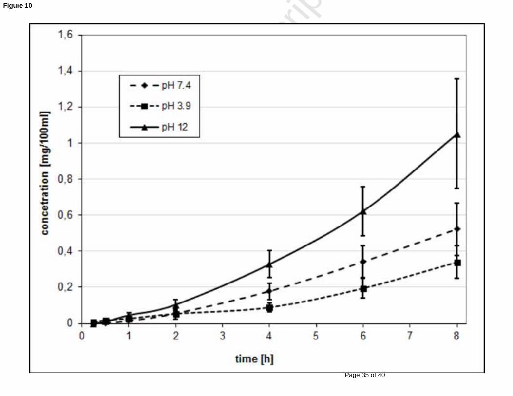
Because of very slow permeation (pH 7.4 donor), the influence of ionization of SUS (the pKa 348
values are 4.21, 5.67, 9.63, >12) on permeation rate was explored. Permeation was evaluated 349
using donor solutions of pH values 3.9, 7.4, 12.0. The highest concentrations in the acceptor 350
phase and highest pseudosteady state fluxes were obtained at pH 12.0, where sumatriptan 351
occurs mostly in the unionized form as a sumatriptan base. The lowest permeation rate was 352
found with donors of pH 3.9, where the sumatriptan succinate molecule is fully ionized (Fig. 353
10; Table 1). It is in good agreement with the theory of passive permeation through most 354
biological membranes. Results of a similar character were obtained in a study with nicotine,355
where the differences of permeation rates at various pH values (expressed as cumulative 356
amounts) were even more significant (Chen L.-L. et al., 1999).357
358
Fig. 10. Permeation of sumatriptan through a porcine sublingual membrane using donor 359
solutions (6%, 0.5 mL) of three different pH (3.9, 7.4 and 12.0)360
361
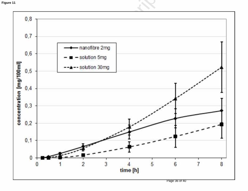
The nanofibrous PVA membranes containing 20% of SUS were placed on the sublingual 362
mucosa as the donor samples, and concentrations of the permeated drug in acceptor 363
compartments were measured. The results showed (Fig. 11) that permeation from the 364
nanofibrous donor was much faster compared to the solution containing 5 mg of the drug in 365
0.5 mL of donor (e.g. 1%), although the amount of the drug in the solution was more than 2 366
times higher than in the nanofibrous membranes. Moreover, within the initial 2 hour period,367
SUS permeation from nanofibres was even faster than from the saturated solution. This 368
finding can be explained by a much higher concentration of the drug presented at the very 369
large interface of nanofibre/mucosa. It is possible to imagine an exposed mucosal layer that is 370
Page 17
Page 16 of 40
Accep
ted
Man
uscr
ipt
16
fully saturated with the drug released from the nanofibres, and an immediate replacement of 371
permeated-off drug from nanofibrous storage. This situation is quite different in comparison372
with the drug solution donor sample. Nanofibres probably served as a reservoir for surface 373
facilitated drug release supplying the carrier/mucosa interface with a higher efficiency than 374
was the case with solutions.375
376
Fig. 11. Permeation of sumatriptan through a porcine sublingual membrane. Donors: PVA 377
nanofibrous membrane 2 mg, solution 1% (sol 5 mg), solution 6% (sol 30 mg).378
379
As results on Fig. 12 show the transmucosal drug permeation was increased about 5 times 380
when the drug is loaded in nanofibrous membranes, compared to the highly concentrated 381
solutions used as a donor. The increase in released and subsequently permeated drug amounts382
in percentages of initially loaded amounts is substantial from a practical point of view.383
384
Fig. 12. Amounts of sumatriptan permeated. Donors: PVA nanofibrous membrane 2 mg, 385
solution 1% (sol 5 mg), solution 6% (sol 30 mg, pH 12) 386
387
We consider the necessary permeated dose of SUS should reach at least 4 mg (or rather 6 mg) 388
of the drug. It represents an equivalent of a subcutaneously administered dose of SUS with a 389
bioavailability of about 96% (Imitrex, 2013).390
Thus, if the permeated amount of SUS from the nanofibrous donor was about 4% of the 391
loaded dose, then about 100 mg of SUS would have to be administered (on 20 cm2, which is 392
the estimated area size of the sublingual mucosa available for administration, and also the 393
size of the intended membrane formulation). This requirement could be technologically 394
realizable. For instance, to obtain 100 mg of API for delivery the nanomembrane weight 395
Page 18
Page 17 of 40
Accep
ted
Man
uscr
ipt
17
would have to be about 250 or 300 mg of nanofibers made of one or more polymers. The 396
membrane would have to be produced of a high drug load and a high mass per area (g/m2). 397
The membranes could be layered and then pressed to fix them. This reservoir would be 398
covered by an impermeable layer on one side. We also take into account a possible addition 399
of an adhesive at the edges of a final preparation to ensure a good and long term contact at a 400
place of absorption. 401
Further improvement could be achieved by the use of a sumatriptan base for the formulation 402
of drug loaded nanofibrous membranes, because this form of the drug is more permeable 403
across the sublingual membrane, as illustrated in Fig. 10. Penetration enhancers are another 404
applicable possibility to improve the permeation rate. They can be directly incorporated into 405
electrospun membranes, and released simultaneously with a drug affecting its permeation.406
407
Permeation of naproxen and naproxen sodium from the concentrated donor solutions through 408
porcine sublingual mucosa showed very good results for both the drugs. The free acid, but 409
also the sodium salt, permeated through the mucosa surprisingly quickly, thus high amounts 410
of the drugs in the acceptor phase were detected.411
Solubility of the salt form in an aqueous medium is several times higher, and could therefore 412
be administered in much higher concentrations and amounts, and was predominantly used for 413
the experiments.414
The increment in concentration of the drug administered in a donor solution on the mucosa 415
increased the permeation rate even more than proportionally, and the highest drug permeation 416
was found using a saturated drug solution (Figs. 13 and 15), as was expected. In the case of 417
the saturated donor solution (10%), more than 70% of the total amount of the drug permeated 418
during 8 hours of the experiment.419
420
Page 19
Page 18 of 40
Accep
ted
Man
uscr
ipt
18
Fig. 13. Permeation of naproxen through a porcine sublingual membrane using graduated 421
concentrations of donor solutions (0.5 mL) - 1, 2, 3.2 and 10%. 422
423
However, when nanofibrous membranes (CHI, PCL) containing 30% NAP were used as a 424
donor, good contact with the absorption mucosa was achieved, and NAP permeated very 425
quickly and more effectively than from the tested solutions. The permeated amount of NAP 426
reached almost identical values from nanofibrous membranes as from donor solutions 427
containing a two times higher amount of the drug. Thus, permeation of NAP from the 428
electrospun membranes was more than 2 times higher in comparison to the solution with the 429
same amount of the drug (Fig. 14).430
431
Fig. 14. Permeation of naproxen through a porcine sublingual membrane using 3.2% solution 432
(sol 16 mg), 2% solution (sol 10 mg), 1% solution (sol 5 mg) and nanofibrous membrane 5433
mg (PCL)434
435
Fig. 15. Amounts of naproxen permeated through porcine sublingual membrane using 10% 436
solution (sol 50 mg), 3.2% solution (sol 16 mg), 2% solution (sol 10 mg), 1% solution (sol 5 437
mg) and nanofibrous membrane 5 mg (PCL)438
439
Table 1. The sublingual permeation flux values. 440
441
It is apparent, by comparing permeability of NAP and SUS that less polar units of NAP 442
permeate through a sublingual membrane more freely than polar SUS. Molecular weights of 443
both drugs are very similar (230 NAP vs. 295 SUS), however there are significant differences 444
in the polarity of the molecule (log P 3.18 NAP vs. 0.93 SUS) that probably cause the 445
Page 20
Page 19 of 40
Accep
ted
Man
uscr
ipt
19
differences in the drug permeation extensions. The results show that permeation through 446
nonpolar routes of this very thin mucosal membrane probably prevails. Naproxen permeated 447
amounts were 35% to 75% of the administered dose, and in the case of sumatriptan, the 448
pertinent values were around 0.5 % to 4.5% of the dose.449
It is important to note that the increase of the drug permeation is much more evident for 450
sumatriptan, which passes reluctantly, than naproxen, which has lower relative increase due 451
to its already high permeation level. Thus, usage of nanofibrous membranes could be more 452
favourable for the drugs with slow permeation of mucosal barriers.453
454
4. CONCLUSIONS455
456
Sumatriptan succinate, naproxen and its sodium salt, as well as their combination were 457
successfully incorporated into nanofibrous membranes via electrospinning manufacturing 458
technology. The membranes obtained showed very good nanofibre diameters and shape 459
homogeneity without any crystal moieties within nanofibrous masses.460
The membranes showed very fast and mutually independent release of sumatriptan succinate, 461
naproxen and naproxen sodium from either hydrophilic PVA, PAA, and chitosan nanofibres,462
or a hydrophobic PCL nanofibrous carrier.463
The used drugs exerted very high differences in in vitro sublingual permeation, but in both 464
cases, it was several times increased using nanofibrous membranes as the drug carrier in 465
comparison to the drug solutions as donors. As the drugs permeated preferentially as non-466
ionized moieties, therefore we would recommend to prepare nanofibrous membranes 467
containing both the drugs in non-ionized forms.468
Page 21
Page 20 of 40
Accep
ted
Man
uscr
ipt
20
The membranes prepared were very flexible and mechanically resistant. Having a drug load 469
capacity of up to 40% of the membrane mass, they could be very advantageous for the 470
formulation of sublingual drug delivery systems.471
472
Acknowledgements473
474
The study was supported by the grants No. SVV 267 001 of the Ministry of Education of the 475
Czech Republic and No. 530812/2012 of Grant Agency of Charles University.476
477
References478
479
Arulmozhi D.K., Veeranjaneyulu A., Bodhankar S.L., 2005. Migraine: Current concepts and 480
emerging therapies. Vascular Pharmacology 43, 176-187.481
Ramdan N. M., Buchanan T. M., 2006. New and future migraine therapy. Pharmacol. & 482
Therap. 112, 199-212.483
Imitrex® - Product monograph, 2013. Glaxo Smith Kline Inc.;484
http://www.gsk.ca/english/docs-pdf/product-monographs/Imitrex.pdf ,(10th.July.2013) 485
Hearnden V., Sankar V., Hull K., Vidović Juras D., Greenberg M., Kerr R., Lockhart P. B., 486
Patel P., Makwana S., Jobanputra U., Ravat M., Ajmera A, Patel M., 2011. Sublingual route 487
for the systemic delivery of ondasentron. Int. J. Drug Dev.& Res. 3(4), 36-44.488
Bayrak Z., Tas C., Tasdemir U., Erol H., Ozkan C. K., Savaser A., Ozkan Y., 2011. 489
Formulation of zolmitriptan sublingual tables prepared by direct compression with different 490
polymers: In vitro and in vivo evaluation. Eur. J. Pharm. Biopharm. 78, 499-505.491
Patel V. F., Liu F., Brown M. B., 2011. Advances in oral transmucosal drug delivery. J. 492
Control. Release 153, 106-116.493
Page 22
Page 21 of 40
Accep
ted
Man
uscr
ipt
21
Nagy Z. K., Nyúl K., Wagner I., Molnár K., Marosi. G., 2010. Electrospun water soluble 494
polymer mat for ultrafast release of Donepezil HCl. Express Polym. Lett. 4, 763-772495
Yu D.-G., Gao D., White K., Branford-White Ch., Lu W.-Y., Zhu L-M., 2010. 496
Multicomponent Amorphous Nanofibers Electrospun from Hot Aqueous Solutions of a 497
Poorly Soluble Drug. Pharm. Res. 27, 2466-2477.498
Yu D.-G., Yang J.-M., Branford-White Ch., Lu P., Zhang L., Zhu L.-M., 2010. Third 499
generation solid dispersions of ferulic acid in electrospun composite nanofibers, Int. J. Pharm. 500
400, 158-164.501
Stranska D., Klabanova D., Dolezal P., Vrbata P., Berka P., Musilova M., 2012. CZ Patent 502
No. 303244 B6.503
Reneker D. H., Chun I., 1996. Nanometre diameter fibres of polymer produced by 504
electrospinning. Nanotechnology 7, 216-223.505
Frenot A., Chronakis I. S., 2003. Polymer nanofibres assembled by electrospinning. Curr. 506
Opin. Colloid. Interface Sci. 8, 64-75.507
Agarwal S, Greiner A., Wendorff J. H., 2013.Functional materials by electrospinning of 508
polymers. Prog. Polym. Sci., http://dx.doi.org/10.1016/j.progpolymsci.2013.02.001.509
Jirsak O., Sanetrnik F., Lukas D., Kotek K., Martinova L., Chaloupek J., 2005. U.S. patent 510
No. WO205024101.511
Leung V., Ko F., 2011. Biomedical applications of nanofibers, Polym. Adv. Technol. 22, 512
350-365.513
Nagy Z. K., Balogh A., Vajna B., Farkas A., Patyi G., Kramarics A., Marosi G., 2012.514
Comparison of electrospun and extruded soluplus®-based solid dosage forms of improved 515
dissolution. J. Pharm. Sci. 101, 322–332.516
Cao H., Liu T., Chew S. Y., 2009. The application of nanofibrous scaffolds in neural tissue 517
engineering. Adv. Drug Deliv. Rev. 61, 1055-1064.518
Page 23
Page 22 of 40
Accep
ted
Man
uscr
ipt
22
Zhang X., Reagan M. R., Kaplan D. L., 2009. Electrospun silk biomaterial scaffolds for 519
regenerative medicine. Adv. Drug Deliv. Rev. 61, 988-1006.520
Sell S. A., McClure M. J., Garg K., Wolfe P. S., Bowlin G. L., 2009. Electrospinning of 521
collagen/biopolymers for regenerative medicine and cardiovascular tissue engineering. Adv. 522
Drug Deliv. Rev. 61, 1007-1019.523
Chakraborty S., Liao I-Ch., Adler A., Leong K. W., 2009. Electrohydrodynamics: A facile 524
technique to fabricate drug delivery systems. Adv. Drug Deliv. Rev. 61, 1043-1054. 525
Meinel A. J., Germershaus O., Luhmann T., Merkle H. P., Meinel L., 2012. Electrospun 526
matrices for localized drug delivery: Current technologies and selected biomedical 527
applications. Eur. J. Pharm. Biopharm. 81, 1-13.528
Kenawy E. R., Bowlin G.L., Mansfield K., Layman J., Simpson D.G., Sanders E.H., Wnek 529
G.E., 2002. Release of tetracycline hydrochloride from electrospun poly(ethylene-co-530
vinylacetate), poly(lactic acid), and a blend. J. Control. Release 81 (1-2), 57-64.531
Kim K., Luu Y.K., Chang C., Fang D.F., Hsiao B.S., Chu B., Hadjiargyrou M., 2004. 532
Incorporation and controlled release of a hydrophilic antibiotic using poly(lactide-co-533
glycolide)-based electrospun nanofibrous scaffolds. J. Control. Release 98, 47-56.534
Taepaiboon P., Rungsardthong U., Supaphol P., 2006. Drug loaded electrospun mats of 535
poly(vinyl alcohol) fibres and their release characteristics of four model drugs. 536
Nanotechnology 17, 2317-2329.537
Kenawy E.-R., Abdel-Hay F.I., El-Newehy M.H., Wnek G.E., 2007. Controlled release of 538
ketoprofen from electrospun poly (vinyl alcohol) nanofibers. Mater. Sci. Eng. A 459 (1-2), 539
390-396.540
Huang L.-Y., Branford-White Ch., Shen X.-X., Yu D.-G., Zhu L.-M., 2012. Time-541
engineeringed biphasic drug release by electrospun nanofiber mesches. 436, 88-96.542
Taepaiboon P., Rungsardthong U., Supaphol P., 2007. Vitamin-loaded electrospun cellulose 543
Page 24
Page 23 of 40
Accep
ted
Man
uscr
ipt
23
acetate nanofibers mats as transdermal and dermal therapeutic agents of vitamin A acid and 544
vitamin E. Eur. J. Pharm. Biopharm. 67, 387-397.545
Madhaiyana K., Sridharb R., Sundarrajanb S., Venugopala J. R., Ramakrishna S., 2013. 546
Vitamin B12 loaded polycaprolactone nanofibers: A novel transdermal route for the water 547
soluble energy supplement delivery. Int. J. Pharm. 444, 70-76.548
Xu X., Chen X., Wang Z., Jing X., 2009. Ultrafine PEG-PLA fibers loaded with both 549
paclitaxel and doxorubicin hydorchoride and their in vitro cytotoxicity. Eur. J. Pharm. 550
Biopharm. 72, 18-25.551
Maretschek S., Greiner A., Kissel T., 2008. Electrospun biodegradable nanofiber nonwovens 552
for controlled release of proteins. J. Control. Release 127, 180-187.553
Han N., Johnson J., Lannutti J. J., Winter J. O., 2012. Hydrogel-electrospun fiber composite 554
materials for hydrophilic protein release. J. Control. Release 158, 165-170.555
Nozal M.J., Bernal J.L., Toribio L., Mart´ın M.T., Diez F.J. J., 2002. Development and 556
validation of an LC assay for sumatriptan succinate residues on surfaces in the manufacture 557
of pharmaceuticals. Pharm. Biomed. Anal. 30, 285-291.558
Femenıa-Font A., Merino V., Rodilla V., Lopez-Castellano A., 2005. High-performance 559
liquid chromatographic determination of sumatriptan after in vitro transdermal diffusion 560
studies. J. Pharm. Biomed. Anal. 37, 621-626.561
Natu M. V., de Sousa H. C., Gil M. H., 2008. Effects of drug solubility, state and loading on 562
controlled release in bicomponent electrospun fibers. Int. J. Pharm. 397, 50-58.563
Srikar R., Yarin A. L., Megaaridis C. M., Bazilevsky A. V., Kelley E., 2008. Desorption-564
limited mechanism of release from polymer nanofibers. Langmuir 24, 965-974.565
Chen L.-L. H., Chetty D. J., Chien Y. W., 1999. A mechanistic analysis to characterize 566
oromucosal permeation properties. Int. J. Pharm. 184, 63-72.567
568
Page 25
Page 24 of 40
Accep
ted
Man
uscr
ipt
24
Table. 1568
Sample (SUS)Flux [µg/cm2/h] Sample (NAPS)
Flux [µg/cm2/h]
nano PVA - 2 mg 4.39 ± 0.56 nano PCL 5 mg 180.59 ± 14.05sol pH 7.4 - 5 mg 2.71 ± 1.00 sol 5 mg 77.30 ± 16.56sol pH 12 - 30 mg 13.47 ± 3.47 sol 10 mg 185.95 ± 19.40sol pH 7.4 - 30 mg 10.32 ± 2.72 sol 16 mg 521.26 ± 14.03sol pH 3.9 - 30 mg 6.43 ± 2.47 sol 50 mg 2277.31 ± 41.71
569
Page 26
Page 25 of 40
Accep
ted
Man
uscr
ipt
~
impermeable cover layernanofibrous layer
mucoadhesive nanofibrous layer
Graphical Abstract
Page 27
Page 26 of 40
Accep
ted
Man
uscr
ipt
Figure 1
Page 28
Page 27 of 40
Accep
ted
Man
uscr
ipt
Figure 2
Page 29
Page 28 of 40
Accep
ted
Man
uscr
ipt
Figure 3
Page 30
Page 29 of 40
Accep
ted
Man
uscr
ipt
Figure 4
Page 31
Page 30 of 40
Accep
ted
Man
uscr
ipt
Figure 5
Page 32
Page 31 of 40
Accep
ted
Man
uscr
ipt
Figure 6
Page 33
Page 32 of 40
Accep
ted
Man
uscr
ipt
Figure 7
Page 34
Page 33 of 40
Accep
ted
Man
uscr
ipt
Figure 8
Page 35
Page 34 of 40
Accep
ted
Man
uscr
ipt
Figure 9
Page 36
Page 35 of 40
Accep
ted
Man
uscr
ipt
Figure 10
Page 37
Page 36 of 40
Accep
ted
Man
uscr
ipt
Figure 11
Page 38
Page 37 of 40
Accep
ted
Man
uscr
ipt
Figure 12
Page 39
Page 38 of 40
Accep
ted
Man
uscr
ipt
Figure 13
Page 40
Page 39 of 40
Accep
ted
Man
uscr
ipt
Figure 14
Page 41
Page 40 of 40
Accep
ted
Man
uscr
ipt
Figure 15