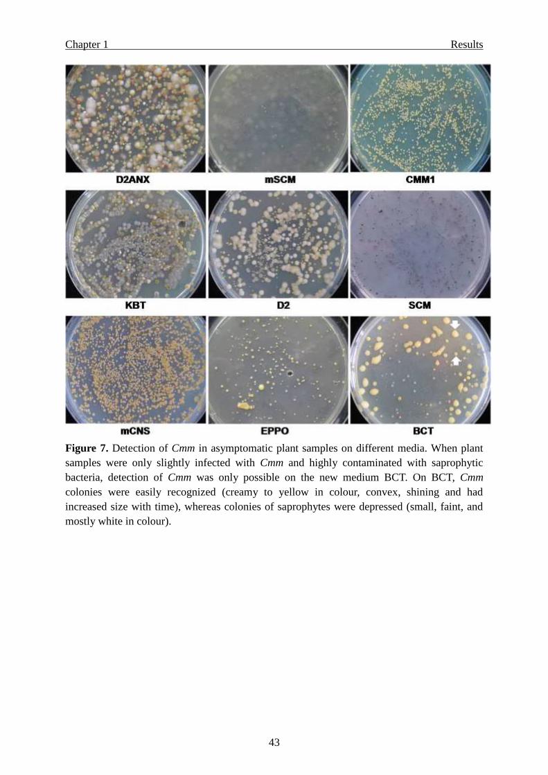
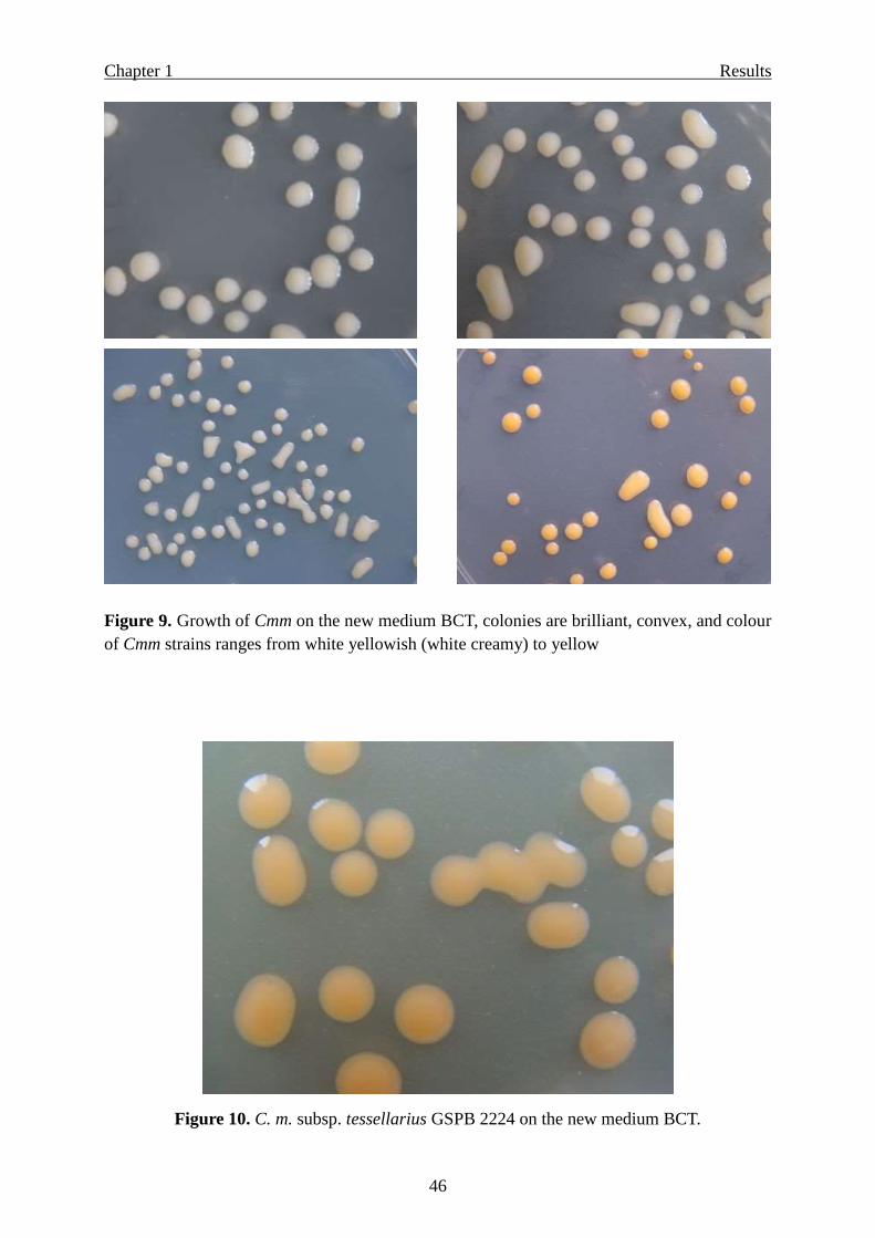
Page 1
Elimination of Clavibacter michiganensis subsp. michiganensis
from tomato cultures and seeds by highly sensitive detection
methods and effective seed treatments
Dissertation
to obtain the PhD degree
at the Faculty of Agricultural Sciences,
Georg-August-University Göttingen, Germany
by
Radwan M. Ftayeh
born in Deir Al-Bakht
Daraa, Syria
Göttingen 2009
Page 2
D7
1. Name of referee: Prof. Dr. Andreas von Tiedemann
2. Name of co-referee: Prof. Dr. Kerstin Wydra
3. Name of co-referee: Prof. Dr. Petr Karlovsky
Date of disputation: January 29, 2010
Page 3
Contents
i
Contents
General Introduction ………………………………………………………………….… 1
Disease history …………………………………………………………………..... 1
Tomato production in Germany …………………………………………………... 3
Symptoms ….…………………………………………………………………..…. 4
Disease epidemiolog …………………………………………………………........ 6
Seed health certification ………………………………………………………….. 7
International requirements ………………………………………………………... 8
Objectives ………………………………………………………………………… 9
Outcomes …………………………………………………………………………. 9
References ……………………………………………………………………….. 11
Chapter 1: Development of new selective and highly sensitive nutrient media for
Clavibacter michiganensis subsp. michiganensis and other subspecies ……………... 15
Summary ……………………………………………………………………………….. 15
Conclusions …………………………………………………………………………….. 16
Introduction ……………………………………………………………………………. 17
Materials and Methods ………………………………………………………………... 19
Bacterial species and strains …………………………………………………….. 19
Antibiotic-resistant mutant of Cmm ……………………………………………... 20
Media and growth conditions …………………………………………………… 20
Selection of the basic medium for Cmm ………………………………………… 22
Screening of antibiotics …………………………………………………………. 22
Susceptibility of accompanying bacteria towards antibiotics …………………… 23
Adjusting the optimum concentrations of inhibitors ……………………………. 23
Determining the plating efficiency (recovery rate) of Cmm strains on semiselective
media …………………...........................…………………………….………….. 24
Evaluation of selectivity and detection sensitivity of semiselective media ……... 24
Results …………………………………………………………………………………... 26
Selecting a new basic medium for Cmm ……………………………………….... 26
Screening of different antibiotics ………………………………………………... 27
Susceptibility of accompanying bacterial species and strains towards
antibiotics ……………………………………………………………………...... 29
Page 4
Contents
ii
Recipes of the new selective media BCT and BCT-2 …………………………… 30
Effect of boric acid………………………………………………………………. 32
Plating efficiency (recovery rate) of Cmm on the published and the new selective
media …………...................................…………………………………………... 35
Selectivity of the new media BCT and BCT-2 ………………………………….. 39
Detection sensitivity of the new media for latent infection by Cmm …………… 42
Selectivity for other pathovars/species of coryneform bacteria ………………… 45
Modifications of the new media BCT and BCT-2 ………………………………. 47
Effect of the buffering system and other fungicides …………………………….. 50
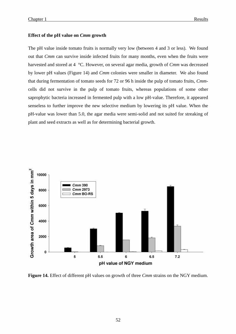
Effect of the pH value on Cmm growth …………………………………………. 52
Effect of fruit juice ………………………………………………………………. 53
Selection of a fungicide …………………………...…………………………….. 53
Discussion …………………………………………………...………………………..… 55
References ……………………………………..………………………………………... 60
Chapter 2: Establishment of a Bio-PCR assay for a sensitive detection of Clavibacter
michiganensis subsp. michiganensis in seed and plant material ……………………. 65
Summary ……………………………………………………………………………….. 65
Introduction ……………………………………………………………………….…… 67
Materials and Methods ………………………………………………………………... 69
Bacterial cultures and growth conditions ……………………………………….. 69
DNA extraction ………………………………………………………………….. 69
Primer design ……………………………………………………………………. 72
Designing new primers based on the publication of Bach et al. (2003) ………… 72
Designing new primers based on the publication of Luo et. al. (2008) …………. 72
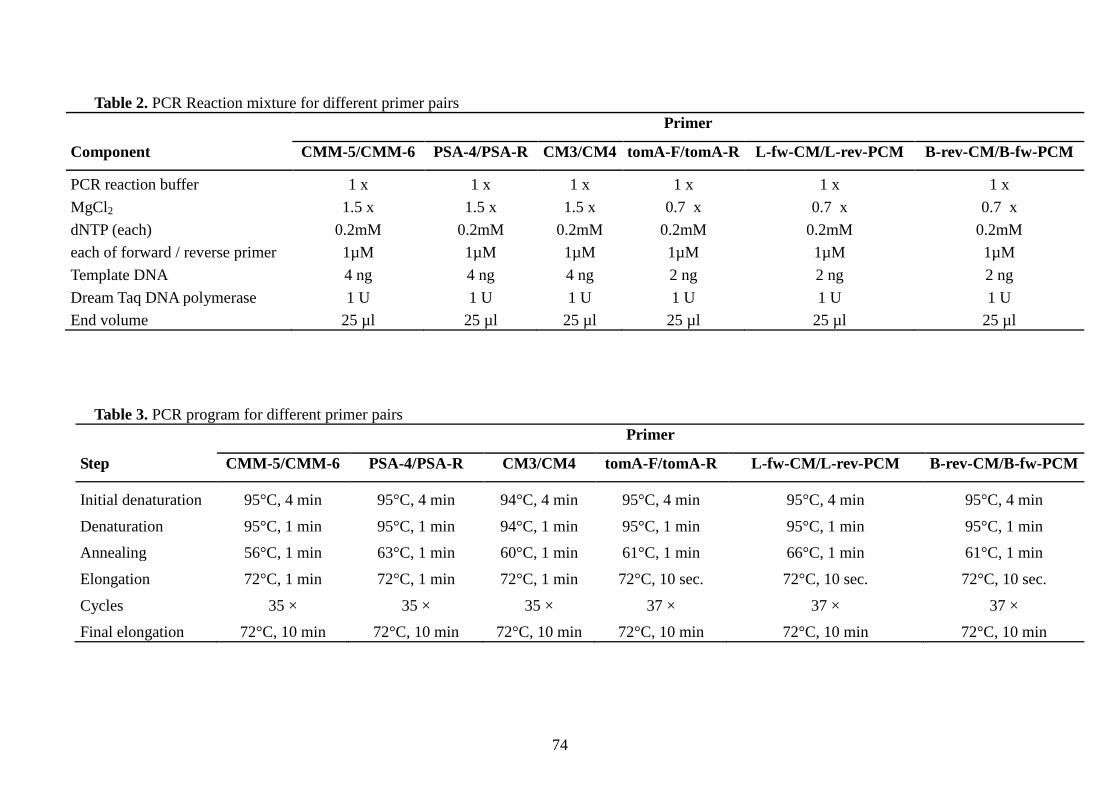
Amplification conditions …................................….........……………………….. 73
- The new primers “B-rev-CM/B-fw-PCM” and “L-fw-CM/L-rev-PCM”; and the
primer set tomA-F/tomA-R (Kleitman et al., 2008) …………………………. 73
- The primers CMM-5/CMM-6 (Dreier et al., 1995); CM3/CM4 (Sousa-Santos
et al., 1997); and PSA-4/PSA-R (Pastrik and Rainey, 1999) …………...……. 73
Direct PCR ………………………………………………………………………. 75
Inhibitor tests ……………………………………………………………………. 75
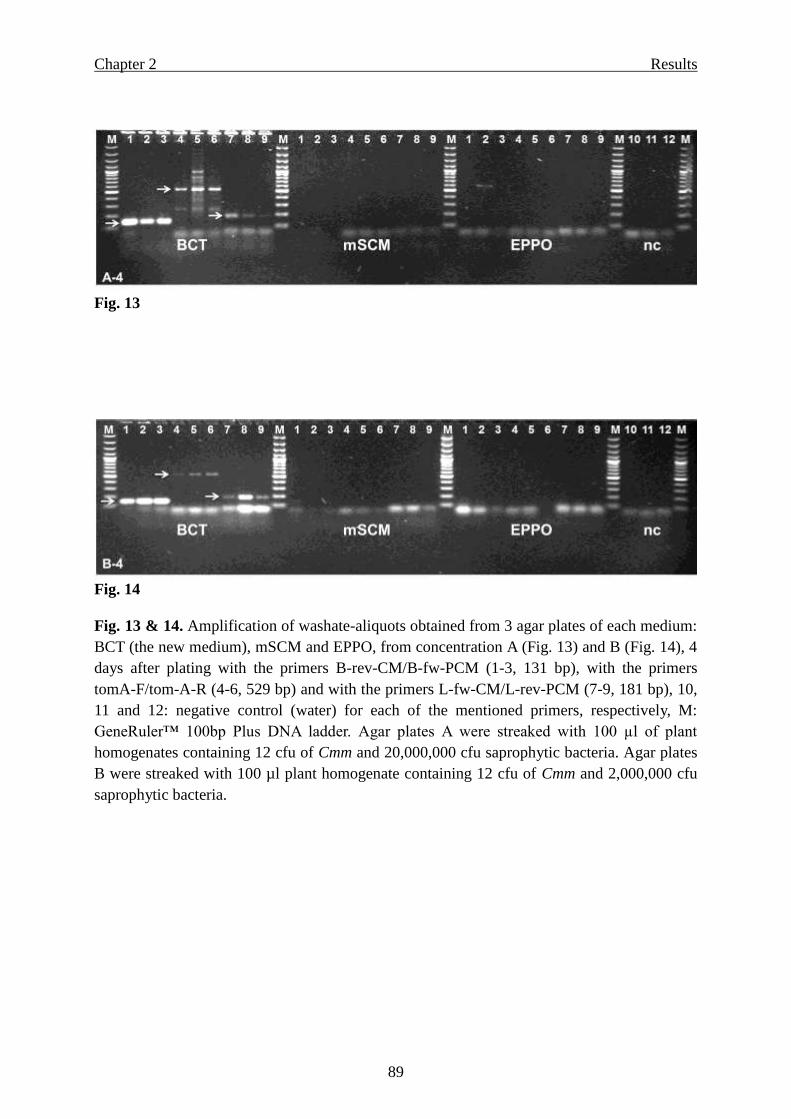
Bio-PCR …………………………………………………………………………. 76
Results ………………………………………………………………………………...… 78
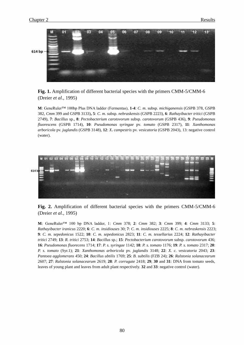
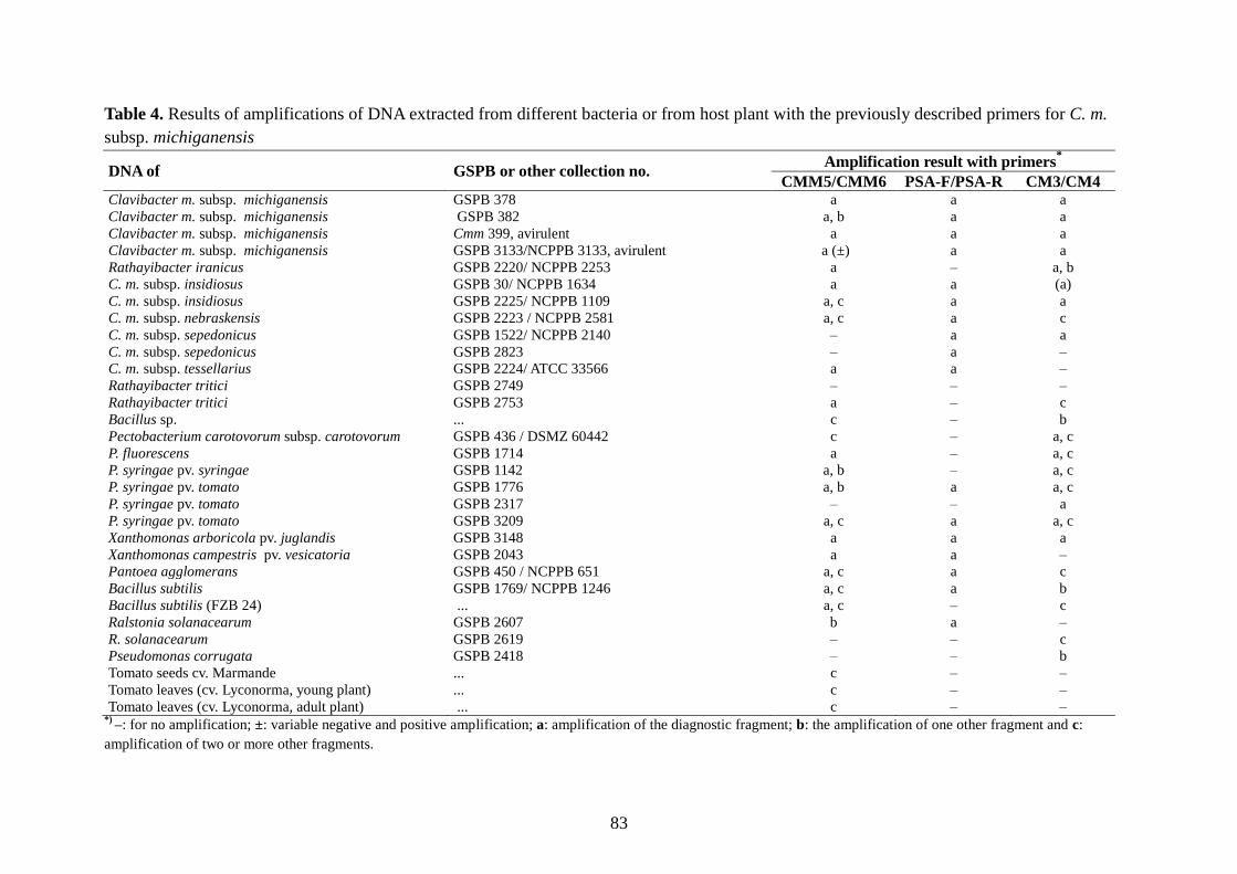
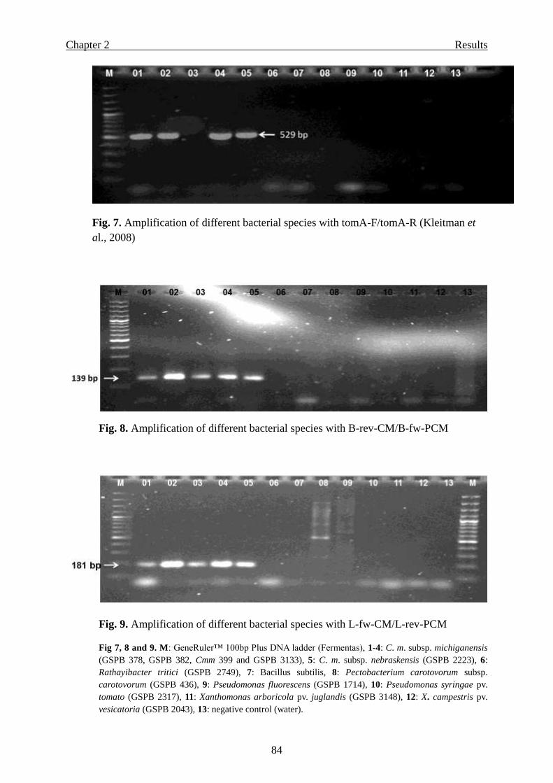
Specificity ……………………………………………………………………..… 78
Page 5
Contents
iii
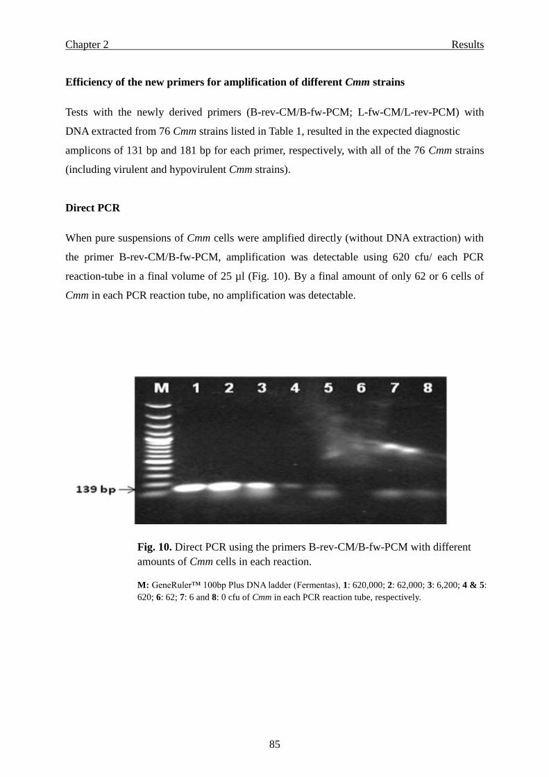
Efficiency of the new primers for amplification of different Cmm strains ……… 85
Direct PCR ………………………………………………...…………………….. 85
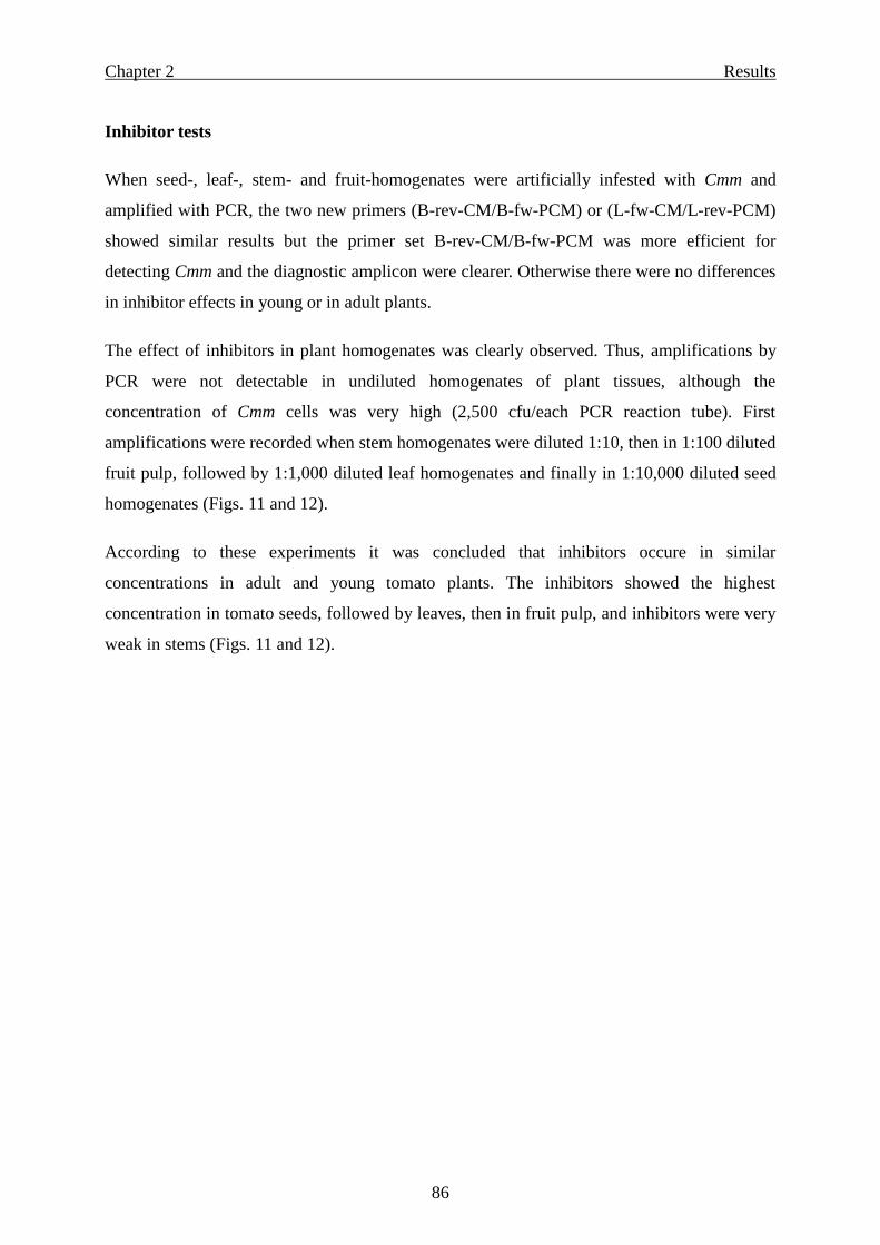
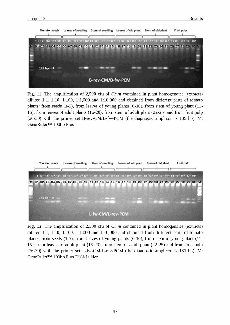
Inhibitor tests ……………………………………………………………………. 86
Bio-PCR …………………………………………………………………………. 88
Discussion ………………………………………………………………………………. 94
References ……………………………………………………………………………... 101
Chapter 3: Efficacy of different disinfection methods for eradication of Clavibacter
michiganensis subsp. michiganensis from tomato seed …………………………….. 107
Summary …………………………………………………………………………….... 107
Introduction …………………………………………………………...……………… 109
Materials and Methods ……………………………………………………...………... 111
Antibiotic-resistant Cmm mutant ………………………………………………. 111
Bacterial inocula and testing of different inoculation methods with Cmm …….. 111
Screening of different chemical and physical seed treatment methods .…......… 113
Seed lots, seed infection and storage conditions ……………………………….. 113
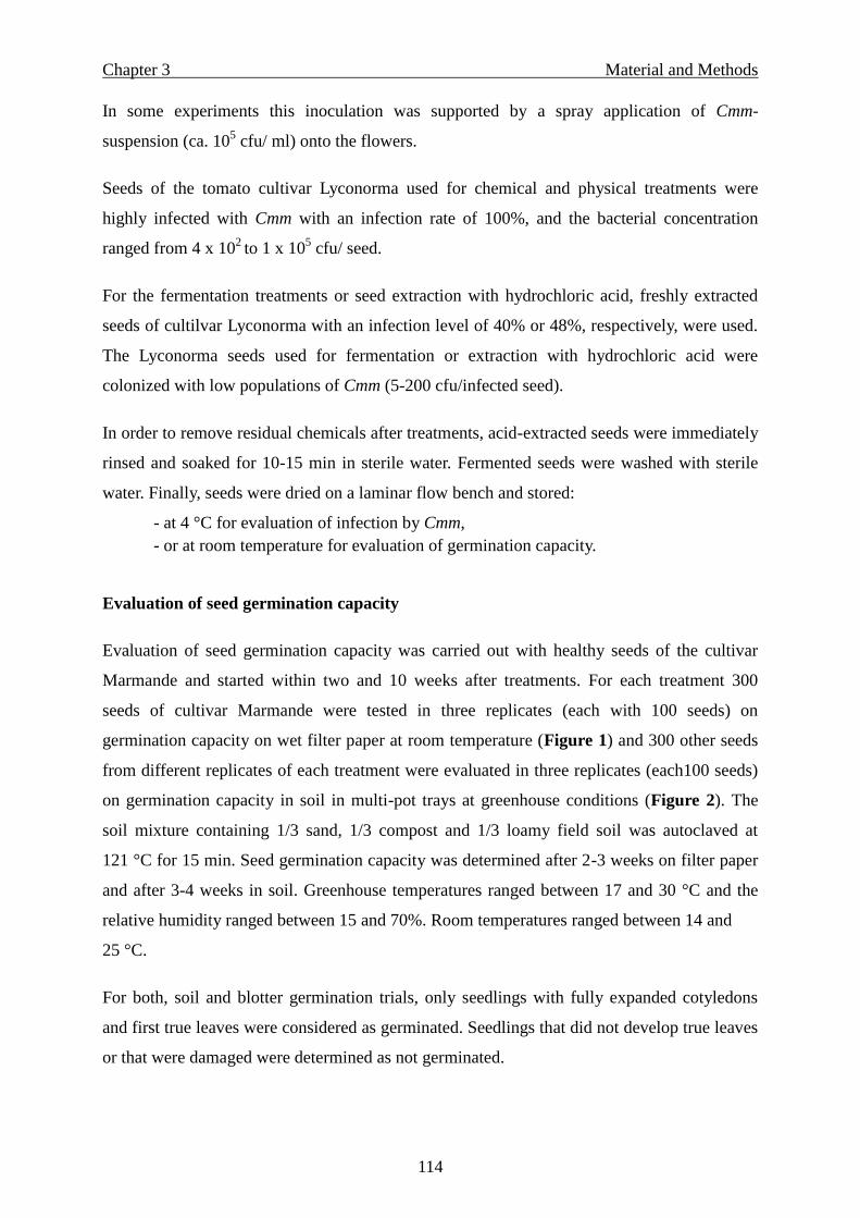
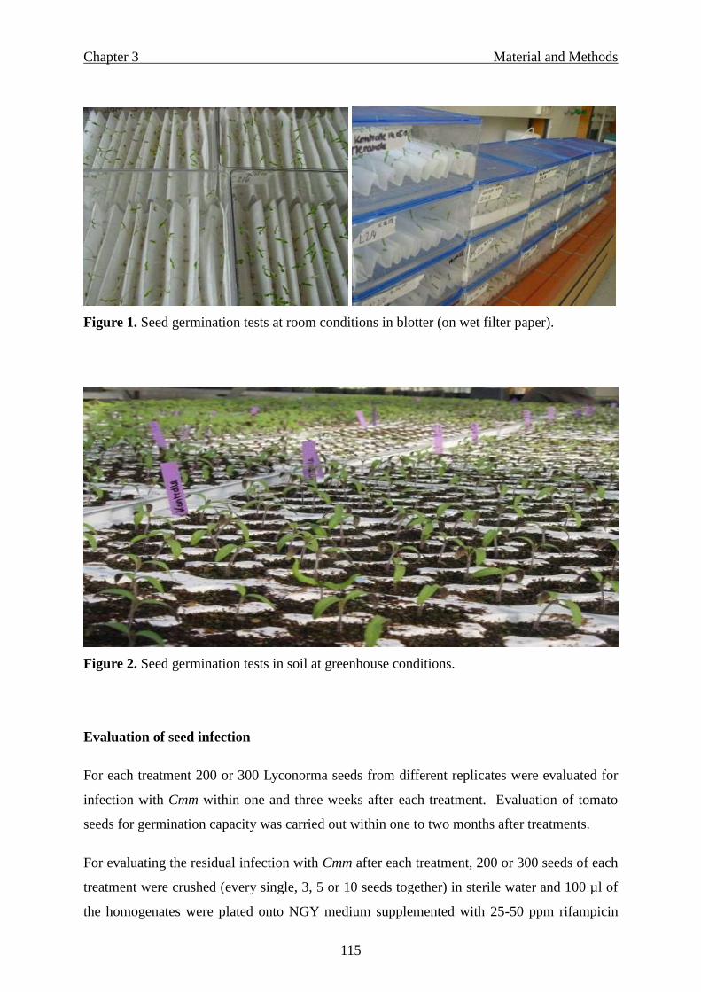
Evaluation of seed germination capacity ………………………………………. 114
Evaluation of seed infection ………………………………………………….... 115
Seed treatments ……………………………………………………………….... 116
Chemical and hot water treatments …………………………………………….. 116
Seed fermentation ……………………………………………………………… 117
Seed extraction with hydrochloric acid ………………………………………... 117
Seed treatments with hot air ……………………………………………………. 118
Statistical analysis ……………………………………………………………… 118
Results …………………………………………………………………………………. 119
Screening of different inoculation methods with Cmm ………………………... 119
Effect of seed treatments towards Cmm bacteria ………………………………. 123
Successful eradication of Cmm from infested seeds by chemical or hot water
treatments .................................................................…………………………… 123
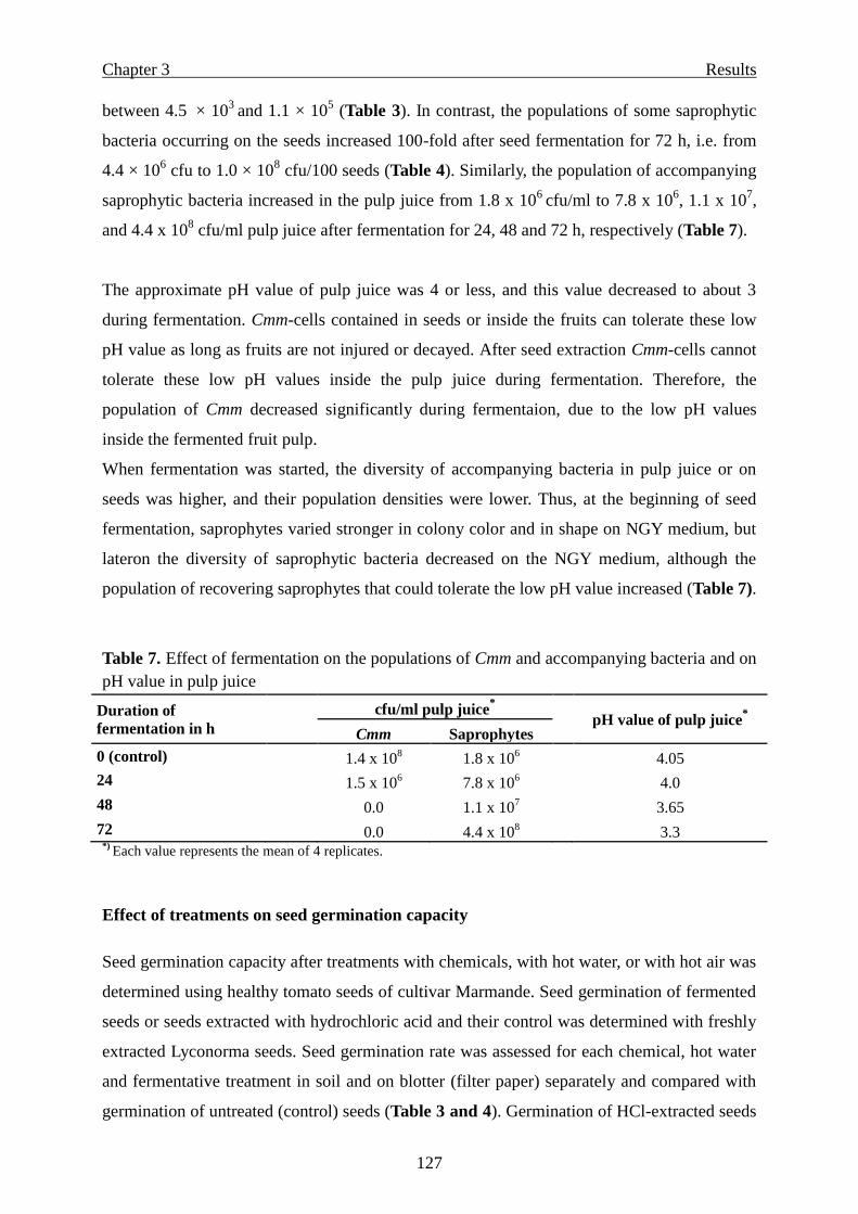
Effect of seed fermentation …………………………………………………….. 125
Seed extraction with hydrochloric acid ………………………………………... 125
Effect of hot air treatments …………………………………………………….. 126
Effect of seed treatments on saprophytic bacteria ……………………………... 126
Effect of treatments on seed germination capacity ……………………………...127
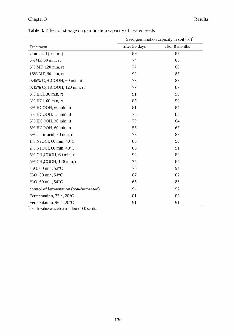
Seed germination capacity at two and eight months after treatments ………..... 128
Page 6
Contents
iv
Discussion ……………………………………………………………………………... 131
References ……………………………………………………………………………... 135
Chapter 4: Occurrence of Clavibacter michiganensis subsp. michiganensis, the causal
agent of bacterial canker of tomato, in Syria ……….……………………………..... 139
Summary ………………………………………………………………………….…... 139
Introduction …………………………………………………………………………... 140
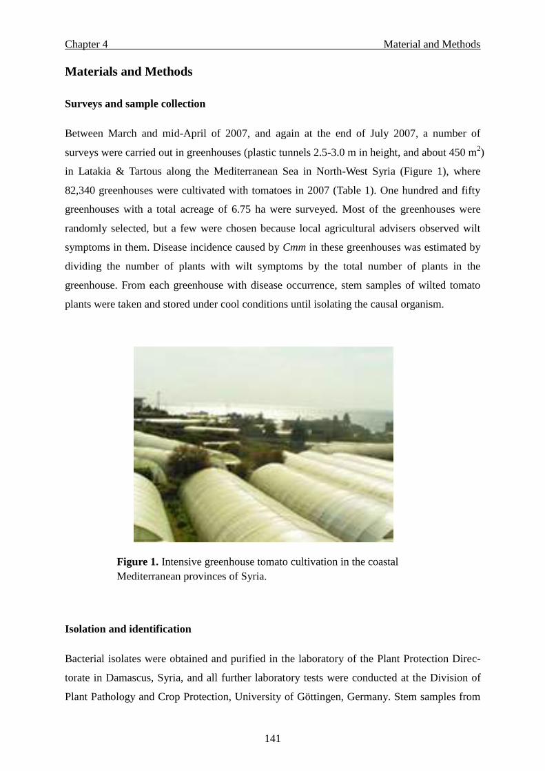
Materials and Methods ………………………………………………………………. 141
Surveys and sample collection …………………………………………………. 141
Isolation and identification …………………………………………………….. 141
Pathogenicity …………………………………………………………………... 142
PCR identification ……………………………………………………………... 143
Results …………………………………………………………………………………. 144
Disease incidence ………………………………………………………………. 144
Isolate identification ………………………………………………………….... 144
Pathogenicity …………………………………………………………………... 145
PCR identification ……………………………………………………………... 145
Discussion ……………………………………………………………………………... 147
References ……………………………………………………………………………... 149
General Discussion …………………………………………………………………… 151
General Summary ……………………………………………………………………. 154
Related publications ………………………………………………………………….. 158
Refereed journals ………………………………………………………………. 158
Presentations at national and international conferences ……………………….. 158
Conferences and workshops attended ………………………………………….. 159
Abbreviations …………………………………………………………………………. 160
Acknowledgements ………………………………………………………………….... 162
Eidesstattliche Erklärung ……………………………………………………………. 164
Curriculum vitae ……………………………………………………………………... 165
Page 7
General Introduction
1
General Introduction
Disease history
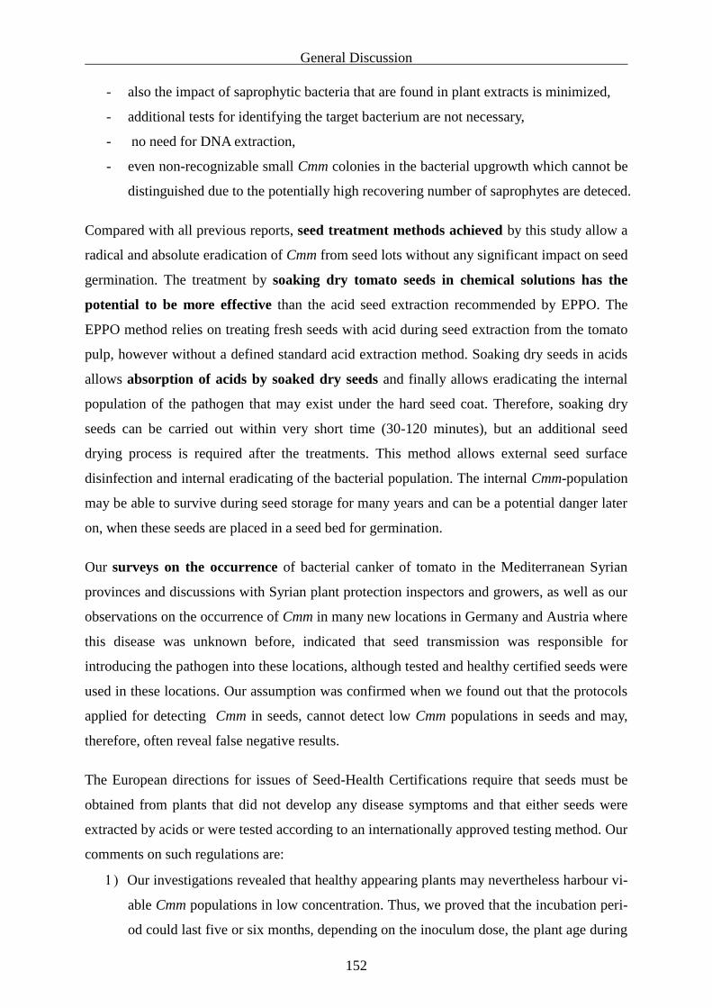
Bacterial canker of tomato, caused by the bacterium Clavibacter michiganensis subsp.
michiganensis (Smith. 1910) Davis et al. 1984 is a serious and destructive disease worldwide.
The disease was at first described by Smith who found it in 1909 in Grand Rapids, Michigan
(Strider, 1969), after which the pathogen spread into nearly all main tomato production areas
world-wide. Recently, the incidence of bacterial canker of tomato increased in Europe and
was newly reported in several countries worldwide causing considerable losses. Therfore, a
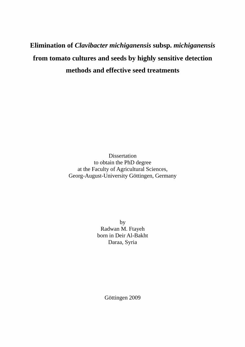
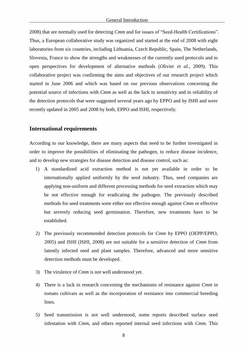
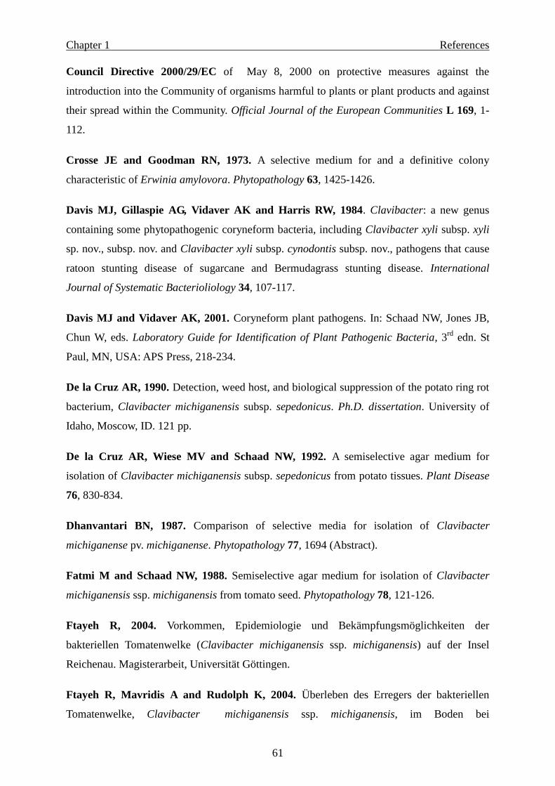
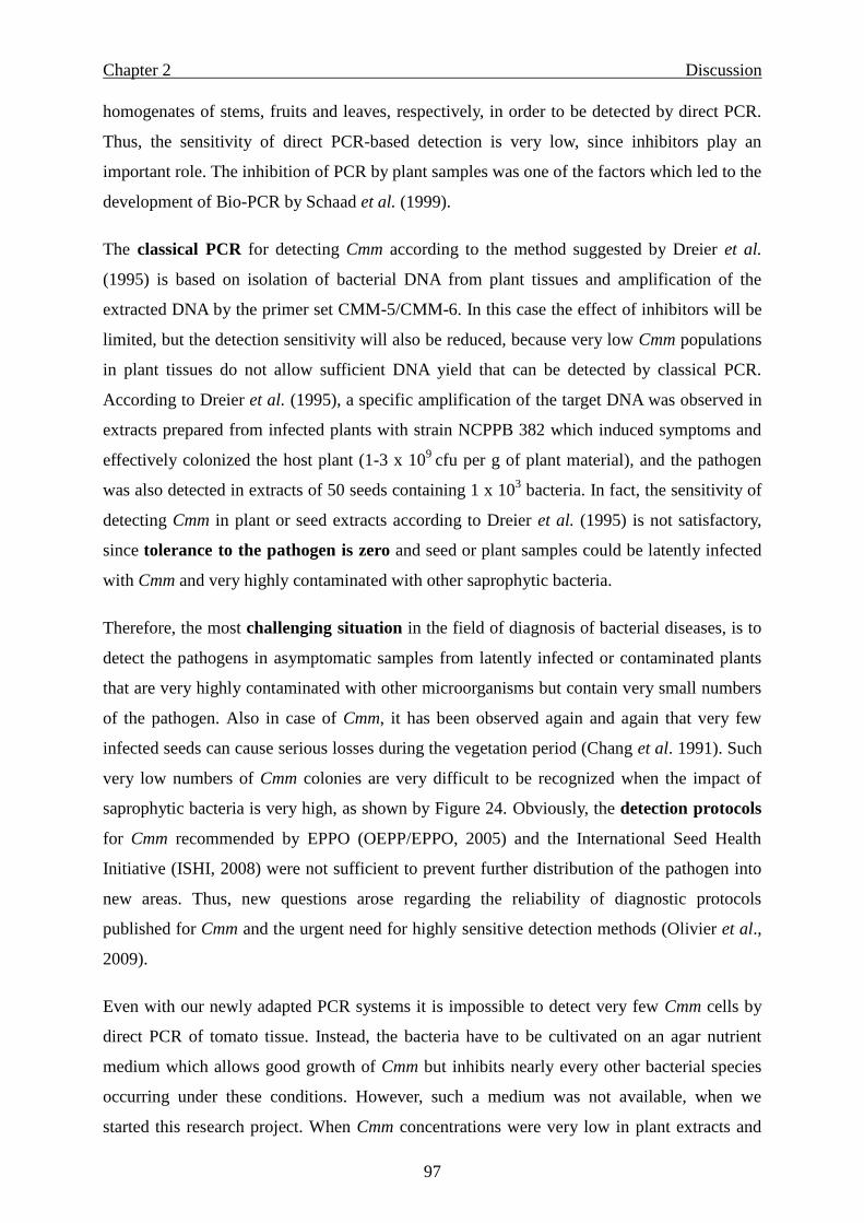
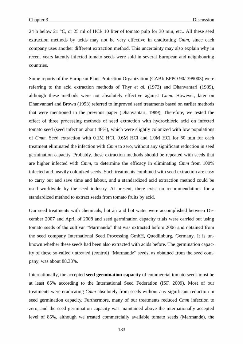
new distribution map of the pathogen (Figure 1) was issued (CABI/EPPO, 2009). The
bacterium is considered as a quarantine organism in the European Union and many other
countries (Council Directive 2000/29/EC; OEPP/EPPO, 1982).
In Germany, the pathogen is known since 1929 (Kotte, 1930; Stapp, 1958), and caused
serious losses in 1978, especially in greenhouses (Griesbach, person. commun.). Recently, the
disease was transmitted in 1998 into the peninsula Reichenau in South Germany in Baden-
Württemberg (Schmidt, 2006, person. commun.) and newly in 2002 into “Knoblauchsland”
near Nürnberg in Bavaria (Maeritz, 2006, personal commun.), also in 2006 into North-Rhine-
Westphalia (Matthäus-Staack and Eickeln, 2006, personal commun.) and very recently again
into new locations of Baden-Württemberg in 2009 (Moltmann, 2009, personal commun.).
Recently, the disease also occurred in neighbouring countries of Germany, such as Austria
(Weber and Fuchs, 2007, personal observation and commun.), Switzerland (Wasserfallen,
2008, personal commun.), the Netherlands, and was newly reported by EPPO (CABI/EPPO,
2009) in several European and non-European countries.
Page 8
2
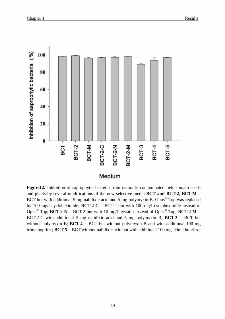
Figure 1. New distribution map of Clavibacter michiganensis subsp. michiganensis, issued by CABI/ EPPO in 2009 (Map no. 26).
Page 9
General Introduction
3
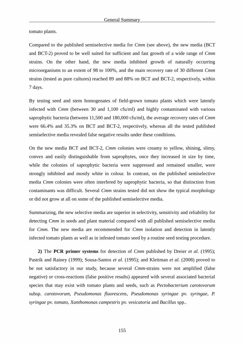
Tomato production in Germany
The total acrage of commercially grown greenhouse tomatoes in Germany ranges between
300 and 400 ha, whereas open field cultivations are little and not important. In 2008, the area
of greenhouse-grown tomato was about 308 ha with a total greenhouse number of 2.808 and a
production of 65.096 ton (Behr, 2009, personal commun. ZMP, 2009).
The largest greenhouse production areas of tomato are located in Baden-Württemberg (79.81
ha), Bavaria (44.7 ha) and North-Rhine-Westphalia (42.8 ha), and additional tomato
cultivation areas exist in all other states of Germany (ZMP, 2009). In Baden-Württemberg,
tomatoes are mostly cultivated in classical normal greenhouses in soil, whereas in North-
Rhine-Westphalia and Bavaria tomatoes are often grown in hydro cultures in so-called “high-
tech” greenhouses using sterile artificial substrates instead of soil, hybrid tomato plants that
are grafted onto basic cultivars with resistance against soil-borne fungal and nematode
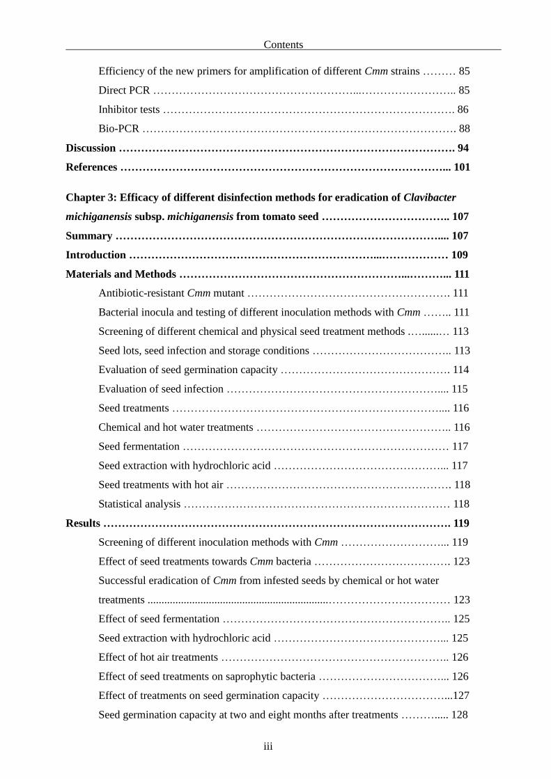
diseases. These tomato cultures require large investments, because of the intensive
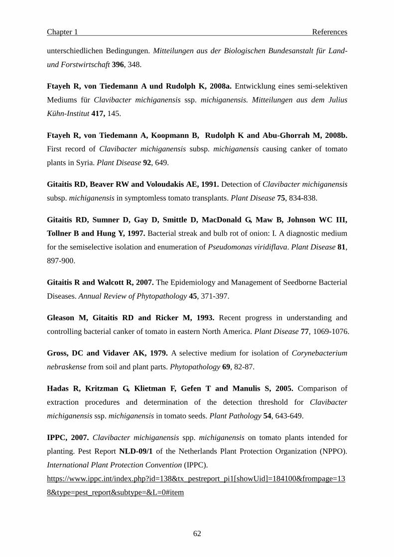
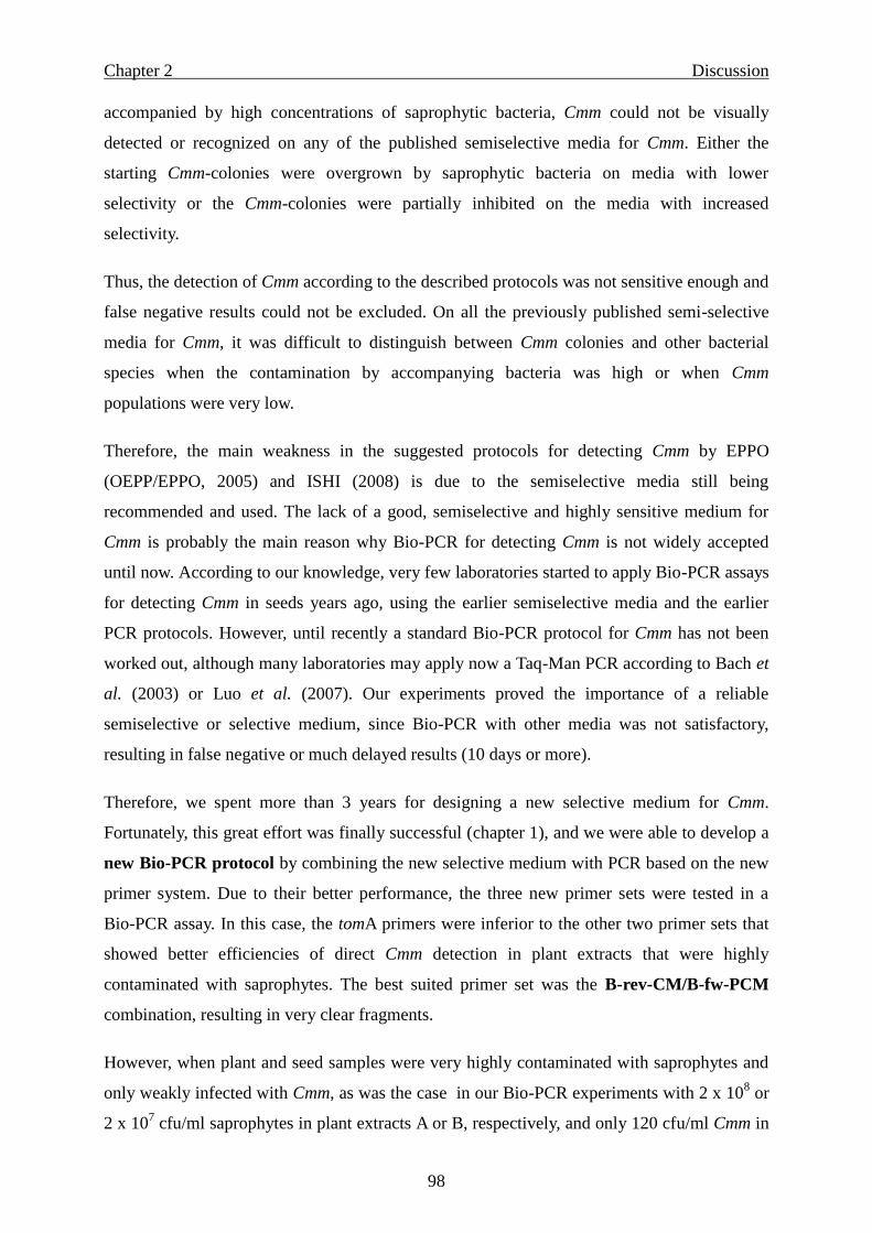
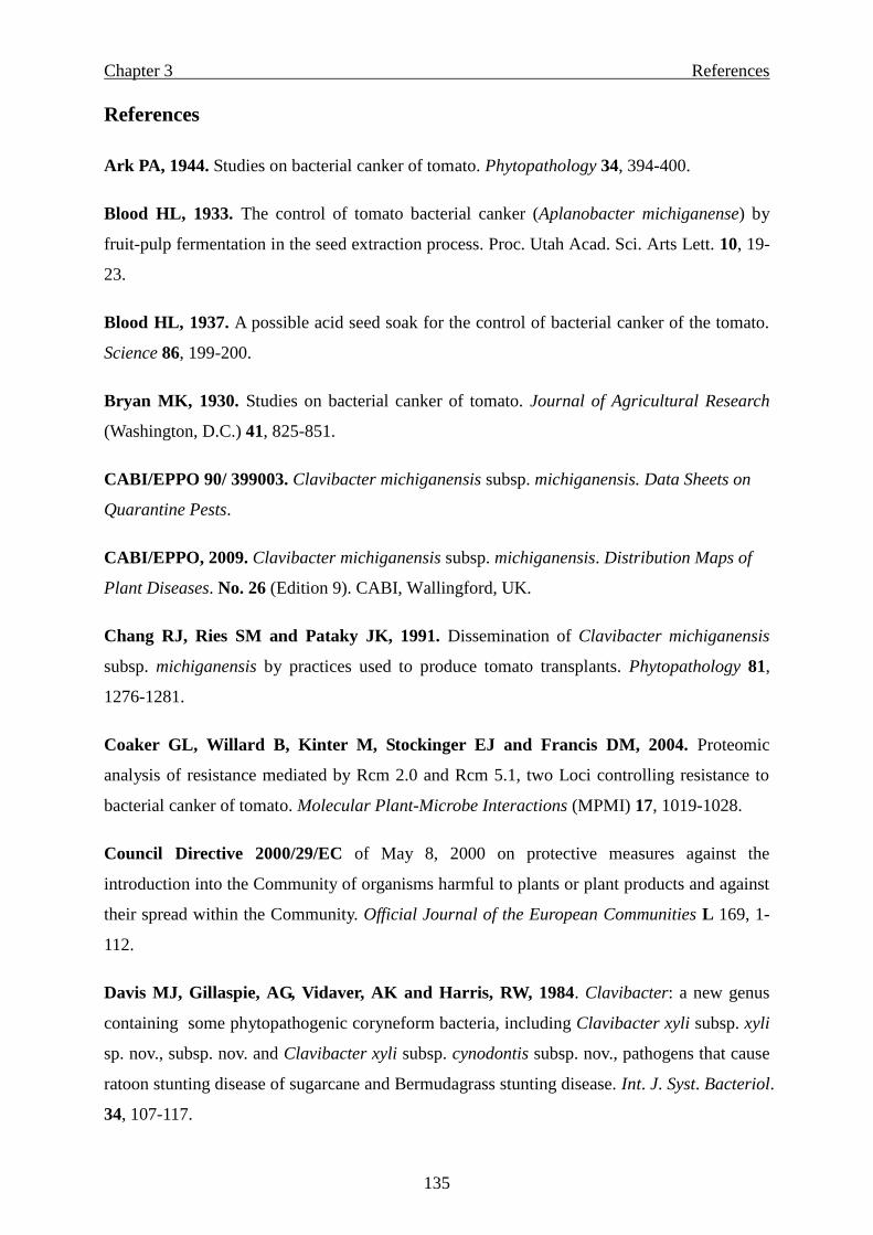
cultivation, e.g. computerized and mechanized watering, air conditioning and fertilizing
(Figure 2). The vegetation period in such high-tech greenhouses lasts 12 months, since two-
month-old transplants are planted in the beginning of January and cultivated in the greenhouse
until the end of November. The tomato plants reach a final length of 10 to 12 m at season’s
end, and during December old plants and the substrate are removed, watering systems and
greenhouse structures are sterilized and greenhouses are prepared again for the new
vegetation period. Some high-tech greenhouses are cultivated with 10,000 to 25,000 plants or
more.
Generally, disease incidence in greenhouses with hydroponic cultures is higher than in
normal greenhouses with soil cultivation, because of the additional infection source by
watering and because plants in these greenhouses are more susceptible to diseases (Figure
9B). In Germany, a primary infection with Clavibacter michiganensis subsp. michiganensis
(Cmm) was recorded during 2006 in some greenhouses with 25,000 plants (in
Knoblauchsland, Bavaria) or with 13,000 plants (in Straelen, North- Rhine-Westphalia) on
only 5 young plants. However, when the hygienic measures were not followed in Straelen
(due to first occurrence of the disease), 80% of all plants (13,000) wilted completely after few
months and the residual plants showed very strong wilting symptoms (Figure 9B). But when
very strict hygienic measures were applied, disease incidence could be kept under 2% in the
greenhouse with 25,000 plants (in Knoblauchsland).
Page 10
General Introduction
4
Figure 2. Intensive hydroponic tomato production in a “high-tech” greenhouse, plants can
reach a length of 10-12 m at season’s end.
Symptoms
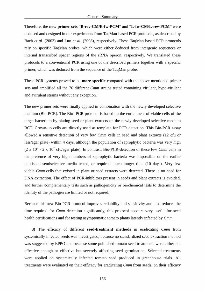
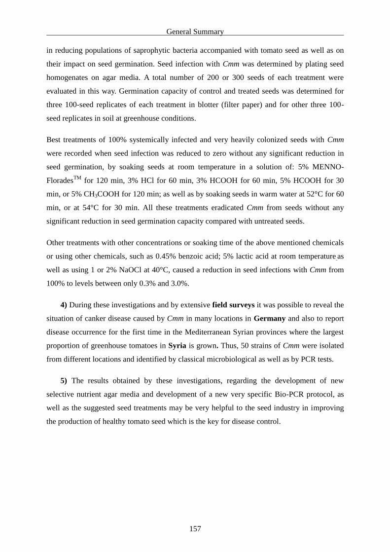
Disease symptoms are variable and seldom appear concomitantly on one plant or in one field
or greenhouse. Typical symptoms include unilateral wilt of leaflets (Figure 3), canker of the
stem (Figure 4), necrosis of leaf margins (Figure 5), and wilting of young plants (Figure 6).
On fruits “bird’s eye spots” may appear (Figure 7 A and B). By cutting the side shoots or the
stem, brown discoloration of the xylem which forms “horseshoe” symptoms may be seen
(Figure 8). Finally the whole plants wilt, in the field (Figure 9A) as well as in the greenhouse
(Figure 9B).
Figure 3. Unilateral wilting of leaflets. Figure 4. Canker of a stem.
Page 11
General Introduction
5
Figure 5. Necrosis on leaf margins. Figure 6. Wilting of a young plant.
Figure 7. Bird’s eye lesions on unripe fruit (A) and ripe fruit (B).
Figure 8. Horseshoe symptom on a side-shoot section.
Page 12
General Introduction
6
Figure 9. Wilting of field tomato plants (A) and all greenhouse tomato plants (B) due to
Clavibacter michiganensis subsp. michiganensis.
Disease epidemiology
The pathogen can survive in the soil in plant debris (Fatmi and Schaad, 2002; Ftayeh, 2004;
Ftayeh et al., 2004; Strider, 1969) and on greenhouse structures (Strider, 1969), but the main
transmission of the disease takes place by contaminated or infected tomato seed or plants.
Disease resistance is known (Coaker et al., 2004; Poysa, 1993; van Steekelenburg, 1985), but
has not been incorporated into commercial tomato cultivars to our knowledge.
The disease can be very destructive, and disease control is not possible once disease appears,
because bactericides for control are not available. The incubation period can last up to 5 or 6
months (Ftayeh et al., 2008a). Therefore, infected and neighbouring plants must be destroyed
immediately when disease symptoms appear, and very strict hygienic measures should be
applied after disease appearance (Strider, 1969).
Thus, the use of pathogen-free seed, whether obtained naturally or by treating seeds with
chemical eradicants, could eliminate a potential source of inocula (Fatmi et al., 1991) and is
considered to be the best strategy for controlling canker disease. Although seed transmission
is less than 1% (Grogan and Kendrick, 1953), already 0.01 to 0.05% of infested seeds can
cause an epidemic in suitable conditions (Chang et al., 1991).
Infested seeds and young plants are responsible for primary infection and disease
transmission into new locations, This may happen even when very strict quarantine measures
are used to control import and export and all kinds of movements of tomato seed, and
Page 13
General Introduction
7
although commercial tomato seed is normally sold together with seed-health certificates
proving that the tomato seed had been certified as pathogen-free according to internationally
standardized testing methods.
Seed health certification
Certification of commercial tomato seeds as pathogen-free can be fulfilled according to the
European Commission Directives 2000/29/EC when:
Tomato seeds are gained from healthy-appearing plants, which did not show any disease
symptoms until the date of seed extraction, and if one of the following conditions is fulfilled:
1) Seeds have been extracted by diluted acids, or
2) Seeds have been tested according to internationally accepted laboratory methods.
However, these directives were insufficient to prevent further spread of the disease in Europe
recently because:
- Healthy appearing plants may be latently infected and the incubation time may extend
more than 5 months (Ftayeh et al., 2008a).
- An internationally accepted standardized seed extraction method by acids is not
available although this method has been required by the European Community and
also recommended by EPPO (Council Directive 2000/29/EC; Petter, 2009, personal
commun.).
- Seed extractions by acids do not ensure an effective and absolute eradication of the
pathogen which is required by the international quarantine regulations for Cmm that
restrict import and export to zero tolerance for Canada, the USA, the EU (Council
Directive 2000/29/EC; Bach et al., 2003) and several other countries in order to
prevent the outbreak of bacterial canker of tomato. Even one contaminated seed in
10,000 must be detectable. Thus, Anwar et al. (2004) and Gitaitis and Walcott (2007)
proved the presence of Cmm in certified commercial tomato seed indicating the need
for more sensitive detection methods.
The recent outbreaks of bacterial canker in the European Community resulted in increased
attention of the national and international plant quarantine and plant protection authorities as
well as the European Plant Protection Organization (EPPO) and the International Seed Health
Initiative (ISHI), concerning the source of inocula in newly infected areas and the reliability
of detection protocols described for Cmm by EPPO (OEPP/EPPO, 2005) and by ISHI (ISHI,
Page 14
General Introduction
8
2008) that are normally used for detecting Cmm and for issues of “Seed-Health Certifications”.
Thus, a European collaborative study was organized and started at the end of 2008 with eight
laboratories from six countries, including Lithuania, Czech Republic, Spain, The Netherlands,
Slovenia, France to show the strengths and weaknesses of the currently used protocols and to
open perspectives for development of alternative methods (Olivier et al., 2009). This
collaborative project was confirming the aims and objectives of our research project which
started in June 2006 and which was based on our previous observations concerning the
potential source of infections with Cmm as well as the lack in sensitivity and in reliability of
the detection protocols that were suggested several years ago by EPPO and by ISHI and were
recently updated in 2005 and 2008 by both, EPPO and ISHI, respectively.
International requirements
According to our knowledge, there are many aspects that need to be further investigated in
order to improve the possibilities of eliminating the pathogen, to reduce disease incidence,
and to develop new strategies for disease detection and disease control, such as:
1) A standardized acid extraction method is not yet available in order to be
internationally applied uniformly by the seed industry. Thus, seed companies are
applying non-uniform and different processing methods for seed extraction which may
be not effective enough for eradicating the pathogen. The previously described
methods for seed treatments were either not effective enough against Cmm or effective
but severely reducing seed germination. Therefore, new treatments have to be
established.
2) The previously recommended detection protocols for Cmm by EPPO (OEPP/EPPO,
2005) and ISHI (ISHI, 2008) are not suitable for a sensitive detection of Cmm from
latently infected seed and plant samples. Therefore, advanced and more sensitive
detection methods must be developed.
3) The virulence of Cmm is not well understood yet.
4) There is a lack in research concerning the mechanisms of resistance against Cmm in
tomato cultivars as well as the incorporation of resistance into commercial breeding
lines.
5) Seed transmission is not well understood, some reports described surface seed
infestation with Cmm, and others reported internal seed infections with Cmm. This
Page 15
General Introduction
9
was the same in old and new reports issued by EPPO. Therefore the exact location of
Cmm on or under the seed coat should be carefully investigated.
Objectives
The objectives of our study were to develop more effective methods in order to eliminate the
pathogen from tomato cultures. These new methods include:
Development of a new selective and highly sensitive nutrient medium for Cmm. The
current available semiselective media for detecting Cmm are the main weaknesses of
the applied detection protocols that are based on plating assays, because these media
often revealed false negative results.
Testing the previously used primers on their specificity for Cmm and searching for
more specific ones which could be used in combination with a potentially developed
new selective medium (Bio PCR).
Selection of the best suited disinfection methods for eradicating Cmm from infected
seeds.
Outcomes
The results of this work can be specified as the following points:
1) A highly sensitive selective medium for detection of Clavibacter michiganensis subsp.
michiganensis has been developed (Chapter 1; Ftayeh et al., 2008c).
2) A Bio-PCR assay for a highly sensitive detection of Cmm was established, based on
utilizing newly adapted primers and a new PCR protocol in combination with the new
selective medium BCT (Chapter 2; Ftayeh et al., 2010b).
3) Numerous seed treatment methods for eradication of Cmm from systemically infected
seeds were investigated, resulting in selection of and very effective methods which
absolutely eradicated the pathogen from seeds without a significant reduction in seed
germination were recorded (Chapter 3, Ftayeh et al., 2008d).
4) The current situation of bacterial canker of tomato in the Syrian Mediterranean strip
provinces and in different locations in Germany was investigated and documented. 50
new Cmm strains were isolated from different German and Syrian locations. Reports
Page 16
General Introduction
10
about disease occurrence in Syria were published (Table 1 of Chapter 2; Chapter 4;
Ftayeh, et al., 2008b; Ftayeh et al., 2010a).
5) Furthermore, many aspects dealt with other investigations which are not included in
this thesis, such as isolation of 45 different antagonists with high efficiency against
Cmm in vitro that could be a potential object for further studies. Other investigations
were carried out on the epidemiology of the pathogen under field and greenhouse
conditions, incubation time of Cmm in tomato plants and its relation to temperatures
and inocula densities, survival of the pathogen in seeds and in binding strings,
population dynamics and spread of the bacterium in planta, as well as the impact of
soil microorganisms on infections via infected seeds (Ftayeh 2004).
The present work may open new ways in understanding, detection, elimination and
management of bacterial canker of tomato caused by Clavibacter michiganensis subsp.
michiganensis.
Page 17
General Introduction References
11
References
Anwar A, van der Zouwen PS, Ilyas S and van der Wolf JM, 2004. Bacterial canker
(Clavibacter michiganensis subsp. michiganensis) of tomato in commercial seed produced in
Indonesia. Plant Dis. 88, 680.
Bach HJ, Jessen I, Schloter M and Munch JC, 2003. A TaqMan-PCR protocol for
quantification and differentiation of the phytopathogenic Clavibacter michiganensis
subspecies. Journal of Microbiological Methods 52, 85-91.
Behr HC, 2009. Agrarmarkt Informations-Gesellschaft mbH. Personal communications.
CABI/EPPO, 2009. Clavibacter michiganensis subsp. michiganensis. Distribution Maps of
Plant Diseases. Map no. 26 (Edition 9). CABI, Wallingford, UK.
Chang RJ, Ries SM and Pataky JK, 1991. Dissemination of Clavibacter michiganensis
subsp. michiganensis by practices used to produce tomato transplants. Phytopathology 81,
1276-1281.
Coaker GL, Willard B, Kinter M, Stockinger EJ and Francis DM, 2004. Proteomic
analysis of resistance mediated by Rcm 2.0 and Rcm 5.1, two loci controlling resistance to
bacterial canker of tomato. Molecular Plant-Microbe Interactions (MPMI) 17, 1019-1028.
Council Directive 2000/29/EC of May 8, 2000 on protective measures against the
introduction into the Community of organisms harmful to plants or plant products and against
their spread within the Community. Official Journal of the European Communities L 169, 1-
112.
Davis MJ, Gillaspie Jr. AG, Vidaver AK and Harris RW, 1984. Clavibacter, a new genus
containing some phytopathogenic coryneform bacteria Clavibacter xyli subsp. xyli sp. nov.,
subsp. nov. and Clavibacter xyli subsp. cynodontis subsp. nov., pathogens that cause ratoon
stunting disease of sugarcane and Bermudagrass stunting disease. International Journal of
Systematic Bacteriology 34, 107-117.
Fatmi M, Schaad NW and Bolkan HA, 1991. Seed treatments for eradicating Clavibacter
michiganensis subsp. michiganensis from naturally infected tomato seeds. Plant Disease 75,
383-385.
Page 18
General Introduction References
12
Fatmi M and Schaad NW, 2002. Survival of Clavibacter michiganensis subsp.
michiganensis in infected tomato stems under natural field conditions in California, Ohio and
Morocco. Plant Pathology 51, 149-154.
Ftayeh R, 2004. Vorkommen, Epidemiologie und Bekämpfungsmöglichkeiten der
bakteriellen Tomatenwelke (Clavibacter michiganensis ssp. michiganensis) auf der Insel
Reichenau. Magisterarbeit, Universität Göttingen.
Ftayeh R, Mavridis A und Rudolph K, 2004. Überleben des Erregers der bakteriellen
Tomatenwelke, Clavibacter michiganensis ssp. michiganensis, im Boden bei
unterschiedlichen Bedingungen. Mitteilungen der Biologischen Bundesanstalt für Land- und
Forstwirtschaft 396, 348.
Ftayeh R, von Tiedemann A und Rudolph K, 2008a. Untersuchungen zum Vorkommen und
Nachweis von Clavibacter michiganensis ssp. michiganensis an Tomatenkulturen.
Nachrichtenblatt des Deutschen Pflanzenschutzdienstes 60, 91.
Ftayeh R, von Tiedemann A, Koopmann B, Rudolph K. and Abu-Ghorrah M, 2008b.
First record of Clavibacter michiganensis subsp. michiganensis causing canker of tomato
plants in Syria. Plant Disease 92, 649.
Ftayeh R, von Tiedemann A und Rudolph K, 2008c. Entwicklung eines semi-selektiven
Mediums für Clavibacter michiganensis ssp. michiganensis. Mitteilungen aus dem Julius
Kühn-Institut 417, 145.
Ftayeh R, von Tiedemann A und Rudolph K, 2008d. Versuche zur Abtötung des
bakteriellen Schaderregers, Clavibacter michiganensis ssp. michiganensis, im Tomatensaatgut.
Mitteilungen aus dem Julius Kühn-Institut 417, 165.
Ftayeh R, von Tiedemann A, Koopmann B, Abu-Ghorrah M and Rudolph K, 2010a.
Occurrence of Clavibacter michiganensis subsp. michiganensis, the causal agent of bacterial
canker of tomato, in Syria. Phytopathologia Mediterranea 49, 174-179.
Ftayeh R, von Tiedemann A, Koopmann B and Rudolph K, 2010b. Reliability and
sensitivity of diagnostic methods for detection of Clavibacter michiganensis subsp.
michiganensis in seeds and plant material. Journal of Plant Diseases and Protection 117, 40.
Page 19
General Introduction References
13
Gitaitis R and Walcott R, 2007. The epidemiology and management of seedborne bacterial
diseases. Annual Review of Phytopathology 45, 371-397.
Grogan RG and Kendrick JB, 1953. Seed transmission, mode of overwintering and spread
of bacterial canker of tomato caused by Corynebacterium michiganese. Phytopathology
Abstracts 43, 473.
ISHI, 2008. Method for the detection of Clavibacter michiganensis subsp. michiganensis on
tomato seed (Version 3). International Seed Health Initiative.
http://www.worldseed.org/isf/ishi_vegetable.html.
Kotte W, 1930. Der Bakterienkrebs der Tomate, eine für Deutschland neue Pflanzenkrankheit.
Zeitschrift für Pflanzenkrankheiten 40, 1-2; 51-56.
Maeritz U, 2006. Gemüseerzeuger-Ring Knoblauchsland, Raiffeisen Str. 200, 90427
Nürnberg. Personal communication.
Matthäus-Staack E and Eickeln B, 2006. Landwirstchaftskammer NRW, Hans-Tenhaeff- Str.
40-42, 47638 Straelen. Personal communication.
Moltmann E, 2009. Landesanstalt für Pflanzenschutz, Reinsburgstr. 107, D-70197 Stuttgart.
Personal communication.
OEPP/EPPO. 1982. Data sheets on quarantine organisms No. 50, Corynebacterium
michiganense. Bulletin OEPP/EPPO Bulletin 12 (1).
OEPP/EPPO, 2005. EPPO standards PM 7/42 diagnostics. Clavibacter michiganensis subsp.
michiganensis. OEPP/EPPO Bulletin 35, 275-283.
Olivier V, Baloche A, Drouin A, Audusseau C, Paillard S and Soubelet H, 2009. Internal
validation and collaborative study on Clavibacter michiganensis subsp. michiganesis in seeds:
an example of a European cooperation. In EPPO Conference on diagnostics and associated
workshops 10 - 15. 05. 2009 in York (UK).
Petter F, 2009. European and Mediterranean Plant Protection Organization (EPPO), 1 rue Le
Nôtre, 75016 Paris, France. E-mail: [email protected] , personal communication.
Poysa V, 1993. Evaluation of tomato breeding lines resistant to bacterial canker. Canadian
Journal of Plant Pathology 15, 301-304.
Page 20
General Introduction References
14
Schmidt U, 2006. Beratungsdienst Reichenau e.V., Marktstr. 1, 78479 Insel Reichenau.
Personal communication.
Smith, EF, ed., 1914. The Grand Rapids tomato disease. In: Bacteria in Relation to Plant
Diseases. Washington, D.C. USA: Carnegie Institute, 161-165.
Stapp C, 1958. Pflanzenpathogene Bakterien, Verlag Paul Parey, Berlin, Deutschland. 259 pp.
Strider DL, 1969. Bacterial canker of tomato caused by Corynebacterium michiganense, a
literature review and bibliography. North Carolina Agric. Exp. Station, Tech. Bul. No. 193,
110 pp.
Van Steekelenburg NAM, 1985. Resistance to Corynebacterium michiganense in tomato
genotypes. Euophytica 34, 245-250.
Wasserfallen A, 2008. Lic. Iur., Dipl. Ing. Agr. ETH, Rechtsanwalt (Landwirtschaftsrecht).
Länggass-Strasse 7, Postfach 7161, CH-3001 Bern., Personal communication.
Weber J and Fuchs R, 2007. Fachabteilung Gartenbau, Landwirtschaftskammer Steiermark,
8010 Graz. Hamerlinggasse 3. Personal communication.
ZMP, 2009. ZMP-Marktbilanz, Gemüse. Statistisches Bundesamt, Fachserie 3, Reihe 3.1.3,
2008.
Page 21
Chapter 1 Summary
15
Chapter 1
Development of new selective and highly sensitive nutrient media for
Clavibacter michiganensis subsp. michiganensis and other subspecies
Summary
All published semiselective media for Clavibacter michiganensis subsp. michiganensis (Cmm)
proved to be not satisfactory for a sensitive detection of Cmm in infected tomato plants and
seeds. Therefore new selective media for Cmm were developed in three steps: 1) Selection of
a basic medium allowing good growth of Cmm but excluding or slowing down several other
bacterial species; 2) screening a wide range of antibiotics and other inhibitors for selective
inhibition of often accompanying bacterial or fungal species; 3) optimizing the composition
of inhibitors and nutrient components.
Initial tests for selection of antibiotics which did not inhibit Cmm were conducted with 30
strains of accompanying pathogenic and non-pathogenic bacterial species isolated from
tomato seeds and plants that were obtained from different locations. For these experiments,
tomato plants were cultivated in the field and artificially inoculated with very low
concentrations of a rifampicin and streptomycin resistant strain of Cmm. These tomato plants
did not develop disease symptoms but were latently infected with the pathogen. On the other
hand, homogenates from leaves, stems or tomato fruits were heavily contaminated with
various microorganisms (bacteria and fungi). The exact concentration of Cmm cells contained
in the homogenates was determined by dilution plating on NGY agar medium supplemented
with rifampicin, streptomycin and a fungicide. Parallely, dilution plating assays from the
same homogenates were conducted on many newly designed compositions for a potential
semiselective medium. The best suited new media were then tested for isolation of Cmm from
naturally infected plants obtained from different locations in Germany, Syria and Austria, in
order to enlarge the diversity of naturally occurring microorganisms on or in tomato plants.
Compared with all previously recommended semiselective media for Cmm, the new media
(BCT and BCT-2) proved to be well suited for sufficient and fast growth of a wide range of
Cmm strains.On the other hand, the new media inhibited growth of naturally occurring
microorganisms to an extent of 98 to 100%. By testing tomato seeds and plants which were
Page 22
Chapter 1 Conclusions
16
latently infected with Cmm and highly contaminated with different saprophytic bacteria, the
Cmm population was always detected on the new media, whereas all published semiselective
media revealed false negative results under these conditions.
Additional tests revealed that the new selective media were also well suited for isolation and
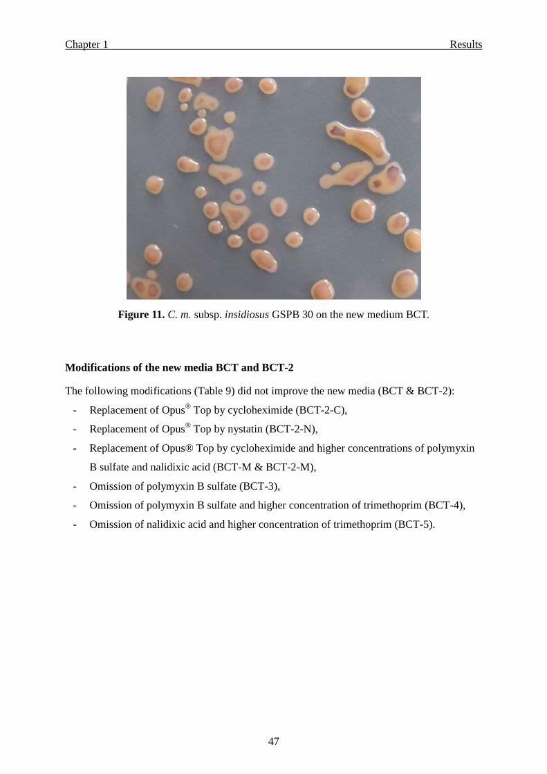
identification of the Clavibacter michiganensis subspecies nebraskensis, insidiosus and
tessellarius, but neither for C. m. ssp. sepedonicus nor for Curtobacterium flaccumfaciens pv.
flaccumfaciens.
Conclusions
The new media BCT and BCT-2 are superior to all published semiselective media for Cmm and
are denoted as selective media because:
the mean plating efficiency amounted up to 89%, all the 30 Cmm strains from a wide
range of different origins grew on the new media (one exception for BCT-2),
high selectivity, accompanying bacterial species occurring on tomato plants and seeds
or obtained from culture collections were inhibited to an extent of 98 to 100%, and
remarkable detection sensitivity. Thus, very low Cmm populations occurring in plant
and seed material in the presence of high concentrations (thousand-fold more) of non-
target accompanying bacteria were detected on the new media but never on the
published semiselective media.
Page 23
Chapter 1 Introduction
17
Introduction
Clavibacter michiganensis subsp. michiganensis (Cmm) (Smith, 1910) Davis et al., 1984 can
cause a very destructive wilt disease of tomato plants, especially in greenhouses. Therefore, the
pathogen has been classified as an A2 quarantine organism by the European Plant Protection
Organization (OEPP/EPPO, 2005; Council Directive 2000/29/EC). The disease may result in
serious losses, and very strict hygienic measures must be applied once it appears (Strider, 1969).
Infested seeds and transplants are responsible for disease transmission into new areas (Chang et
al., 1991; Strider, 1969; Werner et al., 2002), whereas transmission by soil appears to be of
minor importance (Ftayeh, 2004; Ftayeh et al., 2004; Strider, 1969). Thus, indexing of tomato
seed for the canker pathogen is a key for disease control (Biggerstaff et al., 2000).
As few as 0.01-0.05% contaminated seeds or transplants can cause an epidemic in suitable
conditions (Chang et al., 1991). New outbreaks of canker diseases of tomato (Solanum
lycopersicum L) caused by Cmm were recently reported in several locations in Europe,
including Austria, Belgium, Czech Republic, France, Germany, Netherlands, Serbia, Slovakia,
Slovenia and Spain (CABI/ EPPO, 2009), as well as in Syria (Chapetr 4; Ftayeh et al., 2008b),
and several countries worldwide. The disease occurred in some locations for the first time,
although infected plants were originally obtained from tomato seeds and transplants that were
certified as pathogen free. Since health certification documents had been issued according to
international standard detection and testing methods, many questions arose on the reliability of
the presently used diagnostic and detection protocols for Cmm. Due to obvious insufficiencies
of these protocols, the here presented research project was started at the University of
Göttingen in 2006. At the end of 2008, an external evaluation by a European collaborative
study was organised between research institutions as well as seed companies in several
European countries to determine the weaknesses of diagnostic methods and “to open
perspectives for the development of alternative methods” (Olivier et al., 2009).
Protocols for detection of Cmm in tomato seeds and symptomless plant tissues, recommended
by EPPO, the European Plant Protection Organization (OEPP/EPPO, 2005) and by ISHI, the
International Seed Health Initiative (ISHI, 2008) are based on isolation by dilution plating of
seed extracts and tissue homogenates on semiselective media, confirmed by identification tests
of pure bacterial cultures by a pathogenicity test. According to the EPPO protocol, the identity
of the pathogen must be also confirmed by at least one other test, such as biochemical
characteristics, SA-agglutination test, IF test, ELISA, PCR, genomic fingerprinting or SDS-
Page 24
Chapter 1 Introduction
18
PAGE.
Semiselective media are valuable and essential tools in phytobacteriology for disease diagnosis,
indexing and epidemiological studies (Roy and Sasser, 1990). Direct isolations and plating
assays onto semiselective media remain the most widely used detection methods and have
several advantages for detecting bacterial diseases. Plating onto semiselective media is easier
to do, less expensive and results in recovery of viable bacterial cultures that can be used to
determine pathogenicity (Schaad, 1982; Schaad et al., 1997).
Semiselective media are based on knowledge of the nutritional requirements and
physiological tolerances of the target bacterium. This includes choosing suitable carbon and
nitrogen sources that allow growth of the target organism but that are not readily used by
other bacteria, minimizing the growth of non-target organisms. After optimizing carbon and
nitrogen concentrations, inhibitors such as antibiotics and dyes can be incorporated to enhance
selectivity (Gitaitis and Walcott, 2007). Other methods which could increase selectivity of
semiselective media include pH levels (Burbage et al., 1982), osmotic concentrations imposed
by extremely high concentration of sucrose (Crosse and Goodman, 1973) and incubation
temperatures (Gitaitis et al., 1997) that allow growth of the target bacterium but inhibit
growth of the background microflora.
Development of semiselective media for coryneforms is difficult because of their fastidious
nature and inherent susceptibility to antibiotics and inhibitors (De la Cruz et al., 1992).
Semiselective media developed for Cmm differ in basal components and in inhibitors added.
Inhibitors contailned in previously used semiselective media for Cmm include cycloheximide,
polymyxin B sulfate, nalidixic acid, nicotinic acid, nystatin, lithium chloride, boric acid,
potassium tellurite and sodium azide. Inhibitors may differ in mode of action and in their
interactions with components of the basic media, thus effecting selectivity, plating efficiency
and growth speed of the target bacterium and as a result sensitivity and reliability for
detection of Cmm. However, the protocols recently recommended by EPPO and ISHI
(OEPP/EPPO, 2005; ISHI, 2008) for detection of Cmm in tomato seeds and plants are not
sensitive enough, because the suggested semiselective media proved to be not satisfactory.
Therefore, the aim was to develop a new selective and highly sensitive medium that can be
used for routine seed testing and for a reliable isolation and detection of Clavibacter
michiganensis subsp. michiganensis in infested seeds and latently infected plants.
Page 25
Chapter 1 Material and Methods
19
Materials and Methods
Bacterial species and strains
For evaluating the plating efficiency, detection sensitivity and selectivity of semi selective
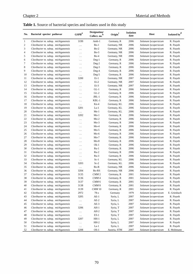
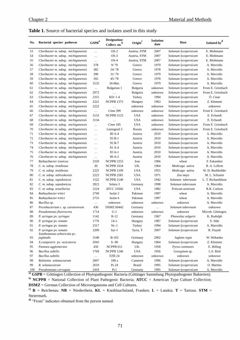
media, 72 bacterial strains were tested. These included 30 Cmm strains that originated from
different countries and were in part self-isolated from different locations in Germany and
Syria or obtained from other bacterial collections (Table 1).
In addition, 42 other pathogenic and non-pathogenic bacterial species or strains were tested.
Pathogenic bacterial species related to Cmm included C. m. subsp. insidiosus, nebraskensis,
sepedonicus, and tessellarius, as well as Curtobacterium flaccumfaciens pv. flaccumfaciens.
As reference, 3 well identified strains of saprophytic or epiphytic bacterial species [Bacillus
subtilis, Pantoea agglomerans (Erwinia herbicola) and Pseudomonas fluorescens] and five
phytopathogenic bacterial species which may occur on tomato plants [Pectobacterium
carotovorum subsp. carotovorum (Erwinia carotovora subsp. carotovora), Pseudomonas
syringae pv. syringae, Pseudomonas syringae pv. tomato, Xanthomonas arboricola pv.
julandis and Xanthomonas campestris pv. vesicatoria] were included. In addition, several
accompanying saprophytic bacterial isolates (S-1 to S-23, listed in Tables 1 and 3) were
obtained from tomato seeds and plants of different origin. Most of these saprophytes were
antagonists of Cmm and were taxonomically identified by gas chromatographic analysis of
their whole cell fatty acid methyl esters (FAME)* as shown in Table 3. The FAME-profile
was achieved by the Hewlett-Packard HP5898A Microbial Identification System (MIS) using
versions 3.80 and 4.01 of the Aerobic Library (TSBA 40) (MIDI Inc., Newark, DE, USA) and
according to the procedure as specified by the manufacturer (Hewlett-Packard, Avondale, PA,
USA).
Organisms not identifiable by FAME analysis were coarsely characterized on the basis of
biochemical or colony morphology features, Gram reaction, and reaction to antibiotics. The
species of these saprophytic bacteria included: Bacillus cereus, B. coagulans, B. licheniformis,
B. pumilus, Microbacterium lacticum, Microbacterium sp., Pantoea agglomerans, Pantoea
sp., Pseudomonas putida, Pseudomonas syringae and Rahnella aquatilis. Even different
isolates of the same species differed in susceptibility to antibiotics.
*) FAME, Fatty Acid Methyl Esters analysis were kindly carried out by Dr. Dieter Felgentreu, Institute for
Ecological Chemistry, Plant Analysis and Stored Product Protection, Julius-Kühn-Institute, Berlin, Germany.
Page 26
Chapter 1 Material and Methods
20
Antibiotic-resistant mutant of Cmm
In order to determine the detection sensitivity of different variants of selective media, it was
necessary to know the exact number of the target Cmm cells existing in plant homogenates.
Therefore, a spontaneous antibiotic-resistant Cmm mutant was selected. This was achieved by
culturing a selected Cmm strain in several passages in NGY liquid medium (see NGY medium
below) containing increasing concentrations of antibiotics. The Cmm strain BO-RS (Table 1)
with resistance to 100 ppm rifampicin and 600 ppm streptomycin was obtained in this way.
Media and growth conditions
All Cmm strains and other bacteria used in this study were cultivated on NGY agar medium
[0.8% nutrient broth (Roth, Karlsruhe, Germany), 1% glucose (AppliChem, Darmstadt,
Germany), 0.3% yeast extract (Roth), pH was adjusted to 7.2; according to Mavridis, person.
commun.].
Only the Pseudomonas spp. were cultivated on NGY or on King’s medium B (King et al.,
1954).
For isolation of the mutant strain BO-RS from seeds and plant samples as well as for
determining its population in infected samples we used the NGY agar medium, supplemented
with 50 ppm rifampicin (25 mg/ml MeOH stock), 200 ppm streptomycin (100 mg/ml water,
stock) and 50 µl/l Opus® Top (50 µl/ml water stock). Bacterial cultures were incubated at
26 °C.
Long-time conservation of bacteria was achieved in 20% glycerol at -80 °C.
Page 27
Chapter 1 Material and Methods
21
Table 1. Origin of bacterial species and strains used to evaluate semiselective media
Bacterial species GSPB no.a
Designation or no.
in other collectionsb
Origin
c Year of
isolation Isolated by
d
Clavibacter m. subsp. michiganensis 3199 Amb-1 Germany, R 2006 R. Ftayeh
Clavibacter m. subsp. michiganensis 3200 Ei-1 Germany, NR 2007 R. Ftayeh
Clavibacter m. subsp. michiganensis … Ei-2 Germany, NR 2007 R. Ftayeh
Clavibacter m. subsp. michiganensis 3201 Lu-1 Germany, KL 2006 R. Ftayeh
Clavibacter m. subsp. michiganensis 3202 Mo-1 Germany, R 2006 R. Ftayeh
Clavibacter m. subsp. michiganensis … Mo-2 Germany, R 2006 R. Ftayeh
Clavibacter m. subsp. michiganensis 3203 Sc-2 Germany, KL 2006 R. Ftayeh
Clavibacter m. subsp. michiganensis 3204 BO-RS Germany, NR 2006 R. Ftayeh
Clavibacter m. subsp. michiganensis 2972 78-s Germany 1979 E. Griesbach
Clavibacter m. subsp. michiganensis 3205 AE-1 Syria, L 2007 R. Ftayeh
Clavibacter m. subsp. michiganensis 3206 AH-1 Syria, T 2007 R. Ftayeh
Clavibacter m. subsp. michiganensis … ES-1 Syria, T 2007 R. Ftayeh
Clavibacter m. subsp. michiganensis 3207 HH-1 Syria, L 2007 R. Ftayeh
Clavibacter m. subsp. michiganensis … La-1 Syria, L 2007 R. Ftayeh
Clavibacter m. subsp. michiganensis 3208 OS-1 Austria, STM 2007 E. Moltmann
Clavibacter m. subsp. michiganensis … OS-2 Austria, STM 2007 E. Moltmann
Clavibacter m. subsp. michiganensis … OS-4 Austria, STM 2007 E. Moltmann
Clavibacter m. subsp. michiganensis 378 9/79 Greece 1979 A. Mavridis
Clavibacter m. subsp. michiganensis 382 24/78 Greece 1978 A. Mavridis
Clavibacter m. subsp. michiganensis 390 31/79 Greece 1979 A. Mavridis
Clavibacter m. subsp. michiganensis 392 45/78 Greece 1978 A. Mavridis
Clavibacter m. subsp. michiganensis … Bulgarian 1 Bulgaria unknown From Griesbach
Clavibacter m. subsp. michiganensis 2973 Cm8 Bulgaria unknown From Griesbach
Clavibacter m. subsp. michiganensis 2315 KD/1-4 Turkey 1994 Ö. Cinar
Clavibacter m. subsp. michiganensis 2221 NCPPB 1573 Hungary 1963 Z. Klement
Clavibacter m. subsp. michiganensis 2222 NCPPB Hungary unknown unknown
Clavibacter m. subsp. michiganensis … 399 Unknown unknown From Griesbach
Clavibacter m. subsp. michiganensis 3133 NCPPB 3123 USA unknown E. Echandi
Clavibacter m. subsp. michiganensis … 185 USA unknown From Griesbach
Clavibacter m. subsp. michiganensis … Leningrad 3 Russia unknown From Griesbach
C. m.subsp. insidiosus 30 NCPPB 1634 UK 1934 From Lelliott
C. m. subsp. nebraskensis 2223 NCPPB 2581 USA 1971 M. L. Schuster
C. m. subsp. sepedonicus 1522 NCPPB 2140, Cs 1 USA 1942 L. T. Richardson
C. m. subsp. sepedonicus 2823 Solara 3 Germany 1998 A. Mavridis
C. m. subsp. tessellarius 2224 ATCC 33566 USA 1982 R.R. Carlson
Curtobacterium f. pv. flaccumfaciens 2218 NCPPB 559 USA 1958 From Lelliott
Bacillus subtilis 1769 NCPPB 1246 USA 1956 L.S. Bird
Bacillus subtilis … FZB 24 Germany unknown unknown
Pectobacterium c. subsp. carotovorum 436 DSMZ 60442 Germany unknown unknown
Pantoea agglomerans 450 NCPPB 651 UK 1985 E. Billing
Pseudomonas corrugata 2418 PC 1 Germany 1995 A. Mavridis
P. fluorescens 1714 G-1 Germany unknown unknown
P. syringae pv.syringae 1142 R - 12 Germany 1967 K. Rudolph
P. syringae pv. tomato 1776 14-1 Hungary 1987 S. Süle
P. syringae pv. tomato 2317 Nr.-1 Turkey 1994 A. Mavridis
P. syringae pv. tomato … Syr-1 Syria 2007 R. Ftayeh
Ralstonia solanacearum 2607 180 a Cameroon 1996 A. Mavridis
Ralstonia solanacearum 2619 Ps 24 Brazil 1995 O. Martins
Xanthomonas arboricola pv. juglandis 3148 B- 102 Germany 2002 W. Wohanka
X. campestris pv. vesicatoria 2043 S-08 Hungary 1964 Z. Klement
22 saprophytic bacteriae … S-1, S-2, ….S-23 Germ. R, NR, KL 2006- 2007 R. Ftayeh
a) GSPB = Göttingen Collection (Sammlung) of Phytopathogenic Bacteria. b) NCPPB = National Collection of Plant Pathogenic Bacteria, UK; ATCC = American Type Culture Collection; DSMZ =
German Collection of Microorganisms and Cell Cultures. c) R = Reichenau; NR = Niederrhein; KL = Knoblauchsland, Franken; L = Latakia; T = Tartous; STM = Steiermark. d) “From” indicates obtained from the person named. e) Saprophytes were isolated from tomato seed and tomato plants and differing in colour, morphology, Gram’s reaction, or
susceptibility to antibiotics, partially identified by fatty acid analysis as shown in Table 3
Page 28
Chapter 1 Material and Methods
22
Selection of the basic medium for Cmm
For selecting a basic medium with high potential plating efficiency of Cmm, compositions of
nine semiselective media were prepared without addition of antibiotics, and the growth of
Cmm was compared with growth on NGY medium. The original nine semiselective media
were: D2 (Kado and Heskett, 1970); KBT (Dhanvantari, 1987); mCNS which was prepared as
suggested by Gitaitis et al. 1991, based on CNS (Gross and Vidaver, 1979) and modified by
omission of lithium chloride and Bravo 6F; D2ANX (Chun, 1982); SCM (Fatmi and Schaad,
1988); mSCM (Waters and Bolkan, 1992); CMM1 (Alvarez and Kaneshiro, 1999); the
recently suggested medium for Cmm by the European Plant Protection Organization
(OEPP/EPPO, 2005), named “EPPO” in our study; and MTNA (Jansing and Rudolph, 1998)
which was developed for Clavibacter michiganensis subsp. sepedonicus. For evaluating the
growth speed of Cmm on these media, bacterial suspensions were prepared in 0.01M MgSO4,
adjusted photometrically to ~108 cfu/ml (OD of 0.06 at 660 nm), and followed by serial
dilution to 250-750 cfu/ml. Finally, 100 µl of each strain were surface streaked with an “L”
shaped glass rod in triplicates per strain onto each of the above described basic media. Growth
areas of Cmm strains were determined in mm2 as average of three replicates on each medium
at the 3rd
and 5th
day after plating.
Growth area = cfu no. x π r2 (Figure 1).
Screening of antibiotics
Forty different antibiotics (Table 2) were initially screened for their inhibitory effect on two
Cmm strains (GSPB 390 and 2973). The screening test was performed according to the
technique of Bauer et al. (1966) by means of commercially available filter discs containing
different concentrations of antibiotics (Oxoid Ltd, England). Bacterial suspensions of the
Cmm strains tested were prepared from 24-hour-old NGY cultures in 0.01M MgSO4. Bacterial
concentrations were photometrically adjusted to approximately 108
cfu/ml using a photometer
(Spectronic 20, Bausch & Lomb), i. e. an optical density of 0.06 at 660 nm, and 150 µl of this
bacterial suspension were streaked onto the surface of NGY medium with a Drigalski spatula.
Within 10 to 20 min discs containing an antibiotic were placed on the agar with sterile forceps
and gently pressed to ensure contact. The plates were kept for two hours at 4 °C to allow
diffusion of antibiotics into the agar before incubating at 26 °C. After incubating at 26 °C for
24-48 h, inhibition’s width around the discs was recorded in mm (Table 2).
Page 29
Chapter 1 Material and Methods
23
Susceptibility of accompanying bacteria towards antibiotics
Antibiotics with no inhibitory effect on Cmm (Table 2) were further tested in several
concentrations in NGY medium on their inhibitory effect against different accompanying
bacteria. Susceptibility testing of accompanying bacteria was carried out to select antibiotics
with potential selectivity. The Cmm strain GSPB 390 was also tested besides the
accompanying bacteria, for determining the maximum concentration of each antibiotic which
caused a strong inhibition of accompanying bacteria while maintaining good growth of Cmm.
Highly concentrated suspensions of Cmm (GSPB 390) and accompanying bacterial species
were prepared and streaked on NGY media with different concentrations of antibiotics, by
dipping a sterile inoculating loop into each bacterial suspension and streaking on NGY media
containing different concentrations of the following antibiotics: aztreaonam, metronidazole,
mupirocin, nalidixic acid, polymyxin B sulfate, trimethoprim and fosfomycin. Agar plates
were incubated at 25 °C for 24-48 h until evaluation (Table 3).
Adjusting the optimum concentrations of inhibitors
Antibiotics inhibiting a wide spectrum of accompanying bacteria, such as trimethoprim,
polymyxin B sulfate and nalidixic acid, were furthermore tested in various combinations and
concentrations with the new basic medium to adjust the optimum concentration of each
antibiotic exerting high selectivity, while maintaining a good growth speed of two Cmm
strains (GSPB 390 and 2073). For this purpose, field tomato seeds and plants that had been
previously inoculated with the double mutant Cmm strain BO-RS (see above) and highly
contaminated with saprophytes were homogenized in sterile water. Aliquots of the
homogenates were streaked on the test plates. For comparison, the homogenates were also
plated on NGY medium supplemented with rifampicin, streptomycin and Opus® Top to
determine the actual number of Cmm cells occurring in the plant homogenates.
Furthermore, homogenates from healthy field plants (collected from different locations in
Germany and Syria) were surface streaked in triplicates onto NGY agar and test compositions
in order to estimate selectivity. Parallely, suspensions of two Cmm strains (GSPB 390 &
GSPB 2973) differing in growth morphology and speed were also streaked, each in triplicates,
onto agar plates with NGY or test compositions to estimate the growth area of Cmm. Only
those compositions which allowed high selectivity concomitantly with large growth areas of
Cmm were selected and modified repeatedly in further experiments.
Finally, the best compositions allowing high selectivity were tested with 30 Cmm strains (see
Page 30
Chapter 1 Material and Methods
24
below).
Determining the plating efficiency (recovery rate) of Cmm strains on semiselective media
Cultures of 30 Cmm strains were grown for 24 h on NGY medium, and bacterial suspensions
in 0.01M MgSO4 containing 100-250 cfu were plated in triplicates on each medium for each
strain. The recovery of Cmm was determined by counting the Cmm colonies of each variant.
To avoid mistakes caused by the possible co-growth of several joining colonies, counting of
colonies was started as soon as possible on each medium (for example on NGY after 48-72 h).
Plating efficiency or recovery rate (Table 4) after 7, 10, 15 and 20 days was expressed in %
recovered CFU of those detected on the NGY medium, i.e.:
Plating efficiency of Cmm (%) = (CFU on test medium/CFU on NGY medium) × 100.
Evaluation of selectivity and detection sensitivity of semiselective media
Selectivity means the suitability of selective media for supporting growth of target micro
organisms or bacteria and preventing growth of nontarget microbes or bacteria.
Detection sensitivity means the lowest number of Cmm CFU occurring in plant homogenates
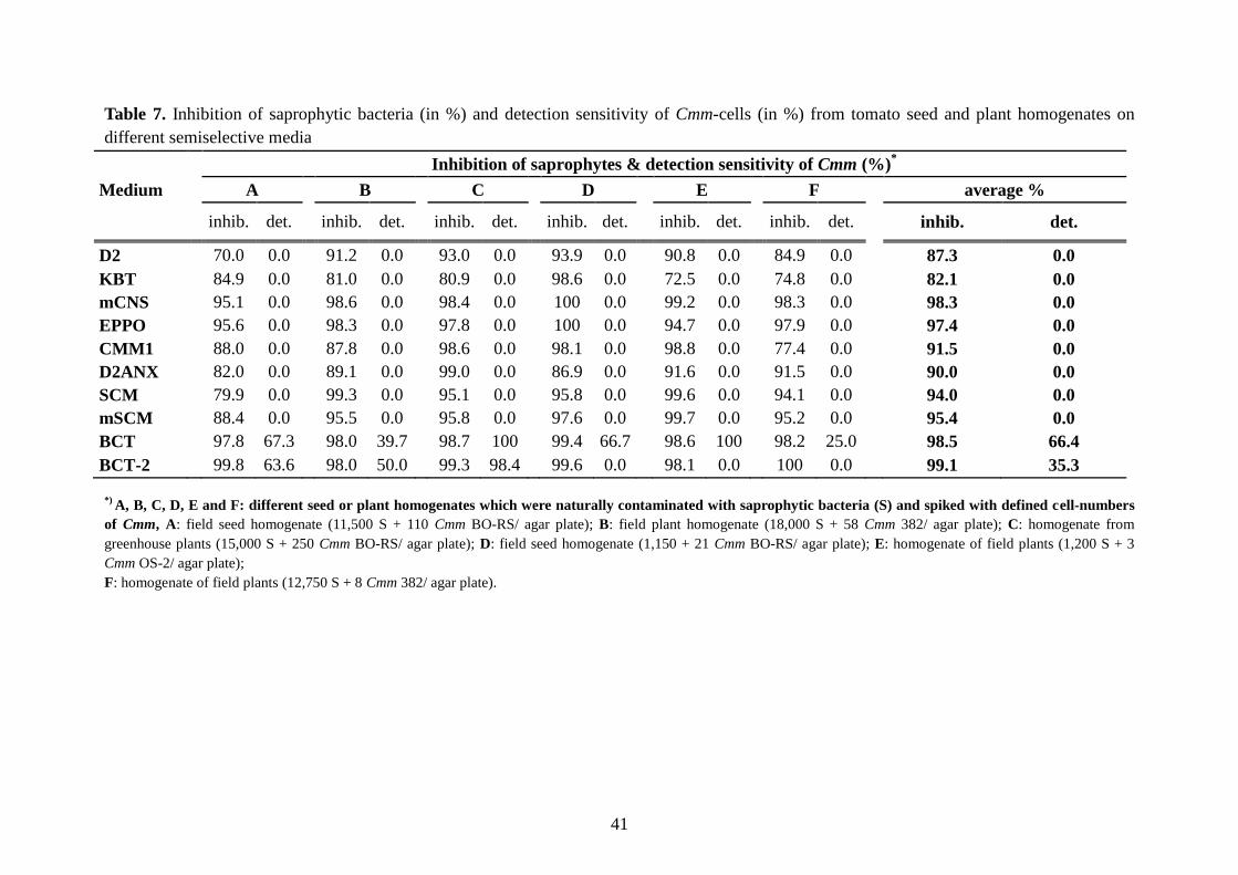
which could be detected in the presence of high concentrations of nontarget bacteria (Table 7
& Figure 8).
Some media, such as mSCM, EPPO and mCNS, showed a rather higher toxicity than
selectivity towards several Cmm strains, resulting in low detection sensitivity. Other media,
such as D2, KBT, SCM, and CMM1, showed less selectivity and detection sensitivity as well,
becuase Cmm growth was inhibited by saprophytic bacteria that rapidly occupied the agar
background.
Thus, it was very important in our study to evaluate both the selectivity and detection
sensitivity of the new media.
For initial evaluation of new medium compositions, field tomato plants were inoculated in
2007 and 2008 with very low concentrations of the double mutant Cmm strain BO-RS (30-50
cfu/ plant). After 30-70 days the field plants were only latently infected with Cmm and never
showed disease symptoms. On the other hand, due to rainy weather conditions, the plants
were highly contaminated by epiphytic or saprophytic microorganisms. Homogenates of plant
stems were streaked on the test media, as well as on NGY agar supplemented with rifampicin,
streptomycin and Opus® Top. In this way it was possible to evaluate detection sensitivity and
selectivity of new medium-compositions.
Page 31
Chapter 1 Material and Methods
25
Concomitantly, infected tomato plant samples which were collected in several locations in
Syria, Germany and Austria or which had been sent to our laboratory in Göttingen between
2006 and 2008 were also evaluated by the medium-compositions being under development.
Finally, the selectivity of all tested semiselective media in comparison with the new media
was evaluated using homogenates of healthy field tomato plants or seed lots which were
highly contaminated with saprophytic bacteria and artificially infested (“spiked”) with
different strains of Cmm. The Cmm strains used were BO-RS, 382 and OS-2. Tomato stems or
seeds were crushed in sterile mortars with sterile water, and serial dilutions were plated on
NGY medium to estimate the density of saprophytic bacteria. Then a defined amount of each
one of the above described Cmm strains was introduced separately into only one of the non-
diluted or 1:10 diluted homogenates, and 100 µl aliquots were plated on each medium. Plates
were incubated at 26 °C. As soon as bacteria began to grow, counting the colonies started for
both, saprophytes and Cmm. Bacteria started to grow on each medium after different intervals
(2 to 15 days).
To compare all media under the same conditions, the final colony number of saprophytes and
Cmm was determined 10 dpi. Cmm-suspected colonies were purified and identified by re-
streaking on new NGY agar plates or on rifampicin-, streptomycin-NGY agar, when the
double mutant was applied.
The selectivity and detection sensitivity of each medium was evaluated as follows:
Selectivity (%) = [(Population of nontarget microbes on NGY - population of nontarget
microbes on test medium) / population of nontarget microbes on NGY] × 100.
Detection sensitivity (%) = The CFU number of target bacteria (Cmm) detected from plant
homogenate or seed extract × 100 / the total CFU number of target bacteria (Cmm) in the
plant homogenate or seed extract.
Page 32
Chapter 1 Results
26
Results
Selecting a new basic medium for Cmm
Three Cmm strains (GSPB 390, GSPB 2973 and Ei-2) with different growth morphology and
growth speed were cultivated on the basic compositions of nine different semiselective media
(without addition of antibiotics). After three and five days, all tested basic media showed
significant differences in growth of Cmm. Compared with NGY agar, the growth of the three
Cmm strains tested was very low or absolutely absent after three and five days on the basic
media of D2, CMM1, SCM, mSCM and EPPO. In comparison to the reference NGY medium
and to all the other tested basic media, the growth of the three Cmm strains was highest on the
basic medium of MTNA after three and five days. On MTNA Cmm colonies appeared earlier
and were larger in diameter (Figure 1). Therefore, the basic MTNA medium which had been
developed for Clavibacter michiganensis subsp. sepedonicus (Jansing and Rudolph, 1998)
was selected and adapted to Cmm by modifying the basic compounds and inhibitors.
Figure 1. Growth areas in mm2 of 3 Cmm strains (as the mean of three replicates for each
strain) on NGY and on different semiselective media (without addition of antibiotics) at the
3rd
and 5th
day after plating. Growth area = number of CFU × π r2 (r: average radius of
colonies in mm).
Page 33
Chapter 1 Results
27
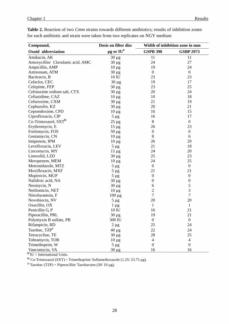
Screening of different antibiotics
Two Cmm strains with different growth speed and growth morphology on NGY medium were
selected for screening 40 different antibiotics. The inhibitory effect of each antibiotic did not
differ strongly against the two Cmm strains tested, but differed between antibiotics. Only co-
trimoxazol which is a combination of trimethoprim and sulfamethoxazole inhibited one Cmm
strain (GSPB 390) but did not inhibit the other strain (GSPB 2973). All the data are shown in
Table 2.
Page 34
Chapter 1 Results
28
Table 2. Reaction of two Cmm strains towards different antibiotics; results of inhibition zones
for each antibiotic and strain were taken from two replicates on NGY medium
Compound, Dosis on filter disc Width of inhibition zone in mm
Oxoid abbreviation µg or IUa
GSPB 390 GSBP 2973
Amikacin, AK 30 µg 11 11
Amoxycillin/ Clavulanic acid, AMC 30 µg 24 27
Ampicillin, AMP 10 µg 19 24
Aztreonam, ATM 30 µg 0 0
Bacitracin, B 10 IU 23 23
Cefaclor, CEC 30 µg 19 17
Cefepime, FEP 30 µg 23 25
Cefotaxime sodium salt, CTX 30 µg 20 24
Ceftazidime, CAZ 10 µg 10 18
Cefuroxime, CXM 30 µg 21 19
Cephazolin, KZ 30 µg 20 21
Cepomdoxime, CPD 10 µg 16 15
Ciprofloxacin, CIP 5 µg 16 17
Co-Trimoxazol, SXTb
25 µg 8 0
Erythromycin, E 15 µg 26 23
Fosfomycin, FOS 50 µg 0 0
Gentamycin, CN 10 µg 8 6
Imipenem, IPM 10 µg 26 20
Levofloxacin, LEV 5 µg 21 18
Lincomycin, MY 15 µg 24 20
Linezolid, LZD 30 µg 25 23
Meropenem, MEM 10 µg 24 25
Metronidazole, MTZ 5 µg 0 0
Moxifloxacin, MXF 5 µg 21 21
Mupirocin, MUP 5 µg 0 0
Nalidixic acid, NA 30 µg 0 0
Neomycin, N 30 µg 6 5
Netilomicin, NET 10 µg 2 3
Nitrofurantoin, F 100 µg 7 7
Novobiocin, NV 5 µg 20 20
Oxacillin, OX 1 µg 1 1
Penicillin G, P 10 IU 16 21
Piperacillin, PRL 30 µg 19 21
Polymyxin B sulfate, PB 300 IU 0 0
Rifampicin, RD 2 µg 25 24
Tazobac, TZPc
40 µg 22 24
Tetracycline, TE 30 µg 28 25
Tobramycin, TOB 10 µg 4 4
Trimethoprim, W 5 µg 0 0
Vancomycin, VA 30 µg 16 16 a)
IU = International Units. b)
Co-Trimoxazol (SXT) = Trimethoprim/ Sulfamethoxazole (1.25/ 23.75 µg).
c) Tazobac (TZP) = Piperacillin/ Tazobactam (30/ 10 µg).
Page 35
Chapter 1 Results
29
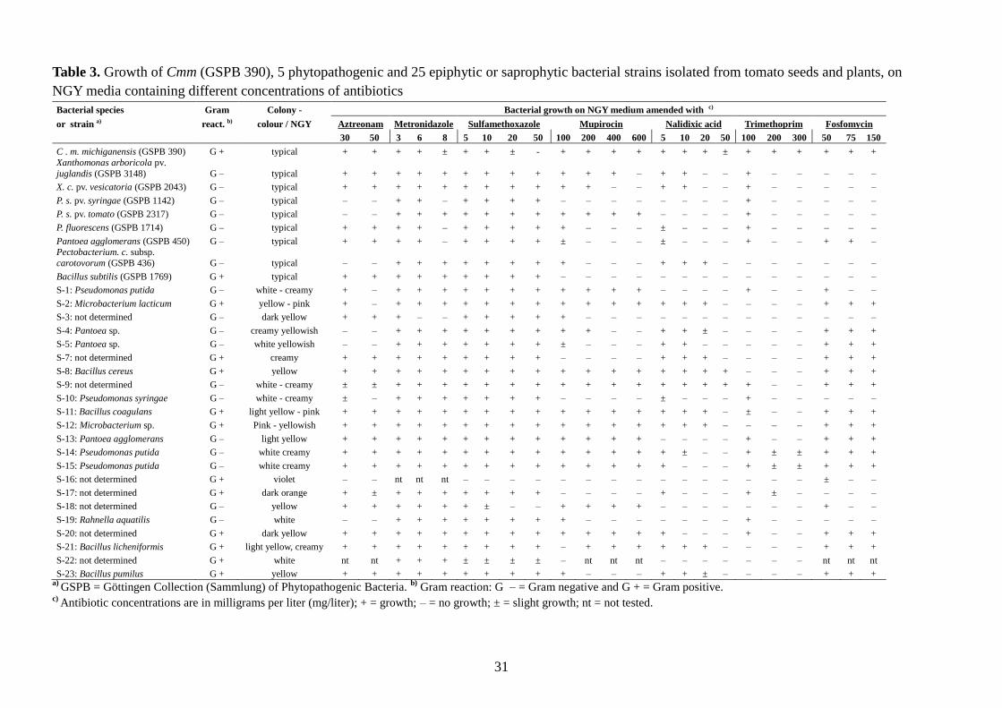
Susceptibility of accompanying bacterial species and strains towards antibiotics
Antibiotics which did not inhibit Cmm, such as aztreonam, fosfomycin, metronidazole,
mupirocin, nalidixic acid, polymyxin B sulfate, sulfamethoxazole and trimethoprim were
tested against accompanying bacteria in order to select the effective ones with a wide
inhibiting spectrum. As shown on Table 3, nalidixic acid (10 and 20 mg/l) and trimethoprim
(100-300mg/l) showed the strongest inhibition spectrum against the accompanying bacterial
species tested, without inhibiting the growth of Cmm. A combination of 20 ppm nalidixic acid
and 100 ppm trimethoprim seemed to inhibit all accompanying bacteria tested. Furthermore,
polymyxin B sulfate was also tested separately and showed a broad inhibitory spectrum of
accompanying bacteria (data not shown).
Therefore, nalidixic acid, trimethoprim and polymyxin B sulfate and the fungicide Opus® Top
were tested furthermore in different compositions in the NGY medium and in different
modifications of the selected basic medium of MTNA. Each composition was tested with two
Cmm strains (GSPB 390 and 2973) for determining the growth speed of Cmm. Concomitantly,
homogenates from naturally or artificially infected field tomato plants and seeds which were
highly contaminated with diverse epiphytic microorganisms, were tested with these
components in order to determine selectivity.
Compositions with low selectivity or low growth speed were excluded. Other compositions
with high growth speed of Cmm and simultaneously high selectivity were further modified. In
this way, every 10-15 days more than 15-20 different compositions were prepared and tested
for growth speed and selectivity. After each experimental block the variants showing the
highest potential for Cmm growth speed combined with a good selectivity were selected and
modified again and again. In this way, the new selective media BCT and BCT-2 were finally
developed.
Page 36
Chapter 1 Results
30
Recipes of the new selective media BCT and BCT-2
Recipe of BCT for one liter: 2.5 g mannitol (Merck); 2.0 g yeast extract (Roth); 1.0 g K2HPO4
(AppliChem); 0.1 g KH2PO4 (Merck); 0.05 g NaCl (Merck); 0.1 g MgSO4 × 7H2O (Merck);
0.015 g MnSO4 × H2O (AppliChem); 0.015 g FeSO4 × 7H2O (Merck); 0.6 g H3BO3
(AppliChem), dissolved in 1 liter deionized H2O. The resulting pH value should be between
7.0 and 7.1. Add 15 g/l agar agar (Roth).
After autoclaving at 121 °C for 15 min, cooling down to 50 °C and under stirring add the
following: 20 mg/l nalidixic acid (AppliChem); 100 mg/l trimethoprim (Fluka); 20 mg/l
polymyxin B sulfate (8,120 international units per milligram, from AppliChem); and 50 µl/l
Opus® Top (commercially available from BSAF and containing 84.0 g/l expoxiconazol and
250.0 g/l fenpropimorph).
Antibiotics and Opus® Top must be added as stock solutions, freshly prepared and kept in
sterile glasses at 4 °C. Stock solution of nalidixic acid (20 mg/ml 0.1N NaOH, filter-
sterilized); trimethoprim (50 mg/ml Dimethyl sulfoxide, must be kept away from light);
poymyxin B sulfate (10 mg/ml water, filter-sterilized); Opus® Top (50 µl/ml sterile water).
Recipe of BCT-2 medium is similar to BCT: instead of 1.0 g K2HPO4 add 2.0 g/l to BCT-2
and instead of 0.1 g KH2PO4 add 0.5 g/l to BCT-2. The resulting pH value of BCT-2 should be
between 7.15 and 7.2.
In most cases, both new selective media BCT & BCT-2 were filled into Petri dishes and stored
for three days at room temperature. In some cases, when both media were used directly after
preparation, we noticed some growth inhibition of several Cmm strains.
Therefore, we recommend using the new selective media BCT & BCT-2 at least three days
after preparation.
Page 37
31
Table 3. Growth of Cmm (GSPB 390), 5 phytopathogenic and 25 epiphytic or saprophytic bacterial strains isolated from tomato seeds and plants, on
NGY media containing different concentrations of antibiotics
Bacterial species Gram Colony - Bacterial growth on NGY medium amended with c)
or strain a) react. b) colour / NGY Aztreonam Metronidazole Sulfamethoxazole Mupirocin Nalidixic acid Trimethoprim Fosfomycin
30 50 3 6 8 5 10 20 50 100 200 400 600 5 10 20 50 100 200 300 50 75 150
C . m. michiganensis (GSPB 390) G + typical + + + + ± + + ± - + + + + + + + ± + + + + + +
Xanthomonas arboricola pv.
juglandis (GSPB 3148) G – typical + + + + + + + + + + + + – + + – – + – – – – –
X. c. pv. vesicatoria (GSPB 2043) G – typical + + + + + + + + + + + – – + + – – + – – – – –
P. s. pv. syringae (GSPB 1142) G – typical – – + + – + + + + – – – – – – – – + – – – – –
P. s. pv. tomato (GSPB 2317) G – typical – – + + + + + + + + + + + – – – – + – – – – –
P. fluorescens (GSPB 1714) G – typical + + + + – + + + + + – – – ± – – – + – – – – –
Pantoea agglomerans (GSPB 450) G – typical + + + + – + + + + ± – – – ± – – – + – – + + – Pectobacterium. c. subsp.
carotovorum (GSPB 436) G – typical – – + + + + + + + + – – – + + + – – – – – – –
Bacillus subtilis (GSPB 1769) G + typical + + + + + + + + + – – – – – – – – – – – – – –
S-1: Pseudomonas putida G – white - creamy + – + + + + + + + + + + + – – – – + – – + – –
S-2: Microbacterium lacticum G + yellow - pink + – + + + + + + + + + + + + + + – – – – + + +
S-3: not determined G – dark yellow + + + – – + + + + + – – – – – – – – – – – – –
S-4: Pantoea sp. G – creamy yellowish – – + + + + + + + + + – – + + ± – – – – + + +
S-5: Pantoea sp. G – white yellowish – – + + + + + + + ± – – – + + – – – – – + + +
S-7: not determined G + creamy + + + + + + + + + – – – – + + + – – – – + + +
S-8: Bacillus cereus G + yellow + + + + + + + + + + + + + + + + + – – – + + +
S-9: not determined G – white - creamy ± ± + + + + + + + + + + + + + + + + – – + + +
S-10: Pseudomonas syringae G – white - creamy ± – + + + + + + + – – – – ± – – – + – – – – –
S-11: Bacillus coagulans G + light yellow - pink + + + + + + + + + + + + + + + + – ± – – + + +
S-12: Microbacterium sp. G + Pink - yellowish + + + + + + + + + + + + + + + + – – – – + + +
S-13: Pantoea agglomerans G – light yellow + + + + + + + + + + + + + – – – – + – – + + +
S-14: Pseudomonas putida G – white creamy + + + + + + + + + + + + + + ± – – + ± ± + + +
S-15: Pseudomonas putida G – white creamy + + + + + + + + + + + + + + – – – + ± ± + + +
S-16: not determined G + violet – – nt nt nt – – – – – – – – – – – – – – – ± – –
S-17: not determined G + dark orange + ± + + + + + + + – – – – + – – – + ± – – – –
S-18: not determined G – yellow + + + + + + ± – – + + + + – – – – – – – + – –
S-19: Rahnella aquatilis G – white – – + + + + + + + + – – – – – – – + – – – – –
S-20: not determined G + dark yellow + + + + + + + + + + + + + + – – – + – – + + +
S-21: Bacillus licheniformis G + light yellow, creamy + + + + + + + + + – + + + + + + – – – – + + +
S-22: not determined G + white nt nt + + + ± ± ± ± – nt nt nt – – – – – – – nt nt nt
S-23: Bacillus pumilus G + yellow + + + + + + + + + + – – – + + ± – – – – + + + a)
GSPB = Göttingen Collection (Sammlung) of Phytopathogenic Bacteria. b)
Gram reaction: G – = Gram negative and G + = Gram positive. c)
Antibiotic concentrations are in milligrams per liter (mg/liter); + = growth; – = no growth; ± = slight growth; nt = not tested.
Page 38
Chapter 1 Results
32
Effect of boric acid
Boric acid has also been used in other semiselective media because of its antimicrobial effect
against saprophytic bacteria. We tested different concentrations of boric acid in the NGY
medium (Figure 2) as well as in the new basic medium (600, 900, 1200, 1500 and 2000 ppm).
By increasing amounts of boric acid in agar media, inhibition of saprophytes was stronger, but
the growth of Cmm was retarded. This means that appearance of Cmm-colonies was delayed
and colony-diameters were smaller resulting in less growth areas. The optimal concentration
of boric acid was determined as 0.6 g/l causing high inhibition of accompanying bacteria by
allowing good growth of Cmm (Figure 3).
Figure 2. Effect of boric acid on growth areas of three Cmm strains (GSPB 390, GSPB 2973
and BO-RS = GSPB 3204) in the NGY medium.
However, when antibiotics were added to the medium the effect of boric acid was contrary.
Thus, without addition of boric acid, the growth of Cmm on the new basic medium containing
different compositions of antibiotics was never satisfactory, and the recovered colony forming
units of Cmm were very low compared with NGY. When very low concentrations of
Page 39
Chapter 1 Results
33
antibiotics were added, the selectivity of the medium was nearly lost completely. However, by
addition of boric acid to some compositions, the recovery rate of Cmm was surprisingly high,
even when increased amounts of antibiotics were added (Figure 4). Thus, by adding of boric
acid together with high amounts of antibiotics a high selectivity of the medium for Cmm could
be achieved.
For understanding the possible interactions between boric acid and different antibiotics and
inhibitors we tested the basic medium with each inhibitor separately, with or without boric
acid. Addition of 0.6 g/liter boric acid to compositions of the basic medium with either Opus®
Top (100 µl/liter), or nalidixic acid (30 mg/liter), or trimethoprim (200 mg/liter), caused a
slight reduction in recovery rate of Cmm compared with the same compositions without boric
acid. Contrary results were obtained in case of polymyxin B sulfate. When the basic medium
contained 30 mg/l polymyxin B sulfate and 0.6 g/l boric acid, the recovery of Cmm was
normal and very high compared with the same composition without boric acid (Figure 5). The
toxicity of polymyxin B sulfate to Cmm appeared to be reduced significantly when boric acid
was added. Obviously, the reason for the very low recovery rate of Cmm without boric acid
was due to the toxic acting of polymyxin B sulfate in the basic medium.
Figure 3. Effect of boric acid (BA) on the growth of three Cmm strains on the new basic
medium BCT (without antibiotics), growth area of each strain represents the mean of three
agar plates after three and five days.
Page 40
Chapter 1 Results
34
Figure 4. Mean number of CFU per agar plate recovered from pure cultures of 13 Cmm
strains (each in three replicates) on the new medium (BCT) with and without boric acid (600
ppm), when ca. 90 cfu were streaked on each Petri dish.
Page 41
Chapter 1 Results
35
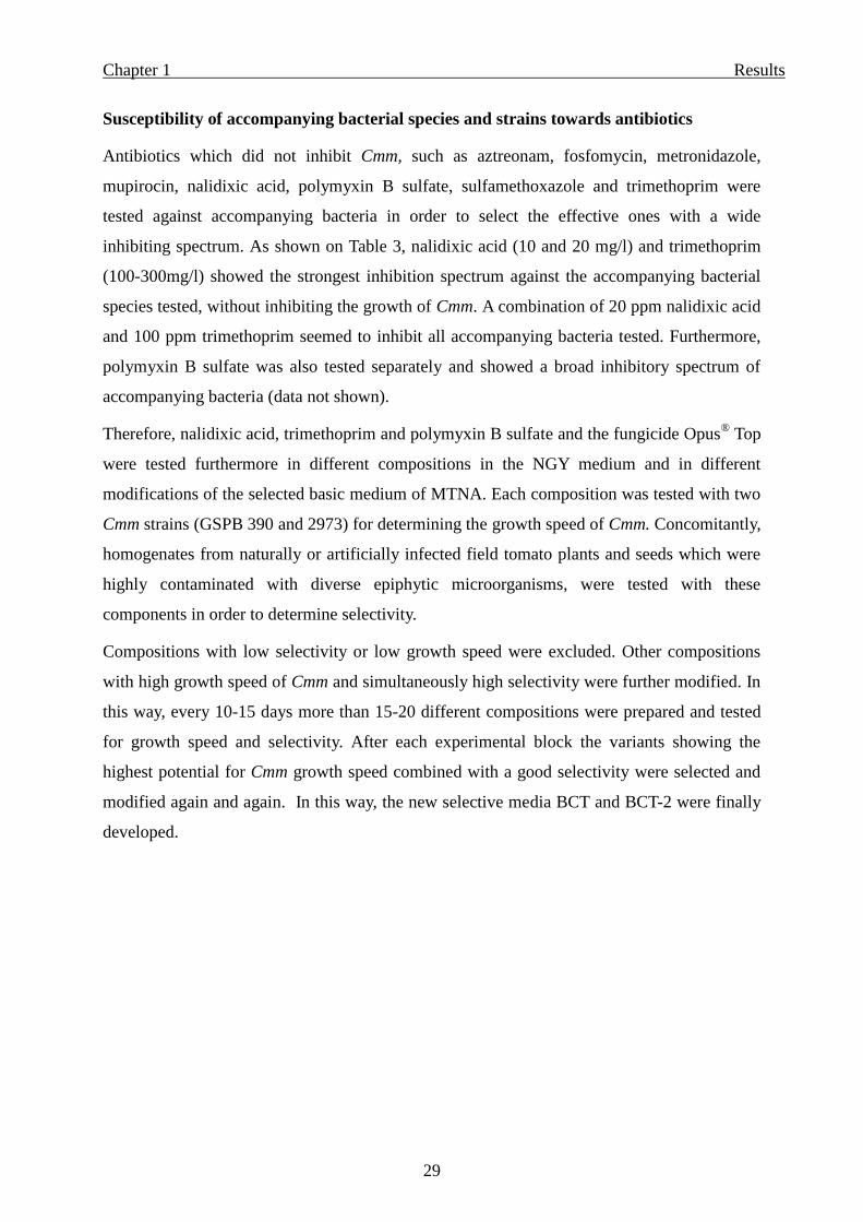
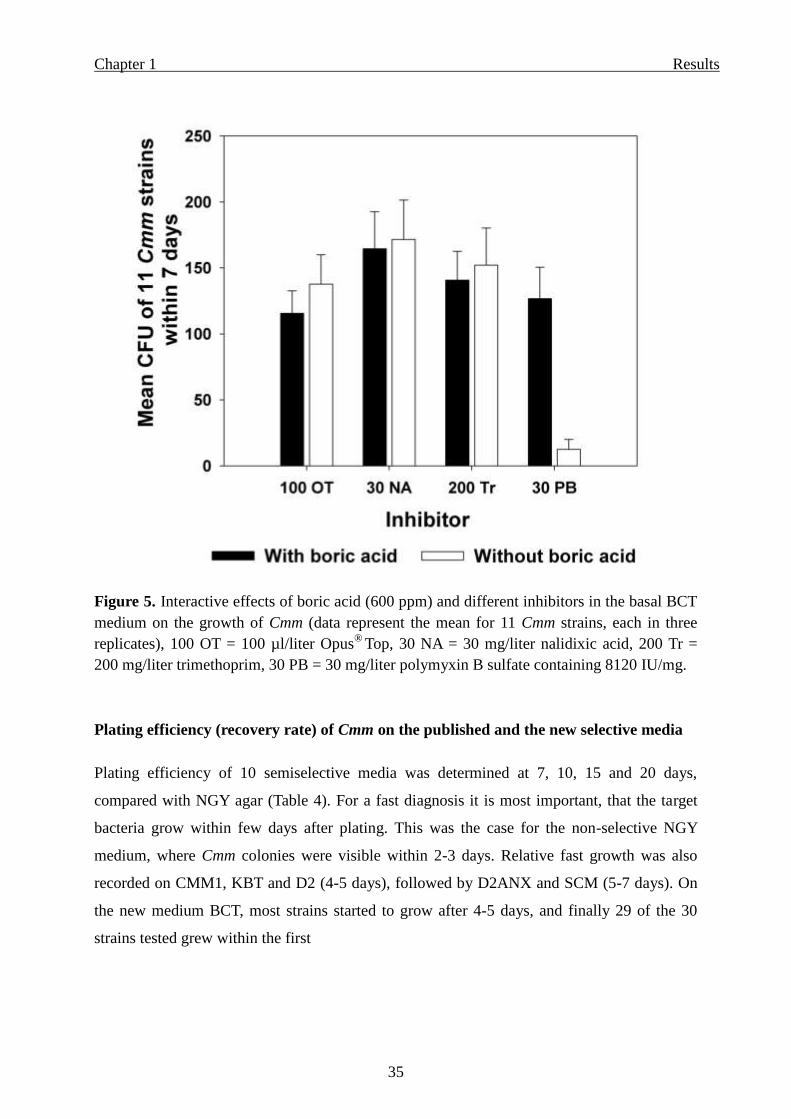
Figure 5. Interactive effects of boric acid (600 ppm) and different inhibitors in the basal BCT
medium on the growth of Cmm (data represent the mean for 11 Cmm strains, each in three
replicates), 100 OT = 100 µl/liter Opus® Top, 30 NA = 30 mg/liter nalidixic acid, 200 Tr =
200 mg/liter trimethoprim, 30 PB = 30 mg/liter polymyxin B sulfate containing 8120 IU/mg.
Plating efficiency (recovery rate) of Cmm on the published and the new selective media
Plating efficiency of 10 semiselective media was determined at 7, 10, 15 and 20 days,
compared with NGY agar (Table 4). For a fast diagnosis it is most important, that the target
bacteria grow within few days after plating. This was the case for the non-selective NGY
medium, where Cmm colonies were visible within 2-3 days. Relative fast growth was also
recorded on CMM1, KBT and D2 (4-5 days), followed by D2ANX and SCM (5-7 days). On
the new medium BCT, most strains started to grow after 4-5 days, and finally 29 of the 30
strains tested grew within the first
Page 42
36
Table 4. Plating efficiency (%) of 30 Cmm strains on different semiselective media compared with the standard NGY medium within
7/ 10/ 15/ 20 days after plating
Strain a)
Plating efficiency d)
(%) within 7/ 10/ 15/ 20 days respectively on
D2 KBT mCNS D2ANX SCM mSCM CMM1 EPPOc)
BCT BCT-2
Amb-1 117/ 117/ 117/ 117 120/ 120/ 120/ 120 0/ 0/ 0/ 0 61/ 61/ 61/ 61 125/ 125/ 125/ 125 0/ 0/ 86/ 113 109/ 109/ 109/ 109 0/ 0/ 0/ 0 67/ 90/ 90/ 90 76/ 76/ 90/ 90
Ei-1 121/ 121/ 121/ 121 123/ 123/ 123/ 123 0/ 0/ 1/ 1 104/ 104/ 104/ 104 119/ 119/ 119/ 119 0/ 0/ 101/ 103 106/ 106/ 106/ 106 0/ 0/ 19/ 43 87/ 109/ 109/ 109 119/ 119/ 119/ 119
Ei-2 110/ 110/ 110/ 110 95/ 95/ 95/ 95 0/ 0/ 0/ 0 13/ 13/ 13/ 13 94/ 94/ 94/ 94 0/ 84/ 84/ 96 89/ 89/ 89/ 89 0/ 0/ 0/ 0 73/ 73/ 73/ 73 98/ 98/ 98/ 98
Lu-1 127/ 127/ 127/ 127 92/ 92/ 92/ 92 0/ 0/ 0/ 0 98/ 98/ 98/ 98 94/ 94/ 94/ 94 0/ 69/ 69/ 98 105/ 105/ 105/ 105 0/ 0/ 0/ 0 102/ 102/ 102/ 102 114/ 114/ 114/ 114
Mo-1 112/ 112/ 112/ 112 89/ 89/ 89/ 89 47/ 47/ 51/ 51 91/ 91/ 91/ 91 105/ 105/ 105/ 105 0/ 0 /113/ 113 93/ 93/ 93/ 93 0/ 0/ 104/ 104 96/ 96/ 96/ 96 97/ 97/ 97/ 97
Mo-2 103/ 103/ 103/ 103 103/ 103/ 103/ 103 0/ 0/ 3/ 6 90/ 90/ 90/ 90 104/ 104/ 104/ 104 0/ 51/ 85/ 102 111/ 111/ 111/ 111 0/ 0/ 0/ 2 95/ 95/ 95/ 95 87/ 87/ 87/ 87
Sc-1 112/ 112/ 112/ 112 106/ 106/ 106/ 106 0/ 0/ 3/ 6 90/ 90/ 90/ 90 107/ 107/ 107/ 107 0/ 35/ 86/ 95 108/ 108/ 108/ 108 0/ 0/ 0/ 0 112/ 112/ 112/ 112 110/ 110/ 110/ 110
BO-RS 91/ 91/ 91/ 91 68/ 68/ 68/ 68 0/ 0/ 0/ 0 36/ 36/ 36/ 36 25/ 35/ 35/ 35 0/ 0/ 0/ 0 67/ 67/ 67/ 67 0/ 0/ 0/ 0 76/ 76/ 76/ 76 71/ 71/ 71/ 71
GSPBb)
2972 84/ 84/ 84/ 84 61/ 61/ 61/ 61 0/ 0/ 0/ 0 107/ 107/ 107/ 107 80/ 80/ 80/ 80 0/ 0/ 0/ 0 97/ 97/ 97/ 97 0/ 0/ 0/ 57 92/ 92/ 92/ 92 81/ 81/ 88/ 88
AE-1 112/ 112/ 112/ 112 87/ 87/ 87/ 87 0/ 0/ 0/ 0 56/ 56/ 56/ 56 102/ 102/ 102/ 102 0/ 10/ 24/ 38 94/ 94/ 94/ 94 0/ 12/ 12/ 21 106/ 106/ 106/ 106 94/ 94/ 94/ 94
AH-1 117/ 117/ 117/ 117 114/ 114/ 114/ 114 0/ 0/ 0/ 0 89/ 89/ 89/ 89 107/ 107/ 107/ 107 0/ 44/ 77/ 89 118/ 118/ 118/ 118 0/ 111/ 111/ 122 100/ 100/ 100/ 100 68/ 68/ 68/ 68
ES-1 98/ 98/ 98/ 98 90/ 90/ 90/ 90 0/ 0/ 0/ 0 95/ 95/ 95/ 95 100/ 100/ 100/ 100 0/ 0/ 99/ 99 90/ 90/ 90/ 90 0/ 0/ 92/ 92 93/ 93/ 93/ 93 79/ 79/ 79/ 79
HH-1 109/ 109/ 109/ 109 99/ 99/ 99/ 99 0/ 0/ 4/ 5 75/ 75/ 75/ 75 104/ 104/ 104/ 104 0/ 76/ 77/ 79 86/ 86/ 86/ 86 0/ 0/ 0/ 0 45/ 45/ 45/ 45 107/ 107/ 107/ 107
La-1 96/ 96/ 96/ 96 64/ 64/ 64/ 64 0/ 0/ 0/ 0 43/ 43/ 43/ 43 89/ 89/ 89/ 89 71/ 79/ 87/ 88 89/ 89/ 89/ 89 0/ 87/ 87/ 91 87/ 87/ 87/ 87 73/ 73/ 73/ 73
OS-1 93/ 93/ 93/ 93 74/ 74/ 74/ 74 0/ 0/ 4/ 4 93/ 93/ 93/ 93 101/ 101/ 101/ 101 0/ 0/ 99/ 99 94/ 94/ 94/ 94 0/ 0/ 79/ 84 96/ 96/ 96/ 96 93/ 93/ 93/ 93
OS-2 91/ 91/ 91/ 91 84/ 84/ 84/ 84 0/ 0/ 0/ 0 81/ 81/ 81/ 81 129/ 129/ 129/ 129 0/ 0/ 103/ 105 91/ 91/ 91/ 91 0/ 0/ 65/ 73 91/ 91/ 91/ 91 93/ 93/ 93/ 93
OS-4 91/ 91/ 91/ 91 87/ 87/ 87/ 87 0/ 0/ 0/ 0 47/ 47/ 47/ 47 120/ 120/ 120/ 120 0/ 89/ 89/ 101 91/ 91/ 91/ 91 0/ 0/ 0/ 10 109/ 109/ 109/ 109 83/ 83/ 83/ 83
GSPB 378 88/ 88/ 88/ 88 88/ 88/ 88/ 88 0/ 0/ 0/ 0 43/ 43/ 43/ 43 93/ 93/ 93/ 93 0/ 76/ 98/ 98 75/ 75/ 75/ 75 0/ 79/ 87/ 88 66/ 66/ 66/ 66 81/ 81/ 81/ 81
GSPB 382 83/ 83/ 83/ 83 55/ 55/ 55/ 55 0/ 0/ 0/ 0 0/ 0/ 0/ 0 65/ 65/ 65/ 65 48/ 67/ 67/ 67 49/ 49/ 49/ 49 0/ 65/ 65/ 66 45/ 45/ 45/ 45 20/ 42/ 46/ 46
GSPB 390 111/ 111/ 111/ 111 102/ 102/ 102/ 102 0/ 0/ 0/ 0 85/ 85/ 85/ 85 88/ 88/ 88/ 88 0/ 76/ 76/ 93 103/ 103/ 103/ 103 0/ 113/ 113/ 116 108/ 108/ 108/ 108 111/ 111/ 111/ 111
GSPB 392 106/ 106/ 106/ 106 91/ 91/ 91/ 91 0/ 0/ 0/ 1 72/ 72/ 72/ 72 92/ 92/ 92/ 92 0/ 0/ 115/ 115 96/ 96/ 96/ 96 0/ 0/ 113/ 113 102/ 102/ 102/ 102 107/ 107/ 107/ 107
Bulgarian 1 97/ 97/ 97/ 97 104/ 104/ 104/ 104 0/ 0/ 0/ 0 79/ 79/ 79/ 79 103/ 103/ 103/ 103 0/ 85/ 102/ 102 87/ 87/ 87/ 87 0/ 0/ 96/ 96 0/ 23/ 58/ 64 0/ 0/ 0/ 0
GSPB 2973 94/ 94/ 94/ 94 99/ 99/ 99/ 99 0/ 7/ 9/ 9 94/ 94/ 94/ 94 82/ 82/ 82/ 82 0/ 0/ 0/ 46 100/ 100/ 100/ 100 0/ 0/ 55/ 73 84/ 84/ 84/ 84 85/ 85/ 85/ 85
GSPB 2315 128/ 128/ 128/ 128 78/ 78/ 78/ 78 0/ 0/ 0/ 4 79/ 79/ 79/ 79 91/ 91/ 91/ 91 0/ 14/ 14/ 101 106/ 106/ 106/ 106 0/ 0/ 0/ 38 102/ 102/ 102/ 102 82/ 82/ 82/ 82
GSPB 2221 103/ 103/ 103/ 103 79/ 79/ 79/ 79 7/ 7/ 13/ 13 86/ 86/ 86/ 86 99/ 99/ 99/ 99 0/ 0/ 95/ 97 98/ 98/ 98/ 98 0/ 0/ 95/ 97 91/ 91/ 91/ 91 90/ 90/ 90/ 90
GSPB 2222 105/ 105/ 105/ 105 61/ 61/ 61/ 61 0/ 0/ 0/ 0 66/ 66/ 66/ 66 90/ 90/ 90/ 90 0/ 0/ 0/ 81 95/ 95/ 95/ 95 0/ 0/ 0/ 22 103/ 103/ 103/ 103 74/ 74/ 74/ 74
399 102/ 102/ 102/ 102 47/ 47/ 47/ 47 0/ 0/ 2/ 7 103/ 103/ 103/ 103 75/ 75/ 75/ 75 0/ 0/ 87/ 87 100/ 100/ 100/ 100 0/ 0/ 99/ 101 97/ 97/ 97/ 97 87/ 87/ 90/ 90
GSPB 3133 103/ 103/ 103/ 103 88/ 88/ 88/ 88 0/ 0/ 0/ 0 23/ 23/ 23/ 23 87/ 87/ 87/ 87 0/ 59/ 59/ 66 92/ 92/ 92/ 92 0/ 0/ 34/ 47 85/ 85/ 85/ 85 94/ 94/ 94/ 94
185 98/ 98/ 98/ 98 81/ 81/ 81/ 81 0/ 0/ 0/ 0 29/ 29/ 29/ 29 98/ 98/ 98/ 98 86/ 98/ 98/ 98 80/ 80/ 80/ 80 0/ 114/ 115/ 115 100/ 100/ 100/ 100 89/ 89/ 89/ 89
Leningrad 3 107/ 107/ 107/ 107 61/ 61/ 61/ 61 0/ 0/ 42/ 43 81/ 81/ 81/ 81 110/ 110/ 110/ 110 0/ 55/ 80/ 81 98/ 98/ 98/ 98 0/ 0/ 83/ 85 84/ 85/ 85/ 85 106/ 106/ 106/ 106 a)
100 to 250 CFU were plated in triplicates onto each medium. b)
GSPB = Göttingen Collection (Sammlung) of Phytopathogenic Bacteria.
c) EPPO = This medium was suggested in 2005 by the European Plant Protection Organization, therefore we named it EPPO.
d) Plating efficiency % = (CFU of Cmm on test medium / CFU of Cmm on NGY) × 100. Each value was derived from triplicates.
Page 43
Chapter 1 Results
37
seven days. Only one Cmm strain grew between 7 and 10 days. On the BCT-2 medium, due to
a higher buffering capacity of this medium, the growth of each Cmm strain was delayed one
day compared with BCT, and just one strain did not grow at all on BCT-2.
In contrast, on mSCM some strains started to grow between 7 and 10 days, other strains
between 10 and 15 days, or between 15 and 20 days, and few strains did not grow at all. On
the EPPO medium, only 7 from 30 strains and other 12 from 30 strains grew after 7 and 10
days, respectively, and 5 strains did not grow after 20 days (Table 4).
Compared with the NGY medium, most tested semiselective media showed significant
differences in recovery rates (Figure 6 and Table 4). Seven and ten days after plating, the
maximum mean number was recorded on D2, followed by SCM, CMM1, BCT, BCT-2, KBT
and D2ANX respectively. On most of the media (D2, CMM1, BCT, BCT-2, KBT and
D2ANX) maximum bacterial growth was recorded within the first seven days (Figure 6). The
media which have been used or recommended recently most often, are mSCM, D2ANX,
SCM, CMM1 and EPPO. However, after 7 and 10 days, the mean recovery rate on medium
mSCM reached only 6% and 29% respectively and was 0% and 17% on the EPPO medium
after 7 and 10 days, respectively. On both of these media, the bacteria reached maximum
growth between 10 and 20 days. Thus, the recovery rates were about 70% and 84% after 15
and 20 days, respectively, on the mSCM medium, and on the EPPO medium only 53% and
62% were reached after 15 and 20 days, respectively. Very little growth was recorded on the
mCNS medium, even after 10 and 20 days only about 2% and 5% cfu, respectively, were
recovered. In contrast, the new media BCT and BCT-2 allowed recovery rates of about 89%
and 88%, respectively within seven days (Table 4).
Page 44
Chapter 1 Results
38
Figure 6. Recovery (CFU) of 30 Cmm strains on the standard NGY medium and different
semiselective media. The starting inoculum contained 100-250 CFU per strain and plate.
Results represent the mean of 30 Cmm strains, each in three replicates. Letters a and b above
each column indicate the significance compared to the NGY medium, a means no significant
difference in the mean number of colony forming units compared with those on the NGY
medium. Statistical analysis was performed by Fisher’s LSD test. P ≤ 0.05; n = 990.
Further tests with 13 selected Cmm strains of different origin (Table 5) revealed sizes of
Cmm- colonies differing strongly between five semiselective media and the new medium BCT
after 7 days. Three media (mCNS, mSCM and EPPO) showed no or very little growth,
whereas the other three media (D2ANX, SCM and BCT) allowed large colony sizes (Table 5).
Page 45
Chapter 1 Results
39
Table 5. Colony diameter (mm) of 13 Cmm strains on 6 selective media 7 days after plating
Strain Diameter of colonies in mm 7 days after plating
mCNS D2ANX SCM mSCM EPPO BCT
Amb-1 0.0 4.0 2.0-4.0 0.0 0.0 2.0-4.0
Ei-1 0.0 nd. 2.0-4.0 ≤ 0.2 0.0 3.0-4.0
Mo-2 0.0 nd. 3.0-5.0 0.0 0.0 3.0
GSPB 2972 0.0 3.0 1.5-2.5 0.0 0.0 1.0-2.2
La-1 0.0 2.5 2.0-5.0 0.0 0.0 3.0-6.0
OS-4 0.0 3.0 3.0 ≤ 0.2 0.0 3.0-5.0
GSPB 382 0.0 0.0 0.5-4.0 0.0 0.0 1.0-2.0
GSPB 392 0.0 nd. 1.0-3.0 0.0 0.0 2.0-4.0
GSPB 2221 0.0 3.0 1.0 ≤ 0.2 0.0 2.0
GSPB 2222 0.0 nd. 2.0-5.0 ≤ 0.5-1.0 0.0 3.0-5.0
399 0.0 3.0 0.5-1.0 ≤ 0.5 0.0 1.0-2.0
GSPB 3133 0.0 4.0 2.0 0.0 0.0 2.0-5.0
Leningrad 3 0.0 2.3 2.0 0.0 0.0 2.0
nd. = not determined
Selectivity of the new media BCT and BCT-2
Accompanying bacterial species and pathovars tested (Table 6), such as Bacillus subtilis,
Pantoea agglomerans, Pectobacterium carotovorum subsp. carotovorum, Pseudomonas
corrugata, P. fluorescens, P. syringae pv. syringae, P. s. pv. tomato, Ralstonia solanacearum,
Xanthomonas arboricola pv. juglandis, Xanthomonas campestris pv. vesicatoria and 22
different saprophytic bacterial isolates from tomato plants grew very well on NGY or KB
medium but were unable to grow on both of the new media BCT and BCT-2.
In contrast, exclusively all the 30 Cmm strains tested grew on the new medium BCT as well
as on NGY, whereas 29 strains out of 30 grew on the new medium BCT-2 (Table 6). Also,
when homogenates from field tomato plants and seeds containing very high levels of
unknown saprophytic bacteria were tested, the new medium BCT showed a very high
selectivity, since more than 98% of accompanying bacterial cells contained in homogenates
were inhibited on the new medium (Table 7 & Fig. 7).
Page 46
Chapter 1 Results
40
Table 6. Growth of 30 Clavibacter michiganensis subsp. michiganensis strains and different
accompanying bacterial species on the new selective media BCT and BCT-2 compared with
NGY agar and King's medium B (KB)
Bacterial species or pathovara Growth on
b
KB/ NGY BCT BCT-2
Clavibacter michiganensis subsp. michiganensis (30 strains) + + (30) + (29)
Bacillus subtilis (FZB 24 & GSPB 1769) + – –
Pectobacterium carotovorum subsp. carotovorum (GSPB 436) + – –
Pantoea agglomerans (GSPB 450) + – –
Pseudomonas corrugata (GSPB 2418) + – –
Pseudomonas fluorescens (GSPB 1714) + – –
Pseudomonas syringae pv. syringae (GSPB 1142) + – –
Pseudomonas syringae pv. tomato (GSPB 1776, GSPB 2317 & Syr 1) + – –
Ralstonia solanacearum (GSPB 2607 & GSPB 2619) + – –
Xanthomonas arboricola pv. juglandis (GSPB 3184) + – –
Xanthomonas campestris pv. vesicatoria (GSPB 2043) + – –
Saprophytic bacteria (22 different isolates)
+ – – a)
Saprophytic bacteria were isolated from tomato seed and tomato plants and differing in colour, morphology,
Gram’s reaction, or susceptibility to antibiotics, partially identified by fatty acid analysis as shown in Table 3. b)
+ = growth; – = no growth.
Page 47
41
Table 7. Inhibition of saprophytic bacteria (in %) and detection sensitivity of Cmm-cells (in %) from tomato seed and plant homogenates on
different semiselective media
Inhibition of saprophytes & detection sensitivity of Cmm (%)*
Medium
A
B
C
D
E
F
average %
inhib. det.
inhib. det.
inhib. det.
inhib. det.
inhib. det.
inhib. det.
inhib. det.
D2
70.0 0.0
91.2 0.0
93.0 0.0
93.9 0.0
90.8 0.0
84.9 0.0
87.3 0.0
KBT
84.9 0.0
81.0 0.0
80.9 0.0
98.6 0.0
72.5 0.0
74.8 0.0
82.1 0.0
mCNS
95.1 0.0
98.6 0.0
98.4 0.0
100 0.0
99.2 0.0
98.3 0.0
98.3 0.0
EPPO
95.6 0.0
98.3 0.0
97.8 0.0
100 0.0
94.7 0.0
97.9 0.0
97.4 0.0
CMM1
88.0 0.0
87.8 0.0
98.6 0.0
98.1 0.0
98.8 0.0
77.4 0.0
91.5 0.0
D2ANX
82.0 0.0
89.1 0.0
99.0 0.0
86.9 0.0
91.6 0.0
91.5 0.0
90.0 0.0
SCM
79.9 0.0
99.3 0.0
95.1 0.0
95.8 0.0
99.6 0.0
94.1 0.0
94.0 0.0
mSCM
88.4 0.0
95.5 0.0
95.8 0.0
97.6 0.0
99.7 0.0
95.2 0.0
95.4 0.0
BCT
97.8 67.3
98.0 39.7
98.7 100
99.4 66.7
98.6 100
98.2 25.0
98.5 66.4
BCT-2
99.8 63.6
98.0 50.0
99.3 98.4
99.6 0.0
98.1 0.0
100 0.0
99.1 35.3
*) A, B, C, D, E and F: different seed or plant homogenates which were naturally contaminated with saprophytic bacteria (S) and spiked with defined cell-numbers
of Cmm, A: field seed homogenate (11,500 S + 110 Cmm BO-RS/ agar plate); B: field plant homogenate (18,000 S + 58 Cmm 382/ agar plate); C: homogenate from
greenhouse plants (15,000 S + 250 Cmm BO-RS/ agar plate); D: field seed homogenate (1,150 + 21 Cmm BO-RS/ agar plate); E: homogenate of field plants (1,200 S + 3
Cmm OS-2/ agar plate);
F: homogenate of field plants (12,750 S + 8 Cmm 382/ agar plate).
Page 48
Chapter 1 Results
42
Detection sensitivity of the new media for latent infection by Cmm
Healthy field tomato plants and seeds naturally contaminated with high levels of saprophytic
bacteria were homogenized in sterile water. Afterwards, defined very small amounts of
Cmm-suspensions were added (“spiked”) to the homogenates. Aliquots (100 µl) of the spiked
homogenates were plated on different semiselective media. As shown in Table 7, Fig. 7 and
Fig. 8, these very small amounts of Cmm-cells were only detected on the newly developed
media BCT and BCT-2, whereas on all the other 8 media Cmm-cells could not be detected at
all. These results were confirmed by repeated re-streaking of suspected colonies on NGY agar
plates. On the other hand, more than 98% of saprophytic bacteria were inhibited on the new
medium BCT, so that between 25% and 100% of the existing Cmm-cells added or contained
in plant and seed homogenates were detected on BCT. The BCT-2 medium showed an even
higher selectivity than BCT, however, the growth of Cmm was delayed compared with BCT.
Therefore, detection of the Cmm-cells added was visibly impossible in some cases, so that the
detection sensitivity of Cmm on BCT-2 was lower than on BCT (Table 7 and Fig. 8).
Thus, our experiments showed very clearly, that all other 8 semiselective media tested
revealed false negative results, because low levels of Cmm-cells were not detectable when the
impact of saprophytes was very high (Table 7, Fig. 7). In other experiments, when the
differences in population densities of saprophytic bacteria and Cmm were lower, by increasing
the amounts of Cmm in plant homogenates, detection of Cmm was possible on some of the
other media. However, distinguishing between Cmm and saprophytes was often difficult, and
in order to prove the presence of Cmm on the other media, dilution re-streaking of bacterial
cultures on NGY medium would have been necessary. Therefore we do not have quantitative
data for such experiments.
Page 49
Chapter 1 Results
43
Figure 7. Detection of Cmm in asymptomatic plant samples on different media. When plant
samples were only slightly infected with Cmm and highly contaminated with saprophytic
bacteria, detection of Cmm was only possible on the new medium BCT. On BCT, Cmm
colonies were easily recognized (creamy to yellow in colour, convex, shining and had
increased size with time), whereas colonies of saprophytes were depressed (small, faint, and
mostly white in colour).
Page 50
Chapter 1 Results
44
Figure 8. Inhibition of naturally occurring saprophytic bacteria and recovery rate of Cmm by
semiselective media compared with the new selective media (BCT and BCT-2) from tomato
plant and seed homogenates spiked with Cmm. Inhibition of saprophytes and recovery rate of
Cmm was determined in % of growth on NGY. The figure shows that, only on BCT and BCT-
2 Cmm was detected, but not on the earlier published semiselective media under these
conditions. The homogenates contained 1,150-18,000 cfu of saprophytes but only 3-250 cfu
of Cmm per Petri plate. Results are the mean of 6 experiments with the following relations of
saprophytes / Cmm: 11,500 / 110; 18,000 / 58; 1,150 / 22; 1,200 / 3; 12,750 / 8 and 15,000 /
250.
Page 51
Chapter 1 Results
45
Selectivity for other pathovars/species of coryneform bacteria
In additional experiments, we tested the new selective medium BCT for detection of different
pathovars of coryneform phytopathogenic bacteria (Table 8, Figs. 10 and 11). The results
revealed that only those bacteria which are very closely related to Cmm according to Davis et
al. (1984) (i.e., the C. michiganensis subspecies tessellarius, insidiosus and nebraskensis)
grew exclusively on the new selective medium BCT wiith a similar appearance as Cmm (Fig.
9).
The appearance of these 3 other subspecies on BCT can be described as follows:
C. m. ssp. nebraskensis (GSPB 2223): colonies similar to Cmm, yellow, convex,
brilliant and slimy colonies, about 2.0-3.0 mm in diameter after 7 days.
C. m. ssp. tessellarius (GSPB 2224): light pink colour, convex, shining and slimy
colonies, diameter of colonies between 1.5-2.5 mm after 7 days (Fig. 10).
C. m. ssp. insidiosus (GSPB 30): convex, shining and pink colonies with violet
internal flecks, diameter of colonies about 1.0-1.8 mm after 7 days (Fig. 11).
Table 8. Growth of other coryneform phytopathogenic bacterial species on NGY and BCT
and colony diameter after 7 days
Bacterial species and GSPB no.
Growth on Colony diameter in mm on Recovery (%)
on BCT NGY BCT NGY BCT
C. m. subsp. insidiosus 30
+ + 2.0 - 5.0 1.0 - 1.8 68.0
C. m. subsp. nebraskensis 2223
+ + 2.0 - 5.0 2.0 - 3.0 99.0
C. m. subsp. tessellarius 2224
+ + 2.0 - 5.0 1.5 - 2.5 98.0
C. m. subsp. sepedonicus 1522
+ – 0.5 0.0 0.0
C. m. subsp. sepedonicus 2823
+ – 0.2 0.0 0.0
Curtobacterium flaccumfaciens
pv. flaccumfaciens 2218
+ – 2.0 - 4.0 0.0 0.0
Page 52
Chapter 1 Results
46
Figure 9. Growth of Cmm on the new medium BCT, colonies are brilliant, convex, and colour
of Cmm strains ranges from white yellowish (white creamy) to yellow
Figure 10. C. m. subsp. tessellarius GSPB 2224 on the new medium BCT.
Page 53
Chapter 1 Results
47
Figure 11. C. m. subsp. insidiosus GSPB 30 on the new medium BCT.
Modifications of the new media BCT and BCT-2
The following modifications (Table 9) did not improve the new media (BCT & BCT-2):
- Replacement of Opus® Top by cycloheximide (BCT-2-C),
- Replacement of Opus® Top by nystatin (BCT-2-N),
- Replacement of Opus® Top by cycloheximide and higher concentrations of polymyxin
B sulfate and nalidixic acid (BCT-M & BCT-2-M),
- Omission of polymyxin B sulfate (BCT-3),
- Omission of polymyxin B sulfate and higher concentration of trimethoprim (BCT-4),
- Omission of nalidixic acid and higher concentration of trimethoprim (BCT-5).
Page 54
48
Table 9. Inhibition of saprophytic bacteria from naturally contaminated field tomato seeds and plants on the new media BCT and BCT-2 and
several modifications of them
Medium Description Inhibition (%)
*
Range Mean
BCT (As described on page 30) 96.62 - 99.96 98.41
BCT-2 (As described on page 30) 97.97 - 99.98 99.02
BCT-M BCT with additional 5 mg/liter of each polymyxin and nalidixic acid,
and Opus® Top was replaced by 100 mg/liter cycloheximide 94.69 - 99.52 96.3
BCT-2-C BCT-2 but Opus Top was replaced by cycloheximide (100 mg/liter) 95.6 - 99.41 96.86
BCT-2-N BCT-2 but Opus®
Top was replaced by nystatin (10mg/liter) 93.53 - 99.37 97.21
BCT-2-M BCT-2-C with additional 5 mg/liter of each polymyxin and nalidixic acid 96.53 - 99.73 98.03
BCT-3 BCT but without polymyxin B sulfate 87.51 - 92.37 89.13
BCT-4 BCT but without polymyxin and with additional 100 mg/liter Trimethoprim 88.45 - 96.8 93.47
BCT-5 BCT but without nalidixic acid and with additional 100 mg/liter trimethoprim 97.16 - 97.47 96.93 *)
Inhibition of saprophytic bacteria indicates selectivity (%) = [(Population of nontarget microbes on NGY - population of nontarget microbes on test medium) / population of
nontarget microbes on NGY] × 100.
Page 55
Chapter 1 Results
49
Figure12. Inhibition of saprophytic bacteria from naturally contaminated field tomato seeds
and plants by several modifications of the new selective media BCT and BCT-2; BCT-M =
BCT but with additional 5 mg nalidixic acid and 5 mg polymyxin B, Opus® Top was replaced
by 100 mg/l cycloheximide; BCT-2-C = BCT-2 but with 100 mg/l cycloheximide instead of
Opus® Top; BCT-2-N = BCT-2 but with 10 mg/l nystatin instead of Opus
® Top; BCT-2-M =
BCT-2-C with additional 5 mg nalidixic acid and 5 mg polymyxin B; BCT-3 = BCT but
without polymyxin B; BCT-4 = BCT but without polymyxin B and with additional 100 mg
trimethoprim.; BCT-5 = BCT without nalidixic acid but with additional 100 mg Trimethoprim.
Page 56
Chapter 1 Results
50
Effect of the buffering system and other fungicides
When the new medium BCT was modified to BCT-2 by increasing the buffering system, the
resulting pH value was increased from 7 - 7.05 in BCT to 7.2 in BCT-2 resulting in increased
selectivity from ~ 98% to ~ 99%, but the appearance of Cmm strains was delayed one day for
each strain, except for strain Bulgarian-1 which was unable to recover on BCT-2.
Since Opus®
Top has a certain general antimicrobial activity; addition of this fungicide
improved the selectivity. By replacing this fungicide with either cycloheximide or nystatin
which are contained in earlier used semiselective mediea, growth speed and recovery rates of
Cmm increased (Fig. 13 & Table 10), but the selectivity was reduced (Fig. 12 & Table 9).
According to our observations it is therefore not recommended to replace Opus® Top by other
fungicides. If Opus® Top is not available it might be replaced by 100 mg/l cycloheximide
(dissolved in MeOH, 100 mg/ml stock) or 10 mg/l nystatin (dissolved in MeOH, 10 mg/ml
stock), However, in this case the whole buffering system should be adjusted as in BCT-2
(BCT-2-C & BCT-2-N), or the amounts of antibiotics should be higher than in BCT (as in
BCT-M).
Figure 13. Recovery rates of 30 Cmm strains on NGY, the new selective media BCT and
BCT-2, and several modifications of them. Results represent the mean of colony forming units
recovered on each agar plate from 30 Cmm strains and three replicates for each strain.
NGY: nutrient broth-glucose-yeast extract agar;
BCT-M: like BCT, but with additional 5 mg/l each of nalidixic acid and polymyxin B, and
Opus® Top was replaced by 100 mg/l cycloheximide;
BCT-2-C: like BCT-2 but Opus®
Top was replaced by 100 mg/l cycloheximide;
BCT-2-N: like BCT-2 but Opus® Top was replaced by 10 mg/l nystatin;
BCT-2-M: Like BCT-2 but with additional 5 mg/l each of nalidixic acid and polymyxin B,
and Opus® Top was replaced by 100 mg/l cycloheximide.
Page 57
51
Table 10. Plating efficiency (%) of 30 Cmm strains on the new medium and different modifications after 7/ 10/ 14/ 20 days respectively, each value
represents the mean of three replicates
Cmm strain Semiselective media
*
BCT BCT-M BCT-2 BCT-2-C BCT-2-N BCT-2-M
Amb-1 117/ 117/ 117/ 117 58/ 103/ 103/ 103 61/ 94/ 94/ 95 77/ 93/ 93/ 93 109/ 123/ 123/ 123 1/ 30/ 46/ 46
Ei-1 93/ 93/ 93/ 93 88/ 88/ 88/ 88 93/ 93/ 93/ 93 88/ 88/ 88/ 88 98/ 98/ 98/ 98 98/ 99/ 99/ 99
Ei-2 107/ 107/ 107/ 107 103/ 103/ 103/ 103 105/ 105/ 105/ 105 107/ 107/ 107/ 107 93/ 93/ 93/ 94 84/ 84/ 84/ 84
Lu-1 90/ 90/ 90/ 90 97/ 97/ 97/ 97 80/ 80/ 80/ 80 127/ 127/ 127/ 127 109/ 109/ 109/ 109 93/ 93/ 93/ 93
MO-1 75/ 75/ 75/ 75 95/ 95/ 95/ 95 84/ 84/ 84/ 84 91/ 91/ 91/ 91 89/ 89/ 89/ 89 91/ 91/ 91/ 91
MO-2 89/ 89/ 89/ 89 107/ 107/ 107/ 107 101/ 101/ 101/ 101 97/ 97/ 97/ 97 90/ 90/ 90/ 90 92/ 92/ 92/ 92
Sc-2 110/ 110/ 110/ 110 109/ 109/ 109/ 109 106/ 106/ 106/ 106 112/ 112/ 112/ 113 107/ 109/ 109/ 109 123/ 123/ 123/ 123
BO-RS 92/ 93/ 93/ 93 130/ 130/ 130/ 130 82/ 83/ 83/ 83 107/ 107/ 107/ 107 102/ 105/ 105/ 105 92/ 94/ 94/ 94
GSPB 2972 101/ 101/ 101/ 101 108/ 108/ 108/ 108 78/ 78/ 78/ 80 105/ 105/ 105/ 105 95/ 95/ 95/ 95 99/ 99/ 99/ 99
AE-1 86/ 86/ 86/ 86 78/ 78/ 78/ 78 61/ 61/ 61/ 61 88/ 88/ 88/ 88 87/ 87/ 87/ 87 77/ 77/ 77/ 77
AH-1 78/ 78/ 78/ 78 93/ 93/ 93/ 93 84/ 84/ 84/ 84 85/ 85/ 85/ 86 96/ 96/ 96/ 97 95/ 95/ 95/ 95
ES-1 90/ 90/ 90/ 90 118/ 118/ 118/ 118 78/ 79/ 79/ 79 105/ 105/ 105/ 105 103/ 104/ 104/ 104 98/ 98/ 98/ 98
HH-1 101/ 101/ 101/ 101 83/ 83/ 83/ 83 95/ 95/ 95/ 95 86/ 86/ 86/ 86 69/ 70/ 70/ 70 92/ 92/ 92/ 92
La-1 71/ 71/ 71/ 71 80/ 80/ 80/ 80 62/ 62/ 62/ 62 72/ 72/ 72/ 73 74/ 74/ 74/ 74 81/ 81/ 81/ 81
OS-1 95/ 95/ 95/ 95 86/ 86/ 86/ 86 87/ 87/ 87/ 87 91/ 91/ 91/ 91 84/ 84/ 84/ 85 98/ 98/ 98/ 98
OS-2 65/ 65/ 65/ 65 75/ 75/ 75/ 75 68/ 68/ 68/ 68 79/ 79/ 79/ 79 79/ 79/ 79/ 79 71/ 71/ 71/ 71
OS-4 96/ 96/ 96/ 96 95/ 95/ 95/ 95 77/ 77/ 77/ 77 90/ 90/ 90/ 90 84/ 84/ 84/ 84 85/ 85/ 85/ 85
GSPB 378 112/ 112/ 112/ 112 100/ 100/ 100/ 100 75/ 75/ 75/ 75 97/ 97/ 97/ 97 93/ 93/ 93/ 93 74/ 85/ 85/ 85
GSPB 382 87/ 98/ 98/ 98 125/ 125/ 125/ 125 35/ 85/ 85/ 90 126/ 126/ 126/ 126 122/ 122/ 122/ 123 90/ 90/ 90/ 90
GSPB 390 84/ 84/ 84/ 84 106/ 106/ 106/ 106 84/ 84/ 84/ 84 98/ 98/ 98/ 98 94/ 94/ 94/ 94 87/ 87/ 87/ 87
GSPB 392 99/ 99/ 99/ 99 87/ 87/ 87/ 87 93/ 93/ 93/ 94 120/ 120/ 120/ 120 93/ 93/ 93/ 93 105/ 105/ 105/ 105
Bulgarian 1 94/ 112/ 112/ 112 102/ 104/ 104/ 104 0/ 0/ 0 / 0 69/ 81/ 81/ 81 59/ 71/ 73/ 73 0/ 59/ 69/ 71
GSPB 2973 78/ 84/ 84/ 84 84/ 84/ 84/ 84 22/ 80/ 80/ 80 86/ 86/ 86/ 86 91/ 91/ 91/ 91 99/ 101/ 101/ 101
GSPB 2315 100/ 100/ 100/ 100 96/ 96/ 96/ 98 108/ 108/ 108/ 108 108/ 108/ 108/ 109 120/ 120/ 120/ 120 104/ 104/ 104/ 104
GSPB 2221 106/ 106/ 106/ 106 96/ 96/ 96/ 96 96/ 96/ 96/ 96 109/ 109/ 109/ 109 100/ 100/ 100/ 100 98/ 98/ 98/ 98
GSPB 2222 79/ 79/ 79/ 79 83/ 83/ 83/ 83 62/ 62/ 62/ 62 91/ 91/ 91/ 92 77/ 77/ 77/ 77 72/ 72/ 72/ 72
399 84/ 84/ 84/ 84 80/ 82/ 83/ 83 87/87/ 90/ 90 2/ 6/ 12/ 12 4/ 15/ 29/ 29 0/ 2/ 2/ 2
GSPG 3133 88/ 88/ 88/ 88 101/ 101/ 101/ 101 113/ 113/ 113/ 135 115/ 115/ 115/ 115 94/ 94/ 94/ 98 99/ 99/ 99/ 99
185 109/ 109/ 109/ 109 101/ 101/ 101/ 101 83/ 84/ 84/ 84 106/ 106/ 106/ 107 97/ 97/97/ 97 97/ 98/ 98/ 98
Leningrad 3 108/ 108/ 108/ 109 97/ 97/ 97/ 97 111/ 111/ 111/ 111 111/ 111/ 11/ 111 100/ 100/ 100/ 101 90/ 90/ 90/ 90 *)
see footnotes from Figure 13
Page 58
Chapter 1 Results
52
Effect of the pH value on Cmm growth
The pH value inside tomato fruits is normally very low (between 4 and 3 or less). We found
out that Cmm can survive inside infected fruits for many months, even when the fruits were
harvested and stored at 4 °C. However, on several agar media, growth of Cmm was decreased
by lower pH values (Figure 14) and Cmm colonies were smaller in diameter. We also found
that during fermentation of tomato seeds for 72 or 96 h inside the pulp of tomato fruits, Cmm-
cells did not survive in the pulp of tomato fruits, whereas populations of some other
saprophytic bacteria increased in fermented pulp with a low pH-value. Therefore, it appeared
senseless to further improve the new selective medium by lowering its pH value. When the
pH-value was lower than 5.0, the agar media were semi-solid and not suited for streaking of
plant and seed extracts as well as for determining bacterial growth.
Figure 14. Effect of different pH values on growth of three Cmm strains on the NGY medium.
Page 59
Chapter 1 Results
53
Effect of fruit juice
Several experiments were conducted trying to develop a semiselective medium on the basis of
host plant extracts, such as tomato juice. We found that on media prepared only of agar and
juice from tomato fruits (100 ml/l), without further additions, Cmm could grow very well. In
other compositions of agar and tomato juice, with or without mannit, with yeast extract and
antibiotics, we noticed that the delayed growth of Cmm by antibiotics was reduced by addition
of tomato juice and that Cmm colonies appeared faster when juice was added to these
compositions. Nevertheless, we stopped completing these experiments by further
modifications, because tomato juice is not always available as a standardized product, can
vary considerably depending on the source of the tomatoes and the procedure to produce the
juice, and tomato juice might also be a very good nutrient source for other accompanying
bacteria.
Selection of a fungicide
During our investigations it was often noticed that while testing other semiselective media for
Cmm that contained cycloheximide or nystatin with homogenates from field plants allowed
fungal growth. Therefore, 29 fungicides were tested for inhibition of these fungi (Table 11).
Results showed that Opus® Top was very effective against the two most often appearing fungi
and did not inhibit Cmm.
In addition, Opus® Top showed antimicrobial effects against some accompanying saprophytic
bacteria. However, Opus® Top may act toxically against Cmm when it is added to other
semiselective media (for example when we replaced cycloheximide by Opus® Top in mSCM
medium) due to some unknown interactions between Opus® Top and basal components of
such media. Thus, Cmm was completely inhibited, when cycloheximide was replaced by
Opus® Top in case of mSCM medium. However, we did not record toxical interactions when
Opus® Top was added to NGY medium or to the new selective media BCT and BCT-2.
In further experiments, Opus® Top was tested in compositions with several antibiotics. The
optimal concentration of Opus® Top was 50 µl/L in the new media BCT and BCT-2. We
added Opus® Top as a diluted stock solution (50 µl/ml sterile water stock) to facilitate the
addition of the exact amount of Opus®
Top, because it is a thick product and cannot be filter-
sterilized.
Page 60
Chapter 1 Results
54
Table 11. Fungicides tested against the two most prevalent appearing fungi
Commercial name Active ingredient content g/l Inhibition of
*
Fungus 1 Fungus 2
Acrobat plus Dimethomorph/ Mancozeb 90 / 600 + –
Afugan Pyrazophos 293 + –
Amistar Azoxystrobin 250 + –
Bayfidan Triadimenol 250 + +
Calixin Tridemorph 750 + +
Capitan Flusilazol 250 + +
Cercobin- M Thiophanate 70% + +
Corbel Fenpropimorph 750 + +
Daconil 2787 extra Chlortalonil 500 + +
Derosal Carbendazim 360 + +
Desmel Propiconazol 250 – +
Euparen WG Dichlofluanid 50% + +
Folicur Tebuconazole 250 + +
Fortress Quinoxyfen 500 + –
Harvesan Flusilazol / Carbendazim 250 / 125 + +
Juwel Top Epoxiconazol/ Fenpropimorph/ Kresoximmethyl 125 / 150/ 125 + +
Maneb80 Spritzpulver Maneb 80% + +
Milgo Ethirimol 280 + –
Opus Top Epoxiconazol / Fenpropimorph 84 / 250 + +
Previcur N Proparmocarb hyd. 722 + –
Ronilan fl. Vinclozolin 500 + –
Saprol Neu Triforin 190 – –
Shirlan Fluazinam 500 – –
Simbo Propiconazol / Fenpropimorph 125 / 300 + +
Sportak alpha Prochloraz / Carbendazim 300 / 80 + +
Sportak delta Prochloraz / Cyproconazol 360 / 48 + +
Sumisclex WG Procymidone 50% + +
Taspa Propiconazol / Difenoconazol 250 / 250 + +
Verisan Iprodion 260 + + *)
+ = Inhibitory effect; – = no inhibitory effect
Page 61
Chapter 1 Discussion
55
Discussion
In recent years, substantial economic losses in commercial tomato cultures caused by Clavi-
bacter michiganensis subsp. michiganensis, the incitant of bacterial canker and wilt, increased
worldwide (Gleason et al.; 1993). High yielding resistant tomato cultivars are not available
(Boelema, 1980; Coaker et al., 2004; Poysa, 1993; van Steekelenburg, 1985), and during to-
mato cultivation the disease cannot be controlled by effective chemicals. Therefore, hygienic
measures are most important for controlling bacterial wilt.
Depending on weather conditions or the micro-climate in greenhouses, plant residues in the
soil may be a source of the primary inoculum (Kleitman et al., 2008). In most cases, however,
transmission by soil appears to be of minor importance (Ftayeh, 2004; Ftayeh et al., 2004).
Thus, recent outbreaks of the disease in greenhouses happened even in tomato cultures on
sterilized artificial substrate (Ftayeh and Maeritz, person. observations). Therefore, a high ef-
fort to detect the pathogen in seeds and transplants is required (ISHI, 2008; Olivier et al.,
2009; Werner et al., 2002). In most cases, the primary infections appeared to originate from
infected seeds or transplants. For instance, in one greenhouse in Germany with tomato hydro
culture the disease was observed in 2006 at first three months after transplanting. In the be-
ginning only 5 tomato plants showed the disease within a total of 25,000 plants (infection ra-
tio 0.02%). Because infected and neighbouring plants were eradicated and hygienic rules were
strictly observed disease incidence could be kept less than 10% until the end of the season. In
another greenhouse with hydro cultivation, the disease started by 5 primary infected plants
within 13,000 (infection ratio of 0.038%) at the beginning of April. Because hygienic
measures were not realized there, 80% of all plants were absolutely wilted in the mid of Octo-
ber, and all other plants showed strong wilt symptoms. Although the necessity of hygienic
measures in tomato cultures under glass should not be neglected, there exists an urgent de-
mand for absolutely pathogen-free tomato seeds and tomato plantlets.
Thus, plant protection inspectors confirmed that disease incidence in hydro-cultures with arti-
ficial substrates was often more destructive than in traditional soil-cultivations, although hy-
dro-cultures are organized very professionally. Infected plants monitored during 2007 in some
locations with no previously disease occurrence were obtained from tomato seeds and plant-
ing material which had been previously tested according to the recommended protocol by EP-
PO (OEPP/EPPO, 2005), indicating that the detection protocol was not reliable (Weber and
Fuchs, personal commun.; IPPC, 2007).
Page 62
Chapter 1 Discussion
56
Therefore, a high effort in this research project dealt with developing a new selective medium
which allows a very sensitive detection of infected seeds or plantlets. At first, all the semise-
lective media recommended for Cmm to our knowledge were scrutinized thoroughly (alt-
hough some of them are not used anymore today). Our investigations revealed that all these
media proved to be not satisfactory for a sensitive detection of Cmm in infected tomato plants
and seeds.
Some semiselective media such as mSCM, mCNS and the recent one suggested by EPPO, are
not suited for sufficient growth of many Cmm strains. These media are rather toxic than selec-
tive, and the growth of many Cmm strains is time consuming. The media appear to be selec-
tive in the beginning, but after about days 7 days of incubation, non-target bacteria start to
grow even before the growth of target Cmm-bacteria starts. Thus, measuring the selectivity of
these media was performed 10 days after plating, since Cmm strains needed at least this time
to be identified, if they grew at all.
Except for the SCM medium, all semiselective media developed for Cmm were not studied
thoroughly on selectivity and plating efficiency. In fact, some semiselective media which were
suggested for Cmm, such as the EPPO medium, mCNS and mSCM were more or less toxic to
many Cmm strains. Other media, such as SCM, CMM1, KBT, D2 and D2ANX showed a high
plating efficiency but a low selectivity. The media mSCM, SCM and D2ANX are the most
cited and applied ones for detection of Cmm, whereas KBT, D2, mCNS are very seldom used
or not used at all today.
Our results concerning the plating efficiency of the medium SCM are similar to those of
Fatmi and Schaad (1998). These authors found that the inhibition of accompanying bacteria
on SCM was more than 98%, and we obtained similar results with some plant samples (Table
7), but with other plant samples inhibition of accompanying bacteria was only 79.9%. We
noted a similar effect for all semiselective media tested, because different seed or plant
samples may also harbour a different diversity of accompanying bacteria.
Many semiselective media, such as mSCM, mCNS, D2ANX, KBT and EPPO were not
evaluated by the original authors on plating efficiency for Cmm, but other authors confirmed
our results in this respect. Thus, Hadas et al. (2005) obtained similar results, since some of
their Cmm-strains tested were not able to grow on D2ANX, CNS, or mSCM, and other Cmm-
strains grew with very low plating efficiency. Recently, Koenraadt et al. (2009) reported that
antagonistic bacteria occurring in tomato seed extracts seriously hampered the recovery of
Page 63
Chapter 1 Discussion
57
Cmm on the semiselective media D2ANX, CMM1, SCM and mSCM.
In summary, some of the previous developed semiselective media for Cmm allowed high
plating efficiency, such as the media D2, KBT, SCM, CMM1 and D2ANX, but these media
showed a very low selectivity level, so that detection and growth of Cmm was inhibited by the
overgrowth of accompanying bacteria. In case of high inhibition of the accompanying bacteria
on semiselective media, such as mSCM, EPPO, and mCNS, this feature was due to a general
toxicity, so that also many Cmm-strains could not grow on these media. Thus, a sensitive
detection with a very low threshold (Hadas et al., 2005) was impossible with any of these
earlier developed semiselective media.
Therefore, we always tested newly designed diverse semiselective media in plant
homogenates containing high concentrations of saprophytic accompanying bacteria but only
very few cells of Cmm. By following this strategy we finally could develop media with high
inhibition of accompanying bacteria but simultaneously allowing high growth speed and
plating efficiency of Cmm.
Developing a selective medium for Cmm was difficult because in nearly all cases some of the
diverse accompanying bacteria existing with tomato seeds and plants showed higher tolerance
towards inhibitors or antibiotics than Cmm. On the other side, also Cmm-strains differed
considerably in sensitivity to inhibitors. Further difficulties arose because Cmm-strains
differed in growth ability on nutrient media.
Development of absolute synthetic selective media for Cmm was impossible because of its
partial fastidious nature. Thus, complete omission of yeast extract was impossible. Unlike
previous semiselective media for Cmm, the new medium BCT contains D(-)-mannit which is
more selective than glucose or sucrose. Mannose is also selective but it does not support
growth of Cmm. These results are similar to those of De la Cruz (1990) and Jansing and
Rudolph (1998) in case of C. michiganensis subsp. sepedonicus. Similarly to the MTNA
medium (Jansing and Rudolph, 1998), our new selective media contain the antibiotic
trimethoprim, as was reported by Ftayeh et al. (2008a). Later on, Koenraadt et al. (2009)
confirmed that trimethoprim was well suited in semiselective media for detection of Cmm.
We excluded sodium azide because we found it to be toxic to Cmm when combined with
other inhibitors. Lithium chloride delayed growth of Cmm, which was also reported for C. m.
ssp. nebraskensis by Smidt and Vidaver (1986). Amendment of the new medium with
potassium tellurite which was used in the SCM medium was not preferable, because it
Page 64
Chapter 1 Discussion
58
caused a gray to black colour of Cmm colonies as well as colonies of saprophytic bacteria
making it impossible to distinguish between Cmm and accompanying bacteria.
Kaup et al. (2005) identified the enzyme tomatinase in Cmm (NCPPB 382) which
deglycosylates α-tomatine to tomatidine. Therefore we tried to develop a semiselective
medium containing α-tomatine which has antimicrobial activity and could only be utilized by
Cmm. However, this approach was not successful, because tomatine failed to be filter-
sterilized and was contaminating the medium. Possibly, we did not follow this strategy
thoroughly enough and it should be further investigated in future studies.
The new medium BCT (with pH value of 7.00 - 7.05) was modified to BCT-2 which is more
basic and has a pH value of 7.2. BCT-2 showed a higher selectivity than BCT, but one Cmm
strain (Bulgarian-1) did not grow at all, and all Cmm strains grew with one day delay
compared to BCT. We recommend use of BCT because it possesses acceptable selectivity,
offers high plating efficiency and fast growth of Cmm and also allows high detection
sensitivity. The medium BCT-2 was modified further by replacing the fungicide Opus®
Top
with cycloheximide or nystatin. Suggested different compositions of BCT-2 with
cycloheximide or nystatin could be useful when the contamination with other saprophytic
bacteria is low. Therefore, one of these other compositions could be used parallely with the
BCT medium. BCT, BCT-2 and the different new modifications might be useful for
application or development of new semiselective media suited for other subspecies of
Clavibacter michiganensis. Although Opus®
Top has antimicrobial effect, it should not be
added to other basal media, without testing its possible toxical effect against different Cmm
strains due to some interactions with media’s components. Thus, Opus® Top prevented growth
of some Cmm strains when it was used instead of cycloheximide in mSCM medium.
On the new media BCT and BCT-2, colour of Cmm colonies ranges from white creamy to
yellow, brilliant and slimy. Cmm-colonies are easily distinguished from saprophytes once they
have increased size by time, while saprophytic bacteria remain smaller, well inhibited and are
mostly white in colour (Figure 7). In contrast, on other semiselective media Cmm colonies
could not always be distinguished from contaminants, since certain Cmm strains do not
always show the typical morphology as has been described on those media, so that some
contaminants seem to be similar to Cmm.
Page 65
Chapter 1 Discussion
59
Because the new media BCT and BCT-2 proved to be superior in selectivity, sensitivity and
reliability for detecting Cmm in seeds and plant material compared to all published
semiselective media, the new media BCT & BCT-2 were appropriately denoted as selective
media.
To our knowledge, no other researcher has reported similar experiments in terms of detecting
Cmm in plant homogenates or seed extracts occurring in low concentrations in the presence of
high concentrations (up to thousand-fold more) of saprophytic bacteria (Table 7 & Figure 8).
Page 66
Chapter 1 References
60
References
Alvarez AM and Kaneshiro W, 1999. Detection and identification of Clavibacter
michiganensis subsp. michiganensis in tomato seed. In: Sheppard JW (ed) Proceedings of the
3rd International Seed Testing Association Plant Disease Committee Seed Health Symposium,
Iowa State University. The International Seed Testing Association, Zurich, Switzerland, pp.
93-97.
Anonymous, 2007. Health and Consumer Protection Directorate-General. Standing
Committee on Plant Health, November 26-27, 2007. D1(2007)/D/ 412946.
Bauer AW, Kirby WMM, Sherris JC and Turck M, 1966. Susceptibility testing by a
standardized single disc method. American Journal of Clinical Pathology 45, 493-496.
Biggerstaff CM, Gleason ML and Braun EJ, 2000. Refinement of a nondestructive tomato
seed assay for Clavibacter michiganensis ssp. michiganensis using seed fiber. Seed Science &
Technology 28, 261-269.
Boelema BH, 1980. Resistance to Corynebacterium michiganense in tomato cultivars and
breeding lines. Phytophylactica 12, 81-82.
Burbage DA, Sasser M and Lumsden RD, 1982. A medium selective for Pseudomonas
cepacia. (Abstr.) Phytopathology 72, 706.
CABI/EPPO, 2009. Clavibacter michiganensis subsp. michiganensis. Distribution Maps of
Plant Diseases. No. 26 (Edition 9). CABI, Wallingford, UK.
Chang RJ, Ries SM and Pataky JK, 1991. Dissemination of Clavibacter michiganensis
subsp. michiganensis by practices used to produce tomato transplants. Phytopathology 81,
1276-1281.
Chun WCC, 1982. Identification and detection of Corynebacterium michiganense in tomato
seed using the indirect enzyme-linked immunosorbent assay. MSc Thesis, University of
Hawaii, Honolulu, HI, USA.
Coaker GL, Willard B, Kinter M, Stockinger EJ and Francis DM, 2004. Proteomic
Analysis of Resistance Mediated by Rcm 2.0 and Rcm 5.1, Two Loci Controlling Resistance
to Bacterial Canker of Tomato. Molecular Plant-Microbe Interactions 17, 1019-1028.
Page 67
Chapter 1 References
61
Council Directive 2000/29/EC of May 8, 2000 on protective measures against the
introduction into the Community of organisms harmful to plants or plant products and against
their spread within the Community. Official Journal of the European Communities L 169, 1-
112.
Crosse JE and Goodman RN, 1973. A selective medium for and a definitive colony
characteristic of Erwinia amylovora. Phytopathology 63, 1425-1426.
Davis MJ, Gillaspie AG, Vidaver AK and Harris RW, 1984. Clavibacter: a new genus
containing some phytopathogenic coryneform bacteria, including Clavibacter xyli subsp. xyli
sp. nov., subsp. nov. and Clavibacter xyli subsp. cynodontis subsp. nov., pathogens that cause
ratoon stunting disease of sugarcane and Bermudagrass stunting disease. International
Journal of Systematic Bacterioliology 34, 107-117.
Davis MJ and Vidaver AK, 2001. Coryneform plant pathogens. In: Schaad NW, Jones JB,
Chun W, eds. Laboratory Guide for Identification of Plant Pathogenic Bacteria, 3rd
edn. St
Paul, MN, USA: APS Press, 218-234.
De la Cruz AR, 1990. Detection, weed host, and biological suppression of the potato ring rot
bacterium, Clavibacter michiganensis subsp. sepedonicus. Ph.D. dissertation. University of
Idaho, Moscow, ID. 121 pp.
De la Cruz AR, Wiese MV and Schaad NW, 1992. A semiselective agar medium for
isolation of Clavibacter michiganensis subsp. sepedonicus from potato tissues. Plant Disease
76, 830-834.
Dhanvantari BN, 1987. Comparison of selective media for isolation of Clavibacter
michiganense pv. michiganense. Phytopathology 77, 1694 (Abstract).
Fatmi M and Schaad NW, 1988. Semiselective agar medium for isolation of Clavibacter
michiganensis ssp. michiganensis from tomato seed. Phytopathology 78, 121-126.
Ftayeh R, 2004. Vorkommen, Epidemiologie und Bekämpfungsmöglichkeiten der
bakteriellen Tomatenwelke (Clavibacter michiganensis ssp. michiganensis) auf der Insel
Reichenau. Magisterarbeit, Universität Göttingen.
Ftayeh R, Mavridis A and Rudolph K, 2004. Überleben des Erregers der bakteriellen
Tomatenwelke, Clavibacter michiganensis ssp. michiganensis, im Boden bei
Page 68
Chapter 1 References
62
unterschiedlichen Bedingungen. Mitteilungen aus der Biologischen Bundesanstalt für Land-
und Forstwirtschaft 396, 348.
Ftayeh R, von Tiedemann A und Rudolph K, 2008a. Entwicklung eines semi-selektiven
Mediums für Clavibacter michiganensis ssp. michiganensis. Mitteilungen aus dem Julius
Kühn-Institut 417, 145.
Ftayeh R, von Tiedemann A, Koopmann B, Rudolph K and Abu-Ghorrah M, 2008b.
First record of Clavibacter michiganensis subsp. michiganensis causing canker of tomato
plants in Syria. Plant Disease 92, 649.
Gitaitis RD, Beaver RW and Voloudakis AE, 1991. Detection of Clavibacter michiganensis
subsp. michiganensis in symptomless tomato transplants. Plant Disease 75, 834-838.
Gitaitis RD, Sumner D, Gay D, Smittle D, MacDonald G, Maw B, Johnson WC III,
Tollner B and Hung Y, 1997. Bacterial streak and bulb rot of onion: I. A diagnostic medium
for the semiselective isolation and enumeration of Pseudomonas viridiflava. Plant Disease 81,
897-900.
Gitaitis R and Walcott R, 2007. The Epidemiology and Management of Seedborne Bacterial
Diseases. Annual Review of Phytopathology 45, 371-397.
Gleason M, Gitaitis RD and Ricker M, 1993. Recent progress in understanding and
controlling bacterial canker of tomato in eastern North America. Plant Disease 77, 1069-1076.
Gross, DC and Vidaver AK, 1979. A selective medium for isolation of Corynebacterium
nebraskense from soil and plant parts. Phytopathology 69, 82-87.
Hadas R, Kritzman G, Klietman F, Gefen T and Manulis S, 2005. Comparison of
extraction procedures and determination of the detection threshold for Clavibacter
michiganensis ssp. michiganensis in tomato seeds. Plant Pathology 54, 643-649.
IPPC, 2007. Clavibacter michiganensis spp. michiganensis on tomato plants intended for
planting. Pest Report NLD-09/1 of the Netherlands Plant Protection Organization (NPPO).
International Plant Protection Convention (IPPC).
https://www.ippc.int/index.php?id=138&tx_pestreport_pi1[showUid]=184100&frompage=13
8&type=pest_report&subtype=&L=0#item
Page 69
Chapter 1 References
63
ISHI (International Seed Health Initiative), 2008. Method for the detection of Clavibacter
michiganensis subsp. michiganensis on tomato seed (Version 3).
http://www.worldseed.org/cms/medias/file/TradeIssues/PhytosanitaryMatters/SeedHealthTesti
ng/ISHI-Veg/Tomato_Cmm_2008.pdf
Jansing H and Rudolph K, 1998. Physiological capabilities of Clavibacter michiganensis
ssp. sepedonicus and development of a semi-selective medium. Journal of Plant Diseases and
Protection 105, 590-601.
Kado CI and Heskett MG, 1970. Selective media for isolation of Agrobacterium,
Corynebacterium, Erwinia, Pseudomonas, and Xanthomonas. Phytopathology 60, 969-976.
Kaup O, Gräfen I, Zellermann EM, Eichenlaub R and Gartemann K-H, 2005.
Identification of a tomatinase in the tomato-pathogenic actinomycete Clavibacter
michiganensis subsp. michiganensis NCPPB382. Molecular Plant-Microbe Interactions 18,
1090-1098.
King EO, Ward MK and Raney DE, 1954. Two simple media for the demonstration of
pyocyanin and fluorescein. Journal of Laboratory and Clinical Medicine 44, 301-307.
Kleitman F, Barash I, Burger A, Iraki N, Falah Y, Sessa G, Weinthal D, Chalupowicz L,
Gartemann K-H, Eichenlaub R and Manulis-Sasson S, 2008. Characterization of a
Clavibacter michiganensis subsp. michiganensis population in Israel. European Journal of
Plant Pathology 121, 463-475.
Koenraadt H, van Vliet A, Neijndorff N and Woudt B, 2009. Improvement of semi-
selective media for the detection of Clavibacter michiganensis subsp. michiganensis in seeds
of tomato. Phytopathology 99, 66.
OEPP/EPPO, 2005. EPPO standards PM 7/42 diagnostics. Clavibacter michiganensis subsp.
michiganensis. OEPP/EPPO Bulletin 35, 275-283
Olivier V, Baloche A, Drouin A, Audusseau C, Paillard S and Soubelet H, 2009. Internal
validation and collaborative study on Clavibacter michiganensis subsp. michiganesis in seeds:
an example of a European cooperation. In EPPO Conference on diagnostics and associated
workshops 10 - 15. 05. 2009 in York (UK).
Page 70
Chapter 1 References
64
Poysa V, 1993. Evaluation of tomato breeding lines resistant to bacterial canker. Canadian
Journal of Plant Pathology 15, 301-304.
Roy MA and Sasser M, 1990. Selective media: principles of design and use. In: Methods in
Phytobacteriology (Klement Z, Rudolph K and Sands DC eds.). pp. 62-65, Akademiai Kiado,
Budapest.
Schaad NW, 1982. Detection of seedborne bacterial plant pathogens. Plant Disease 66, 885-
890.
Smidt ML and Vidaver AK, 1986. Differential effects of lithium chloride on in vitro growth
of Clavibacter michiganense subsp. nebraskense depending upon inoculum source. Applied
and Environmental Microbiology 52, 591-593.
Strider DL, 1969. Bacterial canker of tomato caused by Corynebacterium michiganense, a
literature review and bibliography. North Carolina Agric. Exp. Station, Tech. Bul. No. 193,
110 pp.
Van Steekelenburg NAM, 1985. Resistance to Corynebacterium michiganense in tomato
genotypes. Euophytica 34, 245-250.
Waters CM and Bolkan HA, 1992. An improved semi-selective medium for detecting
Clavibacter michiganensis ssp. michiganensis in tomato seeds. Phytopathology 82, 1072.
Weber J and Fuchs R, 2007. Fachabteilung Gartenbau, Landwirtschaftskammer Steiermark,
8010 Graz. Hamerlinggasse 3. E-Mail: [email protected] ; [email protected] ;
Telefax: 0043-316-8050-1620. Personal communications.
Werner NA, Fulbright DW, Podolsky R, Bell J and Hausbeck MK, 2002. Limiting
populations and spread of Clavibacter michiganensis ssp. michiganensis on seedling tomatoes
in the greenhouse. Plant Disease 86, 535-542.
Page 71
Chapter 2 Summary
65
Chapter 2
Establishment of a Bio-PCR assay for a sensitive detection of Clavibacter
michiganensis subsp. michiganensis in seed and plant material
Summary
A Bio-PCR protocol for a highly sensitive detection of Clavibacter michiganensis subsp.
michiganensis (Cmm), the causal agent of bacterial canker of tomato (Solanum lycopersicum),
was developed. The protocol is based on the enrichment of viable cells of the target bacterium
by plating seed or plant extracts on the newly developed selective medium BCT. Grown up
cells are directly used as template for PCR detection.
However, the PCR primer systems for Cmm published by: Dreier et al. (1995); Pastrik and
Rainey (1999); Sousa-Santos et al. (1995); and Kleitman et al. (2008) proved to be not
satisfactory in our study, because several Cmm-strains were not amplified (false negative) in
some cases, or cross-reactions (false positive results) appeared in other cases with several
associated bacterial species that may exist with tomato plants and seeds, such as
Pectobacterium carotovorum subsp. carotovorum; Pseudomonas fluorescens, Pseudomonas
syringae pv. syringae, P. syringae pv. tomato, Xanthomonas campestris pv. vesicatoria, and
diverse species of saprophytic bacteria.
Therefore, two new primer sets “B-rev-CM/B-fw-PCM” and “L-fw-CM/L-rev-PCM” were
deduced and converted from TaqMan PCR into classical PCR protocols. The new primer pairs
showed to be specific for detecting Cmm and amplified all the 76 Cmm strains tested without
any exception. These Cmm-strains had been obtained from different origins and included
virulent, hypovirulent and avirulent isolates.
This new Bio-PCR protocol allowed a sensitive detection of very small Cmm cell numbers in
plant homogenates (12 cfu or less/agar plate), although the population of saprophytic bacteria
was very high (2 x 10⁶ - 2 x 10
7 cfu/agar plate). Furthermore, a reliable detection was possible
within short time (4-5 days). In contrast, Bio-PCR-detection of these small numbers of Cmm
cells occurring together with very high numbers of saprophytic bacteria was impossible on
most of the earlier published semiselective media or required much longer time (10 days or
Page 72
Chapter 2 Summary
66
more).
Only viable cells of Cmm that exist in plant and seed extracts are detected. There is no need
for DNA extraction. The effect of PCR inhibitors present in seeds and plant extracts is
avoided and further complementary tests such as pathogenicity or biochemical tests to
determine the identity of the pathogen are limited or not required.
Because the new Bio-PCR protocol improves the detection reliability and sensitivity and also
reduces the time for Cmm-detection significantly, the protocol appears to be very useful for
seed health certifications and for testing asymptomatic tomato plants for latent infection by
Cmm.
Page 73
Chatper 2 Introduction
67
Introduction
The phytopathogenic bacterium Clavibacter michiganensis subsp. michiganensis (Cmm) is
classified as an A2 quarantine organism according to the European Plant Protection
Organisation (EPPO). Cmm causes bacterial canker of tomato, which is considered as one of
the most serious bacterial diseases of tomato plants worldwide. As few as 0.01 - 0.05%
contaminated seeds or transplants can cause an epidemic under suitable conditions (Chang et
al., 1991). Therefore, very strict international quarantine regulations have been issued for
controlling the trade of commercial seeds that can be infested with such quarantine organisms.
Molecular-based methods such as PCR or Real-Time PCR are widely used today for detection
of plant pathogens and have greatly improved detection of bacteria in environmental samples.
Classical and Real-Time PCR protocols are available for many different bacteria (Alvarez,
2004; Schaad et al., 2001; Schaad et al., 2003). These protocols can be very specific, but the
sensitivity of PCR is not high enough when compared to agar plating assays. Several factors
may limit the detection by PCR, especially inhibitors often present in plant samples
(Nabizadeh-Ardekani, 1999; Prosen et al., 1993; Rossen et al., 1992; Schaad et al., 1999), or
a relatively low sensitivity due to extremely small sample size requirements (Schaad et al.,
2007; Weller et al., 2000a and 2000b).
The sensitivity of PCR technique can be improved 10 to 100 fold by combination with plating
assays on selective media (Bio-PCR). In this way, viable cells of the target bacterium are
enriched on nutrient agar media and detected in extremely low levels in seeds and other
propagative materials (Ito et al., 1998; Schaad et al., 1995; Schaad & Frederick, 2002; Schaad
et al., 2007).
Bio-PCR considerably increases sensitivity by detecting very small bacterial cell numbers that
exist in asymptomatic plant tissues, also in the presence of numerous other microorganisms
(Schaad et al., 2007; Schaad et al., 1999). Additional advantages of Bio-PCR assays are:
minimizing the effect of plant inhibitors, DNA extraction is not required, and the need for
further complementary tests such as pathogenicity or biochemical tests to determine the
identity of the pathogen is limited.
PCR and Real-time PCR protocols are widely applied and available for most plant bacterial
pathogens, but until recently Bio-PCR assays were applied only for very few phytopathogenic
bacterial species, such as Clavibacter michiganensis subsp. sepedonicus (Schaad et al., 1999),
Page 74
Chatper 2 Introduction
68
Pseudomonas syringae pv. phaseolicola (Schaad et al., 2007), Ralstonia solanacearum (Ito et
al., 1998; Weller et al., 2000a and 2000b), Agrobacterium tumefaciens (Weller and Stead,
2002), Acidovorax avenae subsp. citrulli (Randhawa et al., 2001), Xanthomonas albilineans
(Wang et al., 1999).
For detection of Clavibacter michiganensis subsp. michiganensis by Bio-PCR, Burokiené
published a paper in 2006. However, these results cannot be utilized in practice for detection
of Cmm, because Burokiené used a combination of non-selective media and PCR for
detecting Cmm in very young plants that were artificially inoculated with high Cmm
concentrations (108 cfu/ml). These plants were probably only slightly contaminated with
accompanying microorganisms.
Although the international quarantine regulations for seed trade restrict the occurrence of
Cmm in tomato seeds and transplants for import and export to zero tolerance for the EU,
Canada, the USA (Bach et al., 2003) and many other countries, increased outbreaks of
bacterial canker of tomato were recently recorded in several European countries, including
Austria, Belgium, Czech Republic, France, Netherlands, Serbia, Slovakia, Slovenia and Spain,
and in some more locations world-wide (CABI/EPPO, 2009). Obviously, the current applied
detection protocols for Cmm used for issuing seed heath certificates were not effective to
prevent further distribution of the pathogen into new areas. Therefore, questions arose about
the reliability of the diagnostic protocols that have been described for Cmm. Thus, the urgent
need for highly sensitive detection methods of Cmm in order to prevent any further
distribution of the pathogen via infested seeds and young plants became obvious. As pointed
out by Louws et al. (1999), most sampling protocols cannot detect an infestation or infection
at a threshold of one seed in 10,000, but such a threshold still represents an economic risk.
The objective of our investigations was to significantly improve the detection protocols for
Cmm which have been recommended by EPPO (OEPP/EPPO, 2005). The aim was to develop
a new protocol for detecting Cmm in seed lots and asymptomatic plant tissues. This protocol
should be applicable in a routine laboratory test for a highly sensitive detection of Cmm and
for issuing reliable seed health certificates.
Page 75
Chapter 2 Material and Methods
69
Materials and Methods
Bacterial cultures and growth conditions
Hundred bacterial isolates of different species and origins as listed in Table 1 were tested, the
including 76 Cmm strains and 24 strains of other bacterial species. Most of the Cmm strains
were self-isolated from different locations in Germany or Syria and identified microscopically
by cell shape, size and mobility as well as by biochemical tests, PCR and pathogenicity tests.
Additional Cmm strains were obtained from other bacterial collections.
Pseudomonads were grown on King’s medium B (King et al., 1954) and incubated at 26 °C
for 24 h, whereas all other bacterial strains were grown on NGY medium and incubated for
24-72 h at 26 °C. The NGY medium (Mavridis, personal commun.) contains: 0.8% nutrient
broth (Roth, Karlsruhe, Germany), 1% glucose (AppliChem, Darmstadt, Germany) and 0.3%
yeast extract (Roth). Bacteria were stored on NGY or KB plates at 4 °C for short periods and
at -80 °C in 20% glycerol for long time storage.
For DNA isolation a typical single colony of each bacterial strain was suspended in test-tubes
filled with 12 ml of Rhodes liquid medium (Rhodes, 1959). Test-tubes were incubated
overnight in a circular shaker at 20-26 °C and 1-1.5 ml bacterial suspensions were sedimented
by centrifugation. Sedimented cells were used for DNA isolation directly or stored at -20 °C
until the extraction date.
DNA extraction
DNA of both Gram-positive as well as Gram-negative bacteria was extracted from in-vitro-
grown pure bacterial cultures with the MasterPureTM
Gram Positive DNA Purification Kit
(Epicentre Biotechnologies, Madison, WI, USA). Extracted DNA was resuspended in TE
buffer and stored at -20 °C.
Concentrations of DNA were assessed after standard gel electrophoresis (1.2% w/v of agarose
dissolved in 0.5% TBE-Puffer, pre-stained with 0.3 µg/ml ethidium bromide, 3V/cm, 120 min)
in comparison with different concentrations of Lambda DNA (Fermentas, St. Leon-Rot,
Germany). For routine PCR, working DNA-dilutions of 4 ng/ µl were prepared and stored at
-20 °C.
Page 76
Chapter 2 Material and Methods
70
Table 1. Source of bacterial species and isolates used in this study
No. Bacterial species/ pathovar GSPBa
Designation/
Collect. no.b
Originc
Isolation
date Host Isolated by
d
1 Clavibacter m. subsp. michiganensis 3199 Amb-1 Germany, R 2006 Solanum lycopersicum R. Ftayeh
2 Clavibacter m. subsp. michiganensis … Bo-1 Germany, NR 2006 Solanum lycopersicum R. Ftayeh
3 Clavibacter m. subsp. michiganensis … Bo-2 Germany, NR 2006 Solanum lycopersicum R. Ftayeh
4 Clavibacter m. subsp. michiganensis … Bo-3 Germany, NR 2006 Solanum lycopersicum R. Ftayeh
5 Clavibacter m. subsp. michiganensis … Bo-4 Germany, NR 2006 Solanum lycopersicum R. Ftayeh
6 Clavibacter m. subsp. michiganensis … Deg-1 Germany, R 2006 Solanum lycopersicum R. Ftayeh
7 Clavibacter m. subsp. michiganensis … Deg-2 Germany, R 2006 Solanum lycopersicum R. Ftayeh
8 Clavibacter m. subsp. michiganensis … Deg-3 Germany, R 2006 Solanum lycopersicum R. Ftayeh
9 Clavibacter m. subsp. michiganensis … Deg-4 Germany, R 2006 Solanum lycopersicum R. Ftayeh
10 Clavibacter m. subsp. michiganensis … Deg-5 Germany, R 2006 Solanum lycopersicum R. Ftayeh
11 Clavibacter m. subsp. michiganensis 3200 Ei-1 Germany, NR 2007 Solanum lycopersicum R. Ftayeh
12 Clavibacter m. subsp. michiganensis … Ei-2 Germany, NR 2007 Solanum lycopersicum R. Ftayeh
13 Clavibacter m. subsp. michiganensis … Ei-3 Germany, NR 2007 Solanum lycopersicum R. Ftayeh
14 Clavibacter m. subsp. michiganensis … GL-1 Germany, R 2006 Solanum lycopersicum R. Ftayeh
15 Clavibacter m. subsp. michiganensis … GL-2 Germany, R 2006 Solanum lycopersicum R. Ftayeh
16 Clavibacter m. subsp. michiganensis … JBL-1 Germany, R 2006 Solanum lycopersicum R. Ftayeh
17 Clavibacter m. subsp. michiganensis … KBL-1 Germany, R 2006 Solanum lycopersicum R. Ftayeh
18 Clavibacter m. subsp. michiganensis … Kn-4 Germany, KL 2006 Solanum lycopersicum R. Ftayeh
19 Clavibacter m. subsp. michiganensis 3201 Lu-1 Germany, KL 2006 Solanum lycopersicum R. Ftayeh
20 Clavibacter m. subsp. michiganensis … Me-1 Germany, KL 2006 Solanum lycopersicum R. Ftayeh
21 Clavibacter m. subsp. michiganensis 3202 Mo-1 Germany, R 2006 Solanum lycopersicum R. Ftayeh
22 Clavibacter m. subsp. michiganensis … Mo-2 Germany, R 2006 Solanum lycopersicum R. Ftayeh
23 Clavibacter m. subsp. michiganensis … Mo-3 Germany, R 2006 Solanum lycopersicum R. Ftayeh
24 Clavibacter m. subsp. michiganensis … Mo-4 Germany, R 2006 Solanum lycopersicum R. Ftayeh
25 Clavibacter m. subsp. michiganensis … Mo-5 Germany, R 2006 Solanum lycopersicum R. Ftayeh
26 Clavibacter m. subsp. michiganensis … Mo-6 Germany, R 2006 Solanum lycopersicum R. Ftayeh
27 Clavibacter m. subsp. michiganensis … Mo-7 Germany, R 2006 Solanum lycopersicum R. Ftayeh
28 Clavibacter m. subsp. michiganensis … Mo-8 Germany, R 2006 Solanum lycopersicum R. Ftayeh
29 Clavibacter m. subsp. michiganensis … Ok-1 Germany, R 2006 Solanum lycopersicum R. Ftayeh
30 Clavibacter m. subsp. michiganensis … Ru-1 Germany, R 2006 Solanum lycopersicum R. Ftayeh
31 Clavibacter m. subsp. michiganensis … Ru-2 Germany, R 2006 Solanum lycopersicum R. Ftayeh
32 Clavibacter m. subsp. michiganensis … Ru-3 Germany, R 2006 Solanum lycopersicum R. Ftayeh
33 Clavibacter m. subsp. michiganensis … Sc-1 Germany, KL 2006 Solanum lycopersicum R. Ftayeh
34 Clavibacter m. subsp. michiganensis 3203 Sc-2 Germany, KL 2006 Solanum lycopersicum R. Ftayeh
35 Clavibacter m. subsp. michiganensis … Bo-R Germany, NR 2006 Solanum lycopersicum R. Ftayeh
36 Clavibacter m. subsp. michiganensis 3204 Bo-RS Germany, NR 2006 Solanum lycopersicum R. Ftayeh
37 Clavibacter m. subsp. michiganensis 3135 CMM 2 Germany, R 2001 Solanum lycopersicum R. Ftayeh
38 Clavibacter m. subsp. michiganensis 3136 CMM 4 Germany, R 2001 Solanum lycopersicum R. Ftayeh
39 Clavibacter m. subsp. michiganensis 3137 CMM 6 Germany, R 2001 Solanum lycopersicum R. Ftayeh
40 Clavibacter m. subsp. michiganensis 3138 CMM 8 Germany, R 2001 Solanum lycopersicum R. Ftayeh
41 Clavibacter m. subsp. michiganensis 3139 CMM 10 Germany, R 2001 Solanum lycopersicum R. Ftayeh
42 Clavibacter m. subsp. michiganensis 2972 78-s Germany 1979 Solanum lycopersicum E. Griesbach
43 Clavibacter m. subsp. michiganensis 3205 AE-1 Syria, L 2007 Solanum lycopersicum R. Ftayeh
44 Clavibacter m. subsp. michiganensis … AE-2 Syria, L 2007 Solanum lycopersicum R. Ftayeh
45 Clavibacter m. subsp. michiganensis … AE-3 Syria, L 2007 Solanum lycopersicum R. Ftayeh
46 Clavibacter m. subsp. michiganensis 3206 AH-1 Syria, T 2007 Solanum lycopersicum R. Ftayeh
47 Clavibacter m. subsp. michiganensis … AH-2 Syria, T 2007 Solanum lycopersicum R. Ftayeh
48 Clavibacter m. subsp. michiganensis … ES-1 Syria, T 2007 Solanum lycopersicum R. Ftayeh
49 Clavibacter m. subsp. michiganensis 3207 HH-1 Syria, L 2007 Solanum lycopersicum R. Ftayeh
50 Clavibacter m. subsp. michiganensis … HH-2 Syria, L 2007 Solanum lycopersicum R. Ftayeh
51 Clavibacter m. subsp. michiganensis … La-1 Syria, L 2007 Solanum lycopersicum R. Ftayeh
52 Clavibacter m. subsp. michiganensis 3208 OS-1 Austria, STM 2007 Solanum lycopersicum E. Moltmann
Page 77
Chapter 2 Material and Methods
71
Table 1. Source of bacterial species and isolates used in this study
No. Bacterial species/ pathovar GSPBa
Designation/
Collect. no.b
Originc
Isolation
date Host Isolated by
d
53 Clavibacter m. subsp. michiganensis … OS-2 Austria, STM 2007 Solanum lycopersicum E. Moltmann
54 Clavibacter m. subsp. michiganensis … OS-3 Austria, STM 2007 Solanum lycopersicum E. Moltmann
55 Clavibacter m. subsp. michiganensis … OS-4 Austria, STM 2007 Solanum lycopersicum E. Moltmann
56 Clavibacter m. subsp. michiganensis 378 9/ 79 Greece 1979 Solanum lycopersicum A. Mavridis
57 Clavibacter m. subsp. michiganensis 382 24/ 78 Greece 1978 Solanum lycopersicum A. Mavridis
58 Clavibacter m. subsp. michiganensis 390 31/ 79 Greece 1979 Solanum lycopersicum A. Mavridis
59 Clavibacter m. subsp. michiganensis 392 45/ 78 Greece 1978 Solanum lycopersicum A. Mavridis
60 Clavibacter m. subsp. michiganensis 3132 34-Mut. Greece 1979 Solanum lycopersicum A. Mavridis
61 Clavibacter m. subsp. michiganensis … Bulgarian 1 Bulgaria unknown Solanum lycopersicum From E. Griesbach
62 Clavibacter m. subsp. michiganensis 2973 … Bulgaria unknown Solanum lycopersicum From E. Griesbach
63 Clavibacter m. subsp. michiganensis 2315 KD/ 1-4 Turkey 1994 Solanum lycopersicum Ö. Cinar
64 Clavibacter m. subsp. michiganensis 2221 NCPPB 1573 Hungary 1963 Solanum lycopersicum Z. Klement
65 Clavibacter m. subsp. michiganensis 2222 … unknown unknown Solanum lycopersicum unknown
66 Clavibacter m. subsp. michiganensis … Cmm 399 unknown unknown Solanum lycopersicum From E. Griesbach
67 Clavibacter m. subsp. michiganensis 3133 NCPPB 3123 USA unknown Solanum lycopersicum E. Echandi
68 Clavibacter m. subsp. michiganensis 3134 … USA unknown Solanum lycopersicum E. Echandi
69 Clavibacter m. subsp. michiganensis … Cmm 185 USA unknown Solanum lycopersicum From E. Griesbach
70 Clavibacter m. subsp. michiganensis … Leningrad 3 Russia unknown Solanum lycopersicum From E. Griesbach
71 Clavibacter m. subsp. michiganensis … 80 A-4 Austria 2010 Solanum lycopersicum A. Mavridis
72 Clavibacter m. subsp. michiganensis … 93 B-1 Austria 2010 Solanum lycopersicum A. Mavridis
73 Clavibacter m. subsp. michiganensis … 92 B-7 Austria 2010 Solanum lycopersicum A. Mavridis
74 Clavibacter m. subsp. michiganensis … 81 A-4 Austria 2010 Solanum lycopersicum A. Mavridis
75 Clavibacter m. subsp. michiganensis … 83 A-1 Austria 2010 Solanum lycopersicum A. Mavridis
76 Clavibacter m. subsp. michiganensis … 86 A-2 Austria 2010 Solanum lycopersicum A. Mavridis
77 Rathayibacter iranicus 2220 NCPPB 2253 Iran 1966 wheat F. Eskandari
78 C. m. subsp. insidiosus 30 NCPPB 1634 UK. 1964 Medicago sativa R. A. Lelliott
79 C. m. subsp. insidiosus 2225 NCPPB 1109 USA 1955 Medicago sativa W. H. Burkholder
80 C. m. subsp. nebraskensis 2223 NCPPB 2581 USA 1971 Zea mays M. L. Schuster
81 C. m. subsp. sepedonicus 1522 NCPPB 2140 USA 1942 Solanum tuberosum L. T. Richardidson
82 C. m. subsp. sepedonicus 2823 Solara 3 Germany 1998 Solanum tuberosum A. Mavridis
83 C. m. subsp. tessellarius 2224 ATCC 33566 USA 1982 Triticum aestivum R.R. Carlson
84 Rathayibacter tritici 2749 Isolat 2 Pakistan 1997 wheat A. Mavridis
85 Rathayibacter tritici 2753 Isolat 6 Pakistan 1997 wheat A. Mavridis
86 Bacillus sp. … unknown unknown unknown unknown A. Mavridis
87 Pectobacterium c. sp. carotovorum 436 DSMZ 60442 Germany … Solanum tuberosum unknown
88 Pseudomonas fluorescens 1714 G-1 unknown unknown unknown Microb. Göttingen.
89 P. syringae pv. syringae 1142 R-12 Germany 1967 Phaseolus vulgaris K. Rudolph
90 P. syringae pv. tomato 1776 14-1. Hungary 1987 Solanum lycopersicum S. Süle
91 P. syringae pv. tomato 2317 Nr.-1 Turkey 1994 Solanum lycopersicum A. Mavridis
92 P. syringae pv. tomato 3209 Syr-1 Syria, T 2007 Solanum lycopersicum R. Ftayeh
93
Xanthomonas arboricola pv.
juglandis 3148 B-102 Germany 2002 Juglans regia W. Wohanka
94 X.campestris pv. vesicatoria 2043 S- 08 Hungary 1964 Solanum lycopersicum Z. Klement
95 Pantoea agglomerans 450 NCPPB 651 UK. 1958 Pyrus communis E. Billing
96 Bacillus subtilis 1769 NCPPB 1246 USA 1956 Gossypium sp. L.S. Bird
97 Bacillus subtilis … FZB 24 unknown unknown unknown unknown
98 Ralstonia solanacearum 2607 180 a Cameron 1996 Solanum lycopersicum A. Mavridis
99 R. solanacearum 2619 Ps 24 Brazil 1995 Solanum lycopersicum O. Martins
100 Pseudomonas corrugata 2418 Pc1 Germany 1995 Solanum lycopersicum A. Mavridis a)
GSPB = Göttingen Collection of Phytopathogenic Bacteria (Göttinger Sammlung Phytopathogener Bakterien). b)
NCPPB = National Collection of Plant Pathogenic Bacteria; ATCC = American Type Culture Collection;
DSMZ = German Collection of Microorganisms and Cell Cultures. c)
R = Reichenau. NR = Niederrhein. KL = Knoblauchsland, Franken. L = Latakia. T = Tartous. STM =
Steiermark. d)
“From” indicates obtained from the person named.
Page 78
Chapter 2 Material and Methods
72
Primer Design
Two new primer pairs were derived from PCR assays published by Bach et al. (2003) and Luo
et al. (2008).
Designing new primers based on the publication of Bach et al. (2003)
The original primers of Bach et al. (2003) suggested for the application of TaqMan PCR
technique for different subspecies of Clavibacter michiganensis, were designed by targeting
intergenic spacer sequences of the rRNA operon (ITS) that are contained in all subspecies of
Clavibacter michiganensis and extracted from the Genomatix DiAlign program
(http://genomatix.gsf.de/cgi-bin/dialign/dialign.pl). In case of Cmm the intergenic spacer
sequence was U09379 and U09380. Specificity of PCR was reached by Bach et al. (2003) via
the TaqMan probe.
We used the described reverse primer (B-rev-CM) for our protocol and deduced a primer from
the TaqMan probe (B-fw-PCM). The combination of both primers will result in an amplicon
size of 139 bp.
B*-rev-CM: GGA.GAC.AGA.ATT.GAC.CAA.TGA.T
B-fw-PCM**
: C.CGT.CGT.CCT.GTT.GTG.GAT.G
Designing new primers based on the publication of Luo et al. (2008)
The subspecies-specific primers and probe sequences suggested for Cmm by Luo et al. (2008)
were designed according to the ITS sequences of U09379 and U09380 that were obtained
from the NVBI database and aligned with DNAMAN software (Li & De Boer, 1995a and b).
The Luo protocol is a TaqMan protocol. We converted this protocol to a standard PCR
procedure by using the sequence of the TaqMan probe for primer design. Targeting sufficient
specificity, the new primer sequences derived from Luo et al. (2008) were designed by
elongating the forward primer “Spm4f” of Luo et al. (2008) with three nucleotide bases [GGT]
into L-fw-CM, and the reverse primer sequence, L-rev-PCM, was as reverse sequence of the
specific probe used by Luo et al. (2008). Combining this primer set results in an amplicon of
181bp.
L*-fw-CM
***: TCA.GGC.GTC.TGT.TCT.GGC.GGT
L-rev-PCM**
: GAA.ACC.AGA.CAC.ACC.CAG.AAG.G
*) First letter indicates the initial source of the primer sequence, B = Bach et al. (2003); L = Lou et al. (2008).
**) Indicates deduced primers from the probe sequence of the correspending work.
***) Orignial Luo primer elongated by 3 additional nucleotides.
Page 79
Chapter 2 Material and Methods
73
Amplification conditions
- The new primers “B-rev-CM/B-fw-PCM” and “L-fw-CM/L-rev-PCM”; and the
primer set tomA-F/tomA-R (Kleitman et al., 2008)
Optimization of annealing temperatures and PCR reaction buffers for all primers was
accomplished using a T-Gradient Thermoblock (Biometra, Göttingen, Germany).
Further amplifications were carried out using a PTC-100 Thermo Cycler (MJ Research, INC.,
Watertown, USA) for both primer sets (B-rev-CM/B-Fw-PCM and tomA-F/tomA-R) or using
a Biometra T-Gradient Thermoblock (L-fw-CM/L-rev-PCM).
For B-rev-CM/B-fw-PCM; L-fw-CM/L-rev-PCM; and tomA-F/tomA-R, PCR was performed
in a total volume of 25 µL containing 0.5 µL of template DNA (equal to 2 ng), 1µM of each
forward and reverse primer, 0.7mM MgCl2, 0.2mM dNTPs (Fermentas, St. Leon-Rot,
Germany), 1 x reaction buffer and 1 U of Dream Taq DNA polymerase (MBI Fermentas, St.
Leon-Rot, Germany) as in Table 2.
Initial denaturation at 95 °C for 4 min, 37 cycles of amplification at 95 °C for 1 min, at 61 °C
(B-rev-CM/B-fw-PCM and tomA-F/tomA-R) or at 66 °C (L-fw-CM/L-rev-PCM) for 1 min
and 72 °C for 10 sec. The final elongation step was accomplished at 72 °C for 10 min (Table
3).
- The primers CMM-5/CMM-6 (Dreier et al., 1995); CM3/CM4 (Sousa-Santos et al.,
1997); and PSA-4/PSA-R (Pastrik and Rainey, 1999)
Amplifications were carried out using a PTC-100 Thermo Cycler (MJ Research, Inc) for
CM3/CM4 and PSA-4/PSA-R and a Biometra T-Gradient Thermoblock for CMM5/CMM6.
PCR was performed for these primers in a total volume of 25 µl containing 1 µl of template
DNA (equal to 4 ng), 1µM of each forward and revers primer, 1.5mM MgCl2, 0.2mM dNTPs,
1 x reaction buffer and 1 U of Dream Taq DNA polymerase (the same reaction mixture of the
new primers but with 1.5mM MgCl2 instead of 0.7mM MgCl2 (Table 2)). The PCR program
for each primer is shown in Table 3.
PCR products and DNA markers (GenRulerTM
100 bp DNA Ladder or GenRulerTM
100 bp
Plus DNA Ladder) were separated on 1.5% agarose gels. Gels were pre-stained after cooling
at 55 °C with 0.3 µg/ml ethidium bromide.
Page 80
74
Table 2. PCR Reaction mixture for different primer pairs
Component
Primer
CMM-5/CMM-6 PSA-4/PSA-R CM3/CM4 tomA-F/tomA-R L-fw-CM/L-rev-PCM B-rev-CM/B-fw-PCM
PCR reaction buffer 1 x 1 x 1 x 1 x 1 x 1 x
MgCl2 1.5 x 1.5 x 1.5 x 0.7 x 0.7 x 0.7 x
dNTP (each) 0.2mM 0.2mM 0.2mM 0.2mM 0.2mM 0.2mM
each of forward / reverse primer 1µM 1µM 1µM 1µM 1µM 1µM
Template DNA 4 ng 4 ng 4 ng 2 ng 2 ng 2 ng
Dream Taq DNA polymerase 1 U 1 U 1 U 1 U 1 U 1 U
End volume 25 µl 25 µl 25 µl 25 µl 25 µl 25 µl
Table 3. PCR program for different primer pairs
Step
Primer
CMM-5/CMM-6 PSA-4/PSA-R CM3/CM4 tomA-F/tomA-R L-fw-CM/L-rev-PCM B-rev-CM/B-fw-PCM
Initial denaturation 95°C, 4 min 95°C, 4 min 94°C, 4 min 95°C, 4 min 95°C, 4 min 95°C, 4 min
Denaturation 95°C, 1 min 95°C, 1 min 94°C, 1 min 95°C, 1 min 95°C, 1 min 95°C, 1 min
Annealing 56°C, 1 min 63°C, 1 min 60°C, 1 min 61°C, 1 min 66°C, 1 min 61°C, 1 min
Elongation 72°C, 1 min 72°C, 1 min 72°C, 1 min 72°C, 10 sec. 72°C, 10 sec. 72°C, 10 sec.
Cycles 35 × 35 × 35 × 37 × 37 × 37 ×
Final elongation 72°C, 10 min 72°C, 10 min 72°C, 10 min 72°C, 10 min 72°C, 10 min 72°C, 10 min
Page 81
Chapter 2 Material and Methods
75
Direct PCR
Direct PCR assay with pure bacterial suspension of Cmm was performed using the primer pair
B-rev-CM/B-fw-PCM, without DNA extraction, to determine the minimal concentration of
bacterial cells needed for a visible amplification of the diagnostic amplicon.
Initial Cmm suspension of 2.5 x 10⁸ cfu/ml was prepared in sterile H2O. Dilutions of 1:10
were prepared serially down to 2.5 x 10³ cfu/ml. From each bacterial dilution, 2.5 µl were
added to a final volume of 25 µl for each PCR reaction tube, so that approximately 6; 62; 620;
6,200; 62,000; and 620,000 cfu of Cmm were contained in each PCR reaction.
Inhibitor tests
Inhibitors from plants are often a problem for detecting phytopathogenic bacteria by classical
PCR (Schaad et al., 2007; Schaad et al., 1995; Schaad et al., 1999; Weller et al., 2000a) as
well as for detecting fungi (Zhonghuo and Michaillides, 2006). The occurrence of such
inhibitors was reported by Nabizadeh-Ardekani (1999) in tomato plant homogenates.
For testing whether and which parts of tomato plants contain substances that inhibit the
amplification of PCR, different parts of healthy tomato plants including leaves, stems, seeds
and fruit pulp were tested. Leaves and stem parts were obtained either from 50-day-old young
plants or from 6-month-old plants. Samples of 0.5 g from fresh leaves or stem parts from
young or adult plants, as well as 0.5 g of adult fruit pulp or 25 dried tomato seeds (which were
commercially extracted 3 years earlier and kept at 4 °C) were crushed separately in 2 ml of
TE buffer in sterile mortars. For supplementing the homogenates with defined amounts of
Cmm, a bacterial suspension of 0.06 optical density at 660 nm (~108 cfu/ml) was diluted
1:100, so that a working concentration of ~106 cfu/ml was obtained. Plant homogenates were
diluted so that each PCR reaction tube with a final volume of 25 µl contained either 1:1; 1:10;
1:100; 1:1,000 or 1:10,000 diluted plant extracts (stem, leaves, seeds or fruit pulp) and 2.5 µl
Cmm bacterial suspension of ~106
cfu/ml (equal to 2,500 cfu of Cmm in each PCR reaction
tube).
For inhibitor tests, direct cell PCR (without DNA extraction) was carried out with the newly
derived primers from the Bach and Luo protocols (B-rev-CM/B-fw-PCM) & (L-fw-CM/L-
rev-PCM). Bacterial cells were lysed by an initial denaturation step at 95 °C for 4 min. PCR
amplification products were visualised on 1.5% agarose gel, pre-stained with ethidium
Page 82
Chapter 2 Material and Methods
76
bromide (0.3 µg/ml).
Bio- PCR
Bio-PCR-experiments were accomplished by plating of plant extracts artificially infested with
Cmm on the new selective medium BCT (this thesis chapter 1) and two additional
semiselective media, i.e. the medium recommended recently by EPPO (OEPP/EPPO, 2005),
and the mSCM medium (Waters & Bolkan, 1992), the internationally most often used
medium for seed health certifications and for detection of Cmm. PCR with bacterial up-
growth was carried out using our two new primer sets (B-rev-CM/B-fw-PCM & L-fw-CM/L-
rev-PCM) and the primer pair tomA-F/tomA-R proposed by Kleitman et al. (2008).
From healthy field tomato plants (5 plants) 1 cm stem slices were crushed together in 10 ml
sterile water without primary surface disinfection to ensure high contamination with epiphytic
microorganisms. Population density of saprophytic bacteria was determined by plating of
serial dilutions on NGY medium supplemented with a fungicide (50 µg/liter Opus® Top).
Cmm strain 185 (origin USA) was used because this strain was growing faster than the other
29 Cmm strains tested on the semiselective media mSCM and EPPO. This strain was selected
to give these two media an additional chance for detection of Cmm, since several other Cmm
strains did not grow at all on these two media (Chapter 1, Table 4). Two concentrations of
plant homogenates were used for the Bio-PCR test, undiluted (A) and 1:10 diluted (B).
Defined amounts of Cmm-cells were added to A and B, so that the final concentrations of
saprophytes in A and B were 200,000,000 and 20,000,000 cfu/ml, respectively. In contrast,
only 120 cfu/ml of Cmm were contained in samples A and B.
From these mixtures of plant homogenates and Cmm bacterial cells 0.1 ml aliquots of each
variant were plated in three replicates on the media mSCM, EPPO and the new medium BCT
for each time of evaluation. As positive controls, 0.1 ml of pure Cmm suspension containing
12 cfu of Cmm were plated onto three replicates of each selective medium, to compare growth
of pure Cmm cultures without saprophytes on these agar media.
Bacterial up-growth was washed from three replicates of each tested medium with 4 ml sterile
water after 4, 7, 10 and 13 days of incubation at 26 °C. 1.0-1.5 ml of pooled samples (wash-
aliquots) were put in 2 ml E-cups. Samples were incubated in a water bath at 97-98 °C for 15
min and cooled on ice. Samples were either used directly for Bio-PCR or stored at -20 °C for
later use. An aliquot of 2.5 µl of each variant was added for amplification to 22.5 µl of the
Page 83
Chapter 2 Material and Methods
77
PCR reaction-mixture of each one of the three primers applied, i.e. the two new primers (B-
rev-CM/B-fw-PCM) & (L-fw-CM/L-rev-PCM) and the primer tomA-F/tomA-R. The PCR
reaction mixture and PCR program are shown in Tables 2 and 3.
Amplification by PCR was visualized by gel electrophoresis (3V/cm for 120 min) by loading
6 µl of PCR products on 1.5% agarose gel, pre-stained with 0.3 µg/ml ethidium bromide.
Page 84
Chapter 2 Results
78
Results
Specificity
Initial PCR tests were carried out with DNA isolated from different bacterial species with the
primers developed by Dreier et al. (1995); Pastrik & Rainey (1999) and Sousa-Santos et al.
(1997) for Cmm. These primers may still be used in many laboratories. The results shown in
Figs. 1-6, and summarized in Table 4 revealed strong positive results for several Cmm strains,
but also weak positive results for many other phytopathogenic and non-phytopathogenic
species, such as Bacillus subtilis, Pectobacterium carotovorum subsp. carotovorum,
Pseudomonas syringae pv. syringae, P. fluorecens, P. syringae pv. tomato and Xanthomonas
campestris pv. vesicatoria etc., i.e. false positive results. Even DNA extracted from the host
plant (Solanum lycopersicum) lead to a weak positive reaction. We noticed that generally
increasing the template DNA from accompanying bacteria often resulted in amplification,
while even small DNA amounts of Cmm (1-2 ng/ reaction) were enough for a strong
amplification. Since tomato seeds and plant samples could be strongly colonized with other
pathogenic and non pathogenic bacteria, the use of the old primers may often reveal false
positive results, even when plant and seed samples are Cmm-free. Therefore, we excluded
these primer sets from our further experiments, and instead we searched for other primers that
were more specific.
Two new PCR protocols were established based on TaqMan protocols published by Bach et
al. (2003) and Luo et al. (2008). Besides the two newly derived primer sets we also tested one
primer pair that was suggested recently by Kleitman et al. (2008). The new primers possessed
a much better specificity, since the other tested bacterial species were never amplified (Figs.
7, 8 and 9). Therefore, we used only the new pimers for our further studies.
DNA of the Cmm strain GSPB 3133 (NCPPB 3123) that was reported by Griesbach et al.
(2000a and 2000b) and by Dreier et al. (1995) to be avirulent, was not amplified by Dreier et
al. (1995). This strain was also not amplified in our tests using the primer of Dreier et al.
(Figs. 1 and 2). However, this strain was amplified in other repetitions when more DNA
template was applied (≥ 4.0-8.0 ng/ reaction). The Cmm strain 399 obtained from K. Richter
(the bacterial collection of E. Griesbach) is also considered as avirulent (according to
Griesbach) and was amplified by Dreier et al. (1995). However, this strain was also amplified
in our study with the primer of Dreier et al. (Figs. 1 and 2), but not amplified by the primers
Page 85
Chapter 2 Results
79
tomA-R/tomA-F of Kleitman et al. (Fig. 7). Both of those avirulent Cmm strains (GSPB 3133
& 399) were amplified with our new primer pairs (Figs. 8 and 9) and with the primers of
Pastrik and Rainey (Figs. 3 and 4) & Sousa-Santos et al. (Figs. 5 and 6).
Furthermore, 6 newly isolated and weakly virulent (hypovirulent) Cmm strains from Austria
(80 A-4,; 93 B-1; 92 B-7; 81 A-4; 83 A-1 and 86 A-2) were not amplified by the primer pair
CMM-5/CMM-6 of Dreier et al. (1995), whereas the primer pair tomA-R/tomA-F of
Kleitman et al. (2008) amplified only 4 of the 6 weakly virulent Cmm strains, but two strains
(80 A-4 and 81 A-4) were not amplified. However, with our newly adapted primer pairs all
the six hypovirulent strains were amplified.
Page 86
Chapter 2 Results
80
Fig. 1. Amplification of different bacterial species with the primers CMM-5/CMM-6
(Dreier et al., 1995)
M: GeneRuler™ 100bp Plus DNA ladder (Fermentas), 1-4: C. m. subsp. michiganensis (GSPB 378, GSPB
382, Cmm 399 and GSPB 3133), 5: C. m. subsp. nebraskensis (GSPB 2223), 6: Rathayibacter tritici (GSPB
2749), 7: Bacillus sp., 8: Pectobacterium carotovorum subsp. carotovorum (GSPB 436), 9: Pseudomonas
fluorescens (GSPB 1714), 10: Pseudomonas syringae pv. tomato (GSPB 2317), 11: Xanthomonas
arboricola pv. juglandis (GSPB 3148), 12: X. campestris pv. vesicatoria (GSPB 2043), 13: negative control
(water).
Fig. 2. Amplification of different bacterial species with the primers CMM-5/CMM-6
(Dreier et al., 1995)
M: GeneRuler™ 100 bp DNA ladder, 1: Cmm 378; 2: Cmm 382; 3: Cmm 399; 4: Cmm 3133; 5:
Rathayibacter iranicus 2220; 6: C. m. insidiouses 30; 7: C. m. insidiouses 2225; 8: C. m. nebraskensis 2223;
9: C. m. sepedonicus 1522; 10: C. m. sepedonicus 2823; 11: C. m. tessellarius 2224; 12: Rathayibacter
tritici 2749; 13: R. tritici 2753; 14: Bacillus sp.; 15: Pectobacterium carotovorum subsp. carotovorum 436;
16: Pseudomonas fluorecens 1714; 17: P. s. syringae 1142; 18: P. s. tomato 1176; 19: P. s. tomato 2317; 20:
P. s. tomato (Syr.1); 21: Xanthomonas arboricola pv. juglandis 3148; 22: X. c. vesicatoria 2043; 23:
Pantoea agglomerans 450; 24: Bacillus ubtilis 1769; 25: B. subtilis (FZB 24); 26: Ralstonia solanacearum
2607; 27: Ralstonia solanacearum 2619; 28: P. corrugate 2418; 29; 30 and 31: DNA from tomato seeds,
leaves of young plant and leaves from adult plant respectively. 32 and 33: negative control (water).
Page 87
Chapter 2 Results
81
Fig. 3. Amplification of different bacterial species with the primers PSA-4/ PSA-R
(Pastrik and Rainey, 1999)
M: GeneRuler™ 100bp Plus DNA ladder (Fermentas), 1-4: C. m. subsp. michiganensis (GSPB 378,
GSPB 382, Cmm 399 and GSPB 3133), 5: C. m. subsp. nebraskensis (GSPB 2223), 6: Rathayibacter
tritici (GSPB 2749), 7: Bacillus sp., 8: Pectobacterium carotovorum subsp. carotovorum (GSPB 436), 9:
Pseudomonas fluorescens (GSPB 1714), 10: Pseudomonas syringae pv. tomato (GSPB 2317), 11:
Xanthomonas arboricola pv. juglandis (GSPB 3148), 12: X. campestris pv. vesicatoria (GSPB 2043),
13: negative control (water).
Fig. 4. Amplification of different bacterial species with the primers PSA-4/ PSA-R of
Pastrik and Rainey (1999)
M: GeneRuler™ 100 bp DNA ladder, 1: Cmm 378; 2: Cmm 382; 3: Cmm 399; 4: Cmm 3133; 5:
Rathayibacter iranicus 2220; 6: C. m. insidiosus 30; 7: C. m. insidiosus 2225; 8: C. m. nebraskensis
2223; 9: C. m. sepedonicus 1522; 10: C. m. sepedonicus 2823; 11: C. m. tessellarius 2224; 12:
Rathayibacter tritici 2749; 13: R. tritici 2753; 14: Bacillus sp.; 15: Pectobacterium carotovorum subsp.
carotovorum 436; 16: Pseudomonas fluorescens 1714; 17: P. s. syringae 1142; 18: P. s. tomato 1176; 19:
P. s. tomato 2317; 20: P. s. tomato (Syr.1); 21: Xanthomonas arboricola pv. juglandis 3148; 22: X. c.
vesicatoria 2043; 23: Pantoea agglomerans 450; 24: Bacillus subtilis 1769; 25: B. subtilis (FZB 24); 26:
Ralstonia solanacearum 2607; 27: Ralstonia solanacearum 2619; 28: P. corrugata 2418; 29; 30 and 31:
DNA from tomato seeds, from leaves of young plant and from leaves of adult plant respectively. 32 and
33: negative control (water).
Page 88
Chapter 2 Results
82
Fig. 5. Amplification of different bacterial species with the primers CM3/CM4
(Sousa-Santos et al., 1997)
M: GeneRuler™ 100bp Plus DNA ladder (Fermentas); 1-4: C. m. subsp. michiganensis (GSPB 378,
GSPB 382, Cmm 399 and GSPB 3133), 5: C. m. subsp. nebraskensis (GSPB 2223), 6: Rathayibacter
tritici (GSPB 2749), 7: Bacillus sp., 8: Pectobacterium carotovorum subsp. carotovorum (GSPB 436), 9:
Pseudomonas fluorescens (GSPB 1714), 10: Pseudomonas syringae pv. tomato (GSPB 2317), 11:
Xanthomonas arboricola pv. juglandis (GSPB 3148), 12: X. campestris pv. vesicatoria (GSPB 2043),
13: negative control (water).
Fig. 6. Amplification of different bacterial species with the primers CM3/CM4 (Sousa-
Santos et al., 1997)
M: GeneRuler™ 100 bp DNA ladder, 1: Cmm 378; 2: Cmm 382; 3: Cmm 399; 4: Cmm 3133; 5:
Rathayibacter iranicus 2220; 6: C. m. insidiosus 30; 7: C. m. insidiosus 2225; 8: C. m. nebraskensis 2223; 9:
C. m. sepedonicus 1522; 10: C. m. sepedonicus 2823; 11: C. m. tessellarius 2224; 12: Rathayibacter tritici
2749; 13: R. tritici 2753; 14: Bacillus sp.; 15: Pectobacterium carotovorum subsp. carotovorum 436; 16:
Pseudomonas fluorescens 1714; 17: P. s. syringae 1142; 18: P. s. tomato 1176; 19: P. s. tomato 2317; 20: P.
s. tomato (Syr.1); 21: Xanthomonas arboricola pv. juglandis 3148; 22: X. c. vesicatoria 2043; 23: Pantoea
agglomerans 450; 24: B. subtilis 1769; 25: B. subtilis (FZB 24); 26: Ralstonia solanacearum 2607; 27:
Ralstonia solanacearum 2619; 28: P. corrugata 2418; 29; 30 and 31: DNA from tomato seeds, leaves of
young plant and leaves from adult plant respectively; 32 and 33: negative control (water).
Page 89
83
Table 4. Results of amplifications of DNA extracted from different bacteria or from host plant with the previously described primers for C. m.
subsp. michiganensis
DNA of GSPB or other collection no. Amplification result with primers
*
CMM5/CMM6 PSA-F/PSA-R CM3/CM4 Clavibacter m. subsp. michiganensis GSPB 378 a a a
Clavibacter m. subsp. michiganensis GSPB 382 a, b a a
Clavibacter m. subsp. michiganensis Cmm 399, avirulent a a a
Clavibacter m. subsp. michiganensis GSPB 3133/NCPPB 3133, avirulent a (±) a a
Rathayibacter iranicus GSPB 2220/ NCPPB 2253 a – a, b
C. m. subsp. insidiosus GSPB 30/ NCPPB 1634 a a (a)
C. m. subsp. insidiosus GSPB 2225/ NCPPB 1109 a, c a a
C. m. subsp. nebraskensis GSPB 2223 / NCPPB 2581 a, c a c
C. m. subsp. sepedonicus GSPB 1522/ NCPPB 2140 – a a
C. m. subsp. sepedonicus GSPB 2823 – a –
C. m. subsp. tessellarius GSPB 2224/ ATCC 33566 a a –
Rathayibacter tritici GSPB 2749 – – –
Rathayibacter tritici GSPB 2753 a – c
Bacillus sp. ... c – b
Pectobacterium carotovorum subsp. carotovorum GSPB 436 / DSMZ 60442 c – a, c
P. fluorescens GSPB 1714 a – a, c
P. syringae pv. syringae GSPB 1142 a, b – a, c
P. syringae pv. tomato GSPB 1776 a, b a a, c
P. syringae pv. tomato GSPB 2317 – – a
P. syringae pv. tomato GSPB 3209 a, c a a, c
Xanthomonas arboricola pv. juglandis GSPB 3148 a a a
Xanthomonas campestris pv. vesicatoria GSPB 2043 a a –
Pantoea agglomerans GSPB 450 / NCPPB 651 a, c a c
Bacillus subtilis GSPB 1769/ NCPPB 1246 a, c a b
Bacillus subtilis (FZB 24) ... a, c – c
Ralstonia solanacearum GSPB 2607 b a –
R. solanacearum GSPB 2619 – – c
Pseudomonas corrugata GSPB 2418 – – b
Tomato seeds cv. Marmande ... c – –
Tomato leaves (cv. Lyconorma, young plant) ... c – –
Tomato leaves (cv. Lyconorma, adult plant) ... c – – *)
–: for no amplification; ±: variable negative and positive amplification; a: amplification of the diagnostic fragment; b: the amplification of one other fragment and c:
amplification of two or more other fragments.
Page 90
Chapter 2 Results
84
Fig. 7. Amplification of different bacterial species with tomA-F/tomA-R (Kleitman et
al., 2008)
Fig. 8. Amplification of different bacterial species with B-rev-CM/B-fw-PCM
Fig. 9. Amplification of different bacterial species with L-fw-CM/L-rev-PCM
Fig 7, 8 and 9. M: GeneRuler™ 100bp Plus DNA ladder (Fermentas), 1-4: C. m. subsp. michiganensis
(GSPB 378, GSPB 382, Cmm 399 and GSPB 3133), 5: C. m. subsp. nebraskensis (GSPB 2223), 6:
Rathayibacter tritici (GSPB 2749), 7: Bacillus subtilis, 8: Pectobacterium carotovorum subsp.
carotovorum (GSPB 436), 9: Pseudomonas fluorescens (GSPB 1714), 10: Pseudomonas syringae pv.
tomato (GSPB 2317), 11: Xanthomonas arboricola pv. juglandis (GSPB 3148), 12: X. campestris pv.
vesicatoria (GSPB 2043), 13: negative control (water).
Page 91
Chapter 2 Results
85
Efficiency of the new primers for amplification of different Cmm strains
Tests with the newly derived primers (B-rev-CM/B-fw-PCM; L-fw-CM/L-rev-PCM) with
DNA extracted from 76 Cmm strains listed in Table 1, resulted in the expected diagnostic
amplicons of 131 bp and 181 bp for each primer, respectively, with all of the 76 Cmm strains
(including virulent and hypovirulent Cmm strains).
Direct PCR
When pure suspensions of Cmm cells were amplified directly (without DNA extraction) with
the primer B-rev-CM/B-fw-PCM, amplification was detectable using 620 cfu/ each PCR
reaction-tube in a final volume of 25 µl (Fig. 10). By a final amount of only 62 or 6 cells of
Cmm in each PCR reaction tube, no amplification was detectable.
Fig. 10. Direct PCR using the primers B-rev-CM/B-fw-PCM with different
amounts of Cmm cells in each reaction.
M: GeneRuler™ 100bp Plus DNA ladder (Fermentas), 1: 620,000; 2: 62,000; 3: 6,200; 4 & 5:
620; 6: 62; 7: 6 and 8: 0 cfu of Cmm in each PCR reaction tube, respectively.
Page 92
Chapter 2 Results
86
Inhibitor tests
When seed-, leaf-, stem- and fruit-homogenates were artificially infested with Cmm and
amplified with PCR, the two new primers (B-rev-CM/B-fw-PCM) or (L-fw-CM/L-rev-PCM)
showed similar results but the primer set B-rev-CM/B-fw-PCM was more efficient for
detecting Cmm and the diagnostic amplicon were clearer. Otherwise there were no differences
in inhibitor effects in young or in adult plants.
The effect of inhibitors in plant homogenates was clearly observed. Thus, amplifications by
PCR were not detectable in undiluted homogenates of plant tissues, although the
concentration of Cmm cells was very high (2,500 cfu/each PCR reaction tube). First
amplifications were recorded when stem homogenates were diluted 1:10, then in 1:100 diluted
fruit pulp, followed by 1:1,000 diluted leaf homogenates and finally in 1:10,000 diluted seed
homogenates (Figs. 11 and 12).
According to these experiments it was concluded that inhibitors occure in similar
concentrations in adult and young tomato plants. The inhibitors showed the highest
concentration in tomato seeds, followed by leaves, then in fruit pulp, and inhibitors were very
weak in stems (Figs. 11 and 12).
Page 93
Chapter 2 Results
87
Fig. 11. The amplification of 2,500 cfu of Cmm contained in plant homogenates (extracts)
diluted 1:1, 1:10, 1:100, 1:1,000 and 1:10,000 and obtained from different parts of tomato
plants: from seeds (1-5), from leaves of young plants (6-10), from stem of young plant (11-
15), from leaves of adult plants (16-20), from stem of adult plant (22-25) and from fruit pulp
(26-30) with the primer set B-rev-CM/B-fw-PCM (the diagnostic amplicon is 139 bp). M:
GeneRuler™ 100bp Plus
Fig. 12. The amplification of 2,500 cfu of Cmm contained in plant homogenates (extracts)
diluted 1:1, 1:10, 1:100, 1:1,000 and 1:10,000 and obtained from different parts of tomato
plants: from seeds (1-5), from leaves of young plants (6-10), from stem of young plant (11-
15), from leaves of adult plant (16-20), from stem of adult plant (22-25) and from fruit pulp
(26-30) with the primer set L-fw-CM/L-rev-PCM (the diagnostic amplicon is 181 bp). M:
GeneRuler™ 100bp Plus DNA ladder.
Page 94
Chapter 2 Results
88
Bio-PCR
Samples obtained from three agar plates of each medium (the new medium BCT, mSCM and
EPPO) of each concentration (A & B) and for each time of evaluation (4, 7, 10 and 13 days
after incubation at 26 °C) revealed the first detection results four days after incubation for
both concentrations A (Fig. 13) and B (Fig. 14) only with washate-aliquots taken from all
replicates of the new mediuma BCT nd with all primers. These results proved very clearly that
only the new medium allowed detection of the very small amounts of Cmm on each agar plate
(12 cfu), although the amount of saprophytic bacteria on each agar plate was very high, i. e.
20,000,000 and 2,000,000 cfu for A and B, respectively.
Results after 7 days did not change, the amplification was only positive on all replicates of the
new medium with all primers used, but not for the other media (Figs. 15 & 16). Also on
control plates, when the same amount of Cmm-cells was plated on each agar medium, but
without saprophytes, the amplification was only possible with washates from the new medium
BCT, indicating that the growth of Cmm did not start on both of the other selective media
mSCM and EPPO, even when competing saprophytic microorganisms were not present (Fig.
17). When NGY Petri dishes which had been previously streaked with concentration B were
washed after 7 days, we did not obtain amplifications, indicating that recovery of Cmm was
inhibited by other saprophytes on the non-selective agar medium (Fig. 18). The diagnostic
bands shown on Fig. 18 were obtained only from DNA of Cmm as positive control for
amplifications with each primer.
After 10 days, amplification from washate samples of the EPPO plates started on A with
primers B-rev-CM/B-fw-PCM and L-fw-CM/L-rev-PCM (Fig. 19) and on B with all primers,
whereas the washate samples from the mSCM medium did not show any amplification (Fig.
20). The washate-aliquots of the positive control only showed strong amplifications when
obtained from the new medium BCT and slight amplifications when obtained from the EPPO
medium (Fig. 21), indicating that the mSCM medium did not allow growth of low
concentrated Cmm.
Results did not change after 13 days (Figs. 22 & 23). These results clearly indicate, that 12
cfu of Cmm were not detectable on the widely used medium mSCM, and that this low number
of Cmm cells was under the sensitivity threshold of this medium.
Page 95
Chapter 2 Results
89
Fig. 13
Fig. 14
Fig. 13 & 14. Amplification of washate-aliquots obtained from 3 agar plates of each medium:
BCT (the new medium), mSCM and EPPO, from concentration A (Fig. 13) and B (Fig. 14), 4
days after plating with the primers B-rev-CM/B-fw-PCM (1-3, 131 bp), with the primers
tomA-F/tom-A-R (4-6, 529 bp) and with the primers L-fw-CM/L-rev-PCM (7-9, 181 bp), 10,
11 and 12: negative control (water) for each of the mentioned primers, respectively, M:
GeneRuler™ 100bp Plus DNA ladder. Agar plates A were streaked with 100 µl of plant
homogenates containing 12 cfu of Cmm and 20,000,000 cfu saprophytic bacteria. Agar plates
B were streaked with 100 µl plant homogenate containing 12 cfu of Cmm and 2,000,000 cfu
saprophytic bacteria.
Page 96
Chapter 2 Results
90
Fig. 15
Fig. 16
Fig. 15 & 16. Amplification of washates obtained from 3 agar plates of the new medium BCT,
mSCM and EPPO, from concentration A (Fig. 15) and B (Fig. 16), 7 days after plating with
the primers B-rev-CM/B-fw-PCM (1-3, 131 bp), with the primers tomA-F/tomA-R (4-6, 529
bp) and with the primers L-fw-CM/L-rev-PCM (7-9, 181 bp), 10, 11 and 12: negative control
(water) for each of the mentioned primers, respectively, M: GeneRuler™ 100bp Plus DNA
ladder .
Fig. 17. Amplification of pooled samples of the positive controls obtained from three
replicates of each medium, 7 days after plating, when only 12 cfu of Cmm were plated onto
each agar plate without saprophytes with the primers B-rev-CM/B-fw-PCM (1-3, 131 bp),
with the primers tomA-F/tomA-R (4-6, 529 bp) and with the primers L-fw-CM/L-rev-PCM
(7-9, 181 bp), 10, 11 and 12: negative control (water) for each of the mentioned primers,
respectively. M: GeneRuler™ 100bp Plus DNA ladder.
Page 97
Chapter 2 Results
91
Fig. 18. Amplification of pooled samples obtained from three replicates of non-selective
medium (NGY), 7 days after plating, with the primer B-rev-CM/B-fw-PCM (1-3, 131 bp),
with the primers tomA-F/tomA-R (4-6, 529 bp) and with the primers L-fw-CM/L-rev-PCM
(7-9, 181 bp), pc: positive control for each primer with DNA of Cmm. M: GeneRuler™ 100bp
Plus DNA ladder.
Fig. 19
Fig. 20
Fig. 19 & 20. Amplification of washate aliquots obtained from 3 agar plates of each medium
BCT, mSCM and EPPO, from concentration A (Fig. 19) and concentration B (Fig. 20), 10
days after plating with the primers B-rev-CM/B-fw-PCM (1-3, 131 bp), with the primers
tomA-F/tomA-R (4-6, 529 bp) and with the primers L-fw-CM/L-rev-PCM (7-9, 181 bp), 10,
11 and 12: negative control (water) for each of the mentioned primers, respectively, M:
GeneRuler™ 100bp Plus DNA ladder .
Page 98
Chapter 2 Results
92
Fig 21. Amplification of pooled samples of the positive controls obtained from three
replicates of each medium, 10 days after plating, when only 12 cfu of Cmm were plated onto
each agar plate without saprophytes with the primers B-rev-CM/B-fw-PCM (1-3, 131 bp),
with the primers tomA-F/tomA-R (4-6, 529 bp) and with the primers L-fw-CM/L-rev-PCM
(7-9, 181 bp), 10, 11 and 12: negative control (water) for each of the mentioned primers,
respectively. M: GeneRuler™ 100bp Plus DNA ladder.
Fig 22
Fig. 23
Fig. 22 & 23. Amplification of washate aliquots obtained from 3 agar plates of each medium
BCT, mSCM and EPPO, from each concentration A (Fig. 22) and B (Fig. 23), 13 days after
plating with the primers B-rev-CM/B-fw-PCM (1-3, 131 bp), with the primers tomA-F/tomA-
R (4-6, 529 bp) and with the primers L-fw-CM/L-rev-PCM (7-9, 181 bp), 10, 11 and 12:
negative control (water) for each of the mentioned primers, respectively, M: GeneRuler™
100bp Plus DNA ladder.
Page 99
Chapter 2 Results
93
Fig. 24. Agar plates of NGY medium, the new semiselective medium BCT, mSCM medium
and EPPO medium, when “inoculated” with 20,000,000 cfu of saprophytic bacteria and only
12 cfu of Clavibacter michiganensis subsp. michiganensis.
A visible detection of Cmm on these agar plates was impossible. Only with Bio-PCR it was
possible to detect Cmm on the new medium BCT within 4 days and on the EPPO medium
within 10 and 13 days.
Page 100
Chapter 2 Discussion
94
Discussion
Several PCR primer systems for detection of C. m. subsp. michiganensis (Cmm) have been
described in the literature. Our aim was to carefully check the value of all published primer
pairs in terms of specificity and effectiveness. Finally, the best suited sets were applied in
combination with the newly developed selective medium (BCT) in comparison with other
semiselective media for sensitivity of detecting Cmm in plant or seed material which was
highly contaminated by saprophytic or other accompanying bacteria.
In the first experiments, three primer sets described by Dreier et al. (1995), Pastrik & Rainey
(1999) and Sousa-Santos et al. (1997) were tested. Whereas Dreier et al. derived the primers
from the pat1 gene of Cmm, Pastrik & Rainey used intergenic spacer sequences of the rRNA
cistron to develop Cmm specific primers. The origin of the primers CM3 and CM4 of Sousa-
Santos et al. is a shot gun cloned Cmm DNA-Fragment of a total DNA preparation. To our
knowledge, these three primer sets were never scrutinized for specificity against a broad range
of bacterial species and strains from diverse origin and relatedness to Cmm. Therefore, we
compared these primer sets in a specificity check with several other bacterial species many of
which were probably never checked before in this regard.
It turned out that all three primer sets showed cross-reactions with several accompanying
bacterial species that may occur on tomato plants and seeds, such as Pectobacterium
carotovorum subsp. carotovorum, Pseudomonas fluorescens, Pseudomonas syringae pv.
syringae, P. syringae pv. tomato, Xanthomonas campestris pv. vesicatoria, Bacillus sp. and
various accompanying non-pathogenic bacteria of different species.
Sousa-Santos et al. (1997) reported no amplification of the diagnostic fragment of 645 bp
when the primer set CM3/CM4 was tested with one strain each of: Clavibacter michiganensis
subsp. sepedonicus, C. m. subsp. insidiosus, P. syringae pv. syringae and Ralstonia
solanacearum. In contrast, we found amplifications of the diagnostic fragment of 645 bp with
C. m. sepedonicus (GSPB 1522), but no amplification with the C. m. sepedonicus strain
GSPB 2823. Also, both C. m. insidiosus strains (GSPB 30 and GSPB 2225) were positive as
well as P. syringae pv. syringae (GSPB 1142). No amplicon was detected for two strains of R.
solanacearum (GSPB 2607 and GSPB 2619).
Pastrik and Rainey (1999) reported that there were no cross-reactions with other bacterial
genera using the primers PSA-4 and PSA-R. We obtained the same results with strains of
Page 101
Chapter 2 Discussion
95
Ralstonia solanacearum and Pectobacterium carotovorum spp. carotovorum. However, by
testing different strains of other genera we recorded some amplifications of the diagnostic
band (270 bp) and even stronger amplifications when other subspecies of Clavibacter
michiganensis were tested (Figs. 3 & 4).
Since these three primer sets proved to be not specific, we excluded them from further
experiments, although all or some of them are still being recommended by EPPO
(OEPP/EPPO, 2005). Therefore, we carried out further tests with a primer set described by
Kleitman et al. (2008) derived from the tomatinase gene tomA. In addition, TaqMan based
PCR protocols, as described by Bach et al. (2003) and Luo et al. (2008) were included in our
studies. These protocols rely on specific TaqMan probes, which were either deduced from
intergenic sequences or internal transcribed spacer regions of the rRNA operon, respectively.
We translated these protocols to a conventional PCR using one of the described primers
together with a specific primer, which was deduced from the sequence of the TaqMan probe.
These newly adapted PCR systems proved to be superior when compared with the three
afore-mentioned primer sets (see Figs. 7, 8 and 9). Thus, our results clearly revealed that all
possibly accompanying bacterial species tested, such as Bacillus subtilis, Pectobacterium
carotovorum subsp. carotovorum, Pseudomonas fluorescens, P. syringae pv. tomato and X.
campestris pv. vesicatoria, were not amplified by the newly adapted primer sets. On the other
hand, all the 76 different Cmm strains tested were amplified without any exception by both
new primer sets, i.e. B-rev-CM/B-fw-PCM as well as L-fw-CM/L-rev-PCM.
With the primers tomA-R/tomA-F developed by Kleitman et. al. (2008) all virulent Cmm
strains tested were amplified, except a variable amplification of the avirulent strain 399.
Similar results for the avirulent Cmm strain NCPPB 3123 (GSPB 3133) were reported by
Dreier et al. (1995) with the primer set CMM-5/CMM-6. However, 6 newly obtained
hypovirulent Cmm strains from Austria (80 A-4,; 93 B-1; 92 B-7; 81 A-4; 83 A-1 and 86 A-2)
were not amplified by the primer pair CMM-5/CMM-6 of Dreier et al. (1995), whereas the
primer pair tomA-R/tomA-F of Kleitman et al. (2008) amplified only 4 of the 6 weakly
virulent Cmm strains. Nevertheless, all 6 strains induced typical, but very late disease
symptoms on the tomato cv. Lyconorma (first symptoms were recorded after 3 months).
Accordinly, our newly adapted primer sets B-rev-CM/B-fw-PCM and L-fw-CM/L-rev-PCM
showed a reliable amplification of all the new and hypovirulent 6 Austrian Cmm isolates, as
well as of strain NCPPB 3123. Since these strains were capable to induce typical disease
symptoms on tomato plants in our pathogenicity tests, it is necessary that the occurrence of
Page 102
Chapter 2 Discussion
96
such strains can be detected by a reliable PCR protocol. Also, Kaneshiro and Alvarez (2001)
and Alvarez et al. (2005a and 2005b) pointed out that hypovirulent and avirulent Cmm-strains
potentially play a role in disease development. Especially since these strains can occure rather
frequently, up to 50% of all Cmm strains on tomato seeds, the hypovirulent or avirulent Cmm
strains should not be neglected in diagnosis.
For direct PCR, a significant inhibitory effect of tomato tissues was found, which is
consistent to results reported by Nabizadeh-Ardekani (1999) in tomato, but was also proved
by Prosen et al. (1993) in beans; by Rossen et al. (1992) in food and by Schaad et al. (1999)
in potato tubers. Inhibitors showed the highest concentration in seed-homogenates, followed
by leaves, fruit pulp and stems. Nabizadeh-Ardekani (1999) reported the presence of
inhibitors also in tissues of tomato plants that inhibited detection of Pseudomonas syringae pv.
tomato with direct PCR in non-diluted or slightly diluted plant samples, so that first
amplifications were accomplished in 1:100 diluted plant extracts. But it was not clear,
whether the final concentrations of plant extracts in the PCR reaction mixture were 1:100 or
1:500 diluted. According to our results, homogenates of crushed seeds had to be diluted
1:10,000, leaf homogenates 1:1,000, fruit pulp 1:100 and stem homogenates 1:10-diluted in
order to enable amplifications by direct PCR. In the laboratory, seeds should not be crushed
but soaked in buffer overnight. Then, the buffer should be centrifuged and the supernatant
discarded, whereby the effect of inhibitors could be considerably diminished or omitted in the
sedimented bacterial template, compared to homogenates of crushed seeds. On the other hand,
Hadas et al. (2005) found that grinding of seeds was significantly better for detection of Cmm
in seed lots than other methods based only on seed-soaking. Obviously, Cmm cells attached to
or existing in tomato seeds are only partly released by seed-soaking. However, hybrid tomato
seed is very expensive, so that a non-destructive soaking method has been developed
(Biggerstaff et al., 2000).
By direct PCR with pure cell-suspensions of Cmm (without DNA extraction) using the primer
B-rev-CM/B-fw-PCM, visible amplification was started by a minimum of 620 cfu each in
the PCR reaction-tube with a final volume of 25 µl (Figure 10). Assuming that 620 cfu are
needed for a visible amplification are contained in ~6.0 µl of initial bacterial suspension,
means that the minimal bacterial concentration required for a visible amplification is equal to
about 105 cfu/ml by direct PCR. Due to the possible effect of inhibitors in plant tissue we
recommend dilution of homogenates from stems, fruits or leaves to be 1:10, 1:100, or 1:1,000.
This means that the minimal Cmm concentration should be 106, 10
7 and 10
8 cfu/ml in
Page 103
Chapter 2 Discussion
97
homogenates of stems, fruits and leaves, respectively, in order to be detected by direct PCR.
Thus, the sensitivity of direct PCR-based detection is very low, since inhibitors play an
important role. The inhibition of PCR by plant samples was one of the factors which led to the
development of Bio-PCR by Schaad et al. (1999).
The classical PCR for detecting Cmm according to the method suggested by Dreier et al.
(1995) is based on isolation of bacterial DNA from plant tissues and amplification of the
extracted DNA by the primer set CMM-5/CMM-6. In this case the effect of inhibitors will be
limited, but the detection sensitivity will also be reduced, because very low Cmm populations
in plant tissues do not allow sufficient DNA yield that can be detected by classical PCR.
According to Dreier et al. (1995), a specific amplification of the target DNA was observed in
extracts prepared from infected plants with strain NCPPB 382 which induced symptoms and
effectively colonized the host plant (1-3 x 10⁹
cfu per g of plant material), and the pathogen
was also detected in extracts of 50 seeds containing 1 x 10³ bacteria. In fact, the sensitivity of
detecting Cmm in plant or seed extracts according to Dreier et al. (1995) is not satisfactory,
since tolerance to the pathogen is zero and seed or plant samples could be latently infected
with Cmm and very highly contaminated with other saprophytic bacteria.
Therefore, the most challenging situation in the field of diagnosis of bacterial diseases, is to
detect the pathogens in asymptomatic samples from latently infected or contaminated plants
that are very highly contaminated with other microorganisms but contain very small numbers
of the pathogen. Also in case of Cmm, it has been observed again and again that very few
infected seeds can cause serious losses during the vegetation period (Chang et al. 1991). Such
very low numbers of Cmm colonies are very difficult to be recognized when the impact of
saprophytic bacteria is very high, as shown by Figure 24. Obviously, the detection protocols
for Cmm recommended by EPPO (OEPP/EPPO, 2005) and the International Seed Health
Initiative (ISHI, 2008) were not sufficient to prevent further distribution of the pathogen into
new areas. Thus, new questions arose regarding the reliability of diagnostic protocols
published for Cmm and the urgent need for highly sensitive detection methods (Olivier et al.,
2009).
Even with our newly adapted PCR systems it is impossible to detect very few Cmm cells by
direct PCR of tomato tissue. Instead, the bacteria have to be cultivated on an agar nutrient
medium which allows good growth of Cmm but inhibits nearly every other bacterial species
occurring under these conditions. However, such a medium was not available, when we
started this research project. When Cmm concentrations were very low in plant extracts and
Page 104
Chapter 2 Discussion
98
accompanied by high concentrations of saprophytic bacteria, Cmm could not be visually
detected or recognized on any of the published semiselective media for Cmm. Either the
starting Cmm-colonies were overgrown by saprophytic bacteria on media with lower
selectivity or the Cmm-colonies were partially inhibited on the media with increased
selectivity.
Thus, the detection of Cmm according to the described protocols was not sensitive enough and
false negative results could not be excluded. On all the previously published semi-selective
media for Cmm, it was difficult to distinguish between Cmm colonies and other bacterial
species when the contamination by accompanying bacteria was high or when Cmm
populations were very low.
Therefore, the main weakness in the suggested protocols for detecting Cmm by EPPO
(OEPP/EPPO, 2005) and ISHI (2008) is due to the semiselective media still being
recommended and used. The lack of a good, semiselective and highly sensitive medium for
Cmm is probably the main reason why Bio-PCR for detecting Cmm is not widely accepted
until now. According to our knowledge, very few laboratories started to apply Bio-PCR assays
for detecting Cmm in seeds years ago, using the earlier semiselective media and the earlier
PCR protocols. However, until recently a standard Bio-PCR protocol for Cmm has not been
worked out, although many laboratories may apply now a Taq-Man PCR according to Bach et
al. (2003) or Luo et al. (2007). Our experiments proved the importance of a reliable
semiselective or selective medium, since Bio-PCR with other media was not satisfactory,
resulting in false negative or much delayed results (10 days or more).
Therefore, we spent more than 3 years for designing a new selective medium for Cmm.
Fortunately, this great effort was finally successful (chapter 1), and we were able to develop a
new Bio-PCR protocol by combining the new selective medium with PCR based on the new
primer system. Due to their better performance, the three new primer sets were tested in a
Bio-PCR assay. In this case, the tomA primers were inferior to the other two primer sets that
showed better efficiencies of direct Cmm detection in plant extracts that were highly
contaminated with saprophytes. The best suited primer set was the B-rev-CM/B-fw-PCM
combination, resulting in very clear fragments.
However, when plant and seed samples were very highly contaminated with saprophytes and
only weakly infected with Cmm, as was the case in our Bio-PCR experiments with 2 x 10⁸ or
2 x 10⁷ cfu/ml saprophytes in plant extracts A or B, respectively, and only 120 cfu/ml Cmm in
Page 105
Chapter 2 Discussion
99
variants A or B, it was impossible to recognize Cmm colonies, even though our new medium
was superior in selectivity, detection sensitivity and plating efficiency to all other semi-
selective media, and although Cmm strains grow very fast on this medium (within 4 and 7
days). The only way for detection very few numbers of Cmm cells under these conditions was
to apply Bio-PCR (Figure 24). In other plant extracts with higher numbers of Cmm or with
less numbers of saprophytic bacterial cells, Cmm colonies can be visually recognized very
easily on the new medium compared to the published semiselective media. There may be no
need for Bio-PCR if time is not so important (additional one to three days are needed to
recognize Cmm colonies easily).
Hadas et al. (2005) found out that detection of one infected tomato seed in 10,000 was only
possible by Bio-PCR, and only one sample out of five infected replicates was positive by Bio-
PCR. But Hadas et al. (2005) used the semiselective media CNS (Gross and Vidaver, 1979),
mSCM (Waters and Bolkan, 1992) and D2ANX (Chun, 1982) for their Bio-PCR assay. The
publication of Hadas et al. (2005) and the Bio-PCR assay described by Burokiené (2006) are
the only papers on applying Bio-PCR for detecting Cmm to our knowledge. The Bio-PCR
protocol of Burokiené is not applicable in practice, because this author tested artificially
highly infected young plants by plating of plant extracts on a non-selective medium (yeast
glucose mineral agar, YGMA) prior to PCR. Thus, detection of Cmm was probably only
possible in these experiments, because a high Cmm inoculum of 108 cfu/ml was used for
artificial inoculation of young tomato plants that were not highly contaminated with
saprophytes. Such infections are normally detectable without highly sensitive detection
methods and without Bio-PCR. Therefore, the Bio-PCR method described by Burokiené is
not of any practical impact for detecting Cmm in latently infected plant samples that are
highly contaminated with saprophytic bacteria. In contrast, by our Bio-PCR assay, as few as
120 cfu/ml of Cmm were detectable in the presence of 200,000,000 or 20,000,000 cfu/ml of
saprophytic bacteria in all replicates and at all times of evaluations. Theoretically, only one or
two bacterial cells of the pathogen/ml could be detected by Bio-PCR, since this bacterial cell
can recover on a good selective and sensitive medium, building a colony that contains
millions of bacterial cells that are easily detectable.
For a rapid detection of Cmm, the agricultural advisers often use “Immune Strips” of the
company Linaris (Linaris Biologische Produkte GmbH, Hotelstrasse 11, D-97877 Wertheim-
Bettingen). This test method is proposed for a rapid diagnosis of Cmm-infections of tomato
plants, even before the first symptoms appear. However, in our experiments this test
Page 106
Chapter 2 Discussion
100
procedure proved to be not reliable, and first positive signals were recorded with a minimum
Cmm-population of 105 cfu/ml plant extract. Infection levels of 10
4, 10
3, 10
2 and 10 cfu/ml
were not detected. In comparison, detection of Cmm by the new Bio-PCR assay, or only by
plating plant extracts on the new selective media is much more sensitive.
Visual tests of all other semiselective media developed for Cmm, such as D2ANX (Chun,
1982), CMM1 (Alvarez and Kaneshiro, 1999), mCNS (Gitaitis et al., 1991), SCM (Fatmi and
Schaad, 1988), D2 (Kado and Heskett, 1970), KBT (Dhanvantari, 1987) on their efficiency
for detection of Cmm in latently infected seed and plant materials often revealed false
negative results. Such false negative results occurred because Cmm-colonies were overgrown
and inhibited by saprophytes. Therefore, the use of these published media is not suited for a
sensitive Bio-PCR protocol.
Obviously, the biological amplification (by Bio-PCR), based on application of the new
selective medium in combination with PCR using the new primers improved the detection
sensitivity considerably. Thus, very few bacterial cells contained in plant extracts in the
presence of high populations of numerous other microorganisms allowed detection of very
few non-recognizable Cmm colonies within 4 days (Figure 24).
In very short summary, our new Bio-PCR protocol reduced the time needed for detection of
Cmm, eliminated the effect of inhibitors present in plant tissues and significantly improved
the sensitivity of classical PCR, allowing detection of very small numbers of the target
bacterium, also in presence of high populations of numerous saprophytic bacteria in plant
extracts which prevented recognizing of Cmm colonies on the new selective medium. This
new Bio-PCR protocol is faster than earlier Bio-PCR protocols using other semiselective
media. False negative results were never obtained and all the tested 76 Cmm strains of
different origin were amplified without exception.
Page 107
Chapter 2 References
101
References
Alvarez AM, 2004. Integrated approaches for detection of plant pathogenic bacteria and
diagnosis of bacterial diseases. Annual Review of Phytopathology 42, 339-366.
Alvarez A and Kaneshiro W, 2005a. Virulence in bacterial plant pathogens: significance in
diversity of populations that cause bacterial canker of tomato. Proceedings 20th
Annual Toma-
to Disease Workshop Wooster, Ohio October 20-21, 2005, 11-14.
Alvarez AM, Kaneshiro WS and Vine BG, 2005b. Diversity of Clavibacter michiganensis
subsp. michiganensis populations in tomato seed: what is the significance? Proc. 1st IS on
Tomato Diseases, Eds. M. T. Momol, P. Ji and J. B. Jones. Acta Horticulturae 695, ISHS.
Bach H-J, Jessen I, Schloter M, Munch JC, 2003. A TaqMan-PCR protocol for
quantification and differentiation of the phytopathogenic Clavibacter michiganensis
subspecies. Journal of Microbiological Methods 52, 85-91.
Biggerstaff CM, Gleason ML and Braun EJ, 2000. Refinement of a nondestructive tomato
seed assay for Clavibacter michiganensis ssp. michiganensis using seed fiber. Seed Science &
Technology 28, 261-269.
Burokiené D, 2006. Early detection of Clavibacter michiganensis subsp. michiganensis in
tomato seedlings. Agronomy Research 4 (Special issue), 151-154.
CABI/EPPO, 2009. Clavibacter michiganensis subsp. michiganensis. Distribution Maps of
Plant Diseases. Map no. 26 (Edition 9). CABI, Wallingford, UK.
Chang RJ, Ries SM and Pataky JK, 1991. Dissemination of Clavibacter michiganensis
subsp. michiganensis by practices used to produce tomato transplants. Phytopathology 81,
1276-1281.
Chun WCC, 1982. Identification and detection of Corynebacterium michiganense in tomato
seed using the indirect enzyme-linked immunosorbent assay. MSc Thesis. Honolulu, HI, USA:
University of Hawaii.
Dhanvantari BN, 1987. Comparison of selective media for isolation of Clavibacter
michiganense pv. michiganense. Phytopathology 77, 1694 (Abstract).
Page 108
Chapter 2 References
102
Dreier J, Bermpohl A and Eichenlaub R, 1995. Southern hybridization and PCR for
specific detection of phytopathogenic Clavibacter michiganensis subsp. michiganensis.
Phytopathology 85, 462-468.
Fatmi M and Schaad NW, 1988. Semiselective agar medium for isolation of Clavibacter
michiganensis ssp. michiganensis from tomato seed. Phytopathology 78, 121-126.
Gitaitis RD, Beaver RW and Voloudakis AE, 1991. Detection of Clavibacter michiganensis
subsp. michiganensis in symptomless tomato transplants. Plant Disease 75, 834-838.
Griesbach E, Eisbein K, Krämer I, Müller J and Völksch B, 2000a. Induction of
resistance to bacterial pathogens in the pathosystem tomato/Clavibacter michiganensis subsp.
michiganensis I. Characterization of the resistance induction. Journal of Plant Diseases and
Protection 107, 449-463.
Griesbach E, Eisbein K, Krämer I, Ramm M, Müller B and Völksch B, 2000b. Induction
of resistance to bacterial pathogens in the pathosystem tomato/Clavibacter michiganensis
subsp. michiganensis II. Characterization of the resistance inductor. Journal of Plant Diseases
and Protection 107, 464-483.
Gross DC and Vidaver AK, 1979. A selective medium for isolation of Corynebacterium
nebraskense from soil and plant parts. Phytopathology 69, 82-87.
Hadas R, Kritzman G, Klietman F, Gefen T and Manulis S, 2005. Comparison of
extraction procedures and determination of the detection threshold for Clavibacter
michiganensis ssp. michiganensis in tomato seeds. Plant Pathology 54, 643-649.
ISHI (International Seed Health Initiative), 2008. Method for the detection of Clavibacter
michiganensis subsp. michiganensis on tomato seed (Version 3).
http://www.worldseed.org/isf/ishi_vegetable.html.
Ito S, Ushijma Y, Fujii T, Tanaka S, Kameya-Iwaki M, 1998. Detection of viable cells of
Ralstonia solanacearum in soil using semiselective medium and a PCR technique. Journal of
Phytopathology 146, 379-384.
Kado CI and Heskett MG, 1970. Selective media for isolation of Agrobacterium,
Corynebacterium, Erwinia, Pseudomonas, and Xanthomonas. Phytopathology 60, 969-976.
Page 109
Chapter 2 References
103
Kaneshiro WS and Alvarez AM, 2001. Specificity of PCR and ELISA assays for
hypovirulent and avirulent Clavibacter michiganensis subsp. michiganensis. Phytopatholpgy
91, S46.
Kaneshiro, WS, Mizumoto, CY and Alvarez, AM, 2006. Differentiation of Clavibacter
michiganensis subsp. michiganensis from seed-borne saprophytes using ELISA, Biolog and
16S rDNA sequencing. Eur. J. Plant Pathol. 116, 45-56.
King EO, Ward MK and Raney DE, 1954. Two simple media for the demonstration of
pyocyanin and fluorescin. Journal of Laboratory and Clinical Medicine 44, 301-307.
Kleitman F, Barash I, Burger A, Iraki N, Falah Y, Sessa G, Weinthal D, Chalupowicz L,
Gartemann K-H, Eichenlaub R and Manulis-Sasson S, 2008. Characterization of a
Clavibacter michiganensis subsp. michiganensis population in Israel. European Journal of
Plant Pathology 121, 463-475.
Li X and De Boer S, 1995a. Comparison of 16S ribosomal RNA genes in Clavibacter
michiganensis subspecies with other coryneform bacteria. Canadian Journal of Microbiology
41, 925-929.
Li X and De Boer S, 1995b. Selection of polymerase chain reaction primers from an RNA
intergenic spacer region for specific detection of Clavibacter michiganensis subsp.
sepedonicus. Phytpathology 85, 837-842.
Louws FJ, Rademaker J and de Bruijn F, 1999. The three Ds of PCR-based genomic
analysis of phytobacteria: diversity, detection and disease diagnosis. Annual Review of
Phytopathology 37, 81-125.
Luo LX, Walters C, Bolkan H, Liu XL and Li JQ, 2008. Quantification of viable cells of
Clavibacter michiganensis subsp. michiganensis using a DNA binding dye and a real-time
PCR assay. Plant Pathology 57, 332-337.
Nabizadeh-Ardekani F, 1999. Nachweis bakterieller Krankheiten der Tomate durch die
Polymerase-Kettenreaktion oder serologische Methoden und Bestimmung des
Rassenspektrums von Pseudomonas syringae pv. tomato in der Türkei. Dissertation,
University of Göttingen. ISBN 3-89712-783-0.
Page 110
Chapter 2 References
104
OEPP/EPPO, 2005. EPPO standards PM 7/42 diagnostics. Clavibacter michiganensis subsp.
michiganensis. OEPP/EPPO Bulletin 35, 275-283.
Olivier V, Baloche A, Drouin A, Audusseau C, Paillard S and Soubelet H, 2009. Internal
validation and collaborative study on Clavibacter michiganensis subsp. michiganesis in seeds:
an example of a European cooperation. In EPPO Conference on diagnostics and associated
workshops 10 - 15. 05. 2009 in York (UK).
Pastrik K-H and Rainey FA, 1999. Identification and differentiation of Clavibacter
michiganensis subspecies by polymerase chain reaction-based techniques. Journal of
Phytopathology 147, 687-693.
Prosen D, Hatziloukas E, Schaad NW and Panopoulos NJ, 1993. Specific detection of
Pseudomonas syringae pv. phaseolicola DNA in bean seed by polymerase chain reaction-
based amplification of a phaseolotoxin gene region. Phytopathology 83, 965-970.
Randhawa PS, Pannu SS and Schaad NW, 2001. Improved bio-PCR test for detection of
Acidovorax avenae sunsp. citrulli in watermelon and cantaloupe seeds. APS/MSA/SON Joint
Meeting August 25-29, 2001, Salt Lake City.
Rhodes ME, 1959. The characterization of Pseudomonas fluorescens. Journal of General
Microbiology 21, 221-263.
Rossen L, Norskov P, Holmstrom K and Rasmussen OF, 1992. Inhibition of PCR by
components of food samples, microbial diagnostic assay and DNA-extraction solutions.
Journal of Food Microbiology 17, 37-45.
Schaad NW, Berthier-Schaad Y and Knorr D, 2007. A high throughout membrane BIO-
PCR technique for ultra-sensitive detection of Pseudomonas syringae pv. phaseolicola. Plant
Pathology 56, 1-8.
Schaad NW, Berthier-Schaad Y, Sechler A and Knorr D, 1999. Detection of Clavibacter
michiganensis subsp. sepedonicus in potato tubers by Bio-PCR and automated real-time
fluorescence detection system. Plant Disease 83, 1095-1100.
Schaad NW, Cheong S, Tamaki S, Hatziloukas E and Panopoulas NJ, 1995. A combined
biological amplification (Bio-PCR) technique to detect Pseudomonas syringae pv.
phaseolicola in bean seed extracts. Phytopathology 85, 243-248.
Page 111
Chapter 2 References
105
Schaad NW and Frederick RD, 2002. Real-time PCR and its application for rapid plant
disease diagnostics. Can. J. Plant Pathol. 24, 250-258.
Schaad NW, Frederick RD, Shaw J, Schneider WL, Hickson R, Petrillo MD and Luster
DG, 2003. Advances in molecular-based diagnostics in meeting crop biosecurity and
phytosanitary issues. Annual Review of Phytopathology 41, 305-324.
Schaad NW, Hatziloukas E and Henricksen G, 1996. Development of membrane BIO-PCR
for ultra-sensitive detection of pathogens in environmental samples. American Society of
Microbiology. Annual Meeting, Abstract, p. 391.
Schaad NW, Jones JB and Chun W, eds. 2001. Laboratory Guide for Identification of Plant
Pathogenic Bacteria. St. Paul, MN: APS Press. 3rd. ed.
Schaad NW, Panopoulos NJ and Hatziloukas E, 2002. Direct polymerase chain reaction
assay, or BIO-PCR. U.S. Patent No. 6410223.
Sousa-Santos M, Cruz L, Norskov P and Rasmussen OF, 1997. A rapid and sensitive
detection of Clavibacter michiganensis subsp. michiganensis in tomato seeds by polymerase
chain reaction. Seed Science and Technology 25, 581-584.
Wang ZK, Comstock JC, Hatziloukas E and Schaad NW, 1999. Comparison of PCR, BIO-
PCR, DIA, ELISA and isolation on semiselective medium for detection of Xanthomonas
albilineans, the causal agent of leaf scald of sugarcane. Plant Pathology 48, 245-252.
Waters, CM and Bolkan, HA, 1992. An improved semi-selective medium for detecting
Clavibacter michiganensis ssp. michiganensis in tomato seeds. Phytopathology 82, 1072.
Weller SA, Elphinstone JG, Smith NC, Boonham N and Stead DE, 2000a. Detection of
Ralstonia solanacearum strains with a quantitative, multiplex, real-time, fluorogenic PCR
(TaqMan) assay. Applied and Environmental Microbiology 66, 2853-58.
Weller SA, Elphinstone JG, Smith N and Stead DE, 2000b. Detection of Ralstonia
solanacearum from potato tissue by post enriched TaqManTM
PCR. OEPP/EPPO Bulltein. 30,
381-383.
Weller SA and Stead DE. 2002. Detection of root mat associated Agrobacterium strains from
plant material and other sample types by post-enrichment TaqMan PCR. Journal of Applied
Microbiology 92, 118-126.
Page 112
Chapter 2 References
106
Wilson EE, Zeitoun FM and Fredrickson DL, 1967. Bacterial phloem canker, a new
disease of Persian walnut trees. Phytopathology 57, 618-621.
Zhonghuo M and Michaillides TJ, 2006. Approaches for eliminating PCR inhibitors and
designing PCR primers for the detection of phytopathogenic fungi. Crop Protection 26, 145-
161.
Page 113
Chapter 3 Summary
107
Chapter 3
Efficacy of different disinfection methods for eradication of Clavibacter
michiganensis subsp. michiganensis from tomato seed
Summary
Various chemical, physical and fermentative treatments were tested on their efficacy for
eradicating the phytopathogenic bacterium Clavibacter michiganensis subsp. michiganensis
(Cmm), the causal agent of bacterial canker of tomato, from systemically infected tomato seed.
The pathogen was absolutely eradicated from 100% infected seed by several chemical and
physical treatments without significant reduction in seed germination capacity.
Initial experiments were carried out with healthy tomato seed for adjusting and selecting the
most effective concentrations of chemicals, soaking time and treating temperatures without
reduction of seed germination capacity.
Selected treatments were applied in further experiments parallely on systemically infected
tomato seed produced in greenhouse trials and on healthy seed. All treatments were evaluated
on their efficacy in eradicating Cmm from seed, in reducing populations of saprophytic
bacteria accompanied with tomato seed, and in their impact on seed germination capacity.
Seed infection was determined by the plating assay technique of seed homogenates on agar
media from 200-300 seeds of each treatment. Germination capacity was determined for three
100-seed replicates of each treatment in blotter (filter paper) and for other three 100-seed
replicates in soil at greenhouse conditions.
Complete eradication (100%) of Cmm from seeds without a significant reduchtion of
germination rates was obtained by soaking 100% systemically and very heavily infected
seeds at room temperatures in a solution of:
- 5% MENNO-FloradesTM
for 120 min,
- 3% HCl for 60 min,
- 3% HCOOH for 60 min,
- 5% HCOOH for 30 min,
- 5% CH3COOH for 120 min,
- in warm water at 52 °C for 60 min,
- in warm water at 54 °C for 30 min.
Page 114
Chapter 3 Summary
108
Also, fermentative treatments for 96 h of slightly infected seeds, as well as acid seed ex-
traction with 0.1M, 0.6M or 1.0M HCl for one hour, reduced seed infection from 40% or
48% to zero with each treatment, respectively, without significant reduction of seed germina-
tion capacity.
All the other treatments reduced seed infection with Cmm from 100% to levels between 0.3%
and 3.0%.
All treatments were also evaluated on their efficacy against accompanying saprophytic
bacteria existing in or on tomato seeds. All treatments, except fermentative treatments,
reduced the population of saprophytic bacteria but did not eradicate them absolutely from
seeds. Saprophytic bacteria were much more resistant to all the disinfection treatments by
chemicals, high temperatures or fermentations, compared with Cmm.
When seed germination capacity was determined 8 months after treatment by chemicals, no
reduction in germination rates was recorded.
In addition, hot air treatments of dry and wet seeds at 63-64 °C for 15, 48 and 96 h were
investigated. However, these treatments were not successful.
Thus, several treatments proved to be effective in eradicating Cmm from tomato seed without
significant reduction of seed germination capacity, and these treatments can be recommended
to the seed industry for avoiding disease transmission via infested seeds into new areas.
Page 115
Chapter 3 Introduction
109
Introduction
Clavibacter michiganensis subsp. michiganensis (Cmm), Smith 1910 (Davis et al., 1984) the
causal agent of bacterial canker of tomato (Solanum lycopersicum) is considered an A2
quarantine organism by the European Plant Protection Organization (Council Directive
2000/29/EC; OEPP/EPPO, 1982) and was transmitted worldwide by infested or infected seeds
within the last century into nearly all main tomato production areas. The disease can be very
destructive. The incidence of the disease has increased recently in several European countries
and many other locations worldwide (CABI/EPPO, 2009).
Disease control in the field or in greenhouses is very difficult, once the disease appears,
because bactericides for control are not available. Disease resistance is known (Coaker et al.,
2004; Poysa, 1993; van Steekelenburg, 1985) but has not been incorporated into commercial
tomato cultivars.
Therefore, hygienic measures play an important role in disease control. Infected and
neighboring plants must be destroyed as soon as possible, and very strict hygienic measures
have to be applied after disease appearance (Strider, 1969). The pathogen survives in plant
debris (Farley, 1971; Fatmi and Schaad, 2002; Ftayeh et al., 2004; Gleason et al., 1991;
Moffett and Wood, 1984; Strider, 1967; Strider, 1969). When residues from infected plants are
not decomposed between the vegetation periods, or the bacteria are not killed by high
temperatures in dry soil in warm climates, the disease may be transmitted by the soil (Ftayeh,
2004; Ftayeh et al., 2004). However, disease occurrence was observed even when all possible
hygienic measures were applied and when tomato plants were grown in artificial substrates,
indicating that infected seeds play a very important role in disease-transmission.
Obviously, tomato seeds are the most important inoculum source (Bryan, 1930; Kruger, 1959;
Moffett et al., 1983; Shoemaker and Echandi, 1976; Strider, 1969; Thyr, 1969; Tsiantos, 1987),
and even symptomless young tomato plants can contain high populations of Cmm (Werner et
al., 2002). Seed transmission has been reported to be less than 1% (Grogan and Kendrick,
1953). However, only 0.01 to 0.05% infested seeds can cause an epidemic in suitable
conditions (Chang et al., 1991). Tomato seeds can be surface infested with the pathogen
(Bryan 1930; Shoemaker and Echandi, 1976) or internally infected (Bryan 1930; Moffett et
al., 1983).
Although seed infection or infestation is not well understood, the use of pathogen-free seed,
Page 116
Chapter 3 Introduction
110
whether obtained naturally or by treating seeds with chemical eradicants, could eliminate a
potential source of inocula (Fatmi et al., 1991) and is considered to be the best strategy for
controlling the disease.
Many scientists have worked on treatments of tomato seed for eradication of Cmm, e.g. Ark
(1944); Blood (1933 & 1937); Dhanvantari (1989); Dhanvantari and Brown (1993); Fatmi et
al. (1991); Thyr et al. (1973). However, most of the treatments investigated by previous
researchers were either not effective to eradicate the pathogen absolutely from seeds or were
effective but simultaneously severely reducing seed germination capacity.
The aim of this chapter was to test the efficacy of different treatments for eradicating Cmm
from systemically infected seeds, in order to select effective ones that could be applied in
practice by the seed industry, by nurseries or even by individual growers to eliminate the
pathogen from tomato cultures.
Page 117
Chapter 3 Material and Methods
111
Materials and Methods
Antibiotic resistant Cmm-mutant
From a highly virulent strain of Cmm (BO) we selected the spontaneous mutant BO-RS with
resistance against two antibiotics (100 ppm rifampicin and 600 ppm streptomycin). With this
antibiotic resistant mutant it was much easier to determine bacterial concentrations in seeds,
by plating aliquots of seed homogenates on NGY agar, supplemented with both antibiotics.
The NGY medium contained: 0.8% nutrient broth (Roth, Karlsruhe, Germany), 1% glucose
(AppliChem, Darmstadt, Germany) and 0.3% yeast extract (Roth). The mutant was obtained
by cultivating Cmm strain BO in 100 ml of Rhodes liquid medium (Rhodes, 1959) for 12 h at
25 °C and 110 rpm, using a HT Multitron 2 incubator (Basel, Switzerland) or ETI incubator
from Clim-O-Shake (Adolf Kühner AG, Basel, Switzerland) and then introducing 5 ppm of
rifampicin into the bacterial culture. After 24 h, one ml from this culture was introduced into a
new Erlenmeyer flask with 100 ml Rhodes liquid medium, and after 12 h a higher
concentration of rifampicin was added so that a final concentration of 15 ppm resulted. After
visible growth of the bacterial culture, one ml of this culture was introduced into a new
Erlenmeyer flask, and the process was repeated again with increasing concentrations of
rifampicin of 25, 40, 60, 80 and 100 ppm. Mutation and culture purity were tested on NGY
agar plates with or without rifampicin. In order to obtain the second mutation against
streptomycin, the same procedure was followed with increasing streptomycin concentrations
of 25, 50, 100, 200, 300, 400, 500 and 600 ppm, on the rifampicin-resistant Cmm strain. Thus,
we obtained the Cmm-strain BO-RS which proved to be virulent in pathogenicity tests.
Bacterial inocula and testing of different inoculation methods with Cmm
Inocula were prepared by transferring a loopful of a 24-h-old bacterial culture into 0.01M
MgSO4 and adjusting the optical density (OD) to 0.06 at 660 nm (≈108
cfu/ml) using a
photometer (Spectronic 20, Bausch & Lomb, USA). The needed bacterial concentrations were
prepared by 1:10- serial dilutions.
Different inoculation methods were tested on efficiency for production of Cmm-infected seeds
(Table 1), in order to obtain tomato seeds with a very high infection level with Cmm for our
experiments.
Page 118
Chapter 3 Material and Methods
112
The following inoculation methods were tested (summarized in table 1):
Inoculating the axil of the 3rd
or 4th
leaf of two-month-old plants with 50 µl of a
bacterial suspension of 104
cfu/ml (A1), 106 cfu/ml (A2), or with 10
8 cfu/ml (A3).
Spraying a bacterial suspension of 106 cfu/ml (B1) or 10
8 cfu/ml (B2) onto the
flowers.
Spraying a bacterial suspension of 106 cfu/ml (C1) or 10
8 cfu/ml (C2) onto very
small fruits (directly after pollination).
Inoculating the bases of fruit peduncles (with small two-week-old fruits) with 50 µl
of bacterial suspensions of 104
cfu/ml (D1), 106
cfu/ml (D2) or with 108
cfu/ml (D3).
Injection into small fruits using a syringe with 50 µl of bacterial suspension of 102
cfu/ml (E1) or 104 cfu/ml (E2).
Spraying small fruits using a force-pump with bacterial suspensions of 104
cfu/ml
(F1) or 106
cfu/ml (F2).
Artificial inoculation of tomato seeds under vacuum (G): Tomato seeds were
soaked in a bacterial suspension of 108 cfu/ml for 30 min, then evacuated at -0.95
bar for 15 min followed by release of the vacuum, so that the bacterial suspension
was infiltrated into the seeds.
Tomato seeds obtained after these different inoculation methods were extracted with water,
dried, labelled and stored at 4 °C until evaluation of infection with Cmm. Twenty-five single
seeds from each trial were crushed (each single seed separately) in 0.5 ml sterile water and
100 µl each of three serial dilutions (100, 10
-1 and 10
-2) were plated onto NGY plates,
incubated at 26 °C and finally grown Cmm colonies were counted to determine the efficiency
of each inoculation method for seed infection.
Page 119
Chapter 3 Material and Methods
113
Table 1. Designation of bacterial suspensions of different inoculation methods tested for
production of infected seeds
Inoculation method Designation of bacterial
suspension
Inoculation of leaf axil A1: 104; A2: 10
6; A3: 10
8
Spraying of flowers B1: 106; B2: 10
8
Spraying of small fruits C1: 106; C2: 10
8
Inoculating the basis of fruit peduncles D1: 104; D2: 10
6; D3: 10
8
Injecting small fruits with syringe E1: 102; E2: 10
4
Spraying small fruits with pressure pump F1: 104; F2: 10
6
Infection under air pressure after vacuum release G: 108
(at -0.95 bar for 15 min)
Screening of different chemical and physical seed treatment methods
For adjusting acid concentrations, temperatures and soaking time for each treatment, several
treatment methods using different chemicals or warm water were carried out with healthy
tomato seeds of the cultivar Marmande. Marmande cultivar was obtained from International
Seed Processing GmbH, Quedlinburg, Germany. Pre-treatments were evaluated on their effect
on seed germination capacity on wet filter paper at room temperature.
Treatments with no or with low impact on seed germination capacity were selected and
applied later on Cmm-infected tomato seeds. From these treatments, only those with strongest
concentrations, longest soaking time or highest temperatures with very low or without
significant reductions of seed germination capacity were selected and later applied on
systemically infected seeds to achieve an absolute eradication of Cmm from seeds without
significant reduction in seed germination capacity.
Seed lots, seed infection and storage conditions
For evaluating the efficacy of each treatment, two different seed cultivars were used. Healthy
seeds of the cultivar Marmande were used for evaluating the impact of different treatments on
seed germination capacity. For evaluating the impact of each treatment on infection with Cmm,
systemically infected seeds of the cultivar Lyconorma were used. Seed infection was achieved
in greenhouse trials by inoculating the bases of very small fruit peduncles. Thirty µl of
bacterial suspensions of 104
cfu/ml (strain BO-RS) were placed on the axil of fruit or flower
peduncles (during flowering or one to three days directly after pollination of the first flowers),
and the axils of fruit/ flower peduncles were stabbed by a needle through the suspension drop.
Page 120
Chapter 3 Material and Methods
114
In some experiments this inoculation was supported by a spray application of Cmm-
suspension (ca. 105 cfu/ ml) onto the flowers.
Seeds of the tomato cultivar Lyconorma used for chemical and physical treatments were
highly infected with Cmm with an infection rate of 100%, and the bacterial concentration
ranged from 4 x 10² to 1 x 10
5 cfu/ seed.
For the fermentation treatments or seed extraction with hydrochloric acid, freshly extracted
seeds of cultilvar Lyconorma with an infection level of 40% or 48%, respectively, were used.
The Lyconorma seeds used for fermentation or extraction with hydrochloric acid were
colonized with low populations of Cmm (5-200 cfu/infected seed).
In order to remove residual chemicals after treatments, acid-extracted seeds were immediately
rinsed and soaked for 10-15 min in sterile water. Fermented seeds were washed with sterile
water. Finally, seeds were dried on a laminar flow bench and stored:
- at 4 °C for evaluation of infection by Cmm,
- or at room temperature for evaluation of germination capacity.
Evaluation of seed germination capacity
Evaluation of seed germination capacity was carried out with healthy seeds of the cultivar
Marmande and started within two and 10 weeks after treatments. For each treatment 300
seeds of cultivar Marmande were tested in three replicates (each with 100 seeds) on
germination capacity on wet filter paper at room temperature (Figure 1) and 300 other seeds
from different replicates of each treatment were evaluated in three replicates (each100 seeds)
on germination capacity in soil in multi-pot trays at greenhouse conditions (Figure 2). The
soil mixture containing 1/3 sand, 1/3 compost and 1/3 loamy field soil was autoclaved at
121 °C for 15 min. Seed germination capacity was determined after 2-3 weeks on filter paper
and after 3-4 weeks in soil. Greenhouse temperatures ranged between 17 and 30 °C and the
relative humidity ranged between 15 and 70%. Room temperatures ranged between 14 and
25 °C.
For both, soil and blotter germination trials, only seedlings with fully expanded cotyledons
and first true leaves were considered as germinated. Seedlings that did not develop true leaves
or that were damaged were determined as not germinated.
Page 121
Chapter 3 Material and Methods
115
Figure 1. Seed germination tests at room conditions in blotter (on wet filter paper).
Figure 2. Seed germination tests in soil at greenhouse conditions.
Evaluation of seed infection
For each treatment 200 or 300 Lyconorma seeds from different replicates were evaluated for
infection with Cmm within one and three weeks after each treatment. Evaluation of tomato
seeds for germination capacity was carried out within one to two months after treatments.
For evaluating the residual infection with Cmm after each treatment, 200 or 300 seeds of each
treatment were crushed (every single, 3, 5 or 10 seeds together) in sterile water and 100 µl of
the homogenates were plated onto NGY medium supplemented with 25-50 ppm rifampicin
Page 122
Chapter 3 Material and Methods
116
and 200 ppm streptomycin for evaluating the infection with Cmm. Another 100 µl, each from
the non-diluted and 1:10 diluted homogenates, were plated onto NGY medium without
antibiotics to determine the survival rates of saprophytes. Initially 100 single seeds of each
treatment were ground separately in 0.5 ml sterile water and further on, every 3, 5 or 10 seeds
- (according to the residual infection with Cmm) - were crushed together in one ml sterile
water and 100-200 µl of seed homogenates were plated on the agar media described above to
determine the infection with Cmm or contamination with saprophytes.
Seed treatments
Infected (100%) ‘Lyconorma’ seeds as well as healthy ‘Marmande’ seeds were treated with
chemical solutions, or physically with hot water or hot air. Seed infection was determined
with ‘Lyconorma’ seeds, whereas germination capability was determined with ‘Marmande’
seeds. In other experiments, freshly extracted Lyconorma seeds, 48% or 40% infected with
Cmm, were either extracted with hydrochloric acid for 1 h, or were fermented with the pulp
juice for 72 or 96 h, respectively.
Chemical and hot water treatments
Chemical and hot water treatments were applied on 100% infected ‘cv. Lyconorma’ seeds and
on healthy ‘Marmande’ seeds in parallel (Table 3). These chemical treatments were carried
out using the disinfectant MENNO FloradesTM
; benzoic acid (C6H5COOH); hydrochloric acid
(HCl); formic acid (HCOOH); lactic acid (CH3CHOHCOOH); sodium hypochlorite (NaOCl);
or with acetic acid (CH3COOH).
Solutions of chemicals were freshly prepared and their concentrations were adjusted with
bidest water. Concentrations of sodium hypochlorite (NaOCl) were adjusted depending on
chlorine concentrations (1% or 2% NaOCl means final chlorine concentrations of 1% and 2%).
As shown in Table 3, different soaking times and temperatures were tested for the chemical
treatments.
Chemical treatments were carried out in small Erlenmeyer flasks (50 ml) with a relation of 10
ml of each solution for each gram of seeds, and during seed treatment flasks were covered to
inhibit acid evaporation. In parallel, non-infected seeds of cultivar Marmande were subjected
to the same treatments for determining the possible reduction of seed germination capacity.
Page 123
Chapter 3 Material and Methods
117
Hot water treatments or chemical treatments at high temperatures, such as treatment with
MENNO FloradesTM
or sodium hypochlorite, were performed in Erlenmeyer flasks that were
kept in a digital water bath (Memmert, Germany). Soaking of the seeds was started when the
exact temperatures were reached inside the flasks for avoiding possible differences in
temperatures between water bath and the content of the flasks.
Seeds treated with chemicals were washed immediately with sterile water several times,
soaked in sterile water for 15 min and washed again after soaking to remove residual acid
from the seeds. Seeds treated with hot water were also washed with sterile water for
normalizing seed temperatures.
Seed fermentation
Infected ‘Lyconorma’ seeds were removed from the ripe fruits together with the surrounding
gelatinous pulp. After mixing the pulp on a magnetic stirrer a portion was taken from the juice
as non-fermented control. The other part was fermented at 20 °C for 72 or 96 h. Immediate
analysis of the non-fermented juice revealed that about 40% of the seeds were colonized by a
low level of Cmm (5 to 200 cfu per infected seed). After fermentation, seeds were washed
several times with sterile water, dried on a laminar flow bench and evaluated for Cmm-
infection and germination capacity.
Seed extraction with hydrochloric acid
In separately performed experiments, infected (48%) ‘Lyconorma’ seeds were extracted
together with the surrounding gelatinous pulp. This pulp was mixed well using a magnetic
stirrer and was divided into four portions, one for control, and three portions for the
treatments with HCl. The volume of the three portions was adjusted with water and
supplemented with defined volumes of hydrochloric acid so that final concentrations of 0.1,
0.6 and 1.0M HCl were obtained inside the pulp that was stirred by a magnetic stirrer during
the time of treatment (60 min). Then seeds were filtered in a strainer, washed three times with
sterile water, soaked in sterile water for 15 min, washed again for releasing residual acids and
dried. Finally, seeds were evaluated on germination capacity and infection with Cmm.
Page 124
Chapter 3 Material and Methods
118
Seed treatments with hot air
Hot air treatments of healthy “Marmande” and 100% infected “Lyconorma” seeds were
accomplished in a digital incubator (Memmert, Germany) at 63-64 °C for 15, 48 or 96 h. The
treatments were carried out with dry and wet seeds. For wetting 1-2 ml water were mixed with
1 g seed before beginning the treatment. Seeds were placed in plastic petri dishes to avoid
direct contact with the incubator. After finishing the treatment, Lyconorma seeds were
evaluated for infection with Cmm and Marmande seeds were evaluated for germination
capacity.
Statistical analysis
Results of the different treatments were evaluated for germination capacity in soil or in
blotter separately and analyses were done with ONEWAY ANOVA, using SPSS Statistics 17
(Version 17.0.0), comparing germination capacity after each treatment with the control
(untreated) for each soil or blotter trials separately.
Statistical analyses of the treatments’ effects on infection with Cmm were not done because
the infections were significantly reduced from 100% to values between 3% and zero by all
chemical and physical treatments as listed in Table 3, or from 40% to values between 2% and
zero by seed fermentation compared with non-fermented seeds (Table 4) and from 48% to
zero when seeds were extracted with hydrochloric acid (Table 5).
Page 125
Chapter 3 Results
119
Results
Screening of different inoculation methods with Cmm
Seed infection with Cmm was evaluated for each inoculation method. Inoculation methods
differed strongly in efficacy of seed infection ranging between zero and 100% (Figure 3).
The highest infection rates were obtained by:
- artificial inoculation with air pressure after evacuation of soaked seeds (G: 100%),
- spraying small fruits without pressure (C1: 84% and C2: 92%),
- injection of small fruits with a bacterial suspension (E1: 84% and E2: 68%).
Spraying flowers with 108
cfu/ml (B2) also caused a high infection rate (56%), but all these
inoculation methods have several disadvantages that are listed in Table 2. Therefore, these
methods were not used to produce systemically infected tomato seeds that were needed for
the seed disinfection experiments.
Inoculating the basis of fruit peduncles with a bacterial suspension of 104
cfu/ml (D1) caused
an infection of 68%. This method was modified into (H) by inoculating very small flower
peduncles while in pollination or directly after pollination, supported with spray aplications
of Cmm-suspension (105
cfu/ml) onto the flowers, so that all the seeds were infected (100%)
with a very high level of the Cmm population. This method was used for production of
infected seeds, because it was easier to be carried out than the other methods and closer to
natural infections when plants are injured or cut and the pathogen can infect the plant tissue
systemically.
Induction of birds’ eye spots was only possible when very small fruits were sprayed with a
bacterial suspension of 108 cfu/ml, but not with 10
6 cfu/ml and not with any other inoculation
method (Figure 4). Naturally, birds’ eye spots seem to occur due to external infections when
dew drops containing high bacterial populations fall in early morning from infected plants
onto the small fruits, and the bacteria infect the fruits through stomata (Figure 5 A and B).
This may be the explanation why birds’ eye spots often appeared only on one side of naturally
infected fruits, as shown in Figure 5 (A and B).
Page 126
Chapter 3 Results
120
Figure 3. Seed infection rate with Cmm resulting from different inoculation methods.
Inoculation methods are explained in Table 1.
Figure 4. Formation of bird’s eye spots on a young fruit that was sprayed in a
younger stage with a Cmm-suspension of 108 cfu/ml.
Page 127
Chapter 3 Results
121
Figure 5. Bird’s eye spots concentrated on one side of tomato fruits collected from naturally
infected open field (A), whereas no spots appeared on the other sides of the fruits (B).
Page 128
Chapter 3 Results
122
Table 2. Advantages and disadvantages of different inoculation methods
Inoculation method Advantages Disadvantages
Inoculating leaf axils
Easy to do
Fruits that are far from the in-
oculation site do not become
infected, especially by low in-
ocula
Spraying the flowers By 108 cfu/ml seed infec-
tion was 56%
Difficult to do and time con-
suming, new growing flowers
must be sprayed 1-2 times
weekly
Spray onto small fruits
(diameter ≤ 1.0 cm)
By 108 cfu/ml bird's eye
spots developed and seed
infection was 92%
Time consuming, newly devel-
oping fruits must be sprayed
once a week
Inoculating the basis
of fruit peduncles
Easy, seed infection by
suspensions of 104 cfu/ml
was 68%, but by higher
concentrated suspensions,
seed infection was zero.
Inoculation points in field trials
were infected with Phytophtho-
ra infestans, but the method
was good in greenhouse trials
Injection of small
fruits
High infection rates Fruits remained small produc-
ing seeds very seldom or very
few seeds
Spraying small fruits
with pressure pump
Difficult to do, although fruits
were infested, we never ob-
tained infected seeds
Vacuum inoculation Very fast, easy to do in the
lab, time saving and no
need to inoculate plants,
seeds infection was 100%
Far away from natural infec-
tion ways, seeds might be only
externally infested
Page 129
Chapter 3 Results
123
Effect of seed treatments towards Cmm bacteria
Dry tomato seeds of the cultivar ‘Lyconorma’ infected systemically to 100% with Cmm were
treated chemically (with acids) or physically (with hot water or hot air). The Cmm-population
ranged between 4 × 10²
and 1 × 10⁵ cfu/ seed. The infection rate of other ‘Lyconorma’ freshly
extracted seeds used for fermentative treatments or for extraction with HCl was 40% or 48%,
respectively, with Cmm-populations between 5 and 200 cfu/ infested seed. Except of hot air
treatment of dry seeds, all the treatments tested reduced Cmm-infection to levels between zero
and 3.0%.
Successful eradication of Cmm from infested seeds by chemical or hot water treatments
All the treatments listed in Table 3 differed in efficacy against Cmm. A complete eradication
(100%) of Cmm from seeds, without any significant reduction in seed germination capacity,
was achieved by the following treatments:
- 5% MENNO-FloradesTM
for 120 min at room temperature
- 3% HCl for 60 min at room temperature
- 3% HCOOH for 60 min at room temperature
- 5% HCOOH for 30 min at room temperature
- 5% CH3COOH for 120 min at room temperature
- warm water at 52 °C for 60 min
- warm water at 54 °C for 30 min
All the other treatments reduced Cmm-infection to 0.3-3.0%. The highest residual infection
rate (3%) was recorded for treatments with 1 or 2% NaOCl at 40 °C for 60 min (Table 3).
Page 130
Chapter 3 Results
124
Table 3. Effect of tomato seed treatments with chemical disinfectants or hot water on
infection with Clavibacter michiganensis subsp. michiganensis (Cmm), on seed germination
rate and on population densities of Cmm and seed-associated saprophytic bacteria
Treatmentw
Seed infection with
Cmm (%)x ± std.
dev.
Seed germination (%)y
± std dev.
CFUz / 100 seed
in soil in blotter Cmm ± std. dev. Saprophytes
Untreated seeds (control) 100.0 ± 0.0 88.3a ± 1.2 79.0
d ± 13.2 (2.8 ± 0.9) × 10
6 3.9 × 10
6
5% MF, 60 min, rt 0.5 ± 0.7 82.3a ± 7.4 83.0
d ± 7.0 (2.6 ± 3.6) × 10
3 7.8 × 10
4
5% MF, 120 min, rt 0.0 ± 0.0 80.3a ± 6.7 73.0
d ± 16.5 0.0 ± 0.0 4.9 × 10
4
15% MF, 60 min, rt 0.7 ± 0.6 83.3a ± 11.0 79.0
d ± 4.0 (7.3 ± 13.0) × 10
2 1.1 × 10
4
0.45% C6H5COOH, 60 min, rt 0.5 ± 0.7 84.0a ± 5.3 74.7
d ± 13.5 70.0 ± 99.0 1.1 × 10
5
0.45% C6H5COOH, 120 min, rt 0.5 ± 0.7 85.3a ± 7.6 80.7
d ± 5.5 40.0 ± 56.6 9.4 × 10
4
3% HCl, 30 min, rt 0.3 ± 0.5 90.3a ± 0.6 76.3
d ± 15.0 18.0 ± 35.0 1.1 × 10
5
3% HCl, 60 min, rt 0.0 ± 0.0 88.3a ± 2.9 78.0
d ± 7.0 0.0 ± 0.0 4.8 × 10
3
3% HCOOH, 60 min, rt 0.0 ± 0.0 84.0a ± 3.0 75.0
d ± 5.2 0.0 ± 0.0 1.8 × 10
4
5% HCOOH, 15 min, rt 0.5 ± 0.7 85.0a ± 10.8 71.0
d ± 8.9 50.0 ± 70.7 1.7 × 10
4
5% HCOOH, 30 min, rt 0.0 ± 0.0 82.7a ± 3.2 80.0
d ± 3.5 0.0 ± 0.0 2.6 × 10
4
5% HCOOH, 60 min, rt 0.0 ± 0.0 62.3b ± 6.4 51.7
e ± 17.1 0.0 ± 0.0 1.2 × 10
4
5% lactic acid, 60 min, rt 1.0 ± 1.4 84.3a ± 6.0 81.0
d ± 9.54 (2.0 ± 2.8) × 10
2 3.5 × 10
4
1% NaOCl, 60 min, 40°C 3.0 ± 1.7 86.3a ± 3.2 82.7
d ± 2.1 (3.4 ± 4.1) × 10
2 1.8 × 10
4
2% NaOCl, 60 min, 40°C 3.0 ± 1.4 82.3a ± 14.2 80.3
d ± 7.2 (5.1 ± 2.7) × 10
3 4.5 × 10
3
5% CH3COOH, 60 min, rt 0.3 ± 0.6 90.0a ± 1.7 83.3
d ± 5.0 (4.9 ± 8.5) × 10
2 9.3 × 10
4
5% CH3COOH, 120 min, rt 0.0 ± 0.0 82.7a ± 6.8 81.7
d ± 11.9 0.0 ± 0.0 6.1 × 10
4
H2O, 60 min, 52°C 0.0 ± 0.0 86.0a ± 9.2 80.0
d ± 1.7 0.0 ± 0.0 5.4 × 10
4
H2O, 30 min, 54°C 0.0 ± 0.0 81.3a ± 6.0 67.7
d ± 11.9 0.0 ± 0.0 3.3 × 10
4
H2O, 60 min, 54°C 0.0 ± 0.0 71.7b ± 9.9 59.0
e ± 17.6 0.0 ± 0.0 3.2 × 10
4
w) rt = room temperature.
x) Data represent the mean of seed infection taken from 200 or 300 seeds (cv. Lyconorma) from different
replicates for each treatment; std. dev. = standard deviation. y)
Data represent the mean of germinated seeds (cv. Marmande) from three 100-seed replicates in soil under
greenhouse conditions and other three 100-seed replicates in the blotter test at room temperature; values
followed by the same letters (a or d) do not differ significantly (P ≤ 0.05) when compared to the control,
according to Fisher’s least significant differences (LSD) test. z) Data represent the mean number of colony forming units (CFU) of Cmm or seed-associated saprophytes in 100
seeds; data for Cmm populations were derived from two or three replicates each with 100 seeds and from only
100 seeds for saprophytic bacteria.
Page 131
Chapter 3 Results
125
Effect of seed fermentation
Fermenting seeds for 96 h at 20 °C reduced infection from 40% to zero, whereas fermenting
seeds for 72 h at 20 °C reduced infection from 40% to 2% (Table 4).
The populations of saprophytic bacteria in seeds increased 100-fold after seed fermentation
for 72 h, i. e. from 4.4 × 10⁶ cfu to 1.0 × 10
8 cfu/ 100 seeds (Table 4). However, the diversity
of saprophytes decreased after fermentation for 72 h compared with non-fermented seeds,
because those saprophytes that were resistant to low pH values survived and increased in
population.
Table 4. Effect of seed fermentative treatments (cv. Lyconorma) on infection rate with Clavibacter
michiganensis subsp. michiganensis (Cmm), on germination rate, and on population densities of
Cmm and seed-associated saprophytes
Treatment Infection (%) with
Cmm w
± std. dev.
Germination (%)w
± std. dev.c CFU per 100 seed
in soil
on filter paper Cmmw ± std. dev. saprophytes
y
Control (non-fermented) 40.0 ± 7.71 93.3* ± 0.58 62.0
* ± 35.09 1425 ± 318.2 4.4 × 10
6
Fermentation, 72 h, 20°C 2.0 ± 2.83 85.3* ± 5.13 66.67
* ± 1.15 180.0 ± 254.56 1.1 × 10
8
Fermentation, 96 h, 20°C 0.0 ± 0.0 90.0* ± 1.73 78.8
* ± 5.66 0.0 ± 0.0 not available
w) data represent the mean from three replicates, each wilth 100 seeds; std. dev. = standared deviation.
y) values of saprophytic bacteria were taken from 100 seed only.
*) indicates
no significant differences when compared to the control, according to LSD test
(P ≤ 0.05).
Seed extraction with hydrochloric acid
The tomato seeds used in these experiments were slightly infected with Cmm (48%). By seed
extraction with 0.1, 0.6 or 1.0M hydrochloric acid, seed infection was reduced to zero without
any significant reduction in seed germination capacity compared with the control seeds that
were extracted with water only (Table 5).
Table 5. Effect of seed extractions with hydrochloric acid on the infection rate with Cmm and
on seed germination
Seed-extraction, duration,
temperature
Germination capacity
in soil ± std. dev.
infection % with
Cmm
cfu of Cmm/ 100
seeds ± std. dev.
water (control) 95.59a ± 1.88 48 5095 ± 3296
0.1M HCl, 60 min, rt 97.06a ± 1.13 0.0 0.0 ± 0.0
0.6M HCl, 60 min, rt 95.59a ± 2.47 0.0 0.0 ± 0.0
1.0M HCl, 60 min, rt 97.55a ± 1.88 0.0 0.0 ± 0.0
Same letters following values of seed germination capacity mean no significant differences.
Page 132
Chapter 3 Results
126
Effect of hot air treatments
Treating dry infected seeds with hot air at 63-64 °C for 15, 48 or 96 h did not reduce
germination capacity or infections with Cmm. In contrast, treating wetted seeds with hot air
caused a reduction in seed infection with Cmm, but also in seed germination capacity, to zero
(Table 6). Thus, hot air treatments under the conditions of these experiments cannot be used
for eradicating Cmm from seeds. It is possible that treating wetted seeds at lower temperatures
than 63 °C might reduce or eliminate infection with Cmm without reduction in seed
germination capacity, but we did not test such lower temperatures.
Table 6. Effect of hot air treatment of tomato seed at 63-64 °C on infection with Clavibacter
michiganensis subsp. michiganensis (Cmm) and on the seed germination rate (wet blotter test)
Treatment
Seed infection (%)w
of Seed germination (%)x ± std. dev.
dry seeds wetted seeds dry seeds wetted seeds
untreated 100.0 100.0 88.0 ± 5.66 88.0 ± 5.66
15 h 100.0 0.0 83.0± 2.12 0.0 ± 0.0
48 h 100.0 0.0 80.5 ± 3.54 0.0 ± 0.0
96 h 90.0 0.0 74.0 ± 2.83 0.0 ± 0.0
In all experiments, seeds were either dry or wetted with 1-2 ml water/g seeds directly before starting the
treatment. w)
Data represent the mean of 100 seeds (cv. Lyconorma), systemically infected with Cmm. x)
Data represent the mean of three replicates of 100 seeds each (cv. Marmande).
Effect of seed treatments on saprophytic bacteria
In general, accompanying saprophytic bacteria that exist on seeds were more tolerant to all
chemical, physical and fermentaive treatments tested than Cmm. However, Cmm bacteria
were eradicated from seed by several treatments, whereas saprophytic bacteria were not
eradicated completely by any of the treatments tested. By chemical and physical treatments,
both the diversity and population density of saprohhytic bacteria were reduced but not
compeletly eradicated, whereas by fermentative treatments the diversity of saprophytes was
reduced, but some species increased in population during fermentation.
The populations of saprophytic bacteria decreased by chemical and hot water treatments
between 10 and 1000-fold after treatments, from 3.9 × 10⁶ cfu/ 100 seeds (control) to levels
Page 133
Chapter 3 Results
127
between 4.5 × 10
3 and 1.1 × 10
5 (Table 3). In contrast, the populations of some saprophytic
bacteria occurring on the seeds increased 100-fold after seed fermentation for 72 h, i.e. from
4.4 × 10⁶ cfu to 1.0 × 10
8 cfu/100 seeds (Table 4). Similarly, the population of accompanying
saprophytic bacteria increased in the pulp juice from 1.8 x 10⁶
cfu/ml to 7.8 x 10⁶, 1.1 x 10
7,
and 4.4 x 10⁸ cfu/ml pulp juice after fermentation for 24, 48 and 72 h, respectively (Table 7).
The approximate pH value of pulp juice was 4 or less, and this value decreased to about 3
during fermentation. Cmm-cells contained in seeds or inside the fruits can tolerate these low
pH value as long as fruits are not injured or decayed. After seed extraction Cmm-cells cannot
tolerate these low pH values inside the pulp juice during fermentation. Therefore, the
population of Cmm decreased significantly during fermentaion, due to the low pH values
inside the fermented fruit pulp.
When fermentation was started, the diversity of accompanying bacteria in pulp juice or on
seeds was higher, and their population densities were lower. Thus, at the beginning of seed
fermentation, saprophytes varied stronger in colony color and in shape on NGY medium, but
lateron the diversity of saprophytic bacteria decreased on the NGY medium, although the
population of recovering saprophytes that could tolerate the low pH value increased (Table 7).
Table 7. Effect of fermentation on the populations of Cmm and accompanying bacteria and on
pH value in pulp juice
Duration of
fermentation in h
cfu/ml pulp juice*
pH value of pulp juice*
Cmm Saprophytes
0 (control) 1.4 x 10⁸ 1.8 x 10
6 4.05
24 1.5 x 10⁶
7.8 x 10⁶ 4.0
48 0.0 1.1 x 10⁷ 3.65
72 0.0 4.4 x 10⁸ 3.3
*) Each value represents the mean of 4 replicates.
Effect of treatments on seed germination capacity
Seed germination capacity after treatments with chemicals, with hot water, or with hot air was
determined using healthy tomato seeds of cultivar Marmande. Seed germination of fermented
seeds or seeds extracted with hydrochloric acid and their control was determined with freshly
extracted Lyconorma seeds. Seed germination rate was assessed for each chemical, hot water
and fermentative treatment in soil and on blotter (filter paper) separately and compared with
germination of untreated (control) seeds (Table 3 and 4). Germination of HCl-extracted seeds
Page 134
Chapter 3 Results
128
was determined only in soil and germination of hot air treated seeds was determined only on
blotter. Except for seed treatments with 5% HCOOH for 60 min at room temperature and with
warm water treatment at 54 °C for 60 min (Table 3), or treatment with hot air of wetted seeds
(Table 6), there were no significant differences in germination rates between treated and
untreated seeds in all the experiments. These results were anticipated, because numerous pre-
treatments were initially done with each disinfectant on healthy seeds to adjust and select
concentrations, soaking time and temperatures with potential absolute eradication of Cmm,
but without reduction in seed germination capacity.
However, it should not be disregarded that most of the average values of the seed
germination capacity of treated seeds were lower than those of untreated seeds (Table 3).
Fermentative treatments did not cause a significant reduction in germination rates when
compared with the control in both soil and blotter trials. However, germination rates of
fermented seeds and their control were significantly lower in blotter test compared to
germination rate in soil, because of fungal attack that was recorded in blotter tests (Table 4).
Seed germination capacity at two and eight months after treatments
In one of the treatment trials, seeds were evaluated for germination capacity in soil at
greenhouse conditions nearly two months after the treatments. Another seed-portion of the
same variants was stored at room temperature and evaluated 6 months later (about 8 months
after treatments) on germination capacity under the same conditions to find out whether
germination rates of treated seeds decreased during storage. Surprisingly, seed germination
capacity increased when tested 8 months after treatments in most experiments (Table 8)
comparing germination within 2 months after treatments. In general, no negative effect was
recorded on seed germination capacity by storage at room temperature. The mean germination
rates of all variants after two and eight months are summarized in Figure 6. The increase in
seed germination capacity is probably due to the degradation of the residual chemicals in
seeds during storage, since we did not normalize the pH values of seeds after treatments by
rinsing seeds in 0.1M phosphate buffer (pH 7.0). It is known that seeds must be washed very
well after acid-treatments and soaked in 0.1M phosphate buffer (pH 7.0), because high
residual acid concentrations in seeds reduce the germination capacity when the storage
temperatures are high. In our experiments the storage temperatures ranged between 15-23 °C,
and the seeds were washed well after treatment, but were not soaked in phosphate buffer to
normalize the seed-pH-values.
Page 135
Chapter 3 Results
129
Figure 6. Mean of seed germination capacity from all treatments listed
in Table 8 at two months and 8 months post treatments
Page 136
Chapter 3 Results
130
Table 8. Effect of storage on germination capacity of treated seeds
Treatment
Seed germination capacity in soil (%)*
after 50 days after 8 months
Untreated (control) 89 89
5%MF, 60 min, rt 74 85
5% MF, 120 min, rt 77 88
15% MF, 60 min, rt 92 87
0.45% C6H5COOH, 60 min, rt 78 88
0.45% C6H5COOH, 120 min, rt 77 87
3% HCl, 30 min, rt 91 90
3% HCl, 60 min, rt 85 90
3% HCOOH, 60 min, rt 81 84
5% HCOOH, 15 min, rt 73 88
5% HCOOH, 30 min, rt 79 84
5% HCOOH, 60 min, rt 55 67
5% lactic acid, 60 min, rt 78 85
1% NaOCl, 60 min, 40°C 85 90
2% NaOCl, 60 min, 40°C 66 91
5% CH3COOH, 60 min, rt 92 89
5% CH3COOH, 120 min, rt 75 85
H2O, 60 min, 52°C 76 94
H2O, 30 min, 54°C 87 82
H2O, 60 min, 54°C 65 83
control of fermentation (non-fermented) 94 92
Fermentation, 72 h, 20°C 81 86
Fermentation, 96 h, 20°C 91 91 *)
Each value was obtained from 100 seeds.
Page 137
Chapter 3 Discussion
131
Discussion
Different inoculation methods with Cmm were tested to select the best suited one for
production of highly and systemically infected seeds for carrying out investigations on
different seed treatments. The best method was to inoculate very small (young) peduncles of
fruits or flowers three days after or during pollination of the first lower flowers with a
bacterial suspension of 104 cfu/ml. Thus, 100% infected seeds were obtained when the fruit/
flower peduncles were very young and also when the inoculation method was supported with
a spray application of Cmm-suspension (105
cfu/ ml) onto the flowers.
The typical symptom of birds’ eye spots could be produced only when very small fruits were
sprayed with a bacterial suspension of 108 cfu/ml but not with 10
6 cfu/ml and not with any
other inoculation method. These results were similar to those of Medina-Mora et al. (2001).
Naturally, birds’ eye spots seem to appear when dew drops containing many bacterial cells fall
in early morning from infected plants onto small fruits, so that the bacteria can infect the fruits
through open stomata. This may be the explanation why birds’ eye spots often appeared only
on one side of naturally infected fruits (Figure 5).
By using the double mutant Cmm strain BO-RS (GSPB 3204) that was mutated against 100
ppm rifampicin and 600 ppm streptomycin for seed infection, it was very easy to determine
Cmm population densities in infected seeds by plating seed homogenates on NGY medium
supplemented with 25-50 ppm rifampicin and 200 ppm streptomycin. The combination of
these two antibiotics effectively excluded most of the accompanying saprophytic bacteria in
our experiments. We did not carry out any bio-assay trials in planta for determining seed-
infection with Cmm after treatments, because this method proved to be not reliable in other
studies, since the bacterial cells can mask themselves inside infected plants and the incubation
time could be more than 5-6 months. Using dilution plating of seed homogenates on the above
mentioned medium was much easier, faster and more accurate in order to determine seed-
infections with Cmm when compared with bio-assay trials and allowed a quantative
determination of Cmm populations in seeds. The new selective medium BCT (Chapter 1) was
not yet developed when this study was carried out. Therefore, we used the NGY medium
amended with rifampicin and streptomycin.
We excluded any grow-out assays (i.e. planting of seeds in soil and determination of Cmm
infection according to symptom appearance of canker disease) for evaluating the efficacy of
Page 138
Chapter 3 Discussion
132
treatments. These assays proved not to be reliable in our study, since the incubation period of
Cmm in planta can last 5 or 6 months before latent infections with Cmm can be visually
detected, and because this method does not allow any quantitative determination of bacterial
populations.
Fatmi et al. (1991) mentioned that soaking seeds in warm water at 56 °C for 30 min was
effective in eradicating the pathogen without affecting seed germination. In contrast, we found
in our pre-experiments that seed germination after such treatments was reduced to 1%. This
contradiction could be due to difficulties in adjusting the correct temperature. In our
experiments treated seeds were soaked in Erlenmeyer flasks within a water bath. Soaking the
seeds was started once the adjusted temperature was reached inside the flask, since we
realized that temperature inside the flasks could be 1-2 °C lower than in the surrounding
water-bath.
Treatments with 1% or 2% sodium hypochlorite at 40 °C were not effective enough to
eradicate Cmm from seeds. These results are similar to those from other researchers, such as
Fatmi et al. (1991) and Dhanvantari and Brown (1993). Fatmi et al. (1991) reported that a
treatment with sodium hypochlorite was disinfecting the seed surface but not eradicating the
internal seed infection. This finding corresponds to our results. However, an effetcive seed
treatment must destroy the bacteria on as well as beneath the seed coat (Bryan, 1930; Patino,
1964).
The active substance of MENNO FloradesTM
is 9% benzoic acid. In our experiments we
applied both, benzoic acid (0.45%) as well as MENNO FloradesTM
. In fact 5% of MENNO
FloradesTM
is equivalent to 0.45% benzoic acid, and the treatments with both solutions for 60
or 120 min at room temperature resulted in nearly similar results.
Seed extraction with acid has been recommended by the Council Directive of Eulropean
Communities (Council Directive 2000/29/EC, 2000). In addition, it was also recommended by
the European Plant Protection Organization (EPPO) to obtain tomato seed by acid extraction
(OEPP/EPPO, 1990). However, a uniformly standardized extraction method has never been
developed, which was internationally accepted (Council Directive 2000/29/EC; Petter, 2009,
personal commun.). It is unknown whether this method is applied exactly in the same way by
different seed companies or other laboratories. Thus, different acids, different concentrations
and different treating durations may be applied. For instance, some seed companies use 0.6%
HCl for 1-2 h, 3% tartaric acid (C4H6O6) for 1 h, wet seeds + 0.8% solution of acetic acid for
Page 139
Chapter 3 Discussion
133
24 h below 21 °C, or 25 ml of HCl/ 10 liter of tomato pulp for 30 min, etc.. All these seed
extraction methods by acids may not be very effective in eradicating Cmm, since each
company uses another different extraction method. This uncertainty may also explain why in
recent years latently infected tomato seeds were sold in several European and neighbouring
countries.
Some reports of the European Plant Protection Organization (CABI/ EPPO 90/ 399003) were
referring to the acid extraction methods of Thyr et al. (1973) and Dhanvantari (1989),
although these methods were not absolutely effective against Cmm. However, later on
Dhanvantari and Brown (1993) referred to improved seed treatments based on earlier methods
that were mentioned in the previous paper (Dhanvantari, 1989). Therefore, we tested the
effect of three processing methods of seed extraction with hydrochloric acid on infected
tomato seed (seed infection about 48%), which were slightly colonized with low populations
of Cmm. Seed extraction with 0.1M HCl, 0.6M HCl and 1.0M HCl for 60 min for each
treatment eliminated the infection with Cmm to zero, without any significant reduction in seed
germination capacity. Probably, these extraction methods should be repeated with seeds that
are higher infected with Cmm, to determine the efficacy in eliminating Cmm from 100%
infected and heavily colonized seeds. Such treatments combined with seed extraction are easy
to carry out and save time and labour, and a standardized acid extraction method could be
used worldwide by the seed industry. At present, there exist no recommendations for a
standardized method to extract seeds from tomato fruits by acid.
Our seed treatments with chemicals, hot air and hot water were accomplished between De-
cember 2007 and April of 2008 and seed germination capacity trials were carried out using
tomato seeds of the cultivar “Marmande” that was extracted before 2006 and obtained from
the seed company International Seed Processing GmbH, Quedlinburg, Germany. It is un-
known whether these seeds had been also extracted with acids before. The germination capac-
ity of these so-called untreated (control) “Marmande” seeds, as obtained from the seed com-
pany, was about 88.33%.
Internationally, the accepted seed germination capacity of commercial tomato seeds must be
at least 85% according to the International Seed Federation (ISF, 2009). Most of our
treatments were eradicating Cmm absolutely from seeds without any significant reduction in
seed germination capacity. Furthermore, many of our treatments reduced Cmm infection to
zero, and the seed germination capacity was maintained above the internationally accepted
level of 85%, although we treated commercially available tomato seeds (Marmande), the
Page 140
Chapter 3 Discussion
134
germination capacity of which was only 88% before the treatments.
Our results concerning the seed germination capacity were anticipated, because numerous
pre-treatments were initially done with each disinfectant on healthy seeds to adjust and select
concentrations, soaking time and temperatures with potential absolute eradication of Cmm,
but without reducing the seed germination capacity. However, it should not be neglected that
most of the average values of the seed germination capacity of treated seeds were lower than
those of untreated seeds (Table 3).
The seed extraction processing with hydrochloric acid (0.1, 0.6 or 1.0M HCl) for one hour
was effective in reducing the Cmm-population to zero. However, the seeds that were extracted
with HCl were latently infected and infested with Cmm (infection ratio 48%), and the seed
colonization with Cmm was rather low (about 5.1 × 10³ cfu/ 100 seeds). Nevertheless, we
concluded that soaking dry seeds in acid solutions (as it was done in our main experiments,
Table 3) is much more effective in eradicating Cmm than seed extraction by acids of wet
and freshly extracted seeds, because dry seeds absorb more acid solutions that allow killing
and eradicating the bacteria under the seed coat much more than treating wet seeds during
seed extraction. Thus, these two aspects must be tested again carefully on very strongly
infected seeds. In this way it should be possible to develop a standard method that could really
eradicate the bacteria from seeds. This strategy is now possible and easy, because we
established a suitable inoculation method for production of highly infected seeds with Cmm as
well as a highly sensitive Bio-PCR protocol (chapter 2 of this thesis) for detection of Cmm
that is based on a combination of new specific primers (chapter 2) and the new sensitive and
selective BCT medium developed for Cmm (chapter 1).
Page 141
Chapter 3 References
135
References
Ark PA, 1944. Studies on bacterial canker of tomato. Phytopathology 34, 394-400.
Blood HL, 1933. The control of tomato bacterial canker (Aplanobacter michiganense) by
fruit-pulp fermentation in the seed extraction process. Proc. Utah Acad. Sci. Arts Lett. 10, 19-
23.
Blood HL, 1937. A possible acid seed soak for the control of bacterial canker of the tomato.
Science 86, 199-200.
Bryan MK, 1930. Studies on bacterial canker of tomato. Journal of Agricultural Research
(Washington, D.C.) 41, 825-851.
CABI/EPPO 90/ 399003. Clavibacter michiganensis subsp. michiganensis. Data Sheets on
Quarantine Pests.
CABI/EPPO, 2009. Clavibacter michiganensis subsp. michiganensis. Distribution Maps of
Plant Diseases. No. 26 (Edition 9). CABI, Wallingford, UK.
Chang RJ, Ries SM and Pataky JK, 1991. Dissemination of Clavibacter michiganensis
subsp. michiganensis by practices used to produce tomato transplants. Phytopathology 81,
1276-1281.
Coaker GL, Willard B, Kinter M, Stockinger EJ and Francis DM, 2004. Proteomic
analysis of resistance mediated by Rcm 2.0 and Rcm 5.1, two Loci controlling resistance to
bacterial canker of tomato. Molecular Plant-Microbe Interactions (MPMI) 17, 1019-1028.
Council Directive 2000/29/EC of May 8, 2000 on protective measures against the
introduction into the Community of organisms harmful to plants or plant products and against
their spread within the Community. Official Journal of the European Communities L 169, 1-
112.
Davis MJ, Gillaspie, AG, Vidaver, AK and Harris, RW, 1984. Clavibacter: a new genus
containing some phytopathogenic coryneform bacteria, including Clavibacter xyli subsp. xyli
sp. nov., subsp. nov. and Clavibacter xyli subsp. cynodontis subsp. nov., pathogens that cause
ratoon stunting disease of sugarcane and Bermudagrass stunting disease. Int. J. Syst. Bacteriol.
34, 107-117.
Page 142
Chapter 3 References
136
Dhanvantari BN, 1989. Effect of seed extraction methods and seed treatments on control of
tomato bacterial canker. Canadian Journal of Plant Pathology 11, 400-408.
Dhanvantari BN and Brown RJ, 1993. Improved seed treatments for control of bacterial
canker of tomato. Canadian Journal of Plant Pathology 15, 201-205.
Farley JD, 1971. Recovery of Corynebacterium michiganense from overwintered tomato
stems by the excised petiole inoculation method. Plant Disease Reporter 55, 654-656.
Fatmi M, Schaad NW and Bolkan HA, 1991. Seed treatments for eradicating Clavibacter
michiganensis subsp. michiganensis from naturally infected tomato seeds. Plant Disease 75,
383-385.
Fatmi M and Schaad NW, 2002. Survival of Clavibacter michiganensis subsp.
michiganensis in infected tomato stems under natural field conditions in California, Ohio and
Morocco. Plant Pathology 51, 149-154.
Ftayeh R, 2004. Vorkommen, Epidemiologie und Bekämpfungsmöglichkeiten der
bakteriellen Tomatenwelke (Clavibacter michiganensis ssp. michiganensis) auf der Insel
Reichenau. Magisterarbeit, Universität Göttingen.
Ftayeh R, Mavridis A and Rudolph K, 2004. Überleben des Erregers der bakteriellen
Tomatenwelke, Clavibacter michiganensis ssp. michiganensis, im Boden bei
unterschiedlichen Bedingungen. Mitteilungen der Biologischen Bundesanstalt für Land- und
Forstwirtschaft 396, 348.
Gleason ML, Braun EJ, Carlton WM and Peterson RH, 1991. Survival and dissemination
of Clavibacter michiganensis subsp. michiganensis in tomatoes. Phytopathology 81, 1519-
1523.
Grogan RG and Kendrick JB, 1953. Seed transmission, mode of overwintering and spread
of bacterial canker of tomato caused by Corynebacterium michiganese. Phytopathology
Abstracts 43, 473.
ISF, 2009. Rules and usages for the trade in seeds for sowing purposes. International Seed
Federation. http://www.worldseed.org/cms/medias/file/Rules/Trade/Trade%20Rules_2009.pdf
Kruger W, 1959. The control of tomato canker (Corynebacterium michiganense (Erw. Smith)
Jensen) by means of antibiotics. South African Journal of Agricultural Sciences 2, 195-205.
Page 143
Chapter 3 References
137
Medina-Mora CM, Hausbeck MK and Fulbright DW, 2001. Bird’s eye lesions of tomato
fruit produced by aerosol and direct application of Clavibacter michiganensis ssp.
michiganensis. Plant Disease 85, 88-91.
Moffett ML, Fahy PC and Cartwright D, 1983. Conyrebacterium. Pages 45-65 in: Plant
Bacterial Diseases. A Diagnostic Guide. Fahy PC and Perley, eds.
Moffett ML and Wood BA, 1984. Survival of Corynebacterium michiganense subsp.
michiganense within host debris in soil. Australian Plant Pathology 13, 1-3.
OEPP/EPPO, 1982. Data sheets on quarantine organisms No. 50, Corynebacterium michi-
ganense. Bulletin OEPP/EPPO Bulletin 12 (1).
OEPP/EPPO, 1990. Specific quarantine requirements. EPPO Technical Documents No. 1008.
Patino MG, 1964. Studies on the pathogenicity of Corynebacterium michiganense (E. F.
Smith) Jensen and its transmission into tomato seed. Ph.D. thesis. University of California,
Davis. 57 pp.
Petter F, 2009. European and Mediterranean Plant Protection Organization (EPPO), 1 rue Le
Nôtre, 75016 Paris, France. e-mail: [email protected] . Personal communication.
Poysa V, 1993. Evaluation of tomato breeding lines resistant to bacterial canker. Canadian
Journal of plant pathology 15, 301-304.
Rhodes ME, 1959. The characterization of Pseudomonas fluorescens. Journal of General
Microbiololgy 21, 221-263.
Shoemaker PB and Echandi E, 1976. Seed and bed treatments for bacterial canker of
tomato. Plant Disease Reporter. 60, 163-166.
Strider DL, 1967. Survival studies with the tomato bacterial canker organism.
Phytopathology 57, 1067-1071.
Strider DL, 1969. Bacterial canker of tomato caused by Corynebacterium michiganense, a
literature review and bibliography. North Carolina Agric. Exp. Station, Tech. Bul. No. 193,
110 pp.
Page 144
Chapter 3 References
138
Thyr BD, 1969. Assaying tomato seed for Corynebacterium michiganense. Plant Disease
Reporter 53, 858-860.
Thyr BD, Webb RE, Jaworski CA and Ratcliffe TJ, 1973. Tomato bacterial canker:
Control by seed treatment. Plant Disease Reporter 57, 974-977.
Tsiantos J, 1987. Transmission of bacterium Corynebacterium michiganense pv.
michiganense by seeds. Phyltopathologische Zeitschrift (Journal of Phytopathology) 119,
142-146.
Van Steekelenburg NAM, 1985. Resistance to Corynebacterium michiganense in tomato
genotypes. Euophytica 34, 245-250.
Werner NA, Fulbright DW, Podolsky R, Bell J and Hausbeck MK, 2002. Limiting
populations and spread of Clavibacter michiganensis ssp. michiganensis on seedling tomatoes
in the greenhouse. Plant Disease 86, 535-542.
Page 145
Chapter 4 Summary
139
Chapter 4
Occurrence of Clavibacter michiganensis subsp. michiganensis, the causal
agent of bacterial canker of tomato, in Syria
This paper was accepted for publication in Phytopathologia Mediterranea (2010) 49:172-178, and a related first
report had been previously published in Plant Disease (2008) 92: 649.
Summary
Several surveys were carried out to evaluate the occurrence of bacterial canker of tomato
caused by Clavibacter michiganensis subsp. michiganensis (Cmm) in the North-West
provinces of Syria (Latakia and Tartous). The surveys revealed typical disease symptoms in
greenhouses where the tomato cvs. Dima, Huda and Astona were grown, such as dark brown
to black lesions on the leaf margins, wilting of whole plants, stunting, and vascular
discoloration. The disease incidence in such greenhouses was 15% in the spring of 2007, and
up to 70% by the end of July. Ten isolates obtained from diseased plants at different locations
in these two provinces were identified as Clavibacter michiganensis subsp. michiganensis
using classical microbiological tests as well as PCR. This is the first detailed proof of the
occurrence of bacterial canker of tomato in Syria.
Page 146
Chapter 4 Introduction
140
Introduction
Clavibacter michiganensis subsp. michiganensis (Smith, 1910) Davis et al., 1984 (Cmm)
causes one of the most injurious bacterial diseases of tomato (Solanum lycopersicum L.). It is
listed as an A2 quarantine pathogen by EPPO and now occurs in many tomato-growing areas
worldwide, including the EPPO region (EPPO/CABI, 1998) and many neighbouring countries.
In Syria; too; Cmm is a quarantine organism and imported tomato seeds must be free of this
pathogen. So far the occurrence of the disease in Syria has not been comprehensively studied,
apart from one abstract (Ftayeh et al., 2008). The bacterium causes yield losses of up to 60%
(Griesbach et al., 2000) and it has several alternative host plants, such as Capsicum annuum,
Solanum melongena, S. nigrum and S. triflorum (Strider, 1969). The pathogen survives in
seeds, on greenhouse structures, in plant debris (Strider, 1969; Fatmi & Schaad, 2002), and to
a certain extent in soil (Ftayeh et al., 2004).
Contaminated seeds and young plants are the principal means for long-distance transmission
of the pathogen (Strider, 1969). A minute number of contaminated seeds (1-5 in 10,000) can
cause an epidemic in field-grown tomatoes (Chang et al., 1991; Gitaitis et al., 1991). Even
symptomless tomato seedlings may harbour high populations of Cmm (Werner et al., 2002)
and infect other tomato plants later. Since there are as yet no effective bactericides, or high-
yielding Cmm-resistant tomato cultivars available (Boelema, 1980), strict hygienic measures
are currently the only way to control the disease. Most important is the use of pathogen-free
tomato seeds.
The aim of this study was to survey bacterial canker of tomato in the Syrian provinces Latakia
and Tartous along the Mediterranean Sea, where almost all Syrian greenhouse tomatoes are
grown, destined for the Syrian market in winter and for export.
Table 1. Areas and yield of tomatoes in Syria and in the Syrian provinces Latakia and Tartous
in 2007 (Anonymous, 2007)
Tomato Production
Open field Greenhouse
Syria Latakia
province
Tartous
province Syria
Latakia
province
Tartous
province
Area (ha) 15235 677 404 3759 418 3287
Yield (ton) 731251 13440 6371 501204 55740 438300
Page 147
Chapter 4 Material and Methods
141
Materials and Methods
Surveys and sample collection
Between March and mid-April of 2007, and again at the end of July 2007, a number of
surveys were carried out in greenhouses (plastic tunnels 2.5-3.0 m in height, and about 450 m2)
in Latakia & Tartous along the Mediterranean Sea in North-West Syria (Figure 1), where
82,340 greenhouses were cultivated with tomatoes in 2007 (Table 1). One hundred and fifty
greenhouses with a total acreage of 6.75 ha were surveyed. Most of the greenhouses were
randomly selected, but a few were chosen because local agricultural advisers observed wilt
symptoms in them. Disease incidence caused by Cmm in these greenhouses was estimated by
dividing the number of plants with wilt symptoms by the total number of plants in the
greenhouse. From each greenhouse with disease occurrence, stem samples of wilted tomato
plants were taken and stored under cool conditions until isolating the causal organism.
Figure 1. Intensive greenhouse tomato cultivation in the coastal
Mediterranean provinces of Syria.
Isolation and identification
Bacterial isolates were obtained and purified in the laboratory of the Plant Protection Direc-
torate in Damascus, Syria, and all further laboratory tests were conducted at the Division of
Plant Pathology and Crop Protection, University of Göttingen, Germany. Stem samples from
Page 148
Chapter 4 Material and Methods
142
diseased plants were surface-disinfected with 70% ethanol and homogenized in a sterilized
mortar in sterile water. Serial dilutions (1:10) until 10-5
of the homogenate were made in
0.01M MgSO4, and 0.1 ml from each dilution was plated on NGY agar containing 0.8% nu-
trient broth, 1% glucose, 0.3% yeast extract (Mavridis, University of Göttingen, Germany,
personal communication), as well as on the new selective medium for Cmm (chapter 1, this
thesis). The Petri dishes were incubated at 26°C and evaluated after 3 or 5 days on NGY or
the new selective medium, respectively.
Putative colonies of Cmm were purified by sub-culturing and repeated re-streaking on Petri
dishes containing NGY. Isolates were initially identified on the basis of colony characteristics
and cell morphology (colour, shape, motility and size), Gram’s reaction with 3% KOH
(Gregersen, 1978), and a hypersensitive reaction on the leaves of four-o’clock plants
(Mirabilis jalapa) (Gitaitis, 1990) using bacterial suspensions of 108 cfu/mL prepared
photometrically (Spectronic 20, Bausch & Lomb Inc., Rochetser, NY, USA). Final
identification of the isolates was confirmed by both PCR and pathogenicity tests. At all
identification steps, two reference Cmm strains (2973 and 390) obtained from the Göttinger
Sammlung phytopathogener Bakterien (GSPB), were used as positive controls. As negative
controls, plants were inoculated with sterile 0.01M MgSO4.
Pathogenicity
Pathogenicity of the isolates was tested by mechanically inoculating 6-week-old tomato plants
(cv. Lyconorma). Each isolate and strain was inoculated into three tomato seedlings. The
inoculum was prepared by suspending a loopful of a 24-h-old bacterial culture grown on
NGY in sterile 0.01M MgSO4, and the suspension was adjusted to an optical density of 0.06
at 660 nm (Spectronic 20, Bausch & Lomb Inc., Rochetser, NY, USA) corresponding to about
108 cfu/mL. A 35 µL drop was placed in the axil of the second or third true leaf (Mavridis et
al., 1990). Inoculation was performed by pricking the stem through the drop with a sterile
needle. For negative control, the tomato seedlings were inoculated with sterile 0.01M MgSO4.
The plants were kept at room temperature (18°C) for 12 h and later in a glasshouse at 25/20°C
(day/night) with a relative humidity between 50 and 90%. Plants were checked regularly for
symptom development.
Symptoms were recorded within 10 to 15 days after inoculation. To fulfil Koch´s postulates,
the pathogen was re-isolated and re-identified from the inoculated plants showing disease
symptoms.
Page 149
Chapter 4 Material and Methods
143
PCR identification
DNA of all Cmm strains was isolated from in-vitro-grown pure bacterial strains with the
MasterPure Gram Positive DNA Purification Kit (Epicentre Biotechnologies, Madison, WI,
USA). Concentrations of DNA were assessed after standard gel electrophoresis (1.2% w/v of
agarose dissolved in 0.5% TBE-Puffer, stained with 0.5 µg/mL ethidium bromide, 3 V/cm,
120 min) in comparison with different concentrations of Lambda DNA (MBI Fermentas, St.
Leon-Rot, Germany).
The polymerase chain reaction (PCR) was carried out using the specific primer set PSA-4 and
PSA-R proposed by Pastrik and Rainey (1999). Amplification was performed in a total
volume of 25 µL. The reaction mix contained 1x reaction buffer (10mM Tris-HCl of pH 8.8 at
25°C, 50mM KCl, 0.8% Nonidet P40) and was supplemented with 1.5mM MgCl2, 0.2mM
dNTPs, 1µM of each primer, 1 U Taq DNA polymerase (MBI Fermentas, St. Leon-Rot,
Germany) and 1 ng of template DNA. The PCR profile consisted of an initial denaturation
step at 95°C for 4 min, followed by 35 amplification cycles at 95°C for 1 min, 63°C for 1 min
and 72°C for 1 min. The final elongation step was done at 72°C for 10 min. Amplification
was performed using a PTC 100 thermo cycler (MJ Research, Watertown, MD, USA). PCR
products and the GeneRuler™ 100 bp DNA ladder (MBI Fermentas, St. Leon-Rot, Germany)
were separated on 1.5% agarose gel. Gels were stained in 0.5 µg/ml ethidium bromide
solution for 10 min.
Page 150
Chapter 4 Results
144
Results
Disease incidence
Typical symptoms of bacterial canker were observed in 10 of the 150 greenhouses. Symptoms
such as stunting, dark brown-to-black lesions on the leaf margins (Figure 2 A), and vascular
discoloration followed by wilting (Figure 2 B) were seen on the tomato cvs. Dima, Huda and
Astona. Disease incidence in these greenhouses was estimated at up to 15% by the middle of
April 2007. By the end of July, disease incidence had increased to a maximum of 70% in two
of these greenhouses, to 30-40% in 6 greenhouses, and was still 15% in the remaining two
greenhouses. Obviously, disease incidence varied depending on how actively farmers
destroyed infected and adjacent plants and followed the recommended hygienic measures. In
2008 and 2009 no surveys were conducted. Wilt symptoms were seen by agricultural advisers
(M. Eshbani) in some greenhouses, but laboratory tests for isolation of the causal pathogen
were not done.
Figure 2. Symptoms seen in greenhouses: A, discoloration of leaf margins and B, wilting of
whole plants.
Isolate identification
Ten bacterial isolates, subsequently identified as Clavibacter michiganensis subsp.
michiganensis, were obtained from various greenhouses at different locations in both
provinces: from Ayn Erraheb and Bostan Eljamee in Latakia, and from Banyas, Hryson and
Alkhrab Alshamali in Tartous. Three days after streaking these strains onto NGY and 5 days
after streaking them on the new selective medium, typical Cmm colonies appeared when
Page 151
Chapter 4 Results
145
incubated at 26°C. Colonies were 2 to 3 mm, light yellow, brilliant, convex and slimy, round
or with irregular margins. Cmm colonies on NGY and on the new medium were very similar.
However, the new medium strongly suppressed saprophytic bacteria.
Microscopically, the bacterial cells were coryneform in shape and non-motile. All the isolates
were Gram-positive and induced hypersensitive reactions on four-o’clock plants (Mirabilis
jalapa) within 24 h after inoculation.
Pathogenicity
All the isolates and the reference strains induced the typical symptoms of bacterial canker on
mechanically inoculated young tomato plants in 10 to 15 days. These symptoms included
unilateral wilt of leaflets (Figure 3 A) and cankers on the stems (Figure 3 B) followed finally
by wilting of entire plants. Control plants inoculated with sterile 0.01M MgSO4 solution did
not show any symptoms. In order to fulfill Koch’s postulates, re-isolation and re-identification
of the pathogen was performed from these artificially inoculated plants.
Figure 3. Symptoms seen after inoculation: A, unilateral wilt of leaflets and B, canker on to-
mato stem.
PCR identification
Amplifications using the primer pair PSA-4 and PSA-R produced the expected amplicons of
270 bp with both the two reference strains and all the 10 Syrian isolates (Figure 4), as
described by Pastrik and Rainey (1999).
Page 152
Chapter 4 Results
146
Figure 4. Gel electrophoresis of amplicons after PCR. M, GeneRuler™ 100bp DNA ladder;
01-10, Syrian Cmm isolates; 11 & 12, positive controls (Cmm GSPB 2973 & 390); 13,
negative control (water).
Page 153
Chapter 4 Discussion
147
Discussion
Bacterial canker of tomato has not been reported before in Syria (Ftayeh et al., 2008). Similar
symptoms such as stunting or wilting of tomato plants and discoloration of the vascular
system may have been seen in the past but they were not further investigated in Syria,
probably because they were mistaken for Fusarium wilt (M. Eshbani, personal
communication). In addition, the exchange of information between Syria and the EPPO was
not very intensive in the past. This is therefore the first detailed report and confirmation of
bacterial canker occurring on tomatoes in Syria.
Although the total yield of greenhouse grown tomatoes in Syria is lower than that of field
tomatoes (Table 1), greenhouse tomatoes are economically very important because they are
harvested in winter and represent the only source of fresh tomatoes in winter for the market in
Syria, and they are also exported. The price of fresh tomatoes is much higher in winter than in
summer. Consequently this study focused on greenhouse tomatoes.
The economic losses caused by Cmm in this part of the country can only roughly be estimated.
In the surveys, 150 greenhouses in Latakia and Tartous out of 82,340 existing greenhouses
(Anonymous 2007), or only 0.18% of the total, were carefully inspected for Cmm. Ten
infected greenhouses out of 150 signifies an infection rate of 6.6%. However, since some of
the greenhouses examined were not selected at random but on the basis of information
provided by agricultural advisers, it is assumed that overall only 2% of all greenhouses were
infected with Cmm; or 1,647 greenhouses. In this part of Syria the average yield of tomatoes
per greenhouse is 6 t (Table 1), so that a loss of 20% due to Cmm would amount to 1.2 t per
greenhouse, or 2000 t for all infected greenhouses. Assuming a wholesale selling-price of 0.50
€ per kg for the farmer and a retail market price of 1.00 € per kg, this would signify that Cmm
caused an economic loss of 1 million € to farmers and a loss of 2 million € on the market.
Discussions with Syrian farmers and agricultural advisers revealed that bacterial canker had
not been noticed in this part of the country before. The typical symptoms were certainly not
detected in the year before the present survey was initiated in any of the greenhouses later
found to be infected in the survey. This suggests that the pathogen may have been introduced
recently by infected or contaminated seeds, although the seed from which the diseased tomato
plants were grown had been certified as healthy. The survey also revealed that the disease did
not turn into an epidemic. Instead, disease incidences occurred in diverse locations in both
Page 154
Chapter 4 Discussion
148
provinces Latakia and Tartous, obviously scattered all over this region. These findings also
suggest that bacterial canker when it occurred derived from a very few and only slightly
infested tomato seeds which remained undetected in the tomato seed lots that are regularly
imported from overseas. It is therefore strongly recommended that in future all lots of tomato
seeds and young plants should be carefully inspected for latent infection or contamination by
Cmm before permitting them to enter the country.
After the survey was completed, some recommendations were given to Syrian farmers to help
them manage bacterial canker and avoid further infections. The recommendations were: to
destroy all infected and adjacent plants together with their root system, to disinfect all cutting
tools with 70% ethanol, not to exchange or move tools between greenhouses, and to make all
workers aware of the symptoms of bacterial canker. It is vital to eradicate all plants with their
main root systems at the end of the vegetation period. When severe outbreaks of bacterial
canker occur, the soil should be damped or solarized if possible. And in any case, it is
strongly recommended to use certified healthy seeds every year.
Page 155
Chapter 4 References
149
References
Anonymous, 2007. The Annual Agricultural Abstract 2007. Ministry of Agriculture and
Agrarian Reform, Damascus, Syria.
Boelema BH, 1980. Resistance to Corynebacterium michiganense in tomato cultivars and
breeding lines. Phytophylactica 12, 81-82.
Chang RJ, Ries SM and Pataky JK, 1991. Dissemination of Clavibacter michiganensis
subsp. michiganensis by practices used to produce tomato transplants. Phytopathology 81,
1276-1281.
EPPO/CABI, 1998. Map 253 in: Distribution Maps of Quarantine Pests for Europe. CAB
International, Wallingford, GB.
Fatmi M and Schaad NW, 2002. Survival of Clavibacter michiganensis subsp.
michiganensis in infected tomato stems under natural field conditions in California, Ohio and
Morocco. Plant Pathology 51, 149-154.
Ftayeh R, Mavridis A und Rudolph K, 2004. Überleben des Erregers der bakteriellen
Tomatenwelke, Clavibacter michiganensis ssp. michiganensis, im Boden bei
unterschiedlichen Bedingungen. Mitteilungen der Biologischen Bundesanstalt für Land- und
Forstwirtschaft 396, 348.
Ftayeh R, von Tiedemann A, Koopmann B, Rudolph K and Abu-Ghorrah M, 2008. First
record of Clavibacter michiganensis subsp. michiganensis causing canker of tomato plants in
Syria. Plant Disease 92, 649.
Gitaitis RD, 1990. Induction of a hypersensitive like reaction in four-o’clock by Clavibacter
michiganensis subsp. michiganensis. Plant Disease 74, 58-60.
Gitaitis RD, Beaver RW and Voloudakis AE, 1991. Detection of Clavibacter michiganensis
subsp. michiganensis in symptomless tomato transplants. Plant Disease 75, 834-838.
Gregersen T, 1978. Rapid method for distinction of Gram-positive bacteria. Journal of
Applied Microbiology and Biotechnology 5, 123-127.
Griesbach E, Eisbein K, Krämer I, Müller J and Völksch B, 2000. Induction of resistance
to bacterial pathogens in the pathosystem tomato/Clavibacter michiganensis subsp.
michiganensis. I. Characterization of the resistance inductor. Journal of Plant Diseases and
Protection 107, 449-463.
Page 156
Chapter 4 References
150
Mavridis A, Rudolph K and Vidaver A, 1990. Inoculation of plant tissues, wilt diseases. In:
Methods in Phytobacteriology (Z. Klement, K. Rudolph, D.C. Sands, eds.), Akadémia Kiadó,
Budapest, Hungary, 106-111.
Pastrik KH and Rainey FA, 1999. Identification and differentiation of Clavibacter
michiganensis subspecies by polymerase chain reaction-based techniques. Journal of
Phytopathology 147, 687-693.
Strider DL, 1969. Bacterial Canker of Tomato Caused by Corynebacterium michiganense, a
Literature Review and Bibliography. North Carolina Agricultural Experimental Station, Tech-
nical Bulletin. No. 193, 110 pp.
Werner NA, Fulbright DW, Podolsky R, Bell J and Hausbeck MK, 2002. Limiting
populations and spread of Clavibacter michiganensis ssp. michiganensis on seedling tomatoes
in the greenhouse. Plant Disease 86, 535-542.
Page 157
General Discussion
151
General Discussion
The newly developed selective medium BCT (a selective medium for bacterial canker of
tomato) proved to be superior to all other previously known semiselective media for Cmm in
selectivity, in detection sensitivity and in allowing a fast growth of a very wide range of Cmm
strains without exceptions. In addition, the new selective medium appears to be very
promising for a sensitive detection of other subspecies of Clavibacter michiganensis, too.
Also the newly adapted PCR primers were significantly superior in specificity to the
published ones tested and allowed amplifications of all 76 tested Cmm strains without
exception. Even some so-called avirulent Cmm strains were amplified.
A Bio-PCR protocol for a highly sensitive detection of Clavibacter michiganensis subsp.
michiganensis (Cmm), the causal agent of bacterial canker of tomato (Solanum lycopersicum /
Lycopersicon esculentum) was developed. The protocol is based on the enrichment of viable
cells of the target bacterium by plating seed extracts or plant homogenates on the newly
developed selective medium. Bacterial upgrowth was directly used as template for PCR
detection using new sets of Cmm-specific primers. This Bio-PCR assay allows a sensitive
detection of very few Cmm cells in plant homogenates and seed extracts, even when
populations of saprophytic bacteria are very high. Furthermore, the protocol allows very fast
and early detection of Cmm (within 4 days). In contrast, false negative or significantly
delayed results were obtained using earlier recommended semiselective media. The new Bio-
PCR protocol improves reliability and sensitivity, and also reduces the time for Cmm-
detection significantly, and further additional tests for identifying Cmm are no longer
necessary. The protocol could be useful for issuing seed-health certifications and for testing
asymptomatic tomato plants on latent infection by Cmm.
Using this new Bio-PCR protocol for testing of tomato seed lots may allow detection of only
one infected seed within 10,000 seeds. This high sensitivity in detecting infected seeds was
not possible before, and this may be the reason of further distribution of the pathogen via
infested seeds in recent years, although the seed had been tested and certified as pathogen free
in the past.
Other advantages of the new Bio-PCR for Cmm are:
- the assay needs less time for detection (detection is possible within 4 days),
- the effect of PCR inhibitors that exist in plant and seed extracts is eliminated,
Page 158
General Discussion
152
- also the impact of saprophytic bacteria that are found in plant extracts is minimized,
- additional tests for identifying the target bacterium are not necessary,
- no need for DNA extraction,
- even non-recognizable small Cmm colonies in the bacterial upgrowth which cannot be
distinguished due to the potentially high recovering number of saprophytes are deteced.
Compared with all previous reports, seed treatment methods achieved by this study allow a
radical and absolute eradication of Cmm from seed lots without any significant impact on seed
germination. The treatment by soaking dry tomato seeds in chemical solutions has the
potential to be more effective than the acid seed extraction recommended by EPPO. The
EPPO method relies on treating fresh seeds with acid during seed extraction from the tomato
pulp, however without a defined standard acid extraction method. Soaking dry seeds in acids
allows absorption of acids by soaked dry seeds and finally allows eradicating the internal
population of the pathogen that may exist under the hard seed coat. Therefore, soaking dry
seeds can be carried out within very short time (30-120 minutes), but an additional seed
drying process is required after the treatments. This method allows external seed surface
disinfection and internal eradicating of the bacterial population. The internal Cmm-population
may be able to survive during seed storage for many years and can be a potential danger later
on, when these seeds are placed in a seed bed for germination.
Our surveys on the occurrence of bacterial canker of tomato in the Mediterranean Syrian
provinces and discussions with Syrian plant protection inspectors and growers, as well as our
observations on the occurrence of Cmm in many new locations in Germany and Austria where
this disease was unknown before, indicated that seed transmission was responsible for
introducing the pathogen into these locations, although tested and healthy certified seeds were
used in these locations. Our assumption was confirmed when we found out that the protocols
applied for detecting Cmm in seeds, cannot detect low Cmm populations in seeds and may,
therefore, often reveal false negative results.
The European directions for issues of Seed-Health Certifications require that seeds must be
obtained from plants that did not develop any disease symptoms and that either seeds were
extracted by acids or were tested according to an internationally approved testing method. Our
comments on such regulations are:
Our investigations revealed that healthy appearing plants may nevertheless harbour vi-
able Cmm populations in low concentration. Thus, we proved that the incubation peri-
od could last five or six months, depending on the inoculum dose, the plant age during
Page 159
General Discussion
153
infection and the weather temperatures. We observed that even tomato plants grown
from highly infected seeds did not develop symptoms during the first five months.
These results were confimed by field observations when the new plants showed dis-
ease symptoms at an age of approximately five months (three months after transplant-
ing two month-old plantlets).
Since no standardized and effective acid seed extraction method is recommended, seed
companies process their seed lots differently. Therefore, this weak rule cannot be an
alternative for a scrutinized seed testing protocol in order to certify seeds as pathogen
free.
The recommended seed testing protocols that are based on plating assays of seed ex-
tracts on the old semiselective media often revealed false negative results.
Since such weaknesses of seed health testing are internationally known (Olivier, 2009),
alternative solutions are very urgently required. For the time being it is suggested to require
fulfilling of all criteria to certify seeds as pathogen free, this means:
- seeds must be obtained from plants without any disease symptoms,
- seed extraction by acids is obligatory,
- seeds must be tested for Cmm infestation as thoroughly as possible.
We hope that this thesis can help to understand the present weaknesses of tomato seed health
tests regarding Cmm and that it will be possible in the future to provide pathogen free seed to
the grower which can be obtained by effective seed treatments as well as by highly sensitive
and reliable detection methods.
Page 160
General Summary
154
General Summary
The main objective of these investigations was to improve the diagnostic methods for
Clavibacter michiganensis subsp. michiganensis (Cmm), since the previous detection methods
often failed to detect infections by Cmm in seed lots and asymptomatic plant samples. An
improvement of Cmm-detection was achieved by developing two new sensitive and selective
media for Cmm, deducing and designing two specific primer sets and finally by establishment
of a novel Bio-PCR assay for a sensitive detection of the pathogen. Another objective of the
study was to investigate different seed treatment methods for eradicating Cmm from infected
tomato seed.
1) All the previously published semiselective media for Clavibacter michiganensis subsp.
michiganensis (Cmm) tested, i. e. SCM, mSCM, D2ANX, CMM-1, KBT, D2 and the
semiselective medium recently recommended by EPPO (in 2005) proved to be not
satisfactory for a sensitive detection of Cmm in infected tomato plants and seeds. Therefore
new selective agar media (BCT & BCT-2) for Bacterial Canker of Tomato were developed
in three steps: 1) Selection of a basic medium allowing good growth of Cmm but excluding or
slowing down several other bacterial species; 2) screening a wide range of antibiotics and
other inhibitors for selective inhibition of often accompanying bacterial or fungal species; 3)
optimizing the composition of inhibitors and nutrient components.
Initial tests for selection of antibiotics which did not inhibit Cmm were conducted with 32
strains of accompanying pathogenic and non-pathogenic bacterial species isolated from
tomato seeds and plants that were obtained from different locations. For these experiments,
tomato plants were cultivated in the field and artificially inoculated with very low
concentrations of a rifampicin and streptomycin resistant strain of Cmm. These tomato plants
did not develop disease symptoms but were latently infected with the pathogen. On the other
hand, homogenates from leaves, stems, or tomato fruits from these plants were heavily
contaminated with various microorganisms (bacteria and fungi). The exact concentration of
Cmm cells contained in the homogenates was determined by dilution plating on NGY agar
medium amended with rifampicin, streptomycin and a fungicide. Parallely, dilution plating
assays from the same homogenates were conducted on many newly designed compositions
for a potential semiselective medium. The best suited new media were then tested for isolation
of Cmm from naturally infected plants obtained from different locations in Germany, Syria
and Austria, in order to enlarge the diversity of naturally occurring microorganisms on or in
Page 161
General Summary
155
tomato plants.
Compared to the published semiselective media for Cmm (see above), the new media (BCT
and BCT-2) proved to be well suited for sufficient and fast growth of a wide range of Cmm
strains. On the other hand, the new media inhibited growth of naturally occurring
microorganisms to an extent of 98 to 100%, and the main recovery rate of 30 different Cmm
strains (tested as pure cultures) reached 89 and 88% on BCT and BCT-2, respectively, within
7 days.
By testing seed and stem homogenates of field-grown tomato plants which were latently
infected with Cmm (between 30 and 1,100 cfu/ml) and highly contaminated with various
saprophytic bacteria (between 11,500 and 180,000 cfu/ml), the average recovery rates of Cmm
were 66.4% and 35.3% on BCT and BCT-2, respectively, whereas all the tested published
semiselective media revealed false negative results under these conditions.
On the new media BCT and BCT-2, Cmm colonies were creamy to yellow, shining, slimy,
convex and easily distinguishable from saprophytes, once they increased in size by time,
while the colonies of saprophytic bacteria were suppressed and remained smaller, were
strongly inhibited and mostly white in colour. In contrast, on the published semiselective
media Cmm colonies were often interfered by saprophytic bacteria, so that distinction from
contaminants was difficult. Several Cmm strains tested did not show the typical morphology
or did not grow at all on some of the published semiselective media.
Summarizing, the new selective media are superior in selectivity, sensitivity and reliability for
detecting Cmm in seeds and plant material compared with all published semiselective media
for Cmm. The new media are recommended for Cmm isolation and detection in latently
infected tomato plants as well as in infested tomato seed by a routine seed testing procedure.
2) The PCR primer systems for detection of Cmm published by Dreier et al. (1995);
Pastrik and Rainey (1999); Sousa-Santos et al. (1995); and Kleitman et al. (2008) proved to
be not satisfactory in our study, because several Cmm-strains were not amplified (false
negative) or cross-reactions (false positive results) appeared with several associated bacterial
species that may exist with tomato plants and seeds, such as Pectobacterium carotovorum
subsp. carotovorum, Pseudomonas fluorescens, Pseudomonas syringae pv. syringae, P.
syringae pv. tomato, Xanthomonas campestris pv. vesicatoria and Bacillus spp..
Page 162
General Summary
156
Therefore, the new primer sets “B-rev-CM/B-fw-PCM” and “L-fw-CM/L-rev-PCM” were
deduced and designed in our experiments from TaqMan-based PCR protocols, as described by
Bach et al. (2003) and Luo et al. (2008), respectively. These TaqMan based PCR protocols
rely on specific TaqMan probes, which were either deduced from intergenic sequences or
internal transcribed spacer regions of the rRNA operon, respectively. We translated these
protocols to a conventional PCR using one of the described primers together with a specific
primer, which was deduced from the sequence of the TaqMan probe.
These PCR systems proved to be more specific compared with the above mentioned primer
sets and amplified all the 76 different Cmm strains tested containing virulent, hypo-virulent
and avirulent strains without any exception.
The new primer sets were finally applied in combination with the newly developed selective
medium (Bio-PCR). The Bio- PCR protocol is based on the enrichment of viable cells of the
target bacterium by plating seed or plant extracts on the newly developed selective medium
BCT. Grown-up cells are directly used as template for PCR detection. This Bio-PCR assay
allowed a sensitive detection of very few Cmm cells in seed and plant extracts (12 cfu or
less/agar plate) within 4 days, although the population of saprophytic bacteria was very high
(2 x 10⁶ - 2 x 10
7 cfu/agar plate). In contrast, Bio-PCR-detection of these few Cmm cells in
the presence of very high numbers of saprophytic bacteria was impossible on the earlier
published semiselective media tested, or required much longer time (10 days). Very few
viable Cmm-cells that existed in plant or seed extracts were detected. There is no need for
DNA extraction. The effect of PCR-inhibitors present in seeds and plant extracts is avoided,
and further complementary tests such as pathogenicity or biochemical tests to determine the
identity of the pathogen are limited or not required.
Because this new Bio-PCR protocol improves reliability and sensitivity and also reduces the
time required for Cmm detection significantly, this protocol appears very useful for seed
health certifications and for testing asymptomatic tomato plants latently infected by Cmm.
3) The efficacy of different seed-treatment methods in eradicating Cmm from
systemically infected seeds was investigated, because no standardized seed extraction method
was suggested by EPPO and because some published tomato seed treatments were either not
effective enough or effective but severely affecting seed germination. Selected treatments
were applied on systemically infected tomato seed produced in greenhouse trials. All
treatments were evaluated on their efficacy for eradicating Cmm from seeds, on their efficacy
Page 163
General Summary
157
in reducing populations of saprophytic bacteria accompanied with tomato seed as well as on
their impact on seed germination. Seed infection with Cmm was determined by plating seed
homogenates on agar media. A total number of 200 or 300 seeds of each treatment were
evaluated in this way. Germination capacity of control and treated seeds was determined for
three 100-seed replicates of each treatment in blotter (filter paper) and for other three 100-
seed replicates in soil at greenhouse conditions.
Best treatments of 100% systemically infected and very heavily colonized seeds with Cmm
were recorded when seed infection was reduced to zero without any significant reduction in
seed germination, by soaking seeds at room temperature in a solution of: 5% MENNO-
FloradesTM
for 120 min, 3% HCl for 60 min, 3% HCOOH for 60 min, 5% HCOOH for 30
min, or 5% CH3COOH for 120 min; as well as by soaking seeds in warm water at 52°C for 60
min, or at 54°C for 30 min. All these treatments eradicated Cmm from seeds without any
significant reduction in seed germination capacity compared with untreated seeds.
Other treatments with other concentrations or soaking time of the above mentioned chemicals
or using other chemicals, such as 0.45% benzoic acid; 5% lactic acid at room temperature as
well as using 1 or 2% NaOCl at 40°C, caused a reduction in seed infections with Cmm from
100% to levels between only 0.3% and 3.0%.
4) During these investigations and by extensive field surveys it was possible to reveal the
situation of canker disease caused by Cmm in many locations in Germany and also to report
disease occurrence for the first time in the Mediterranean Syrian provinces where the largest
proportion of greenhouse tomatoes in Syria is grown. Thus, 50 strains of Cmm were isolated
from different locations and identified by classical microbiological as well as by PCR tests.
5) The results obtained by these investigations, regarding the development of new
selective nutrient agar media and development of a new very specific Bio-PCR protocol, as
well as the suggested seed treatments may be very helpful to the seed industry in improving
the production of healthy tomato seed which is the key for disease control.
Page 164
Related Publications
158
Related Publications
Refereed journals
Ftayeh RM, von Tiedemann A, Koopmann B, Abu-Ghorrah M and Rudolph K,
2010. Occurrence of Clavibacter michiganensis subsp. michiganensis, the causal
agent of bacterial canker of tomato, in Syria. Phytopathologia Mediterranea 49,
172-178.
Ftayeh R, von Tiedemann A, Koopmann B, Abu-Ghorrah M and Rudolph K, 2008.
First record of Clavibacter michiganensis subsp. michiganensis causing canker of
tomato plants in Syria. Plant Disease 92, 649.
Ftayeh RM, von Tiedemann A and Rudolph KWE. A new selective medium for
isolation of Clavibacter michiganensis subsp. michiganensis from tomato plants
and seed. Accepted by Phytopathology.
Presentations at national and international conferences
Ftayeh R, von Tiedemann A, Koopmann B and Rudolph K, 2010. Reliability and
sensitivity of diagnostic methods for detection of Clavibacter michiganensis subsp.
michiganensis in seeds and plant material. J. Plant Disease and Protection 117, 40.
Oral presentation on the 30th
workshop "Phytobacteriology" of the German
Phytopathological Society (DPG) in Dossenheim, 2009.
Ftayeh R, von Tiedemann A und Rudolph K, 2008. Untersuchungen zum Vorkommen
und Nachweis von Clavibacter michiganensis ssp. michiganensis an Tomatenkulturen.
Nachrichtenbl. Deut. Pflanzenschutzd. 60, 91. Oral presentaion on the 28th
workshop
"Phytobacteriology" of the German Phytopathological Society (DPG) in Quedlinburg,
2007.
Ftayeh R, von Tiedemann A und Rudolph K, 2008. Entwicklung eines semi-selektiven
Mediums für Clavibacter michiganensis ssp. michiganensis. (Development of a new
semi-selective medium for Clavibacter michiganensis ssp. michiganensis). Mitt.
Julius-Kühn-Institut 417, 145. Oral presentation on the 56th
German Plant Protection
Conference in Kiel, 2008.
Ftayeh R, von Tiedemann A und Rudolph K, 2008. Versuche zur Abtötung des
bakteriellen Schaderregers, Clavibacter michiganensis ssp. michiganensis, im
Tomatensaatgut. (Investigations on eradication of the phytopathogenic bacterium,
Clavibacter michiganensis ssp. michiganensis, in tomato seeds). Mitt. Julius-Kühn-
Institut 417, 165. Oral presentation on the 56th
German Plant Protection Conference in
Kiel, 2008.
Page 165
Related Publications
159
Conferences and workshops attended
Sept. 01 - Sept. 04, 2009: Participation in the 30th meeting of the working group
Phytobacteriology in Dossenheim, one presentation. J. of
Plant Disease and Protection 117, 40.
Sept. 22 - Sept. 25, 2008: Participation in the 56th German Plant Protection
Conference in Kiel, two presentations. Mitt. Julius Kühn-
Institut 417, 145 and 165.
Sept. 14 - Sept. 15, 2007: Participation in the 28th meeting of the working group
Phytobacterioloy in Quedlinburg, one presentation.
Nachrichtenbl. Deut. Pflanzenschutzd. 60, 91.
Page 166
Abbreviations
160
Abbreviations
°C: degree Celsius
µg: microgram (10-6
g)
µl: microliter (10-6
l)
BCT/ BCT-2: new selective media for Bacterial Canker of Tomato
bidest.: bidestillata = double-distilled
bp: base pair
cfu: colony forming units
cfu no.: number of colony forming units
cm: centimetre (10-2 m)
Cmm: Clavibacter michiganensis subsp. michiganensis
cv.: cultivar
DNA: deoxyribonucleic acid
dNTP: deoxynucleotide triphosphates
EDTA: ethylenediaminetetraacetate
e.g.: exempli graciā (for example)
et al.: et alii, (and others)
etc.: et cetera = and so on
EV: end volume
FAME: fatty acid methyl esters
Fig.: figure
g: gram
h: hours
ha: hectare (10,000 m2)
i.e.: id est (that is; in other words)
IU: international unit
l: litre
m: metre
M: molar
MF: MENNO FloradesTM
mg: milligram (10-3
g)
min: minute
MIS: Microbial Identification System (Hewlett-Packard HP5898A)
Page 167
Abbreviations
161
ml: millilitre (10-3
l)
mm: millimetre (10-3
m)
mM: millimolar (10-3
molar)
µM: micromolar (10-6
molar)
mm2: square millimetre
ng: nanogram (10-9
g)
nm: nanometre (10-9
)
OD: optical density
p: pico (10-12
)
PCR: polymerase chain reaction
pmol: picomole (10-12
)
ppm: parts per million (10-6
)
pv.: pathovar
r: radius
rt: room temperature
sec: second
std. dev.: standard deviation
subsp.: subspecies
TBE-buffer: tris-borate-EDTA-buffer
Tris: tris-(hydroxymethyl)-aminomethan
U: unite
V: Volt
w/v: weight/volume
Page 168
Acknowledgements
162
Acknowledgements
My special thanks go to Prof. Dr. Andreas von Tiedemann, my supervisor, for believing in my
skills and abilities and for his continuous support.
My profound thanks go to Dr. Klaus Rudolph for devising the topic of this research study and
for his scientific supervision, fruitful discussions and endless support.
I also want to thank Prof. Dr. Kerstin Wydra and Prof. Dr. Petr Karlovsky for accepting to be
my referee and co-referee.
Special thanks go to Dr. Birger Koopmann who supported the molecular genetic work during
this study in designing the new primers, and for his helpful suggestions.
My special thanks are due to Dr. Athanassios Mavridis for providing several bacterial strains
and for his interesting discussions.
I would like to thank Dr. Esther Moltmann who arranged many valuable contacts and
supported this work with many suggestions.
I am very grateful to the German Academic Exchange Service (DAAD), the Hanns-Seidel-
Stiftung (HSS) and the University of Göttingen for financial support.
Many thanks go to Dr. Dieter Felgentreu, Institute for Ecological Chemistry, Plant Analysis
and Stored Product Protection, Julius-Kühn-Institute, Berlin for his kindly support in carrying
out the Fatty Acid Methyl Esters analyses (FAME) of several new bacterial isolates used in
this study.
I would like to thank Dr. Klaus Richter, Dr. Erika Griesbach and the Göttingen Collection
(Sammlung) of Phytopathogenic Bacterial (GSPB) for supporting me with several reference
bacterial strains.
I want to thank Dr. Mahmoud Abu-Ghorrah (University of Damascus), Dr. Jamal Hajjar, Mr.
Khaled Horani and Mr. Mhd. Alsayed (Plant Protection Directorate in Damascus) for
supporting my field surveys in the Mediterranean Provinces of Syria and for offering
laboratory facilities.
I thank Prof. Dr. med. Helmut Eiffert (Institute of Medical Microbiology, University of
Page 169
Acknowledgements
163
Göttingen) for supplying many different antibiotics used for the initial screening tests.
I am thankful to Dr. Dirk Willers (GTZ) for his help to get financial support to start my MSc.
study, the basis of this research.
Many thanks go to Claudia Nordmann, Dagmar Tacke and Jacqueline Platzek for their
excellent technical support and contributions in several laboratory, field and greenhouse trials.
I appreciate the help of all my colleagues and the working staff in the Division of Plant
Pathology and Crop Protection as well as in the Department of Crop Sciences, University of
Göttingen for the nice working and social atmosphere.
I want to thank all of my friends and my family members who stood behind me and
encouraged me to complete this research project.
My special love and thanks go to my parents who helped and encouraged me throughout my
education and to my sister, Samar, and her family who supported me in all steps of my life
and were there whenever I needed them.
My endless love and thanks go to my children, Anas, Manhal and Owais, the best motivation
to complete this work.
Page 170
Eidesstattliche Erklärung
164
Eidesstattliche Erklärung
Hiermit erkläre ich eidesstattlich, dass diese Dissertation selbständig und ohne unerlaubte Hilfe
angefertigt wurde.
Göttingen, im Januar 2010
Page 171
Curriculum Vitae
165
Curriculum Vitae
Personal data
Name: Radwan M. Ftayeh
Date & Place of Birth: 1967 in Deir Al-Bakht, Daraa, Syria
E-Mail: [email protected] or [email protected]
Educational background:
1973 - 1979: Primary school in Damascus
1979 - 1985: Secondary school in Damascus
1985 - 1990: Student at the Faculty of Agricultural Sciences, University of
Damascus, Syria.
Nov. 01, 95 - Feb. 28, 1997: Advanced professional training in the Federal Republic of
Germany, supported by the German Foundation for
International Development/ Centre for Food, Rural
Development and the Environment (DSE)/ ZEL).
July 17, 2004: Master of agricultural sciences (MSc. agr.), at the Institute of
Plant Pathology and Plant Protection, University of Göttingen,
Germany.
Since June 01, 2006: PhD student at Department of Crop Sciences, Division of Plant
Pathology and Crop Protection, University of Göttingen,
Germany.
Professional experience:
May 26, 1991: Permanent position at the Ministry of Agriculture and Agrarian
Reform (MAAR), Directorate of Plant Protection, Damascus,
Syria.
March 01, 1997 - Feb. 28, 2000: Participation in a German/ Syrian cooperation project,
supported by the German Agency for Technical Cooperation
GTZ (Deutsche Gesellschaft für technische Zusammenarbeit)
in the field of plant quarantine, Damascus, Syria.
Since June, 2006: Research Assistant at the Department of Crop Sciences,
Division of Plant Pathology and Crop Protection, Georg-
August-University of Göttingen, Germany.