Department of Anatomy, Biology and Medicine (Dermatology and Biochemistry), Faculty of Medicine, Oita University,Hasama-machi, Yufu 879-5593, Japan1; Department of Molecular Medicine, Max Planck Institute for Biochemistry,
82152 Martinsried, Germany2; and Department of Molecular Morphology, Kitasato University GraduateSchool of Medicine, Sagamihara, Kanagawa 228-8555, Japan3
Received 25 February 2005/Returned for modification 15 April 2005/Accepted 8 October 2005
Epiplakin (EPPK) was originally identified as a human epidermal autoantigen. To identify the function ofepiplakin, we generated epiplakin “knockout” mice. These mice developed normally, with apparently normalepidermis and hair. Electron microscopy after immunostaining revealed the presence of EPPK adjacent tokeratin filaments in wild-type mice, suggesting that epiplakin might associate with keratin. The appearanceand localization of keratin bundles in intact epidermal keratinocytes of EPPK�/� mice were similar to thosein wild-type mice. Wounds on the backs of EPPK�/� mice closed more rapidly than those on the backs ofwild-type and heterozygous mice. The outgrowth of keratinocytes from skin explants from knockout mice wasenhanced compared to outgrowth from explants from wild-type mice, even in the presence of mitomycin C,suggesting that the difference in keratinocyte outgrowth might be due to a difference in the speed of migrationof keratinocytes. At wound edges in wild-type mice, EPPK was expressed in proliferating keratinocytes inconjunction with keratin 6. In EPPK�/� mice, no similar proliferating keratinocytes were observed, butmigrating keratinocytes weakly expressed keratin 6. EPPK was coexpressed with keratin 6 in some keratino-cytes in explant cultures from wild mice. We propose that EPPK might be linked functionally with keratin 6.
Epiplakin (EPPK) was originally identified as an autoanti-gen that reacted with serum from an individual with subepi-dermal blistering disease (5, 6). Human EPPK is a 552-kDaprotein that is expressed not only in sheets of epidermis andthe esophagus, but also in the outer root sheath of hair folliclesand in mucous epithelial cells (7).
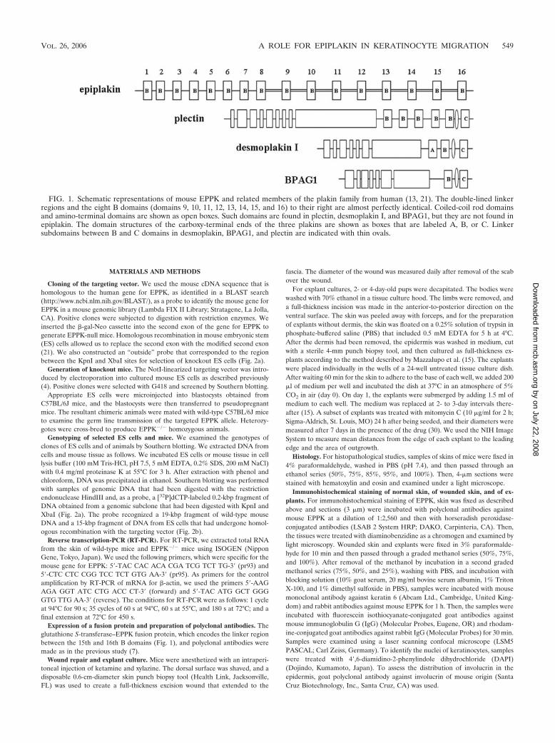
Epiplakin is homologous to plectin and other members ofthe plakin family, but it belongs to a novel category of plakinsbecause of the following unusual features (Fig. 1). Humanepiplakin has 13 domains, and mouse epiplakin has 16 do-mains, that are homologous to the B domain, which is one ofthe plakin repeat domains (PRDs) found in the carboxy-ter-minal region of desmoplakin, and these domains are distrib-uted along the amino acid sequence with relatively uniformspacing (7, 21). The amino acid sequences of the last five(human) or eight (mouse) of these B domains, starting fromthe carboxyl terminus, together with their associated linkerregions, are particularly strongly conserved. Epiplakin lacksthe coiled-coil rod domain and the amino-terminal domainthat are found in all other known members of the plakinfamily. Furthermore, there is no dimerization motif in theentire amino acid sequence. Thus, it is likely that EPPK existsin vivo as a single-chain structure (7). The unique features ofthe repeated structures in EPPK undoubtedly contribute to theprotein’s function in vivo.
It seems likely that the carboxy-terminal regions, including
PRDs and linker regions, of proteins in the plakin family, suchas desmoplakin, BPAG1 (an autoantigen of bullous pemphi-goid), and plectin (a protein responsible for epidermolysis bul-losa with muscular dystrophy), bind to intermediate filaments(8, 12, 17, 18, 22, 23, 28). However, EPPK has only one kind ofPRD, a B domain, and connecting linker domains, as notedabove, and in vitro experiments have demonstrated that theEPPK plakin repeat domain with its connecting linker domainbinds to intermediate filaments (10).
It is unclear why autoantibodies against epiplakin are asso-ciated with subepidermal blistering disease. Gene targeting ofBPAG1 and of plectin, both of which are members of theplakin family, causes skin fragility and the formation of blisters(9, 16). We designed the present study to examine what hap-pens when expression of EPPK is disrupted in mice.
The open reading frame that encodes EPPK is transcribed asa single exon in human and mouse (21, 25), although in themouse, the 5�-terminal untranslated mRNA is encoded by anadjacent exon (referred to here as the first exon). Thus, nosplice variants of EPPK should exist, depending on the tissue inwhich EPPK is expressed. Moreover, there are no genes fororthlogs with structural homology to EPPK that might com-pensate for the absence of EPPK, other than the gene forplectin, in the mouse and human genomes. Thus, we were ableto examine whether a change in phenotype would occur whenwe inactivated the gene for EPPK in mice.
We report here the first gene targeting of EPPK, to ourknowledge, and an analysis of some aspects of the phenotypeof EPPK knockout mice. We also examined the localization ofepiplakin in the murine epidermis by immunostaining andelectron microscopy.
* Corresponding author. Mailing address: Department of Anatomy,Biology and Medicine (Dermatology), Oita University, Idaigaoka 1-1,Hasama-machi, Yufu 879-5593, Japan. Phone: 81-97-586-5882. Fax:81-97-586-5889. E-mail: [email protected].
Cloning of the targeting vector. We used the mouse cDNA sequence that ishomologous to the human gene for EPPK, as identified in a BLAST search(http://www.ncbi.nlm.nih.gov/BLAST/), as a probe to identify the mouse gene forEPPK in a mouse genomic library (Lambda FIX II Library; Stratagene, La Jolla,CA). Positive clones were subjected to digestion with restriction enzymes. Weinserted the �-gal-Neo cassette into the second exon of the gene for EPPK togenerate EPPK-null mice. Homologous recombination in mouse embryonic stem(ES) cells allowed us to replace the second exon with the modified second exon(21). We also constructed an “outside” probe that corresponded to the regionbetween the KpnI and XbaI sites for selection of knockout ES cells (Fig. 2a).
Generation of knockout mice. The NotI-linearized targeting vector was intro-duced by electroporation into cultured mouse ES cells as described previously(4). Positive clones were selected with G418 and screened by Southern blotting.
Appropriate ES cells were microinjected into blastocysts obtained fromC57BL/6J mice, and the blastocysts were then transferred to pseudopregnantmice. The resultant chimeric animals were mated with wild-type C57BL/6J miceto examine the germ line transmission of the targeted EPPK allele. Heterozy-gotes were cross-bred to produce EPPK�/� homozygous animals.
Genotyping of selected ES cells and mice. We examined the genotypes ofclones of ES cells and of animals by Southern blotting. We extracted DNA fromcells and mouse tissue as follows. We incubated ES cells or mouse tissue in celllysis buffer (100 mM Tris-HCl, pH 7.5, 5 mM EDTA, 0.2% SDS, 200 mM NaCl)with 0.4 mg/ml proteinase K at 55°C for 3 h. After extraction with phenol andchloroform, DNA was precipitated in ethanol. Southern blotting was performedwith samples of genomic DNA that had been digested with the restrictionendonuclease HindIII and, as a probe, a [32P]dCTP-labeled 0.2-kbp fragment ofDNA obtained from a genomic subclone that had been digested with KpnI andXbaI (Fig. 2a). The probe recognized a 19-kbp fragment of wild-type mouseDNA and a 15-kbp fragment of DNA from ES cells that had undergone homol-ogous recombination with the targeting vector (Fig. 2b).
Reverse transcription-PCR (RT-PCR). For RT-PCR, we extracted total RNAfrom the skin of wild-type mice and EPPK�/� mice using ISOGEN (NipponGene, Tokyo, Japan). We used the following primers, which were specific for themouse gene for EPPK: 5�-TAC CAC ACA CGA TCG TCT TG-3� (pr93) and5�-CTC CTC CGG TCC TCT GTG AA-3� (pr95). As primers for the controlamplification by RT-PCR of mRNA for �-actin, we used the primers 5�-AAGAGA GGT ATC CTG ACC CT-3� (forward) and 5�-TAC ATG GCT GGGGTG TTG AA-3� (reverse). The conditions for RT-PCR were as follows: 1 cycleat 94°C for 90 s; 35 cycles of 60 s at 94°C, 60 s at 55°C, and 180 s at 72°C; and afinal extension at 72°C for 450 s.
Expression of a fusion protein and preparation of polyclonal antibodies. Theglutathione S-transferase–EPPK fusion protein, which encodes the linker regionbetween the 15th and 16th B domains (Fig. 1), and polyclonal antibodies weremade as in the previous study (7).
Wound repair and explant culture. Mice were anesthetized with an intraperi-toneal injection of ketamine and xylazine. The dorsal surface was shaved, and adisposable 0.6-cm-diameter skin punch biopsy tool (Health Link, Jacksonville,FL) was used to create a full-thickness excision wound that extended to the
fascia. The diameter of the wound was measured daily after removal of the scabover the wound.
For explant cultures, 2- or 4-day-old pups were decapitated. The bodies werewashed with 70% ethanol in a tissue culture hood. The limbs were removed, anda full-thickness incision was made in the anterior-to-posterior direction on theventral surface. The skin was peeled away with forceps, and for the preparationof explants without dermis, the skin was floated on a 0.25% solution of trypsin inphosphate-buffered saline (PBS) that included 0.5 mM EDTA for 5 h at 4°C.After the dermis had been removed, the epidermis was washed in medium, cutwith a sterile 4-mm punch biopsy tool, and then cultured as full-thickness ex-plants according to the method described by Mazzalupo et al. (15). The explantswere placed individually in the wells of a 24-well untreated tissue culture dish.After waiting 60 min for the skin to adhere to the base of each well, we added 200�l of medium per well and incubated the dish at 37°C in an atmosphere of 5%CO2 in air (day 0). On day 1, the explants were submerged by adding 1.5 ml ofmedium to each well. The medium was replaced at 2- to 3-day intervals there-after (15). A subset of explants was treated with mitomycin C (10 �g/ml for 2 h;Sigma-Aldrich, St. Louis, MO) 24 h after being seeded, and their diameters weremeasured after 7 days in the presence of the drug (30). We used the NIH ImageSystem to measure mean distances from the edge of each explant to the leadingedge and the area of outgrowth.
Histology. For histopathological studies, samples of skins of mice were fixed in4% paraformaldehyde, washed in PBS (pH 7.4), and then passed through anethanol series (50%, 75%, 85%, 95%, and 100%). Then, 4-�m sections werestained with hematoxylin and eosin and examined under a light microscope.
Immunohistochemical staining of normal skin, of wounded skin, and of ex-plants. For immunohistochemical staining of EPPK, skin was fixed as describedabove and sections (3 �m) were incubated with polyclonal antibodies againstmouse EPPK at a dilution of 1:2,560 and then with horseradish peroxidase-conjugated antibodies (LSAB 2 System HRP; DAKO, Carpinteria, CA). Then,the tissues were treated with diaminobenzidine as a chromogen and examined bylight microscopy. Wounded skin and explants were fixed in 3% paraformalde-hyde for 10 min and then passed through a graded methanol series (50%, 75%,and 100%). After removal of the methanol by incubation in a second gradedmethanol series (75%, 50%, and 25%), washing with PBS, and incubation withblocking solution (10% goat serum, 20 mg/ml bovine serum albumin, 1% TritonX-100, and 1% dimethyl sulfoxide in PBS), samples were incubated with mousemonoclonal antibody against keratin 6 (Abcam Ltd., Cambridge, United King-dom) and rabbit antibodies against mouse EPPK for 1 h. Then, the samples wereincubated with fluorescein isothiocyanate-conjugated goat antibodies againstmouse immunoglobulin G (IgG) (Molecular Probes, Eugene, OR) and rhodam-ine-conjugated goat antibodies against rabbit IgG (Molecular Probes) for 30 min.Samples were examined using a laser scanning confocal microscope (LSM5PASCAL; Carl Zeiss, Germany). To identify the nuclei of keratinocytes, sampleswere treated with 4�,6-diamidino-2-phenylindole dihydrochloride (DAPI)(Dojindo, Kumamoto, Japan). To assess the distribution of involucrin in theepidermis, goat polyclonal antibody against involucrin of mouse origin (SantaCruz Biotechnology, Inc., Santa Cruz, CA) was used.
FIG. 1. Schematic representations of mouse EPPK and related members of the plakin family from human (13, 21). The double-lined linkerregions and the eight B domains (domains 9, 10, 11, 12, 13, 14, 15, and 16) to their right are almost perfectly identical. Coiled-coil rod domainsand amino-terminal domains are shown as open boxes. Such domains are found in plectin, desmoplakin I, and BPAG1, but they are not found inepiplakin. The domain structures of the carboxy-terminal ends of the three plakins are shown as boxes that are labeled A, B, or C. Linkersubdomains between B and C domains in desmoplakin, BPAG1, and plectin are indicated with thin ovals.
VOL. 26, 2006 A ROLE FOR EPIPLAKIN IN KERATINOCYTE MIGRATION 549
Electron microscopy. Small blocks of mouse skin were placed in Zamboni’sfixative at 4°C for 2 h and then treated as described previously by Yamamoto-Hinoet al. (31). In brief, cryosections on glass slides were incubated overnight withantibodies against mouse EPPK or mouse monoclonal antibody against keratin 6,which had been diluted 100-fold. Then they were treated with antibodies raised ingoat against rabbit or mouse IgG, which had been conjugated with 0.8-nm particlesof colloidal gold (Aurion Co., Ltd., Amsterdam, The Netherlands). The sectionswere soaked in a mixture of 2.5% glutaraldehyde, 0.2% tannic acid, and 1% OsO4
in 0.1 M phosphate buffer for 30 min at room temperature and then rinsed intriple-distilled water. They were then incubated with a solution for silver enhance-ment (Aurion Co., Ltd.), embedded in a 1% solution of chitosan, fixed in 2.5%glutaraldehyde, and subjected to routine processing. Finally, the thin sections werestained with uranyl acetate and lead citrate and examined with an electron micro-scope (JEM 1230; JEOL Ltd., Tokyo, Japan).
RESULTS
Generation of mice with an inactivated gene for EPPK. Ourstrategy for disruption of the expression of the gene for EPPKinvolved construction of a gene-targeting vector that wouldallow us to generate EPPK-null mice. We anticipated that the
mutation would result in a premature termination codon inexon 2 at a distance of approximately 0.5 kbp from the ATGcodon. Thus, almost every function of mouse EPPK would bedisrupted (Fig. 2a). After electroporation of ES cells with thetargeting vector, we selected positive clones by growing cells inmedium that contained the neomycin analog G418. We con-firmed that homologous recombination had occurred betweenthe wild-type allele and the targeting vector by Southern blot-ting using DNA digested with KpnI and XbaI. A 15-kbp frag-ment indicated the presence of the disrupted allele, whereas a19-kbp fragment corresponded to the wild-type allele (Fig. 2b).
We microinjected ES cells with the appropriately disruptedgene into C57BL/6J blastocysts. The resultant chimeric malesprovided evidence of germ line transmission of the ES cellgenome, as revealed by the agouti coat color of offspring fromchimeras. An inbred line was established by mating the chime-ras with 129/Sv mice. Heterozygous animals were also gener-ated with a pure 129/Sv background.
FIG. 2. (a) The �-gal-Neo cassette was inserted into the second exon of the mouse gene for EPPK to generate the targeting vector used forgeneration of EPPK�/� mice. As a result of homologous recombination in mouse ES cells, this construct replaced the second exon of the wild-typegene. The “outside” probe was generated by cleavage at the KpnI and XbaI sites for selection of ES cells and mice by genomic Southern blotting.K, KpnI; H, HindIII; Xb, XbaI; Xh, XhoI. (b) Southern blotting yielded a 19-kbp DNA fragment in the case of the wild-type allele and a 15-kbpfragment in the case of the mutant allele. Heterozygous animals harbored both alleles. (c) Wild-type and heterozygous skin cells expressed EPPKmRNA, whereas EPPK�/� cells did not express this mRNA. All cells expressed �-actin mRNA.(d) Histology of skin of the feet. Staining withhematoxylin and eosin revealed the absence of blistering in both wild-type and EPPK�/� mice (top). Immunostaining with polyclonal antibodiesagainst the linker region of EPPK was negative in EPPK�/� skin, while it was positive in wild-type skin (bottom). Bars � 50 �m.
We confirmed that the mice lacked a gene for full-lengthEPPK by genomic Southern blotting, RT-PCR, and immuno-staining with polyclonal antibodies (Fig. 2b, c, and d).
EPPK�/� mice resembled wild-type mice. The EPPK�/�
mice resembled wild-type mice in terms of gross phenotype,and light microscopy revealed the absence of blistering andskin fragility (Fig. 2d). Mice that were homozygous for thetargeted allele were born at the expected Mendelian fre-quency. They developed normally and were healthy and fertile.In mice, EPPK has been found by immunostaining, not only inskin, but also in hair follicles, the esophagus, and the simpleepithelium of the digestive organs, as is also the case in humans(7, 21). However, there were no abnormalities in such tissuesof EPPK�/� mice at the macroscopic and light-microscopiclevels (data not shown). Under the electron microscope, therewere no apparent differences between basal and spinous ker-atinocytes, respectively, of EPPK�/� mice and wild-type mice(Fig. 3a, b, c, and d). Immunoreactivity specific for EPPK wasdetected adjacent to keratin filaments in wild-type mice, sug-
gesting that epiplakin might be associated with keratin. How-ever, the appearance and localization of keratin bundles inintact epidermal keratinocytes of EPPK�/� mice were similarto those in wild-type mice. Immunoreactivity in wild-type micewas detected from the basal cells to the granular cells and wasnot concentrated in desmosomes or hemidesmosomes (datanot shown).
EPPK�/� mice exhibited slightly enhanced wound closure.In EPPK�/� mice, wound closure was almost complete on day10, whereas in wild-type and heterozygous mice, wound closureoccurred 1 or 2 days later (Fig. 4). We postulated that themigration and/or proliferation of mouse keratinocytes mighthave been accelerated in the absence of EPPK. We confirmedour observations under the light microscope. Enhanced woundclosure was evident in EPPK�/� mice even when we comparedwounds without peeling off the scabs. In EPPK�/� mice, ele-vated numbers of keratinocytes were observed, and they mi-grated under the fibrin clot on day 3. By contrast, in wild-typemice, fewer keratinocytes were localized on the upper side of
FIG. 3. Electron micrographs of keratinocytes from the dorsal epidermis with low (a and b) and high (c and d) magnification. There are noapparent abnormalities in the keratin filaments and desmosomes (white arrows) of wild-type mice (a and c) and EPPK�/� mice (b and d). Silvergrains are visible adjacent to keratin bundles (black arrows). Nu, nucleus. Bars � 1 �m (a and b) and 100 nm (c and d).
VOL. 26, 2006 A ROLE FOR EPIPLAKIN IN KERATINOCYTE MIGRATION 551
the wound’s edge on day 3. On day 4, the difference betweenthe two lines of mice was quite marked (Fig. 5). In wild-typemice, the edges of wounds invaginated sharply, the epidermiswas hypertrophic, a few keratinocytes made a wall, and smallpopulations of keratinocytes migrated under the clot. A largescab covered the ulcer. By contrast, in EPPK�/� mice, weobserved decreased hypertrophy of the epidermis at thewound’s edge, and the leading edge extended a long distancecompared with that in wild-type mice. The scab peeled awayfrom the edge of the wound, becoming smaller and shifting tothe center of the wound.
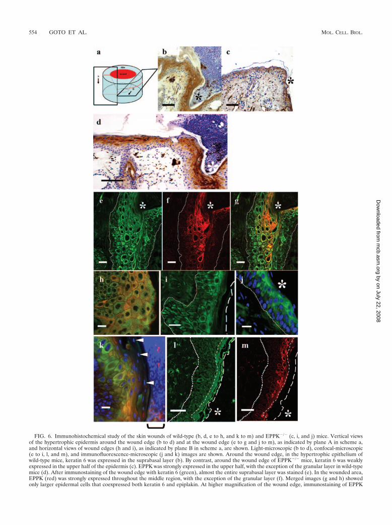
During wound healing, synthesis of keratin 6 is induced insuprabasal keratinocytes. In the wound edge, keratin 6 wasstrongly expressed in wild-type mice, while in EPPK�/� mice,keratin 6 expression was much weaker (Fig. 6b and c). Next, weexamined the expression of EPPK and compared it to that ofkeratin 6 in wild-type mice by immunohistochemical staining.Around the region of the wound, EPPK was expressed in theupper half of the epidermis, but expression of EPPK was sup-pressed in the granular layer of the epidermis, as is the case innormal epithelium (Fig. 6d). On the basilar side of the supra-basal layer of the epidermis at the wound’s edge, where keratin6 was expressed and EPPK was expressed only weakly, kera-tinocytes were small (Fig. 6e and f). In the upper layer of theepidermis, EPPK was expressed strongly, especially in largerkeratinocytes, but unevenly (Fig. 6f, g, and h). In the granularlayer at the edge of the wound in wild-type mice, neitherepiplakin nor keratin 6 was expressed (Fig. 6k). By contrast, inEPPK�/� mice, the epithelium was only slightly hypertrophic(Fig. 6i) and keratin 6 was weakly expressed in the upper layerof the wounded area (Fig. 6j). In the upper and lower regions,the keratinocytes exhibited none of the morphological changesseen in wild-type mice. At the leading edge, only keratin 6 wasexpressed, even in wild-type mice, and there were no morpho-logical differences between wild-type and knockout mice. In-volucrin was distributed in the suprabasal layer in the epider-mis, including the granular layer, while EPPK was expressedonly in the upper half-layer of the epidermis, with the excep-tion of the granular layer (Fig. 6l and m).
After immunostaining, electron microscopy revealed that at
wound edges in EPPK�/� mice, antibodies against keratin 6reacted far less strongly than in wild-type mice. This observa-tion suggested that, in EPPK�/� mice, the expression of ker-atin 6 was suppressed at the wound edge (Fig. 7).
On the appearance of the final scar, the epidermis in wild-type mice was more hypertrophic than that in EPPK�/� mice(data not shown).
EPPK�/� keratinocytes had an enhanced capacity for epi-thelialization. Epiplakin is expressed only in the epithelium innormal skin. Thus, it is likely that EPPK contributes to theepithelial closure of wounds. To assess the wound epithelial-ization potential in EPPK�/� mice, we used a skin explantassay in which cultured explants mimic the behavior of kera-tinocytes at the edges of skin wounds in vivo. This assay allowsthe qualitative and quantitative assessment of the potential ofkeratinocytes for epithelialization while avoiding the effects ofdermis-mediated contraction of the wound bed (3, 15). Wefound that EPPK�/� explants exhibited a statistically signifi-cant 1.2-fold enhancement of epithelial outgrowth comparedwith explants from wild-type and heterozygous mice (Fig. 8aand b).
Enhanced outgrowth of the EPPK�/� keratinocytes resultedfrom migration. Reepithelialization of skin wounds in vivoresults from increases in mitotic activity and the migration ofkeratinocytes that are located at wound margins (14). Simi-larly, over a period of 7 days in culture, migration and mitosiscontribute equally to the outgrowth of keratinocytes from skinexplants (27). To determine whether migration or mitosis wasinvolved in the effect of EPPK, we assessed the outgrowth ofkeratinocytes after treatment of explants with mitomycin C, anirreversible inhibitor of mitosis (27). Relative to the wild-type andheterozygous controls, treated and untreated EPPK�/� explantsexhibited similar outgrowth of keratinocytes in terms of bothdistance and area (Fig. 8c and d). However, treated EPPK�/�
skin explants exhibited a statistically significant 1.5-fold enhance-ment of epithelial outgrowth compared to wild-type and heterozy-gous explants. Untreated EPPK�/� skin explants exhibited a sta-tistically significant 1.2-fold enhancement of epithelial outgrowthcompared with wild-type and heterozygous explants.
Epiplakin was coaligned with keratin 6 in some keratino-cytes in wild-type explants. In order to investigate the colocal-ization of epiplakin and keratin 6, we examined explant cul-tures with a confocal microscope after double immunostainingwith antibodies against keratin 6 and EPPK. In wild-type ex-plants, epiplakin was distributed as granules or a reticularnetwork in almost all keratinocytes, and keratin 6 was distrib-uted as a fine reticular network and only partially as filamentsin some keratinocytes. In some keratinocytes, EPPK was colo-calized with keratin 6 (Fig. 9a to f). Since actin filaments arethought to be involved in cell migration, we observed explantsfrom wild-type mice after double immunostaining with anti-bodies against EPPK and actin. Epiplakin was not colocalizedwith actin (data not shown). In EPPK�/� explants, keratin 6was distributed as filaments that seemed to be stretched in alarge number of keratinocytes (Fig. 9g and h).
DISCUSSION
Epiplakin is a member of the plakin family, to which plectinand BPAG1 also belong. Mice that lack plectin or BPAG1
FIG. 4. Comparison of wound closure processes between wild-type,heterozygous, and homozygous mice. In EPPK�/� mice, wound clo-sure occurred slightly faster than in wild-type and heterozygous mice(n � 28 for wild-type, 56 for heterozygous, and 20 for EPPK�/� mice).The error bars represent standard deviations.
have fragile skin (1, 9). However, EPPK�/� mice exhibited noskin fragility up to 2 years of age, and light microscopy revealedno signs of blistering of the skin. Moreover, under the electronmicroscope, we found no abnormalities in the epidermal ker-atinocytes. The absence of EPPK might have an effect on skinfragility different from that of the absence of BPAG1 andplectin because the latter proteins are localized in hemidesmo-somes in basal cells and connect intermediate filaments andhemidesmosomes (9, 16). Ultrastructural analysis suggestedthat EPPK is not concentrated in desmosomes and hemides-mosomes but is associated with keratin filaments in the cyto-plasm of epidermal keratinocytes. Jang et al. reported recentlythat in cultured HeLa cells, HaCaT cells, and human keratin-
ocytes, EPPK is colocalized with keratin and vimentin, and indot blot experiments, the plakin repeat domain with its linkerbound to the intermediate filaments (10). These data supportthe validity of our findings.
Epiplakin and plectin have a common domain structure attheir carboxyl termini, but plectin did not appear to compen-sate for the loss of EPPK, since in EPPK�/� mice, the patternof immunostaining for plectin was similar to that in wild-typemice (data not shown).
Epiplakin is expressed not only in the skin, but also in hairfollicles, the esophagus, and the simple epithelium of digestiveorgans (7, 21). Jang et al. suggested, from knock-down exper-iments, that the effect of elimination of EPPK might be more
FIG. 5. Histological analysis of skin wounds on the backs of wild-type (a, c, and e) and EPPK�/� (b, d, and f) mice. On day 4, the edges ofwounds in wild-type mice invaginated sharply, and the epidermis was hypertrophic at the margins (a). A large scab covered the ulcer. In EPPK�/�
mice, there was decreased hypertrophy of the epidermis at the wound’s edge, and the leading edge extended a long distance compared with thatin wild-type mice. The scab was also smaller than that in wild-type mice (b). *, wound edge; arrows, leading edge. Higher-magnification views ofthe wound edge (c and d) and of the leading edge (e and f) are shown. Bars � 100 �m.
VOL. 26, 2006 A ROLE FOR EPIPLAKIN IN KERATINOCYTE MIGRATION 553
FIG. 6. Immunohistochemical study of the skin wounds of wild-type (b, d, e to h, and k to m) and EPPK�/� (c, i, and j) mice. Vertical viewsof the hypertrophic epidermis around the wound edge (b to d) and at the wound edge (e to g and j to m), as indicated by plane A in scheme a,and horizontal views of wound edges (h and i), as indicated by plane B in scheme a, are shown. Light-microscopic (b to d), confocal-microscopic(e to i, l, and m), and immunofluorescence-microscopic (j and k) images are shown. Around the wound edge, in the hypertrophic epithelium ofwild-type mice, keratin 6 was expressed in the suprabasal layer (b). By contrast, around the wound edge of EPPK�/� mice, keratin 6 was weaklyexpressed in the upper half of the epidermis (c). EPPK was strongly expressed in the upper half, with the exception of the granular layer in wild-typemice (d). After immunostaining of the wound edge with keratin 6 (green), almost the entire suprabasal layer was stained (e). In the wounded area,EPPK (red) was strongly expressed throughout the middle region, with the exception of the granular layer (f). Merged images (g and h) showedonly larger epidermal cells that coexpressed both keratin 6 and epiplakin. At higher magnification of the wound edge, immunostaining of EPPK
apparent in the simple epithelium than in keratinocytes orHaCaT cells (10). However, in EPPK�/� mice, we found noabnormalities in such tissues by light microscopy (data notshown). Moreover, although it has been reported that EPPK iscross-linked with trichohyalin in mouse hair follicles (24), thehairs of EPPK�/� mice seemed normal. In our study, theimmunoreactivity of EPPK was not detected in the granularlayer of the epidermis. Jang et al. suggested that in the laterstages of keratinocyte differentiation, epiplakin continues to besynthesized with some degradation (10). This discrepancymight have occurred because the EPPK epitope was maskeddue to transglutaminase-mediated cross-linking, while the in-volucrin epitope was not disturbed in the granular layer.
The enhanced wound healing in EPPK�/� mice was unex-pected. When the skin is undamaged, EPPK appears to beunnecessary, and other molecules can perform its role in itsabsence. However, when the integrity of normal skin is dis-rupted, proliferating keratinocytes express enhanced amountsof EPPK. At the wound edge in wild-type mice, expression ofkeratin 6 was induced first in relatively small keratinocytes inthe suprabasal portion of the epidermis. Later, expression ofEPPK was induced in larger keratinocytes. The edge of thewound was fixed by these cells as if by a wall. In EPPK�/� mice,we found no such large keratinocytes and no wall at thewound’s edge, and the keratinocytes appeared to have mi-grated more rapidly under the scab. As the leading edges
progressed, the scab peeled off from the wound’s edge andregressed. At the front of the leading edge, only keratin 6 andno epiplakin was expressed, and the cells were similar in wild-type and EPPK�/� mice. The data from skin explant culturessupported the observed difference in wound healing betweenwild-type and EPPK�/� mice. In the presence of mitomycin C,which inhibits mitosis, EPPK�/� explants also exhibited en-hanced outgrowth compared to wild-type and heterozygousexplants. It seems likely that the contribution of EPPK�/�
keratinocytes to the enhanced outgrowth of explants was dueto the migration of keratinocytes rather than to their prolifer-ation. Some proliferating and migrating cells came from theouter root sheaths of hair follicles, but the patterns of immu-nostaining of keratin 6 and EPPK were essentially similar tothose in cells of epidermal origin.
The mechanism responsible for the faster migration of ker-atinocytes is unknown. Epiplakin was expressed in proliferat-ing large keratinocytes, namely, differentiated keratinocytes atthe wound’s edge, in wild-type mice, but at the wound’s edge inEPPK�/� mice no such large keratinocytes were observed. Themigratory process is presumably mediated by actin filamentsand microtubules (11). However, EPPK was apparently notcoaligned with actin or with microtubules (unpublished data).Spectraplakin binds to actin and microtubules and perhaps tointermediate filaments (11, 20), while EPPK has neither anactin-binding site nor a microtubule-binding domain. How-
was more evident in larger keratinocytes, but staining was uneven (h; right, scab side) At wound edges in EPPK�/� mice, the epidermis was onlyslightly hypertrophied, and keratin 6 was weakly expressed in the upper region of the epidermis (i and j; stained for keratin 6 and with DAPI). Afterstaining with DAPI (blue), in addition to double immunostaining, neither keratin 6 nor epiplakin was detected in the granular layer, as indicatedby arrowheads (k; the square bracket indicates the scab). Involucrin (green in panel l) was distributed in the suprabasal layer of the epidermis,including the granular layer, while epiplakin (red in panel m) was expressed in the upper half-layer of the epidermis, with the exception of thegranular layer. *, wound edge; arrowheads, nuclei in the granular layer. The dotted lines represent basal lamina, long-dashed lines represent thesurface of the stratum corneum, and short-dashed lines represent the borders of the malpighian and granular layers of the epidermis. Bars � 50�m (b, c, l, and m), 100 �m (d), and 20 �m (e to k).
FIG. 7. Electron micrographs of suprabasilar keratinocytes at wound edges. Immunostaining for keratin 6 (silver grains indicated by arrows)was much weaker in EPPK�/� mice (b) than in wild-type mice (a). Nu, nucleus. Bars � 1 �m.
VOL. 26, 2006 A ROLE FOR EPIPLAKIN IN KERATINOCYTE MIGRATION 555
ever, EPPK might interact with and coordinate the activities ofother molecules, including members of the spectraplakin fam-ily.
Wound healing is a complex process. With respect to theexpression of keratin after epidermal injury, it is known thatkeratins 6, 16, and 17 are expressed in suprabasal keratinocytesat the wound’s edge and that such expression is suppressedonce the epidermis has covered the wound (2, 3, 19). Enhancedhealing of wounds was observed in keratin 6 (K6�/�) knockoutmice, but such mice died soon after birth as a result of thefragility of the stress-bearing epithelium within the oral mu-cosa (29, 30). The common phenotype, namely, faster closureof wounds in EPPK�/� and K6�/��/� mice, and the inducedexpression of both proteins at the edges of wounds suggest thatEPPK might be linked functionally with keratin 6. In some cellsin our explant cultures, EPPK and keratin 6 were colocalized,and in EPPK�/� mice, the expression of keratin 6 seemed to besuppressed in the wounded area. Epiplakin might enhance thefunctions of keratin 6 during wound healing, and the proteinsmight collaborate and contribute to the reinforcement re-quired for resistance to increased mechanical stress. The lo-calization patterns of K6 in the wild type and EPPK�/� ex-plants were different. These data also suggest that EPPK mighttherefore work in collaboration with K6, although we have nodirect evidence that EPPK in fact interacts with K6.
The putative type I partners of keratins 6, 16, and 17 (19)might function with EPPK. In this context, the delayed healingof wounds that is induced by elevated levels of keratin 16 isintriguing (26, 27).
We tried to detect the differences in expression pattern ofother types of keratin, e.g., K5, -14, and -10, between wild-typeand EPPK�/� mice. The staining patterns of K5, -14, and -10in EPPK�/� mice were similar to those of wild-type mice, andthe expression of K10 was lowered at the wound edges.
Despite several reports of the effects on wound healing of adeficiency in or overexpression of keratin in mice, as describedabove, there are no reports, to our knowledge, of the geneticmodification of the expression of any members of the plakinfamily of proteins and its effect on wound healing. OurEPPK�/� mice should help us to clarify the involvement ofintermediate filaments in the migration of keratinocytes.
In summary, we generated EPPK knockout mice by inacti-vating the single mouse gene for EPPK. EPPK�/� mice had noapparent phenotypic abnormalities, but the closure of woundson their backs was slightly accelerated compared with that inwild-type and heterozygous mice. In the skin explant assay,EPPK�/� explants also exhibited accelerated migration of ker-atinocytes compared with that in explants of wild-type andheterozygous skin. This phenomenon might involve changes ininteractions between EPPK and intermediate filaments, suchas keratin. This is the first report, to our knowledge, of the
7 days, after incubation with or without mitomycin C from 24 h on-ward, are shown for heterozygous, EPPK�/�, and wild-type explants.(c) Comparison of the extents of outgrowth (P � 0.05; for treatment ofexplants with mitomycin C, n � 15 for wild-type, heterozygous, andEPPK�/� explants). (d) Comparison of areas of outgrowth (P � 0.05;for treatment of explants with mitomycin C, n � 7 for wild-type, 10 forheterozygous, and 8 for EPPK�/� explants).
FIG. 8. Comparison of wild-type, heterozygous, and EPPK�/� skinexplants in terms of the distance between keratinocytes located at thedistal edge of the outgrowth and the edge of each explant. The errorbars represent standard deviations. Asterisks indicate a statisticallysignificant difference from the wild type (P � 0.01 or P � 0.05, asindicated below; Student’s t test). (a) Comparison of the extents ofoutgrowth (P � 0.01; n � 148 for wild-type, 143 for heterozygous, and144 for EPPK�/� explants). (b) Comparison of areas of outgrowth (P� 0.01; n � 71 for wild-type, 63 for heterozygous, and 68 for EPPK�/�
explants). The extents and total areas of keratinocyte outgrowths after
generation of EPPK knockout mice, and these mice shouldhelp us to analyze the functions of other members of the plakinfamily, since EPPK is an extreme example of the family, beingcomposed almost exclusively of plakin repeat domains.
ACKNOWLEDGMENTS
We are most grateful to the late Rupert Timpl of the Max-PlanckInstitute for Biochemistry (Germany; Project QLK3-CT2000-00084 ofTimpl) for encouraging our research; to Katsushi Owaribe (NagoyaUniversity) for the kind gift of plectin-specific antibodies; and to Sa-
toko Sato, Aiko Yasuda, and Hiroaki Kawasato for technical assis-tance.
This work was supported in part by grants 1370892 and 15591184 toS.F. from the Ministry of Education, Culture, Science and Technologyof Japan.
REFERENCES
1. Andra, K., H. Lassmann, R. Bittner, S. Shorny, R. Fassler, F. Propst, and G.Wiche. 1997. Targeted inactivation of plectin reveals essential function inmaintaining the integrity of skin, muscle, and heart cytoarchitecture. GenesDev. 11:3143–3156.
2. Coulombe, P. A. 1997. Towards a molecular definition of keratinocyte acti-
FIG. 9. Immunolocalization of keratin 6 in keratinocytes of explants from wild-type (a to f) and EPPK�/� (g and h) mice. In wild-type explants,keratin 6 (green) was distributed as fine reticular networks and only partly as fine filaments in some keratinocytes (a), and epiplakin (red) wasdistributed as granules or reticular networks in almost all keratinocytes (b). In the merged image (c), epiplakin was colocalized with keratin 6 andappeared yellow. At higher magnification, in the keratinocyte indicated by an arrow in panel c, keratin 6 (d) and epiplakin (e) were colocalized,and fine filaments or reticular networks were yellow in the merged image (f). In EPPK�/� explants, a large number of keratinocytes containedweakly stained filamentous and apparently stretched keratin 6 (g). At higher magnification, in the keratinocytes from EPPK�/� explants, thekeratin 6 was distributed in a more filamentous and less reticular pattern (h). Nuclei in all panels except panels c and f were counterstained withDAPI. Bars � 50 �m (a to c and g) and 20 �m (d to f and h).
VOL. 26, 2006 A ROLE FOR EPIPLAKIN IN KERATINOCYTE MIGRATION 557
vation after acute injury to stratified epithelia. Biochem. Biophys. Res. Com-mun. 236:231–238.
3. Coulombe, P. A. 2003. Wound epithelialization: accelerating the pace ofdiscovery. J. Investig. Dermatol. 37:219–230.
4. Fassler, R., and M. Meyer. 1995. Consequences of lack of beta 1 integringene expression in mice. Genes Dev. 9:1896–1908.
5. Fujiwara, S., H. Shinkai, S. Takayasu, K. Owaribe, S. Tsukita, and T.Kageshita. 1992. A case of subepidermal blister disease associated withautoantibody against 450kD protein. J. Dermatol. 19:610–613.
6. Fujiwara, S., K. Kohno, A. Iwamatsu, I. Naito, and H. Shinkai. 1996. Iden-tification of a 450-kDa human epidermal autoantigen as a new member ofthe plectin family. J. Investig. Dermatol. 106:1125–1130.
7. Fujiwara, S., N. Takeo, Y. Otani, D. A. D. Parry, M. Kunimatsu, R. Lu, M.Sasaki, N. Matsuo, M. Khaleduzzaman, and H. Yoshioka. 2001. Epiplakin,a novel member of the plakin family originally identified as a 450-kDahuman epidermal autoantigen. J. Biol. Chem. 276:13340–13347.
8. Green, K. J., D. A. D. Parry, P. M. Steinert, M. L. A. Virata, R. M. Wagner,B. D. Angst, and L. A. Nilles. 1990. Structure of the human desmoplakins.Implications for function in the desmosomal plaque. J. Biol. Chem. 265:2603–2612.
9. Guo, L., L. Degenstein, J. Dowling, Q.-C. Yu, R. Wollmann, B. Perman, andE. Fuchs. 1995. Gene targeting of BPAG1: abnormalities in mechanicalstrength and cell migration in stratified epithelia and neurologic degenera-tion. Cell 81:233–243.
10. Jang, S.-I., A. Kalinin, K. Takahashi, L. N. Marekov, and P. M. Steinert.2005. Characterization of human epiplakin: RNAi-mediated epiplakin de-pletion leads to the disruption of keratin and vimentin IF networks. J. CellSci. 118:781–793.
11. Kodama, A., T. Lechler, and E. Fuchs. 2004. Coordinating cytoskeletal tracksto polarize cellular movements. J. Cell Biol. 167:203–207.
12. Leung, C. L., D. Sun, and R. K. H. Liem. 1999. The intermediate filamentprotein peripherin is the specific interaction partner of mouse BPAG1-n(Dystonin) in neurons. J. Cell Biol. 144:435–446.
13. Leung, C. L., K. J. Green, and R. K. H. Liem. 2002. Plakins: a family ofversatile cytolinker proteins. Trends Cell Biol. 12:37–45.
14. Martin, P. 1997. Wound healing—aiming for perfect skin regeneration.Science 276:75–81.
15. Mazzalupo, S., M. J. Wawersik, and P. A. Coulumbe. 2002. An ex vivo assayto assess the potential of skin keratinocytes for wound epithelialization.J. Investig. Dermatol. 118:866–870.
16. McLean, W. H., L. Pulkkinen, F. J. Smith, E. L. Rugg, E. B. Lane, F.Bullrich, R. E. Burgeson, S. Amano, D. L. Hudson, K. Owaribe, J. A.McGrath, J. R. McMillan, R. A. Eady, I. M. Leigh, A. M. Christiano, and J.Uitto. 1996. Loss of plectin causes epidermolysis bullosa with musculardystrophy: cDNA cloning and genomic organization. Genes Dev. 10:1724–1735.
17. Meng, J.-J., E. A. Bornslaeger, K. J. Green, P. M. Steinert, and W. Ip. 1997.Two-hybrid analysis reveals fundamental differences in direct interactionsbetween desmoplakin and cell type-specific intermediate filaments. J. Biol.Chem. 272:21495–21503.
18. Nikolic, B., E. M. Nulty, B. Mir, and G. Wiche. 1996. Basic amino acidresidue cluster within nuclear targeting sequence motif is essential for cyto-plasmic plectin-vimentin network junctions. J. Cell Biol. 134:1455–1467.
19. Paladini, R. D., K. Takahashi, N. S. Bravo, and P. A. Coulombe. 1996. Onsetof re-epithelialization after skin injury correlates with a reorganization ofkeratin filaments in wound edge keratinocytes: defining a potential role forkeratin 16. J. Cell Biol. 132:381–397.
20. Roper, K., S. L. Gregory, and N. H. Brown. 2002. The ‘Spectraplakins’:cytoskeletal giants with characteristics of both spectrin and plakin families.J. Cell Sci. 115:4215–4225.
21. Spazierer, D., P. Fuchs, V. Proll, L. Janda, S. Oehler, I. Fischer, R. Haupt-mann, and G. Wiche. 2003. Epiplakin gene analysis in mouse reveals a singleexon encoding a 725-kDa protein with expression restricted to epithelialtissues. J. Biol. Chem. 278:31657–31666.
22. Stappenbeck, T. S., E. A. Bornslaeger, C. M. Corcoran, H. H. Luu, M. L.Virata, and K. J. Green. 1993. Functional analysis of desmoplakin domains:specification of the interaction with keratin versus vimentin intermediatefilament networks. J. Cell Biol. 123:691–705.
23. Steinbock, F. A., B. Nikolic, P. A. Coulombe, E. Fuchs, P. Traub, and G.Wiche. 2000. Dose-dependent linkage, assembly inhibition and disassemblyof vimentin and cytokeratin 5/14 filaments through plectin’s intermediatefilament-binding domain. J. Cell Sci. 113:483–491.
24. Steinert, P. M., D. A. D. Parry, and L. N. Marekov. 2003. Trichohyalinmechanically strengthens the hair follicle. J. Biol. Chem. 278:41409–41419.
25. Takeo, N., W. Wang, N. Matsuo, H. Sumiyoshi, H. Yoshioka, and S. Fuji-wara. 2003. Structure and heterogeneity of the human gene for epiplakin(EPPK1). J. Investig. Dermatol. 121:1224–1226.
26. Wawersik, M., and P. A. Coulombe. 2000. Forced expression of keratin 16alters the adhesion, differentiation, and migration of mouse skin keratino-cytes. Mol. Biol. Cell 11:3315–3327.
27. Wawersik, M. J., S. Mazzalupo, D. Nguyen, and P. A. Coulombe. 2001.Increased levels of keratin 16 alter epithelialization potential of mouse skinkeratinocytes in vivo and ex vivo. Mol. Biol. Cell 12:3439–3450.
28. Wiche, G., D. Gromov, A. Donovan, M. J. Castanon, and E. Fuchs. 1993.Expression of plectin mutant cDNA in cultured cells indicates a role ofCOOH-terminal domain in intermediate filament association. J. Cell Biol.121:607–619.
29. Wong, P., E. Colucci-Guyon, K. Takahashi, C. Gu, C. Babinet, and P. A.Coulombe. 2000. Introducing a null mutation in the mouse K6� and K6�genes reveals their essential structural role in the oral mucosa. J. Cell Biol.150:921–928.
30. Wong, P., and P. A. Coulombe. 2003. Loss of keratin 6 (K6) proteins revealsa function for intermediate filaments during wound repair. J. Cell Biol.163:327–337.
31. Yamamoto-Hino, M., A. Miyawaki, A. Segawa, E. Adachi, S. Yamashina, T.Fujimoto, T. Sugiyama, T. Furuichi, M. Hasegawa, and K. Mikoshiba. 1998.Apical vesicles bearing inositol 1,4,5-triphosphate receptors in the Ca2�
initiation site of ductal epithelium of submandibular gland. J. Cell Biol.141:135–142.