Elucidation of Structure-activity Relationship of 2-Quinolone Derivatives and Exploration of Their Antitumor Potential Through Bax-induced Apoptotic Pathway Nitesh Kumar 1 , Vasanth P. Raj 2 , B. S. Jayshree 3 , Sidhartha S. Kar 3 , Arvind Anandam 1 , Seeja Thomas 3 , Prateek Jain 2 , Amita Rai 4 and C. M. Rao 1, * 1 Department of Pharmacology, Manipal College of Pharmaceutical Sciences, Manipal University, Manipal-576104, Karnataka, India 2 Department of Pharmaceutical Biotechnology, Manipal College of Pharmaceutical Sciences, Manipal University, Manipal-576104, Karnataka, India 3 Department of Pharmaceutical Chemistry, Manipal College of Pharmaceutical Sciences, Manipal University, Manipal-576104, Karnataka, India 4 Grace College of Pharmacy, Palakkad-678004, Kerala, India *Corresponding author: C. M. Rao, [email protected]3-Aryl-2-quinolone derivates were extensively investigated for their inhibition of farnesyl trans- ferase. Taking this as a cue, we studied the other possible mechanism of antitumor activity of 2- quinolone derivates. A series of new 2-quinolone derivatives have been synthesized and screened for their cytotoxicity by trypan blue assay on Ehr- lich ascites carcinoma cells and MTT assay on MCF-7 cells. Compound 1a (nJST) was found to be more effective in both studies with the lowest CTC 50 value among all nine synthesized com- pounds. This compound was further screened on four different cell lines, viz. human breast adeno- carcinoma (MCF-7, MDA-MB-231), colon cancer (HCT-15), murine melanoma (B16F10) cell lines for 24 and 48 h. The CTC 50 value of the compound was found to be <10 lM. Compound 1a induced DNA damage which was revealed by DNA frag- mentation studies and further confirmed by nuclear staining. The compound also showed sig- nificant elevation in Bax and reduction Bcl-2 gene expression levels. Acute toxicity study in mice indicated that the compound is safe till 2000 mg ⁄ kg. Two different doses 50 and 100 mg ⁄ kg were selected and studied in Ehrlich ascites carcinoma model of cancer and have shown significant improvement in survival time and hematological parameters. Key words: 2-quinolone, cytotoxicity, DNA fragmentation, Ehrlich ascites carcinoma, MTT assay Received 25 November 2011, revised 8 February 2012 and accepted for publication 24 April 2012 Treatment for cancer is still being a challenge for medical world. Many researches are going on to get a safe and effective treat- ment for this disease. 2-Quinolones are one of the molecules in this category. Joseph et al. initiated the exploration of anticancer poten- tial of this moiety by reporting a series of 2-quinolones with 3-aryl and N-alkyl substitutions of which 12 compounds were exhibiting cytotoxicity (CTC 50 ) of more than 10 lM on MCF-7 (human breast cancer) cell line. However, these compounds were found to be non- toxic in in vivo toxicity determination and effective in in vivo model of MXT mouse mammary adenocarcinoma (1). The leading molecule of this category, tipifarnib, is still in clinical trial stage. This molecule is also have 3-aryl and N-methylation sub- stitutions and exhibits cytotoxicity in breast cancer cell lines MDA- MB-231 and BT-474 with a CTC 50 value <30 lM (2). Moreover, it is active orally and causes apoptosis in myeloid leukemia cell line (3). Apoptosis plays a central role in study of carcinogenesis and drug development for the cancer therapy. It is a regulated evolutionary con- served programme of cell suicide. Disturbance in this physiological pro- gramme prolongs the life of cell and leads to carcinogenesis. In cancer cells, the apoptosis diminishes and causes dominance of anti-apoptotic protein. Mitochondrial-mediated apoptosis is controlled by anti-apopto- tic (Bcl-2) and proapoptotic (Bax and Bad) proteins of Bcl-2 family. Over- expression of Bcl-2 occurs in 40–80% of human breast cancers (4). These studies added rising interest in developing and evaluating anticancer activity of 2-quinolone derivatives through apoptotic path- way. Among quinolones, most of the anticancer compounds have a 3-aryl substitution. However, to our knowledge nobody has explored the anticancer activity of 3-methyl substituted quinolones. Previously, we synthesized and evaluated compounds 1b–i and identified them as promising antimicrobial and antioxidant agents (5). But none of the compounds was tested for their cytotoxic potential against can- cer cell lines. With this rationale and our continuing effort to explore the biological activity of 2-quinolones, we report here the synthesis and pharmacological evaluation of the compounds 1a–i containing in their structures a 2-quinolone moiety as the pharmacophore and a functionally diverse side chain through amide linkage as the lipophi- licity contributor to the scaffold (Figure 1). Present study was also 291 Chem Biol Drug Des 2012; 80: 291–299 Research Article ª 2012 John Wiley & Sons A/S doi: 10.1111/j.1747-0285.2012.01402.x
Transcript
Elucidation of Structure-activity Relationship of2-Quinolone Derivatives and Exploration of TheirAntitumor Potential Through Bax-inducedApoptotic Pathway
Nitesh Kumar1, Vasanth P. Raj2,B. S. Jayshree3, Sidhartha S. Kar3,Arvind Anandam1, Seeja Thomas3,Prateek Jain2, Amita Rai4 and C. M. Rao1,*
1Department of Pharmacology, Manipal College of PharmaceuticalSciences, Manipal University, Manipal-576104, Karnataka, India2Department of Pharmaceutical Biotechnology, Manipal College ofPharmaceutical Sciences, Manipal University, Manipal-576104,Karnataka, India3Department of Pharmaceutical Chemistry, Manipal College ofPharmaceutical Sciences, Manipal University, Manipal-576104,Karnataka, India4Grace College of Pharmacy, Palakkad-678004, Kerala, India*Corresponding author: C. M. Rao, [email protected]
3-Aryl-2-quinolone derivates were extensivelyinvestigated for their inhibition of farnesyl trans-ferase. Taking this as a cue, we studied the otherpossible mechanism of antitumor activity of 2-quinolone derivates. A series of new 2-quinolonederivatives have been synthesized and screenedfor their cytotoxicity by trypan blue assay on Ehr-lich ascites carcinoma cells and MTT assay onMCF-7 cells. Compound 1a (nJST) was found to bemore effective in both studies with the lowestCTC50 value among all nine synthesized com-pounds. This compound was further screened onfour different cell lines, viz. human breast adeno-carcinoma (MCF-7, MDA-MB-231), colon cancer(HCT-15), murine melanoma (B16F10) cell lines for24 and 48 h. The CTC50 value of the compoundwas found to be <10 lM. Compound 1a inducedDNA damage which was revealed by DNA frag-mentation studies and further confirmed bynuclear staining. The compound also showed sig-nificant elevation in Bax and reduction Bcl-2 geneexpression levels. Acute toxicity study in miceindicated that the compound is safe till2000 mg ⁄ kg. Two different doses 50 and100 mg ⁄ kg were selected and studied in Ehrlichascites carcinoma model of cancer and haveshown significant improvement in survival timeand hematological parameters.
Key words: 2-quinolone, cytotoxicity, DNA fragmentation, Ehrlichascites carcinoma, MTT assay
Received 25 November 2011, revised 8 February 2012 and accepted forpublication 24 April 2012
Treatment for cancer is still being a challenge for medical world.Many researches are going on to get a safe and effective treat-ment for this disease. 2-Quinolones are one of the molecules in thiscategory. Joseph et al. initiated the exploration of anticancer poten-tial of this moiety by reporting a series of 2-quinolones with 3-aryland N-alkyl substitutions of which 12 compounds were exhibitingcytotoxicity (CTC50) of more than 10 lM on MCF-7 (human breastcancer) cell line. However, these compounds were found to be non-toxic in in vivo toxicity determination and effective in in vivo modelof MXT mouse mammary adenocarcinoma (1).
The leading molecule of this category, tipifarnib, is still in clinicaltrial stage. This molecule is also have 3-aryl and N-methylation sub-stitutions and exhibits cytotoxicity in breast cancer cell lines MDA-MB-231 and BT-474 with a CTC50 value <30 lM (2). Moreover, it isactive orally and causes apoptosis in myeloid leukemia cell line (3).
Apoptosis plays a central role in study of carcinogenesis and drugdevelopment for the cancer therapy. It is a regulated evolutionary con-served programme of cell suicide. Disturbance in this physiological pro-gramme prolongs the life of cell and leads to carcinogenesis. In cancercells, the apoptosis diminishes and causes dominance of anti-apoptoticprotein. Mitochondrial-mediated apoptosis is controlled by anti-apopto-tic (Bcl-2) and proapoptotic (Bax and Bad) proteins of Bcl-2 family. Over-expression of Bcl-2 occurs in 40–80% of human breast cancers (4).
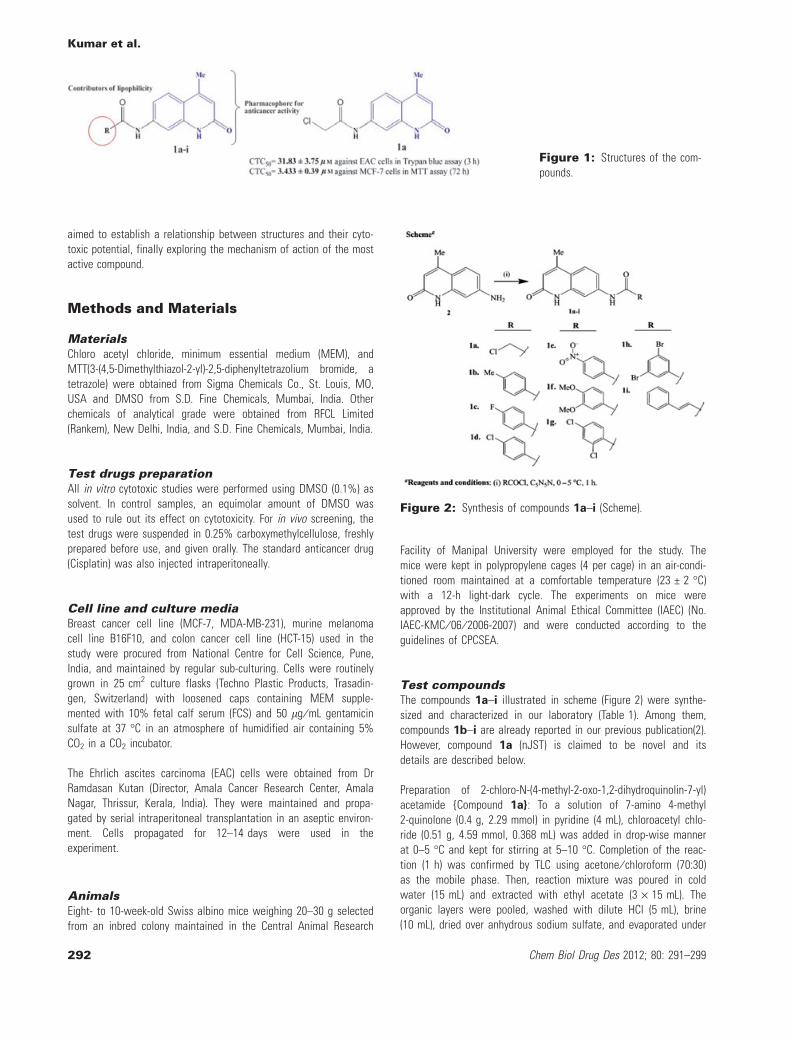
These studies added rising interest in developing and evaluatinganticancer activity of 2-quinolone derivatives through apoptotic path-way. Among quinolones, most of the anticancer compounds have a3-aryl substitution. However, to our knowledge nobody has exploredthe anticancer activity of 3-methyl substituted quinolones. Previously,we synthesized and evaluated compounds 1b–i and identified themas promising antimicrobial and antioxidant agents (5). But none ofthe compounds was tested for their cytotoxic potential against can-cer cell lines. With this rationale and our continuing effort to explorethe biological activity of 2-quinolones, we report here the synthesisand pharmacological evaluation of the compounds 1a–i containingin their structures a 2-quinolone moiety as the pharmacophore and afunctionally diverse side chain through amide linkage as the lipophi-licity contributor to the scaffold (Figure 1). Present study was also
291
Chem Biol Drug Des 2012; 80: 291–299
Research Article
ª 2012 John Wiley & Sons A/S
doi: 10.1111/j.1747-0285.2012.01402.x
aimed to establish a relationship between structures and their cyto-toxic potential, finally exploring the mechanism of action of the mostactive compound.
Methods and Materials
MaterialsChloro acetyl chloride, minimum essential medium (MEM), andMTT(3-(4,5-Dimethylthiazol-2-yl)-2,5-diphenyltetrazolium bromide, atetrazole) were obtained from Sigma Chemicals Co., St. Louis, MO,USA and DMSO from S.D. Fine Chemicals, Mumbai, India. Otherchemicals of analytical grade were obtained from RFCL Limited(Rankem), New Delhi, India, and S.D. Fine Chemicals, Mumbai, India.
Test drugs preparationAll in vitro cytotoxic studies were performed using DMSO (0.1%) assolvent. In control samples, an equimolar amount of DMSO wasused to rule out its effect on cytotoxicity. For in vivo screening, thetest drugs were suspended in 0.25% carboxymethylcellulose, freshlyprepared before use, and given orally. The standard anticancer drug(Cisplatin) was also injected intraperitoneally.
Cell line and culture mediaBreast cancer cell line (MCF-7, MDA-MB-231), murine melanomacell line B16F10, and colon cancer cell line (HCT-15) used in thestudy were procured from National Centre for Cell Science, Pune,India, and maintained by regular sub-culturing. Cells were routinelygrown in 25 cm2 culture flasks (Techno Plastic Products, Trasadin-gen, Switzerland) with loosened caps containing MEM supple-mented with 10% fetal calf serum (FCS) and 50 lg ⁄ mL gentamicinsulfate at 37 �C in an atmosphere of humidified air containing 5%CO2 in a CO2 incubator.
The Ehrlich ascites carcinoma (EAC) cells were obtained from DrRamdasan Kutan (Director, Amala Cancer Research Center, AmalaNagar, Thrissur, Kerala, India). They were maintained and propa-gated by serial intraperitoneal transplantation in an aseptic environ-ment. Cells propagated for 12–14 days were used in theexperiment.
AnimalsEight- to 10-week-old Swiss albino mice weighing 20–30 g selectedfrom an inbred colony maintained in the Central Animal Research
Facility of Manipal University were employed for the study. Themice were kept in polypropylene cages (4 per cage) in an air-condi-tioned room maintained at a comfortable temperature (23 € 2 �C)with a 12-h light-dark cycle. The experiments on mice wereapproved by the Institutional Animal Ethical Committee (IAEC) (No.IAEC-KMC ⁄ 06 ⁄ 2006-2007) and were conducted according to theguidelines of CPCSEA.
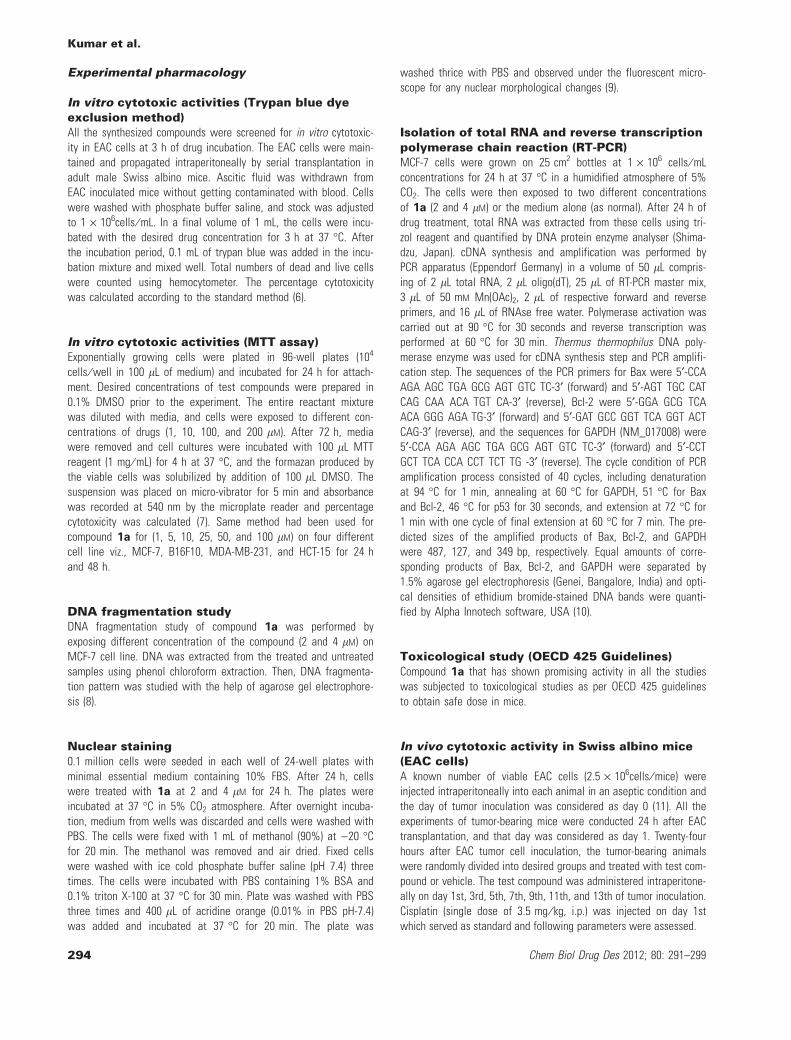
Test compoundsThe compounds 1a–i illustrated in scheme (Figure 2) were synthe-sized and characterized in our laboratory (Table 1). Among them,compounds 1b–i are already reported in our previous publication(2).However, compound 1a (nJST) is claimed to be novel and itsdetails are described below.
Preparation of 2-chloro-N-(4-methyl-2-oxo-1,2-dihydroquinolin-7-yl)acetamide {Compound 1a}: To a solution of 7-amino 4-methyl2-quinolone (0.4 g, 2.29 mmol) in pyridine (4 mL), chloroacetyl chlo-ride (0.51 g, 4.59 mmol, 0.368 mL) was added in drop-wise mannerat 0–5 �C and kept for stirring at 5–10 �C. Completion of the reac-tion (1 h) was confirmed by TLC using acetone ⁄ chloroform (70:30)as the mobile phase. Then, reaction mixture was poured in coldwater (15 mL) and extracted with ethyl acetate (3 · 15 mL). Theorganic layers were pooled, washed with dilute HCl (5 mL), brine(10 mL), dried over anhydrous sodium sulfate, and evaporated under
Figure 1: Structures of the com-pounds.
Figure 2: Synthesis of compounds 1a–i (Scheme).
Kumar et al.
292 Chem Biol Drug Des 2012; 80: 291–299
vacuum to afford 0.36-g crude oil. Crude compound was purified bycolumn chromatography using silica 100–200 and pet. ether ⁄ ethylacetate (6:4) as the mobile phase to afford 0.144 g off white solid,Yield: 25%; Melting Point: > 310 �C; Rf: 0.7 (acetone ⁄ chloroform,70:30). Kmax (methanol): 254 nm. IR (KBr, per cm): 3216 (N-H str),3018 (Ar-H str), 2968 (-CH3 str), 2852 (-CH2 str), 1676 (-CO str of
Antitumor Potential Through Bax-induced Apoptotic Pathway
Chem Biol Drug Des 2012; 80: 291–299 293
Experimental pharmacology
In vitro cytotoxic activities (Trypan blue dyeexclusion method)All the synthesized compounds were screened for in vitro cytotoxic-ity in EAC cells at 3 h of drug incubation. The EAC cells were main-tained and propagated intraperitoneally by serial transplantation inadult male Swiss albino mice. Ascitic fluid was withdrawn fromEAC inoculated mice without getting contaminated with blood. Cellswere washed with phosphate buffer saline, and stock was adjustedto 1 · 106cells ⁄ mL. In a final volume of 1 mL, the cells were incu-bated with the desired drug concentration for 3 h at 37 �C. Afterthe incubation period, 0.1 mL of trypan blue was added in the incu-bation mixture and mixed well. Total numbers of dead and live cellswere counted using hemocytometer. The percentage cytotoxicitywas calculated according to the standard method (6).
In vitro cytotoxic activities (MTT assay)Exponentially growing cells were plated in 96-well plates (104
cells ⁄ well in 100 lL of medium) and incubated for 24 h for attach-ment. Desired concentrations of test compounds were prepared in0.1% DMSO prior to the experiment. The entire reactant mixturewas diluted with media, and cells were exposed to different con-centrations of drugs (1, 10, 100, and 200 lM). After 72 h, mediawere removed and cell cultures were incubated with 100 lL MTTreagent (1 mg ⁄ mL) for 4 h at 37 �C, and the formazan produced bythe viable cells was solubilized by addition of 100 lL DMSO. Thesuspension was placed on micro-vibrator for 5 min and absorbancewas recorded at 540 nm by the microplate reader and percentagecytotoxicity was calculated (7). Same method had been used forcompound 1a for (1, 5, 10, 25, 50, and 100 lM) on four differentcell line viz., MCF-7, B16F10, MDA-MB-231, and HCT-15 for 24 hand 48 h.
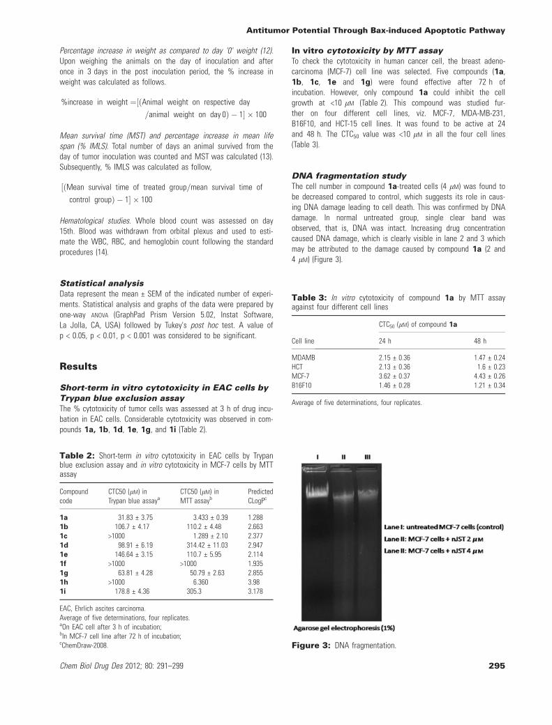
DNA fragmentation studyDNA fragmentation study of compound 1a was performed byexposing different concentration of the compound (2 and 4 lM) onMCF-7 cell line. DNA was extracted from the treated and untreatedsamples using phenol chloroform extraction. Then, DNA fragmenta-tion pattern was studied with the help of agarose gel electrophore-sis (8).
Nuclear staining0.1 million cells were seeded in each well of 24-well plates withminimal essential medium containing 10% FBS. After 24 h, cellswere treated with 1a at 2 and 4 lM for 24 h. The plates wereincubated at 37 �C in 5% CO2 atmosphere. After overnight incuba-tion, medium from wells was discarded and cells were washed withPBS. The cells were fixed with 1 mL of methanol (90%) at )20 �Cfor 20 min. The methanol was removed and air dried. Fixed cellswere washed with ice cold phosphate buffer saline (pH 7.4) threetimes. The cells were incubated with PBS containing 1% BSA and0.1% triton X-100 at 37 �C for 30 min. Plate was washed with PBSthree times and 400 lL of acridine orange (0.01% in PBS pH-7.4)was added and incubated at 37 �C for 20 min. The plate was
washed thrice with PBS and observed under the fluorescent micro-scope for any nuclear morphological changes (9).
Isolation of total RNA and reverse transcriptionpolymerase chain reaction (RT-PCR)MCF-7 cells were grown on 25 cm2 bottles at 1 · 106 cells ⁄ mLconcentrations for 24 h at 37 �C in a humidified atmosphere of 5%CO2. The cells were then exposed to two different concentrationsof 1a (2 and 4 lM) or the medium alone (as normal). After 24 h ofdrug treatment, total RNA was extracted from these cells using tri-zol reagent and quantified by DNA protein enzyme analyser (Shima-dzu, Japan). cDNA synthesis and amplification was performed byPCR apparatus (Eppendorf Germany) in a volume of 50 lL compris-ing of 2 lL total RNA, 2 lL oligo(dT), 25 lL of RT-PCR master mix,3 lL of 50 mM Mn(OAc)2, 2 lL of respective forward and reverseprimers, and 16 lL of RNAse free water. Polymerase activation wascarried out at 90 �C for 30 seconds and reverse transcription wasperformed at 60 �C for 30 min. Thermus thermophilus DNA poly-merase enzyme was used for cDNA synthesis step and PCR amplifi-cation step. The sequences of the PCR primers for Bax were 5¢-CCAAGA AGC TGA GCG AGT GTC TC-3¢ (forward) and 5¢-AGT TGC CATCAG CAA ACA TGT CA-3¢ (reverse), Bcl-2 were 5¢-GGA GCG TCAACA GGG AGA TG-3¢ (forward) and 5¢-GAT GCC GGT TCA GGT ACTCAG-3¢ (reverse), and the sequences for GAPDH (NM_017008) were5¢-CCA AGA AGC TGA GCG AGT GTC TC-3¢ (forward) and 5¢-CCTGCT TCA CCA CCT TCT TG -3¢ (reverse). The cycle condition of PCRamplification process consisted of 40 cycles, including denaturationat 94 �C for 1 min, annealing at 60 �C for GAPDH, 51 �C for Baxand Bcl-2, 46 �C for p53 for 30 seconds, and extension at 72 �C for1 min with one cycle of final extension at 60 �C for 7 min. The pre-dicted sizes of the amplified products of Bax, Bcl-2, and GAPDHwere 487, 127, and 349 bp, respectively. Equal amounts of corre-sponding products of Bax, Bcl-2, and GAPDH were separated by1.5% agarose gel electrophoresis (Genei, Bangalore, India) and opti-cal densities of ethidium bromide-stained DNA bands were quanti-fied by Alpha Innotech software, USA (10).
Toxicological study (OECD 425 Guidelines)Compound 1a that has shown promising activity in all the studieswas subjected to toxicological studies as per OECD 425 guidelinesto obtain safe dose in mice.
In vivo cytotoxic activity in Swiss albino mice(EAC cells)A known number of viable EAC cells (2.5 · 106cells ⁄ mice) wereinjected intraperitoneally into each animal in an aseptic condition andthe day of tumor inoculation was considered as day 0 (11). All theexperiments of tumor-bearing mice were conducted 24 h after EACtransplantation, and that day was considered as day 1. Twenty-fourhours after EAC tumor cell inoculation, the tumor-bearing animalswere randomly divided into desired groups and treated with test com-pound or vehicle. The test compound was administered intraperitone-ally on day 1st, 3rd, 5th, 7th, 9th, 11th, and 13th of tumor inoculation.Cisplatin (single dose of 3.5 mg ⁄ kg, i.p.) was injected on day 1stwhich served as standard and following parameters were assessed.
Kumar et al.
294 Chem Biol Drug Des 2012; 80: 291–299
Percentage increase in weight as compared to day '0' weight (12).Upon weighing the animals on the day of inoculation and afteronce in 3 days in the post inoculation period, the % increase inweight was calculated as follows.
%increase in weight ¼½ðAnimal weight on respective day
=animal weight on day 0Þ � 1� � 100
Mean survival time (MST) and percentage increase in mean lifespan (% IMLS). Total number of days an animal survived from theday of tumor inoculation was counted and MST was calculated (13).Subsequently, % IMLS was calculated as follow,
½ðMean survival time of treated group=mean survival time of
control groupÞ � 1� � 100
Hematological studies. Whole blood count was assessed on day15th. Blood was withdrawn from orbital plexus and used to esti-mate the WBC, RBC, and hemoglobin count following the standardprocedures (14).
Statistical analysisData represent the mean € SEM of the indicated number of experi-ments. Statistical analysis and graphs of the data were prepared byone-way ANOVA (GraphPad Prism Version 5.02, Instat Software,La Jolla, CA, USA) followed by Tukey's post hoc test. A value ofp < 0.05, p < 0.01, p < 0.001 was considered to be significant.
Results
Short-term in vitro cytotoxicity in EAC cells byTrypan blue exclusion assayThe % cytotoxicity of tumor cells was assessed at 3 h of drug incu-bation in EAC cells. Considerable cytotoxicity was observed in com-pounds 1a, 1b, 1d, 1e, 1g, and 1i (Table 2).
In vitro cytotoxicity by MTT assayTo check the cytotoxicity in human cancer cell, the breast adeno-carcinoma (MCF-7) cell line was selected. Five compounds (1a,1b, 1c, 1e and 1g) were found effective after 72 h ofincubation. However, only compound 1a could inhibit the cellgrowth at <10 lM (Table 2). This compound was studied fur-ther on four different cell lines, viz. MCF-7, MDA-MB-231,B16F10, and HCT-15 cell lines. It was found to be active at 24and 48 h. The CTC50 value was <10 lM in all the four cell lines(Table 3).
DNA fragmentation studyThe cell number in compound 1a-treated cells (4 lM) was found tobe decreased compared to control, which suggests its role in caus-ing DNA damage leading to cell death. This was confirmed by DNAdamage. In normal untreated group, single clear band wasobserved, that is, DNA was intact. Increasing drug concentrationcaused DNA damage, which is clearly visible in lane 2 and 3 whichmay be attributed to the damage caused by compound 1a (2 and4 lM) (Figure 3).
Table 2: Short-term in vitro cytotoxicity in EAC cells by Trypanblue exclusion assay and in vitro cytotoxicity in MCF-7 cells by MTTassay
EAC, Ehrlich ascites carcinoma.Average of five determinations, four replicates.aOn EAC cell after 3 h of incubation;bIn MCF-7 cell line after 72 h of incubation;cChemDraw-2008.
Table 3: In vitro cytotoxicity of compound 1a by MTT assayagainst four different cell lines
Antitumor Potential Through Bax-induced Apoptotic Pathway
Chem Biol Drug Des 2012; 80: 291–299 295
Nuclear stainingAlteration in the nuclear structure of the drug-treated cells is oneof the important aspects to study nuclear morphology. To visualizenuclear morphology, nuclear staining was carried out using dye acri-dine orange. The study was carried out against MCF-7 cells.Nucleus in control cells was very much intact, round, or oval inshape, without any condensation and blabbing. Cytoplasm was nor-mal with intact cell membrane and cytoplasm disintegration wasnot observed. When cells were treated with compound 1a (2 and4 lM), they showed typical apoptotic morphological changes suchas nuclear and cytoplasmic condensation, loss of cell volume, andnuclear fragmentation (Figure 4).
Reverse transcriptase PCR analysisReverse transcriptase PCR was used to analyze the levels of Baxand Bcl-2 mRNA levels in MCF-7 cells treated with compound 1a
(2 and 4 lM). Amplification of Bax and Bcl-2 was carried out withhelp of specific primers using cDNA as a template strand preparedfrom MCF-7 cells, and the specificity was confirmed using agarosegel electrophoresis.
Bax and Bcl-2 mRNA expressionThe mRNA levels of Bax and Bcl-2 from MCF-7 cells are shown inFigure 5. The levels of Bax in MCF-7 cells with compound 1a
treatment increased significantly compared with untreated normalcells (p < 0.001). Compound 1a at 4 lM concentration caused asignificant decrease of Bcl-2 mRNA levels when compared with thatof the normal cells (p < 0.001).
Change in body weight in EAC inoculated miceA significant gain in body weight was observed in EAC inoculatedcontrol mice, while maximum gain (47%) was observed within15 days. Cisplatin administration on day 1st significantly(p < 0.05) reduced the elevated body weight. Drug treatment(compound 1a at 50 mg ⁄ kg and 100 mg ⁄ kg) was monitored everyalternate day for body weight. In the treated mice, no significantchange in body weight was observed till 5th day. All the treat-ment showed a significant reduction in bodyweight compared tocontrol from day 9 onwards. On day 7, only cisplatin treatmentand 1a treatment at 100 mg ⁄ kg have shown the significantreduction in body weight compared to sham control (p < 0.05)(Figure 6).
Effect on survival time in EAC inoculated miceIn EAC inoculated control mice, MST was found to be 16.16, whilemedian survival time was 16. In cisplatin treatment, a significantincrease in MST (38.16) and % IMLS (138.1) was observed. 1a hassignificantly increased MST to 24.16 days (p < 0.05) and 27.5 days(p < 0.01), while % IMLS was 48.5 and 70.1 days for 50 mg ⁄ kgand 100 mg ⁄ kg, respectively. Median survival time for cisplatin,compound 1a at 50 and 100 mg ⁄ kg was found to be 40, 24, and27 day, respectively (Figures 7 and 8).
Effect of synthesized compounds onhematological parameters in EAC inoculatedmiceWBC increased about twofold in control EAC inoculated mice,which was significantly (p < 0.001) reversed by cisplatin treatmentand 1a treatment at 100 mg ⁄ kg (p < 0.01). Tumor induction incontrol mice reduced the RBC count compared to normal animals(p < 0.01), which was significantly improved by tested compound
Figure 4: Nuclear Staining.
Figure 5: Bax and Bcl-2 expression.Figure 6: Percentage increase in body weight. All the valuesare mean € SEM of six animals.
Kumar et al.
296 Chem Biol Drug Des 2012; 80: 291–299
at 100 mg ⁄ kg (p < 0.05). Tumor development in the animalscaused significant anemia (decrease in hemoglobin content andRBC count), which was significantly (p < 0.01) reversed bycisplatin treatment and compound 1a (100 mg ⁄ kg) (p < 0.01) (Fig-ure 9).
Discussion
ChemistryThe solution phase syntheses of compounds 1a–i are depicted inscheme (Figure 2). Previously reported by Jayshree et al., 2010,compounds 1b–i were resynthesized and confirmed by comparingwith the reported Rf values and melting points. Novel analog of theseries compound 1a was synthesized by acetylating 7-amino-4-methylquinolin-2(1H)-one (compound 2) using the same protocolapplied for the preparation of compounds 1b–i and characterizedby 1HNMR, GC-MS, IR, and UV.
PharmacologyAnticancer drug development has become an extremely competitiveand expensive process with high rate of failures. So, the presentstudy was designed to explore the possible in vitro and in vivo anti-cancer activity of already existing quinolone compounds, which have
previously been screened for other activity (5). Generally, anticanceractivity of most of the chemotherapeutic agents is owing to theircytotoxicity (15). Trypan blue exclusion assay and MTT assay arethe widely used method for evaluation of cytotoxicity.
Therefore, a series of nine novel substituted N-(4-methyl-2-oxo-1,2dihydroquinolin-7-yl) amides were synthesized and subjected to Try-pan blue assay against EAC cells and MTT assay against MCF-7cell line. The CTC50 was determined after 3 h of incubation in Try-pan blue assay and after 72 h in MTT assay. CTC50 of our com-pounds are summarized in Table 2. Of the nine compoundssynthesized, compound 1a was found to be most active with CTC50
of 31.83 lM against EAC cells and compound 1c was the mostpotent (CTC50 = 1.289 lM) to inhibit the proliferation of MCF-5 celllines. Compounds with electron-withdrawing group substituted atthe phenyl ring appear to be more activity against MCF-7 cell line.However, functional modifications on the phenyl ring show a com-plex relationship with the anticancer activity against EAC cells inTrypan blue assay.
Figure 9: Hematological parameters. All the values aremean € SEM of three animals, where *p < 0.05, **p < 0.01,***p < 0.001 are compared to control and ap < 0.05, bp < 0.01,cp < 0.001 are compared to Sham animals.
Figure 8: Percentage survival. The data represent six animals ineach group.
Antitumor Potential Through Bax-induced Apoptotic Pathway
Chem Biol Drug Des 2012; 80: 291–299 297
Structure-activity relationshipsTo explore the structure-activity relationships (SAR) of substitutedN-(4-methyl-2-oxo-1,2 dihydroquinolin-7-yl) amides, functional modi-fications have been carried out at the amino group.
The compound 1a with haloalkyl chain attached to the parentnucleus through an amide linkage at amino group was consideredas a reasonable starting point for SAR studies. It showed signifi-cant anticancer activity of CTC50 = 31.83 lM against EAC cells inTrypan blue assay and CTC50 = 3.433 lM against MCF-7 cell line inMTT assay. Encouraged with this result, a more lipophilic(ClogP = 2.663) '4-toluyl' substituent was introduced in place ofhaloalkyl substituent of compound 1a to afford compound 1b. Toour surprise, this modification drastically lowered the anticanceractivity in multifold against both the cancer cells. Because the elec-tron-donating substituent on phenyl ring was inactive, analogs of1b in which the 4-methyl substituent on phenyl ring was alteredwith electron-withdrawing group like 4-fluorine (compound 1c) and4-chlorine (compound 1d) to investigate the cytotoxicity. Remark-ably, compound 1c exhibited an excellent improvement in CTC50 of1.289 lM against MCF-7 cell line where as compound 1d showeda little improvement in CTC50 of 98.91 lM against the proliferationof EAC cells. The promising results of compound 1c and 1d
prompted us to probe the effect of a charged –NO2 group in placeof halogens on the phenyl ring. Nevertheless, it was failed toimprove the anticancer activity. Introduction of dimethoxy functional-ity in the phenyl ring of our moiety also resulted in equally frustrat-ing activity (CTC50 h 1000 lM against both the cells). Discouragedwith the insensitivity of electron-donating functionality on antican-cer activity, strong electron-withdrawing functional groups like –Cland –Br were placed twice in the phenyl to afford compounds 1g
and 1h, respectively. Interestingly, with this simple modification,compound 1h showed a CTC50 of 6.360 lM against MCF-7 celllines, whereas compound 1g was moderately active against boththe cancer cells. However, introduction of a spacer in betweenamide functionality and the phenyl ring of our moiety obliteratedthe anticancer activity. On the contrary, our SAR study reveals thatnature and position of functional groups on the phenyl ring influ-ence the anticancer activity of N-(4-methyl-2-oxo-1,2 dihydroquino-lin-7-yl) amides. In addition, we speculate that the functional groupthat increases the acidity of our parent nucleus is essential for theanticancer activity. However, in our SAR study, we could not estab-lish a strong correlation between anticancer activity and Log P ofthe compounds.
As compound 1a has shown better cytotoxicity than other com-pounds (CTC50 = 3.433 € 0.39 lM), it has been screened further onfour cell lines including MCF-7 for 24 and 48 h. It has also shownpromising result on the remaining cell lines at both time intervalswith CTC50 <10 lM. This compound has been further studied forDNA fragmentation assay on MCF-7 cell line. It has shown a bandwith laddering, which indicates its action responsible for DNAdamage. Nuclear staining experiments were performed using acri-dine orange staining dye. Compound 1a at 2 and 4 lM concentra-tion was tested against MCF-7 cells, and nuclear staining imagesshowed induction of apoptosis such as membrane blebbing,cytoplasmic condensation, and nuclear fragmentation. Furtherreverse transcriptase PCR (RT-PCR) was performed to confirm the
involvement of apoptosis in MCF-7 cells. RNA was isolated fromMCF-7 cells with and without treatment with 1a 2 and 4 lM. Prim-ers specific for Bax and Bcl-2 were used to confirm the involvementof apoptosis. RT-PCR results showed upregulation of Bax expressionand downregulation of Bcl-2 expression when compared with thatof untreated MCF-7 cells. It has been reported that there is astrong relationship in apoptosis and farnesyl transferase inhibition(16–18). Hence, this finding supports that molecule 1a may be afarnesyl transferase inhibitor.
Ehrlich tumor is a hurriedly growing carcinoma with very aggressivebehavior (19). It can grow in almost all strains of mice. The tumorimplantation induces a local inflammatory reaction, with increasingvascular permeability, resulting in an intense edema formation, cel-lular migration, and progressive ascitic fluid formation (20). The as-citic fluid is essential for tumor growth, as it constitutes a directnutritional source for tumor cells. Drug treatment was given on 1st,3rd, 5th, 7th, 9th, 11th, and 13th day of inoculation and checkedfor anticancer activity at these time points. Compound 1a has beenfound to be significantly effective in reducing the body weight andalso improved MST significantly compared to control EAC injectedmice. Prolongation in the life span of tumor inoculated mice is areliable criterion for judging the anticancer activity of any drug (13).In untreated mice, EAC inoculation causes 100% mortality within20 days, which is supported by our present data. An enhancementof life span by 25% or more over that of control was considered aseffective antitumor response (13). Treatment with compound 1a at50 and 100 mg ⁄ kg has delayed the onset of mortality andincreased the life span of EAC inoculated mice by 49.48% and70.1%, respectively.
Significant decrease in hemoglobin and RBC (owing to hypoxic con-dition) with parallel increase in WBC [may be owing to the influ-ence of tumor cell (antigen) on the immune system] has beenobserved in EAC inoculated control mice. Compound 1a at100 mg ⁄ kg significantly reduced the elevated WBC count andincreased RBC number compared to EAC inoculated control mice.The reversal of WBC indicates protective action on the hemopoieticsystem and supports the antitumor activity of the compound. Usu-ally, in cancer chemotherapy, the major problems that are encoun-tered include myelosuppression and anemia (21). The results hadclearly shown that compound 1a at 100 mg ⁄ kg was found to besignificantly active in improving the hemoglobin content and RBCcount to normal levels.
Conclusion
The study shows that 2-quinolone without 3-aryl substitution canalso be a potential moiety for anticancer research. Upregulationof Bax expression and downregulation of BCl-2 expression incancer cells is a very important event in apoptosis. Hence, wepropose Bax-induced apoptosis as a potential alternativemechanism of 2-quinolone derivatives other than farnesyl transfer-ase inhibition. Further, they have the significant cytotoxic poten-tial and are safe as indicated by acute toxicity activity.Compound 1a merits further investigation to explore in molecularmechanisms.
Kumar et al.
298 Chem Biol Drug Des 2012; 80: 291–299
Acknowledgment
We thank All India Council of Technical Education (AICTE) for sup-porting in vitro studies (8023 ⁄ BOR ⁄ RID ⁄ RPS-154 ⁄ 2007-08).
References
1. Joseph B., Darro F., B�hard A., Lesur B., Collignon F., Decaestec-ker C., Frydman A., Guillaumet G., Kiss R. (2002) 3-Aryl-2-quino-lone derivatives: synthesis and characterization of in vitro and invivo antitumor effects with emphasis on a new therapeuticaltarget connected with cell migration. J Med Chem;45:2543–2555.
2. Izbicka E., Campos D., Patnaik A., Carrizales G. (2005) Biomar-kers of anticancer activity of R115777 (Tipifarnib, Zarnestra) inhuman breast cancer models in vitro. Anticancer Res;25:3215–3223.
3. Morgan M.A., Dolp O., Reuter C.W. (2001) Cell-cycle-dependentactivation of mitogen-activated protein kinase kinase (MEK-1 ⁄ 2)in myeloid leukemia cell lines and induction of growth inhibitionand apoptosis by inhibitors of RAS signaling. Blood;97:1823–1834.
4. Binder C., Marx D., Binder L., Schauer A., Hiddemann W. (1996)Expression of Bax in relation to Bcl-2 and other predictiveparameters in breast cancer. Ann Oncol;7:129–133.
5. Jayashree B.S., Thomas S., Nayak Y. (2010) Design and synthe-sis of 2-quinolones as antioxidants and antimicrobials: a rationalapproach. Med Chem Res;19:193–209.
6. Sheeja K.R., Kuttan G., Kuttan R. (1997) Cytotoxic and antitumoractivity of berberin. Amala Res Bull;17:73–76.
7. Denizot F., Lang R. (1986) Rapid colorimetric assay for cellgrowth and survival. Modifications to the tetrazolium dye proce-dure giving improved sensitivity and reliability. J Immunol Meth-ods;89:271–277.
8. Raj P.V., Nitesh K., Chandrashekhar K.R., Rao C.M., Rao J.V.,Udupa N. (2010a) Effect of lecithin and silymarin on D-galactos-amine induced toxicity in isolated hepatocytes and rats. Indian JClin Biochem;25:169–174.
9. Zainal Ariffin S.H., Wan Omar W.H., Zainal Ariffin Z., SafianM.F., Senafi S., Megat Abdul Wahab R. (2009) Intrinsic anticar-cinogenic effects of Piper sarmentosum ethanolic extract on ahuman hepatoma cell line. Cancer Cell Int;9:6.
10. Raj P.V., Nitesh K., Gang S.S., Jagani H.V., Chandrashekhar H.R.,Rao J.V., Rao C.M., Udupa N. (2010b) Protective role of catechinin D-galactosamine induced hepatotoxicity through p53 depen-dent pathway. Indian J Clin Biochem;25:349–356.
11. Jagetia G.C., Baliga M.S. (2003) Modulation of antineoplasticactivity of cyclophosphamide by Alstonia scholaris in the Ehrlichascites carcinoma-bearing mice. J Exp Ther Oncol;3:272–282.
13. Hazra B., Sarkar R., Bhattacharyya S., Roy P. (2002) Tumor inhibi-tory activity of chicory root extract against Ehrlich ascites carci-noma in mice. Fitoterapia;73:730–733.
14. Brandao R., Borges L.P., de Oliveira R., Rocha J.B., NoqueiraC.W. (2008) Diphenyl diselenide protects against hematologicaland immunological alterations induced by mercury in mice. JBiochem Mol Toxicol;22:311–319.
15. Jackson J.K., Gleave M.E., Yago V., Beraldi E., Hunter W.L., BurtH.M. (2000) The suppression of human prostate tumor growth inmice by the intratumoral injection of a slow-release polymericpaste formulation of Paclitaxel. Cancer Res;60:4146–4151.
16. Manne V., Lee F.Y., Bol D.K., Gullo-Brown J., Fairchild C.R., Lom-bardo L.J., Smykla R.A., Vite G.D., Wen M.L., Yu C., Wong T.W.,Hunt J.T. (2004) Apoptotic and cytostatic farnesyltransferaseinhibitors have distinct pharmacology and efficacy profiles intumor models. Cancer Res;64:3974–3980.
17. Rose W.C., Lee F.Y., Fairchild C.R., Lynch M., Monticello T., Kra-mer R.A., Manne V. (2001) Preclinical antitumor activity of BMS-214662, a highly apoptotic and novel farnesyltransferase inhibi-tor. Cancer Res;61:7507–7517.
18. Gomez-Benito M., Marzo I., Anel A., Naval J. (2005) Farnesyl-transferase inhibitors BMS-214662 induces apoptosis in mye-loma cells through PUMA up-regulation, Bax and Bak activation.And Mcl-1 elimination. Mol Pharmacol;67:1991–1998.
19. Segura J.A., Barbero L.G., Marquez J. (2000) Ehrlich ascitestumour unbalances splenic cell populations and reduces respon-siveness of T cells to Staphylococcus aureus enterotoxin B stim-ulation. Immunol Lett;74:111–115.
20. Fecchio D., Sirois P., Russo M., Jancar S. (1990) Studies oninflammatory response induced by Ehrlich tumor in mice perito-neal cavity. Inflammation;14:125–132.


![BAX.0001 [AV 591+B ] Rev-01.doc](https://static.documents.pub/doc/80x56/55cf905d550346703ba539e7/bax0001-av-591b-rev-01doc.jpg)


