8/18/18 1 Early Embryology Kristine Krafts, M.D. • In general, what happens in the first and second phases of the embryonic period? • What happens during week 1 and 2? • Describe the main events in gastrulation. • How does the neural tube form, and what happens to it? Embryology Lecture Objectives • What is the fate of the endoderm, mesoderm and ectoderm? • What are the pharyngeal arches? What structures arise from each arch, groove, and cleft? • What structures do neural crest cells form? major stuff forms from pouches? Embryology Lecture Objectives General overview of prenatal development Embryonic period phase 1 • Formation of bilaminar disk • Formation of trilaminar disk (gastrulation) Embryonic period phase 2 • Formation of neural tube • Differentiation of mesoderm • Folding of embryo • Formation of pharyngeal arches Embryology Lecture Outline General overview of prenatal development Embryology Lecture Outline Prenatal Development Fertilization 1 2 3 4 Phase 1 Cellular proliferation and migration 5 6 7 8 Phase 2 Differentiation of internal & external structures 40 Phase 3 Growth and maturation Embryo Fetus 0 Delivery
Transcript
8/18/18
1
Early EmbryologyKristine Krafts, M.D.
• In general, what happens in the first and second phases of the embryonic period?
• What happens during week 1 and 2?
• Describe the main events in gastrulation.
• How does the neural tube form, and what happens to it?
Embryology Lecture Objectives
• What is the fate of the endoderm, mesoderm and ectoderm?
• What are the pharyngeal arches? What structures arise from each arch, groove, and cleft?
• What structures do neural crest cells form? major stuff forms from pouches?
Embryology Lecture Objectives
General overview of prenatal development
Embryonic period phase 1• Formation of bilaminar disk• Formation of trilaminar disk (gastrulation)
Embryonic period phase 2• Formation of neural tube• Differentiation of mesoderm• Folding of embryo• Formation of pharyngeal arches
Embryology Lecture Outline
General overview of prenatal development
Embryology Lecture Outline Prenatal Development
Fertilization
1 2 3 4
Phase 1Cellular
proliferation and migration
5 6 7 8
Phase 2Differentiation of
internal & external structures
40
Phase 3Growth and
maturation
Embryo Fetus
0
Delivery
8/18/18
2
This YouTube video is awesome at explaining early embryonic development:
http://www.youtube.com/watch?v=rN3lep6roRI General overview of prenatal development
Embryonic period phase 1
Embryology Lecture Outline
Prenatal Development
Fertilization
1 2 3 4
Phase 1Cellular
proliferation and migration
5 6 7 8
Phase 2Differentiation of
internal & external structures
40
Phase 3Growth and
maturation
Embryo Fetus
0
Delivery
General overview of prenatal development
Embryonic period phase 1• Formation of bilaminar disk
Embryology Lecture Outline
Week 1: Differentiation of Morula into Blastocyst
Morula Blastocyst
Week 2: Formation of Bilaminar Germ Disk
epiblast
hypoblast
8/18/18
3
General overview of prenatal development
Embryonic period phase 1• Formation of bilaminar disk• Formation of trilaminar disk (gastrulation)
Embryology Lecture Outline
"It is not birth, marriage, or death, but gastrulation which is truly the most important time in your life.”
- Lewis Wolpert (1986)
Gastrulation: formation of primitive streak
primitive streak
primitive node
epiblast
Gastrulation: movement and differentiation of epiblast cells
Epiblast cells give rise to all three germ cell layers!(the hypoblast does NOT turn into endoderm)
Bilaminar germ disk
EpiblastHypoblast
Endoderm EndodermMesoderm
Primitive streak
Ectoderm
Gastrulation: formation of notochord
The notochord is super important because it tells the three layers what to do next.
General overview of prenatal development
Embryonic period phase 1• Formation of bilaminar disk• Formation of trilaminar disk (gastrulation)
Embryonic period phase 2
Embryology Lecture Outline Prenatal Development
Fertilization
1 2 3 4
Phase 1Cellular
proliferation and migration
5 6 7 8
Phase 2Differentiation of
internal & external structures
40
Phase 3Growth and
maturation
Embryo Fetus
0
Delivery
8/18/18
4
General overview of prenatal development
Embryonic period phase 1• Formation of bilaminar disk• Formation of trilaminar disk (gastrulation)
Embryonic period phase 2• Formation of neural tube
Embryology Lecture OutlineFormation of Neural Tube
Formation of the Neural Tube
General overview of prenatal development
Embryonic period phase 1• Formation of bilaminar disk• Formation of trilaminar disk (gastrulation)
Embryonic period phase 2• Formation of neural tube• Differentiation of mesoderm
Embryology Lecture Outline
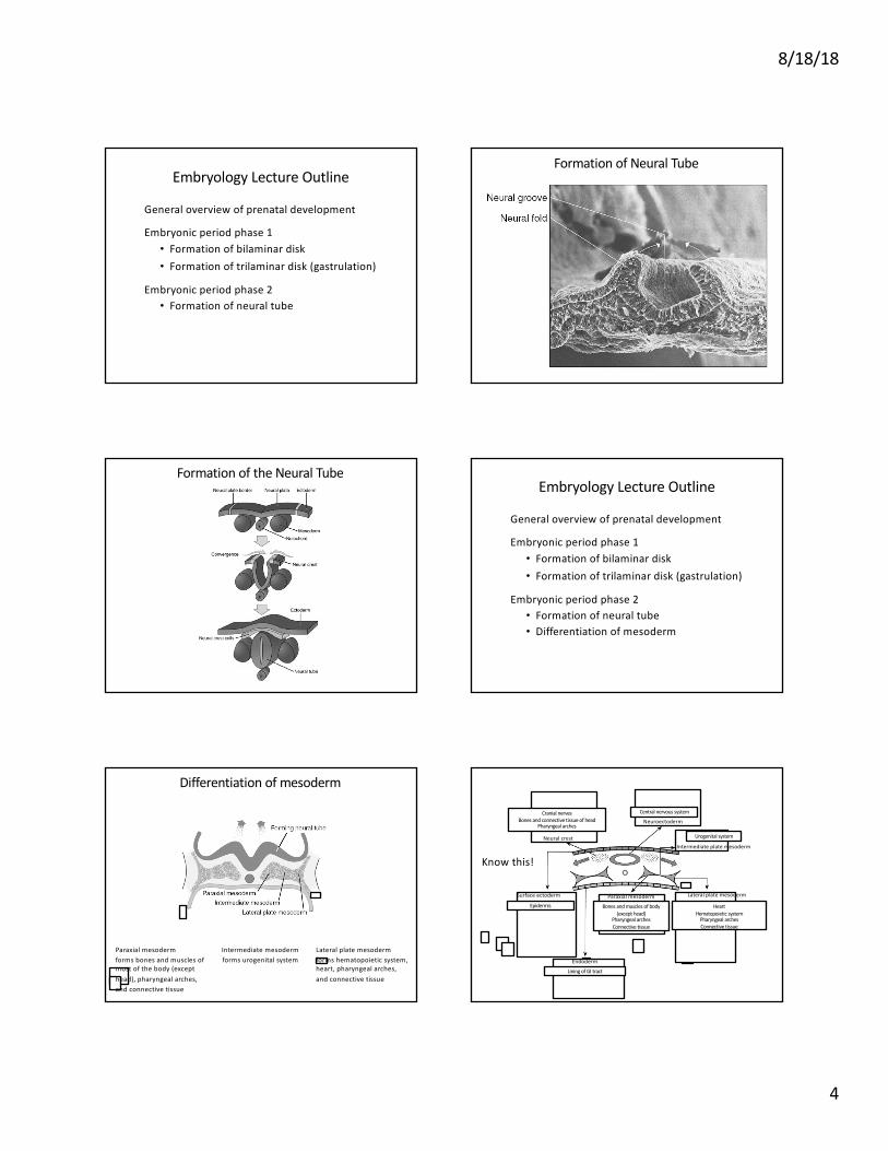
Differentiation of mesoderm
Paraxial mesoderm forms bones and muscles of most of the body (except head), pharyngeal arches, and connective tissue
Intermediate mesoderm forms urogenital system
Lateral plate mesoderm forms hematopoietic system, heart, pharyngeal arches, and connective tissue