Title Embryonic lethality in mice lacking the nuclear factor of activated t cells 5 protein due to impaired cardiac development and function Author(s) Mak, MC; Lam, KM; Chan, PK; Lau, YB; Tang, WH; Yeung, PKK; Ko, BCB; Chung, SMS; Chung, SK Citation Plos One, 2011, v. 6 n. 7 Issued Date 2011 URL http://hdl.handle.net/10722/137191 Rights Creative Commons: Attribution 3.0 Hong Kong License
Transcript
TitleEmbryonic lethality in mice lacking the nuclear factor ofactivated t cells 5 protein due to impaired cardiac developmentand function
Rights Creative Commons: Attribution 3.0 Hong Kong License
Embryonic Lethality in Mice Lacking the Nuclear Factorof Activated T Cells 5 Protein Due to Impaired CardiacDevelopment and FunctionMan Chi Mak1., Ka Man Lam1., Ping Kei Chan1, Yu Bond Lau1, Wai Ho Tang2, Patrick Ka Kit Yeung1, Ben
Chi Bun Ko3, Stephen Man Sum Chung2, Sookja Kim Chung1*
1 Department of Anatomy, Li Ka Shing Faculty of Medicine, The University of Hong Kong, Hong Kong SAR, China, 2 Division of Science and Technology, United
International College, Zhuhai, Guandong, China, 3 Department of Anatomical and Cellular Pathology, The Chinese University of Hong Kong, Hong Kong SAR, China
Abstract
Nuclear factor of activated T cells 5 protein (NFAT5) is thought to be important for cellular adaptation to osmotic stressby regulating the transcription of genes responsible for the synthesis or transport of organic osmolytes. It is alsothought to play a role in immune function, myogenesis and cancer invasion. To better understand the function ofNFAT5, we developed NFAT5 gene knockout mice. Homozygous NFAT5 null (NFAT52/2) mouse embryos failed todevelop normally and died after 14.5 days of embryonic development (E14.5). The embryos showed peripheral edema,and abnormal heart development as indicated by thinner ventricular wall and reduced cell density at the compact andtrabecular areas of myocardium. This is associated with reduced level of proliferating cell nuclear antigen and increasedcaspase-3 in these tissues. Cardiomyocytes from E14.5 NFAT52/2 embryos showed a significant reduction of beatingrate and abnormal Ca2+ signaling profile as a consequence of reduced sarco(endo)plasmic reticulum Ca2+-ATPase(SERCA) and ryanodine receptor (RyR) expressions. Expression of NFAT5 target genes, such as HSP 70 and SMIT werereduced in NFAT52/2 cardiomyocytes. Our findings demonstrated an essential role of NFAT5 in cardiac developmentand Ca2+ signaling. Cardiac failure is most likely responsible for the peripheral edema and death of NFAT52/2 embryosat E14.5 days.
Citation: Mak MC, Lam KM, Chan PK, Lau YB, Tang WH, et al. (2011) Embryonic Lethality in Mice Lacking the Nuclear Factor of Activated T Cells 5 Protein Due toImpaired Cardiac Development and Function. PLoS ONE 6(7): e19186. doi:10.1371/journal.pone.0019186
Editor: Saverio Bellusci, Children’s Hospital Los Angeles, United States of America
Received December 22, 2010; Accepted March 22, 2011; Published July 12, 2011
Copyright: � 2011 Mak et al. This is an open-access article distributed under the terms of the Creative Commons Attribution License, which permits unrestricteduse, distribution, and reproduction in any medium, provided the original author and source are credited.
Funding: This work was supported by the Research Grant Council Grants The University of Hong Kong 7504/06M. The funders had no role in study design, datacollection and analysis, decision to publish, or preparation of the manuscript.
Competing Interests: The authors have declared that no competing interests exist.
Nuclear factor of activated T cells 5 protein (NFAT5), also
called tonicity element binding protein (TonEBP) [1] or osmotic
response element binding protein (OREBP) [2], is a member of the
Rel family of transcription factor with a conserved DNA binding
Rel domain [3]. Although NFAT5 has similar a DNA binding
domain as other NFATs (NFAT1–4), which are regulated by
calcium/calcineurin and primarily involved in the regulation of
cytokine and other genes important for the immune response in T
lymphocytes [4], its regulation and biological functions are quite
different from the other NFATs. When there is hypertonic stress,
NFAT5 is translocated to the nucleus and regulates gene tran-
scriptions, which are responsible for the import or synthesis of
organic osmolytes such as myo-inositol, betaine, taurine, and
sorbitol [5]. NFAT5 mRNA is also stabilized by hypertonic stress,
leading to increased synthesis of NFAT5 [6]. Activated NFAT5
regulates the transcription of sodium/myo-inositol cotransporter
(SMIT), sodium-chloride-betaine cotransporter (BGT1), and
taurine transporter (TauT), which are responsible for the cellular
uptake of myo-inositol, betaine and taurine, respectively. Tran-
scription of aldose reductase (AR), which is involved in the
synthesis of sorbitol, is also regulated by NFAT5 [7]. Apart from
the osmoprotective genes, heat shock protein 70 (HSP70) gene also
contains an osmotic response element (ORE), and its expression is
regulated by NFAT5 under hypertonic stress [8]. The critical role
of NFAT5 in osmoprotection has been demonstrated in NFAT5
knockout mice [9,10]. The vast majority of the NFAT5 null mice
died at the embryonic stage, and the few that survived to adult
stage, exhibited kidney atrophy in the medulla with reduced level
of AR, BGT1, and SMIT [10].
Besides the renal medulla, where the epithelial cells are
constantly exposed to hypertonicity, NFAT5 mRNA has also
been detected in the brain, heart and T lymphocytes [11,12],
suggesting it may have functions other than osmoprotection. In T
lymphocytes, NFAT5 can be induced by both hypertonicity and
mitogen [12]. NFAT5 is detected in some transformed cells which
are integrin-mediated in carcinoma metastasis [13]. Moreover,
several studies have suggested that NFAT5 plays a role in cell
differentiation [10,14,15]. To better understand the physiological
functions of this protein, NFAT5 knockout mice were generated
and used in the present study. Here we show that the embryonic
lethality for NFAT5 null mutant NFAT52/2 mice is likely due to
impaired cardiac development.
PLoS ONE | www.plosone.org 1 July 2011 | Volume 6 | Issue 7 | e19186
Results
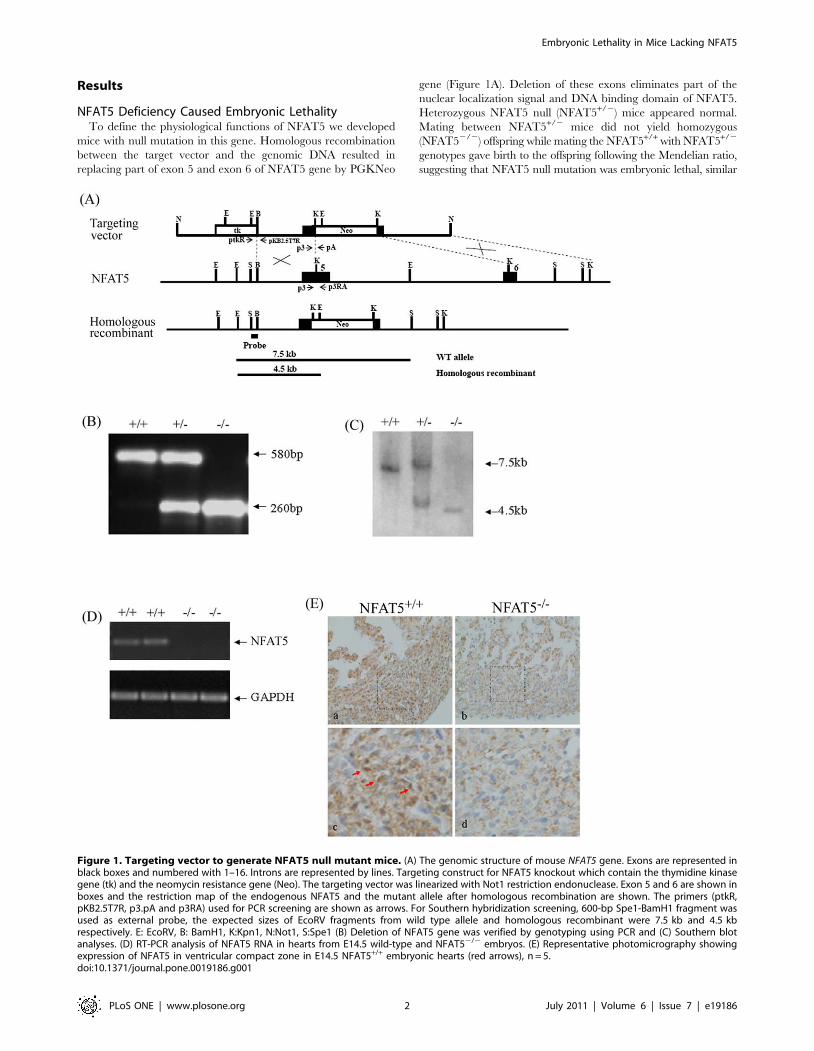
NFAT5 Deficiency Caused Embryonic LethalityTo define the physiological functions of NFAT5 we developed
mice with null mutation in this gene. Homologous recombination
between the target vector and the genomic DNA resulted in
replacing part of exon 5 and exon 6 of NFAT5 gene by PGKNeo
gene (Figure 1A). Deletion of these exons eliminates part of the
nuclear localization signal and DNA binding domain of NFAT5.
Heterozygous NFAT5 null (NFAT5+/2) mice appeared normal.
Mating between NFAT5+/2 mice did not yield homozygous
(NFAT52/2) offspring while mating the NFAT5+/+ with NFAT5+/2
genotypes gave birth to the offspring following the Mendelian ratio,
suggesting that NFAT5 null mutation was embryonic lethal, similar
Figure 1. Targeting vector to generate NFAT5 null mutant mice. (A) The genomic structure of mouse NFAT5 gene. Exons are represented inblack boxes and numbered with 1–16. Introns are represented by lines. Targeting construct for NFAT5 knockout which contain the thymidine kinasegene (tk) and the neomycin resistance gene (Neo). The targeting vector was linearized with Not1 restriction endonuclease. Exon 5 and 6 are shown inboxes and the restriction map of the endogenous NFAT5 and the mutant allele after homologous recombination are shown. The primers (ptkR,pKB2.5T7R, p3.pA and p3RA) used for PCR screening are shown as arrows. For Southern hybridization screening, 600-bp Spe1-BamH1 fragment wasused as external probe, the expected sizes of EcoRV fragments from wild type allele and homologous recombinant were 7.5 kb and 4.5 kbrespectively. E: EcoRV, B: BamH1, K:Kpn1, N:Not1, S:Spe1 (B) Deletion of NFAT5 gene was verified by genotyping using PCR and (C) Southern blotanalyses. (D) RT-PCR analysis of NFAT5 RNA in hearts from E14.5 wild-type and NFAT52/2 embryos. (E) Representative photomicrography showingexpression of NFAT5 in ventricular compact zone in E14.5 NFAT5+/+ embryonic hearts (red arrows), n = 5.doi:10.1371/journal.pone.0019186.g001
Embryonic Lethality in Mice Lacking NFAT5
PLoS ONE | www.plosone.org 2 July 2011 | Volume 6 | Issue 7 | e19186
to that reported for NFAT5 null mice in another study [9]. To
determine the onset of embryonic lethality, the genotypes of
embryos of NFAT5+/2 matings were determined at different stages
of development. As shown in Table 1, at E14.5 the ratio of
NFAT5+/+, NFAT5+/2 and NFAT52/2 genotypes showed no
deviation from Mendelian transmission. However, from E17.5
onwards, NFAT52/2 genotype was underrepresented, suggesting
reabsorption of mal-developed NFAT5 null embryos in the uterus.
No NFAT5 mRNA or protein was detected in the heart of E14.5
wall and loosely packed cells organization in the compact and
trabecular zone (Figure 2B b and d).
Abnormal Cardiomyocyte Proliferation and Apoptosis inNFAT52/2 Embryos
To further understand the structural abnormalities of the hearts
of the NFAT52/2 mice, sections of hearts from the E14.5 embryos
were stained with antibodies against the proliferating cell nuclear
antigen (PCNA), a marker for proliferating cells. The number of
PCNA-positive staining cells in the compact and trabecular zone
was significantly reduced in NFAT52/2 embryos compared with
NFAT5+/+ embryos (Figure 2C), suggesting less proliferating
cardiomyocyte in NFAT5 null embryos. Bcl-2, Bax and cleaved
caspase-3 staining was also preformed to determine whether
abnormal regulation of apoptosis also contributed to the thinner
ventricular wall and loosely packed cells organization in the
trabeculae in the NFAT5 null embryos’ hearts. The Bcl-2 positive
cells were found in the compact zone and trabeculae of the
NFAT5+/+ hearts (Figure 2D a), but rarely seen in that of
NFAT52/2 hearts (Figure 2D b). Bax positive cells were also
found in the compact zone and trabeculae of the hearts, however,
there was no significant difference between the NFAT5+/+ and
NFAT52/2 hearts (Figure 2D c and d). On the other hand, the
number of cleaved caspase-3 positive cells was increased signi-
ficantly in the compact zone, trabeculae and epicardium of the
NFAT52/2 embryos when compared with that of the NFAT5+/+
embryos (Figure 2D e and f ).
Reduced Beating Rate in NFAT52/2 EmbryonicCardiomyocytes
To determine if deficient of NFAT5 would affect the function of
the cardiomyocytes, cardiomyocytes were isolated from the hearts of
E14.5 NFAT5+/+ and NFAT52/2 embryos. Cardiomyocyte were
cultured for 4 days and the beating rate of the synchronized cells was
measured. The number of beats per minute in NFAT52/2
cardiomyocytes was reduced significantly compared with that of
the NFAT5+/+ cardiomyocytes (Figure 3A).
Effect of NFAT5 Deficiency on Intracellular CalciumSignaling
To determine if the reduced beating rate of the NFAT52/2
cardiomyocytes was due to the abnormal Ca2+ signaling, intracel-
lular Ca2+ pulses, [Ca2+]i, in NFAT52/2 and NFAT5+/+
cardiomyocytes were monitored using Ca2+ indicator, fura-2. When
compared with the NFAT5+/+ cardiomyocytes, the Ca2+ oscillation
profile of the NFAT52/2 cardiomyocytes appeared oscillating
irregularly (Figure 3B). In line with reduced beating rate results, the
peak-to-peak time of Ca2+ wave in NFAT52/2 cardiomyocytes was
increased. The amount of Ca2+ in the cytoplasm, represented by the
amplitude of [Ca2+]i, was similar between NFAT52/2 and
NFAT5+/+ cardiomyocytes. The rate of Ca2+ released from the
sarcoplasmic reticulum (SR), represented by the time to peak of
[Ca2+]I, was increased by 40% in NFAT52/2 cardiomyocytes
compared with that of wildtype cells (Figure 3C). The removal of
cytoplasmic Ca2+, represented by the decay of [Ca2+]i, was also
increased in NFAT52/2 cardiomyocytes. The t50 of [Ca2+]i decay
was increased to 126% in the NFAT5+/+ cardiomyocytes
(Figure 3D).
Effect of NFAT5 Deficiency on the Expression of RyR andSERCA in Ventricular Cardiomyocytes
Since RyR and SERCA are the major proteins involved in Ca2+
release and uptake into SR, respectively. The mRNA levels of
these two proteins in NFAT5+/+ and NFAT52/2 cardiomyocytes
were determined by semi quantitative RT-PCR (Figure 4A).
There was a trend of reduction in SERCA mRNA level in
NFAT52/2 cardiomyocytes, however, the difference was not
statistically significant (Figure 4B). The RyR mRNA level, on the
other hand, was significantly reduced in NFAT52/2 cardiomyo-
cytes (Figure 4C). The reduced expression of these proteins is
likely to contribute to contractile dysfunction in NFAT52/2
cardiomyocytes.
Downregulation of HSP 70 and SMIT mRNA Expression inNFAT52/2 Cardiomyocytes
NFAT5 is a key factor that enhances the transcription of AR,
SMIT, TauT and HSP70 when cells are under hypertonic stress.
To determine if NFAT5 deficiency affects the expression of these
genes under isotonic condition, the mRNA levels of these genes in
NFAT52/2 and NFAT5+/+ cardiomyocytes were determined by
real-time RT-PCR. The levels of AR and TauT mRNA were
decrease in NFAT52/2 cells, but the difference was not
statistically significant. HSP 70 and SMIT mRNA levels were
reduced significantly in NFAT52/2 cardiomyocytes (Table 2),
indicating that even under isotonic condition, NFAT5 is involved
in the transcription of these genes.
Discussion
A previous study has shown that NFAT5 null mice with a
deletion of the sixth exon in the NFAT5 allele, which encodes the
Table 1. Embryonic lethality of NFAT52/2 mice.
Day NFAT5+/+ NFAT5+/2 NFAT52/2
E14.5 29(29) 75(58) 35(29)
E17.5 14(14) 27(28) 0(14)
P18–21 70(70) 139(140) 3(70)
The numbers of mice of the indicated genotypes obtained from heterozygousintercrosses. Numbers in parentheses indicate the expected number based onpredicted Mendelian inheritance.doi:10.1371/journal.pone.0019186.t001
Embryonic Lethality in Mice Lacking NFAT5
PLoS ONE | www.plosone.org 3 July 2011 | Volume 6 | Issue 7 | e19186
DNA-binding loop of NFAT5, shows kidney abnormalities [9]. Go
et al., 2004 generated other NFAT5 mutant mice with exons 6 and
7 deletions, which eliminated a region of the NFAT5 protein
important for the site-specific DNA-binding transcription factor.
Immune function impairment is shown in these mice. In the present
study, we have generated the NFAT5 null mice with partial exon 5
and 6 deletions. Similar reports for NFAT5 deficient mice [9,10],
we found that NFAT5 deficiency is embryonic lethal. A reduction in
the number of NFAT52/2 embryos after E14.5 days suggests that
these embryos died soon after E14.5 and were reabsorbed in the
uterus. In the previous studies, only adult mice were used to
investigate the role of NFAT5, and the importance of embryonic
lethality was not addressed. The kidney defects reported in newborn
NFAT5 deficient mice were not the cause of embryonic lethality
because the kidney was not yet functioning at that stage [9]. In the
present study, we show that the most prominent feature of the E14.5
Figure 2. Peripheral edema in embryos at E14.5d. (A) Edema is apparent in the E14.5 NFAT52/2 embryos (b) compared with a wild-typelittermate (a). The arrow indicate the outer skin layer. (B) Histological comparison of NFAT5+/+ (a and c) and NFAT52/2 (b and d) embryonic heart at14.5 days. (C) Representative photomicrograph showing proliferating cell nuclear antigen (PCNA)-positive cells (arrows) in compact zone andtrabecular region in E14.5 NFAT5+/+ and NFAT52/2 hearts. (D) Representative photomicrography showing the (a and b) Bcl-2, (c and d) Bax and (e andf) cleaved caspase-3 staining (arrows) on E14.4 NFAT5+/+and NFAT52/2 hearts. Com, compact layer; E, endocardial cushion; Endo, endocardium; Ep,epicardium; LV, left ventricle; Tr, trabecular. n = 5 Scale bar = 50 mm.doi:10.1371/journal.pone.0019186.g002
Embryonic Lethality in Mice Lacking NFAT5
PLoS ONE | www.plosone.org 4 July 2011 | Volume 6 | Issue 7 | e19186
NFAT52/2 embryos is peripheral edema. Increased permeability
of the blood vasculature might contribute to this edema. Animal
models with increased vascular permeability showed signs of edema
with extensive hemorrhage [16,17]. However, this may not be the
mechanism that caused edema in NFAT52/2 embryos as they did
not exhibit any obvious hemorrhage. Edema and mid-gestation
lethality are signs of congestive cardiac failure, which results in back-
pressure that extrudes circulation fluid into the tissues [18]. To
investigate if this was the cause of edema in NFAT52/2 embryos,
the structure of the hearts and the function of cardiomyocytes was
examined. The developed hearts of E14.5 NFAT52/2 embryos
appeared abnormal with a thinner ventricular wall and lower cell
density at the compact and trabecular zone of myocardium. The
functions of cardiomyocytes in these embryos also seemed impaired,
as indicated by a slower beat rate. Taken together, these facts
suggest that cardiac failure contributed to the edema and lethality
for NFAT52/2 embryos.
The exact mechanism by which NFAT5 affects cardiac
development is still largely unknown. A recent study has reported
that NFAT52/2 T lymphocytes undergo cell cycle arrest in G1/S
and G2/M, which is associated with the reduced expression of
cyclins E1, A2 and B1 [19]. In the present study, PCNA-positive
staining cells in the compact and trabecular zone were significantly
reduced in NFAT52/2 embryos compared with NFAT5+/+
embryos, suggesting that the cardiomyocytes were in a non-active
proliferative stage, which is in agreement with the study indicating
that NFAT5 deficiency causes cell cycle arrest. Together with the
observed decrease in Bcl-2 and the increased cleaved caspase-3
expressions in cardiac tissues of NFAT52/2 embryos, the thinner
ventricular wall and reduced cell density in the cardiac tissues of
NFAT52/2 embryos is due to the increase in apoptosis and
the decrease in cell proliferation [20]. Cardiomyocytes from
NFAT52/2 embryos exhibited a reduced spontaneous beat rate.
This was associated with an abnormal Ca2+ signaling profile with
increased time-to-peak and increased time-of-decay for the Ca2+
signal, resulting in a slower beat rate. This is probably due to
decreased expressions of RyR and SERCA, as shown in the pre-
sent study, which are the main proteins responsible for releasing
Ca2+ from the SR to the sarcoplasm and for the uptaking of Ca2+
from the sarcoplasm to the SR, respectively.
Whether NFAT5 directly affect the expression of Bcl-2, PCNA,
SERCA or RyR is not clear. NFAT5 is thought to be activated by
Figure 3. Beating rate of cardiomyocytes from E14.5 embryos. (A) The beating rate is presented in total counts of beating in cardiomyocytesper minute. Data presented as mean 6 S.E.M. **P,0.01, one-way ANOVA. (B) Ca2+ records from a rested (not paced) NFAT5+/+ and NFAT52/2
cardiomyocytes. Effect of NFAT5 on amplitude of [Ca2+]i transient (C), time to peak (D) and time to 50% decay (t50) in single ventricularcardiomyocytes. Data presented as mean 6 S.E.M. $13 cardiomyocytes were measured from each individual animal. NFAT5+/+, n = 6; NFAT52/2, n = 5*P,0.05; ***P,0.0001, student t-test.doi:10.1371/journal.pone.0019186.g003
Embryonic Lethality in Mice Lacking NFAT5
PLoS ONE | www.plosone.org 5 July 2011 | Volume 6 | Issue 7 | e19186
hypertonicity. In the present study, however, when cardiomyo-
cytes were cultured in isotonic condition, the expression levels of
SMIT and HSP 70, which are known to be regulated by NFAT5
under hypertonic condition, were reduced in NFAT52/2
cardiomyocytes, indicating that NFAT5 is also activated under
isotonic condition. This is in agreement with the study indicating
that NFAT5 expression is found in the nucleus under isotonic
condition [21]. The speculation is that NFAT5 might also regu-
late the expression of Bcl-2, PCNA, SERCA and RyR during
embryonic development.
Decreased expression of SMIT and HSP 70 might also con-
tribute to the impaired function or development of the heart in
NFAT52/2 embryos. SMIT is responsible for importing myoino-
sitol (MI) into the cells. MI is the precursor for the synthesis of
inositol-3-phosphate (IP3), a signal transduction molecule involved
in the regulation of many cellular functions, including the release
of Ca2+ from the SR, and thus cardiac contraction [22,23]. A
reduced level of SMIT might lead to a lower IP3 level and impair
cardiac contractility. HSP 70 is important for cell division during
embryonic development [24] as it triggers the differentiation of the
mesenchymal stem cells to form myocytes. Mice lacking HSP 70
show a mild cardiac hypertrophy and impaired cardiac contractile
function [25]. It has been suggested that HSP 70 plays a role in
Ca2+ signaling by interacting with Ca2+ handling proteins such as
RyR, SERCA and NCX [26]. Thus, NFAT5 might also affect
cardiac development and function by regulating the expression
level of HSP 70.
Previous studies show that NFAT5 is important for kidney
function [9] and T-cell development in the thymus [10,27]. In the
present study, we have demonstrated that NFAT5 also plays an
important role in cardiac development and function. Embryonic
lethality for NFAT5 deficient mice is most likely due to the
impaired development and function of the heart.
Materials and Methods
Mice were housed under diurnal lighting condition and allowed
free access to food and water. The protocol of this study was
reviewed and approved by the Committe on the Use of Live Animals
in Teaching and Research in The University of Hong Kong.
Generation of NFAT52/2 MiceThe gene-targeting vector was constructed as shown in Figure 1A.
It contains the thymidine kinase gene (tk), and a 2.5 kb of NFAT5
genomic DNA with part of exon 5 and 6 replaced by neomycin
resistance gene (Neo). The NFAT5 targeting vector was transfected
into AB2.2 embryonic stem (ES) cells by electroporation. ES
cells with the targeting vector integrated into their genome by
homologous recombination were selected by the addition of G418
and FIAU in the culture medium after transfection [28]. Two of the
independent ES clones with the targeting vector integrated into the
genome were injected into blastocytes from C57BL/6J mice. The
blastocytes were implanted into the uterus of ICR foster mothers to
carry the embryos to term. Mice with deletion of part of exon 5 and
6 of NFAT5 gene were identified by PCR using the following
primers, p3: 59-AGGCACACAGTCTTGTACATCTCAC-39;
p3RA: 59-CCTCTATGCCTAACCATACATAA-39 and pA: 59-
GATCAGCAGCCTCTGTTCCA-39 (Figure 1B and C). Chimeric mice
derived from blastocyte injections of gene-targeted NFAT5+/2 ES
cells were backcrossed to C57BL/6 mice once, and germline
transmission was verified by using PCR and southern blot
hybridization (Figure 1C).
To generate NFAT52/2 embryos, timed heterozygous matings
were set up, and the morning of vaginal plug detection was
Figure 4. Effect of NFAT5 on the mRNA expression of RyR andSERCA in embryonic cardiomyocytes. (A) Representative semi-quantitative RT-PCR showing the relative mRNA abundance of SERCAand RyR in NFAT52/2 cardiomyocytes compared to wild-type control.(B) Histogram showing the relative quantification of SERCA of semi-quantitative RT-PCR. (C) Histogram showing the relative quantificationof RyR of semi-quantitative RT-PCR. Data presented as mean 6 S.E.M.**P,0.05, student t-test, n = 3.doi:10.1371/journal.pone.0019186.g004
Table 2. Real-time PCR analysis of genes downstream toNFAT5 in embryonic cardiomyocytes.
mRNA NFAT5+/+ NFAT52/2
AR 1.2360.53 0.1760.08
HSP70 1.0060.04 0.3060.10**
SMIT 1.0060.02 0.3060.12**
TauT 1.1160.35 0.6860.04
Values are means 6 S.E.M.**P,0.01 vs NFAT5+/+ control, student t-test, n = 3.doi:10.1371/journal.pone.0019186.t002
Embryonic Lethality in Mice Lacking NFAT5
PLoS ONE | www.plosone.org 6 July 2011 | Volume 6 | Issue 7 | e19186
considered to be embryonic day 0.5 (E0.5 d). The genotypes of the
embryos were identified by PCR amplification.
Histological and Immunohistochemical AnalysesFor histological studies, 7 mm sections of paraffin-embedded
hearts from 14.5 days old embryos (E14.5) were fixed with 4%
paraformaldehyde, and stained with hematoxylin and eosin (H&E).
For immunohistochemical (IHC) analyses, embryonic heart sections
were incubated with antibodies against Bax, Bcl-2, cleaved caspase-
3 (1:200; Cell signaling, St Louis, MO, USA), PCNA and NFAT5
(TonEBP, 1:400, a generous gift from Prof. H.M. Kwon, University
of Maryland). The presence of antibody binding was visualized by
Vectastain ABC kit (Vector Laboratories, Burlingame, CA, USA)
with 3,39-diaminobenzidine tetrahydrochloride (Zymed, South San
Francisco, CA, USA). Photomicrographs were taken with a Zeiss
Axiophot microscope. For controls, adjacent sections were used but
without primary antibodies.
Primary Cultures of Embryonic CardiomyocytesEmbryonic cardiomyocytes isolation was performed as de-
scribed previously [29]. Hearts of E14.5 embryos were dissected
and kept in ice-cold Moscona’s solution (136.8 mM NaCl,
28.6 mM KCl, 11.9 mM NaHCO3, mM 9.4 glucose, and
0.08 mM NaH2PO4, pH7.4). Atria were removed from the
ventricles under a dissecting microscope (Olympus). Isolated
ventricles were rinsed with ice-cold Hank’s balanced salt solution
(HBSS). The ventricles were minced and incubated with HBSS
with 100 U/ml collagenase (type II, Worthington) in a 37uC water
bath for 1 hour. At 15-min intervals, the tissues were pipetted up
and down to dissociate the cells. The process was repeated until all
tissues were dissociated. Cells were pelleted, resuspended in culture
medium (DMEM supplemented with 10% FBS, 100 U/ml
streptomycin and 100 U/ml penicillin, pH 7.4). Cells were plated
at the density of 200 cells/mm2 on cell culture dishes pre-coated
with 50 mg/ml of type I collagen (Vitrogen) in DMEM for 1 hour
at 37uC. Synchronized beating cardiomyocytes colonies were
selected for beating rate count. The rate of beating was presented
in numbers of beating/minute.
Measurement of [Ca2+]i Transients in the SingleVentricular Myocytes
[Ca2+]i transients were measured using a spectrofluorometric
method with fura-2 AM as the Ca2+ indicator. Ventricular
myocytes were incubated with 5 mm fura-2 AM for 30 min.
Fluorescent signals obtained at 340-nm with 380-nm excitation
wavelengths were recorded and stored in the computer for data
processing and analysis.
RNA Isolation, cDNA Preparation and Real-time PCR forMeasurement of Abundance of Specific RNAs
Total RNA was isolated from ventricular myocytes using a
hsp70 is essential to mitosis during early cleavage of Paracentrotus lividusembryos: the blockage of constitutive hsp70 impairs mitosis. Biochem Biophys
Res Commun 260: 143–149.25. Kim YK, Suarez J, Hu Y, McDonough PM, Boer C, et al. (2006) Deletion of the
inducible 70-kDa heat shock protein genes in mice impairs cardiac contractilefunction and calcium handling associated with hypertrophy. Circulation 113:
2589–2597.
26. Liu J, Kam KW, Borchert GH, Kravtsov GM, Ballard HJ, et al. (2006) Further
study on the role of HSP70 on Ca2+ homeostasis in rat ventricular myocytessubjected to simulated ischemia. Am J Physiol Cell Physiol 290: C583–591.
27. Trama J, Go WY, Ho SN (2002) The osmoprotective function of the NFAT5
transcription factor in T cell development and activation. J Immunol 169:5477–5488.
28. Ho HT, Chung SK, Law JW, Ko BC, Tam SC, et al. (2000) Aldose reductase-deficient mice develop nephrogenic diabetes insipidus. Mol Cell Biol 20:
5840–5846.
29. Evans HJ, Sweet JK, Price RL, Yost M, Goodwin RL (2003) Novel 3D culturesystem for study of cardiac myocyte development. Am J Physiol Heart Circ
Physiol 285: H570–578.30. Cai Q, Ferraris JD, Burg MB (2004) Greater tolerance of renal medullary cells
for a slow increase in osmolality is associated with enhanced expression ofHSP70 and other osmoprotective genes. Am J Physiol Renal Physiol 286:
F58–67.
Embryonic Lethality in Mice Lacking NFAT5
PLoS ONE | www.plosone.org 8 July 2011 | Volume 6 | Issue 7 | e19186


![Tamoxifen-inducible cardiac-specific Cre transgenic mouse ...Gene knockout experiments have shown that abnormal development of the heart is the main cause of embryonic lethality [2],](https://static.documents.pub/doc/80x56/60b0d139548266047877d3c8/tamoxifen-inducible-cardiac-specific-cre-transgenic-mouse-gene-knockout-experiments.jpg)


