1
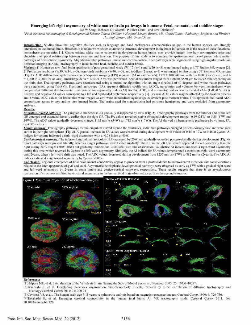
Emerging left-right asymmetry of white matter brain pathways in humans: Fetal, neonatal, and toddler stages Jae W Song 1 , Rebecca D Folkerth 2 , P. Ellen Grant 1 , and Emi Takahashi 1 1 Fetal-Neonatal Neuroimaging & Developmental Science Center, Children's Hospital Boston, Boston, MA, United States, 2 Pathology, Brigham And Women's Hospital, Boston, MA, United States Introduction: Studies show that cognitive abilities such as language and hand preferences, characteristics unique to the human species, are strongly lateralized in the human brain. However, it is unknown whether asymmetric structural development in the brain influences or is the result of these functional hemispheric asymmetries [1]. Characterizing white matter pathways in developing human brains may provide insight into how asymmetries arise and elucidate a temporal relationship between structure and function. The purpose of this study is to compare the spatio-temporal development white matter pathways of hemispheric asymmetry. Migration-related pathways, limbic and cortico-cortical fiber pathways were segmented using high-angular resolution diffusion imaging (HARDI) tractography in intact human fetal, neonate, and toddler brains. Method: 1) Human ex vivo fetal brain specimens of post-gestational week (W) 17 (n=1) and W20 (n=3) were imaged using a 4.7T Bruker MR system [2]. 2) Premature newborns (W30, W34; n=3), term-birth newborns (W40; n=3), and toddlers (2years; n=1) were imaged in vivo using a 3T Siemens MR system (Fig. 1). A 3D diffusion-weighted spin-echo echo-planar imaging (EPI) sequence (61 measurements), TR/TE 1000/40 ms, with b = 8,000 (for ex vivo) and b = 1,000 to 3,000 (for in vivo), small/large delta = 12.0/24.2 ms was performed. Spatial resolution ranged from 400x500x550 μm to 2x2x2 mm depending on the brain size. Tractography pathways were reconstructed using a streamline algorithm with an angle threshold of 40 degrees, and white matter pathways were segmented using TrackVis. Fractional anisotropy (FA), apparent diffusion coefficients (ADC), trajectories and volumes between hemispheres were compared at different developmental time points. An asymmetry index (AI) for FA, ADC, and volumetric values was calculated (AI= (L-R)/0.5(L+R)). Positive and negative AI values correspond to a left and right-sided preference, respectively [3]. Because ADC values may be affected by the fixation process and b-value, ADC values for brains that were imaged in vivo were standardized against age-equivalent post-mortem brains. This approach facilitated ADC comparisons across in vivo and ex vivo imaged brains. The brains used for standardizing had only one hemisphere and were excluded from asymmetry analyses. Results: Migration-related pathway: The ganglionic eminence (GE) gradually disappeared by 40W (Fig. 2). Tractography pathways from the anterior end of the left GE emerged and extended dorsally earlier than the right GE. The FA values remained stable throughout development (range: 0.19 (21W) to 0.25 (17W and 34W)). The ADC values gradually decreased (range: 1162 mm 2 /s (34W) to 1712 mm 2 /s (17W)). The AI showed no hemispheric preference by volume, FA, or ADC metrics. Limbic pathway: Tractography pathways for the cingulum curved around the ventricles, individual pathways emerged postero-dorsally first and were seen earlier in the right hemisphere (Fig. 3). A gradual increase in FA values was observed during development with values of 0.15 at 17W to 0.48 at 2years. AI indices for volume indicated a right-ward asymmetry with a -0.78 index at 40W. Cortico-cortical pathway: The inferior longitudinal fasciculus (ILF) appeared by 20W and gradually extended postero-dorsally during development (Fig. 4). Short pathways were present laterally, whereas longer pathways were located medially. The ILF in the left hemisphere appeared thicker posteriorly than the right during early stages (20W, 30W) but gradually thinned out. Consistent with this observation, volumetric AI indices indicated a right-ward asymmetry during this time, which reversed by 2years to a left-ward asymmetry. Similarly, the AI indices for FA values demonstrated a consistent right-ward asymmetry until 2years, when a left-ward shift was noted. The ADC values decreased during development from 1235 mm 2 /s (17W) to 882 mm 2 /s (2years). The ADC AI indices indicated a right-ward asymmetry by 2years (-0.07). Conclusion: Regional emergence of fetal brain axonal connectivity appear to proceed from a postero-dorsal to antero-ventral direction with local variations related to the later appearance of gyri and sulci. Asymmetric hemispheric developmental pathways were observed as early as 17W with a gradual right-ward and left-ward asymmetry by 2years in some limbic and cortico-cortical pathways, respectively. These results suggest that there is an asynchronous maturation of structures resulting in structural asymmetry in the human fetal brain observed as early as the second trimester. References: [1]Halpern ME, et al. Lateralization of the Vertebrate Brain: Taking the Side of Model Systems. J Neurosci 2005. 25: 10351-10357. [2]Takahashi E, et al. Developing neocortex organization and connectivity in cats revealed by direct correlation of diffusion tractography and histology.Cerebral Cortex 2011: 21; 200-211. [3]Caviness VS, et al.. The human brain age 7-11 years: A volumetric analysis based on magnetic resonance images. Cerebral Cortex 1996: 6: 726-736. [4]Takahashi E, et al. Emerging cerebral connectivity in the human fetal brain: An MR tractography study. Cerebral Cortex 2011. doi: 10.1093/cercor/bhr126. 3156 Proc. Intl. Soc. Mag. Reson. Med. 20 (2012)