37
Endobronchial Ultrasound in the Diagnosis & Staging of Lung Cancer Dr Richard Booton PhD FRCP ESMO-Christie Lung Cancer Course Manchester 2017

Endobronchial Ultrasound in the Diagnosis & Staging of Lung Cancer
Dr Richard Booton PhD FRCP
ESMO-Christie Lung Cancer Course
Manchester 2017
Overview
• What is Endobronchial Ultrasound?
• Why & When Do We Use It?
• Comparison of Non-Invasive/ Minimally Invasive Staging
• Performance Characteristics
• How to Handle Negative or Inadequate Results
• Quality Assurance
• Case Based

DB 85yr old male Ex-smoker 22 years (40 pack years) Retired Roofer MRC3 WHO PS1 FEV1 1.8L(90%) FVC 3.15L (114%) DLCO 49% Kco 59%
October 2012 Cough – few months
Minor haemoptysis
CXR 29/10/2012: Normal
January 2013 Wt loss 9lbs, anorexia, SOBOE CXR 18/01/2013:
Increased density inferior right hilum
CXR 18/01/13

Considerations
What Next? 1. Choose investigations that give the most information about diagnosis and staging with the least risk to the patient (NICE 2011) 2. All cases to be proved microscopically 3. TNM clinical and pathological classification 4. Attribute group staging 5. Where there is doubt, the patient should receive the benefit of doubt
• Stage determines treatment/ prognosis
• Which Investigation(s), which order?
• Impact of comorbidity/ age - safety of investigations - potential treatments/ radical vs palliative - physiological fitness
• EDD: Estimated Date of Discussion at MDT
• Sample type/ size/ quality
(Thorax 2003;58:711–720)


Endobronchial Ultrasound
• Linear
• Radial (and adjunct to navigational bronchoscopy)
• Tissue Diagnosis
• Staging & Depth of Invasion
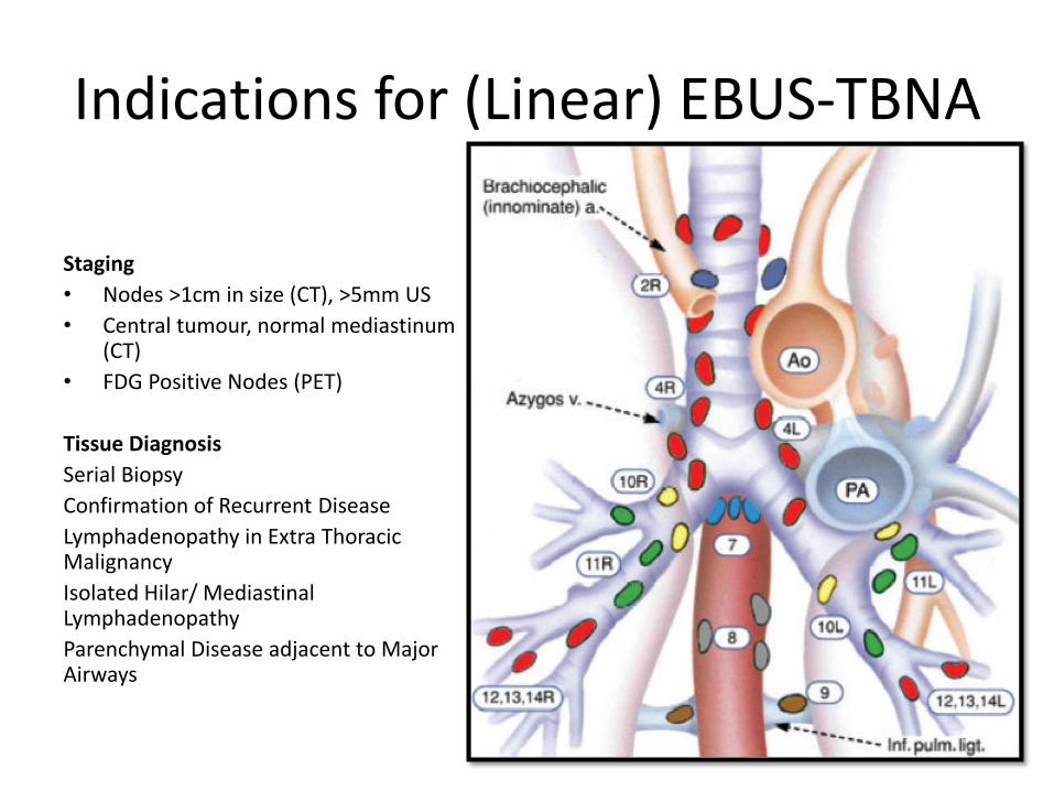
Indications for (Linear) EBUS-TBNA
Staging
• Nodes >1cm in size (CT), >5mm US
• Central tumour, normal mediastinum (CT)
• FDG Positive Nodes (PET)
Tissue Diagnosis
Serial Biopsy
Confirmation of Recurrent Disease
Lymphadenopathy in Extra Thoracic Malignancy
Isolated Hilar/ Mediastinal Lymphadenopathy
Parenchymal Disease adjacent to Major Airways
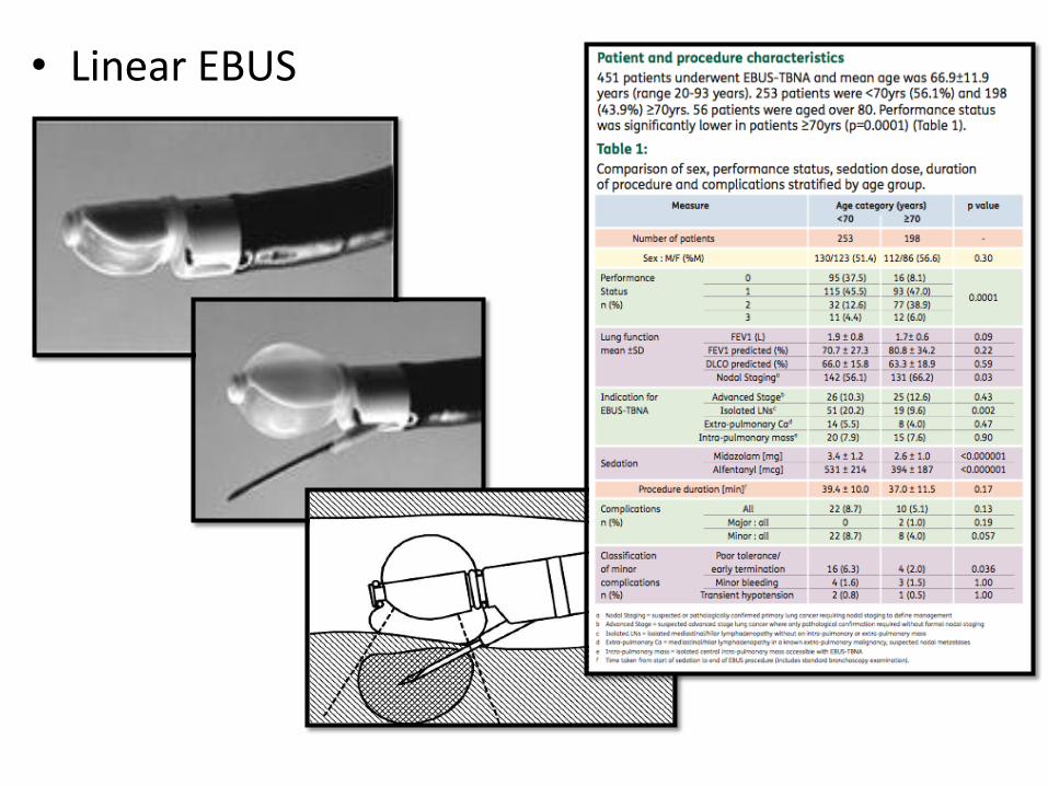
• Linear EBUS
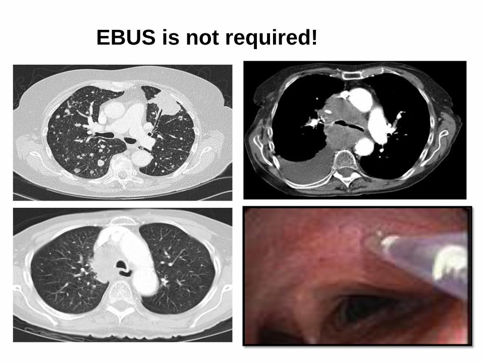
EBUS is not required!

Non-Invasive Staging of the Mediastinum
• Provides clarity of the pulmonary abnormality
• Categories defined by anatomic characteristics (size, location, extent)
• CT is inexpensive, widely available
• In combination with clinical history and examination, can decide which other tests indicated
• Subsequent invasive tests driven by anatomic characteristics
Silvestri et al CHEST 2007; 132:178S–201S
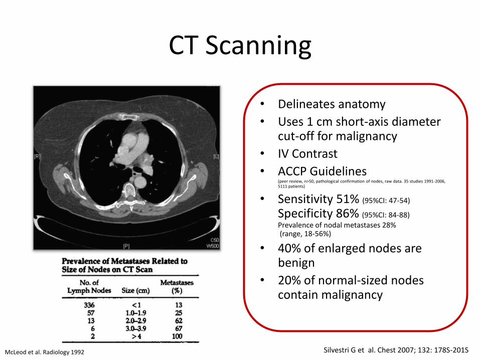
CT Scanning
• Delineates anatomy
• Uses 1 cm short-axis diameter cut-off for malignancy
• IV Contrast
• ACCP Guidelines (peer review, n>50, pathological confirmation of nodes, raw data. 35 studies 1991-2006, 5111 patients)
• Sensitivity 51% (95%CI: 47-54)
Specificity 86% (95%CI: 84-88)
Prevalence of nodal metastases 28% (range, 18-56%)
• 40% of enlarged nodes are benign
• 20% of normal-sized nodes contain malignancy
McLeod et al. Radiology 1992 Silvestri G et al. Chest 2007; 132: 178S-201S

• Based on biological activity of neoplastic cells ie function not anatomy
• No standardised quantitative criteria
• Qualitative assessment of lesion vs background
• Low limit of spatial resolution (7-10mm)
• ACCP Guideline (peer review, n>20, pathological nodal confirmation, raw data. 44 studies, 1994-2006, 2865 patients)
• Sensitivity 74% (95%CI: 69-79)
Specificity 85% (95%CI: 82-88)
Prevalence mediastinal metastases 29% (range, 5-64%)
• Inaccurate in upto 25% of nodes >1cm
• Whole body study Unexpected M1 in Stage 1: 7.5%, Stage II: 18%, Stage III: 24%
• PET positive mediastinal nodes (SUVmax>2.5) requires invasive sampling (NICE, ACCP, ESTS) before surgery is ruled out
18FDG-PET Scanning
Int J Radiat Oncol Biol Phys. 2001 Jun 1;50(2):287-93
J Natl Cancer Inst 2007;99: 1753 – 67

Summary ROC Characteristics for CT & PET CT
CT Scanning PET Scanning
Positive Likelihood Ratio 3.4 Negative Likelihood Ratio 0.6 Positive Likelihood Ratio 4.9
Negative Likelihood Ratio 0.3
There is no node size that can reliably determine tumor stage and operability. Where CT scan criteria for metastatic node are met, the clinician must still prove beyond a reasonable doubt that the node is indeed malignant.
PET is more accurate than CT scanning, but far from perfect

Utility of Linear EBUS

EBUS-TBNA: Systematic Review & Meta-analysis
Thorax 2009;64:757–762.

Quality Assurance
ACCP Simulation Based Accreditation
ERS – no statement
BTS Quality Standards

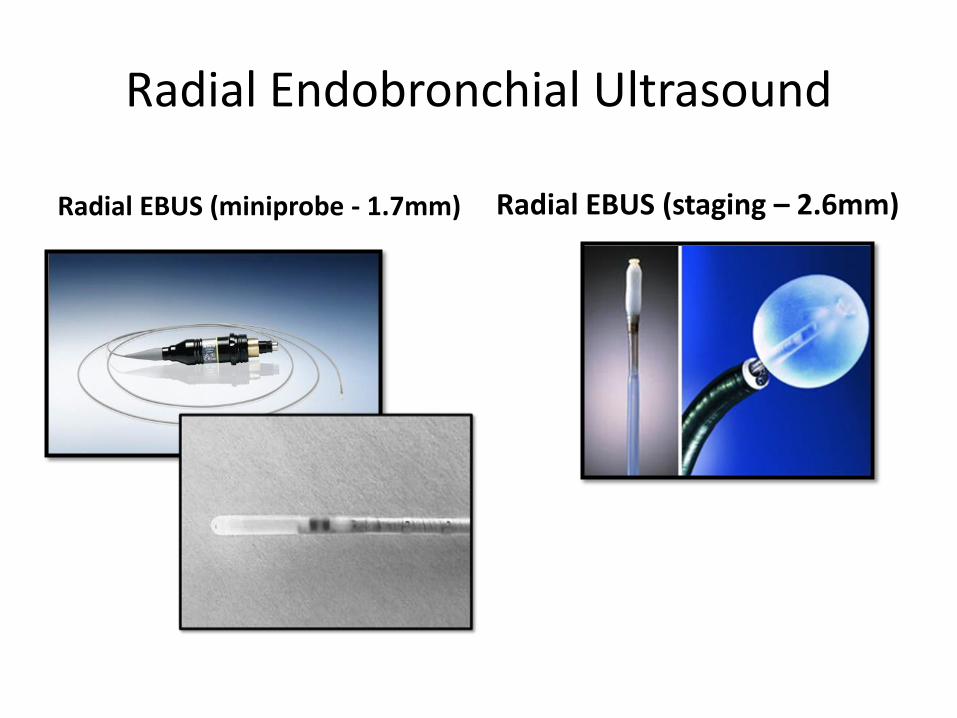
Radial Endobronchial Ultrasound
Radial EBUS (miniprobe - 1.7mm) Radial EBUS (staging – 2.6mm)

Transbronchial Biopsy?
n=293, randomised study
• Useful - with limited lung function - CT biopsy risky
• Add guidesheath - <2cm
CHEST 2005; 128:3551–3557
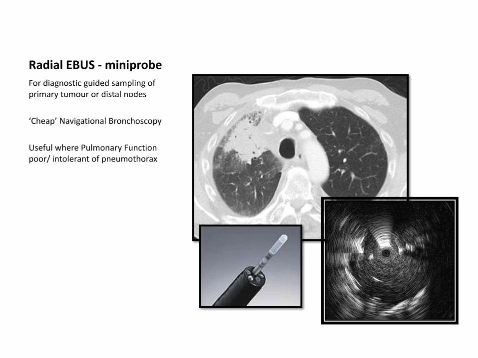
Radial EBUS - miniprobe
For diagnostic guided sampling of primary tumour or distal nodes
‘Cheap’ Navigational Bronchoscopy
Useful where Pulmonary Function poor/ intolerant of pneumothorax

• Lesion amenable to Biopsy in 96/117 (82%)
• 68% malignant
• 50% false negative rate
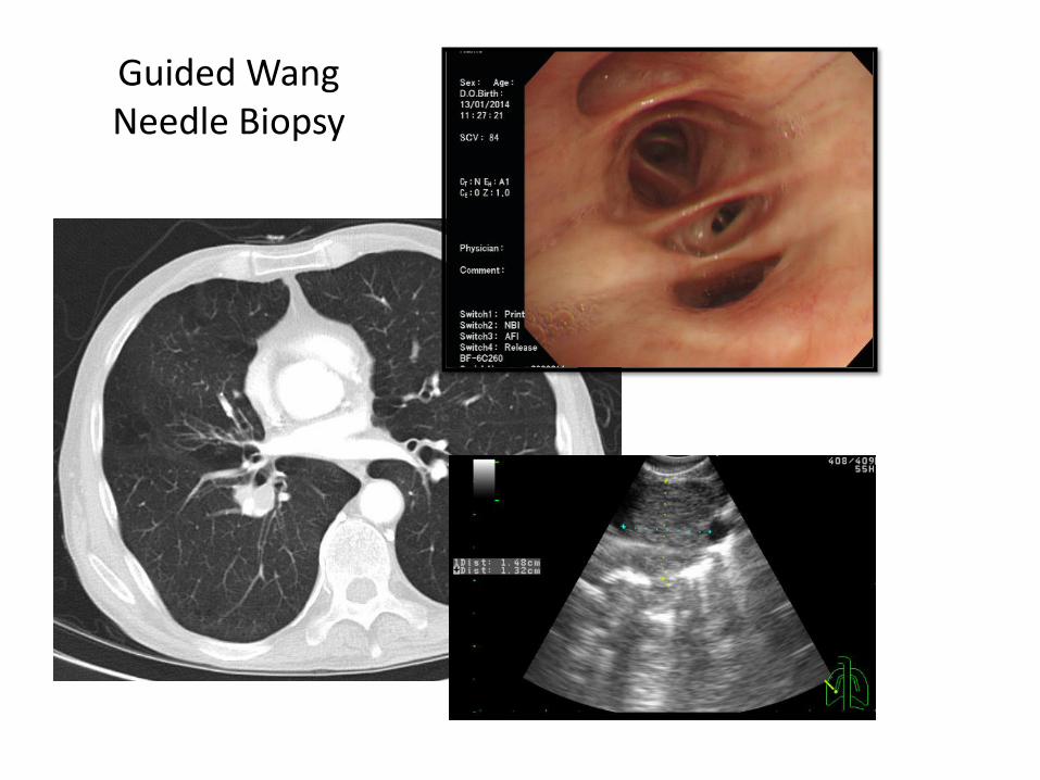
Guided Wang Needle Biopsy


Navigational Bronchoscopy
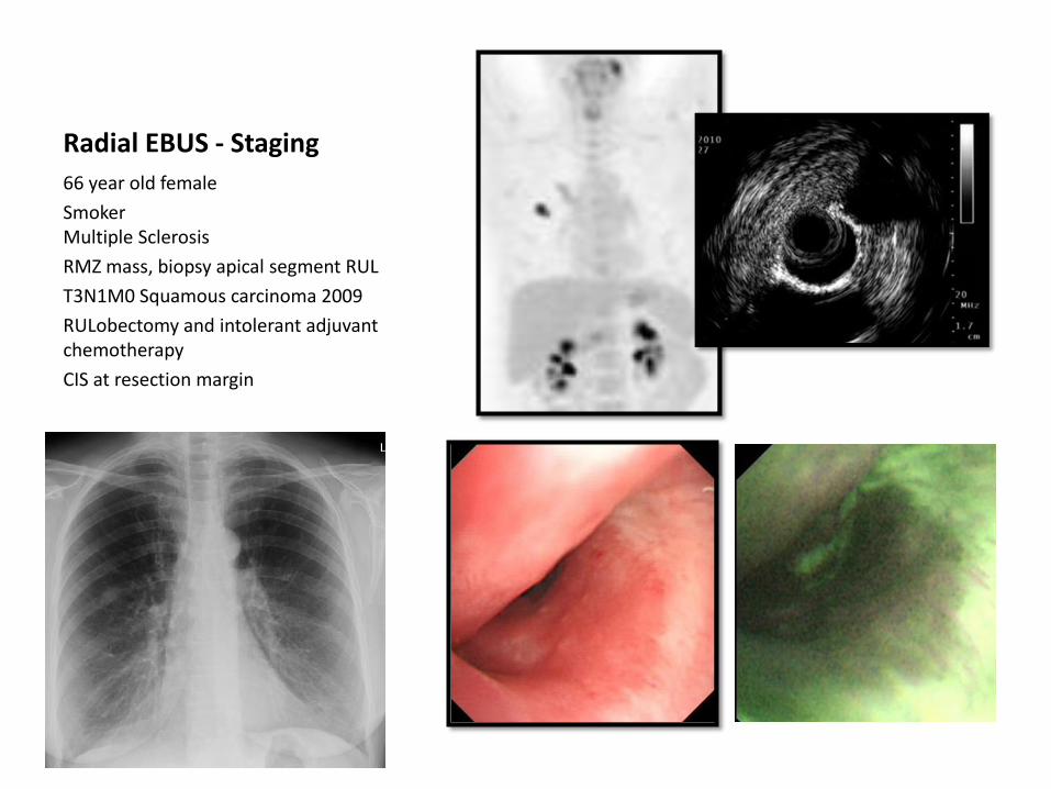
Radial EBUS - Staging
66 year old female
Smoker Multiple Sclerosis
RMZ mass, biopsy apical segment RUL
T3N1M0 Squamous carcinoma 2009
RULobectomy and intolerant adjuvant chemotherapy
CIS at resection margin
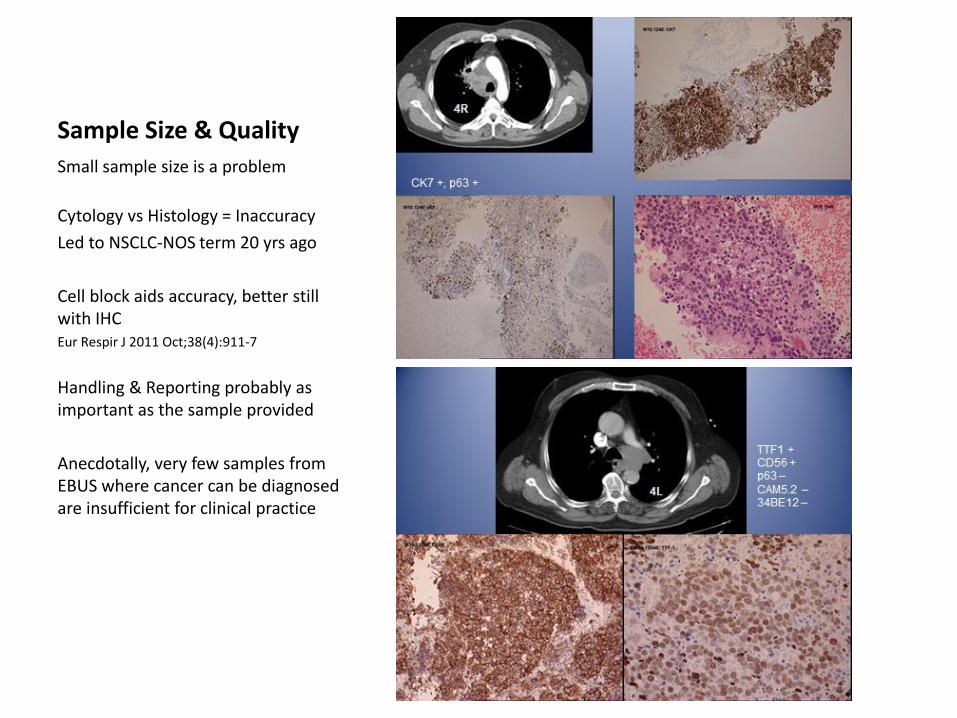
Sample Size & Quality
Small sample size is a problem
Cytology vs Histology = Inaccuracy
Led to NSCLC-NOS term 20 yrs ago
Cell block aids accuracy, better still with IHC Eur Respir J 2011 Oct;38(4):911-7
Handling & Reporting probably as important as the sample provided
Anecdotally, very few samples from EBUS where cancer can be diagnosed are insufficient for clinical practice
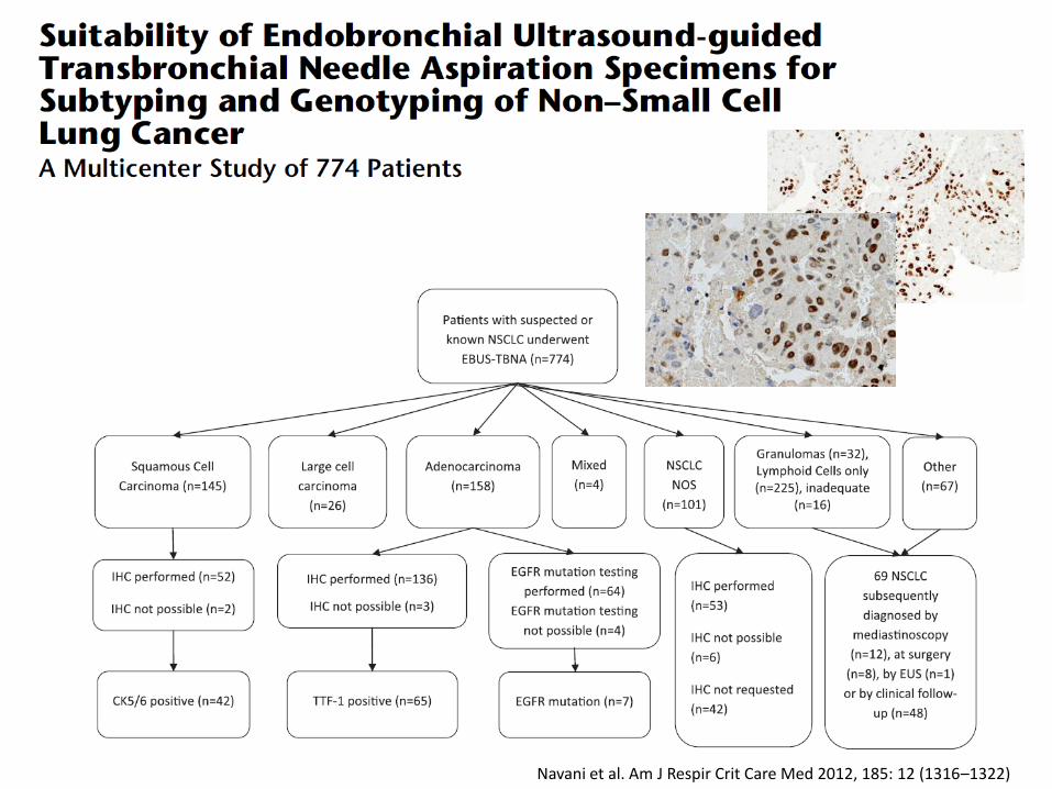
Navani et al. Am J Respir Crit Care Med 2012, 185: 12 (1316–1322)

EBUS samples can successfully be used in the majority (>95%) mutation profiling and gene rearrangement studies

DB 85yr old male Ex-smoker 22 years (40 pack years) Retired Roofer MRC3 WHO PS1 FEV1 1.8L(90%) FVC 3.15L (114%) DLCO 49% Kco 59%
October 2012 Cough – few months
Minor haemoptysis
CXR 29/10/2012: Normal
January 2013 Wt loss 9lbs, anorexia, SOBOE CXR 18/01/2013:
Increased density inferior right hilum
CXR 18/01/13

DB 85 year old male Ex-smoker 22 years
Pre-clinic:
CT scan of thorax (21 January 2013)
1. Attenuation RBI and RML
2. 3.1x2.2cm Subpleural Mass medial RML
3. Associated consolidation and GGO RML
4. Centrilobular Emphysema
5. 11R 1.4cm 7 1.0cm 4R Suspicious by number (<1cm) R SCF 5mm
6. Bilateral Pleural Plaques Probable basal subpleural fibrosis (Stable) Small HH Liver cyst segment 2 Normal R adrenal Thickening of body of L adrenal 11mm
Conclusion: Subpleural mass with hilar/mediastinal nodes and bulky L adrenal (T2aN3Mx). Emphysema, pleural plaques and asbestosis.
CXR 18/01/13
CT Scan 21/01/13
OPC 22/01/13

DB 85 year old male Ex-smoker 22 years
31 January 2013 US Neck
Cluster of prominent nodes identified in the right supraclavicular region (level 4), largest 17x11mm. A smaller adjacent node measures 14x8mm and has loss of fatty hilum with distorted architecture.
No evidence of cervical lymphadenopathy (levels 1-6)
FNAC performed.
CXR 18/01/13
CT Scan 21/01/13
Neck US 31/01/13
OPC 22/01/13

DB 85 year old male Ex-smoker 22 years
FNAC R Neck Level 4 nodes
Numerous small lymphocytes and occasional large lymphocytes, confirming aspiration from a lymph node. No malignant cells are seen.

DB 85 year old male Ex-smoker 22 years
FDG PET-CT: 6 February 2013
1. Occlusive lesion RML Bronchus 10mm (SUV max 8.5)
2. Distal collapse/ consolidation RML (SUVmax upto 12)
3. Several non-enlarged ipsilateral and contralateral nodes (SUVmax 2.5-4.4) Station 7 & RSCF SUVmax 3.5
Staging: T2aN2M0
CXR 18/01/13
CT Scan 21/01/13
PET-CT 06/02/13
Neck US 31/01/13
OPC 22/01/13

DB 85 year old male Ex-smoker 22 years
Bronchoscopy + EBUS-TBNA
7 February 2013
1. Endobronchial tumour distal RBI, probably originating from RML.
2. 4L, 2R, 4R and 7 assessed Features of benign disease including central hilar structure, <1cm short axis.
RBI Biopsy: Pieces of bronchial mucosa infiltrated by tumour of variable appearance. There is insitu dysplastic squamous epithelium and an isolated fragment of keratinising malignant squamous epithelium. There are infiltrating components with trabecular, insular and small cell appearances, positive for Cam5.2, 34bE12, p63 and CK5/6 and negative for TTF1, napsin
and CD56. Pure squamous cell carcinoma with poorly differentiated component
CK 5/6
p63
CXR 18/01/13
CT Scan 21/01/13
PET-CT 06/02/13
Neck US 31/01/13
OPC 22/01/13
EBUS 07/02/13

DB 85 year old male Ex-smoker 22 years
Lymph node FNAC: 2R, 4R/L and 7
Lymph node aspirate. No malignant cells identified
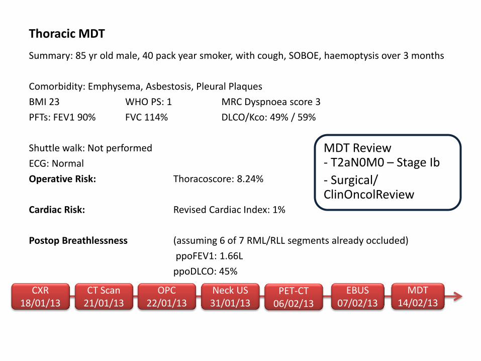
Summary: 85 yr old male, 40 pack year smoker, with cough, SOBOE, haemoptysis over 3 months
Comorbidity: Emphysema, Asbestosis, Pleural Plaques
BMI 23 WHO PS: 1 MRC Dyspnoea score 3
PFTs: FEV1 90% FVC 114% DLCO/Kco: 49% / 59%
Shuttle walk: Not performed
ECG: Normal
Operative Risk: Thoracoscore: 8.24%
Cardiac Risk: Revised Cardiac Index: 1%
Postop Breathlessness (assuming 6 of 7 RML/RLL segments already occluded)
ppoFEV1: 1.66L
ppoDLCO: 45%
MDT Review - T2aN0M0 – Stage Ib
- Surgical/ ClinOncolReview
Thoracic MDT
CXR 18/01/13
CT Scan 21/01/13
PET-CT 06/02/13
Neck US 31/01/13
OPC 22/01/13
MDT 14/02/13
EBUS 07/02/13

In Summary…..
• Endobronchial Ultrasound comes in several forms, with differing indications
• EBUS has extended our diagnostic and staging reach, with better patient tolerability
• EBUS-TBNA is the preferred staging modality compared with image based technologies or mediastinoscopy, but…
• Combination of PET findings & US nodal characteristics may be useful in ‘inadequate’ sampling
• Quality Assurance is key/ mandatory; individual professional societies need to consider key performance outcomes
• Tissue profiling & needs of molecular precision medicine is supported by minimally invasive cytological techniques


