Page 1
1
Endocrine disrupting organotin compounds are potent inducers
of adipogenesis in vertebrates
Running title: Organotins as inducers of adipogenesis
Felix Grün1, Hajime Watanabe
2, Zamaneh Zamanian
1, Lauren Maeda
1, Kayo Arima
1, Ryan
Chubacha1, David M. Gardiner
1, Jun Kanno
3, Taisen Iguchi
2, and Bruce Blumberg
1
1 Department of Developmental & Cell Biology, University of California Irvine, Irvine CA
92697-2300.
2 National Institutes of Natural Sciences, National Institute for Basic, Biology, Okazaki Institute
for Integrative Bioscience, 5-1 Higashiyama, Myodaiji, Okazaki 444-8787, Japan.
3
Division of Cellular & Molecular Toxicology, Biological Safety Research Center, National
Institute of Health Sciences, Setagaya-ku, Tokyo 158-8501, Japan.
"This is an un-copyedited author manuscript copyrighted by The Endocrine Society. This may
not be duplicated or reproduced, other than for personal use or within the rule of “Fair Use of
Copyrighted Materials” (section 107, Title 17, U.S. Code) without permission of the copyright
owner, The Endocrine Society. From the time of acceptance following peer review, the full text
of this manuscript is made freely available by The Endocrine Society at
http://www.endojournals.org/. The final copy edited article can be found at
http://www.endojournals.org/. The Endocrine Society disclaims any responsibility or liability for
errors or omissions in this version of the manuscript or in any version derived from it by the
National Institutes of Health or other parties.”
Address correspondence and reprints to: Bruce Blumberg, Department of Developmental & Cell
Biology, UCI, 2113 McGaugh Hall, Irvine CA 92697-2300. Tel. (949) 824-8573; Fax. (949)
824-4709; E-mail: [email protected]
Key words: organotin, tributyltin, adipogenesis, retinoid x receptor, peroxisome proliferator
activated receptor.
F.G., H.W., Z.Z., L.M., K.A., R.C., D.M.G., J.K., T.I. have nothing to declare. B.B. is a named
inventor on U.S. patents US 5,861,274, US 6,200,802 and US 6,815,168.
Grant Support: This work was supported by grants from the U.S. EPA (STAR R830686) and
NIH (GM-60572) to B.B., the University of California Toxic Substances Research & Training
Program to F.G.( UC-37579) and funding from the Ministries of Education, Culture, Sports,
Science and Technology, Environment and Health Labor and Welfare, Japan to T. I.
Molecular Endocrinology. First published April 13, 2006 as doi:10.1210/me.2005-0367
Copyright (C) 2006 by The Endocrine Society
Page 2
2
Abstract
Dietary and xenobiotic compounds can disrupt endocrine signaling, particularly of steroid
receptors and sexual differentiation. Evidence is also mounting that implicates environmental
agents in the growing epidemic of obesity. Despite a long-standing interest in such compounds,
their identity has remained elusive. Here we show that the persistent and ubiquitous
environmental contaminant, tributyltin chloride (TBT)**
, induces the differentiation of
adipocytes in vitro and increases adipose mass in vivo. TBT is a dual, nanomolar affinity ligand
for both the retinoid ‘X’ receptor (RXR) and the peroxisome proliferator activated receptor γ
(PPARγ). TBT promotes adipogenesis in the murine 3T3-L1 cell model and perturbs key
regulators of adipogenesis and lipogenic pathways in vivo. Moreover, in utero exposure to TBT
leads to strikingly elevated lipid accumulation in adipose depots, liver, and testis of neonate mice
and results in increased epididymal adipose mass in adults. In the amphibian Xenopus laevis,
ectopic adipocytes form in and around gonadal tissues following organotin, RXR or PPARγ
ligand exposure. TBT represents, to our knowledge, the first example of an environmental
endocrine disrupter that promotes adipogenesis through RXR and PPARγ activation.
Developmental or chronic lifetime exposure to organotins may therefore act as a chemical
stressor for obesity and related disorders.
Introduction
Organotins are a diverse group of widely distributed environmental pollutants. Tributyltin
chloride (TBT) and bis(triphenyltin) oxide (TPTO), have pleiotropic adverse effects on both
invertebrate and vertebrate endocrine systems. Organotins were first used in the 1960s as
antifouling agents in marine shipping paints although such use has been restricted in recent
Page 3
3
years. Organotins persist as prevalent contaminants in dietary sources, such as fish and shellfish,
and through pesticide use on high value food crops (1, 2). Additional human exposure to
organotins may occur through their use as antifungal agents in wood treatments, industrial water
systems and textiles. Mono- and diorganotins are prevalently used as stabilizers in the
manufacture of polyolefin plastics (PVC), which introduces the potential for transfer by contact
with drinking water and foods.
Exposure to organotins such as TBT and TPTO results in imposex, the abnormal induction of
male sex characteristics in female gastropod mollusks (3, 4). Bioaccumulation of organotins
decreases aromatase activity leading to a rise in testosterone levels that promotes development of
male characteristics (5). Imposex results in impaired reproductive fitness or sterility in the
affected animals and is one of the clearest examples of environmental endocrine disruption. TBT
exposure also leads to masculinization of at least two fish species (6, 7), but TBT is only
reported to have modest adverse effects on mammalian male and female reproductive tracts and
does not alter sex ratios (8, 9). Instead hepatic-, neuro- and immunotoxicity appear to be the
predominant effects of organotin exposure (10). Hence, the current mechanistic understanding of
the endocrine disrupting potential of organotins is based on their direct actions on the levels or
activity of key steroid regulatory enzymes such as aromatase and more general toxicity mediated
via damage to mitochondrial functions and subsequent cellular stress responses (11-15).
However, it remains an open question whether in vivo organotins act primarily as protein and
enzyme inhibitors, or rather mediate their endocrine disrupting effects at the transcriptional level.
Recent work has shown that aromatase mRNA levels can be down regulated in human ovarian
granulosa cells by treatment with organotins or ligands for the nuclear hormone receptors,
retinoid X receptor (RXRs) or peroxisome proliferator activated receptor gamma (PPARγ) (16-
Page 4
4
18). Furthermore, Nishikawa et al. have demonstrated that the gastropod T. clavigara RXR
homolog is responsive to 9-cis RA and TBT, and 9-cis RA can also induce imposex (19)
suggesting a conserved transcriptional mechanism for TBT action across phyla. These ligand
dependent transcription factors belong to the nuclear hormone receptor superfamily – a group of
~150 members (48 human genes) that includes the estrogen receptor (ER), androgen receptor
(AR), glucocorticoid receptor (GR), thyroid hormone receptor (TR), vitamin D receptor (VDR),
retinoic acid receptors (RARs and RXRs), peroxisome proliferator-activated receptors (PPARs)
and numerous orphan receptors. We were therefore intrigued by the similar effects of TBT and
RXR/PPARγ ligands on mammalian aromatase mRNA expression and hypothesized that TBT
might be exerting some of its biological effects via transcriptional regulation of gene expression
through activation of one or more nuclear hormone receptors.
Our results show that organotins such as TBT, are indeed potent and efficacious agonistic
ligands of the vertebrate nuclear receptors, retinoid X receptors (RXRs) and PPARγ. The
physiological consequences of receptor activation predict that permissive RXR heterodimer
target genes and downstream signaling cascades are sensitive to organotin misregulation.
Consistent with this prediction we observe that organotins phenocopy the effects of RXR and
PPARγ ligands using in vitro and in vivo models of adipogenesis. Therefore, TBT and related
organotin compounds are the first of a potentially new class of environmental endocrine
disrupters that targets adipogenesis by modulating the activity of key regulatory transcription
factors in the adipogenic pathway, RXRα and PPARγ. The existence of such xenobiotic
compounds was previously hypothesized (20, 21). Our results suggest that developmental
exposure to TBT and its congeners that activate RXR/PPARγ might be expected to increase the
Page 5
5
incidence of obesity in exposed individuals and that chronic lifetime exposure could act as a
potential chemical stressor for obesity and obesity related disorders.
RESULTS
Organotins are agonists of vertebrate RXR and RXR-permissive heterodimers
Many known or suspected environmental endocrine disrupting chemicals (EDCs) mimic
natural lipophilic hormones that act through members of the superfamily of nuclear receptor
transcription factors (22, 23). In a screen of high priority endocrine disrupting chemicals (EDC)
against a bank of vertebrate nuclear receptor ligand binding domains (NR-LBDs), we observed
that organotins, specifically tributyltin chloride (TBT) and bis(triphenyltin) oxide (TPTO), could
fully activate an RXRα ligand binding domain construct (GAL4-RXRα) in transient transfection
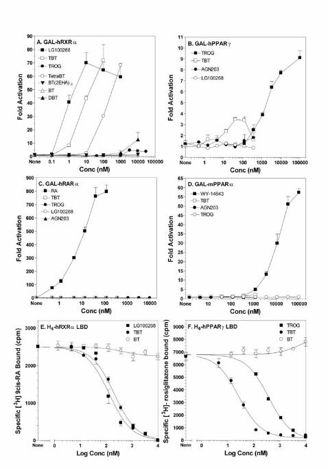
assays. Both TBT and TPTO were as potent (EC50 ~3-10 nM) as 9-cis retinoic acid, an
endogenous RXR ligand and approximately 2-5-fold less potent than the synthetic RXR-specific
ligands LG100268 (EC50 ~ 2 nM) or AGN195203 (EC50 ~ 0.5 nM) (Fig 1A, Table 2). Maximal
activation for TBT reached the same levels as LG100268 or AGN195203.
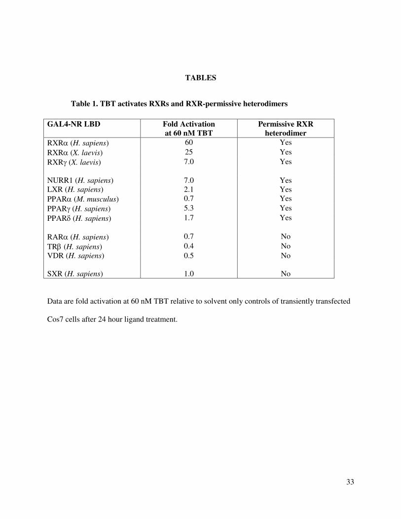
We next tested whether activation by TBT was unique to RXRα only, restricted to RXR
heterodimer complexes or a general nuclear receptor transcriptional response (Fig. 1 B-D and
Table 1). TBT activated RXRα and RXRγ from the amphibian Xenopus laevis in addition to
human RXRs (Table 1). Our results are consistent with recent findings by Nishikawa et al. that
organotins promote activation of all 3 human RXR subtypes in a yeast two-hybrid screen (19,
24). We also observed significant activation of receptors typically considered to be permissive
heterodimeric partners of RXR including human PPARγ (Fig. 1B ~30% maximal activation of
10 µM troglitazone, but note that activation is compromised by cellular toxicity above 100 nM),
Page 6
6
PPARδ, liver X receptor (LXR) and the orphan receptor NURR1. In contrast, typical non-
permissive partners such as retinoic acid receptors (RARs), thyroid hormone receptor (TRβ) and
vitamin D receptor (VDR) failed to show activation by organotins (Fig. 1C and Table 1). Murine
PPARα was also not activated by TBT although it was fully activated by its specific synthetic
agonist WY-14643 (Fig. 1D). The steroid and xenobiotic receptor (SXR) was likewise
unresponsive. The orphan receptor NURR1, which has no discernable ligand pocket and is
believed to be ligand-independent (25), was nevertheless activated 7 to 10-fold at 100 nM TBT.
Similarly, other RXR-specific ligands, e.g. LG100268, activated NURR1 to the same degree
suggesting that this response occurred through NURR1’s heterodimeric partner RXR as has been
previously described (25, 26). Like other RXR-specific ligands, tributyltin was also able to
promote the ligand dependent recruitment of nuclear receptor co-factors such as ACTR, SRC-1
and PBP in mammalian two-hybrid interaction assays (data not shown). We infer from these
results that nuclear receptor activation by TBT activation is specific to a small subset of
receptors and not a consequence of a general effect on the cellular transcriptional machinery.
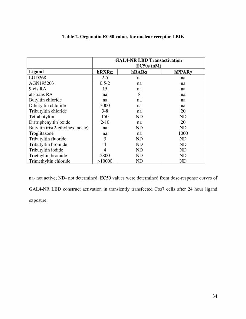
We next investigated the relationship between the structure of the tin compounds and RXR
activation by testing the response of GAL4-RXRα to mono-, di-, tri- and tetra-substituted
butyltin, branched side chains, variations in the alkyl chain length and changes in the halide
component (Fig. 1 A and Table 2). Overall, trialkyltin compounds were the most effective with
nanomolar EC50 values. Monobutyltin gave no significant activation whereas dibutyltin was
moderately active in the µM range (Fig. 1 A and Table 2). Tetrabutyltin was 20-fold less potent
than TBT whereas the branched side-chain butyltin tris(2-ethylhexanoate) (BT(2-EHA)3) was
inactive (Table 2). Although activation by dialkyltins is weaker than that of TBT, it is potentially
Page 7
7
significant due to their widespread use in the manufacture of polyvinyl chloride (PVC) plastics
and greater solubility than TBT.
The effect of the hydrocarbon chain length was very pronounced, suggesting an important
structure activity relationship. A reduction in hydrophobicity from butyl to ethyl side chains
raised the EC50 value by almost a 1000-fold into the micromolar range. Trimethyltin was weakly
active only above 100 µM (Table 2). Substitution of the halide component had no significant
effect on the EC50 values for TBT, probably due to the lability of the halide atom through
exchange in aqueous tissue culture media where chloride ions are prevalent.
TBT is a potent ligand of both RXRα and PPARγ.
Many, if not most, natural and synthetic nuclear receptor agonists act as ligands that
specifically interact with their cognate receptor ligand binding domains. We therefore performed
equilibrium competition binding experiments with purified histidine-tagged human RXRα (H6-
RXRα) and PPARγ (H6-PPARγ) ligand binding domains to determine if the potent and specific
activation of these receptors by TBT was due to direct ligand-receptor interaction (Fig. 1E-F).
The equilibrium binding curves indicate that TBT is a high affinity, competitive ligand for 9-
cis RA bound RXRα. The inhibition equilibrium dissociation constant was calculated by the
Chang-Prusoff method (Ki equals Kd) as 12.5 nM (10-15 nM - 95% confidence interval) (Table
3). By comparison, the value obtained for the synthetic RXR agonist LG100268 was 7.5 nM
which compared favorably with its published value of ~ 3 ± 1 nM (27). Therefore, the
identification of TBT as an RXR ligand expands the molecular definition of known “rexinoids”
(agonists able to activate RXR) to include this structurally unique class of organotin compounds.
Page 8
8
Somewhat surprisingly, we also observed potent specific competitive binding by TBT for
rosiglitazone bound to human PPARγ LBD (Fig. 2B). The deduced Ki of 20 nM (17-40 nM -
95% confidence interval) was slightly higher than for RXRα but significantly better than the Ki
for the PPARγ agonist troglitazone which yielded a Ki of 300 nM, consistent with its published
Kd (28). The Kd values for TBT binding to RXRα (12.5 nM) and PPARγ (20 nM) are also in
close agreement with EC50 values obtained from transient transfection assays using GAL4-
RXRα and GAL4-PPARγ constructs (Table 2).
Taken together, these data show that organotins such as TBT, although structurally distinct
from previously described natural or synthetic ligands, can interact with RXRα and PPARγ, via
direct ligand binding to induce productive receptor:coactivator interactions and promote
transcription in a concentration dependent manner. Organotins are therefore potent nanomolar
receptor activators on par with synthetic RXR and PPARγ ligands such as LG100268,
AGN195203 and thiazolidinediones.
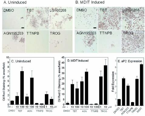
TBT promotes adipogenesis in the murine 3T3-L1 cell model.
Numerous studies have demonstrated the critical role played by RXRα:PPARγ signaling in
regulation of mammalian adipogenesis (29-31). In the murine 3T3-L1 preadipocyte cell model,
adipogenic signals induce early key transcriptional regulators such as CCAAT/enhancer binding
proteins C/EBPβ and δ that lead to mitotic clonal expansion of growth arrested preadipocytes
and induction of the late differentiation factors C/EBPα and PPARγ (32-34). The combination
of C/EBPα expression together with PPARγ signaling efficiently drives terminal adipocyte
differentiation and lipid accumulation. We therefore tested whether TBT signaling through
RXR:PPARγ could promote adipogenesis in the murine 3T3-L1 differentiation assay and
Page 9
9
compared its effect to other RXR-specific or PPARγ ligands (Fig. 2). Undifferentiated 3T3-L1
cells were cultured for 1 week in the presence of ligands either with or without a prior 2 day
treatment with MDIT (an adipogenic sensitizing cocktail of IBMX, dexamethasone, insulin and
T3) (35). Cells were then scored for lipid accumulation using Oil Red O staining to determine the
degree of terminal adipocyte differentiation. TBT was as effective as LG100268 or AGN195203
in promoting differentiation in the absence of MDIT treatment, increasing the number of
differentiated adipocytes about 7-fold over solvent only controls (Fig. 2A, C). The PPARγ
agonist troglitazone was a weak inducer in the absence of MDIT. Prior treatment with MDIT
increased the response to TBT, LG100268 and AGN195203 a further 3-5 fold (Fig. 2B,D).
MDIT treatment also boosted the response to troglitazone to equivalent levels as expected from
published studies showing that combination treatment with PPARγ ligands promote efficient
adipocyte differentiation (36-38). In contrast the RAR-agonist TTNPB inhibited the
differentiation of 3T3-L1 cells consistent with previously published data that showed RAR
signaling blocks adipogenesis during the early stages of differentiation in vitro and can modulate
adiposity and whole body weight in vivo (39-41). The differential response of 3T3-L1 cells to
receptor selective retinoids indicates that TBT favors RXR homodimer or permissive RXR-
heterodimer rather than RXR:RAR signaling in this cell model.
Adipocyte differentiation by TBT was accompanied by direct transcriptional effects on
RXR:PPARγ targets such as adipocyte-specific fatty acid binding protein (aP2) mRNA. The aP2
promoter contains response elements sensitive to CCAAT/enhancer binding protein factors
(C/EBPs) and RXRα:PPARγ signaling (42). QRT-PCR analysis showed aP2 levels were
elevated by TBT treatment approximately 5-fold at 24 hrs (Fig. 2E) and 45-fold at 72 hrs (data
not shown). LG100268, troglitazone and MDIT treatment also increased aP2 expression at these
Page 10
10
time points whereas the RAR agonist TTNPB was inhibitory, consistent with the observed
cellular responses.
TBT induces adipogenic regulators and markers of RXRα:PPARγ signaling in vivo.
The ability of organotins to regulate RXRα:PPARγ target genes and key modulators of
adipogenesis and lipid homeostasis in vivo has not previously been examined. Therefore, we next
asked whether TBT could perturb expression of critical transcriptional mediators of adipogenesis
such as RXRα, PPARγ, C/EBPα/β/δ and sterol regulatory element binding factor 1 (Srebf1) as
well as known target genes of RXRα:PPARγ signaling from liver, epididymal adipose tissue and
testis of six week old male mice dosed for 24 hours with TBT (0.3 mg/kg b.w.), AGN195203
(0.3 mg/kg b.w.), troglitazone (3 mg/kg b.w) or vehicle (corn oil) administered by intraperitoneal
injection. TBT either had no effect or weakly repressed RXRα and PPARγ transcription in liver
(Fig. 3A, B). A more pronounced decrease was observed for RXRα, PPARγ, C/EBPα and
C/EBPδ in adipose tissue and testis (Fig. 3B,C). In contrast, TBT, AGN195203 and troglitazone
significantly induced expression of the early adipogenic transcription factor C/EBPβ in liver and
testis whereas it was more weakly induced in adipose tissue. Induction was strongest in testis
where TBT and troglitazone increased expression greater than 10-fold and AGN195203
increased expression 60-fold compared to vehicle controls (Fig. 3C). In addition to C/EBPβ, the
proadipogenic transcription factor Srebf1 was also significantly increased in adipose tissue by all
three receptor ligands and weakly induced in liver.
We also observed coordinate changes in several well characterized direct target genes of
RXR:PPARγ signaling. Fatty acid transport protein (Fatp) acts as a key control point for
regulation of cellular fatty acid content. The Fatp promoter contains a functional PPRE shown to
Page 11
11
be sensitive to RXR:PPARγ signaling in 3T3-L1 adipocytes and white fat (43-46). Fatp mRNA
levels were up regulated 2-3 fold in liver and epididymal adipose tissue but not testis by TBT,
AGN195203 and troglitazone (Fig. 5A,B). Similarly, the PPARγ target phosphoenolpyruvate
carboxykinase 1 (PEPCK/Pck1) (47), the rate-limiting step in hepatic gluconeogenesis and
adipose glyceroneogenesis, was up regulated in liver and adipose tissues by TBT or troglitazone
treatment.
Signaling through RXR:PPARγ, RXR:LXR and ADD1/Srebf1 in hepatocytes has been
shown to modulate fatty acid synthesis through transcriptional control of acetyl-CoA carboxylase
(Acac), the rate limiting step in long chain fatty acid synthesis (48, 49), as well as fatty acid
synthase (Fasn) (50-53). Hepatic expression of both Acac and Fasn was unregulated between
1.5-2.5-fold by TBT, AGN195203 and troglitazone. Therefore, the coordinate increased
expression of Fatp, Pck1, Acac and Fasn in liver suggests that TBT stimulates fatty acid uptake
and triglyceride synthesis. Similar changes have been reported in the induction of hepatic
steatosis by overactive PPARγ signaling (49, 54).
Taken together, these data show that TBT exposure induces lipogenic RXR:PPARγ target
gene expression in adipose tissue and liver, and modulates associated early adipocyte
differentiation factors such as C/EBPβ and Srebf1. We inferred from these data that organotins
are potential adipogenic agents in vivo.
Developmental exposure to TBT disrupts lipid homeostasis and adipogenesis in vertebrates.
Based on its molecular pharmacology, ability to induce 3T3-L1 adipocyte differentiation and
in vivo transcriptional responses, we reasoned that TBT would disrupt normal endocrine control
over lipid homeostasis and impact adipogenesis particularly when exposure occurred during
Page 12
12
sensitive periods of development. We therefore tested this hypothesis in two vertebrate model
systems, mouse and Xenopus laevis, during embryogenesis.
Pregnant C57BL/6 mice were injected daily from gestational day 12-18 with TBT (0.05 or
0.5 mg/kg body weight i.p.) dissolved in sesame oil or vehicle alone. Pups were then sacrificed at
birth and histological sections prepared from liver, testis, mammary gland and inguinal adipose
tissue. Sections were stained with Oil Red O to assess changes in total tissue lipid accumulation.
TBT exposure caused a disorganization of hepatic (Fig. 4A,B) and gonadal (Fig. 4 C,D)
architecture and significantly increased Oil Red O staining in treated animals versus controls.
Liver sections exhibited signs of steatosis consistent with the misregulation of fatty acid uptake
and synthesis observed using molecular markers. In addition Oil Red O positive staining in
mammary, and inguinal adipose (Fig. 4E-H) tissues was dramatically elevated reflecting either
an increase in lipid accumulation or an increase in mature adipocytes.
To determine whether exposure induced long term changes in growth or adipose tissue, we
followed mice from birth to adulthood after in utero exposure to TBT as indicated above. At
birth, mice were cross-fostered to unexposed dams and total body weight recorded until 10
weeks of age (Fig. 5A). Growth curves for male and female pups showed a slight trend for lower
total body weight consistent with published observations (9) but were not statistically significant
at 10 weeks (Control vs TBT: male 26.00g ± 0.70, n=9 vs 25.53g ± 0.39, n=10, p=0.583; female
21.22g ± 0.41, n =10, vs 20.24g ± 0.24, n=10, p = 0.0529). Males were sacrificed at 10 weeks
and epidydimal fat pads weighed (Fig. 5B). Adipose mass in TBT treated males was increased
significantly by 20 % over controls (Control vs TBT: 0.30g ± 0.020, n=9 vs 0.36g ± 0.018, n
=10, p=0.0374). These data support the conclusion that TBT can increase body adiposity without
Page 13
13
overtly increasing total body weight. Similar lipid accumulation and changes in adipose tissue
mass have also been observed following TZD or rexinoid treatment (55-57).
We had previously observed that TBT activated Xenopus RXRs (Table 1) and reasoned that
the strong conservation in vertebrate nuclear receptor signaling pathways should result in
consistent responses to organotin and RXR/PPARγ ligands across diverse vertebrate species. We
therefore tested chronic exposure to environmentally relevant low doses of TBT (1-10 nM), the
RXR-specific ligands LG100268 and AGN195203 (10-100 nM), troglitazone (0.1-1 µM) and
estradiol (1-10 nM) on developing Xenopus laevis tadpoles from stage 48 to metamorphs. To
determine the effectiveness of these doses in Xenopus laevis tadpoles we used aromatase
expression as a molecular marker since activity and expression is sensitive to endocrine
disruption by organotins and RXR/PPARγ ligands in mammals (17, 18). Xenopus aromatase
expression was similarly repressed 2 to 3-fold by 10 nM TBT, AGN195203, LG100268 or 1 µM
troglitazone at stage 56 tadpoles (Fig. 6A) and at all subsequent stages. Despite significant ligand
induced aromatase downregulation, neither sex ratios nor the time required to reach
metamorphosis were altered (data not shown). Xenopus liver and kidney also exhibited no gross
structural abnormalities at the doses given.
However, consistent with the testis and adipose results from mice presented above, we
observed a dose-dependent increase in ectopic adipocyte formation posterior to the fat-bodies in
and around the gonads of both sexes following TBT or RXR/PPARγ ligand exposure (Fig. 6B).
In contrast, estradiol treated animals did not show increased adipocyte formation compared to
controls. At 10 nM TBT, 10 nM AGN195203 or 1 µM troglitazone, ectopic adipocytes were
observed in approximately 45-60% of animals. At the highest dose of TBT in males, testicular
Page 14
14
tissue was interspersed with, or replaced by adipocytes along the anterior-posterior axis (Fig.
6D,E,G).
The concordant changes observed in Xenopus aromatase expression, gonadal adipocyte
formation and increased murine adiposity following exposure to TBT, RXR and PPARγ ligands
are therefore consistent with a common mechanism of action through RXR:PPARγ activation,
supporting the conclusion that endocrine disruption via nuclear receptor transcriptional
regulation is a novel and key feature of organotin toxicity.
DISCUSSION
We have shown above that TBT is a potent inducer of adipogenesis, in vitro and in vivo, by
acting as a novel, high-affinity xenobiotic ligand for RXRα and PPARγ. The ability of
organotins to bind and activate these receptors, in particular the RXRs which exhibit very
restricted ligand specificity, is unexpected given the radically different chemical composition and
3D-molecular structure of organotins when compared to known natural and synthetic nuclear
receptor ligands. Typically, RXR ligands comprise of a carboxylic acid functional group and a
3D-molecular shape that mimics 9-cis RA. Structure-activity profiles indicate distinct structural
preferences for organotins but also a relatively broad accommodation for agonist activity that is
not easily reconciled with the classical ligand binding model. Organotins may therefore interact
somewhat differently than previously described RXR/PPARγ ligands with receptor LBDs to
induce productive conformational changes required for co-activator recruitment. However, the
binding data indicate that TBT is a potent and efficacious ligand for both RXRs and PPARγ that
interacts, at least partially, with the same receptor binding sites of other high-affinity ligands and
Page 15
15
promotes the necessary co-factor interactions required for agonist activation. In Kanayama et al.,
TBT was only effective in co-activator recruitment assays with PPARγ above 10 µM in vitro.
However, in accord with our findings, they show that TBT activated PPARγ significantly at
nanomolar concentrations in transfection assays. This may reflect a limitation of preference in
the co-factor used in vitro. Alternatively, the lower maximal activation observed with TBT on
PPARγ in cells (~30% at 100 nM TBT cf troglitazone) is consistent with one of two possibilities:
either non-specific cellular toxicity at high levels or activation as a partial agonist.
The ability of TBT to act as a dual ligand for permissive heterodimers such as
RXRα:PPARγ, which can be activated by specific ligands for either receptor, also raises the
possibility for additive or synergistic effects that might increase the potency of these compounds
in vivo at low doses for this specific signaling pathway. Of note is that receptor activation is
observed at nanomolar concentrations, whereas other mechanisms of toxicity and endocrine
disruption e.g. direct inhibition of aromatase activity, typically occur in the micromolar range.
Furthermore, the activation of other permissive RXR heterodimeric partners, e.g. LXR and
NURR1, suggests that organotins may act more widely to disrupt multiple nuclear receptor
mediated hormonal signaling pathways.
The biological consequences of organotin activation of the RXR:PPARγ signaling pathway
are predictable and should follow known aspects of RXR/PPARγ biology. The RXR:PPARγ
pathway plays a key role in adipocyte differentiation and energy storage, and is central to the
control of whole body metabolism (58). PPARγ activation increases the expression of genes that
promote fatty acid storage and represses genes that induce lipolysis in adipocytes in white
adipose tissue (59). PPARγ such as the thiazolidinediones can modulate insulin sensitivity due to
these effects on the adipocyte, reversing insulin resistance in the whole body by sensitizing the
Page 16
16
muscle and liver tissue to insulin (60). However, a consequence of this increase in whole body
insulin sensitivity is that fat mass is increased through the promotion of triglyceride storage in
adipocytes. Evidence is also mounting that depot-specific remodeling and adipocyte numbers
increase following thiazolidinedione treatment (55-57). Therefore, PPARγ agonists comprise a
class of pharmaceutical therapies for type 2 diabetes that can also promote obesity by increasing
fat storage. Likewise, retinoid X receptor (RXR) ligands also act as insulin-sensitizing agonists
in rodents (61) underscoring the permissive nature of the PPARγ:RXR heterodimer and the
potential effects on diabetes and obesity of both PPARγ and RXR agonists.
Our data are consistent with recent studies that organotins can mediate some of their
endocrine disruption effects by transcriptional regulation through nuclear receptors, in particular
RXR:PPARγ signaling (17-19, 24). Consequently, TBT exposure can promote adipocyte
differentiation in the same manner as other RXR or PPARγ ligands in vitro using the standard
murine 3T3-L1 cell model and in vivo through increased adiposity following intra-uterine
organotin exposure in newborn mice. It is currently unknown whether the increased adiposity in
vivo results from an increase in adipocyte precursor cell number, enhanced adipocyte
differentiation from the same number of precursors, an increase in adipocyte size without an
increase in number or some combination of these.
The prevailing epidemiological data ascribes high density caloric and/or fatty diets coupled
with decreased physical activity as the root causes for the rise in obesity rates in the general
population (62). The contribution of genetic components is less clear. While genetic variation
contributes to an individual’s propensity to develop obesity, the rapid worldwide increase in
obesity suggests that interaction with the modern environment exposes inherent genetic
differences. The Barker hypothesis postulates that in utero fetal nutritional status is a potential
Page 17
17
risk factor for metabolic syndrome diseases (63-67). In this view, developmental metabolic
programming of a thrifty phenotype limits the range in adaptive responses to the environment,
e.g. diet and exercise, in later life (68). Experimental evidence from animal models lends support
to this hypothesis (69). Plausible mechanisms include imprinting of obesity sensitive hormonal
pathways or changes in cell type and number e.g. adipocytes, established during development.
Others, however, argue that the environment plays another role in obesity. Since the increase
in obesity rates parallels the rapid growth in the use of industrial chemicals over the past 40
years, it is plausible and provocative to associate in utero or chronic lifetime exposure to
chemical triggers present in the modern environment with this epidemic. Hence, an “obesogen”
model predicts the existence of xenobiotic chemicals that inappropriately regulate lipid
metabolism and adipogenesis to promote obesity. Several recent studies serve as “proof-of-
principle” for such a hypothesis. Environmental estrogens such as bisphenol A and nonylphenol,
for instance, can promote adipocyte differentiation or proliferation in murine cell lines (70, 71).
Furthermore, epidemiological studies link maternal smoking during pregnancy to an elevated
risk of childhood obesity (72-76).
Seen in this context, we propose that organotins such as TBT and its congeners are chemical
stressors or “obesogens” that activate RXR:PPARγ signaling to promote long term changes in
adipocyte number and/or lipid homeostasis following developmental or chronic lifetime
exposure.
Page 18
18
MATERIALS AND METHODS
Plasmids and transfections
pCMX-GAL4 and pCMX-VP16 plasmid fusion constructs to nuclear receptors ligand
binding domains and coactivators (GAL4-hRARα, hRXRα, -xRXRα/γ, -hPPARγ, -mPPARα, -
hSXR, -NURR1, -VDR, -LXR, -hACTR, -hPBP, -hSrc1, hTIF2) have been previously described
(77-82) . Transfections were performed in Cos7 cells essentially as described in (83) using
MH200-x4-TK-Luc as reporter and normalized to pCMX-β-galactosidase controls. Briefly, Cos7
cells were seeded at 5000 cells/well in 96-well tissue culture plates in 10 % FBS/DMEM and
transfected for 8 hrs with 11 µg/plate of DNA/calcium phosphate precipitate mix (MH200x4-
TK-Luc: CMX-β-galactosidase: nuclear receptor/coactivator effector(s) at a ratio of 5:5:1). Cells
were washed free of precipitate with phosphate buffered saline (PBS) and media replaced with
serum free ITLB/DMEM medium (84) plus ligands for an additional 24 hours prior to assays for
luciferase and β-galactosidase activity. All transfection data points were performed in triplicate
and all experiments were repeated at least three times.
Quantitative real-time PCR analyses
Total cellular RNA from C57BL/6 mouse and Xenopus laevis tissues was isolated with
Trizol reagent and reversed transcribed with oligo dT and Superscript II (Invitrogen, CA)
according to the manufacturer’s instructions. Triplicate cDNA samples (50 ng/reaction) were
analyzed by QRT-PCR on a DNA Engine Opticon thermal cycler (MJ Research/Biorad) using
SYBR Green chemistry (PerkinElmer Life Sciences, MA). Fold changes in expression levels
were calculated after normalization to histone Hist2h4 using the delta-delta cycle threshold
Page 19
19
method (85). Gene specific primers were as follows - Hist2h4 F
5’CCCGTGGTGTGCTGAAGGTGTT3’, R 5’GA ATTGAAGCGGCGGCGTCTA3’; RXRα F
5’CGGCTGCTCAGGGTACTTGTGTTT3’; R 5’ CGGCTGCTCAGGGTACTTGTGTTT3’;
PPARγ F 5’TGGGTGAAACTCTGGGAGATTC3’, R 5’
AATTTCTTGTGAAGTGCTCATAGGC3’; C/EBPα F 5’CCAAGAAGTCGGTGGACAAGA
3’, R 5’CGGTCATTGTCACTGGTCAACT3’; C/EBPβ F 5’GCCCGCCGCCTTTAGACC 3’, R
5’CGCTCGTGCTCGCCAATG3’; C/EBPδ F 5’AACCCGCGGCCTTCTACGAG3’, R
5’ACGGCGGCCATGGAGTCAAT3’; aP2 F 5’GAATTCGATGAAATCACCGCA3’, R
5’CTCTTTATTGTGGTCGACTTTCCA3’; FATP F 5’AGCCGCTTCT
GGGATGACTGTGT3’, R 5’ACCGAAGCGCTGCGTGAACTC3’; ACS F 5’CCCAGCCAGT
CCCCACCAG3’, R 5’CACACCACTCAGGCTCACACTCGT3’, FASN F
5’TCGGGTGTGGTG GGTTTGGTGAAT3’; R 5’ACTTGGGGGCGTG AGATGTGTTGC3’;
ACAC F 5’GGATGGCAGCTCTGGAGGTGTATG3’, R 5’TGTCCTTAAG
CTGGCGGTGTTGTA; Pck1 F 5’CTGGCAGCATGGGGTGTTTGTAGG3’, R
5’TGCCGAAGTTGTAGCCGAAGAAGG3’; Srebf1 F 5’GCCCC TGCCCACCTCAAACCT3’,
R 5’ACTGGCACGGGCATCCTTCCTC3’; Xenopus EF1α F 5’GAT
CCCAGGAAAGCCAATGTGC3’, R 5’CCGGATCCTGCTGCCTTCTTCT3’; Xenopus CYP19
(aromatase) – F 5’GTCTGGATTAATGGCGAGGAAACA3’, R
5’CTGATGAAGTATGGCCGAATGACC3’.
Ligand binding
Histidine-tagged RXRα ligand binding domain (H6-RXRα LBDs) was expressed and
purified from pET15b(+) vector in BL21(DE3) pLysS bacteria cultures after induction with 1
Page 20
20
mM IPTG for 3hrs at 37 oC (86). Purified H6-PPARγ was purchased from Invitrogen. Proteins
were bound to 96-well Nickel Chelate Flashplates (PerkinElmer Life Sciences, MA) at 100
µg/ml overnight at 4 oC and washed 5 times with 200 µl/well Flashplate Assay Buffer (20 mM
HEPES pH 7.9, 100 mM KCl, 0.1 % CHAPS, 0.1 mM DTT). Competition assays typically used
1-5 nM [3H]-9-cis-RA (Perkin Elmer Life Sciences, MA) or 10-50 nM [
3H]-rosiglitazone
(American Radiochemicals Inc., MO) plus cold competitor ligands in Flashplate Assay Buffer at
concentrations indicated in the figures. Plates were incubated at room temperature, protected
from light and read after 4 hours on a Packard Topcount scintillation counter. Specific bound
cpm were determined by subtraction of cpm from uncoated wells at each ligand concentration.
Data were analyzed with GraphPad Prism 4.0 (GraphPad, CA) using a one-site competition
binding equation to determine Ki values for competitor ligands; Kd values of 1.4 and 41 nM for
9-cis-RA and rosiglitazone for their respective receptors were used in the calculations (87, 88).
3T3-L1 Cell Assays
3T3-L1 (ATCC, VA) cells were maintained as sub-confluent cultures by passage every 3
days from cultures seeded at 5000 cells/cm2 in 8 % calf serum/DMEM medium. For
differentiation assays, cells were seeded at 15x103 cells/well into 24-well tissue culture plates in
8% fetal bovine serum/DMEM medium, cultures grown for 2 days and then treated with the
indicated RXR, RAR and PPAR ligands either with or without MDIT (100 µM IBMX, 100 nM
dexamethasone, 0.1 ng/ml insulin and 2 nM T3 thyroid hormone) induction cocktail. Media and
ligand treatments were renewed every 2 days. After 1 week, cells were scored for adipocyte
differentiation by Oil Red staining for lipid droplet accumulation. Cultures were washed with
phosphate buffered saline (PBS), fixed with 10 % formaldehyde for 15 minutes, washed with
Page 21
21
distilled water and stained with filtered Oil Red solution (4g/L, 60 % isopropanol) for 15 mins.
Excess stain was removed by washing three times with distilled water. Three random fields from
each well were photographed under phase contrast and analyzed in ImageJ. Images were
converted into high contrast black and white images to visualize lipid droplets and scored as the
percentage area/field. Data are shown as the mean ± S.E.M from three wells per treatment. The
method was validated by extraction of Oil Red O from stained cells into 100 % isopropanol and
quantitated by absorbance at 540nm on a spectrophotometer.
In vivo animal exposure experiments
C57BL/6J mice were housed under a 12-hour light/dark cycle. Pregnant mice were dosed by
intraperitoneal injection with TBT (0.05 or 0.5 mg/kg body weight (b.w.) or vehicle (sesame oil)
from E12 every 24 hours until the day before delivery. Neonates were sacrificed at the day of
delivery and analyzed. The samples were embedded in OCT and sectioned (12 mm) using a
cryostat. Sections were fixed on slides with 4% paraformaldehyde for 10 min and rinsed in
phosphate-buffered saline (PBS). The slides were then sequentially washed with distilled water
and 60 % of isopropanol and stained with Oil Red O (4g/L, 60 % isopropanol). After washing
with 60 % isopropanol and distilled water, the slides were counterstained with hematoxylin.
Sections were evaluated and photographed using a Zeiss microscope.
For long-term growth studies, pups were cross-fostered to unexposed C57BL/6 dams after
birth and litter sizes kept constant at 8 pups/dam (control, 2 male + 2 female; TBT treated, 2
male + 2 female). Animals were weaned at 3 weeks of age and maintained on standard rodent
chow. Total body weight was followed until 10 weeks of age. Males were then sacrificed,
epidydimal fat pads dissected and weighed.
Page 22
22
Xenopus laevis tadpoles were sorted at stage 48 (89) and maintained in 1 L glass tanks in 20
% Holtfreter’s buffered salt solution (90) at a density of 10 tadpoles/tank on a diet of ground
Tetramin Fish Flakes and spirulina. Compounds prepared in dimethylsulfoxide (DMSO) as 105-
fold stock solutions were tested on triplicate tanks and dosed by static renewal after weekly
water changes. Metamorphs at stage 64 were transferred to individual containers and fed frozen
brine shrimp for 2 weeks until stage 66. Froglets were euthanized with 250 mg/L MS222 in 20%
Holtfreter’s solution and then scored for gonadal abnormalities and inter-renal/gonadal adipocyte
formation under a dissecting microscope. Kidneys with attached gonads and livers were fixed in
10 % formalin-phosphate buffered saline, embedded in parrafin and sectioned at 15 µm
thickness. Sections were developed with Mallory’s trichrome stain.
All animal experiments were approved and performed in accordance with IACUC protocols.
Page 23
23
FOOTNOTES
*This work was supported by grants from the U.S. EPA (STAR R830686) and NIH (GM-60572)
to B.B., from the Ministries of Education, Culture, Sports, Science and Technology,
Environment and Health Labor and Welfare, Japan to T.I. and from the University of California
Toxic Substance Research & Tranining Program to F.G. We thank Drs. I. Blitz, K. Cho, C. Zhou
and T. Osborne for critical reading and comments on the manuscript, Dr. C. Li (Expression
Technologies) for the H6-RXRα LBD construct and Dr. R. Chandraratna (Allergan
Pharmaceuticals) for AGN203 and LG268).
**Abbreviations used are: TBT, tributyltin chloride; TPTO, triphenyltin oxide; RXR, retinoid x
receptor; PPAR, peroxisome proliferator-activated receptor; DMEM, Dulbecco's modified
Eagle's medium, FBS, fetal bovine serum; 9-cis RA, 9-cis retinoic acid; T3, 3,5,3'-triiodo-L-
thyronine; MDIT, 3-isobutyl-1-methylxanthine, dexamethasone, insulin and 3,5,3'-triiodo-L-
thyronine adipocyte differentiation mix; Cos7, transformed green monkey kidney fibroblast cell
line; 3T3-L1, embryonic murine preadipocyte fibroblast cell line; OCT, optimal cutting
temperature embedding compound.
Page 24
24
REFERENCES
1. Appel KE 2004 Organotin compounds: toxicokinetic aspects. Drug Metab Rev 36:763-
86
2. Golub M, Doherty J 2004 Triphenyltin as a potential human endocrine disruptor. J
Toxicol Environ Health B Crit Rev 7:281-95
3. Blaber SJM 1970 The occurrence of a penis-like outgrowth behind the right tentacle in
spent females of Nucella lapillus. Proceedings of the Malacological Society of London
39:231-233
4. Gibbs P, Bryan G 1986 Reproductive failure in populations of the dog-whelk, Nucella
lapillus, caused by imposex induced by tributyltin from antifouling paints. Journal of the
Marine Biological Association of the United Kingdom 66
5. Matthiessen P, Gibbs P 1998 Critical appraisal of the evidence for tributyltin-mediated
endocrine disruption in mollusks. Environ Toxicol Chem 17:37-43
6. Shimasaki Y, Kitano T, Oshima Y, Inoue S, Imada N, Honjo T 2003 Tributyltin
causes masculinization in fish. Environ Toxicol Chem 22:141-4
7. McAllister BG, Kime DE 2003 Early life exposure to environmental levels of the
aromatase inhibitor tributyltin causes masculinisation and irreversible sperm damage in
zebrafish (Danio rerio). Aquat Toxicol 65:309-16
8. Omura M, Ogata R, Kubo K, Shimasaki Y, Aou S, Oshima Y, Tanaka A, Hirata M,
Makita Y, Inoue N 2001 Two-generation reproductive toxicity study of tributyltin
chloride in male rats. Toxicol Sci 64:224-32
9. Ogata R, Omura M, Shimasaki Y, Kubo K, Oshima Y, Aou S, Inoue N 2001 Two-
generation reproductive toxicity study of tributyltin chloride in female rats. J Toxicol
Environ Health A 63:127-44
10. Boyer IJ 1989 Toxicity of dibutyltin, tributyltin and other organotin compounds to
humans and to experimental animals. Toxicology 55:253-98
11. Heidrich DD, Steckelbroeck S, Klingmuller D 2001 Inhibition of human cytochrome
P450 aromatase activity by butyltins. Steroids 66:763-9
12. Cooke GM 2002 Effect of organotins on human aromatase activity in vitro. Toxicol Lett
126:121-30
13. Powers MF, Beavis AD 1991 Triorganotins inhibit the mitochondrial inner membrane
anion channel. J Biol Chem 266:17250-6
14. Gennari A, Viviani B, Galli CL, Marinovich M, Pieters R, Corsini E 2000 Organotins
induce apoptosis by disturbance of [Ca(2+)](i) and mitochondrial activity, causing
oxidative stress and activation of caspases in rat thymocytes. Toxicol Appl Pharmacol
169:185-90
15. Philbert MA, Billingsley ML, Reuhl KR 2000 Mechanisms of injury in the central
nervous system. Toxicol Pathol 28:43-53
16. Mu YM, Yanase T, Nishi Y, Waseda N, Oda T, Tanaka A, Takayanagi R, Nawata H
2000 Insulin sensitizer, troglitazone, directly inhibits aromatase activity in human ovarian
granulosa cells. Biochem Biophys Res Commun 271:710-3.
17. Mu YM, Yanase T, Nishi Y, Takayanagi R, Goto K, Nawata H 2001 Combined
treatment with specific ligands for PPARgamma:RXR nuclear receptor system markedly
Page 25
25
inhibits the expression of cytochrome P450arom in human granulosa cancer cells. Mol
Cell Endocrinol 181:239-48.
18. Saitoh M, Yanase T, Morinaga H, Tanabe M, Mu YM, Nishi Y, Nomura M, Okabe
T, Goto K, Takayanagi R, Nawata H 2001 Tributyltin or triphenyltin inhibits
aromatase activity in the human granulosa-like tumor cell line KGN. Biochem Biophys
Res Commun 289:198-204
19. Nishikawa J, Mamiya S, Kanayama T, Nishikawa T, Shiraishi F, Horiguchi T 2004
Involvement of the retinoid X receptor in the development of imposex caused by
organotins in gastropods. Environ Sci Technol 38:6271-6
20. Baillie-Hamilton PF 2002 Chemical toxins: a hypothesis to explain the global obesity
epidemic. J Altern Complement Med 8:185-92
21. Heindel JJ 2003 Endocrine disruptors and the obesity epidemic. Toxicol Sci 76:247-9
22. Jacobs MN, Lewis DF 2002 Steroid hormone receptors and dietary ligands: a selected
review. Proc Nutr Soc 61:105-22
23. Watanabe H, Iguchi T, Morohashi K 2002 [Endocrine disruptors and nuclear
receptors]. Nippon Rinsho 60:397-403
24. Kanayama T, Kobayashi N, Mamiya S, Nakanishi T, Nishikawa J 2005 Organotin
compounds promote adipocyte differentiation as agonists of the peroxisome proliferator-
activated receptor gamma/retinoid X receptor pathway. Mol Pharmacol 67:766-74
25. Wang Z, Benoit G, Liu J, Prasad S, Aarnisalo P, Liu X, Xu H, Walker NP,
Perlmann T 2003 Structure and function of Nurr1 identifies a class of ligand-
independent nuclear receptors. Nature 423:555-60
26. Aarnisalo P, Kim CH, Lee JW, Perlmann T 2002 Defining requirements for
heterodimerization between the retinoid X receptor and the orphan nuclear receptor
Nurr1. J Biol Chem 277:35118-23
27. Boehm MF, Zhang L, Zhi L, McClurg MR, Berger E, Wagoner M, Mais DE, Suto
CM, Davies JA, Heyman RA, et al. 1995 Design and synthesis of potent retinoid X
receptor selective ligands that induce apoptosis in leukemia cells. J Med Chem 38:3146-
55
28. Yu C, Chen L, Luo H, Chen J, Cheng F, Gui C, Zhang R, Shen J, Chen K, Jiang H,
Shen X 2004 Binding analyses between Human PPARgamma-LBD and ligands. Eur J
Biochem 271:386-97
29. Forman BM, Tontonoz P, Chen J, Brun RP, Spiegelman BM, Evans RM 1995 15-
Deoxy-delta 12, 14-prostaglandin J2 is a ligand for the adipocyte determination factor
PPAR gamma. Cell 83:803-12
30. Schoonjans K, Staels B, Auwerx J 1996 The peroxisome proliferator activated
receptors (PPARS) and their effects on lipid metabolism and adipocyte differentiation.
Biochim Biophys Acta 1302:93-109
31. Kersten S 2002 Peroxisome proliferator activated receptors and obesity. Eur J Pharmacol
440:223-34
32. Lane MD, Tang QQ, Jiang MS 1999 Role of the CCAAT enhancer binding proteins
(C/EBPs) in adipocyte differentiation. Biochem Biophys Res Commun 266:677-83
33. Tang QQ, Otto TC, Lane MD 2003 CCAAT/enhancer-binding protein beta is required
for mitotic clonal expansion during adipogenesis. Proc Natl Acad Sci U S A 100:850-5
Page 26
26
34. Tang QQ, Lane MD 1999 Activation and centromeric localization of CCAAT/enhancer-
binding proteins during the mitotic clonal expansion of adipocyte differentiation. Genes
Dev 13:2231-41
35. Rubin CS, Hirsch A, Fung C, Rosen OM 1978 Development of hormone receptors and
hormonal responsiveness in vitro. Insulin receptors and insulin sensitivity in the
preadipocyte and adipocyte forms of 3T3-L1 cells. J Biol Chem 253:7570-8
36. Kletzien RF, Clarke SD, Ulrich RG 1992 Enhancement of adipocyte differentiation by
an insulin-sensitizing agent. Mol Pharmacol 41:393-8
37. Kletzien RF, Foellmi LA, Harris PK, Wyse BM, Clarke SD 1992 Adipocyte fatty
acid-binding protein: regulation of gene expression in vivo and in vitro by an insulin-
sensitizing agent. Mol Pharmacol 42:558-62
38. Tafuri SR 1996 Troglitazone enhances differentiation, basal glucose uptake, and Glut1
protein levels in 3T3-L1 adipocytes. Endocrinology 137:4706-12
39. Xue JC, Schwarz EJ, Chawla A, Lazar MA 1996 Distinct stages in adipogenesis
revealed by retinoid inhibition of differentiation after induction of PPARgamma. Mol
Cell Biol 16:1567-75
40. Kawada T, Kamei Y, Sugimoto E 1996 The possibility of active form of vitamins A
and D as suppressors on adipocyte development via ligand-dependent transcriptional
regulators. Int J Obes Relat Metab Disord 20 Suppl 3:S52-7.
41. Kawada T, Kamei Y, Fujita A, Hida Y, Takahashi N, Sugimoto E, Fushiki T 2000
Carotenoids and retinoids as suppressors on adipocyte differentiation via nuclear
receptors. Biofactors 13:103-9
42. Tontonoz P, Graves RA, Budavari AI, Erdjument-Bromage H, Lui M, Hu E,
Tempst P, Spiegelman BM 1994 Adipocyte-specific transcription factor ARF6 is a
heterodimeric complex of two nuclear hormone receptors, PPAR gamma and RXR alpha.
Nucleic Acids Res 22:5628-34.
43. Martin G, Schoonjans K, Lefebvre AM, Staels B, Auwerx J 1997 Coordinate
regulation of the expression of the fatty acid transport protein and acyl-CoA synthetase
genes by PPARalpha and PPARgamma activators. J Biol Chem 272:28210-7
44. Motojima K, Passilly P, Peters JM, Gonzalez FJ, Latruffe N 1998 Expression of
putative fatty acid transporter genes are regulated by peroxisome proliferator-activated
receptor alpha and gamma activators in a tissue- and inducer-specific manner. J Biol
Chem 273:16710-4
45. Frohnert BI, Hui TY, Bernlohr DA 1999 Identification of a functional peroxisome
proliferator-responsive element in the murine fatty acid transport protein gene. J Biol
Chem 274:3970-7
46. Martin G, Poirier H, Hennuyer N, Crombie D, Fruchart JC, Heyman RA, Besnard
P, Auwerx J 2000 Induction of the fatty acid transport protein 1 and acyl-CoA synthase
genes by dimer-selective rexinoids suggests that the peroxisome proliferator-activated
receptor-retinoid X receptor heterodimer is their molecular target. J Biol Chem
275:12612-8
47. Tontonoz P, Hu E, Devine J, Beale EG, Spiegelman BM 1995 PPAR gamma 2
regulates adipose expression of the phosphoenolpyruvate carboxykinase gene. Mol Cell
Biol 15:351-7
Page 27
27
48. Magana MM, Lin SS, Dooley KA, Osborne TF 1997 Sterol regulation of acetyl
coenzyme A carboxylase promoter requires two interdependent binding sites for sterol
regulatory element binding proteins. J Lipid Res 38:1630-8
49. Schadinger SE, Bucher NL, Schreiber BM, Farmer SR 2005 PPAR{gamma}2
regulates lipogenesis and lipid accumulation in steatotic hepatocytes. Am J Physiol
Endocrinol Metab
50. Tontonoz P, Kim JB, Graves RA, Spiegelman BM 1993 ADD1: a novel helix-loop-
helix transcription factor associated with adipocyte determination and differentiation.
Mol Cell Biol 13:4753-9
51. Kim JB, Spiegelman BM 1996 ADD1/SREBP1 promotes adipocyte differentiation and
gene expression linked to fatty acid metabolism. Genes Dev 10:1096-107
52. Joseph SB, Laffitte BA, Patel PH, Watson MA, Matsukuma KE, Walczak R, Collins
JL, Osborne TF, Tontonoz P 2002 Direct and indirect mechanisms for regulation of
fatty acid synthase gene expression by liver X receptors. J Biol Chem 277:11019-25
53. Seo JB, Moon HM, Kim WS, Lee YS, Jeong HW, Yoo EJ, Ham J, Kang H, Park
MG, Steffensen KR, Stulnig TM, Gustafsson JA, Park SD, Kim JB 2004 Activated
liver X receptors stimulate adipocyte differentiation through induction of peroxisome
proliferator-activated receptor gamma expression. Mol Cell Biol 24:3430-44
54. Yu S, Matsusue K, Kashireddy P, Cao WQ, Yeldandi V, Yeldandi AV, Rao MS,
Gonzalez FJ, Reddy JK 2003 Adipocyte-specific gene expression and adipogenic
steatosis in the mouse liver due to peroxisome proliferator-activated receptor gamma1
(PPARgamma1) overexpression. J Biol Chem 278:498-505
55. Hallakou S, Doare L, Foufelle F, Kergoat M, Guerre-Millo M, Berthault MF, Dugail
I, Morin J, Auwerx J, Ferre P 1997 Pioglitazone induces in vivo adipocyte
differentiation in the obese Zucker fa/fa rat. Diabetes 46:1393-9
56. de Souza CJ, Eckhardt M, Gagen K, Dong M, Chen W, Laurent D, Burkey BF 2001
Effects of pioglitazone on adipose tissue remodeling within the setting of obesity and
insulin resistance. Diabetes 50:1863-71
57. Smith SR, De Jonge L, Volaufova J, Li Y, Xie H, Bray GA 2005 Effect of
pioglitazone on body composition and energy expenditure: a randomized controlled trial.
Metabolism 54:24-32
58. Auwerx J 1999 PPARgamma, the ultimate thrifty gene. Diabetologia 42:1033-49
59. Ferre P 2004 The biology of peroxisome proliferator-activated receptors: relationship
with lipid metabolism and insulin sensitivity. Diabetes 53 Suppl 1:S43-50
60. Day C 1999 Thiazolidinediones: a new class of antidiabetic drugs. Diabet Med 16:179-
92
61. Mukherjee R, Davies PJ, Crombie DL, Bischoff ED, Cesario RM, Jow L, Hamann
LG, Boehm MF, Mondon CE, Nadzan AM, Paterniti JR, Jr., Heyman RA 1997
Sensitization of diabetic and obese mice to insulin by retinoid X receptor agonists. Nature
386:407-10.
62. Hill JO, Peters JC 1998 Environmental contributions to the obesity epidemic. Science
280:1371-4
63. Barker DJ, Bull AR, Osmond C, Simmonds SJ 1990 Fetal and placental size and risk
of hypertension in adult life. Bmj 301:259-62
64. Phillips DI, Hirst S, Clark PM, Hales CN, Osmond C 1994 Fetal growth and insulin
secretion in adult life. Diabetologia 37:592-6
Page 28
28
65. Martyn CN, Barker DJ, Jespersen S, Greenwald S, Osmond C, Berry C 1995
Growth in utero, adult blood pressure, and arterial compliance. Br Heart J 73:116-21
66. Yajnik C 2000 Interactions of perturbations in intrauterine growth and growth during
childhood on the risk of adult-onset disease. Proc Nutr Soc 59:257-65
67. Barker DJ, Martyn CN, Osmond C, Hales CN, Fall CH 1993 Growth in utero and
serum cholesterol concentrations in adult life. Bmj 307:1524-7
68. Lucas A 1998 Programming by early nutrition: an experimental approach. J Nutr
128:401S-406S
69. Armitage JA, Khan IY, Taylor PD, Nathanielsz PW, Poston L 2004 Developmental
Programming of Metabolic Syndrome by Maternal Nutritional Imbalance; How Strong is
the Evidence from Experimental Models in Mammals? J Physiol
70. Masuno H, Kidani T, Sekiya K, Sakayama K, Shiosaka T, Yamamoto H, Honda K
2002 Bisphenol A in combination with insulin can accelerate the conversion of 3T3-L1
fibroblasts to adipocytes. J Lipid Res 43:676-84
71. Masuno H, Okamoto S, Iwanami J, Honda K, Shiosaka T, Kidani T, Sakayama K,
Yamamoto H 2003 Effect of 4-nonylphenol on cell proliferation and adipocyte
formation in cultures of fully differentiated 3T3-L1 cells. Toxicol Sci 75:314-20
72. Toschke AM, Koletzko B, Slikker W, Jr., Hermann M, von Kries R 2002 Childhood
obesity is associated with maternal smoking in pregnancy. Eur J Pediatr 161:445-8
73. von Kries R, Toschke AM, Koletzko B, Slikker W, Jr. 2002 Maternal smoking during
pregnancy and childhood obesity. Am J Epidemiol 156:954-61
74. Oken E, Huh SY, Taveras EM, Rich-Edwards JW, Gillman MW 2005 Associations
of maternal prenatal smoking with child adiposity and blood pressure. Obes Res 13:2021-
8
75. Power C, Jefferis BJ 2002 Fetal environment and subsequent obesity: a study of
maternal smoking. Int J Epidemiol 31:413-9
76. Hill SY, Shen S, Locke Wellman J, Rickin E, Lowers L 2005 Offspring from families
at high risk for alcohol dependence: increased body mass index in association with
prenatal exposure to cigarettes but not alcohol. Psychiatry Res 135:203-16
77. Umesono K, Murakami KK, Thompson CC, Evans RM 1991 Direct repeats as
selective response elements for the thyroid hormone, retinoic acid, and vitamin D3
receptors. Cell 65:1255-66
78. Perlmann T, Rangarajan PN, Umesono K, Evans RM 1993 Determinants for selective
RAR and TR recognition of direct repeat HREs. Genes Dev 7:1411-22
79. Blumberg B, Mangelsdorf DJ, Dyck JA, Bittner DA, Evans RM, De Robertis EM
1992 Multiple retinoid-responsive receptors in a single cell: families of retinoid "X"
receptors and retinoic acid receptors in the Xenopus egg. Proc Natl Acad Sci U S A
89:2321-5
80. Blumberg B, Bolado J, Jr., Derguini F, Craig AG, Moreno TA, Chakravarti D,
Heyman RA, Buck J, Evans RM 1996 Novel retinoic acid receptor ligands in Xenopus
embryos. Proc Natl Acad Sci U S A 93:4873-8
81. Blumberg B, Sabbagh W, Jr., Juguilon H, Bolado J, Jr., van Meter CM, Ong ES,
Evans RM 1998 SXR, a novel steroid and xenobiotic-sensing nuclear receptor. Genes
Dev 12:3195-205
82. Tabb MM, Sun A, Zhou C, Grun F, Errandi J, Romero K, Pham H, Inoue S,
Mallick S, Lin M, Forman BM, Blumberg B 2003 Vitamin K2 regulation of bone
Page 29
29
homeostasis is mediated by the steroid and xenobiotic receptor SXR. J Biol Chem
278:43919-27
83. Grun F, Blumberg B 2003 Identification of novel nuclear hormone receptor ligands by
activity-guided purification. Methods Enzymol 364:3-24
84. Grun F, Venkatesan RN, Tabb MM, Zhou C, Cao J, Hemmati D, Blumberg B 2002
Benzoate X receptors alpha and beta are pharmacologically distinct and do not function
as xenobiotic receptors. J Biol Chem 277:43691-7
85. Livak KJ, Schmittgen TD 2001 Analysis of relative gene expression data using real-
time quantitative PCR and the 2(-Delta Delta C(T)) Method. Methods 25:402-8
86. Bourguet W, Ruff M, Chambon P, Gronemeyer H, Moras D 1995 Crystal structure of
the ligand-binding domain of the human nuclear receptor RXR-alpha. Nature 375:377-82.
87. Allegretto EA, McClurg MR, Lazarchik SB, Clemm DL, Kerner SA, Elgort MG,
Boehm MF, White SK, Pike JW, Heyman RA 1993 Transactivation properties of
retinoic acid and retinoid X receptors in mammalian cells and yeast. Correlation with
hormone binding and effects of metabolism. J Biol Chem 268:26625-33
88. Lehmann JM, Moore LB, Smith-Oliver TA, Wilkison WO, Willson TM, Kliewer SA
1995 An antidiabetic thiazolidinedione is a high affinity ligand for peroxisome
proliferator-activated receptor gamma (PPAR gamma). J Biol Chem 270:12953-6
89. Nieuwkoop P, Faber J 1994 Normal Table of Xenopus laevis (Daudin). 2nd ed. Garland
Publishing Inc., New York
90. Rugh R 1962 Experimental embryology: techniques and procedures., 3rd ed. Burgess
Publishing Co., Minneapolis, MN
Page 30
30
FIGURE LEGENDS
Figure 1 Organotins are agonist ligands of RXRαααα and PPARγ.γ.γ.γ. Organotins are high affinity
ligand agonists of RXRα and PPARγ. (A-D) Activation of GAL4-hRXRα, -hPPARγ, -hRARα
or -hPPARα in transiently transfected Cos7 cells by organotins and receptor specific ligands.
Data represent reporter luciferase activity normalized to β-galactosidase and plotted as the
average fold activation ± S.E.M. (n=3) relative to solvent only controls from representative
experiments. (E-F) Competition binding curves of histidine-tagged RXRα or PPARγ LBDs with
tributytlin chloride (TBT). Data shown are from a representative experiment analyzed in
GraphPad Prism 4.0 (GraphPad, CA) and Ki values deduced (Table 3).
Figure 2 Tributyltin induces adipogenesis in 3T3-L1 cells. Uninduced (A) and MDIT induced
(B) 3T3-L1 cultures grown for 1 week in the presence of vehicle (DMSO) or ligands were
analyzed for mature adipocyte differentiation by Oil Red O staining. Scale bar represents 100
µm. (C and D) The percentage area stained was determined by automated analysis of random
fields (n=9) from high contrast dissecting scope photographs of monolayers analyzed in ImageJ;
1-100 nM of TBT, AGN195203 and TTNPB or 1-10 µM troglitazone. (E) Quantitative real-time
PCR (QRT-PCR) of adipocyte specific fatty acid binding protein aP2 (aP2/Fabp4) expression
levels in post-confluent 3T3-L1 cells treated with the indicated ligands for 24 hours. Data were
normalized to GAPDH controls and plotted as average fold induction ± S.E.M. (n=3).
Figure 3 In vivo induction of adipogenic modulators and RXR:PPARγγγγ target genes.
C57BL/6 male mice (3 animals/treatment) were dosed with TBT (0.3 mg/kg b.w.), AGN195203
(0.3 mg/kg), troglitazone (3 mg/kg b.w.) or vehicle (corn oil) only by intrapertioneal injection.
Page 31
31
Animals were sacrificed after 24 hours, dissected and cDNA prepared from liver, epidydimal fat
pad or testis for QRT-PCR analysis. Expression levels were normalized to histone Hist2h4 and
shown as the average fold change ± S.E.M. (n=3) compared to vehicle (corn oil) controls.
Control v ligand treatments were analyzed by the unpaired Student’s t-test; * p<0.1, ** p<0.05.
Figure 4 In utero exposure to TBT increases adiposity in mouse liver, testis and adipose
depots. Histological sections (12 µm) of newborn mouse liver (A,B), testis (C,D), inguinal
adipose (E,F) and mammary adipose (G,H) stained with Oil Red O and counterstained with
hematoxylin following in utero exposure to vehicle only (sesame oil) (A,C,E,G) or 0.5 mg/kg
b.w. TBT (B,D,F,H) given s.c. daily from E12-18. Scale bar represents 100 µm.
Figure 5 In utero exposure to TBT increases adipose mass but not body weight in adult
mice. (A) Growth curves of C57BL/6 male and female pups exposed to control (sesame oil) or
TBT in utero (E-12-18). Data are mean ± SEM (n= 10). (B) Epidydimal fat pad weights from
control or TBT treated males at 10 weeks. *Epidydimal adipose mass from exposed males was
~20 % greater (Control vs TBT: 0.30g ± 0.020, n=9 vs 0.36g ± 0.018, n =10, * p=0.0374). Data
represent mean ± SEM n=9-10.
Figure 6 Endocrine disruption of RXR:PPARγγγγ signaling and ectopic induction of
adipocytes in Xenopus laevis by TBT. (A) Expression levels of Xenopus aromatase (XCYP19)
were determined in tadpoles (stage 56) by QRT-PCR after 24 hours exposure to vehicle only
(DMSO) or the indicated ligands. Expression was normalized to Xenopus EF1α and expressed as
average fold change in expression ± S.E.M. (n= 9) relative to vehicle controls. (B) Xenopus
laevis tadpoles were dosed weekly under static renewal conditions with indicated ligands from
Page 32
32
stage 48 (prior to gonadogenesis) until stage 64 (metamorphic climax). Metamorphs (stage 66)
were scored for ectopic adipocyte patches on gonads and urogenital ducts. Data are shown as the
percentage of metamorphs exhibiting ectopic adipocyte patches posterior to the fat bodies; mean
± S.D. from triplicate tanks. (C-E) Dissecting microscope photographs of kidneys (k), testis (t)
and fat bodies (fb) from DMSO control, 10 nM TBT and 1 µM troglitazone treated male
metamorphs. Multiple ectopic adipocyte patches (red arrows) are present posterior to the fat
bodies along the anterior-posterior axis of gonads in TBT (D) and troglitazone (E) treated
animals but not controls (C). Histological sections of kidneys and gonads from the same control
(F) and 10 nM TBT (G) treated males at the level indicated by the white line in C and D.
Gonadal and connective tissue was either completely replaced by or interspersed with adipocytes
(red arrows) in TBT treated animals. Sections were developed with Mallory’s trichrome stain.
Scale bars represent 100 µm.
Page 33
33
TABLES
Table 1. TBT activates RXRs and RXR-permissive heterodimers
GAL4-NR LBD Fold Activation
at 60 nM TBT
Permissive RXR
heterodimer
RXRα (H. sapiens) 60 Yes
RXRα (X. laevis) 25 Yes
RXRγ (X. laevis) 7.0 Yes
NURR1 (H. sapiens) 7.0 Yes
LXR (H. sapiens) 2.1 Yes
PPARα (M. musculus) 0.7 Yes
PPARγ (H. sapiens) 5.3 Yes
PPARδ (H. sapiens) 1.7 Yes
RARα (H. sapiens) 0.7 No
TRβ (H. sapiens) 0.4 No
VDR (H. sapiens) 0.5 No
SXR (H. sapiens) 1.0 No
Data are fold activation at 60 nM TBT relative to solvent only controls of transiently transfected
Cos7 cells after 24 hour ligand treatment.
Page 34
34
Table 2. Organotin EC50 values for nuclear receptor LBDs
GAL4-NR LBD Transactivation
EC50s (nM)
Ligand hRXRαααα hRARαααα hPPARγγγγ
LGD268 2-5 na na
AGN195203 0.5-2 na na
9-cis RA 15 na na
all-trans RA na 8 na
Butyltin chloride na na na
Dibutyltin chloride 3000 na na
Tributyltin chloride 3-8 na 20
Tetrabutyltin 150 ND ND
Di(triphenyltin)oxide 2-10 na 20
Butyltin tris(2-ethylhexanoate) na ND ND
Troglitazone na na 1000
Tributyltin fluoride 3 ND ND
Tributyltin bromide 4 ND ND
Tributyltin iodide 4 ND ND
Triethyltin bromide 2800 ND ND
Trimethyltin chloride >10000 ND ND
na- not active; ND- not determined. EC50 values were determined from dose-response curves of
GAL4-NR LBD construct activation in transiently transfected Cos7 cells after 24 hour ligand
exposure.
Page 35
35
Table 3 Tributyltin chloride binding constants (Kd) for hRXRαααα and hPPARγγγγ LBDs
Receptor Competitive Inhibiition Binding Constants
Ki (nM ± 95 % CI)
Ligand H6-RXRαααα H6-PPARγγγγ Published
TBT 12.5 (10-15) 20 (17-40) -
LG100268 7.5 (6-10) ND 3± 1a
Troglitazone ND 300 (270-335) 300 ± 30b
Competition binding curves were determined at constant [3H]-specific ligand concentrations (20
nM 9cis-RA, Kd = 1.4 nM (87) or rosiglitazone, Kd = 41 nM (88) with increasing cold
competitor ligands over the range indicated in Fig. 1 E,F. Data were anlayzed in GraphPad Prism
by non-linear regression of a competitive one-site binding equation (Chang-Prusoff method) to
determine Ki values ± 95% confidence interavsl (n=3). a RXRα:LG100268 Kd = 3±1 nM (27);
b
PPARγ:troglitazone Kd = 300 ± 30 (28).
Page 41
F Gt
t
k k
t
C D fb
k k
fb Efb
k
t
- 10 100 100 1000 100.0
0.2
0.4
0.6
0.8
1.0
1.2
DMSO TB
TAGN203LG268TR
OG E 2- 1 3 10 10 100 100 1000 1 10
0
10
20
30
40
50
60
70
80
90
100
TBT AGN203 TROG E2DMSO
% M
etam
orph
s w/
Ect
opic
Adip
ocyt
esA B
Fold
Exp
ress
ion
nM nM


![IKEA Restricted Substance List[7]Organotin compounds No kind of organotin compounds are allowed to be used. Contamination limit values: • for DBT and for TBT: 0.2 mg/kg each. •](https://static.documents.pub/doc/80x56/5f883696032a016b424b4adc/ikea-restricted-substance-list7-organotin-compounds-no-kind-of-organotin-compounds.jpg)


