61
Endoplasmic Reticulum Stress and Lipid Metabolism Biochemistry Research International Guest Editors: Huiping Zhou, Kezhong Zhang, Sabina Janciauskiene, and Xiaokun Li

Endoplasmic Reticulum Stress and Lipid Metabolism
Biochemistry Research International
Guest Editors: Huiping Zhou, Kezhong Zhang, Sabina Janciauskiene, and Xiaokun Li

Endoplasmic Reticulum Stress and LipidMetabolism
Biochemistry Research International
Endoplasmic Reticulum Stress and LipidMetabolism
Guest Editors: Huiping Zhou, Kezhong Zhang,Sabina Janciauskiene, and Xiaokun Li

Copyright © 2012 Hindawi Publishing Corporation. All rights reserved.
This is a special issue published in “Biochemistry Research International.” All articles are open access articles distributed under theCreative Commons Attribution License, which permits unrestricted use, distribution, and reproduction in any medium, provided theoriginal work is properly cited.

Editorial Board
K. Ravi Acharya, UKVernon E. Anderson, USADouglas A. Andres, USAHans-Jurgen Apell, GermanyIan M. Armitage, USAGeorge S. Baillie, UKJames D. Baleja, USALeonard J. Banaszak, USAYechezkel Barenholz, IsraelMartin Berchtold, DenmarkSanford I. Bernstein, USAPhillip I. Bird, AustraliaTerry M. Bricker, USAD. N. Brindley, CanadaDavid Ronald Brown, UKSamuel Butcher, USAPi-Wan Cheng, USARoberta Chiaraluce, ItalyD. M. Clarke, CanadaG. Marius Clore, USAMarco Colombini, USAAnita H. Corbett, USAGraham Cote, CanadaTrevor Creamer, USATimothy A. Cross, USAZheng Cui, USAFrancesca Cutruzzola, ItalyDavid L. Daleke, USA
A. I. de Kroon, The NetherlandsShoukat Dedhar, CanadaPaul W. Doetsch, USAZiad Fajloun, LebanonRobert O. Fox, USAStefano Gianni, ItalyPaul R. Gooley, AustraliaAngela M. Gronenborn, USAJan-Ake Gustafsson, SwedenJ. A. Hamilton, USAAndreas Holzenburg, USAP. Lynne Howell, CanadaJ. Justin Hsuan, UKPaul W. Huber, USAJerome Hui, UKJean-Michel Jault, FranceEdna S. Kaneshiro, USAH. Ke, USASharon Kelly, UKA. Kuksis, CanadaJohn E. Ladbury, UKJ. Gordon Lindsay, UKR. J. Linhardt, USADavid W. Litchfield, CanadaGary A. Lorigan, USAMin Lu, USAJan A. Miernyk, USANikolai N. Modyanov, USA
Peter Moody, UKSimon J. Morley, UKTzi Bun NG, Hong KongEmil Pai, CanadaStefano Pascarella, ItalyGeorge Perry, USARona Ruth Ramsay, UKNeale Ridgway, CanadaStephen Robbins, CanadaNicoletta Sacchi, USAGary S. Shaw, CanadaBrian Shilton, CanadaMenachem Shoham, USAE. E. Strehler, USAAndrei Surguchov, USABirte Svensson, DenmarkLukas K. Tamm, USABernardo Trigatti, CanadaWilliam S. Trimble, CanadaVito Turk, SloveniaVinzenz Unger, USAVladimir Uversky, USAHans J. Vogel, CanadaMark von Itzstein, AustraliaJohn Voss, USAJoel H. Weiner, CanadaStephan Wilkens, USA
Contents
Endoplasmic Reticulum Stress and Lipid Metabolism, Huiping Zhou, Kezhong Zhang,Sabina Janciauskiene, and Xiaokun LiVolume 2012, Article ID 257528, 2 pages
The Myocardial Unfolded Protein Response during Ischemic Cardiovascular Disease, Edward B. ThorpVolume 2012, Article ID 583170, 7 pages
Endoplasmic Reticulum Stress-Associated Lipid Droplet Formation and Type II Diabetes,Xuebao Zhang and Kezhong ZhangVolume 2012, Article ID 247275, 5 pages
ER Stress and Lipid Metabolism in Adipocytes, Beth S. Zha and Huiping ZhouVolume 2012, Article ID 312943, 9 pages
Endoplasmic Reticulum Stress and Lipid Metabolism: Mechanisms and Therapeutic Potential,Sana Basseri and Richard C. AustinVolume 2012, Article ID 841362, 13 pages
UPR-Mediated Membrane Biogenesis in B Cells, Joseph W. Brewer and Suzanne JackowskiVolume 2012, Article ID 738471, 7 pages
Mechanisms of Alcohol-Induced Endoplasmic Reticulum Stress and Organ Injuries, Cheng JiVolume 2012, Article ID 216450, 12 pages

Hindawi Publishing CorporationBiochemistry Research InternationalVolume 2012, Article ID 257528, 2 pagesdoi:10.1155/2012/257528
Editorial
Endoplasmic Reticulum Stress and Lipid Metabolism
Huiping Zhou,1, 2 Kezhong Zhang,3, 4 Sabina Janciauskiene,5 and Xiaokun Li6
1 Department of Microbiology and Immunology, School of Medicine, Virginia Commonwealth University,Richmond, VA 23298, USA
2 Hunter Holmes McGuire VA Medical Center, Richmond, VA 23298, USA3 Center for Molecular Medicine and Genetics, Wayne State University School of Medicine, Detroit, MI 48201, USA4 Department of Immunology and Microbiology, Wayne State University School of Medicine, Detroit, MI 48201, USA5 Department of Respiratory Medicine, Hannover Medical School, Feodor Lynen Street 23, 30625 Hannover, Germany6 College of Pharmacy, Wenzhou Medical College, Wenzhou, Zhejiang 325035, China
Correspondence should be addressed to Huiping Zhou, [email protected]
Received 12 April 2012; Accepted 12 April 2012
Copyright © 2012 Huiping Zhou et al. This is an open access article distributed under the Creative Commons Attribution License,which permits unrestricted use, distribution, and reproduction in any medium, provided the original work is properly cited.
Endoplasmic reticulum (ER) is an elaborate cellularorganelle essential for protein folding, calcium homeosta-sis, and lipid biosynthesis. Disruption of ER homeostasisimposes stress on the ER and subsequently leads to accumu-lation of unfolded or misfolded proteins in the ER lumen—acondition termed ER stress. In response to ER stress, a groupof intracellular signaling pathways originated from the ER,collectively termed ER stress response, are activated to altertranscriptional and translational programs in the stressedcells. ER stress response has been linked to various humandiseases associated with dyslipidemia, such as inflammatorydiseases, obesity, diabetes, alcoholic and nonalcoholic liverdiseases, and cardiovascular diseases. Understanding theimpact of ER stress signaling pathways on lipid metabolismwill provide important information for the prevention andtreatment of these common human diseases in the modernworld.
The paper by C. Ji provided an updated overview of thepotential mechanisms involved in alcohol-induced ER stressand organ injuries. The alcoholic injuries and roles ofER stress in major organs including liver, pancreas, brain,and heart were discussed. In addition, several importantmechanisms underlying alcohol-induced ER stress weredescribed in detail.
The paper by S. Basseri and R. C. Austin provided anoverview of recent findings related to ER stress and hepaticlipid metabolism. Liver is the central organ involved inlipid metabolism. Disruption of hepatic lipid metabolismhas been linked to various metabolic diseases. Regulation of
hepatic lipid metabolism and ER-stress signaling pathwayswas described. Furthermore, the therapeutic potential oftargeting ER stress signaling pathways in dyslipidemia andobesity was discussed.
The paper by E. B. Thorp discussed the Unfolded-Protein-Response-(UPR)-induced apoptosis in ischemic car-diovascular disease. Ischemia is the major cause of heartfailure which is secondary to dyslipidemia, atherosclerosis,and myocardial infarction. The ER stress signaling pathwaysin cardiomyocyte and role of ER stress in ischemia-inducedapoptosis were discussed.
The paper by J. W. Brewer and S. Jackowski focused onthe physiological UPR in the regulation of lipid synthesisand membrane biogenesis during the differentiation ofB lymphocytes into antibody-secreting plasma cells. Thispaper described the current understanding of the relation-ship between the UPR, lipid biosynthesis, and organellebiogenesis in activated B cells. In particular, the authorsprovided up-to-date information regarding the roles andmechanisms of the UPR signaling pathways in regulatingphosphatidylcholine synthesis and ER biosynthesis.
The paper by X. Zhang and K. Zhang discussed thelinks between ER stress, lipid droplet formation, and typeII diabetes. The excessive deposition of lipid droplets inadipocytes, hepatocytes, and macrophages has been recog-nized as a feature of many metabolic diseases, includingtype II diabetes. Increasing evidence suggests that ERstress response regulates the lipid droplet formation that isassociated with the pathogenesis of type II diabetes. This

2 Biochemistry Research International
paper summarized the recent advances in understanding ERstress-associated mechanisms in lipid droplet formation andits involvement in type II diabetes.
The paper by B. S. Zha and H. Zhou provided an updatedoverview of ER stress response in lipid metabolism inadipocytes. Adipocytes are one of the major cell typesinvolved in the pathogenesis of the metabolic syndrome.Recent advances in identifying the role of ER stress inregulating lipid metabolism in adipocytes and potentiallinks among ER stress, inflammation, and autophagy werediscussed.
Huiping ZhouKezhong Zhang
Sabina JanciauskieneXiaokun Li

Hindawi Publishing CorporationBiochemistry Research InternationalVolume 2012, Article ID 583170, 7 pagesdoi:10.1155/2012/583170
Review Article
The Myocardial Unfolded Protein Response duringIschemic Cardiovascular Disease
Edward B. Thorp
Department of Pathology and Feinberg Cardiovascular Research Institute, Feinberg School of Medicine, Northwestern University,300 East Superior Street, Tarry Building 3-705, Chicago, IL 60611, USA
Correspondence should be addressed to Edward B. Thorp, [email protected]
Received 6 December 2011; Accepted 10 January 2012
Academic Editor: Huiping Zhou
Copyright © 2012 Edward B. Thorp. This is an open access article distributed under the Creative Commons Attribution License,which permits unrestricted use, distribution, and reproduction in any medium, provided the original work is properly cited.
Heart failure is a progressive and disabling disease. The incidence of heart failure is also on the rise, particularly in the elderly ofindustrialized societies. This is in part due to an increased ageing population, whom initially benefits from improved, and life-extending cardiovascular therapy, yet ultimately succumb to myocardial failure. A major cause of heart failure is ischemia secon-dary to the sequence of events that is dyslipidemia, atherosclerosis, and myocardial infarction. In the case of heart failure postmyo-cardial infarction, ischemia can lead to myocardial cell death by both necrosis and apoptosis. The extent of myocyte death post-infarction is associated with adverse cardiac remodeling that can contribute to progressive heart chamber dilation, ventricularwall thinning, and the onset of loss of cardiac function. In cardiomyocytes, recent studies indicate that myocardial ischemic injuryactivates the unfolded protein stress response (UPR) and this is associated with increased apoptosis. This paper focuses on the inter-section of ischemia, the UPR, and cell death in cardiomyocytes. Targeting of the myocardial UPR may prove to be a viable targetfor the prevention of myocyte cell loss and the progression of heart failure due to ischemic injury.
1. Introduction
Heart failure (HF) is a common condition and leading causeof hospitalization in the United States and developed coun-tries. HF can be debilitating and lead to reduced cardiacoutput, physical disability, and mortality. The numbers of HFcases in the USA are increasing, in line with a rise in theelderly population who are at increased risk [1, 2]. Commoncauses of HF include ischemic heart disease (including myo-cardial infarction), hypertension, cardiomyopathy, and val-vular heart disease. In the case of ischemic heart disease andmyocardial infarction (MI), advances in patient care havereduced the risk of susceptibility to MI and of immediatedeath. Thus, while there has been an increase in the numberswho initially survive an acute MI, this improvement hasbeen offset by more survivors progressing to HF [3]. Thisdeterioration often leads to left ventricular systolic dysfunc-tion and can be linked to the initial cardiac damage andremodeling early after myocardial ischemia. Thus, new thera-peutic targets and treatments are needed to combat the mor-bidity and mortality caused after MI-induced HF.
1.1. Cardiomyocyte Death in HF. To date, the failure of aheart to deliver blood that is sufficient for the metabolicneeds of the body is largely irreversible. Loss of cardiomy-ocytes by cell death contributes to reduced cardiac output. Inthe case of myocardial infarction, acute ischemia can lead tosignificant levels of cardiomyocyte death. Myocardial ische-mia after MI is a significant cellular stress that promotescardiomyocyte death by either necrosis or apoptosis [4]. Inpatients, increased myocardial apoptosis has been associatedwith unfavorable ventricular remodeling and early symp-toms of post MI heart failure [5]. Adverse cardiac remodelinginvolves scar and fibrous tissue formation, whereby thechambers of the heart enlarge and contractility become lessefficient [6]. At the cellular level, death of the cardiomyocytedepends on the duration of ischemia and also on the capa-city of the myocyte to respond to the ischemic stress. Nume-rous cellular responses have been identified in cardiomy-ocytes under ischemic stress and HF. For example, autophagyis activated in HF and may suppress hypertrophy throughincreased protein degradation [7]. Accumulating evidence
2 Biochemistry Research International
indicates that another significant stress response in car-diomyocytes can affect cell survival. During ischemia, theunfolded protein response (UPR) or integrated stress res-ponse is activated in myocytes, as described below.
1.2. The UPR. In noncardiac cells, the UPR signals from theendoplasmic reticulum [8], which is responsible for the syn-thesis and folding of proteins, as well as calcium storage andother signaling pathways. Under conditions that perturbendoplasmic reticulum homeostasis, the ER has the capacityto adapt and activate the UPR to compensate and attemptto restore organelle equilibrium [8]. The function of theUPR is to protect the ER from normal and pathophysiolo-gical perturbations in development and disease that includeelevated protein synthesis, disruption of ER calcium home-ostasis, changes in redox potential, and disturbances in thephysical properties of the ER membrane bilayer [9, 10]. TheUPR is composed of three main signaling branches. Theseinclude inositol-requiring enzyme-1 (IRE-1) [11] activatingtranscription factor-6 (ATF6) [12] and PKR-like eukaryoticinitiation factor 2 kinase (PERK) [13]. Activation of the UPRregulates multiple compensatory gene expression pathways,including induction of protein-folding chaperones, phos-pholipid biosynthesis, oxidoreductases, and the promotionof terminally misfolded protein degradation, through theER-associated degradation pathway (ERAD) [8, 14, 15]. TheUPR also exerts translational control by phosphorylatingthe eukaryotic initiating factor eIF2α and selectively reducesprotein translation to lessen the load on the ER [16]. Thesecompensatory pathways act first in an attempt to reconstitutecell and ER homeostasis. If homeostasis is restored, thisinduces a negative feedback of the UPR [17]. If disequilib-rium persists, proapoptotic pathways can be induced [18], asdiscussed below.
1.3. SR/ER and the UPR. Within the cardiomyocyte, the sar-coplasmic reticulum (SR) is a specialized endoplasmic retic-ulum and extensive network within the cell that regulatescalcium (Ca2+) flux and excitation contraction coupling.Under conditions of heart disease, the SR is expanded, con-sistent with a compensatory response to stress [19]. Throughthe years, the terms sarcoplasmic reticulum and endoplas-mic reticulum have been used interchangeably. Indeed,numerous canonical ER proteins, including protein chap-erones, can be found in myocytes after relatively crudebiochemical fractionations of the SR. Such ER proteins thathave been identified in cardiac tissue include Bip, Grp94,calnexin, PDI, and others [20–22]. Cardiomyocytes, likeother cells, require these proteins and chaperones to pro-mote protein folding and other housekeeping functionssynonymous with the ER. In addition to encoding canon-ical ER-resident proteins, cardiomyocytes can also activatethe UPR in response to characteristic UPR inducers, suchas protein-folding disequilibrium. For example, the Lys-Asp-Glu-Leu (KDEL) receptor, an ER retrieval receptor forprotein chaperones, promotes chaperone accumulation inthe ER/early secretory pathway. In an experimental mo-del of forced gene activation, transgenic expression of adysfunctional KDEL receptor induced UPR markers in
myocardial tissue [23]. Such protein-folding disorders in theheart have also been linked to cardiomyocyte death, as trans-genic overexpression of preamyloid oligomers induces apop-tosis in cardiomyocytes [24]. In another example of myocar-dial protein dysregulation, a R120G mutation in CryAB(crystallin, alpha B), a small heat shock protein, is linkedto familial cardiomyopathy. This mutation induces CryABprotein aggregation and in mice, overexpression of R120Gmutant CryAB induces cardiomyopathy, whereas overexpres-sion of its wild-type counterpart does not [25]. Furthermore,conditions of increased protein synthesis, such as duringhypertrophy, appear to activate the UPR [26]. Some haveinterestingly suggested that the SR and ER are spatially andfunctionally distinct [27, 28]. Regardless of this distinction,cardiomyocyte stress induces the UPR, and conditions thatcan adversely affect protein folding, similar to in noncar-diomyocytes, are toxic in the myocardium and linked toactivation of UPR pathways.
1.4. Ischemic Stress. In experimental models of myocardialischemia, activation of UPR chaperones has been shown tooccur during development of ischemic heart disease [29].Ischemia is a major contributor to heart failure, and thereduction in supply of oxygen to the heart is a significantstress on myocardial tissue. Even prior to myocardial infarc-tion, expanding atherosclerotic plaque in coronary arteriesreduces blood flow and oxygen in downstream coronarytissue. Loss of perfusion leads to a drop in oxygen and a tran-sition to glycolytic energy production. Ischemic myocardiumis characterized by reduced oxidative phosphorylation andincreased anaerobic metabolism [30]. Reliance on glycolysisand accumulation of inorganic phosphate also lead to cellularacidification through increases in lactic acid production [31].These factors in combination can significantly compromisecellular energy production by reducing generation of adeno-sine triphosphate (ATP). Ischemia also contributes to mito-chondria dysfunction. In heart cells, mitochondria swell andrelease cytochrome C, contributing to contractile dysfunc-tion [32]. When prolonged, ischemia will promote caspase-mediated apoptosis in cardiomyocytes. In vitro, ischemiacan be simulated through deprivation of serum, glucose, andoxygen (SGO). Ischemia, in other tissues, has been shownto lead to the impairment of protein folding in the ER,leading to activation of the UPR. Hypoxia alone leads to dys-functional disulfide bond generation by oxygen-dependentprotein disulfide isomerase and this in turn leads to proteinmisfolding and activation of the UPR [33]. Reoxygenationeffects on the UPR in cardiomyocytes also are a significantfactor [34]. Below, we highlight how ischemia can lead tomodulation of the UPR in the heart. Ischemia has beenlinked to the activation of all three arms of the UPR as des-cribed next.
2. The Cardiomyocyte UPR
2.1. The Cardiomyocyte IRE-1α Pathway and Ischemia. TheER transmembrane protein IRE-1α is homodimerized dur-ing ER stress to induce autophosphorylation. Homodimer-ization is induced by sequestration of GRP78/Bip through
Biochemistry Research International 3
Hypoxia
Dyslipidemia
Atherosclerosis and MI
Apoptosis
Heart failure
Cell survival
ATF6 PERK
JNK
CHOPERAD
↓Aminoacids ↓Glucose
∗∗∗(Ischemia)∗∗∗
Cardiomyocyte
SR/ER
Ire-1α
sXbp-1
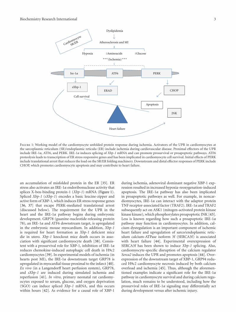
Figure 1: Working model of the cardiomyocyte unfolded protein response during ischemia. Activators of the UPR in cardiomyocytes atthe sarcoplasmic reticulum (SR)/endoplasmic reticulu (ER) include ischemia during cardiovascular disease. Proximal effectors of the UPRinclude IRE-1α, ATF6, and PERK. IRE-1α induces splicing of Xbp-1 mRNA and can promote prosurvival or proapoptotic pathways. ATF6proteolysis leads to transcription of ER stress responsive genes and has been implicated in cardiomyocyte cell survival. Initial effects of PERKinclude translational arrest that reduces the load on the SR/ER folding machinery. Downstream and distal effector responses of PERK includeCHOP, which promotes cardiomyocyte apoptosis and may contribute to heart failure.
an accumulation of misfolded protein in the ER [35]. ERstress also activates an IRE-1α endoribonuclease activity thatsplices X-box-binding protein-1 (Xbp-1) mRNA (Figure 1).Spliced Xbp-1 (sXbp-1) encodes a basic leucine-zipper andactive form of XBP-1, which induces ER stress response genes[36, 37] that escape PERK-mediated translational arrest(discussed below). The requirement for the UPR in theheart and the IRE-1α pathway begins during embryonicdevelopment. GRP78 (guanine-nucleotide-releasing protein78), an IRE-1α and ATF6 downstream target, is upregulatedin the embryonic mouse myocardium. In addition, Xbp-1is required for heart formation as Xbp-1 deficient micedie in utero. Xbp-1 knockout mice death occurs in asso-ciation with significant cardiomyocyte death [38]. Consis-tent with a prosurvival role for XBP-1, inhibition of IRE-1αreduces chemokine-induced autophagic cell death in H9c2cardiomyocytes [39]. In experimental models of ischemia (inhearts post MI), the IRE-1α downstream target GRP78 isupregulated in myocardial tissue proximal to the infarct [40].Ex vivo (in a Langendorff heart perfusion system), GRP78,and sXbp-1 are induced during simulated ischemia andreperfusion [41]. In vitro, primary neonatal rat cardiomy-ocytes exposed to serum, glucose, and oxygen deprivation(SGO) can induce spliced Xbp-1 mRNA, and this occurswithin hours [42]. As evidence for a causal role of XBP-1
during ischemia, adenoviral dominant negative XBP-1 exp-ression resulted in increased hypoxia-reoxygenation-inducedapoptosis. The IRE-1α pathway has also been implicatedin proapoptotic pathways as well. For example, in noncar-diomyocytes, IRE-1α can interact with the adaptor proteinTNF receptor-associated factor (TRAF2). IRE-1α and TRAF2subsequently act on ASK1 (mitogen-activated protein kinasekinase kinase), which phosphorylates proapoptotic JNK [43].Less is known regarding how such a proapoptotic IRE-1αpathway may function in cardiomyocytes. In addition, cal-cium dysregulation is an important component of ischemicheart failure and upregulation of sarco/endoplasmic retic-ulum calcium-ATPase isoform 3f (SERCA3f) is associatedwith heart failure [44]. Experimental overexpression ofSERCA3f has been shown to induce Xbp-1 splicing. Also,cardiomyocyte-specific disruption of the calcium regulatorSerca2 induces the UPR and promotes apoptosis [44]. Over-expression of the downstream target of XBP-1, GRP94 redu-ced H9c2 cardiomyocyte necrosis induced by both calciumoverload and ischemia [45]. Thus, although the aforemen-tioned examples indicate a significant role for the IRE-1αpathway in cardiomyocyte survival and during calcium regu-lation, much remains to be understood, including how theprosurvival roles of IRE-1α signaling may differentially actduring development versus after ischemic injury.

4 Biochemistry Research International
2.2. Activating Transcription Factor 6 (ATF6) in the Heart. Onactivation of the UPR, ATF6 travels to the Golgi, where itscleavage leads to the translocation of its cytosolic fragmentto the nucleus and binding to ER stress response elements(ERSEs). Cleaved ATF6 then promotes transcription ofER-targeted genes, such as the ER chaperone, GRP78. Inmice after MI, inhibition of ATF6 activation with 4-(2-aminoethyl) benzenesulfonyl fluoride, an inhibitor of ATF6,impaired cardiac function and increased mortality. In con-trast, cardiac function after MI was improved in mice exp-ressing a constitutively active mutant of Atf6, compared withwild-type littermates [46] and consistent with a protectiverole. In primary murine cardiac myocytes exposed to oxygenand nutrient deprivation, membrane-associated ATF6 wasreduced with a concomitant increase in nuclear ATF6 [47].This ischemia-induced event was accompanied by ATF6binding to the ERSE of GRP78, transcriptional upregulationof GRP78 and was reversible by simulated reperfusion invitro. More importantly, a dominant-negative form of ATF6prevented inducement of Grp78 and promoted cardiomy-ocyte cell death, indicating a prosurvival role for ATF6. ATF6has also been shown to induce ER-associated degradation(ERAD). ERAD has been shown to alleviate ER stress bydegrading misfolded protein in the ER [15]. Interestingly,Belmont et al. discovered that Derlin-3, a component ofERAD, is induced by ATF6 in the mouse heart [48]. Further-more, overexpression of Derlin-3 protected cardiomyocytesin vitro from simulated ischemia-induced apoptosis. Inanother article by Belmont, transcriptional profiling inden-tified modulatory calcineurin interacting protein-1 (MCIP1),also known as regulator of calcineurin 1 (RCAN1), as anovel ATF6-inducible gene. They found that ATF6 was ableto induce RCAN1 in cultured cardiac myocytes and thatadenoviral overexpression of activated ATF6 further inducedRCAN1 and modulated cell growth [49]. Thus, ATF6 is indu-ced under ischemic conditions and can play a role to helpprotect cardiomyocyte survival. Interestingly, an ATF6 iso-form and other ATF6-related proteins may play a role inregulating the UPR, however, their full roles in cardiomy-ocytes remain undetermined and should be subject of futureinvestigation [50].
2.3. Cardiomyocyte PERK (dsRNA-Activated Protein Kinase-Like Endoplasmic Reticulum Kinase). Though the IRE-1αand ATF6 branches have for the most part been associ-ated with prosurvival roles in cardiomyocytes, prolongedactivation of the PERK/ATF4/CHOP pathway is principallyimplicated in cardiomyocyte cell death. Downstream ofPERK, phosphorylation of eIF2α can be detected as early asone hour after ischemia in vitro in cardiomyocytes [42].Eukaryotic translation initiation factor 2α (eIF2α) phospho-rylation leads to a transient downregulation of the majorityof protein synthesis through inhibition of cap-dependentprotein translation. Only transcripts encoded by ER stressresponse genes are induced, reducing the demands on theER. This may have implications in prevention of cardiachypertrophy and is part of the initial compensatory pathwayof the PERK branch towards promoting survival. Underprolonged ER stress, C/EBP homologous protein (CHOP) is
induced. Myocardial tissue from patients with heart failureexhibits increased Chop mRNA. Okada et al. reported thatprolonged ER stress occurs in hypertrophic and failing heartsafter aortic constriction [51]. Also, Chop deficiency reducescardiac apoptosis in a pressure overload model of heartdisease [52]. CHOP has also been implicated in dilated car-diomyopathy [23]. In vitro, in heart cells, prolonged ER stressinduced by ischemia promoted the activation of CHOP [42],processing of procaspase-12 and induction of apoptosis.Consistent with activation by ischemia, Chop transcription isalso regulated by amino acid starvation. For example, an up-stream cis amino acid response element in Chop has beenfound to bind activating transcription factor 2 (ATF-2) andexpression of ATF-2 is required for the transcriptional acti-vation of Chop by leucine starvation in vitro [53]. In supportof this pathway being activated during ischemia, ATF-2 isstabilized by hypoxia [54]. More recently, prostatic androgenrepressed message-1 or PARM was identified to be predomi-nantly expressed in cardiomyocytes and a negative regulatorof CHOP-mediated apoptosis [55]. Finally, Chop deficiencyhas been shown to reduce myocardial reperfusion injury in amouse model of MI [56]. Future studies in vivo are warrantedto separate the effects of CHOP after ischemia as opposed toafter reperfusion.
3. Discussion
Although treatments for heart failure have advanced, theincidence of HF is still rising and new therapies remain animportant goal. There is now mounting evidence of a sig-nificant role for the UPR in cardiomyocytes during ischemicheart disease. Much remains to be understood with respectto how individual branches of the UPR differentially or syn-ergistically contribute to progression of heart failure and howthese pathways differ from requirements of the UPR duringdevelopment. In addition, the therapeutic and prophylacticpotential of modulating the heart is far from complete. Somehave suggested that ischemic preconditioning of the heartand activation of the UPR may promote cardiac cell survival.Interesting proofs of principle have been published. Forexample, in vitro, overexpression of ER-stress-induced Grp94has been shown to inhibit cardiomyocyte necrosis after cal-cium overload and simulated ischemia [45]. Overexpressionof GRP78 has also been shown to have an effect. ForcedGRP78 expression inhibited apoptosis in rat ventricularmyocytes [57]. Also, preconditioning of H9c2 neonatal car-diomyocytes cells with the ER-stressor tunicamycin has beenshown to protect against ATP deletion [58]. These arelaudable starts, but much work remains to be done. Futurequestions remain. For example: What is the effect of chemicalchaperones on cardiac stress pathways and cardiac function[59]? Future studies will also be required to dissect the effectsof cell-specific deletion of UPR genes in the heart, includingcardiomyocytes, myofibroblasts, and inflammatory cells thatinfiltrate into the myocardium after injury. UPR-targetedtherapies may be realized by promoting the cytoprotectivefunction of the UPR in the myocyte. Such an approachmay induce UPR-specific ER chaperones and downstream

Biochemistry Research International 5
prosurvival pathways that work to enhance cardiac functionand prevent cardiomyocyte death.
Abbreviations
ATF6: Activating transcription factor-6BIP: Binding immunoglobulin proteineIF2α: Eukaryotic initiation factor 2ER: Endoplasmic reticulumERAD: ER-associated degradation pathwayGrp94: Glucose-regulated proteinHF: Heart failureIRE-1: Includes inositol-requiring enzyme-1 (IRE-1)MI: Myocardial infarctionPERK: PKR-like eukaryotic initiation factor 2 kinasePDI: Protein disulfide isomeraseSR: Sarcoplasmic reticulumUPR: Unfolded protein response.
Acknowledgments
The author acknowledges and appreciates previous discus-sions with Ira Tabas, MD, PhD. Funding is got from NIH4R00HL097021-03 Grant from the NHLBI (to ET).
References
[1] J. J. V. McMurray and M. A. Pfeffer, “Heart failure,” The Lancet,vol. 365, no. 9474, pp. 1877–1889, 2005.
[2] J. A. Ezekowitz and P. Kaul, “The epidemiology and manage-ment of elderly patients with myocardial infarction or heartfailure,” Heart Failure Reviews, vol. 15, no. 5, pp. 407–413,2010.
[3] V. L. Roger, A. S. Go, D. M. Lloyd-Jones et al., “Heart diseaseand stroke statistics-2011 update: a report from the AmericanHeart Association,” Circulation, vol. 123, pp. e18–e209, 2011.
[4] R. S. Y. Foo, K. Mani, and R. N. Kitsis, “Death begets failure inthe heart,” Journal of Clinical Investigation, vol. 115, no. 3, pp.565–571, 2005.
[5] A. Abbate, G. G. L. Biondi-Zoccai, R. Bussani et al., “Increasedmyocardial apoptosis in patients with unfavorable left ventric-ular remodeling and early symptomatic post-infarction heartfailure,” Journal of the American College of Cardiology, vol. 41,no. 5, pp. 753–760, 2003.
[6] G. W. Dorn, “Apoptotic and non-apoptotic programmed car-diomyocyte death in ventricular remodelling,” CardiovascularResearch, vol. 81, no. 3, pp. 465–473, 2009.
[7] W. Martinet, M. W. M. Knaapen, M. M. Kockx, and G. R.Y. De Meyer, “Autophagy in cardiovascular disease,” Trends inMolecular Medicine, vol. 13, no. 11, pp. 482–491, 2007.
[8] D. Ron and P. Walter, “Signal integration in the endoplasmicreticulum unfolded protein response,” Nature Reviews Molec-ular Cell Biology, vol. 8, no. 7, pp. 519–529, 2007.
[9] G. S. Hossain, J. V. van Thienen, G. H. Werstuck et al.,“TDAG51 is induced by homocysteine, promotes detachment-mediated programmed cell death, and contributes to the deve-lopment of atherosclerosis in hyperhomocysteinemia,” TheJournal of Biological Chemistry, vol. 278, no. 32, pp. 30317–30327, 2003.
[10] Y. Ma and L. M. Hendershot, “The unfolding tale of the un-folded protein response,” Cell, vol. 107, no. 7, pp. 827–830,2001.
[11] J. S. Cox, C. E. Shamu, and P. Walter, “Transcriptional induc-tion of genes encoding endoplasmic reticulum resident pro-teins requires a transmembrane protein kinase,” Cell, vol. 73,no. 6, pp. 1197–1206, 1993.
[12] H. Yoshida, K. Haze, H. Yanagi, T. Yura, and K. Mori, “Iden-tification of the cis-acting endoplasmic reticulum stress res-ponse element responsible for transcriptional induction ofmammalian glucose-regulated proteins. Involvement of basicleucine zipper transcription factors,” The Journal of BiologicalChemistry, vol. 273, no. 50, pp. 33741–33749, 1998.
[13] H. P. Harding, Y. Zhang, and D. Ron, “Protein translation andfolding are coupled by an endoplasmic-reticulum-residentkinase,” Nature, vol. 397, no. 6716, pp. 271–274, 1999.
[14] R. Friedlander, E. Jarosch, J. Urban, C. Volkwein, and T. Som-mer, “A regulatory link between ER-associated protein deg-radation and the unfolded-protein response,” Nature CellBiology, vol. 2, no. 7, pp. 379–384, 2000.
[15] K. J. Travers, C. K. Patil, L. Wodicka, D. J. Lockhart, J. S.Weissman, and P. Walter, “Functional and genomic analysesreveal an essential coordination between the unfolded proteinresponse and ER-associated degradation,” Cell, vol. 101, no. 3,pp. 249–258, 2000.
[16] H. P. Harding, Y. Zhang, A. Bertolotti, H. Zeng, and D. Ron,“Perk is essential for translational regulation and cell survivalduring the unfolded protein response,” Molecular Cell, vol. 5,no. 5, pp. 897–904, 2000.
[17] P. I. Merksamer, A. Trusina, and F. R. Papa, “Real-time redoxmeasurements during endoplasmic reticulum stress revealinterlinked protein folding functions,” Cell, vol. 135, no. 5, pp.933–947, 2008.
[18] I. Tabas and D. Ron, “Integrating the mechanisms of apoptosisinduced by endoplasmic reticulum stress,” Nature Cell Biology,vol. 13, no. 3, pp. 184–190, 2011.
[19] B. J. Maron, V. J. Ferrans, and W. C. Roberts, “Ultrastructuralfeatures of degenerated cardiac muscle cells in patients withcardiac hypertrophy,” American Journal of Pathology, vol. 79,no. 3, pp. 387–434, 1975.
[20] P. Volpe, A. Villa, P. Podini et al., “The endoplasmic reticulum-sarcoplasmic reticulum connection: distribution of endo-plasmic reticulum markers in the sarcoplasmic reticulum ofskeletal muscle fibers,” Proceedings of the National Academy ofSciences of the United States of America, vol. 89, no. 13, pp.6142–6146, 1992.
[21] S. E. Cala, C. Ulbright, J. S. Kelley, and L. R. Jones, “Purifi-cation of a 90-kDa protein (band VII) from cardiac sarcoplas-mic reticulum. Identification as calnexin and localization ofcasein kinase II phosphorylation sites,” Journal of BiologicalChemistry, vol. 268, no. 4, pp. 2969–2975, 1993.
[22] J. A. Barnes and I. W. Smoak, “Immunolocalization and heartlevels of GRP94 in the mouse during post-implantation devel-opment,” Anatomy and Embryology, vol. 196, no. 4, pp. 335–341, 1997.
[23] H. Hamada, M. Suzuki, S. Yuasa et al., “Dilated cardiomyopa-thy caused by aberrant endoplasmic reticulum quality controlin mutant KDEL receptor transgenic mice,” Molecular andCellular Biology, vol. 24, no. 18, pp. 8007–8017, 2004.
[24] J. S. Pattison, A. Sanbe, A. Maloyan, H. Osinska, R. Klevitsky,and J. Robbins, “Cardiomyocyte expression of a polyglutaminepreamyloid oligomer causes heart failure,” Circulation, vol.117, no. 21, pp. 2743–2751, 2008.

6 Biochemistry Research International
[25] X. Wang, H. Osinska, R. Klevitsky et al., “Expression ofR120G-αB-crystallin causes aberrant desmin and αB-crys-tallin aggregation and cardiomyopathy in mice,” CirculationResearch, vol. 89, no. 1, pp. 84–91, 2001.
[26] J. G. Dickhout, R. E. Carlisle, and R. C. Austin, “Interrelation-ship between cardiac hypertrophy, heart failure, and chronickidney disease: endoplasmic reticulum stress as a mediator ofpathogenesis,” Circulation Research, vol. 108, no. 5, pp. 629–642, 2011.
[27] N. Mesaeli, K. Nakamura, M. Opas, and M. Michalak, “Endo-plasmic reticulum in the heart, a forgotten organelle?” Molec-ular and Cellular Biochemistry, vol. 225, no. 1-2, pp. 1–6,2001.
[28] M. Michalak and M. Opas, “Endoplasmic and sarcoplasmicreticulum in the heart,” Trends in Cell Biology, vol. 19, no. 6,pp. 253–259, 2009.
[29] A. Azfer, J. Niu, L. M. Rogers, F. M. Adamski, and P. E.Kolattukudy, “Activation of endoplasmic reticulum stress res-ponse during the development of ischemic heart disease,”American Journal of Physiology, vol. 291, no. 3, pp. H1411–H1420, 2006.
[30] B. E. Sobel, “Salient biochemical features in ischemic myo-cardium,” Circulation Research, vol. 35, no. 3, pp. 173–181,1974.
[31] R. M. Graham, D. P. Frazier, J. W. Thompson et al., “A uni-que pathway of cardiac myocyte death caused by hypoxia-aci-dosis,” Journal of Experimental Biology, vol. 207, no. 18, pp.3189–3200, 2004.
[32] T. Iwai, K. Tanonaka, R. Inoue, S. Kasahara, N. Kamo, andS. Takeo, “Mitochondrial damage during ischemia determinespost-ischemic contractile dysfunction in perfused rat heart,”Journal of Molecular and Cellular Cardiology, vol. 34, no. 7, pp.725–738, 2002.
[33] Y. Shimizu and L. M. Hendershot, “Oxidative folding: cellularstrategies for dealing with the resultant equimolar productionof reactive oxygen species,” Antioxidants and Redox Signaling,vol. 11, no. 9, pp. 2317–2331, 2009.
[34] J. J. Martindale, R. Fernandez, D. Thuerauf et al., “Endoplas-mic reticulum stress gene induction and protection fromischemia/reperfusion injury in the hearts of transgenic micewith a tamoxifen-regulated form of ATF6,” Circulation Re-search, vol. 98, no. 9, pp. 1186–1193, 2006.
[35] M. Schroder and R. J. Kaufman, “The mammalian unfoldedprotein response,” Annual Review of Biochemistry, vol. 74, pp.739–789, 2005.
[36] C. Sidrauski and P. Walter, “The transmembrane kinase Ire1pis a site-specific endonuclease that initiates mRNA splicing inthe unfolded protein response,” Cell, vol. 90, no. 6, pp. 1031–1039, 1997.
[37] M. Calfon, H. Zeng, F. Urano et al., “IRE1 couples endoplas-mic reticulum load to secretory capacity by processing theXBP-1 mRNA,” Nature, vol. 415, no. 6867, pp. 92–96, 2002.
[38] T. Masaki, M. Yoshida, and S. Noguchi, “Targeted disruptionof CRE-Binding factor TREB5 gene leads to cellular necrosisin cardiac myocytes at the embryonic stage,” Biochemical andBiophysical Research Communications, vol. 261, no. 2, pp. 350–356, 1999.
[39] C. W. Younce and P. E. Kolattukudy, “MCP-1 causes cardiomy-oblast death via autophagy resulting from ER stress causedby oxidative stress generated by inducing a novel zinc-fingerprotein, MCPIP,” Biochemical Journal, vol. 426, no. 1, pp. 43–53, 2010.
[40] D. J. Thuerauf, M. Marcinko, N. Gude, M. Rubio, M. A.Sussman, and C. C. Glembotski, “Activation of the unfoldedprotein response in infarcted mouse heart and hypoxic cultu-red cardiac myocytes,” Circulation Research, vol. 99, no. 3, pp.275–282, 2006.
[41] X. Qi, A. Vallentin, E. Churchill, and D. Mochly-Rosen,“δPKC participates in the endoplasmic reticulum stress-in-duced response in cultured cardiac myocytes and ischemicheart,” Journal of Molecular and Cellular Cardiology, vol. 43,no. 4, pp. 420–428, 2007.
[42] E. Szegezdi, A. Duffy, M. E. O’Mahoney et al., “ER stress con-tributes to ischemia-induced cardiomyocyte apoptosis,” Bio-chemical and Biophysical Research Communications, vol. 349,no. 4, pp. 1406–1411, 2006.
[43] F. Urano, X. Wang, A. Bertolotti et al., “Coupling of stress inthe ER to activation of JNK protein kinases by transmembraneprotein kinase IRE1,” Science, vol. 287, no. 5453, pp. 664–666,2000.
[44] S. Dally, V. Monceau, E. Corvazier et al., “Compartmental-ized expression of three novel sarco/endoplasmic reticulumCa2+ATPase 3 isoforms including the switch to ER stress,SERCA3f, in non-failing and failing human heart,” Cell Cal-cium, vol. 45, no. 2, pp. 144–154, 2009.
[45] M. Vitadello, D. Penzo, V. Petronilli et al., “Overexpressionof the stress protein Grp94 reduces cardiomyocyte necrosisdue to calcium overload and simulated ischemia,” The FASEBJournal, vol. 17, no. 8, pp. 923–925, 2003.
[46] H. Toko, H. Takahashi, Y. Kayama et al., “ATF6 is importantunder both pathological and physiological states in the heart,”Journal of Molecular and Cellular Cardiology, vol. 49, no. 1, pp.113–120, 2010.
[47] S. Doroudgar, D. J. Thuerauf, M. C. Marcinko, P. J. Belmont,and C. C. Glembotski, “Ischemia activates the ATF6 branchof the endoplasmic reticulum stress response,” Journal of Bio-logical Chemistry, vol. 284, no. 43, pp. 29735–29745, 2009.
[48] P. J. Belmont, W. J. Chen, M. N. San Pedro et al., “Roles forendoplasmic reticulum-associated degradation and the novelendoplasmic reticulum stress response gene derlin-3 in theischemic heart,” Circulation Research, vol. 106, no. 2, pp. 307–316, 2010.
[49] P. J. Belmont, A. Tadimalla, W. J. Chen et al., “Coordinationof growth and endoplasmic reticulum stress signaling by regu-lator of calcineurin 1 (RCAN1), a novel ATF6-inducible gene,”Journal of Biological Chemistry, vol. 283, no. 20, pp. 14012–14021, 2008.
[50] C. C. Glembotski, “The role of the unfolded protein responsein the heart,” Journal of Molecular and Cellular Cardiology, vol.44, no. 3, pp. 453–459, 2008.
[51] K. I. Okada, T. Minamino, Y. Tsukamoto et al., “Prolongedendoplasmic reticulum stress in hypertrophic and failing heartafter aortic constriction: possible contribution of endoplasmicreticulum stress to cardiac myocyte apoptosis,” Circulation,vol. 110, no. 6, pp. 705–712, 2004.
[52] H. Y. Fu, K. I. Okada, Y. Liao et al., “Ablation of C/EBP homol-ogous protein attenuates endoplasmic reticulum-mediatedapoptosis and cardiac dysfunction induced by pressure over-load,” Circulation, vol. 122, no. 4, pp. 361–369, 2010.
[53] A. Bruhat, C. Jousse, V. Carraro, A. M. Reimold, M. Ferrara,and P. Fafournoux, “Amino acids control mammalian genetranscription: activating transcription factor 2 is essential forthe amino acid responsiveness of the CHOP promoter,” Mole-cular and Cellular Biology, vol. 20, no. 19, pp. 7192–7204, 2000.

Biochemistry Research International 7
[54] J. H. Choi, H. K. Cho, Y. H. Choi, and J. H. Cheong, “Activatingtranscription factor 2 increases transactivation and proteinstability of hypoxia-inducible factor 1α in hepatocytes,” Bio-chemical Journal, vol. 424, no. 2, pp. 285–296, 2009.
[55] K. Isodono, T. Takahashi, H. Imoto et al., “PARM-1 is an endo-plasmic reticulum molecule involved in endoplasmic reticu-lum stress-induced apoptosis in rat cardiac myocytes,” PloSOne, vol. 5, no. 3, article e9746, 2010.
[56] Y. Miyazaki, K. Kaikita, M. Endo et al., “C/EBP homologousprotein deficiency attenuates myocardial reperfusion injuryby inhibiting myocardial apoptosis and inflammation,” Arte-riosclerosis, Thrombosis, and Vascular Biology, vol. 31, no. 5, pp.1124–1132, 2011.
[57] H. Y. Fu, T. Minamino, O. Tsukamoto et al., “Overexpressionof endoplasmic reticulum-resident chaperone attenuates car-diomyocyte death induced by proteasome inhibition,” Cardio-vascular Research, vol. 79, no. 4, pp. 600–610, 2008.
[58] P. L. Zhang, M. Lun, J. Teng et al., “Preinduced molecularchaperones in the endoplasmic reticulum protect cardiomy-ocytes from lethal injury,” Annals of Clinical and LaboratoryScience, vol. 34, no. 4, pp. 449–457, 2004.
[59] E. Erbay, V. R. Babaev, J. R. Mayers et al., “Reducing endoplas-mic reticulum stress through a macrophage lipid chaperonealleviates atherosclerosis,” Nature Medicine, vol. 15, no. 12, pp.1383–1391, 2009.

Hindawi Publishing CorporationBiochemistry Research InternationalVolume 2012, Article ID 247275, 5 pagesdoi:10.1155/2012/247275
Review Article
Endoplasmic Reticulum Stress-Associated Lipid DropletFormation and Type II Diabetes
Xuebao Zhang1 and Kezhong Zhang1, 2, 3
1 Center for Molecular Medicine and Genetics, The Wayne State University School of Medicine, 540 East Canfield Avenue,Detroit, MI 48201, USA
2 Department of Immunology and Microbiology, The Wayne State University School of Medicine, Detroit, MI 48201, USA3 Karmanos Cancer Institute, The Wayne State University School of Medicine, Detroit, MI 48201, USA
Correspondence should be addressed to Kezhong Zhang, [email protected]
Received 2 September 2011; Revised 14 November 2011; Accepted 15 November 2011
Academic Editor: Huiping Zhou
Copyright © 2012 X. Zhang and K. Zhang. This is an open access article distributed under the Creative Commons AttributionLicense, which permits unrestricted use, distribution, and reproduction in any medium, provided the original work is properlycited.
Diabetes mellitus (DM), a metabolic disorder characterized by hyperglycemia, is caused by insufficient insulin production dueto excessive loss of pancreatic β cells (type I diabetes) or impaired insulin signaling due to peripheral insulin resistance (type IIdiabetes). Pancreatic β cell is the only insulin-secreting cell type that has highly developed endoplasmic reticulum (ER) to cope withhigh demands of insulin synthesis and secretion. Therefore, ER homeostasis is crucial to the proper function of insulin signaling.Accumulating evidence suggests that deleterious ER stress and excessive intracellular lipids in nonadipose tissues, such as myocyte,cardiomyocyte, and hepatocyte, cause pancreatic β-cell dysfunction and peripheral insulin resistance, leading to type II diabetes.The excessive deposition of lipid droplets (LDs) in specialized cell types, such as adipocytes, hepatocytes, and macrophages,has been found as a hallmark in ER stress-associated metabolic diseases, including obesity, diabetes, fatty liver disease, andatherosclerosis. However, much work remains to be done in understanding the mechanism by which ER stress response regulatesLD formation and the pathophysiologic role of ER stress-associated LD in metabolic disease. This paper briefly summarizes therecent advances in ER stress-associated LD formation and its involvement in type II diabetes.
1. Introduction to ER Stress
ER is an intracellular organelle where dynamic protein fold-ing and assembly, storing cellular calcium, and lipid biosyn-thesis occur. A variety of biochemical or pathophysiologicalstimuli can interrupt protein folding process in the ER bydisrupting protein glycosylation, disulfide bond formation,or ER calcium pool. These disruptions can cause the accum-ulation of unfolded or misfolded proteins in the ER lumen,a condition termed as “ER stress” [1, 2]. To protect cellsfrom proteotoxicity caused by ER stress, the unfolded proteinresponse (UPR) is activated through attenuating general pro-tein translation, increasing in protein folding capacity, andexpediting degradation of misfolded proteins. Three majorER stress sensors or transducers have been found: inositol-requiring 1α (IRE1α), double-stranded RNA-dependent pro-tein kinase- (PKR-) like ER kinase (PERK), and activatingtranscription factor 6 (ATF6), which have been comprehen-
sively reviewed [2, 3]. The UPR signaling, mediated throughER stress sensors, modulates transcriptional and translationprograms in cells under ER stress. As a double-edged sword,the UPR provides survival signals at the initial phase of stressresponse, leading to cell adaption to ER stress [1, 2, 4].When ER stress gets prolonged, the UPR can induce celldeath programs to kill the stressed cells. In recent years, thescope and consequence of ER stress and UPR have beensignificantly expanded. Many pathophysiologic stimuli, suchas oxidative stress, proinflammatory stimuli, fatty acids, andenergy fluctuations, can directly or indirectly cause ER stressand the UPR activation in specialized cell types, such asmacrophages, hepatocytes, and pancreatic β cells [2, 5]. TheUPR signaling is fundamental to the initiation and progressof a variety of diseases, including metabolic disease, cancer,cardiovascular disease, and neurodegenerative disease [2, 6,7].

2 Biochemistry Research International
2. LD Formation
LD, also known as adiposome or fat body, has been foundubiquitously present in lipid-overloaded cells from yeast tomammals [8, 9]. For a long time, LD was thought simply asan inert lipid storage reservoir since its earliest descriptionin 19th century. The discovery of perilipin, an LD-associatedprotein that coats LD in adipocytes, makes researchers tochallenge the understanding of LD as lipid storage [10].LD is now recognized as a dynamic organelle composedof a monolayer phospholipid, embedded with numerousproteins without transmembrane spanning domains, and ahydrophobic core that contains triacylglycerols (TGs) andsterol esters [11, 12]. TGs are the key neutral lipid requiredfor LDs formation in adipocytes. Deletion of genes encodingenzymes responsible for neutral lipid synthesis eliminatedLDs formation [13]. Evidence showed that, without DGATenzymes, LDs cannot form in adipocytes. Therefore, bysegregation of extra TG or hydrophobic molecules into LDs,cells are protected from lipotoxicity. These features make LDa regulatory organelle in lipid homeostasis. The biogenesisand assembly of LD are still largely unknown. It has beensuggested that ER is the site where LD is synthesized andassembled. Over ninety percent of LDs were found in closeapposition to the ER [14]. ER budding model, Bicelle model,and vesicular budding model have been suggested to explainhow LD is formed in ER [15]. Perhaps, the most acceptedmodel is ER budding model in which LD originated betweenthe two leaflets of ER bilayer buds into the cytosol. Newlyformed LD can increase its size (0.2 μm–20 μm in diameter)by homotypic fusion that depends on microtubule system,most likely motor protein dynein. Under this mechanism, thegrowth of LD may proceed without ongoing biosynthesis ofTGs and sterol esters [16, 17].
3. ER Stress and LD Formation
LD formation has been proposed as an exit model inthe removal of unfolded or misfolded proteins or someubiquitinated proteins from the ER [18, 19]. LD may serveas a transient depot to sequester unfolded or misfolded aswell as excessive proteins to alleviate ER stress (Figure 1).Diverse groups of LD-associated proteins were found in yeastS. cerevisiae, Drosophila embryos, and human hepatocyte cellline Huh7 [20–22]. Some of the LD-associated proteins, suchas Acl-CoA synthetases, lanosterol synthetase, and GAPDH,are conserved from yeast to human. The proteins detected inLD seem to be specific, since the organelle-specific proteins,including lactate dehydrogenase (LDH) (cytosolic marker),integrin (plasma membrane marker), calnexin (ER marker),and GS28 (Golgi marker), were hardly detected in LDfractions [22]. Interestingly, a number of proteins whichwere thought to be organelle-specific, including histones(nucleus), caveolins (plasma membrane), HSP70 (cytosol),ApoB (ER), and Nir2 (Golgi), were detected in LD fraction[23]. Furthermore, LD dynamically interacts with ER, per-oxisomes, mitochondria, and plasma membrane [15]. LDcan be transported along microtubules, following the sameway that the ER, Golgi, and mitochondria were positioned
and delivered [24]. It was proposed that the dynamicalinteractions between LD and the other compartments facil-itate the exchange of proteins and lipids in cells. The LDis functionally and structurally similar to the extracellularcounterpart of lipoprotein particles [15, 21]. This notionwas supported by the finding that LD provides a platformfor degradation of excessive ApoB protein by convergingubiquitin-proteasomal and autophagy-lysosomal pathways,thereby preventing cytotoxicity resulted from aggregation ofexcessive proteins [25]. Previous studies have shown thatdisruption of ER functions leads to the accumulation ofintracellular lipids [26–28]. Disrupted protein glycosylationor ER-associated protein degradation by ER stress-inducingreagents, such as tunicamycin and brefeldin, has beendemonstrated to increase LD accumulation in budding yeastSaccharomyces cerevisiae or mammalian cells [28, 29]. Previ-ously, it is known that intracellular LD formation is throughthe lipogenic program activated by sterol regulatory element-binding proteins (SREBPs). Recent study suggested thatmore ER-localized, stress-responsive protein factors, suchas hepatocyte-specific cAMP responsive element-bindingprotein (CREBH), can also regulate lipogenic programsto promote LD formation under metabolic stress signals,such as insulin and saturated fatty acids [30]. Moreover,ER stress response may directly facilitate LD synthesis andassembly as a mechanism to defend intracellular stress [29,31] (Figure 1). This is consistent with the observations thatlipids can be recruited to the stressed cells to sequestermisfolded proteins in the ER at the early stage of ER stressand that the ER is expanded significantly to alleviated ERstress independent of the UPR [23, 32].
4. LD Formation and Type II Diabetes
Previous studies demonstrated that excessive accumulationof lipids in peripheral tissues is closely associated withinsulin resistance in type II diabetes [33, 34]. Although ERstress and UPR pathways in metabolic disease have beenextensively reviewed, ER stress-associated LD formation,which is independent of UPR pathway, did not draw muchattention. The interaction between LD and mitochondrialmight affect the peripheral tissue insulin resistance [35, 36](Figure 1). Recent studies indicated that insulin resistanceis not simply associated with the amount of intracellularlipids. Despite elevated lipids content in skeletal muscle ofthe trained enduring athletes, the insulin-signal in these indi-viduals is still markedly sensitive [36]. The combination ofweight loss and physical activity in obesity improves insulinsensitivity and reduces the size of LD, but not the overallintramyocellular lipid [37]. One possible explanation forthese phenomena is that increased mitochondrial oxidativeactivity for lipid oxidation may decrease insulin resistance.This is supported by the facts that lower oxidative capacity isfound in insulin resistant skeletal muscle and that exercisecan improve the capacity for lipid oxidation [36]. Severalmitochondrial proteins including prohibitin, a subunit ofATP synthase, and pyruvate carboxylase were identified inLD fractions by proteomic analysis [35]. In addition, numer-ous lipid metabolic enzymes, such as hormone-sensitive

Biochemistry Research International 3
LD formation UPR
Mitochondria LD interaction
Oxidative activity
Oxidative activity decompensation
Insulin resistance
Diabetes
ER stress
Figure 1: Interactions between ER stress, oxidative stress, and lipid droplets in type II diabetes. LD, lipid droplet; UPR, unfolded proteinresponse.
lipase, lanosterol synthase, and acyl-CoA synthetase, werealso found to be associated with LD complex, and the overallLD protein composition can be changed in response tolipolysis stimulation [35, 38]. Despite these observations,further study is required to explore how mitochondriacommunicate and interact with LD in metabolic processes.
Fat-specific protein 27 (Fsp27) is a member of celldeath-inducing DNA fragmentation factor family proteinsthat is localized to LD. Fsp27 plays an important role inlipid storage and mitochondrial activity in adipocytes [39–41]. Genetic depletion of Fsp27 in mice is characterized byincreased glucose uptake, improved insulin sensitivity, andsignificantly increased mitochondrial metabolism [39, 40].Small sizes of LDs and increased mitochondrial activitywere found in Fsp27-deficient white adipocytes, suggestingthat ectopic LD formation represents an imbalance betweenlipid supply and lipid oxidation in peripheral tissue. Likely,LD-associated proteins and the interactions between LDand the other intracellular organelles may play direct rolesin the pathogenesis of diabetes [42]. Type II diabetes isoften correlated with increased serum levels of proinflamma-tory cytokines secreted by ER stress-activated macrophage.Previous research demonstrated that the proinflammatorycytokine TNFα blunts the insulin signaling pathway thereforecausing insulin resistance by activating the JNK1/2 signalingpathway which is involved in serine phosphorylation ofIRS1 (insulin receptor substrate 1) [43, 44]. However, a newstudy by Ranjit found that proinflammatory cytokines, suchas TNFα, IL1β, and INFγ, act on lipolysis by decreasingthe expression of FSP27 and the size of LD in adipocytes[45]. Since decreased FSP27 is evidenced to improve insulin
resistance and LDs, it is likely that the proinflammatorycytokines play double-edged roles in type II diabetes.
5. Conclusion
Accumulating evidence demonstrated a strong link betweenER stress, LD formation, and type II diabetes. It is importantto note that ER stress response is a fundamental stresssignaling underlying many life styles, such as air pollution,chronic alcohol consumption, and smoking, which may beassociated with the development of metabolic disease [46–48]. Therefore, for the future research, it is important todelineate ER mechanisms in LD formation that is associatedwith the development of type II diabetes. Key questionsinclude what is the mechanism by which ER stress regulatesLD formation? Is there any ER chaperones or UPR targetspresent in the LD complex? Does ER stress-associated LDformation provide survival or devastating pathways in theprogression of type II diabetes? Is it possible to modulate LDformation by targeting ER stress signaling? Answering thesequestions will benefit and direct the future understandingand treatment of type II diabetes and the other types ofmetabolic disease.
Acknowledgments
Portion of the research work in the Zhang laboratory issupported by American Heart Association Grants 09GRNT-2280479; National Institutes of Health Grants DK090313 andES017829 (K. Zhang)

4 Biochemistry Research International
References
[1] K. Zhang and R. J. Kaufman, “Signaling the unfolded proteinresponse from the endoplasmic reticulum,” Journal of Biologi-cal Chemistry, vol. 279, no. 25, pp. 25935–25938, 2004.
[2] D. Ron and P. Walter, “Signal integration in the endoplasmicreticulum unfolded protein response,” Nature Reviews Molec-ular Cell Biology, vol. 8, no. 7, pp. 519–529, 2007.
[3] M. Schroder and R. J. Kaufman, “The mammalian unfoldedprotein response,” Annual Review of Biochemistry, vol. 74, pp.739–789, 2005.
[4] C. Rubio, D. Pincus, A. Korennykh, S. Schuck, H. El-Samad,and P. Walter, “Homeostatic adaptation to endoplasmic retic-ulum stress depends on Ire1 kinase activity,” Journal of Cell Bi-ology, vol. 193, no. 1, pp. 171–184, 2011.
[5] K. Zhang and R. J. Kaufman, “From endoplasmic-reticu-lum stress to the inflammatory response,” Nature, vol. 454, no.7203, pp. 455–462, 2008.
[6] H. Yoshida, “ER stress and diseases,” FEBS Journal, vol. 274,no. 3, pp. 630–658, 2007.
[7] R. J. Kaufman, “Orchestrating the unfolded protein responsein health and disease,” Journal of Clinical Investigation, vol.110, no. 10, pp. 1389–1398, 2002.
[8] M. K. Clausen, K. Christiansen, P. K. Jensen, and O. Behnke,“Isolation of lipid particles from baker’s yeast,” FEBS Letters,vol. 43, no. 2, pp. 176–179, 1974.
[9] K. Christiansen and P. K. Jensen, “Membrane-bound lipidparticles from beef heart chemical composition and structure,”Biochimica et Biophysica Acta, vol. 260, no. 3, pp. 449–459,1972.
[10] A. S. Greenberg, J. J. Egan, S. A. Wek, N. B. Garty, E. J.Blanchette-Mackie, and C. Londos, “Perilipin, a major hor-monally regulated adipocyte-specific phosphoprotein associ-ated with the periphery of lipid storage droplets,” Journal ofBiological Chemistry, vol. 266, no. 17, pp. 11341–11346, 1991.
[11] D. A. Brown, “Lipid droplets: proteins floating on a pool offat,” Current Biology, vol. 11, no. 11, pp. R446–R449, 2001.
[12] P. T. Bozza and J. P. B. Viola, “Lipid droplets in inflammationand cancer,” Prostaglandins Leukotrienes and Essential FattyAcids, vol. 82, no. 4–6, pp. 243–250, 2010.
[13] C. A. Harris, J. T. Haas, R. S. Streeper et al., “DGAT enzymesare required for triacylglycerol synthesis and lipid droplets inadipocytes,” Journal of Lipid Research, vol. 52, no. 4, pp. 657–667, 2011.
[14] K. M. Szymanski, D. Binns, R. Bartz et al., “The lipodystrophyprotein seipin is found at endoplasmic reticulum lipid dropletjunctions and is important for droplet morphology,” Proceed-ings of the National Academy of Sciences of the United States ofAmerica, vol. 104, no. 52, pp. 20890–20895, 2007.
[15] Y. Guo, K. R. Cordes, R. V. Farese Jr., and T. C. Walther, “Lipiddroplets at a glance,” Journal of Cell Science, vol. 122, no. 6, pp.749–752, 2009.
[16] P. Bostrom, L. Andersson, L. Li et al., “The assembly oflipid droplets and its relation to cellular insulin sensitivity,”Biochemical Society Transactions, vol. 37, no. 5, pp. 981–985,2009.
[17] P. Bostrom, M. Rutberg, J. Ericsson et al., “Cytosolic lipiddroplets increase in size by microtubule-dependent complexformation,” Arteriosclerosis, Thrombosis, and Vascular Biology,vol. 25, pp. 1945–1951, 2005.
[18] H. L. Ploegh, “A lipid-based model for the creation of an es-cape hatch from the endoplasmic reticulum,” Nature, vol. 448,no. 7152, pp. 435–438, 2007.
[19] I. Z. Hartman, P. S. Liu, J. K. Zehmer et al., “Sterol-induced dislocation of 3-Hydroxy-3-methylglutaryl coenzymea reductase from endoplasmic reticulum membranes into thecytosol through a subcellular compartment resembling lipiddroplets,” Journal of Biological Chemistry, vol. 285, no. 25, pp.19288–19298, 2010.
[20] K. Athenstaedt, D. Zweytick, A. Jandrositz, S. D. Kohlwein,and G. Daum, “Identification and characterization of majorlipid particle proteins of the yeast Saccharomyces cerevisiae,”Journal of Bacteriology, vol. 181, no. 20, pp. 6441–6448, 1999.
[21] S. Cermelli, Y. Guo, S. P. Gross, and M. A. Welte, “The lipid-droplet proteome reveals that droplets are a protein-storagedepot,” Current Biology, vol. 16, no. 18, pp. 1783–1795, 2006.
[22] Y. Fujimoto, H. Itabe, J. Sakai et al., “Identification of ma-jor proteins in the lipid droplet-enriched fraction isolatedfrom the human hepatocyte cell line HuH7,” Biochimica etBiophysica Acta, vol. 1644, no. 1, pp. 47–59, 2004.
[23] M. A. Welte, “Proteins under new management: lipid dropletsdeliver,” Trends in Cell Biology, vol. 17, no. 8, pp. 363–369,2007.
[24] M. A. Welte, S. Cermelli, J. Griner et al., “Regulation of lipid-droplet transport by the perilipin homolog LSD2,” Current Bi-ology, vol. 15, no. 14, pp. 1266–1275, 2005.
[25] Y. Ohsaki, J. Cheng, A. Fujita, T. Tokumoto, and T. Fujimoto,“Cytoplasmic lipid droplets are sites of convergence of protea-somal and autophagic degradation of apolipoprotein B,” Mol-ecular Biology of the Cell, vol. 17, no. 6, pp. 2674–2683, 2006.
[26] A. J. Kim, Y. Shi, R. C. Austin, and G. H. Werstuck, “Valproateprotects cells fom ER stress-induced lipid accumulation andapoptosis by inhibiting glycogen synthase kinase-3,” Journal ofCell Science, vol. 118, no. 1, pp. 89–99, 2005.
[27] G. H. Werstuck, S. R. Lentz, S. Dayal et al., “Homocysteine-induced endoplasmic reticulum stress causes dysregulation ofthe cholesterol and triglyceride biosynthetic pathways,” Jour-nal of Clinical Investigation, vol. 107, no. 10, pp. 1263–1273,2001.
[28] W. Fei, H. Wang, C. Bielby, and H. Yang, “Conditions of endo-plasmic reticulum stress stimulate lipid droplet formation inSaccharomyces cerevisiae,” Biochemical Journal, vol. 424, no.1, pp. 61–67, 2009.
[29] J.-S. Lee, R. Mendez, H. H. Heng, Z. Yang, and K. Zhang,“Pharmacological ER stress promotes hepatic lipogenesis andlipid droplet formation,” American Journal of Translational Re-search, vol. 4, pp. 102–113, 2012.
[30] C. Zhang, G. Wang, Z. Zheng et al., “Endoplasmic reticulum-tethered transcription factor cAMP responsive element-bind-ing protein, hepatocyte specific, regulates hepatic lipogenesis,fatty acid oxidation, and lipolysis upon metabolic stress inmice,” Hepatology. In press.
[31] K. Zhang, S. Wang, J. Malhotra et al., “The unfolded proteinresponse transducer IRE1alpha prevents ER stress-induced he-patic steatosis,” The EMBO Journal, vol. 30, pp. 1357–1375,2011.
[32] S. Schuck, W. A. Prinz, K. S. Thorn, C. Voss, and P. Walter,“Membrane expansion alleviates endoplasmic reticulum stressindependently of the unfolded protein response,” Journal ofCell Biology, vol. 187, no. 4, pp. 525–536, 2009.
[33] S. E. Thomas, L. E. Dalton, M. L. Daly, E. Malzer, and S. J.Marciniak, “Diabetes as a disease of endoplasmic reticulumstress,” Diabetes/Metabolism Research and Reviews, vol. 26, no.8, pp. 611–621, 2010.
[34] D. Scheuner and R. J. Kaufman, “The unfolded proteinresponse: a pathway that links insulin demand with β-cell

Biochemistry Research International 5
failure and diabetes,” Endocrine Reviews, vol. 29, no. 3, pp.317– 333, 2008.
[35] D. L. Brasaemle, G. Dolios, L. Shapiro, and R. Wang, “Pro-teomic analysis of proteins associated with lipid droplets ofbasal and lipolytically stimulated 3T3-L1 adipocytes,” Journalof Biological Chemistry, vol. 279, no. 5, pp. 46835–46842, 2004.
[36] B. H. Goodpaster, J. He, S. Watkins, and D. E. Kelley, “Skeletalmuscle lipid content and insulin resistance: evidence for a pa-radox in endurance-trained athletes,” Journal of Clinical Endo-crinology and Metabolism, vol. 86, no. 12, pp. 5755–5761, 2001.
[37] J. He, B. H. Goodpaster, and D. E. Kelley, “Effects of weightloss and physical activity on muscle lipid content and dropletsize,” Obesity Research, vol. 12, no. 5, pp. 761–769, 2004.
[38] J. G. Granneman, H. P. Moore, R. L. Granneman, A. S.Greenberg, M. S. Obin, and Z. Zhu, “Analysis of lipolyticprotein trafficking and interactions in adipocytes,” Journal ofBiological Chemistry, vol. 282, no. 8, pp. 5726–5735, 2007.
[39] Z. Nian, Z. Sun, L. Yu, S. Y. Toh, J. Sang, and P. Li, “Fat-specificprotein 27 undergoes ubiquitin-dependent degradation regu-lated by triacylglycerol synthesis and lipid droplet formation,”Journal of Biological Chemistry, vol. 285, no. 13, pp. 9604–9615, 2010.
[40] S. Y. Toh, J. Gong, G. Du et al., “Up-regulation of mitochon-drial activity and acquirement of brown adipose tissue-likeproperty in the white adipose tissue of Fsp27 deficient mice,”PLoS One, vol. 3, no. 8, Article ID e2890, 2008.
[41] N. Nishino, Y. Tamori, S. Tateya et al., “FSP27 contributes toefficient energy storage in murine white adipocytes by pro-moting the formation of unilocular lipid droplets,” Journal ofClinical Investigation, vol. 118, no. 8, pp. 2808–2821, 2008.
[42] M. A. Abdul-Ghani and R. A. Defronzo, “Pathogenesis ofinsulin resistance in skeletal muscle,” Journal of Biomedicineand Biotechnology, vol. 2010, Article ID 476279, 19 pages,2010.
[43] H. A. Tuttle, G. Davis-Gorman, S. Goldman, J. G. Copeland,and P. F. McDonagh, “Proinflammatory cytokines are increas-ed in type 2 diabetic women with cardiovascular disease,”Journal of Diabetes and Its Complications, vol. 18, no. 6, pp.343–351, 2004.
[44] S. Fernandez-Veledo, R. Vila-Bedmar, I. Nieto-Vazquez, andM. Lorenzo, “C-Jun N-terminal kinase 1/2 activation by tumornecrosis factor-α induces insulin resistance in human visceralbut not subcutaneous adipocytes: reversal by liver X receptoragonists,” Journal of Clinical Endocrinology and Metabolism,vol. 94, no. 9, pp. 3583–3593, 2009.
[45] S. Ranjit, E. Boutet, P. Gandhi et al., “Regulation of fat specificprotein 27 by isoproterenol and TNF-α to control lipolysis inmurine adipocytes,” Journal of Lipid Research, vol. 52, no. 2,pp. 221–236, 2011.
[46] S. Laing, G. Wang, T. Briazova et al., “Airborne particu-late matter selectively activates endoplasmic reticulum stressresponse in the lung and liver tissues,” American Journal ofPhysiology, vol. 299, no. 4, pp. C736–C749, 2010.
[47] S. J. Pandol, F. S. Gorelick, A. Gerloff, and A. Lugea, “Alcoholabuse, endoplasmic reticulum stress and pancreatitis,” Diges-tive Diseases, vol. 28, no. 6, pp. 776–782, 2010.
[48] E. Jorgensen, A. Stinson, L. Shan, J. Yang, D. Gietl, and A.P. Albino, “Cigarette smoke induces endoplasmic reticulumstress and the unfolded protein response in normal and mali-gnant human lung cells,” BMC Cancer, vol. 8, article 229, 2008.

Hindawi Publishing CorporationBiochemistry Research InternationalVolume 2012, Article ID 312943, 9 pagesdoi:10.1155/2012/312943
Review Article
ER Stress and Lipid Metabolism in Adipocytes
Beth S. Zha1 and Huiping Zhou1, 2
1 Department of Microbiology and Immunology, School of Medicine, Virginia Commonwealth University, 1217 East Marshall Street,MSB no. 533, Richmond, VA 23298, USA
2 Department of Internal Medicine, McGuire Veterans Affairs Medical Center, Richmond, VA 23298, USA
Correspondence should be addressed to Huiping Zhou, [email protected]
Received 11 September 2011; Accepted 28 October 2011
Academic Editor: Kezhong Zhang
Copyright © 2012 B. S. Zha and H. Zhou. This is an open access article distributed under the Creative Commons AttributionLicense, which permits unrestricted use, distribution, and reproduction in any medium, provided the original work is properlycited.
The role of endoplasmic reticulum (ER) stress is a rapidly emerging field of interest in the pathogenesis of metabolic diseases.Recent studies have shown that chronic activation of ER stress is closely linked to dysregulation of lipid metabolism in severalmetabolically important cells including hepatocytes, macrophages, β-cells, and adipocytes. Adipocytes are one of the majorcell types involved in the pathogenesis of the metabolic syndrome. Recent advances in dissecting the cellular and molecularmechanisms involved in the regulation of adipogenesis and lipid metabolism indicate that activation of ER stress plays a centralrole in regulating adipocyte function. In this paper, we discuss the current understanding of the potential role of ER stress inlipid metabolism in adipocytes. In addition, we touch upon the interaction of ER stress and autophagy as well as inflammation.Inhibition of ER stress has the potential of decreasing the pathology in adipose tissue that is seen with energy overbalance.
1. Introduction
In the last two decades, the complexity of adipose tissuehas finally become apparent. Investigations surrounding thebiological impact of obesity, insulin resistance, and the met-abolic syndrome have surged, resulting in a more intricateunderstanding of “fat.” Adipose tissue (AT) is not only highlyspecialized to store long-term energy, but is also a centralendocrine organ. Therefore, AT is inherently involved in theinterplay of inflammatory cascades and energy metabolism,which are important players in metabolic disorders. Evenmore, sick fat, or adiposopathy, has now been coined anindependent endocrine disease [1].
Adiposopathy can occur environmentally through over-nutrition. Adipocytes store extra energy in the form of tri-glycerides (TG) inside cytosolic organelles (lipid droplets,or LD). When there is a continuous need to store TGs,adipocytes must expand in size while continuously beingstressed to synthesize more proteins for LD formation.There is an inherent threshold at which adipocytes be-come too stressed, secrete multiple cytokines, and can nolonger expand. The cytokines released activate residentmacrophages and call in circulating macrophages, which
begin to attempt to engulf these cells, forming the signature“crownlike structures” found in obese tissue [2].
During this cascade, increased cytokines can increaseadipocyte lipolysis. Increased lipolysis leads to an increase ofcirculating free fatty acids (FFA) that are deposited in muscleand liver (“lipid dumping”) and results in a decreased insulinsensitivity in these tissues (reviewed in [3]). Particularly,FFA from visceral AT is directly deposited into the portalvein, increasing the risk of fatty liver disease. This may bethe underlying basis of current clinical understanding thatincreased visceral fat is a high-risk factor for cardiovasculardisease [4, 5].
An increase in FFA release is not only induced by aninflammatory state in AT, but also cellular insulin insensi-tivity. For this reason, most literature focusing on adipocytedysregulation in metabolic disease concentrates on thenutrient sensing pathways. However, another importantpathway involved in adipocyte pathology is the induction ofendoplasmic reticulum (ER) stress. In the past, overstimu-lation of ER stress has been linked to diseases of geneticsand aging (reviewed in [6]), but may in fact be involved inmore environmentally induced diseases as well. This paper

2 Biochemistry Research International
discusses the recent understanding regarding the role of ERstress in regulating lipid metabolism in adipocytes and theclinical consequence therein.
2. ER Stress in the Adipocyte
Numerous cellular pathways can be altered in times of stress,leading to cellular aberrations and dysfunction. However,in the realm of overnutrition, ER stress is arguably themost common and important [7–10]. The ER is centralfor protein folding, secretions (e.g., cytokines), calciumhomeostasis, and lipid synthesis. In the adipocyte, the ERis directly involved with LD formations and maintenance oflipid homeostasis.
Inducing ER stress is relatively effortless via depletionof ER calcium stores, changes in ER lipid membrane com-position, reactive oxygen species (ROS), or accumulation ofmisfolded and/or unfolded proteins. When triggered, the ERsignals to the cell through the unfolded protein response(UPR) to aid in increased production of proteins needed forprotein folding, while decreasing transcription and increas-ing degradation of other nonessential proteins. If the UPRis unable to return the ER to homeostatic conditions, it willtrigger apoptosis.
A central component of the UPR is an ER chaperoneprotein, BiP/GRP78. In homeostatic conditions, BiP/GRP78is bound to three ER membrane resident proteins. An insultthat alters ATP in the lumen decreases calcium, or increases ademand for protein folding causes GRP78 to unbind. Thesethree proteins, ER transmembrane kinase/endoribonucleaseIRE1, double-stranded RNA-activated protein kinase-like ERkinase (PERK), and activating transcription factor 6 (ATF-6), trigger a cascade upon their release, which ultimatelyleads to the activation of transcription factors that upregulateprotein chaperones, proteasome components, and withcontinuous activation, turns on GADD-153/CHOP (C/EBPhomologous protein), a major transcriptional factor respon-sible for ER-stress-induced apoptosis.
2.1. IRE1. Upon release from GRP78, IRE1 transautophos-phorylates, activating its RNase activity. The activated IRE1specifically acts on its downstream target X-box-bindingprotein 1 (XBP1) and removes a 26 base pair intron sequenceof XBP1 resulting in the formation of spliced XBP1 (XBP1s).There are multiple targets of XBP1s, such as ER protein chap-erones and proteins involved in ER-associated degradation(ERAD) [11–13]. However, beyond the traditional genes itactivates, the biological function of XBP1s has now beenshown to be more diverse.
In fact, XBP1’s ability to induce many ER proteins, andincrease expansion of the rough ER [14] has demonstrated itsnecessity in ER biogenesis. Specific and elaborate knockoutmodels have demonstrated this further; when the ER waspoorly developed, secretory cells subsequently failed tofunction [15, 16]. Sriburi et al. have found that overex-pression of XBP1s in preadipocytes induces upregulation ofthe rate-limiting enzyme in phosphatidylcholine synthesis(CTP: phosphocholine cytidylyltransferase or CCT) [14,17]. As this is the major phospholipid found in the ER
membrane, it follows that XBP1 increases ER biogenesis byboth stimulation of ER proteins and membrane components.
This activity of XBP1 is most likely not cell specific, dueto the already described centrality of this transcription factorin secretory cell types and hepatocytes. What is of interest inadipocytes, however, is the close interplay of ER biogenesisand LD formations. LDs, as mentioned previously, are acentral organelle in adipocytes, though they also are foundto a much lesser extent in other cells such as hepatocytesand macrophages. LDs are known to contain a core of tria-cylglycerols and cholesterol, but the multiple proteins foundin their phospholipid monolayer are only beginning to beunderstood [18]. Although it is already known that the ERassembles and processes the lipids and proteins needed forLD formation, it is not fully known how they are transferred.The formation of a naıve LD is hypothesized to occur whenneutral lipids accumulate at the ER membrane and thenbud off. However, others propose LDs form as a bicelle orvesicular budding. In addition, the ER may in fact remainlinked to LDs, allowing free exchange of proteins [19, 20].
Beyond the debate on whether these two organelles arephysically linked, there is no dispute on the centrality ofCCT. When CCT is limited, LDs begin to fuse due to lessphosphatidylcholine on their surface [21]. Even more, whenone gene of CCT was knocked down 60% in drosophila, therewas a significant increase of triacylglycerol content [21]. Thismay be a compensation in which diacylglycerols normallyutilized in the CCT pathway are now channeled to neutrallipids in the LDs. Nonetheless, the main end is larger anddenser LDs with less active CCT.
The link between CCT, LDs, and the UPR is most likelythe foundation of the essential nature of the IRE1-XBP1pathway in adipogenesis. XBP1-shRNA-treated preadipo-cytes fail to differentiate, and only transduction of the XBP1s
rescued cells [22]. In vivo mouse models are more difficultto handle, as the full XBP1 knockout die in utero [15]. Tocircumvent this, one group has placed a liver-specific XBP1gene into this model, but even these mice die during theneonatal starvation period [16]. These mice are smaller witha negligible white adipose mass, even compared to theirheterozygous counterparts.
The mechanism underlying XBP1’s significant role maybe due to the upregulation of CCAAT/enhancer-bindingprotein-α (C/EBPα) [22]. CCAAT/enhancer-binding pro-teins are essential transcription factors in adipogenesis, withβ and δ being major players in early differentiation and αessential in mid- to late differentiation. Sha et al. found thatXBP1s upregulates C/EBPα, and C/EBPβ increases transcrip-tion of XBP1 [22]. Therefore, XBP1 is integral in the loop oftranscriptional activation of adipocyte differentiation as wellas the functional maturation of LD formation.
2.2. PERK. The PERK-eIF2α pathway is another UPR leginvolved in adipogenesis. When released, PERK transauto-phosphorylates leading to activation of its kinase domain.The major result of this is phosphorylation of eukaryotictranslation initiation factor 2α (eIF2α). In the phospho-rylated state, this essential component of the translationalmachinery cannot recycle GTP, inhibiting general translation
Biochemistry Research International 3
but at the same time increasing the translation of mRNAswhich contain internal ribosome entry sites, such as ATF-4,BiP/GRP78, and SREBP-1 [23–25].
Activating transcription factor (ATF)-4 is a well-studiedprotein involved in the UPR (reviewed in [26]). This tran-scription factor is heavily involved in increasing amino acidmetabolism and protein transport [27, 28]. Importantly,ATF-4 also upregulates stress-related transcription factorsATF-3 and CHOP. CHOP is a central transcription factorinvolved in cellular perturbations, including inhibition ofadipocyte differentiation [29–31], and ultimately inducingapoptosis. However, there is still necessity of balance asalthough high induction of ATF-4 will lead to CHOP activa-tion, complete absence will affect AT lipogenesis [32]. Morestudies are needed to fully understand the role of ATF-4 inlipogenesis in adipocytes.
In contrast, more is understood about SREBPs (sterolregulatory element-binding proteins). SREBPs are additionaltranscription factors found in the ER membrane. There arethree isoforms- SREBP-1a, -1c, and -2. SREBP-1c is involvedin fatty acid synthesis and lipogenesis, -2 in cholesterolsynthesis, and -1a in both pathways. The SREBPs are retainedin the ER via insulin-induced gene (Insig) binding toSREBP-cleavage-activating-protein-(SCAP-) bound SREBP.At times of sensed decreases in cholesterol or fatty acids,SCAP-SREBP dissociates from Insig and relocates to theGolgi where SREBP is cleaved by two site proteases (S1Pand S2P). The mature form of SREBP further translocatesto the nucleus, activating genes involved in cholesteroland lipid metabolism, such as 3-hydroxy-3-methylgutaryl-CoA (HMG-CoA) synthase, HMG-CoA reductase, squalenesynthase, acetyl-CoA carboxylase (ACC), and fatty acidsynthase (FAS). Therefore, disruption of ER homeostasis notonly alters protein production, but also affects cholesteroland fatty acid synthesis.
Normally, SREBPs are released when there is a sense ofdepletion of cholesterol or lipids in the ER membrane. How-ever, SREBP1 processing is also regulated through PERK-eIF2α. In fact, knockout of PERK substantially decreasesactive SREBP1 in mammary glands [33]. This is most likelya result from the recent finding that SREBP1 contains aninternal ribosome entry site [23]. Therefore, activation of ERstress will redundantly lead to active SREBP1 through bothupregulation of translation and release of protein from themembrane.
In adipocytes of the SREBPs isoform, -1c is the mosthighly expressed. SREBP1c is an essential transcription factorduring adipogenesis (and thus has a dual name of adipocytedetermination and differentiation 1/ADD1). Likewise, thePERK pathway has also been found to be important duringdifferentiation of adipocytes in vitro [33]. Overexpressionof ADD1/SREBP1c leads to an increase of LD formation inpreadipocytes, while conditional overexpression in mouseAT inhibits normal mass growth [34]. In addition, SREBP1chas been shown to directly activate C/EBPβ [35], furthersupporting its role in adipogenesis. The contradictory resultsdemonstrated with the above mouse models may demon-strate the balance needed by all transcription factors forfunctional and normal AT.
2.3. ATF-6. There are two genes encoding ATF-6, α, andβ. The α isoform is a strong transcriptional activator [36],and the form classically studied during UPR activation.When ATF-6 is released from GRP78, it is translocated tothe Golgi via a localization signal that was hidden whenin the bound form. In the Golgi, ATF-6 is cleaved by thesame proteases that process SREBPs, releasing the activecytoplasmic domain, which is a transcription factor. Here,ATF6α heterodimerizes with XBP1 and upregulates geneswith the ER stress response element (ERSE) in their promot-ers, including GRP78 [37] and other ER chaperone proteins,CHOP, and even XBP1 (reviewed in [38]).
In the realm of UPR activation altering lipid metabolismin adipocytes, not much has been noted in the literatureconcerning ATF6. Knockout mouse models of either ATF6αor β do not show any striking physiological changes, buthave allowed for the clarification that ATF6α is the moreessential isoform for the ER stress pathway [39], though βis also involved [36]. Some work has recently demonstratedthat ATF6 activation plays a role in the liver to control lipiddeposition [40, 41] through inhibition of SREBP-2 [42].What is of more importance in the adipocyte is the directfunction of ATF6 to upregulate XBP1, described above ascentral in adipogenesis.
ATF6α heterodimerizes with XBP1s in the nucleus toactivate genes downstream of UPR activation. However, itis currently not shown if this relationship is also requiredfor upregulation of C/EBPα, or CCT activity. More investiga-tions are needed to completely elucidate the direct functionof ATF6 in adipocyte lipid metabolism.
3. Autophagy, the UPR, and LipidMetabolism Dysregulation
Autophagy is a self-protective cellular pathway activated bymultiple stimuli including viral infection, perceived star-vation, organelle dysfunction, and ER stress (discussedbelow). However, just as in the case of UPR, autophagy hasthe ability to increase cellular damage or cell death whenoverstimulated. The multifaceted autophagic pathway iscontinuously being studied, as is the capacity of this processto help regulate metabolism in mammalian cells. In the pastfew years, an expanding area of research has unfurled aroundautophagy and lipid metabolism regulation. In hepatocytes,autophagosomes aid in the control of lipid accumulationsby delivering LDs to lysosomes [43]. Similarly, in neuronsaltered autophagy leads to lipid accumulation [44]. Due toits obvious role in lipid metabolism, Singh and colleagueshave now coined this leg of autophagy as lipophagy, in whichlipid droplets are degraded through autophagy rather thanlipolysis [45].
Further, components of the autophagosome may benecessary for lipid droplet formations [46]. This link wasfound through the microtubule-associated protein 1A/1Blight chain 3 (LC3), an essential protein in the autophagypathway. At induction of autophagy, a double membranesequesters components of the cytoplasm through the coor-dination of multiple proteins and membrane expansion.During the initial stages, cytosolic LC3-I is activated through
4 Biochemistry Research International
other autophagic-specific proteins by cleavage and lipidation,converting it to membrane-bound LC3-II. Shibata et al. havefound that LC3-II does not only colocalize to autophago-somes (the specific autophagy sequestering vacuoles), butalso to LDs in hepatocytes and cardiac myocytes [46]. Thissame group has also demonstrated that LC3 colocalizesto LDs in differentiating adipocytes by using LC3-siRNA[47]. The siRNA of LC3 drastically decreased the ability ofadipogenesis [47]. LC3-II has been shown to have tetheringcapacity to help the fusion of autophagosomes to lysosomes[48]. Therefore, there is a hypothesis that LC3-II is acting tobring LDs into the autophagosome pathway for downstreamlipid breakdown [43, 49]. This would provide anotherpathway of lipid flux beyond lipases acting directly on theLD.
Knockout models have demonstrated how essentialautophagy is in adipogenesis. Baerga et al. were able toestablish this by first showing the significant increase ofautophagosome formations during induction of adipogene-sis, followed by the inhibition of differentiation in a knockoutatg5 mouse model [50]. Atg5 encodes a protein that isrequired similarly to LC3 for the maturation of the pre-autophagosome. Using this model, Baerga et al. saw bothin vitro and in vivo that inhibition of autophagy restrainedmaturation of preadipocytes, resulting in a marked reductionof WAT in neonatal mice (this mouse model is not able tosurvive the neonatal starvation period). Of most interest,in the knockout mouse embryonic fibroblasts inducedto differentiate, cells that began to mature died throughapoptosis, while those in the same culture that did notbegin to differentiate remained alive. This study was followedby another with adipose-specific deletion of atg7 [51],the gene encoding an essential protein upstream of Atg5.Interestingly, WAT tissue of this knockout model was morecharacteristic of BAT in both morphology (smaller cellsand LDs) and enzyme levels. The importance of Atg7 inadipogenesis was confirmed by Singh et al. who knockeddown the same gene, but used slightly different cell linesand mouse model [43]. However, both groups came uponthe same finding that the autophagic pathway is essential inadipogenesis.
The trigger of autophagy activation during adipogenesisis currently not known. However, PPARγ, an essentialtranscription factor of adipogenesis, may be involved. In onecancer cell line, it was found that PPARγ agonists can activatethe autophagy pathway [52]. Yet, there is another study thatcontradicts these findings [53], and such investigations havenot yet been repeated in an adipocyte model. Nonetheless,the summation of above experiments does demonstrate thatautophagy is essential in adipogenesis, and without, maycause a transdifferentiation of WAT to BAT. On the otherhand, a decrease of autophagy in the liver leads to lipidoverload in hepatocytes. Intuitively, the difference lies in thebiology of the two cell types, where adipocytes are normallystoring lipids and hepatocytes are not. In metabolic diseasestates, such as the metabolic syndrome, it is easy to conceivehow dysregulation of autophagy could ultimately lead to fattyliver with increased TG storage in the liver and decreasedstorage in AT.
4. Autophagy and ER Stress
Autophagy and ER stress pathways are not disconnected fromone another as previously assumed. In contrast, activationof both can aid in cell survival at times of stress. Forone example, autophagy offers an alternative pathway fordegradation of proteins when ER-activated proteasomes canno longer handle the load [54–58]. In addition, activation ofcell death of each pathway may be interlinked. While classicknowledge is based on ER stress activating apoptosis throughCHOP upregulation and autophagy-mediated cell death viaa completely separate process, recent findings demonstratethat these two cell death pathways are interlinked.
In more noxious circumstances, it has been shown thatcell death through prolonged UPR activation can occurthrough autophagy-induced cell death [55]. Likewise, inhibi-tion of autophagy increases cell viability with prolonged ERstress [59–61]. However, in nutrient overload and metabolicdisorders, impaired autophagy can increase ER stress [62],perhaps due to decreased aberrant protein degradation andenergy turnover needed to maintain ER homeostasis. Thiscomplex crosstalk of ER stress with the autophagy pathway isnot yet well understood. Recently, it was found that ER stressactivation can inhibit Akt phosphorylation, the upstreaminducer of autophagy at times of perceived starvation [63].However, the responsible protein(s) are still not known andmay even be cell-type specific [64].
Another link is hypothesized to occur through the PERKpathway of the UPR [65, 66]. Some studies have shown thatPERK phosphorylation of eIF2α leads to an upregulation ofLC3 [58]. Yet, it has not been shown if this is directly fromeIF2α phosphorylation inducing LC3 translation, or throughATF-4 activation increasing Atg12 transcription [67, 68]. Infact, our current studies suggest that HIV Protease inhibitor(PI)-induced activation of autophagy is closely linked to ERstress via the ATF-4 pathway. We have found that those HIVPIs that induce metabolic side effects in the clinic also induceER stress and autophagy in hepatocytes and adipocytes. Thecorresponding activation of autophagy seems to be one of theunderlying factors by which HIV PIs induce dysregulation oflipid metabolism.
Recent studies have shown a strong link between ac-tivation of ER stress, increased autophagy induction, andincreased SREBP activity leading to lipid overload in hep-atocytes [69], although a mechanism remains to be deter-mined. One group of investigators has demonstrated thecapability of SREBP-2 to directly upregulate the expressionof autophagy essential proteins [70], giving significanceto a previous finding that cholesterol depletion leads toautophagy induction in multiple cell lines [71]. Additionally,knockdown of SREBP-2 decreased LC3 association with LDsin hepatocytes [70]. Although SREBPs are not a currentforefront of proposed activators of autophagy, it is probablethat in times of cellular lipid depletion, LDs are processed formore essential cellular requirements, and this pathway can beactivated through ER stress-induced activation of SREBPs.Although these investigations have not been completed inadipocytes, our laboratory has found that in addition toHIV PIs inducing ER-stress and autophagy in adipocytes,
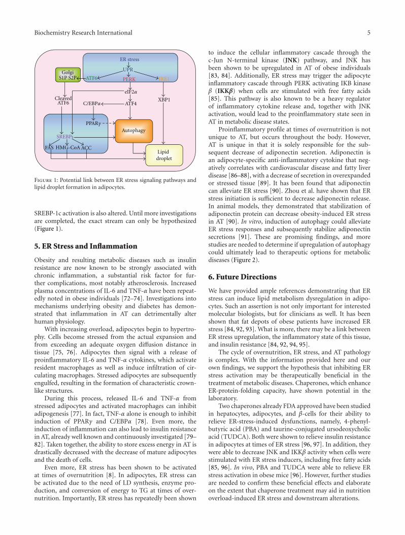
Biochemistry Research International 5
UPR
ATF6 IRE1PERK
ATF4XBP1
GolgiS1P S2P
CleavedATF6
SREBPs
FAS HMG-CoA ACC
Autophagy
eIF2α
C/EBPα
ER stress
Lipiddroplet
PPARγ
Figure 1: Potential link between ER stress signaling pathways andlipid droplet formation in adipocytes.
SREBP-1c activation is also altered. Until more investigationsare completed, the exact stream can only be hypothesized(Figure 1).
5. ER Stress and Inflammation
Obesity and resulting metabolic diseases such as insulinresistance are now known to be strongly associated withchronic inflammation, a substantial risk factor for fur-ther complications, most notably atherosclerosis. Increasedplasma concentrations of IL-6 and TNF-α have been repeat-edly noted in obese individuals [72–74]. Investigations intomechanisms underlying obesity and diabetes has demon-strated that inflammation in AT can detrimentally alterhuman physiology.
With increasing overload, adipocytes begin to hypertro-phy. Cells become stressed from the actual expansion andfrom exceeding an adequate oxygen diffusion distance intissue [75, 76]. Adipocytes then signal with a release ofproinflammatory IL-6 and TNF-α cytokines, which activateresident macrophages as well as induce infiltration of cir-culating macrophages. Stressed adipocytes are subsequentlyengulfed, resulting in the formation of characteristic crown-like structures.
During this process, released IL-6 and TNF-α fromstressed adipocytes and activated macrophages can inhibitadipogenesis [77]. In fact, TNF-α alone is enough to inhibitinduction of PPARγ and C/EBPα [78]. Even more, theinduction of inflammation can also lead to insulin resistancein AT, already well known and continuously investigated [79–82]. Taken together, the ability to store excess energy in AT isdrastically decreased with the decrease of mature adipocytesand the death of cells.
Even more, ER stress has been shown to be activatedat times of overnutrition [8]. In adipocytes, ER stress canbe activated due to the need of LD synthesis, enzyme pro-duction, and conversion of energy to TG at times of over-nutrition. Importantly, ER stress has repeatedly been shown
to induce the cellular inflammatory cascade through thec-Jun N-terminal kinase (JNK) pathway, and JNK hasbeen shown to be upregulated in AT of obese individuals[83, 84]. Additionally, ER stress may trigger the adipocyteinflammatory cascade through PERK activating IKB kinaseβ (IKKβ) when cells are stimulated with free fatty acids[85]. This pathway is also known to be a heavy regulatorof inflammatory cytokine release and, together with JNKactivation, would lead to the proinflammatory state seen inAT in metabolic disease states.
Proinflammatory profile at times of overnutrition is notunique to AT, but occurs throughout the body. However,AT is unique in that it is solely responsible for the sub-sequent decrease of adiponectin secretion. Adiponectin isan adipocyte-specific anti-inflammatory cytokine that neg-atively correlates with cardiovascular disease and fatty liverdisease [86–88], with a decrease of secretion in overexpandedor stressed tissue [89]. It has been found that adiponectincan alleviate ER stress [90]. Zhou et al. have shown that ERstress initiation is sufficient to decrease adiponectin release.In animal models, they demonstrated that stabilization ofadiponectin protein can decrease obesity-induced ER stressin AT [90]. In vitro, induction of autophagy could alleviateER stress responses and subsequently stabilize adiponectinsecretions [91]. These are promising findings, and morestudies are needed to determine if upregulation of autophagycould ultimately lead to therapeutic options for metabolicdiseases (Figure 2).
6. Future Directions
We have provided ample references demonstrating that ERstress can induce lipid metabolism dysregulation in adipo-cytes. Such an assertion is not only important for interestedmolecular biologists, but for clinicians as well. It has beenshown that fat depots of obese patients have increased ERstress [84, 92, 93]. What is more, there may be a link betweenER stress upregulation, the inflammatory state of this tissue,and insulin resistance [84, 92, 94, 95].
The cycle of overnutrition, ER stress, and AT pathologyis complex. With the information provided here and ourown findings, we support the hypothesis that inhibiting ERstress activation may be therapeutically beneficial in thetreatment of metabolic diseases. Chaperones, which enhanceER-protein-folding capacity, have shown potential in thelaboratory.
Two chaperones already FDA approved have been studiedin hepatocytes, adipocytes, and β-cells for their ability torelieve ER-stress-induced dysfunctions, namely, 4-phenyl-butyric acid (PBA) and taurine-conjugated ursodeoxycholicacid (TUDCA). Both were shown to relieve insulin resistancein adipocytes at times of ER stress [96, 97]. In addition, theywere able to decrease JNK and IKKβ activity when cells werestimulated with ER stress inducers, including free fatty acids[85, 96]. In vivo, PBA and TUDCA were able to relieve ERstress activation in obese mice [96]. However, further studiesare needed to confirm these beneficial effects and elaborateon the extent that chaperone treatment may aid in nutritionoverload-induced ER stress and downstream alterations.

6 Biochemistry Research International
IL-6TNF-α
MCP1
AdiponectinER stress
IRE1 PERK
JNK AP-1
Macrophages
Inflammation
Adipocyte
Adiposeinflammation
Fatty liver
Atherosclerosis
IKKβ
Figure 2: ER-stress-induced inflammation in adipocytes and macrophages contributes to atherosclerosis and fatty liver diseases.
Inhibiting ER stress activation may be the key to anapproach for metabolic syndrome therapy. However, morequestions remain in this field. Namely, the role of all partsof the UPR in adipocyte lipid metabolism needs to beuncovered, and the mechanism intertwining ER stress andautophagy needs to be further elucidated. Understandingthese missing components will allow not only further un-derstanding of key lipid pathways in a central metabolic celltype, but also help determine the best approach that can beutilized for clinical metabolic dysfunctions in patients withaltered AT physiology.
Abbreviations
ACC: Acetyl-CoA carboxylaseAdd1: Adipocyte determination and
differentiation 1AT: Adipose tissueATF: Activating transcription factorCHOP: C/EBP homologous proteinCCT: CTP phosphocholine
cytidylyltransferaseeIF2α: Eukaryotic translation initiation
factorER: Endoplasmic reticulumFAS: Fatty acid synthaseFFA: Free fatty acidsInsig: Insulin-induced geneHMG-CoAR: 3-Hydroxy-3-methylgutaryl-CoA
reductaseIKKβ: IKB kinase βIRE1: Inositol requiring enzyme 1IRS1: Insulin response substrateJNK: cJun N-terminal kinaseLC3: Microtubule associated light chain
proteinLD: Lipid dropletPBA: Protein-1 namely 4-phenylbutyric
acid
PERK: PKR-like eukaryotic initiation factor2α kinase
SCAP: SREBP cleavage activating proteinSREBP: Sterol regulatory element binding
proteinTUDCA: Taurine-conjugated ursodeoxycholic
acidTG: TriglycerideUPR: Unfolded protein responseXBP1: X-box binding protein.
References
[1] H. E. Bays, J. M. Gonzalez-Campoy, R. R. Henry et al., “Is adi-posopathy (sick fat) an endocrine disease?” International Jour-nal of Clinical Practice, vol. 62, no. 10, pp. 1474–1483, 2008.
[2] B. K. Surmi and A. H. Hasty, “Macrophage infiltration intoadipose tissue: initiation, propagation and remodeling,” Fu-ture Lipidology, vol. 3, no. 5, pp. 545–556, 2008.
[3] S. Mittra, V. S. Bansal, and P. K. Bhatnagar, “From a glucocen-tric to a lipocentric approach towards metabolic syndrome,”Drug Discovery Today, vol. 13, no. 5-6, pp. 211–218, 2008.
[4] M. Kabir, K. J. Catalano, S. Ananthnarayan et al., “Molecularevidence supporting the portal theory: a causative link be-tween visceral adiposity and hepatic insulin resistance,” Amer-ican Journal of Physiology, vol. 288, no. 2, pp. E454–E461, 2005.
[5] H. Yoshii, T. K. T. Lam, N. Gupta et al., “Effects of portal freefatty acid elevation on insulin clearance and hepatic glucoseflux,” American Journal of Physiology, vol. 290, no. 6, pp.E1089–E1097, 2006.
[6] H. Yoshida, “ER stress and diseases,” FEBS Journal, vol. 274,no. 3, pp. 630–658, 2007.
[7] S. Alhusaini, K. McGee, B. Schisano et al., “Lipopolysaccha-ride, high glucose and saturated fatty acids induce endoplas-mic reticulum stress in cultured primary human adipocytes:salicylate alleviates this stress,” Biochemical and BiophysicalResearch Communications, vol. 397, no. 3, pp. 472–478, 2010.
[8] G. Boden, X. Duan, C. Homko et al., “Increase in endoplasmicreticulum stress-related proteins and genes in adipose tissueof obese, insulin-resistant individuals,” Diabetes, vol. 57, no. 9,pp. 2438–2444, 2008.

Biochemistry Research International 7
[9] M. F. Gregor, L. Yang, E. Fabbrini et al., “Endoplasmic reticu-lum stress is reduced in tissues of obese subjects after weightloss,” Diabetes, vol. 58, no. 3, pp. 693–700, 2009.
[10] N. K. Sharma, S. K. Das, A. K. Mondal et al., “Endoplas-mic reticulum stress markers are associated with obesity innondiabetic subjects,” Journal of Clinical Endocrinology andMetabolism, vol. 93, no. 11, pp. 4532–4541, 2008.
[11] H. Yoshida, T. Matsui, A. Yamamoto, T. Okada, and K. Mori,“XBP1 mRNA is induced by ATF6 and spliced by IRE1 inresponse to ER stress to produce a highly active transcriptionfactor,” Cell, vol. 107, no. 7, pp. 881–891, 2001.
[12] H. Yoshida, T. Matsui, N. Hosokawa, R. J. Kaufman, K. Nagata,and K. Mori, “A time-dependent phase shift in the mammalianunfolded protein response,” Developmental Cell, vol. 4, no. 2,pp. 265–271, 2003.
[13] A. H. Lee, N. N. Iwakoshi, and L. H. Glimcher, “XBP-1regulates a subset of endoplasmic reticulum resident chaper-one genes in the unfolded protein response,” Molecular andCellular Biology, vol. 23, no. 21, pp. 7448–7459, 2003.
[14] R. Sriburi, S. Jackowski, K. Mori, and J. W. Brewer, “XBP1: alink between the unfolded protein response, lipid biosynthesis,and biogenesis of the endoplasmic reticulum,” Journal of CellBiology, vol. 167, no. 1, pp. 35–41, 2004.
[15] A. M. Reimold, N. N. Iwakoshi, J. Manis et al., “Plasma cell dif-ferentiation requires the transcription factor XBP-1,” Nature,vol. 412, no. 6844, pp. 300–307, 2001.
[16] A. H. Lee, G. C. Chu, N. N. Iwakoshi, and L. H. Glimcher,“XBP-1 is required for biogenesis of cellular secretory machin-ery of exocrine glands,” EMBO Journal, vol. 24, no. 24, pp.4368–4380, 2005.
[17] R. Sriburi, H. Bommiasamy, G. L. Buldak et al., “Coordinateregulation of phospholipid biosynthesis and secretory pathwaygene expression in XBP-1(S)-induced endoplasmic reticulumbiogenesis,” Journal of Biological Chemistry, vol. 282, no. 10,pp. 7024–7034, 2007.
[18] R. V. Farese Jr. and T. C. Walther, “Lipid droplets finally get alittle R-E-S-P-E-C-T,” Cell, vol. 139, no. 5, pp. 855–860, 2009.
[19] T. C. Walther and R. V. Farese Jr., “The life of lipid droplets,”Biochimica et Biophysica Acta, vol. 1791, no. 6, pp. 459–466,2009.
[20] H. Robenek, I. Buers, M. J. Robenek et al., “Topography oflipid droplet-associated proteins: insights from freeze-fracturereplica immunogold labeling,” Journal of Lipids, vol. 2011,Article ID 409371, 10 pages, 2011.
[21] Y. Guo, T. C. Walther, M. Rao et al., “Functional genomicscreen reveals genes involved in lipid-droplet formation andutilization,” Nature, vol. 453, no. 7195, pp. 657–661, 2008.
[22] H. Sha, Y. He, H. Chen et al., “The IRE1α-XBP1 pathway ofthe unfolded protein response is required for adipogenesis,”Cell Metabolism, vol. 9, no. 6, pp. 556–564, 2009.
[23] F. Damiano, S. Alemanno, G. V. Gnoni, and L. Siculella,“Translational control of the sterol-regulatory transcriptionfactor SREBP-1 mRNA in response to serum starvation or ERstress is mediated by an internal ribosome entry site,” Bio-chemical Journal, vol. 429, no. 3, pp. 603–612, 2010.
[24] D. J. DeGracia, R. Kumar, C. R. Owen, G. S. Krause, and B.C. White, “Molecular pathways of protein synthesis inhibitionduring brain reperfusion: implications for neuronal survivalor death,” Journal of Cerebral Blood Flow and Metabolism, vol.22, no. 2, pp. 127–141, 2002.
[25] Q. Yang and P. Sarnow, “Location of the internal ribosomeentry site in the 5’ non-coding region of the immunoglobulin
heavy-chain binding protein (BiP) mRNA: evidence for specif-ic RNA-protein interactions,” Nucleic Acids Research, vol. 25,no. 14, pp. 2800–2807, 1997.
[26] K. Ameri and A. L. Harris, “Activating transcription factor 4,”International Journal of Biochemistry and Cell Biology, vol. 40,no. 1, pp. 14–21, 2008.
[27] D. T. Rutkowski and R. J. Kaufman, “All roads lead to ATF4,”Developmental Cell, vol. 4, no. 4, pp. 442–444, 2003.
[28] R. C. Wek, H. Y. Jiang, and T. G. Anthony, “Coping with stress:EIF2 kinases and translational control,” Biochemical SocietyTransactions, vol. 34, no. 1, pp. 7–11, 2006.
[29] N. Batchvarova, X. Z. Wang, and D. Ron, “Inhibition of adi-pogenesis by the stress-induced protein CHOP (Gadd153),”EMBO Journal, vol. 14, no. 19, pp. 4654–4661, 1995.
[30] S. L. Clarke, C. E. Robinson, and J. M. Gimble, “CAAT/Enhancer binding proteins directly modulate transcriptionfrom the peroxisome proliferator-activated receptor γ2 pro-moter,” Biochemical and Biophysical Research Communications,vol. 240, no. 1, pp. 99–103, 1997.
[31] G. Adelmant, J. D. Gilbert, and S. O. Freytag, “Human trans-location liposarcoma-CCAAT/enhancer binding protein (C/EBP) homologous protein (TLS-CHOP) oncoprotein preventsadipocyte differentiation by directly interfering with C/EBPβfunction,” Journal of Biological Chemistry, vol. 273, no. 25, pp.15574–15581, 1998.
[32] C. Wang, Z. Huang, Y. Du, Y. Cheng, S. Chen, and F. Guo,“ATF4 regulates lipid metabolism and thermogenesis,” CellResearch, vol. 20, no. 2, pp. 174–184, 2010.
[33] E. Bobrovnikova-Marjon, G. Hatzivassiliou, C. Grigoriadou etal., “PERK-dependent regulation of lipogenesis during mousemammary gland development and adipocyte differentiation,”Proceedings of the National Academy of Sciences of the UnitedStates of America, vol. 105, no. 42, pp. 16314–16319, 2008.
[34] I. Shimomura, R. E. Hammer, J. A. Richardson et al., “Insulinresistance and diabetes mellitus in transgenic mice expressingnuclear SREBP-1c in adipose tissue: model for congenitalgeneralized lipodystrophy,” Genes and Development, vol. 12,no. 20, pp. 3182–3194, 1998.
[35] S. L. Lay, I. Lefrere, C. Trautwein, I. Dugail, and S. Krief,“Insulin and sterol-regulatory element-binding protein-1c(SREBP-1c) regulation of gene expression in 3T3-L1 adipo-cytes: identification of CCAAT/enhancer-binding protein β asan SREBP-1c target,” Journal of Biological Chemistry, vol. 277,no. 38, pp. 35625–35634, 2002.
[36] D. J. Thuerauf, M. Marcinko, P. J. Belmont, and C. C. Glem-botski, “Effects of the isoform-specific characteristics of AT-F6α and ATF6β on endoplasmic reticulum stress responsegene expression and cell viability,” Journal of Biological Chem-istry, vol. 282, no. 31, pp. 22865–22878, 2007.
[37] K. Haze, H. Yoshida, H. Yanagi, T. Yura, and K. Mori, “Mam-malian transcription factor ATF6 is synthesized as a trans-membrane protein and activated by proteolysis in response toendoplasmic reticulum stress,” Molecular Biology of the Cell,vol. 10, no. 11, pp. 3787–3799, 1999.
[38] E. L. Davenport, G. J. Morgan, and F. E. Davies, “Untanglingthe unfolded protein response,” Cell Cycle, vol. 7, no. 7, pp.865–869, 2008.
[39] Y. Adachi, K. Yamamoto, T. Okada, H. Yoshida, A. Harada,and K. Mori, “ATF6 is a transcription factor specializing inthe regulation of quality control proteins in the endoplasmicreticulum,” Cell Structure and Function, vol. 33, no. 1, pp. 75–89, 2008.
8 Biochemistry Research International
[40] D. T. Rutkowski, J. Wu, S. H. Back et al., “UPR pathwayscombine to prevent hepatic steatosis caused by ER stress-mediated suppression of transcriptional master regulators,”Developmental Cell, vol. 15, no. 6, pp. 829–840, 2008.
[41] K. Yamamoto, K. Takahara, S. Oyadomari et al., “Induction ofliver steatosis and lipid droplet formation in ATF6α-knockoutmice burdened with pharmacological endoplasmic reticulumstress,” Molecular Biology of the Cell, vol. 21, no. 17, pp. 2975–2986, 2010.
[42] M. Schroder and R. J. Kaufman, “ER stress and the unfoldedprotein response,” Mutation Research, vol. 569, no. 1-2, pp. 29–63, 2005.
[43] R. Singh, S. Kaushik, Y. Wang et al., “Autophagy regulates lipidmetabolism,” Nature, vol. 458, no. 7242, pp. 1131–1135, 2009.
[44] M. Martinez-Vicente, Z. Talloczy, E. Wong et al., “Cargorecognition failure is responsible for inefficient autophagy inHuntington’s disease,” Nature Neuroscience, vol. 13, no. 5, pp.567–576, 2010.
[45] R. Singh, “Autophagy and regulation of lipid metabolism,”Results and Problems in Cell Differentiation, vol. 52, pp. 35–46,2010.
[46] M. Shibata, K. Yoshimura, N. Furuya et al., “The MAP1-LC3conjugation system is involved in lipid droplet formation,”Biochemical and Biophysical Research Communications, vol.382, no. 2, pp. 419–423, 2009.
[47] M. Shibata, K. Yoshimura, H. Tamura et al., “LC3, a microtu-bule-associated protein1A/B light chain3, is involved in cyto-plasmic lipid droplet formation,” Biochemical and BiophysicalResearch Communications, vol. 393, no. 2, pp. 274–279, 2010.
[48] H. Nakatogawa, Y. Ichimura, and Y. Ohsumi, “Atg8, a ubiqui-tin-like protein required for autophagosome formation, medi-ates membrane tethering and hemifusion,” Cell, vol. 130, no.1, pp. 165–178, 2007.
[49] J. Kovsan, N. Bashan, A. S. Greenberg, and A. Rudich, “Poten-tial role of autophagy in modulation of lipid metabolism,”American Journal of Physiology, vol. 298, no. 1, pp. E1–E7,2010.
[50] R. Baerga, Y. Zhang, P. H. Chen, S. Goldman, and S. Jin,“Targeted deletion of autophagy-related 5 (atg5) impairs ad-ipogenesis in a cellular model and in mice,” Autophagy, vol. 5,no. 8, pp. 1118–1130, 2009.
[51] Y. Zhang, S. Goldman, R. Baerga, Y. Zhao, M. Komatsu, andS. Jin, “Adipose-specific deletion of autophagy-related gene 7(atg7) in mice reveals a role in adipogenesis,” Proceedings of theNational Academy of Sciences of the United States of America,vol. 106, no. 47, pp. 19860–19865, 2009.
[52] J. Zhou, W. Zhang, B. Liang et al., “PPARγ activation inducesautophagy in breast cancer cells,” International Journal ofBiochemistry and Cell Biology, vol. 41, no. 11, pp. 2334–2342,2009.
[53] J. Yan, H. Yang, G. Wang et al., “Autophagy augmented bytroglitazone is independent of EGFR transactivation and cor-related with AMP-activated protein kinase signaling,” Auto-phagy, vol. 6, no. 1, pp. 67–73, 2010.
[54] T. Kawakami, R. Inagi, H. Takano et al., “Endoplasmic reticu-lum stress induces autophagy in renal proximal tubular cells,”Nephrology Dialysis Transplantation, vol. 24, no. 9, pp. 2665–2672, 2009.
[55] T. Yorimitsu, U. Nair, Z. Yang, and D. J. Klionsky, “Endoplas-mic reticulum stress triggers autophagy,” Journal of BiologicalChemistry, vol. 281, no. 40, pp. 30299–30304, 2006.
[56] W. X. Ding and X. M. Yin, “Sorting, recognition and activationof the misfolded protein degradation pathways through
macroautophagy and the proteasome,” Autophagy, vol. 4, no.2, pp. 141–150, 2008.
[57] W. X. Ding, H. M. Ni, W. Gao et al., “Linking of autophagy toubiquitin-proteasome system is important for the regulationof endoplasmic reticulum stress and cell viability,” AmericanJournal of Pathology, vol. 171, no. 2, pp. 513–524, 2007.
[58] Y. Kouroku, E. Fujita, I. Tanida et al., “ER stress (PERK/eIF2αphosphorylation) mediates the polyglutamine-induced LC3conversion, an essential step for autophagy formation,” CellDeath and Differentiation, vol. 14, no. 2, pp. 230–239, 2007.
[59] J. Price, A. K. Zaidi, J. Bohensky, V. Srinivas, I. M. Shapiro,and H. Ali, “Akt-1 mediates survival of chondrocytes fromendoplasmic reticulum-induced stress,” Journal of CellularPhysiology, vol. 222, no. 3, pp. 502–508, 2010.
[60] L. Qin, Z. Wang, L. Tao, and Y. Wang, “ER stress negativelyregulates AKT/TSC/mTOR pathway to enhance autophagy,”Autophagy, vol. 6, no. 2, pp. 239–247, 2010.
[61] M. Ogata, S. I. Hino, A. Saito et al., “Autophagy is activatedfor cell survival after endoplasmic reticulum stress,” Molecularand Cellular Biology, vol. 26, no. 24, pp. 9220–9231, 2006.
[62] L. Yang, P. Li, S. Fu, E. S. Calay, and G. S. Hotamisligil,“Defective hepatic autophagy in obesity promotes ER stressand causes insulin resistance,” Cell Metabolism, vol. 11, no. 6,pp. 467–478, 2010.
[63] A. K. Gupta, B. Li, G. J. Cerniglia, M. S. Ahmed, S. M.Hahn, and A. Maity, “The HIV protease inhibitor nelfinavirdownregulates Akt phosphorylation by inhibiting proteasomalactivity and inducing the unfolded protein response,” Neopla-sia, vol. 9, no. 4, pp. 271–278, 2007.
[64] S. M. Schleicher, L. Moretti, V. Varki, and B. Lu, “Progress inthe unraveling of the endoplasmic reticulum stress/autophagypathway and cancer: implications for future therapeuticapproaches,” Drug Resistance Updates, vol. 13, no. 3, pp. 79–86, 2010.
[65] W. Jia, R. M. Loria, M. A. Park, A. Yacoub, P. Dent, andM. R. Graf, “The neuro-steroid, 5-androstene 3β,17α diol;induces endoplasmic reticulum stress and autophagy throughPERK/eIF2α signaling in malignant glioma cells and trans-formed fibroblasts,” International Journal of Biochemistry andCell Biology, vol. 42, no. 12, pp. 2019–2029, 2010.
[66] K. W. Kim, L. Moretti, L. R. Mitchell, D. K. Jung, and B. Lu,“Endoplasmic reticulum stress mediates radiation-inducedautophagy by perk-eIF2α in caspase-3/7-deficient cells,” Onco-gene, vol. 29, no. 22, pp. 3241–3251, 2010.
[67] T. Rzymski, M. Milani, L. Pike et al., “Regulation of autophagyby ATF4 in response to severe hypoxia,” Oncogene, vol. 29, no.31, pp. 4424–4435, 2010.
[68] M. Milani, T. Rzymski, H. R. Mellor et al., “The role of ATF4stabilization and autophagy in resistance of breast cancer cellstreated with Bortezomib,” Cancer Research, vol. 69, no. 10, pp.4415–4423, 2009.
[69] S. Nishina, M. Korenaga, I. Hidaka et al., “Hepatitis C virusprotein and iron overload induce hepatic steatosis through theunfolded protein response in mice,” Liver International, vol.30, no. 5, pp. 683–692, 2010.
[70] Y.-K. Seo, T.-I. Jeon, H. K. Chong, J. Biesinger, X. Xie, and T.F. Osborne, “Genome-wide localization of SREBP-2 in hepaticchromatin predicts a role in autophagy,” Cell Metabolism, vol.13, no. 4, pp. 367–375, 2011.
[71] J. Cheng, Y. Ohsaki, K. Tauchi-Sato, A. Fujita, and T. Fujimoto,“Cholesterol depletion induces autophagy,” Biochemical andBiophysical Research Communications, vol. 351, no. 1, pp. 246–252, 2006.

Biochemistry Research International 9
[72] G. P. Van Guilder, G. L. Hoetzer, J. J. Greiner, B. L. Stauffer,and C. A. DeSouza, “Influence of metabolic syndrome on bio-markers of oxidative stress and inflammation in obese adults,”Obesity, vol. 14, no. 12, pp. 2127–2131, 2006.
[73] C. Tsigos, I. Kyrou, E. Chala et al., “Circulating tumor necrosisfactor alpha concentrations are higher in abdominal versusperipheral obesity,” Metabolism, vol. 48, no. 10, pp. 1332–1335, 1999.
[74] P. A. Kern, S. Ranganathan, C. Li, L. Wood, and G. Ranganat-han, “Adipose tissue tumor necrosis factor and interleukin-6expression in human obesity and insulin resistance,” AmericanJournal of Physiology, vol. 280, no. 5, pp. E745–E751, 2001.
[75] R. W. O’Rourke, “Inflammation in obesity-related diseases,”Surgery, vol. 145, no. 3, pp. 255–259, 2009.
[76] G. H. Goossens, “The role of adipose tissue dysfunction in thepathogenesis of obesity-related insulin resistance,” Physiologyand Behavior, vol. 94, no. 2, pp. 206–218, 2008.
[77] B. Gustafson, A. Hammarstedt, C. X. Andersson, and U.Smith, “Inflamed adipose tissue: a culprit underlying the met-abolic syndrome and atherosclerosis,” Arteriosclerosis, Throm-bosis, and Vascular Biology, vol. 27, no. 11, pp. 2276–2283,2007.
[78] L. Meng, J. Zhou, H. Sasano, T. Suzuki, K. M. Zeitoun, and S.E. Bulun, “Tumor necrosis factor α and interleukin 11 secretedby malignant breast epithelial cells inhibit adipocyte differen-tiation by selectively down-regulating CCAAT/enhancer bind-ing protein α and peroxisome proliferator-activated receptorγ: mechanism of desmoplastic reaction,” Cancer Research, vol.61, no. 5, pp. 2250–2255, 2001.
[79] J. P. Bastard, M. Maachi, C. Lagathu et al., “Recent advancesin the relationship between obesity, inflammation, and insulinresistance,” European Cytokine Network, vol. 17, no. 1, pp. 4–12, 2006.
[80] G. S. Hotamisligil, “Inflammation and metabolic disorders,”Nature, vol. 444, no. 7121, pp. 860–867, 2006.
[81] R. Monteiro and I. Azevedo, “Chronic inflammation in obesityand the metabolic syndrome,” Mediators of Inflammation, vol.2010, Article ID 289645, 10 pages, 2010.
[82] H. Xu, G. T. Barnes, Q. Yang et al., “Chronic inflammation infat plays a crucial role in the development of obesity-relatedinsulin resistance,” Journal of Clinical Investigation, vol. 112,no. 12, pp. 1821–1830, 2003.
[83] J. Hirosumi, G. Tuncman, L. Chang et al., “A central, role forJNK in obesity and insulin resistance,” Nature, vol. 420, no.6913, pp. 333–336, 2002.
[84] G. Boden and S. Merali, “Measurement of the increase inendoplasmic reticulum stress-related proteins and genes inadipose tissue of obese, insulin-resistant individuals,” Methodsin Enzymology, vol. 489, pp. 67–82, 2011.
[85] P. Jiao, J. Ma, B. Feng et al., “FFA-induced adipocyte inflam-mation and insulin resistance: involvement of ER stress andIKKβ pathways,” Obesity, vol. 19, no. 3, pp. 483–491, 2011.
[86] S. A. Polyzos, J. Kountouras, C. Zavos, and E. Tsiaousi, “Therole of adiponectin in the pathogenesis and treatment of non-alcoholic fatty liver disease,” Diabetes, Obesity and Metabolism,vol. 12, no. 5, pp. 365–383, 2010.
[87] S. A. Polyzos, J. Kountouras, and C. Zavos, “Nonalcoholic fattyliver disease: the pathogenetic roles of insulin resistance andadipocytokines,” Current Molecular Medicine, vol. 9, no. 3, pp.299–314, 2009.
[88] E. Ros, “Nuts and novel biomarkers of cardiovascular disease,”American Journal of Clinical Nutrition, vol. 89, no. 5, pp.1649S–1656S, 2009.
[89] P. A. Kern, G. B. Di Gregorio, T. Lu, N. Rassouli, and G.Ranganathan, “Adiponectin expression from human adiposetissue: relation to obesity, insulin resistance, and tumornecrosis factor-α expression,” Diabetes, vol. 52, no. 7, pp.1779–1785, 2003.
[90] L. Zhou, M. Liu, J. Zhang, H. Chen, L. Q. Dong, and F.Liu, “DsbA-L alleviates endoplasmic reticulum stress-inducedadiponectin downregulation,” Diabetes, vol. 59, no. 11, pp.2809–2816, 2010.
[91] L. Zhou and F. Liu, “Autophagy: roles in obesity-inducedER stress and adiponectin downregulation in adipocytes,”Autophagy, vol. 6, no. 8, pp. 1196–1197, 2010.
[92] M. Kars, L. Yang, M. F. Gregor et al., “Tauroursodeoxycholicacid may improve liver and muscle but not adipose tissueinsulin sensitivity in obese men and women,” Diabetes, vol. 59,no. 8, pp. 1899–1905, 2010.
[93] M. Miranda, X. Escote, V. Ceperuelo-Mallafre et al., “Relationbetween human LPIN1, hypoxia and endoplasmic reticulumstress genes in subcutaneous and visceral adipose tissue,”International Journal of Obesity, vol. 34, no. 4, pp. 679–686,2010.
[94] N. Marsollier, P. Ferre, and F. Foufelle, “Novel insights in theinterplay between inflammation and metabolic diseases: a rolefor the pathogen sensing kinase PKR,” Journal of Hepatology,vol. 54, no. 6, pp. 1307–1309, 2011.
[95] G. S. Hotamisligil, “Inflammation and endoplasmic reticulumstress in obesity and diabetes,” International Journal of Obesity,vol. 32, no. 7, pp. S52–S54, 2008.
[96] U. Ozcan, E. Yilmaz, L. Ozcan et al., “Chemical chaperonesreduce ER stress and restore glucose homeostasis in a mousemodel of type 2 diabetes,” Science, vol. 313, no. 5790, pp. 1137–1140, 2006.
[97] Y. Nakatani, H. Kaneto, D. Kawamori et al., “Involvement ofendoplasmic reticulum stress in insulin resistance and diabe-tes,” Journal of Biological Chemistry, vol. 280, no. 1, pp. 847–851, 2005.
Hindawi Publishing CorporationBiochemistry Research InternationalVolume 2012, Article ID 841362, 13 pagesdoi:10.1155/2012/841362
Review Article
Endoplasmic Reticulum Stress and Lipid Metabolism:Mechanisms and Therapeutic Potential
Sana Basseri and Richard C. Austin
Division of Nephrology, Department of Medicine, McMaster University and St. Joseph’s Healthcare Hamilton,50 Charlton Avenue East, Hamilton, ON, Canada L8N 4A6
Correspondence should be addressed to Richard C. Austin, [email protected]
Received 10 September 2011; Accepted 18 October 2011
Academic Editor: Kezhong Zhang
Copyright © 2012 S. Basseri and R. C. Austin. This is an open access article distributed under the Creative Commons AttributionLicense, which permits unrestricted use, distribution, and reproduction in any medium, provided the original work is properlycited.
The endoplasmic reticulum (ER) plays a crucial role in protein folding, assembly, and secretion. Disruption of ER homeostasismay lead to accumulation of misfolded or unfolded proteins in the ER lumen, a condition referred to as ER stress. In response toER stress, a signal transduction pathway known as the unfolded protein response (UPR) is activated. UPR activation allows thecell to cope with an increased protein-folding demand on the ER. Recent studies have shown that ER stress/UPR activation plays acritical role in lipid metabolism and homeostasis. ER-stress-dependent dysregulation of lipid metabolism may lead to dyslipidemia,insulin resistance, cardiovascular disease, type 2 diabetes, and obesity. In this paper, we examine recent findings illustrating theimportant role ER stress/UPR signalling pathways play in regulation of lipid metabolism, and how they may lead to dysregulationof lipid homeostasis.
1. Introduction
The liver plays a central role in whole body lipid homeostasis.Metabolic signals such as carbohydrates and dietary fattyacids regulate hepatic gene expression leading to glycolyticand lipogenic signalling pathways. In addition, the pancre-atic hormones, insulin and glucagon, play a pivotal rolein the transcriptional and posttranslational regulation oflipogenesis and lipid oxidation [1]. Lipogenesis, the processof de novo lipid biosynthesis, occurs when an excess ofcarbohydrates is consumed, or when circulating insulinlevels are high. Carbohydrates undergo glycolysis to generateacetyl-CoA molecules which are the building blocks for fattyacid (FA) synthesis. Following esterification, one glycerolmolecule and three FA chains produce triacylglycerol (TG)molecules which are transported in apoB containing verylow-density lipoprotein (VLDL) particles [2] to the adiposetissue for long-term storage. Under fasting conditions wheninsulin levels are low and glucagon levels are high, FAoxidation or lipolysis occurs which allows for mobilizationof FA and uptake by the liver [3]. However, disruption inthese homeostatic mechanisms may lead to the development
of dyslipidemia, insulin resistance, fatty liver, and excessadipose mass, ultimately causing cardiovascular disease anddiabetes.
In recent years, increasing evidence suggests that ERstress and UPR activation can regulate cellular processesbeyond ER protein folding and can play crucial roles inlipid metabolism [4–10]. ER stress, which occurs due todisruption in ER protein-folding capacity, leads to activationof an evolutionarily conserved UPR signalling system inorder to restore ER homeostasis [11]. Accumulating evidencesuggests that activation of the UPR pathways can modulatelipid metabolism by controlling the transcriptional regula-tion of lipogenesis. Excess adipose mass and obesity are adirect consequence of increased de novo lipogenesis and TGstorage in the adipose tissue. The presence of ER stress hasbeen observed in various tissues from obese mice [12, 13]and humans [14–17]. UPR activation has also been linkedto fatty liver disease where lipid droplets accumulate inhepatocytes. The role of ER stress and UPR pathways in thedevelopment of fatty liver disease has been under intenseinvestigation (reviewed in [18]). Here, we aim to examine theevidence regarding the role of UPR pathways in modulating
2 Biochemistry Research International
the transcriptional regulation of lipid metabolism. Further-more, potential therapeutic approaches targeting the ERstress response in obesity and dyslipidemia will be discussed.
2. Transcriptional Regulation ofLipid Metabolism
A number of key transcription factors have been iden-tified which regulate hepatic lipogenesis and fatty acidoxidation. These include sterol-regulatory-element-bindingprotein-1c (SREBP-1c), liver X receptor (LXR), peroxisome-proliferator-activated receptors (PPARs), and carbohydrate-responsive-element-binding protein (ChREBP). Enzymessuch as glucokinase (GK), liver pyruvate kinase (LPK),acetyl CoA carboxylase (ACC), fatty acid synthase (FAS),and stearoyl CoA desaturase-1 (SCD-1) are critical for thebiochemical conversion of glucose into fatty acids and TG[19, 20].
SREBP-1c, a member of the SREBP family of transcrip-tion factors, is thought to be the main driving force forhepatic lipogenesis and development of fatty liver diseaseknown as hepatic steatosis [21, 22]. There are three isoforms,SREBP-1a, -1c and -2. SREBPs are synthesized as inactiveprecursors bound to the ER membrane [23]. While SREBP-1a/-2 upregulate cholesterol synthesis genes, SREBP-1c isresponsible for the regulation of genes involved in FA andTG synthesis pathways [24]. Under sterol-replete conditions,SREBPs are held in the ER through their interaction withSCAP, an anchoring molecule, and Insig, an ER trans-membrane protein. The SREBP-SCAP complex is releasedfrom Insig upon sterol-deplete conditions. SCAP assistsin the transport of SREBP from the ER to the golgi forcleavage by site 1 and 2 proteases [25]. Following proteolyticcleavage, the active mature form of SREBP translocatesinto the nucleus where it induces genes required for lipidbiosynthesis and uptake [26].
SREBP-2 is the main transcription factor responsiblefor regulating the cholesterol biosynthetic pathways [27].Cholesterol is the precursor for steroid biosynthesis and playsan important role in membrane biology. Excess unesterifiedintracellular cholesterol can lead to membrane disruptionsand cellular toxicity and hence must be tightly regulated[28]. Therefore, under sterol-deplete conditions, SREBP-2 iscleaved and translocates to the nucleus allowing for expres-sion of its target genes, including HMG-CoA reductase,the rate-limiting enzyme in cholesterol biosynthesis [29, 30].
SREBP-1c is the predominant isoform and the mainregulator of lipid synthesis in the liver [31]. Overexpressionof the active form of SREBP-1 in the liver leads to hepaticsteatosis due to increased lipid synthesis, uptake, and TGaccumulation [32], while loss of SREBP-1 has been linked tomarked reduction in both lipogenesis and hepatic steatosis[22, 33]. Interestingly, the proteolytic cleavage of SREBP-1c isnot affected by sterol depletion [34]. Proteolytic cleavage andactivation of SREBP-1c is stimulated by insulin [35]. Insulin-mediated SREBP-1c activation occurs through insulin recep-tor substrate-1 (IRS1) and activation of its downstreamtargets protein kinase B (PKB/Akt) and mammalian target of
rapamycin complex 1 (mTORC1) [34]. Although the exactmechanism by which insulin stimulates SREBP-1c cleavageis not entirely understood, it has been shown that insulinleads to phosphorylation of the ER-bound inactive SREBP-1c, increasing its posttranslational processing [36]. Further-more, insulin represses Insig2 mRNA which is thought toenhance SREBP-1c activation [37, 38].
SREBP-1c activity may also be induced through the nu-clear hormone receptor peroxisome-proliferator-activatedreceptor-γ (PPARγ) [39] as well as liver X receptor (LXR)activity [20], both of which play a critical role in lipogen-esis. Ligand-activated nuclear PPARγ heterodimerizes withretinoid X receptors (RXRs) resulting in expression of itstarget genes such as CD36, a fatty acid transport proteininvolved in the transport and metabolism of intracellular FA[40]. Ultimately, PPARγ activity allows for transcription ofgenes involved in promoting lipogenesis [41]. In addition,positive feedback loops have been identified where SREBP-1c activity increases the formation of PPARγ ligands whichlead to its activation [42]. PPARγ also leads to LXRα geneexpression which is a potent activator of SREBP-1c targetgenes [40]. PPARα activity, on the other hand, regulatesperoxisomal, microsomal, and mitochondrial FA oxidationpathways by transcriptionally regulating enzymes involvedin these pathways [40]. Interestingly, LXR competes withPPARα for RXRα heterodimerization, thereby repressingRXRα-PPARα signalling. This in turn suppresses LXR-SREBP-1c activity [40]. This crosstalk would ensure thatlipogenic and lipolytic pathways are not simultaneouslyactivated. Finally, lipogenic and glycolytic gene expressionmay also be regulated by ChREBP, a transcription factorresponsive to high glucose levels and important in regulatingthe expression of LPK, an enzyme required for hepaticglycolysis [43].
In addition to regulation of lipogenic and lipolyticpathways, fatty acid uptake and lipoprotein secretion are alsoimportant for lipid homeostasis. Expression of PPARα, forexample, leads to mobilization and transport of catabolizedfatty acids by inducing expression of enzymes such asfatty-acid-binding protein (FABP) and fatty-acid translocase(FAT) [40]. Fatty acids undergo esterification to form TGwhich can be exported out of the liver as VLDL parti-cles. ApoB is the key component of VLDL particles andmicrosomal triacylglycerol transfer protein (MTP) allows forthe addition of TG to apoB, forming the VLDL particle.However, the overall rate of VLDL assembly depends onthe rate of apoB synthesis in the ER [40].
3. The ER and UPR Activation
The ER is a membranous organelle with several criticalcellular functions. First, it is the site where nascent polypep-tides fold into their proper conformation and any necessaryposttranslational modifications such as glycosylation anddisulphide bond formation take place. This task is accom-plished by ER resident chaperones and foldases and proteindisulphide isomerases (PDI) [11]. Second, phospholipidsynthesis takes place in the ER which allows for expansion of

Biochemistry Research International 3
lipid bilayers in the cell [1]. Third, the ER is a major storagesite for calcium ions which are required for cellular signallingprocesses [44]. Fourth, enzymes such as cytochrome p450 inthe ER allow for efficient metabolism of drugs [45].
A number of physiological, pharmacological, and patho-logical conditions are known to disrupt ER homeostasis andaffect its protein-folding capacity. The inability of the cell toefficiently fold and secrete proteins is defined as ER stress.Cells have evolved mechanisms to adapt to adverse condi-tions in order to maintain homeostasis and survive. One suchcoping mechanism is UPR activation in response to ER stressconditions [46, 47]. Activation of the UPR ultimately resultsin (i) enhancement of ER protein-folding capacity throughexpansion of the ER and increased expression of chaperonesand foldases, (ii) inhibition of protein translation, and (iii)ER-associated protein degradation (ERAD) of misfoldedproteins [48]. If ER stress conditions are not resolved, ER-stress-induced cell death may ensue. Generally, ER-stress-associated cell death occurs through caspase activation [49,50]; however, caspase-independent necrosis and autophagyhave also been observed [51].
The UPR in mammalian cells is composed of threesignalling branches which are initiated by three ER trans-membrane sensors, inositol-requiring protein 1 (IRE1),double-stranded RNA-dependent protein kinase-like ERkinase (PERK), and activating transcription factor 6 (ATF6).Activation of these sensors is dependent on the dissociationof the ER-resident chaperone glucose-regulated protein of78 kDa (GRP78), also known as BiP, from their luminaldomains [52]. This occurs during ER stress conditions whenGRP78 is required for the folding of proteins in the ERand thus is recruited away from IRE1, PERK, and ATF6,thereby activating the UPR. Activation of the UPR pathwaysis often used as an indicator of ER stress due to thetechnical difficulties in directly measuring compromised ERintegrity or protein aggregates in the ER [1]. Figure 1 depictsan overview of mammalian UPR signalling pathways.
Homodimerization and autophosphorylation of PERKfollowing dissociation of GRP78 leads to its kinase activity.PERK phosphorylates the α subunit of eukaryotic initiationfactor 2 (eIF2) resulting in translational attenuation [53].Translation of certain mRNAs with short open readingframes in the 5′-UTR is enhanced by phosphorylation ofeIF2α. ATF4 is an example of such mRNA and its expres-sion results in activation of C/EBP homologous protein(CHOP) which is a proapoptotic transcription factor [54].GADD34 (growth-arrest and DNA-damage-inducible pro-tein 34) is induced by CHOP, which acts to dephosphorylateeIF2α as a negative feedback loop and relieve the cell ofthe translational repression during prolonged ER stress [55].
Similar to PERK, IRE1 is a type 1 transmembrane ser-ine/threonine receptor protein kinase/endonuclease whichupon dissociation of GRP78 homodimerizes leading toautophosphorylation and activation of its kinase and endori-bonuclease functions [48]. Unfolded proteins may alsodirectly bind to IRE1 promoting its homodimerization andautophosphorylation [56–58]. Activation of IRE1 results insplicing of XBP1 mRNA, a process by which a 26-nucleotidesequence of XBP1 mRNA is excised leading to a shift in its
reading frame. Unlike the unspliced XBP1 protein, whichis rapidly degraded, spliced XBP1 (XBP1s) encodes a bZIPtranscription factor with a potent transactivation domain[59]. XBP1s translocates to the nucleus where it leads toexpression of a number of UPR target genes includinggenes involved in protein folding and secretion, proteindegradation and ER translocation [1, 60]. Consistent withits transcriptional target genes, XBP1 is required for thesecretory function of certain highly secretory cell types suchas antibody-producing plasma cells [61].
ATF6, the third arm of the UPR, is comprised oftwo transmembrane bZIP transcription factors, ATF6α andATF6β, which under normal conditions are held in the ERin a complex with GRP78 [62]. ER stress and dissociationof GRP78 from ATF6 leads to its translocation to the Golgiwhere it is cleaved by site 1 and site 2 proteases, a processsimilar to that of the SREBPs. The sequential proteolysis byS1P and S2P leads to the release of the N-terminal cytosolicdomain of ATF6 which then upon entry into the nucleusactivates UPR target genes [63]. Among these target genesare XBP1, CHOP, and ER chaperones such as GRP78 whichallow the ER to cope with the increased protein-foldingdemand [62, 64]. Interestingly, ATF6 and XBP1 possess verysimilar DNA-binding specificity [60] and can heterodimerizesuggesting that they may have common target genes [65].
4. ER Stress and Lipid Metabolism
It is has been known for about a decade that ER stress canlead to altered lipid metabolism and hepatic steatosis. A studyby our group demonstrated that homocysteine-induced ERstress can lead to hepatic steatosis and altered cholesteroland TG biosynthetic pathways, both in cultured cells and inlivers of hyperhomocysteinemic mice [66]. Overexpressionof GRP78, which attenuates ER stress and UPR activation,has been shown to decrease hepatic steatosis by reducingSREBP-1c activity [5]. More recently, specific arms of theUPR and their downstream signalling molecules have beenexamined in cell culture and animal models to deciphertheir function and role in lipid metabolism. It is now wellestablished that various components of the UPR signallingnetwork play a role in the regulation of lipid metabolism [4–10]. Figure 2 summarizes the interactions between variouscomponents of UPR signalling and lipid metabolism.
4.1. PERK Pathway. Activation of PERK is transient and hasoften been difficult to detect [10], but recently the Phos-tag gel approach has proven to be a successful tool fordetection of PERK phosphorylation [67]. Furthermore, thephosphorylation status of eIF2α, a downstream target ofPERK, is often assessed to monitor PERK activity. Changes innutritional status such as fasting and feeding result in alteredphosphorylation status of eIF2α. Fasting followed by 4 hoursof feeding leads to an increase in phospho-eIF2α levels in theliver, which were even greater in high-fat-diet-fed mice [10].To study the effects of compromised PERK-eIF2a-dependentUPR signalling, transgenic mice with enforced expression ofGADD34 were generated [10]. GADD34, by associating with
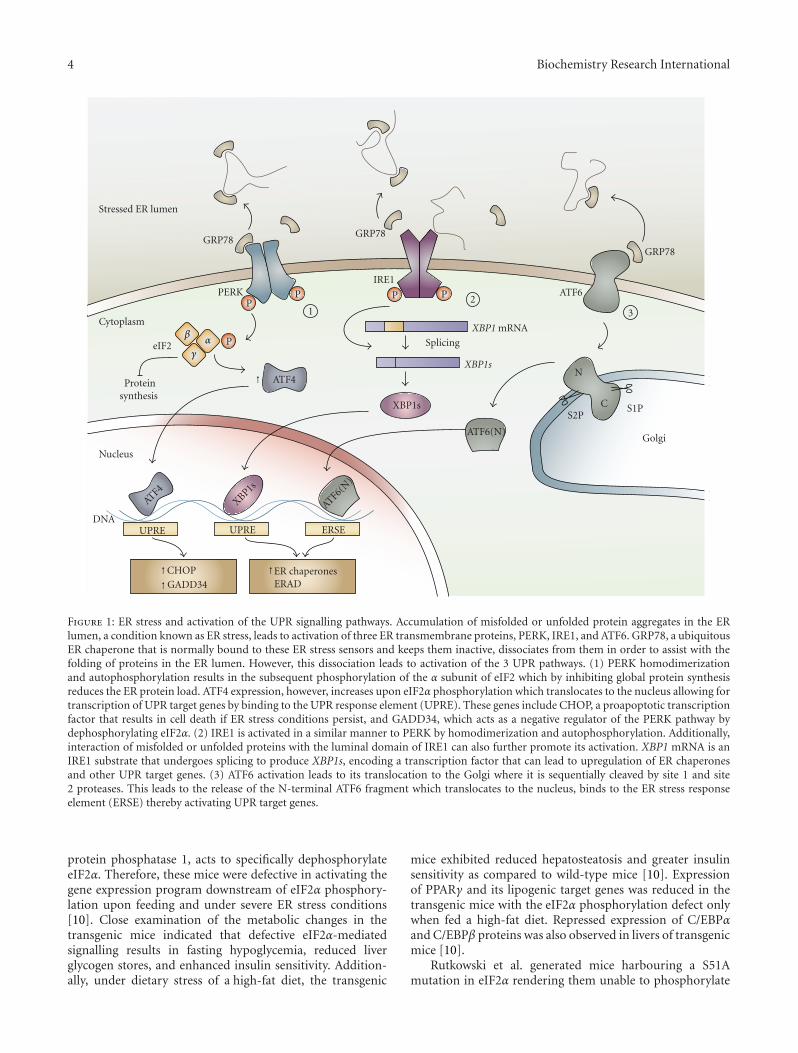
4 Biochemistry Research International
PP P P
P
ATF4
GRP78GRP78
GRP78
PERKIRE1
ATF6
Splicing
XBP1 mRNA
XBP1s
ATF6(N)
N
C S1PS2P
ATF4XBP1s
ATF6(N)
CHOP
GADD34ER chaperonesERAD
12
3
DNAUPREUPRE ERSE
Nucleus
XBP1s
eIF2
Proteinsynthesis
Stressed ER lumen
Cytoplasm
Golgi
↑
↑
↑↑
αβ
γ
Figure 1: ER stress and activation of the UPR signalling pathways. Accumulation of misfolded or unfolded protein aggregates in the ERlumen, a condition known as ER stress, leads to activation of three ER transmembrane proteins, PERK, IRE1, and ATF6. GRP78, a ubiquitousER chaperone that is normally bound to these ER stress sensors and keeps them inactive, dissociates from them in order to assist with thefolding of proteins in the ER lumen. However, this dissociation leads to activation of the 3 UPR pathways. (1) PERK homodimerizationand autophosphorylation results in the subsequent phosphorylation of the α subunit of eIF2 which by inhibiting global protein synthesisreduces the ER protein load. ATF4 expression, however, increases upon eIF2α phosphorylation which translocates to the nucleus allowing fortranscription of UPR target genes by binding to the UPR response element (UPRE). These genes include CHOP, a proapoptotic transcriptionfactor that results in cell death if ER stress conditions persist, and GADD34, which acts as a negative regulator of the PERK pathway bydephosphorylating eIF2α. (2) IRE1 is activated in a similar manner to PERK by homodimerization and autophosphorylation. Additionally,interaction of misfolded or unfolded proteins with the luminal domain of IRE1 can also further promote its activation. XBP1 mRNA is anIRE1 substrate that undergoes splicing to produce XBP1s, encoding a transcription factor that can lead to upregulation of ER chaperonesand other UPR target genes. (3) ATF6 activation leads to its translocation to the Golgi where it is sequentially cleaved by site 1 and site2 proteases. This leads to the release of the N-terminal ATF6 fragment which translocates to the nucleus, binds to the ER stress responseelement (ERSE) thereby activating UPR target genes.
protein phosphatase 1, acts to specifically dephosphorylateeIF2α. Therefore, these mice were defective in activating thegene expression program downstream of eIF2α phosphory-lation upon feeding and under severe ER stress conditions[10]. Close examination of the metabolic changes in thetransgenic mice indicated that defective eIF2α-mediatedsignalling results in fasting hypoglycemia, reduced liverglycogen stores, and enhanced insulin sensitivity. Addition-ally, under dietary stress of a high-fat diet, the transgenic
mice exhibited reduced hepatosteatosis and greater insulinsensitivity as compared to wild-type mice [10]. Expressionof PPARγ and its lipogenic target genes was reduced in thetransgenic mice with the eIF2α phosphorylation defect onlywhen fed a high-fat diet. Repressed expression of C/EBPαand C/EBPβ proteins was also observed in livers of transgenicmice [10].
Rutkowski et al. generated mice harbouring a S51Amutation in eIF2α rendering them unable to phosphorylate

Biochemistry Research International 5
↓
Stressed ER lumen
PP P P
ATF4
GRP78 GRP78
GRP78
PERK
IRE1
ATF6
XBP1sATF6(N)
HepaticapoB
secretion
Fatty acidoxidation
Lipogenicgene
expression
Lipogenicgene
expression
SREBP
Hepatic lipid accumulation
P
CHOP
Insig1
Under severe ER stress
GADD34
PGC1αPPARα
C/EBPα
PPARγ
C/EBPαC/EBPβ
eIF2αeIF2α
Figure 2: Crosstalk between UPR signalling pathways and lipogenesis. Phosphorylation of eIF2α and activation of the PERK pathway underhigh-fat diet-induced ER stress conditions allow for enhanced lipogenesis by inducing C/EBPα and decreasing Insig1 protein translationwhich increases activation of SREBP. However, under severe or prolonged ER stress conditions, CHOP expression may lead to dysregulationof the C/EBPs. Similarly, high-carbohydrate-diet-induced ER stress conditions depend on XBP1 for expression of lipogenic genes andincrease of C/EBPα activity, both of which promote lipogenesis. However, severe ER stress conditions, imposed by tunicamycin, lead toXBP1-mediated inhibition of lipogenic gene expression. Furthermore, both XBP1 and ATF6 are important for apolipoprotein B secretionfrom hepatocytes and activation of fatty acid oxidation pathways (PPARα, PGC1α) under such conditions. These pathways culminate inattenuation of lipogenesis and prevention of fatty liver disease under severe ER stress.
eIF2α and therefore allowing constitutive expression of theunphosphorylated form of eIF2α [7]. This transgenic mousemodel was also utilized to examine the PERK/eIF2α arm ofthe UPR and its role in ER-stress-mediated hepatic lipoge-nesis [7]. Similar to the findings by Oyadomari et al., micewith constitutive eIF2α expression also exhibited suppressedhepatic C/EBPα protein expression. In contrast, however,after a tunicamycin challenge, these mice developed fattyliver [7]. These differences point to the source and severityof ER stress (chronic and adaptive dietary stress versusdirect and acute ER stress challenge) as important factors inthe regulation of lipid metabolism.
Another recent study examined the role of PERK inthe regulation of lipogenesis in adipocytes and mammary
epithelial cells [4]. Absence of PERK in mouse embryonicfibroblasts differentiating into adipocytes and in mammaryepithelium attenuated lipogenesis and expression of genessuch as SREBP1, SCD1, FAS, and ACL [4]. As a result, themammary glands from PERK-deficient mothers had lowerTG and FA content which lead to growth retardation in thepups. This study also demonstrated that SREBP1 activation isdependent on decreased Insig1 translation which occurs dueto PERK and eIF2α-dependent translational attenuation [4].
Due to its upstream open reading frames, the ATF4mRNA is among the transcripts that escape the globaltranslational attenuation that occurs upon phosphorylationof eIF2α. ATF4-knockout mice exhibit smaller white adiposetissues relative to total body weight [68], which prompted
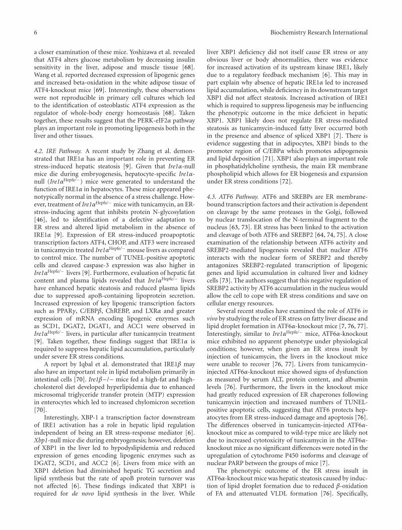
6 Biochemistry Research International
a closer examination of these mice. Yoshizawa et al. revealedthat ATF4 alters glucose metabolism by decreasing insulinsensitivity in the liver, adipose and muscle tissue [68].Wang et al. reported decreased expression of lipogenic genesand increased beta-oxidation in the white adipose tissue ofATF4-knockout mice [69]. Interestingly, these observationswere not reproducible in primary cell cultures which ledto the identification of osteoblastic ATF4 expression as theregulator of whole-body energy homeostasis [68]. Takentogether, these results suggest that the PERK-eIF2α pathwayplays an important role in promoting lipogenesis both in theliver and other tissues.
4.2. IRE Pathway. A recent study by Zhang et al. demon-strated that IRE1α has an important role in preventing ERstress-induced hepatic steatosis [9]. Given that Ire1α-nullmice die during embryogenesis, hepatocyte-specific Ire1α-null (Ire1αHepfe/−) mice were generated to understand thefunction of IRE1α in hepatocytes. These mice appeared phe-notypically normal in the absence of a stress challenge. How-ever, treatment of Ire1αHepfe/− mice with tunicamycin, an ER-stress-inducing agent that inhibits protein N-glycosylation[46], led to identification of a defective adaptation toER stress and altered lipid metabolism in the absence ofIRE1α [9]. Expression of ER stress-induced proapoptotictranscription factors ATF4, CHOP, and ATF3 were increasedin tunicamycin treated Ire1αHepfe/− mouse livers as comparedto control mice. The number of TUNEL-positive apoptoticcells and cleaved caspase-3 expression was also higher inIre1αHepfe/− livers [9]. Furthermore, evaluation of hepatic fatcontent and plasma lipids revealed that Ire1αHepfe/− livershave enhanced hepatic steatosis and reduced plasma lipidsdue to suppressed apoB-containing lipoprotein secretion.Increased expression of key lipogenic transcription factorssuch as PPARγ, C/EBPβ, ChREBP, and LXRα and greaterexpression of mRNA encoding lipogenic enzymes suchas SCD1, DGAT2, DGAT1, and ACC1 were observed inIre1αHepfe/− livers, in particular after tunicamycin treatment[9]. Taken together, these findings suggest that IRE1α isrequired to suppress hepatic lipid accumulation, particularlyunder severe ER stress conditions.
A report by Iqbal et al. demonstrated that IRE1β mayalso have an important role in lipid metabolism primarily inintestinal cells [70]. Ire1β−/− mice fed a high-fat and high-cholesterol diet developed hyperlipidemia due to enhancedmicrosomal triglyceride transfer protein (MTP) expressionin enterocytes which led to increased chylomicron secretion[70].
Interestingly, XBP-1 a transcription factor downstreamof IRE1 activation has a role in hepatic lipid regulationindependent of being an ER stress-response mediator [6].Xbp1-null mice die during embryogenesis; however, deletionof XBP1 in the liver led to hypodyslipidemia and reducedexpression of genes encoding lipogenic enzymes such asDGAT2, SCD1, and ACC2 [6]. Livers from mice with anXBP1 deletion had diminished hepatic TG secretion andlipid synthesis but the rate of apoB protein turnover wasnot affected [6]. These findings indicated that XBP1 isrequired for de novo lipid synthesis in the liver. While
liver XBP1 deficiency did not itself cause ER stress or anyobvious liver or body abnormalities, there was evidencefor increased activation of its upstream kinase IRE1, likelydue to a regulatory feedback mechanism [6]. This may inpart explain why absence of hepatic IRE1α led to increasedlipid accumulation, while deficiency in its downstream targetXBP1 did not affect steatosis. Increased activation of IRE1which is required to suppress lipogenesis may be influencingthe phenotypic outcome in the mice deficient in hepaticXBP1. XBP1 likely does not regulate ER stress-mediatedsteatosis as tunicamycin-induced fatty liver occurred bothin the presence and absence of spliced XBP1 [7]. There isevidence suggesting that in adipocytes, XBP1 binds to thepromoter region of C/EBPα which promotes adipogenesisand lipid deposition [71]. XBP1 also plays an important rolein phosphatidylcholine synthesis, the main ER membranephospholipid which allows for ER biogenesis and expansionunder ER stress conditions [72].
4.3. ATF6 Pathway. ATF6 and SREBPs are ER membrane-bound transcription factors and their activation is dependenton cleavage by the same proteases in the Golgi, followedby nuclear translocation of the N-terminal fragment to thenucleus [63, 73]. ER stress has been linked to the activationand cleavage of both ATF6 and SREBP2 [64, 74, 75]. A closeexamination of the relationship between ATF6 activity andSREBP2-mediated lipogenesis revealed that nuclear ATF6interacts with the nuclear form of SREBP2 and therebyantagonizes SREBP2-regulated transcription of lipogenicgenes and lipid accumulation in cultured liver and kidneycells [73]. The authors suggest that this negative regulation ofSREBP2 activity by ATF6 accumulation in the nucleus wouldallow the cell to cope with ER stress conditions and save oncellular energy resources.
Several recent studies have examined the role of ATF6 invivo by studying the role of ER stress on fatty liver disease andlipid droplet formation in ATF6α-knockout mice [7, 76, 77].Interestingly, similar to Ire1αHepfe/− mice, ATF6α-knockoutmice exhibited no apparent phenotype under physiologicalconditions; however, when given an ER stress insult byinjection of tunicamycin, the livers in the knockout micewere unable to recover [76, 77]. Livers from tunicamycin-injected ATF6α-knockout mice showed signs of dysfunctionas measured by serum ALT, protein content, and albuminlevels [76]. Furthermore, the livers in the knockout micehad greatly reduced expression of ER chaperones followingtunicamycin injection and increased numbers of TUNEL-positive apoptotic cells, suggesting that ATF6 protects hep-atocytes from ER stress-induced damage and apoptosis [76].The differences observed in tunicamycin-injected ATF6α-knockout mice as compared to wild-type mice are likely notdue to increased cytotoxicity of tunicamycin in the ATF6α-knockout mice as no significant differences were noted in theupregulation of cytochrome P450 isoforms and cleavage ofnuclear PARP between the groups of mice [7].
The phenotypic outcome of the ER stress insult inATF6α-knockout mice was hepatic steatosis caused by induc-tion of lipid droplet formation due to reduced β-oxidationof FA and attenuated VLDL formation [76]. Specifically,
Biochemistry Research International 7
there was sustained expression of CHOP in the livers ofATF6α-knockout mice compared to wild-type mice as wellas a decrease in PPARα expression and apoB-100 proteinlevels, favouring the accumulation of lipids in the liver [7,76]. De novo lipogenesis was ruled out as a mechanism forthe increased lipid droplet accumulation in livers from thesemice as expression of lipogenic genes (SCD1, FASN, andDGAT2) was suppressed in tunicamycin-injected ATF6α-knockout mouse livers [7]. In addition, while steatosis wasthe most evident phenotype, upon closer examination itwas discovered that after 48 hours of tunicamycin treat-ment, ATF6α-knockout mice became profoundly resistantto exogenous insulin [7]. This finding is intuitive given thatER stress can lead to insulin resistance [12]. Taken together,the findings from these studies suggest that loss of ATF6predisposes the liver to stress-induced insulin resistance andlipid accumulation.
The studies to date suggest that lipogenic genes and lipidmetabolism are differentially regulated under physiologicalconditions such as high-carbohydrate or high-fat diet feedingin comparison to acute or unresolved ER stress conditionsthat arise when mice are injected with tunicamycin. Forexample, while XBP1 increases hepatic de novo lipogenesis,its upstream kinase IRE1α, or ATF6 which shares DNA-binding sites with XBP1, was protective against hepatic lipidaccumulation. Indeed, a recent study by Rutkowski et al.demonstrated that it is the acute or unresolved form ofER stress that leads to hepatic steatosis [7]. Injection oftunicamycin in mice deficient in one of the UPR sig-nalling components led to chronic upregulation of CHOP,defective eIF2α phosphorylation, and decreased C/EBPαgene expression [7]. CHOP was reported to be at leastpartially responsible for the suppression of gene expressionseen in tunicamycin-injected mice with compromised UPRsignalling [7]. While wild-type mice exhibited rapid buttransient CHOP induction, ATF6-knockout and Ire1αHepfe/−
mice presented with persistent upregulation of CHOP andnuclear localization [7]. CHOP can heterodimerize withthe C/EBP family of transcription factors in the nucleusrepressing their target gene expression [78]. Negative regu-lation of C/EBPα by prolonged nuclear CHOP expressiondue to unresolved ER stress appears to play a key rolein the profound metabolic disruption under severe ERstress conditions which results in fatty liver disease. Indeed,the promoter region of both Srebp1 and Pparα possessespotential binding sites for C/EBPα [7]. The differential effectsof acute/unresolved ER stress conditions in comparison todiet-induced adaptive ER stress conditions also explain whyphosphorylation of eIF2α can lead to hepatic steatosis inone study model while defective eIF2α phosphorylation canaccelerate lipid accumulation and steatosis in another study.
5. The Impact of Lipids on ER Stress
The relationship between ER stress and lipid metabolismis bidirectional. While activation of ER stress pathways canresult in lipogenesis and altered lipid homeostasis, lipidsand aberrant lipid metabolism can also cause ER stress [79–82]. Saturated fatty acids such as palmitate and stearate
are known inducers of ER stress in various cell types andcan modulate survival and apoptotic signals in the cell[81, 82]. A recent study carried out comparative proteomicand lipidomic analysis of fractionated ER from lean andobese liver tissues [79]. The results suggested enrichmentof metabolic enzymes involved in lipid metabolism anda downregulation of ER-associated protein synthesis genes inthe obese ER proteome. These findings implied that the ERin obese liver cells shifts from being the major site of proteinsynthesis to carrying out lipid synthesis and lipid metabolismfunctions [79]. Furthermore, the analysis revealed that thereis a greater proportion of de novo synthesized saturatedfatty acids incorporated into hepatic ER lipids than dietarypolyunsaturated fatty acids. Another interesting finding wasthe increased proportion of phosphatidylcholine (PC) incomparison to phosphatidylethanolamine (PE), both abun-dant ER membrane phospholipids, in the liver ER from obesemice [79]. The increased PC/PE ratio led to perturbation inthe calcium transport activity of the SERCA pump resultingin impaired ER calcium retention. Since ER calcium isimportant for ER homeostasis and chaperone function, suchchanges in calcium concentrations would lead to proteinmisfolding and ER stress. This appears to be a plausiblemechanism for hepatic ER stress in obesity [79]. Hepatic ERstress can promote de novo lipogenesis and insulin resistanceas described above which then in turn may lead to furtherexacerbation of the ER stress situation, creating a viciouscycle.
6. Therapeutic Potential Targeting ER Stressin Dyslipidemia and Obesity
ER stress and UPR activation have been implicated inthe pathogenesis of a number of diseases such as diabetes,obesity, cancer, renal, cardiovascular, and neurodegenerativediseases as well as fatty liver disease [48, 83–86]. As such,potential ways of attenuating ER stress and UPR activationwould provide opportunities in pharmacological interven-tion in a wide array of diseases. A recent study revealed forthe first time in humans that obese insulin-resistant subjectsexpress markers of ER stress in their white adipose tissue[17]. Similarly, an association between ER stress and obesitywas also found in obese nondiabetic subjects [15]. Gastricbypass surgery-mediated weight loss in obese patients waseffective at reducing ER stress in adipose and liver tissuesand improved insulin sensitivity [16]. Furthermore, whenER stress was reduced by hepatic overexpression of GRP78 inob/ob mice, hepatic TG and cholesterol content was reducedand insulin sensitivity improved [5]. These findings togetherwith data in rodents indicating the presence of ER stress intissues of obese animals [12, 13] suggest a strong associationbetween ER stress and obesity. Therefore, the ER serves asan important new treatment target against obesity and itsmetabolic complications.
The use of small molecules called chemical chaper-ones has been examined in a number of disease modelsas potential tools for lowering ER stress and preventingthe activation of UPR pathways. These chaperones similar
8 Biochemistry Research International
to molecular chaperones nonselectively stabilize mutantproteins and assist in their folding and translocation acrossmembranes [87]. Most chemical chaperones are osmolytesand equilibrate cellular osmotic pressure. These can becategorized into 3 classes: carbohydrates (such as glyceroland sorbitol), amino acids (such as glycine and taurine), andmethylamines (such as betaine) [87, 88]. The drawback tothe use of most chemical chaperones is their nonspecificityand high-dose requirement for effective protein foldingproperties. However, two such chemical chaperones, 4-phenylbutyric acid (4-PBA) and tauroursodeoxycholic acid(TUDCA) have been approved by the US Food and DrugAdministration (FDA) and are used in humans. Currently, 4-PBA is approved for use in children with urea-cycle disordersas an ammonia scavenger, while TUDCA is being tested forits liver-protecting properties in cholestatic liver disease inhumans [87].
The low-molecular-weight fatty acid 4-PBA has beentested in a number of disease models for its ability to facilitateprotein folding and trafficking, ultimately relieving ER stress[13, 89–98]. The chaperoning property of 4-PBA was firstidentified when investigating its effect on the translocationand trafficking of a mutant cystic fibrosis transmembraneconductance regulator protein (CFTR). Addition of 4-PBA tothe cells allowed for stabilization of the mutant CFTR proteinand facilitated their translocation to the cell membrane[99]. In addition to its chaperone properties, 4-PBA alsopossesses HDAC inhibitor activity and is under investigationas an anticancer drug [100–102].
Another effective reagent that has been shown to havechaperone properties is TUDCA, which can be classifiedas a hydrophilic endogenous bile acid [87]. TUDCA hasantiapoptotic properties by reducing calcium efflux andblocking ER-stress-mediated caspase-12 activation [103].Furthermore, TUDCA also activates cell survival pathwayssuch as PI3K signalling, thereby inhibiting cell death [104].Apart from these signalling properties, TUDCA can inter-act with the mineralocorticoid receptor and promotes itsdissociation from cytosolic chaperones thereby preventingits translocation to the nucleus for transcriptional activity.In the case of primary neurons, addition of TUDCAwas effective at preventing amyloid beta-peptide-inducedapoptosis through its chaperoning properties [105].
In recent years, several studies have identified beneficialeffects of 4-PBA and TUDCA supplementation on insulinresistance, obesity, and diabetes. Oral administration of4-PBA and TUDCA to obese and insulin-resistant ob/obmice normalized hyperglycemia, restored insulin sensitivityin the liver, muscle, and white adipose, and diminishedfatty liver disease [92]. Our group examined the effect of4-PBA supplementation on diet-induced obesity. For thispurpose, C57BL/6 mice were placed on a high-fat diet withor without 4-PBA supplementation in the drinking water.Mice treated with 4-PBA gained significantly less weight,exhibited lower plasma glucose, TG, and leptin levels, andhad smaller adipocytes as compared to mice on a high-fatdiet alone [89]. Chemical chaperones also have chaperoneactivity within the central nervous system [13, 106]. Leptin,an adipocyte-derived hormone which acts on hypothalamic
neurons to suppress appetite, is important in regulatingenergy expenditure and body weight [107]. ER stress maybe one of the factors resulting in leptin resistance in thebrain, as injection of tunicamycin, an ER-stressor-inducedhypothalamic ER stress, increased food consumption andweight gain despite elevated blood leptin concentrations[13]. Both 4-PBA and TUDCA were shown to be effec-tive at lowering hypothalamic ER stress and increasingthe sensitivity of neurons to leptin, thereby reducing bodyweight in genetic and diet-induced obesity models [107].In the context of atherosclerosis, 4-PBA was effective atprotecting macrophages against palmitate-induced ER stressand apoptosis in culture [108]. A reduction in ER stressand apoptosis was also observed in the macrophages withinthe atherosclerotic lesions of mice treated with 4-PBA, whichwere smaller in size [108]. These findings indicate that 4-PBA treatment can protect cells from the deleterious effectsof lipid accumulation on disease progression.
ER stress has been linked to fatty liver disease and liverinjury [109, 110]. Lipid-induced ER stress inhibits apoB100secretion in liver cells promoting the development of steato-sis [111]. Treatment of hepatoma cells with 4-PBA leadsto the inhibition of lipid-induced ER stress and enhancedapoB100 secretion [111]. Consistent with the studies onmacrophages and progression of atherosclerosis, alleviatinglipid-induced ER stress in hepatocytes also protects the cellsfrom ER-associated apoptosis [112]. Since hepatocellularinjury and damage can lead to progression of fatty liverdisease into steatohepatitis [113, 114], blocking ER stressserves as an important treatment strategy [95]. A recentstudy examined the effects of oral administration of TUDCAon hepatic steatosis and hepatic gene expression in ob/obmice [115]. Yang et al. found a significant decrease in liverfat content and reduced expression of genes involved in denovo lipogenesis with TUDCA treatment [115]. However,they did not find any differences in body weight or insulinsensitivity over the three-week duration of the study. Exam-ination of the effects of orally administered TUDCA oninsulin sensitivity in obese human subjects revealed a 30%improvement in insulin sensitivity in muscle and liver tissuesbut no alterations in hepatic TG content were observed[116]. The differences in the mechanism of action betweenoral treatment and intraperitoneally injected TUDCA mayexplain some of these contrasting outcomes [115].
The effectiveness of chemical chaperones such as 4-PBAand TUDCA as a treatment strategy for dyslipidemia, cardio-vascular disease, diabetes, and obesity require further studyin human subjects. Both 4-PBA and TUDCA have additionalfunctions which may be directly or indirectly alleviating ERstress conditions. Investigation into the discovery of newchemical and biological approaches to enhance ER functionand facilitate the trafficking of proteins would be usefulfor treating ER-stress-related diseases. Furthermore, waysof targeting specific UPR pathways would allow for betterspecificity in targeting ER stress in various disease states[87]. Currently, small molecules that can target IRE1α andalter its endonuclease activity offer hope for further study.These kinase-inhibiting RNase attenuators can also selec-tively enhance XBP1 mRNA splicing and lead to prevention

Biochemistry Research International 9
of apoptotic cell death, while attenuating IRE1α-mediateddecay of mRNA such as those encoding ER chaperones [117].The recent finding that unfolded peptides can directly bindto IRE1 and promote its oligomerization and activationsuggests that compounds that can target its peptide-bindinggroove and oligomerization interface may be effective atregulating IRE1 activity [58]. Finally, given the challengeswith directly measuring ER stress, assay systems which canassess actual cellular ER stress will prove to be useful [118].
7. Conclusions
A growing body of evidence links ER stress and UPRactivation to diseases associated with lipid metabolism.The UPR signalling pathways and activation of transcrip-tion factors such as XBP1 and ATF6 have novel roles incontrolling the transcriptional regulation of lipogenesis.While IRE1α itself is protective against ER-stress-inducedlipogenesis and hepatic steatosis, its downstream mediatorXBP1 promotes transcription of genes involved in fattyacid and cholesterol biosynthesis. Phosphorylation of eIF2αdownstream of PERK affects the transcriptional activity ofC/EBPs, PPARγ, and SREBP-1c thereby leading to lipidaccumulation and hepatic steatosis under high-fat-diet con-ditions. Similar to IRE1α, ATF6α also protects against ERstress-induced steatosis and lipid droplet formation in mice.Furthermore, nuclear ATF6 attenuates SREBP2-mediatedlipogenesis. The exact mechanisms by which ER stress sig-nalling pathways affect lipid homeostasis are incompletelyunderstood. Given the temporal differences in the activationof the three arms of the UPR, a closer examination of eachbranch of the UPR will allow for a better understanding ofhow various components of this signalling network impacton lipogenesis and disease progression. Such studies willfurther enhance our understanding of biological and phar-macological tools needed to effectively treat ER-associateddiseases.
Acknowledgments
The authors would like to thank Hamed Basseri (Facultyof Medicine, University of Ottawa) for his assistance indesigning and preparing the figures. R. C. Austin is sup-ported by grants from the Heart and Stroke Foundationof Ontario and the Canadian Institutes of Health Research.Financial support from St. Joseph’s Healthcare Hamilton isalso acknowledged. R. C. Austin is a Career Investigatorof the Heart and Stroke Foundation of Ontario and holdsthe Amgen Canada Research Chair in nephrology. S. Basseriis supported by a doctoral award from the Heart and StrokeFoundation of Canada.
References
[1] A. H. Lee and L. H. Glimcher, “Intersection of the unfoldedprotein response and hepatic lipid metabolism,” Cellular andMolecular Life Sciences, vol. 66, no. 17, pp. 2835–2850, 2009.
[2] N. O. Davidson and G. S. Shelness, “Apolipoprotein B:mRNA editing, lipoprotein assembly, and presecretory
degradation,” Annual Review of Nutrition, vol. 20, pp. 169–193, 2000.
[3] A. Canbay, L. Bechmann, and G. Gerken, “Lipid metabolismin the liver,” Zeitschrift fur Gastroenterologie, vol. 45, no. 1,pp. 35–41, 2007.
[4] E. Bobrovnikova-Marjon, G. Hatzivassiliou, C. Grigoriadouet al., “PERK-dependent regulation of lipogenesis duringmouse mammary gland development and adipocyte differ-entiation,” Proceedings of the National Academy of Sciencesof the United States of America, vol. 105, no. 42, pp. 16314–16319, 2008.
[5] H. L. Kammoun, H. Chabanon, I. Hainault et al., “GRP78expression inhibits insulin and ER stress-induced SREBP-1cactivation and reduces hepatic steatosis in mice,” The Journalof Clinical Investigation, vol. 119, no. 5, pp. 1201–1215, 2009.
[6] A. H. Lee, E. F. Scapa, D. E. Cohen, and L. H. Glimcher,“Regulation of hepatic lipogenesis by the transcription factorXBP1,” Science, vol. 320, no. 5882, pp. 1492–1496, 2008.
[7] D. T. Rutkowski, J. Wu, S. H. Back et al., “UPR pathwayscombine to prevent hepatic steatosis caused by ER stress-mediated suppression of transcriptional master regulators,”Developmental Cell, vol. 15, no. 6, pp. 829–840, 2008.
[8] R. Sriburi, S. Jackowski, K. Mori, and J. W. Brewer, “XBP1: alink between the unfolded protein response, lipid biosynthe-sis, and biogenesis of the endoplasmic reticulum,” Journal ofCell Biology, vol. 167, no. 1, pp. 35–41, 2004.
[9] K. Zhang, S. Wang, J. Malhotra et al., “The unfolded proteinresponse transducer IRE1α prevents ER stress-induced hep-atic steatosis,” The EMBO Journal, vol. 30, no. 7, pp. 1357–1375, 2011.
[10] S. Oyadomari, H. P. Harding, Y. Zhang, M. Oyadomari, andD. Ron, “Dephosphorylation of translation initiation factor2α enhances glucose tolerance and attenuates hepatosteatosisin mice,” Cell Metabolism, vol. 7, no. 6, pp. 520–532, 2008.
[11] X. Shen, K. Zhang, and R. J. Kaufman, “The unfolded proteinresponse—a stress signaling pathway of the endoplasmicreticulum,” Journal of Chemical Neuroanatomy, vol. 28, no.1-2, pp. 79–92, 2004.
[12] U. Ozcan, Q. Cao, E. Yilmaz et al., “Endoplasmic reticulumstress links obesity, insulin action, and type 2 diabetes,”Science, vol. 306, no. 5695, pp. 457–461, 2004.
[13] L. Ozcan, A. S. Ergin, A. Lu et al., “Endoplasmic reticulumstress plays a central role in development of leptin resistance,”Cell Metabolism, vol. 9, no. 1, pp. 35–51, 2009.
[14] A. T. Sage, S. Holtby-Ottenhof, Y. Shi, S. Damjanovic, A.M. Sharma, and G. H. Werstuck, “Metabolic syndromeand acute hyperglycemia are associated with endoplasmicreticulum stress in human mononuclear cells,” Obesity. Inpress.
[15] N. K. Sharma, S. K. Das, A. K. Mondal et al., “Endoplasmicreticulum stress markers are associated with obesity innondiabetic subjects,” Journal of Clinical Endocrinology andMetabolism, vol. 93, no. 11, pp. 4532–4541, 2008.
[16] M. F. Gregor, L. Yang, E. Fabbrini et al., “Endoplasmic retic-ulum stress is reduced in tissues of obese subjects after weightloss,” Diabetes, vol. 58, no. 3, pp. 693–700, 2009.
[17] G. Boden, X. Duan, C. Homko et al., “Increase in endoplas-mic reticulum stress-related proteins and genes in adiposetissue of obese, insulin-resistant individuals,” Diabetes, vol.57, no. 9, pp. 2438–2444, 2008.
[18] C. L. Gentile, M. Frye, and M. J. Pagliassotti, “Endoplasmicreticulum stress and the unfolded protein response innonalcoholic fatty liver disease,” Antioxidants and RedoxSignaling, vol. 15, no. 2, pp. 505–521, 2011.
10 Biochemistry Research International
[19] K. H. Kim, M. J. Song, E. J. Yoo, S. S. Choe, S. D. Park,and J. B. Kim, “Regulatory role of glycogen synthase kinase3 for transcriptional activity of ADD1/SREBP1c,” Journal ofBiological Chemistry, vol. 279, no. 50, pp. 51999–52006, 2004.
[20] J. B. Seo, H. M. Moon, W. S. Kim et al., “Activated liver Xreceptors stimulate adipocyte differentiation through induc-tion of peroxisome proliferator-activated receptor gammaexpression receptor γ expression,” Molecular and CellularBiology, vol. 24, no. 8, pp. 3430–3444, 2004.
[21] H. Guillou, P. G. Martin, and T. Pineau, “Transcriptionalregulation of hepatic fatty acid metabolism,” Sub-CellularBiochemistry, vol. 49, pp. 3–47, 2008.
[22] N. Yahagi, H. Shimano, A. H. Hasty et al., “Absenceof sterol regulatory element-binding protein-1 (SREBP-1)ameliorates fatty livers but not obesity or insulin resistance inLepob/Lepob mice,” Journal of Biological Chemistry, vol. 277,no. 22, pp. 19353–19357, 2002.
[23] M. S. Brown and J. L. Goldstein, “A proteolytic pathway thatcontrols the cholesterol content of membranes, cells, andblood,” Proceedings of the National Academy of Sciences of theUnited States of America, vol. 96, no. 20, pp. 11041–11048,1999.
[24] J. D. Horton, “Sterol regulatory element-binding proteins:transcriptional activators of lipid synthesis,” BiochemicalSociety Transactions, vol. 30, no. 6, pp. 1091–1095, 2002.
[25] A. Nohturfft, R. A. Debose-Boyd, S. Scheek, J. L. Goldstein,and M. S. Brown, “Sterols regulate cycling of SREBP cleavage-activating protein (SCAP) between endoplasmic reticulumand Golgi,” Proceedings of the National Academy of Sciences ofthe United States of America, vol. 96, no. 20, pp. 11235–11240,1999.
[26] D. Eberle, B. Hegarty, P. Bossard, P. Ferre, and F. Foufelle,“SREBP transcription factors: master regulators of lipidhomeostasis,” Biochimie, vol. 86, no. 11, pp. 839–848, 2004.
[27] R. Sato, “Sterol metabolism and SREBP activation,” Archivesof Biochemistry and Biophysics, vol. 501, no. 2, pp. 177–181,2010.
[28] F. R. Maxfield and I. Tabas, “Role of cholesterol and lipidorganization in disease,” Nature, vol. 438, no. 7068, pp. 612–621, 2005.
[29] J. D. Norton, I. Shimomura, M. S. Brown, R. E. Hammer,J. L. Goldstein, and H. Shimano, “Activation of cholesterolsynthesis in preference to fatty acid synthesis in liver andadipose tissue of transgenic mice overproducing sterol reg-ulatory element-binding protein-2,” The Journal of ClinicalInvestigation, vol. 101, no. 11, pp. 2331–2339, 1998.
[30] Y. Sakakura, H. Shimano, H. Sone et al., “Sterol regula-tory element-binding proteins induce an entire pathway ofcholesterol synthesis,” Biochemical and Biophysical ResearchCommunications, vol. 286, no. 1, pp. 176–183, 2001.
[31] N. Higuchi, M. Kato, Y. Shundo et al., “Liver X receptorin cooperation with SREBP-1c is a major lipid synthesisregulator in nonalcoholic fatty liver disease,” HepatologyResearch, vol. 38, no. 11, pp. 1122–1129, 2008.
[32] H. Shimano, J. D. Horton, R. E. Hammer, I. Shimomura, M.S. Brown, and J. L. Goldstein, “Overproduction of cholesteroland fatty acids causes massive liver enlargement in transgenicmice expressing truncated SREBP-1a,” The Journal of ClinicalInvestigation, vol. 98, no. 7, pp. 1575–1584, 1996.
[33] H. Shimano, N. Yahagi, M. Amemiya-Kudo et al., “Sterolregulatory element-binding protein-1 as a key transcriptionfactor for nutritional induction of lipogenic enzyme genes,”Journal of Biological Chemistry, vol. 274, no. 50, pp. 35832–35839, 1999.
[34] P. Ferre and F. Foufelle, “Hepatic steatosis: a role for de novolipogenesis and the transcription factor SREBP-1c,” Diabetes,Obesity and Metabolism, vol. 12, supplement 2, pp. 83–92,2010.
[35] M. Kohjima, N. Higuchi, M. Kato et al., “SREBP-1c, regulatedby the insulin and AMPK signaling pathways, plays a rolein nonalcoholic fatty liver disease,” International Journal ofMolecular Medicine, vol. 21, no. 4, pp. 507–511, 2008.
[36] C. R. Yellaturu, X. Deng, L. M. Cagen et al., “Insulin enhan-ces post-translational processing of nascent SREBP-1c bypromoting its phosphorylation and association with COPIIvesicles,” Journal of Biological Chemistry, vol. 284, no. 12, pp.7518–7532, 2009.
[37] D. Yabe, R. Komuro, G. Liang, J. L. Goldstein, and M. S.Brown, “Liver-specific mRNA for insig-2 down-regulated byinsulin: implications for fatty acid synthesis,” Proceedingsof the National Academy of Sciences of the United States ofAmerica, vol. 100, no. 6, pp. 3155–3160, 2003.
[38] C. R. Yellaturu, X. Deng, E. A. Park, R. Raghow,and M. B. Elam, “Insulin enhances the biogenesis ofnuclear sterol regulatory element-binding protein (SREBP)-1c by posttranscriptionaldown-regulation of insig-2A andits dissociation from SREBP cleavage-activating protein(SCAP)·SREBP-1c complex,” Journal of Biological Chemistry,vol. 284, no. 46, pp. 31726–31734, 2009.
[39] S. E. Schadinger, N. L.R. Bucher, B. M. Schreiber, andS. R. Farmer, “PPARγ2 regulates lipogenesis and lipidaccumulation in steatotic hepatocytes,” American Journal ofPhysiology, vol. 288, no. 6, pp. E1195–E1205, 2005.
[40] P. Nguyen, V. Leray, M. Diez et al., “Liver lipid metabolism,”Journal of Animal Physiology and Animal Nutrition, vol. 92,no. 3, pp. 272–283, 2008.
[41] Y. L. Zhang, A. Hernandez-Ono, P. Siri et al., “Aberranthepatic expression of PPARγ2 stimulates hepatic lipogenesisin a mouse model of obesity, insulin resistance, dyslipidemia,and hepatic steatosis,” Journal of Biological Chemistry, vol.281, no. 49, pp. 37603–37615, 2006.
[42] B. M. Spiegelman, E. Hu, J. B. Kim, and R. Brun, “PPARγ andthe control of adipogenesis,” Biochimie, vol. 79, no. 2-3, pp.111–112, 1997.
[43] M. Malaguarnera, M. Di Rosa, F. Nicoletti, and L. Malaguar-nera, “Molecular mechanisms involved in NAFLD progres-sion,” Journal of Molecular Medicine, vol. 87, no. 7, pp. 679–695, 2009.
[44] B. Targos, J. Baranska, and P. Pomorski, “Store-operated cal-cium entry in physiology and pathology of mammalian cells,”Acta Biochimica Polonica, vol. 52, no. 2, pp. 397–409, 2005.
[45] E. P. A. Neve and M. Ingelman-Sundberg, “Cytochrome P450proteins: retention and distribution from the endoplasmicreticulum,” Current Opinion in Drug Discovery and Develop-ment, vol. 13, no. 1, pp. 78–85, 2010.
[46] R. J. Kaufman, “Stress signaling from the lumen of the endo-plasmic reticulum: coordination of gene transcriptional andtranslational controls,” Genes and Development, vol. 13, no.10, pp. 1211–1233, 1999.
[47] A. A. Welihinda, W. Tirasophon, and R. J. Kaufman, “Thecellular response to protein misfolding in the endoplasmicreticulum,” Gene Expression, vol. 7, no. 4-6, pp. 293–300,1999.
[48] T. Hosoi and K. Ozawa, “Endoplasmic reticulum stress indisease: mechanisms and therapeutic opportunities,” ClinicalScience, vol. 118, no. 1, pp. 19–29, 2010.
Biochemistry Research International 11
[49] J. Hitomi, T. Katayama, M. Taniguchi, A. Honda, K.Imaizumi, and M. Tohyama, “Apoptosis induced by endo-plasmic reticulum stress depends on activation of caspase-3via caspase-12,” Neuroscience Letters, vol. 357, no. 2, pp. 127–130, 2004.
[50] T. Nakagawa, H. Zhu, N. Morishima et al., “Caspase-12 mediates endoplasmic-reticulum-specific apoptosis andcytotoxicity by amyloid-β,” Nature, vol. 403, no. 6765, pp.98–103, 2000.
[51] E. Ullman, Y. Fan, M. Stawowczyk, H. M. Chen, Z. Yue,and W. X. Zong, “Autophagy promotes necrosis in apoptosis-deficient cells in response to ER stress,” Cell Death andDifferentiation, vol. 15, no. 2, pp. 422–425, 2008.
[52] L. M. Hendershot, “The ER chaperone BiP is a masterregulator of ER function,” Mount Sinai Journal of Medicine,vol. 71, no. 5, pp. 289–297, 2004.
[53] H. P. Harding, Y. Zhang, A. Bertolotti, H. Zeng, and D. Ron,“Perk is essential for translational regulation and cell survivalduring the unfolded protein response,” Molecular Cell, vol. 5,no. 5, pp. 897–904, 2000.
[54] H. P. Harding, I. Novoa, Y. Zhang et al., “Regulatedtranslation initiation controls stress-induced gene expressionin mammalian cells,” Molecular Cell, vol. 6, no. 5, pp. 1099–1108, 2000.
[55] S. J. Marciniak, C. Y. Yun, S. Oyadomari et al., “CHOPinduces death by promoting protein synthesis and oxidationin the stressed endoplasmic reticulum,” Genes and Develop-ment, vol. 18, no. 24, pp. 3066–3077, 2004.
[56] J. J. Credle, J. S. Finer-Moore, F. R. Papa, R. M. Stroud, andP. Walter, “On the mechanism of sensing unfolded proteinin the endoplasmic reticulum,” Proceedings of the NationalAcademy of Sciences of the United States of America, vol. 102,no. 52, pp. 18773–18784, 2005.
[57] Y. Kimata, Y. Ishiwata-Kimata, T. Ito et al., “Two regulatorysteps of ER-stress sensor Ire1 involving its cluster formationand interaction with unfolded proteins,” Journal of CellBiology, vol. 179, no. 1, pp. 75–86, 2007.
[58] B. M. Gardner and P. Walter, “Unfolded proteins are Ire1-activating ligands that directly induce the unfolded proteinresponse,” Science, vol. 333, no. 6051, pp. 1891–1894, 2011.
[59] D. J. Todd, A. H. Lee, and L. H. Glimcher, “The endoplasmicreticulum stress response in immunity and autoimmunity,”Nature Reviews Immunology, vol. 8, no. 9, pp. 663–674, 2008.
[60] A. H. Lee, N. N. Iwakoshi, and L. H. Glimcher, “XBP-1 regulates a subset of endoplasmic reticulum residentchaperone genes in the unfolded protein response,” Molecularand Cellular Biology, vol. 23, no. 21, pp. 7448–7459, 2003.
[61] A. M. Reimold, N. N. Iwakoshi, J. Manis et al., “Plasmacell differentiation requires the transcription factor XBP-1,”Nature, vol. 412, no. 6844, pp. 300–307, 2001.
[62] H. Yoshida, T. Okada, K. Haze et al., “ATF6 activated by prot-eolysis binds in the presence of NF-Y (CBF) directly to thecis-acting element responsible for the mammalian unfoldedprotein response,” Molecular and Cellular Biology, vol. 20, no.18, pp. 6755–6767, 2000.
[63] J. Ye, R. B. Rawson, R. Komuro et al., “ER stress induces cleav-age of membrane-bound ATF6 by the same proteases thatprocess SREBPs,” Molecular Cell, vol. 6, no. 6, pp. 1355–1364,2000.
[64] K. Haze, H. Yoshida, H. Yanagi, T. Yura, and K. Mori,“Mammalian transcription factor ATF6 is synthesized asa transmembrane protein and activated by proteolysis inresponse to endoplasmic reticulum stress,” Molecular Biologyof the Cell, vol. 10, no. 11, pp. 3787–3799, 1999.
[65] K. Yamamoto, T. Sato, T. Matsui et al., “Transcriptionalinduction of mammalian ER quality control proteins ismediated by single or combined action of ATF6α and XBP1,”Developmental Cell, vol. 13, no. 3, pp. 365–376, 2007.
[66] G. H. Werstuck, S. R. Lentz, S. Dayal et al., “Homocysteine-induced endoplasmic reticulum stress causes dysregulationof the cholesterol and triglyceride biosynthetic pathways,”The Journal of Clinical Investigation, vol. 107, no. 10, pp.1263–1273, 2001.
[67] L. Yang, Z. Xue, Y. He, S. Sun, H. Chen, and L. Qi, “Aphos-tag-based approach reveals the extent of physiologicalendoplasmic reticulum stress,” PLoS One, vol. 5, no. 7, ArticleID e11621, 2010.
[68] T. Yoshizawa, E. Hinoi, Y. J. Dae et al., “The transcriptionfactor ATF4 regulates glucose metabolism in mice throughits expression in osteoblasts,” The Journal of Clinical Investi-gation, vol. 119, no. 9, pp. 2807–2817, 2009.
[69] C. Wang, Z. Huang, Y. Du, Y. Cheng, S. Chen, and F. Guo,“ATF4 regulates lipid metabolism and thermogenesis,” CellResearch, vol. 20, no. 2, pp. 174–184, 2010.
[70] J. Iqbal, K. Dai, T. Seimon et al., “IRE1β inhibits chylomicronproduction by selectively degrading MTP mRNA,” CellMetabolism, vol. 7, no. 5, pp. 445–455, 2008.
[71] H. Sha, Y. He, H. Chen et al., “The IRE1α-XBP1 pathway ofthe unfolded protein response is required for adipogenesis,”Cell Metabolism, vol. 9, no. 6, pp. 556–564, 2009.
[72] R. Sriburi, H. Bommiasamy, G. L. Buldak et al., “Coordi-nate regulation of phospholipid biosynthesis and secretorypathway gene expression in XBP-1(S)-induced endoplasmicreticulum biogenesis,” Journal of Biological Chemistry, vol.282, no. 10, pp. 7024–7034, 2007.
[73] L. Zeng, M. Lu, K. Mori et al., “ATF6 modulates SREBP2-mediated lipogenesis,” The EMBO Journal, vol. 23, no. 4, pp.950–958, 2004.
[74] S. M. Colgan, D. Tang, G. H. Werstuck, and R. C. Austin,“Endoplasmic reticulum stress causes the activation of sterolregulatory element binding protein-2,” International Journalof Biochemistry and Cell Biology, vol. 39, no. 10, pp. 1843–1851, 2007.
[75] J. N. Lee and J. Ye, “Proteolytic activation of sterol regulatoryelement-binding protein induced by cellular stress throughdepletion of Insig-1,” Journal of Biological Chemistry, vol. 279,no. 43, pp. 45257–45265, 2004.
[76] K. Yamamoto, K. Takahara, S. Oyadomari et al., “Inductionof liver steatosis and lipid droplet formation in ATF6α-knockout mice burdened with pharmacological endoplasmicreticulum stress,” Molecular Biology of the Cell, vol. 21, no. 17,pp. 2975–2986, 2010.
[77] J. Wu, D. T. Rutkowski, M. Dubois et al., “ATF6α optimizeslong-term endoplasmic reticulum function to protect cellsfrom chronic stress,” Developmental Cell, vol. 13, no. 3, pp.351–364, 2007.
[78] N. Batchvarova, X. Z. Wang, and D. Ron, “Inhibition of adi-pogenesis by the stress-induced protein CHOP (Gadd153),”The EMBO Journal, vol. 14, no. 19, pp. 4654–4661, 1995.
[79] S. Fu, L. Yang, P. Li et al., “Aberrant lipid metabolism disruptscalcium homeostasis causing liver endoplasmic reticulumstress in obesity,” Nature, vol. 473, no. 7348, pp. 528–531,2011.
[80] C. L. Gentile, M. A. Frye, and M. J. Pagliassotti, “Fatty acidsand the endoplasmic reticulum in nonalcoholic fatty liverdisease,” BioFactors, vol. 37, no. 1, pp. 8–16, 2011.
[81] W. Guo, S. Wong, W. Xie, T. Lei, and Z. Luo, “Palmitate mod-ulates intracellular signaling, induces endoplasmic reticulum

12 Biochemistry Research International
stress, and causes apoptosis in mouse 3T3-L1 and rat primarypreadipocytes,” American Journal of Physiology, vol. 293, no.2, pp. E576–E586, 2007.
[82] Y. Wei, D. Wang, F. Topczewski, and M. J. Pagliassotti, “Sat-urated fatty acids induce endoplasmic reticulum stress andapoptosis independently of ceramide in liver cells,” AmericanJournal of Physiology, vol. 291, no. 2, pp. E275–E281, 2006.
[83] J. G. Dickhout and J. C. Krepinsky, “Endoplasmic reticulumstress and renal disease,” Antioxidants and Redox Signaling,vol. 11, no. 9, pp. 2341–2352, 2009.
[84] N. Kaplowitz, A. T. Tin, M. Shinohara, and C. Ji, “Endo-plasmic reticulum stress and liver injury,” Seminars in LiverDisease, vol. 27, no. 4, pp. 367–377, 2007.
[85] T. Minamino and M. Kitakaze, “ER stress in cardiovasculardisease,” Journal of Molecular and Cellular Cardiology, vol. 48,no. 6, pp. 1105–1110, 2010.
[86] R. C. Austin, “The unfolded protein response in health anddisease,” Antioxidants and Redox Signaling, vol. 11, no. 9, pp.2279–2287, 2009.
[87] F. Engin and G. S. Hotamisligil, “Restoring endoplasmicreticulum function by chemical chaperones: an emergingtherapeutic approach for metabolic diseases,” Diabetes, Obe-sity and Metabolism, vol. 12, no. 2, pp. 108–115, 2010.
[88] W. J. Welch and C. R. Brown, “Influence of molecular andchemical chaperones on protein folding,” Cell Stress andChaperones, vol. 1, no. 2, pp. 109–115, 1996.
[89] S. Basseri, S. Lhotak, A. M. Sharma, and R. C. Austin, “Thechemical chaperon 4-phenylbutyrate inhibits adipogenesisby modulating the unfolded protein response,” Journal ofLipid Research, vol. 50, no. 12, pp. 2486–2501, 2009.
[90] X. Kuang, W. Hu, M. Yan, and P. K. Y. Wong, “Phenylbutyricacid suppresses protein accumulation-mediated ER stress inretrovirus-infected astrocytes and delays onset of paralysis ininfected mice,” Neurochemistry International, vol. 57, no. 7,pp. 738–748, 2010.
[91] T. Mizukami, K. Orihashi, B. Herlambang et al., “Sodium 4-phenylbutyrate protects against spinal cord ischemia by inhi-bition of endoplasmic reticulum stress,” Journal of VascularSurgery, vol. 52, no. 6, pp. 1580–1586, 2010.
[92] U. Ozcan, E. Yilmaz, L. Ozcan et al., “Chemical chaperonesreduce ER stress and restore glucose homeostasis in a mousemodel of type 2 diabetes,” Science, vol. 313, no. 5790, pp.1137–1140, 2006.
[93] X. Qi, T. Hosoi, Y. Okuma, M. Kaneko, and Y. Nomura,“Sodium 4-phenylbutyrate protects against cerebral ischemicinjury,” Molecular Pharmacology, vol. 66, no. 4, pp. 899–908,2004.
[94] S. M. Rahman, I. Qadri, R. C. Janssen, and J. E. Friedman,“Fenofibrate and PBA prevent fatty acid-induced loss ofadiponectin receptor and pAMPK in human hepatoma cellsand in hepatitis C virus-induced steatosis,” Journal of LipidResearch, vol. 50, no. 11, pp. 2193–2202, 2009.
[95] M. Vilatoba, C. Eckstein, G. Bilbao et al., “Sodium 4-phenylbutyrate protects against liver ischemia reperfusioninjury by inhibition of endoplasmic reticulum-stress medi-ated apoptosis,” Surgery, vol. 138, no. 2, pp. 342–351, 2005.
[96] J. C. Wiley, C. Pettan-Brewer, and W. C. Ladiges, “Phenyl-butyric acid reduces amyloid plaques and rescues cognitivebehavior in AD transgenic mice,” Aging Cell, vol. 10, no. 3,pp. 418–428, 2011.
[97] C. Xiao, A. Giacca, and G. F. Lewis, “Sodium phenylbutyrate,a drug with known capacity to reduce endoplasmic reticulumstress, partially alleviates lipid-induced insulin resistance and
β-cell dysfunction in humans,” Diabetes, vol. 60, no. 3, pp.918–924, 2011.
[98] G. S. Zode, M. H. Kuehn, D. Y. Nishimura et al., “Reductionof ER stress via a chemical chaperone prevents diseasephenotypes in a mouse model of primary open angleglaucoma,” The Journal of Clinical Investigation, vol. 121, no.9, pp. 3542–3553, 2011.
[99] M. Lim, K. McKenzie, A. D. Floyd, E. Kwon, and P. L.Zeitlin, “Modulation of ΔF508 cystic fibrosis transmembraneregulator trafficking and function with 4-phenylbutyrateand flavonoids,” American Journal of Respiratory Cell andMolecular Biology, vol. 31, no. 3, pp. 351–357, 2004.
[100] A. Ricobaraza, M. Cuadrado-Tejedor, S. Marco, I. Perez-Otano, and A. Garcıa-Osta, “Phenylbutyrate rescues den-dritic spine loss associated with memory deficits in a mousemodel of Alzheimer disease,” Hippocampus. In press.
[101] I. B. Appelskog, O. Ammerpohl, I. G. Svechnikova, W. O.Lui, P. M. Almqvist, and T. J. Ekstrom, “Histone deacety-lase inhibitor 4-phenylbutyrate suppresses GAPDH mRNAexpression in glioma cells,” International journal of oncology,vol. 24, no. 6, pp. 1419–1425, 2004.
[102] M. Jung, “Inhibitors of histone deacetylase as new anticanceragents,” Current Medicinal Chemistry, vol. 8, no. 12, pp.1505–1511, 2001.
[103] S. Sola, X. Ma, R. E. Castro, B. T. Kren, C. J. Steer, and C.M. Rodrigues, “Ursodeoxycholic acid modulates E2F-1 andp53 expression through a caspase-independent mechanismin transforming growth factor beta1-induced apoptosis of rathepatocytes,” The Journal of Biological Chemistry, vol. 278,no. 49, pp. 48831–48838, 2003.
[104] M. H. Schoemaker, L. Conde De La Rosa, M. Buist-Homanet al., “Tauroursodeoxycholic acid protects rat hepatocytesfrom bile acid-induced apoptosis via activation of survivalpathways,” Hepatology, vol. 39, no. 6, pp. 1563–1573, 2004.
[105] S. Sola, J. D. Amaral, P. M. Borralho et al., “Functionalmodulation of nuclear steroid receptors by tauroursodeoxy-cholic acid reduces amyloid β-peptide-induced apoptosis,”Molecular Endocrinology, vol. 20, no. 10, pp. 2292–2303,2006.
[106] M. Inden, Y. Kitamura, H. Takeuchi et al., “Neurodegener-ation of mouse nigrostriatal dopaminergic system inducedby repeated oral administration of rotenone is preventedby 4-phenylbutyrate, a chemical chaperone,” Journal ofNeurochemistry, vol. 101, no. 6, pp. 1491–1504, 2007.
[107] J. M. Friedman, “Leptin, leptin receptors, and the control ofbody weight,” Nutrition Reviews, vol. 56, no. 2, pp. S38–S46,1998.
[108] E. Erbay, V. R. Babaev, J. R. Mayers et al., “Reducingendoplasmic reticulum stress through a macrophage lipidchaperone alleviates atherosclerosis,” Nature Medicine, vol.15, no. 12, pp. 1383–1391, 2009.
[109] C. D. Anderson, G. Upadhya, K. D. Conzen et al., “Endoplas-mic reticulum stress is a mediator of posttransplant injury inseverely steatotic liver allografts,” Liver Transplantation, vol.17, no. 2, pp. 189–200, 2011.
[110] D. Wang, Y. Wei, and M. J. Pagliassotti, “Saturated fatty acidspromote endoplasmic reticulum stress and liver injury in ratswith hepatic steatosis,” Endocrinology, vol. 147, no. 2, pp.943–951, 2006.
[111] T. Ota, C. Gayet, and H. N. Ginsberg, “Inhibition ofapolipoprotein B100 secretion by lipid-induced hepaticendoplasmic reticulum stress in rodents,” The Journal ofClinical Investigation, vol. 118, no. 1, pp. 316–332, 2008.

Biochemistry Research International 13
[112] D.-S. Kim, S.-K. Jeong, H.-R. Kim, D.-S. Kim, S.-W. Chae,and H.-J. Chae, “Metformin regulates palmitate-inducedapoptosis and ER stress response in HepG2 liver cells,”Immunopharmacology and Immunotoxicology, vol. 32, no. 2,pp. 251–257, 2010.
[113] A. D. Tevar, C. N. Clarke, R. Schuster, J. Wang, M. J. Edwards,and A. B. Lentsch, “The effect of hepatic ischemia reperfusioninjury in a murine model of nonalcoholic steatohepatitis,”Journal of Surgical Research, vol. 169, no. 1, pp. e7–e14, 2011.
[114] J. Jou, S. S. Choi, and A. M. Diehl, “Mechanisms of diseaseprogression in nonalcoholic fatty liver disease,” Seminars inLiver Disease, vol. 28, no. 4, pp. 370–379, 2008.
[115] J. S. Yang, J. T. Kim, J. Jeon et al., “Changes in hepatic geneexpression upon oral administration of taurine-conjugatedursodeoxycholic acid in ob/ob mice,” PLoS One, vol. 5, no.11, Article ID e13858, 2010.
[116] M. Kars, L. Yang, M. F. Gregor et al., “Tauroursodeoxycholicacid may improve liver and muscle but not adipose tissueinsulin sensitivity in obese men and women,” Diabetes, vol.59, no. 8, pp. 1899–1905, 2010.
[117] D. Han, A. G. Lerner, L. Vande Walle et al., “IRE1α kinaseactivation modes control alternate endoribonuclease outputsto determine divergent cell fates,” Cell, vol. 138, no. 3, pp.562–575, 2009.
[118] P. I. Merksamer, A. Trusina, and F. R. Papa, “Real-time redoxmeasurements during endoplasmic reticulum stress revealinterlinked protein folding functions,” Cell, vol. 135, no. 5,pp. 933–947, 2008.
Hindawi Publishing CorporationBiochemistry Research InternationalVolume 2012, Article ID 738471, 7 pagesdoi:10.1155/2012/738471
Review Article
UPR-Mediated Membrane Biogenesis in B Cells
Joseph W. Brewer1 and Suzanne Jackowski2
1 Department of Microbiology and Immunology, College of Medicine, University of South Alabama, Mobile, AL 36688, USA2 Department of Infectious Diseases, St. Jude Children’s Research Hospital, Memphis, TN 38105, USA
Correspondence should be addressed to Joseph W. Brewer, [email protected] Suzanne Jackowski, [email protected]
Received 5 August 2011; Accepted 25 August 2011
Academic Editor: Kezhong Zhang
Copyright © 2012 J. W. Brewer and S. Jackowski. This is an open access article distributed under the Creative CommonsAttribution License, which permits unrestricted use, distribution, and reproduction in any medium, provided the original work isproperly cited.
The unfolded protein response (UPR) can coordinate the regulation of gene transcription and protein translation to balancethe load of client proteins with the protein folding and degradative capacities of the ER. Increasing evidence also implicates theUPR in the regulation of lipid synthesis and membrane biogenesis. The differentiation of B lymphocytes into antibody-secretingcells is marked by significant expansion of the ER, the site for antibody synthesis and assembly. In activated B cells, the demand formembrane protein and lipid components leads to activation of the UPR transcriptional activator XBP1(S) which, in turn, initiates acascade of biochemical events that enhance supplies of phospholipid precursors and build machinery for the synthesis, maturation,and transport of secretory proteins. The alterations in lipid metabolism that occur during this developmental transition and theimpact of membrane phospholipid restriction on B cell secretory characteristics are discussed in this paper.
1. Introduction
Activated B lymphocytes proliferate and proceed alongdistinct developmental pathways that determine their func-tion and fate. Specifically, responding B cells can rapidlydifferentiate in extrafollicular sites into short-lived antibody-secreting cells that predominantly secrete IgM antibodies [1].Alternatively, responding B cells can enter germinal centers,undergo somatic hypermutation and isotype switching,and then become memory B cells or long-lived antibody-secreting cells [2]. Extrinsic factors, including the nature ofthe antigen and T cell help in the form of membrane-boundmolecules and soluble cytokines, play key roles in regulatingB cell responses. However, intrinsic signals are also pivotalin directing the fate of responding B cells as evidenced bythe critical role of the unfolded protein response (UPR)transcription factor XBP1(S) in driving the differentiationof antibody-secreting cells [3, 4], the effectors of humoralimmunity. Here, we discuss the current understanding ofthe relationship between the UPR, lipid biosynthesis andorganelle biogenesis in activated B cells.
2. Lipid Supply and Demand
B lymphocytes proliferate and differentiate into antibody-secreting cells upon interaction with specific antigen orcertain Toll-like receptor (TLR) ligands. When B cells arestimulated to enter the cell cycle and proliferate, the mech-anisms that control the membrane phospholipid supply inrapidly dividing cells are engaged. The division of one cellinto two daughter cells requires a doubling of membranecontent during cell cycle progression [5]. Phosphatidyl-choline (PtdCho) is the major membrane phospholipid inmammalian cells and is a precursor to the two other mostabundant membrane phospholipids, sphingomyelin (SM)[6] and phosphatidylethanolamine (PtdEtn) [7]. PtdCho andthe other phospholipids accumulate in a periodic mannerduring S phase, coincident with DNA synthesis. The netincrease in membrane PtdCho results from an interactionbetween cell cycle-dependent oscillations in the rates ofPtdCho biosynthesis and degradation. PtdCho synthesisis stimulated very early during G1 phase [8–10], but isaccompanied by rapid PtdCho turnover. Two phospholipaseshave been implicated in the PtdCho turnover associated with

2 Biochemistry Research International
Cho P-Cho CDP-Cho PtdCho SM
DAG Cer
FA
PtdEtn
TAG
Etn P-Etn CDP-Etn
PtdSer
CKCCT CPT SMsyn
XBP1(S)?
Figure 1: Activation of membrane phospholipid synthesis. Expression of XBP1(S) stimulates de novo fatty acid (FA) synthesis and thenew FAs are incorporated into diacylglycerol (DAG) and ceramide (Cer), immediate precursors of phosphatidylcholine (PtdCho), andsphingomyelin (SM) phospholipids, respectively. The mechanism of stimulation by XBP1(S) has not yet been defined. Elevation of the DAGlevel alters the membrane lipid composition which leads to activation of the choline cytidylyltransferase (CCT) enzymes which produceCDP-choline (CDP-Cho). The DAG and CDP-Cho precursors are converted to PtdCho by the choline phosphotransferase (CPT) enzymes.Excess DAG which is not incorporated into phospholipid, is redirected and incorporated into triacylglycerol (TAG) which can accumulate inlipid droplets. PtdCho conversion to SM is mediated by sphingomyelin synthase (SMsyn). PtdCho conversion to phosphatidylethanolamine(PtdEtn) is routed through phosphatidylserine (PtdSer). PtdEtn can also be synthesized from ethanolamine (Etn) and DAG by the alternativeCDP-ethanolamine (CDP-Etn) pathway. Elevation of all three phospholipids, PtdCho, SM, and PtdEtn, contributes to membrane biogenesisduring B cell activation. Cho, choline; P-Cho, phosphocholine; CK, choline kinase; Etn, ethanolamine; P-Etn, phosphoethanolamine.
cell cycle progression, the group VIA calcium-independentphospholipase A2 [11] and the neuropathy target esterase[12]. Near the G1/S transition, PtdCho turnover is dimin-ished substantially, yielding a net increase in membranePtdCho. Toward the latter part of the cell cycle, prior tocytokinesis, PtdCho synthesis is downregulated [5]. Thiscyclic variation in the supply of membrane phospholipid forcell proliferation is maintained in the absence of differentia-tion.
B cells are unique, however, and in addition to prolifera-tion also undergo a subcellular membrane expansion as theydifferentiate into antibody-secreting cells after stimulation.There is a major increase in synthesis and secretion ofimmunoglobulin (Ig) heavy (H) and light (L) chains [13].Nascent Ig chains are cotranslationally translocated intothe endoplasmic reticulum (ER), an oxidizing, calcium-rich environment containing many resident molecular chap-erones and folding enzymes [14]. Within this specializedprotein folding compartment, H and L chains are assembledinto functional antibodies. Induction of high-rate Ig syn-thesis during the differentiation process is accompanied byexpansion of the rough ER membrane, at least 3- to 4-foldin surface area and volume [15, 16]. Thus, both proliferationand differentiation require an increased supply of phospho-lipids to fuel membrane and organelle biogenesis. To meetthis demand, the synthesis of phospholipids, particularlyPtdCho, increases when B cells are activated [15, 17].
3. Phosphatidylcholine Synthesis
The predominant means for PtdCho biosynthesis in mam-malian cells proceeds via the three steps of the cytidinediphosphocholine (CDP-choline) pathway [18] (Figure 1).First, choline kinase (CK) phosphorylates choline in thepresence of ATP to yield phosphocholine. CKα and CKβare two isoforms which are soluble proteins found in thecytosol [19, 20]. Second, choline cytidylyltransferase (CCT)converts phosphocholine to CDP-choline in the presenceof CTP, and this is the rate-limiting step in the pathway[21]. In every cell type examined thus far, including Bcells [17], CCT catalyzes the slow step in the pathwayand thereby determines the rate of PtdCho formation.Comparatively small amounts of CDP-choline are found incells, in relation to other phospholipid precursors, as CDP-choline is utilized almost immediately after it is made. CCT,including all mammalian isoforms, transiently associateswith the ER membrane and the lipid composition of theER membrane governs CCT association and activity [22].Elevated expression of CCT stimulates PtdCho synthesis butoften does not result in an increased amount of cellularPtdCho in most proliferating cells due to compensatory ele-vation of PtdCho turnover mediated by phospholipases [23,24]. Third, the phosphocholine moiety of CDP-choline istransferred to diacylglycerol (DAG), producing PtdCho. Thisfinal step can be catalyzed by either cholinephosphotrans-ferase (CPT1) or choline/ethanolaminephosphotransferase(CEPT1), a bifunctional enzyme that can synthesize both

Biochemistry Research International 3
choline- and ethanolamine-containing phospholipids. TheCPT enzymes are integral membrane proteins, and the CPT1is found with the Golgi apparatus while the CEPT1 associateswith the ER [25, 26]. Here, we refer to the activities ofCPT1 [27] and CEPT1 [28] collectively as CPT activity. Thelocations of the CPT enzymes designate the subcellular sitesof membrane biogenesis; however, enforced overexpressionof CPT activity does not enhance PtdCho synthesis [29, 30].Rather, the supply of CDP-choline and DAG determine theamount of PtdCho. Thus, elevated expression of the CPTenzymes can be considered as a marker for Golgi and/orER membrane expansion, but not necessarily as a driver ofmembrane phospholipid synthesis.
In lipopolysaccharide- (LPS-) stimulated splenic B cells,CK activity remains fairly constant, CCT activity modestlyincreases ≈2-fold, and CPT activity increases ≈6-fold [15].These modulations of the CDP-choline pathway enzymesin LPS-stimulated splenic B cells correlate with a 6- to 7-fold increase in PtdCho synthesis [15, 31]. Our studies usingthe CH12 B cell lymphoma indicate that increased CCTactivity is pivotal for enhanced flux through the CDP-cholinepathway in LPS-stimulated B cells [17]. In this system,the CCT expression and enzyme specific activity do notincrease when assayed under optimal in vitro conditionsfollowing LPS stimulation. However, radiolabeling experi-ments of stimulated cells demonstrate that the formation ofCDP-choline is substantially enhanced, indicating allostericactivation of CCT by membrane lipids. Indeed, microsomallipids isolated from stimulated cells contain an elevatedamount of DAG and significantly stimulate the activity ofpurified recombinant CCT, compared to lipids isolated fromunstimulated cells. Thus, in this case, the formation ofDAG is key to stimulation of PtdCho synthesis: first, byactivating CCT, and second, by providing substrate for theCPT enzymes. The CCT, in turn, governs the fate of theDAG as DAG is incorporated either into phospholipid underpermissive CCT conditions or into triacylglycerol (TAG)when the CCT activity is reduced [32] (Figure 1).
4. A “Physiologic” UPR
ER stress occurs when the load of client proteins exceedsthe folding capacity of the ER, a condition that can becatastrophic if unresolved. To rebalance load with capacity inthe ER, thereby relieving ER stress, the UPR can slow the flowof nascent polypeptides into the ER lumen and enhance theER machinery needed for folding and/or disposal of clientproteins [33, 34]. The mammalian UPR is orchestrated bya trio of signaling pathways that are separately initiated bythree ubiquitously expressed ER transmembrane proteins:PERK (PKR-like ER kinase) [35, 36], ATF6 (activating tran-scription factor 6) α and β [37, 38], and IRE1 (first identifiedin a yeast mutant with inositol requiring phenotype) α and β[39, 40]. The activation status and role of each UPR pathwayhas been examined during the differentiation of antibody-secreting B cells.
The PERK protein possesses a serine/threonine kinasedomain in its cytoplasmic region through which it mediates
translational attenuation [35, 36]. Upon activation, PERKphosphorylates the α subunit of eIF-2 (eukaryotic initiationfactor-2) on serine 51, thereby impeding formation of trans-lation initiation complexes and slowing the flow of nascentpolypeptides into the ER [41, 42]. PERK does not appear tobe activated during the differentiation of antibody-secretingB cells [43, 44]. In support of this concept, studies of gene-targeted mice reveal that the PERK pathway is dispensable forantibody secretion [43].
ATF6α and ATF6β are type II ER transmembrane pro-teins [37, 38]. Upon UPR activation, ATF6 traffics from theER to the Golgi complex where it is clipped by the Site-1 andSite-2 proteases [45, 46]. Once liberated from the membraneby this process of intramembrane proteolysis, the cytosolicN-terminal domain of ATF6 moves into the nucleus whereit functions as a transcriptional activator of genes encodingER resident molecular chaperones, folding enzymes andcomponents involved in ER-associated degradation (ERAD)of misfolded proteins [37, 38, 47–49]. While ATF6α and β areboth functional, only ATF6α appears essential for inductionof ER stress responsive genes and survival of cells subjectedto ER stress conditions [48, 49]. Overexpression of activeATF6α is sufficient to drive synthesis of fatty acids andphospholipids and to induce expansion of rough ER [50],suggesting that this UPR pathway might participate in thedifferentiation of antibody-secreting B cells. Indeed, ATF6αis activated in LPS-stimulated B cells [43, 51, 52]. However,recent studies of ATF6α-deficient mice indicate that ATF6α,like PERK, is dispensable for the differentiation of antibody-secreting B cells (Brewer et al., manuscript in preparation).
The IRE1 proteins contain a serine-threonine kinasemodule and a C-terminal endoribonuclease domain in theircytoplasmic regions [39, 40]. Upon activation, IRE1 executessite-specific cleavage of Xbp1 (X-box binding protein 1)mRNA. A 26-nt intron is excised and an undefined mech-anism then ligates the resulting 5′ and 3′ fragments, yieldinga spliced Xbp1 mRNA with an altered reading frame [53–55].Both unspliced and UPR-spliced Xbp1 transcripts encodebasic leucine zipper (bZIP) transcription factors, XBP1(U)and XBP1(S), respectively. The XBP1(S) factor exhibitsenhanced transactivating capacity and greater stability ascompared to XBP1(U) [53–56]. Like ATF6α, XBP1(S) issufficient to upregulate synthesis of fatty acids and phos-pholipids and to drive expansion of rough ER [30, 50].Xbp1 is essential for optimal induction of genes encodingproteins that function throughout the secretory pathway andfor proper development of the ER in a variety of specializedsecretory cell types [57, 58]. When B cells are stimulatedto secrete antibody, Xbp1 mRNA increases and undergoesUPR-mediated splicing to yield XBP1(S) [3, 52, 53], a factorrequired for the generation of antibody-secreting B cells[3, 4]. Thus, the physiologic UPR of activated B cells featuresthe IRE1/XBP1 pathway.
5. XBP1(S), Lipid Synthesis, and ER Biogenesis
Xbp1 is required for embryonic development [59]; thus, therole of this UPR transcription factor in lymphocytes was

4 Biochemistry Research International
first investigated using the Rag-2 complementation system[4]. Those experiments revealed that XBP1-deficient B cellsare markedly defective in antibody secretion in vivo inresponse to immunization and in vitro in response to LPS.Importantly, it was shown that XBP1(S), but not XBP1(U),effectively restores the ability of XBP1-deficient B cells tosecrete antibody in response to LPS in vitro [3] and issufficient to drive ER expansion [30, 58]. More recently, theCre-loxP system has been employed for selective deletionof Xbp1 in B cells and studies using this system havecorroborated the earlier findings [60, 61]. Using this system,the abundance of PtdCho was shown to increase in LPS-stimulated XBP1-deficient B cells, but to a lesser degreethan in wild-type cells [62]. The levels of PtdCho, SM, andphosphatidylinositol were significantly reduced in activatedXBP1-deficient B cells, but PtdEtn, phosphatidylserine, andphosphatidylglycerol were similar to corresponding amountsin wild-type activated B cells. In addition, a meager, butdiscernible, expansion of the rough ER was observed in LPS-stimulated XBP1-deficient B cells [62].
PtdCho is most drastically affected by XBP1 deficiencybecause it is the most abundant phospholipid of the ERmembranes. SM is derived directly from PtdCho, wherethe phosphocholine headgroup of PtdCho is transferredto ceramide by the SM synthase [63] (Figure 1). Thus, areduction in PtdCho availability would be reflected by areduction in SM. The pathway for PtdCho conversion toPtdEtn is not as direct, however, and a second pathwayof PtdEtn synthesis via CDP-ethanolamine can bypass adeficiency in PtdCho [64]. Thus, the amount of PtdEtnis less affected following activation of XBP1-deficient Bcells and PtdEtn increases to almost the same extent asin activated wild-type B cells. On the other hand, theenforced expression of XBP1(S) in NIH-3T3 fibroblastsleads to a substantial increase in PtdEtn [30], augmentingthe XBP1(S)-independent mechanism(s) of lipogenesis. Thede novo synthesis of ceramides, key precursors in SMproduction, is upregulated upon LPS stimulation [65] andcontributes to the increase in SM. Inhibition of ceramideformation impairs ER expansion and protein glycosylationin the ER lumen [65], suggesting a link among theseprocesses. These data establish that XBP1 is required formaximal increases in PtdCho, SM, and rough ER in LPS-stimulated B cells, but the mechanisms by which XBP1mediates these events remain to be elucidated. The schemein Figure 1 shows a cascade of biochemical events whichillustrates how XBP1(S) stimulation of fatty acid synthesis[50] is a key feature that drives membrane phospholipidexpansion in B cells [17]. Furthermore, these data suggestthat XBP1-independent mechanisms, as yet undefined, mustalso contribute to the regulation of PtdCho synthesis and ERbiogenesis during the differentiation process.
It has been proposed that the escalation of Ig synthesisin differentiating B cells taxes the protein folding machineryof the ER and, consequently, triggers the UPR [3]. Thismodel was supported by an experiment showing reducedinduction of XBP1(S) in B cells that had undergone ex vivoCre-mediated deletion of IgH chain prior to LPS stimulation[3]. In contrast, recent studies have shown strong induction
of XBP1(S) in μs−/− B cells stimulated with LPS [60, 62],
indicating that increased synthesis of soluble μ H chainsis not a prerequisite for UPR activation. In keeping withthese data, we previously showed that synthesis of XBP1(S)precedes induction of maximal Ig translation in LPS-stimulated CH12 B cells [52], indicating that the IRE1/XBP1pathway is activated at an earlier stage of the differentiationprocess. What then is the signal(s) for UPR activation instimulated B cells? This remains a fundamental question, andits answer is integral to understanding the mechanisms thatdrive development of antibody-secreting B cells.
6. Phosphatidylcholine Synthesis andUPR Signaling
Mammals express three CCT isoforms that are similar inenzymatic activity and regulation. CCTα is encoded bythe Pcyt1a gene whereas CCTβ2 and CCTβ3 are encodedby alternatively spliced transcripts from the Pcyt1b gene[66]. CCTα is predominantly expressed in most tissues,including B cells [17], and is required for early embryonicdevelopment [67]. Tissue-specific deletion of the Pcyt1a geneusing the Cre-loxP system has revealed critical roles forCCTα in specialized secretory cells, including surfactant lipidproduction and secretion by alveolar epithelial cells [68],assembly and secretion of lipoproteins by hepatocytes [69],and cytokine secretion by activated macrophages [70]. Werecently showed that selective deletion of CCTα significantlyhampers the ability of B cells to upregulate PtdCho synthesisupon stimulation, and interestingly, this correlates withheightened induction of the IRE1/XBP1 branch of the UPR
[31].When challenged with a T cell-dependent protein anti-
gen, the animals harboring CCTα-deficient B cells wereunable to produce normal levels of IgG but secreted hyper-levels of IgM [31]. The correlation between the reducedPtdCho synthesis and elevated IgM secretion in the CCTα-deficient B cells was counterintuitive, however, based onthe implied need for membrane PtdCho expansion dur-ing plasma cell differentiation. Investigation of the UPRcomponents revealed that the impaired production of Ptd-Cho triggers IRE-mediated splicing of Xbp1 mRNA earlyafter activation, thereby promoting differentiation of IgM-secreting cells. The inability of CCTα-deficient B cells toundergo isotype switching correlates with a proliferationdefect. However, blocking proliferation by a different mech-anism did not elicit XBP1(S) activation, supporting theidea that the early and potent induction of XBP1(S) byPtdCho deficiency in CCTα-deficient B cells accelerates andaugments the transition into antibody secretion. From theseobservations, we propose that the IRE1/XBP1 branch of theUPR responds to increased demand for phospholipids aswell as increased demand on the protein folding capacityof the ER (Figure 2). In agreement, restriction of eitherPtdCho [71] or fatty acid synthesis [72] has been shown toelicit activation of UPR components in other systems. It isintriguing to speculate that lipid supply might function as

Biochemistry Research International 5
IRE1
ER
Protein biosynthetic,maturation,
and transport machinery
Demand
Demand
?
?
?
XBP1(S)
CDP-choline pathway
Gene transcription
Figure 2: XBP1(S), lipids, and secretory pathway machinery in ER biogenesis. In activated B cells, we propose that increased demand forlipids as well as increased demand on the protein folding capacity of the ER promotes induction of the XBP1(S) transcriptional activatorvia the IRE1/XBP1 branch of the UPR. The means by which these demands are sensed by the IRE1/XBP1 pathway remain unclear. XBP1(S),via transcriptional control, upregulates expression of a large cohort of proteins involved in the synthesis, maturation, and transport of cargoproteins within the secretory pathway. Much of this secretory machinery localizes to the ER. XBP1(S), via mechanisms that are poorlyunderstood, also drives lipid biosynthesis, including production of the major phospholipid PtdCho by the CDP-choline pathway. Thus,XBP1(S) coordinates mechanisms that supply both the lipid and protein components necessary for construction of the ER.
a metabolic cue for induction of the IRE1/XBP1 pathway inactivated B cells.
Acknowledgment
The authors are supported in part by National Institutes ofHealth (NIH) Grants GM061970 (J. W. Brewer), GM062896(S. Jackowski), and by ALSAC (S. Jackowski).
References
[1] D. Tarlinton, A. Radbruch, F. Hiepe, and T. Dorner, “Plasmacell differentiation and survival,” Current Opinion in Immunol-ogy, vol. 20, no. 2, pp. 162–169, 2008.
[2] L. J. McHeyzer-Williams and M. G. McHeyzer-Williams,“Antigen-specific memory B cell development,” Annual Reviewof Immunology, vol. 23, pp. 487–513, 2005.
[3] N. N. Iwakoshi, A. H. Lee, P. Vallabhajosyula, K. L. Otipoby, K.Rajewsky, and L. H. Glimcher, “Plasma cell differentiation andthe unfolded protein response intersect at the transcriptionfactor XBP-I,” Nature Immunology, vol. 4, no. 4, pp. 321–329,2003.
[4] A. M. Reimold, N. N. Iwakoshi, J. Manis et al., “Plasmacell differentiation requires the transcription factor XBP-1,”Nature, vol. 412, no. 6844, pp. 300–307, 2001.
[5] S. Jackowski, “Coordination of membrane phospholipid syn-thesis with the cell cycle,” Journal of Biological Chemistry, vol.269, no. 5, pp. 3858–3867, 1994.
[6] D. R. Voelker and E. P. Kennedy, “Cellular and enzymicsynthesis of sphingomyelin,” Biochemistry, vol. 21, no. 11, pp.2753–2759, 1982.
[7] D. R. Voelker, “Phosphatidylserine functions as the majorprecursor of phosphatidylethanolamine in cultured BHK 21cells,” Proceedings of the National Academy of Sciences of theUnited States of America, vol. 81, no. 9 I, pp. 2669–2673, 1984.
[8] T. G. Tessner, C. O. Rock, G. B. Kalmar, R. B. Cornell, andS. Jackowski, “Colony-stimulating factor 1 regulates CTP:
phosphocholine cytidylytransferase mRNA levels,” Journal ofBiological Chemistry, vol. 266, no. 25, pp. 16261–16264, 1991.
[9] I. C. Northwood, A. H. Y. Tong, B. Crawford, A. E. Drobnies,and R. B. Cornell, “Shuttling of CTP:phosphocholine cytidy-lyltransferase between the nucleus and endoplasmic reticulumaccompanies the wave of phosphatidylcholine synthesis dur-ing the G0 → G1 transition,” Journal of Biological Chemistry,vol. 274, no. 37, pp. 26240–26248, 1999.
[10] M. N. P. Ng, T. E. Kitos, and R. B. Cornell, “Contributionof lipid second messengers to the regulation of phosphatidyl-choline synthesis during cell cycle re-entry,” Biochimica etBiophysica Acta, vol. 1686, no. 1-2, pp. 85–99, 2004.
[11] A. D. Manguikian and S. E. Barbour, “Cell cycle dependence ofgroup VIA calcium-independent phospholipase A2 activity,”Journal of Biological Chemistry, vol. 279, no. 51, pp. 52881–52892, 2004.
[12] O. Zaccheo, D. Dinsdale, P. A. Meacock, and P. Glynn,“Neuropathy target esterase and its yeast homologue degradephosphatidylcholine to glycerophosphocholine in living cells,”Journal of Biological Chemistry, vol. 279, no. 23, pp. 24024–24033, 2004.
[13] K. L. Calame, K. I. Lin, and C. Tunyaplin, “Regulatorymechanisms that determine the development and function ofplasma cells,” Annual Review of Immunology, vol. 21, pp. 205–230, 2003.
[14] E. van Anken and I. Braakman, “Versatility of the endoplasmicreticulum protein folding factory,” Critical Reviews in Bio-chemistry and Molecular Biology, vol. 40, no. 4, pp. 191–228,2005.
[15] J. S. Rush, T. Sweitzer, C. Kent, G. L. Decker, and C. J.Waechter, “Biogenesis of the endoplasmic reticulum in acti-vated B lymphocytes: temporal relationships between theinduction of protein N-glycosylation activity and the biosyn-thesis of membrane protein and phospholipid,” Archives ofBiochemistry and Biophysics, vol. 284, no. 1, pp. 63–70, 1991.
[16] D. L. Wiest, J. K. Burkhardt, S. Hester, M. Hortsch, D. I. Meyer,and Y. Argon, “Membrane biogenesis during B cell differen-tiation: most endoplasmic reticulum proteins are expressed

6 Biochemistry Research International
coordinately,” Journal of Cell Biology, vol. 110, no. 5, pp. 1501–1511, 1990.
[17] P. Fagone, R. Sriburi, C. Ward-Chapman et al., “Phospho-lipid biosynthesis program underlying membrane expansionduring B-lymphocyte differentiation,” Journal of BiologicalChemistry, vol. 282, no. 10, pp. 7591–7605, 2007.
[18] C. Kent, “Eukaryotic phospholipid biosynthesis,” AnnualReview of Biochemistry, vol. 64, pp. 315–343, 1995.
[19] G. Wu and D. E. Vance, “Choline kinase and its function,”Biochemistry and Cell Biology, vol. 88, no. 4, pp. 559–564, 2010.
[20] C. Aoyama, H. Liao, and K. Ishidate, “Structure and functionof choline kinase isoforms in mammalian cells,” Progress inLipid Research, vol. 43, no. 3, pp. 266–281, 2004.
[21] C. Kent, “CTP:phosphocholine cytidylyltransferase,” Biochim-ica et Biophysica Acta, vol. 1348, no. 1-2, pp. 79–90, 1997.
[22] G. S. Attard, R. H. Templer, W. S. Smith, A. N. Hunt, andS. Jackowski, “Modulation of CTP:phosphocholine cytidylyl-transferase by membrane curvature elastic stress,” Proceedingsof the National Academy of Sciences of the United States ofAmerica, vol. 97, no. 16, pp. 9032–9036, 2000.
[23] C. J. Walkey, G. B. Kalmar, and R. B. Cornell, “Overexpressionof rat liver CTP:phosphocholine cytidylyltransferase acceler-ates phosphatidylcholine synthesis and degradation,” Journalof Biological Chemistry, vol. 269, no. 8, pp. 5742–5749, 1994.
[24] I. Baburina and S. Jackowski, “Cellular responses to excessphospholipid,” Journal of Biological Chemistry, vol. 274, no. 14,pp. 9400–9408, 1999.
[25] A. L. Henneberry, M. M. Wright, and C. R. McMaster, “Themajor sites of cellular phospholipid synthesis and moleculardeterminants of fatty acid and lipid head group specificity,”Molecular Biology of the Cell, vol. 13, no. 9, pp. 3148–3161,2002.
[26] P. Fagone and S. Jackowski, “Membrane phospholipid syn-thesis and endoplasmic reticulum function,” Journal of lipidresearch, vol. 50, pp. S311–S316, 2009.
[27] A. L. Henneberry, G. Wistow, and C. R. McMaster, “Cloning,genomic organization, and characterization of a human cho-linephosphotransferase,” Journal of Biological Chemistry, vol.275, no. 38, pp. 29808–29815, 2000.
[28] A. L. Henneberry and C. R. McMaster, “Cloning and expres-sion of a human choline/ethanolaminephosphotransferase:synthesis of phosphatidylcholine and phosphatidylethanola-mine,” Biochemical Journal, vol. 339, no. 2, pp. 291–298, 1999.
[29] M. M. Wright, A. L. Henneberry, T. A. Lagace, N. D. Ridgway,and C. R. McMaster, “Uncoupling farnesol-induced apoptosisfrom its inhibition of phosphatidylcholine synthesis,” Journalof Biological Chemistry, vol. 276, no. 27, pp. 25254–25261,2001.
[30] R. Sriburi, H. Bommiasamy, G. L. Buldak et al., “Coordinateregulation of phospholipid biosynthesis and secretory pathwaygene expression in XBP-1(S)-induced endoplasmic reticulumbiogenesis,” Journal of Biological Chemistry, vol. 282, no. 10,pp. 7024–7034, 2007.
[31] P. Fagone, C. Gunter, C. R. Sage, K. E. Gunn, J. W. Brewer,and S. Jackowski, “CTP:phosphocholine cytidylyltransferase αis required for B-cell proliferation and class switch recombi-nation,” Journal of Biological Chemistry, vol. 284, no. 11, pp.6847–6854, 2009.
[32] S. Jackowski, J. Wang, and I. Baburina, “Activity of the phos-phatidylcholine biosynthetic pathway modulates the distribu-tion of fatty acids into glycerolipids in proliferating cells,”Biochimica et Biophysica Acta, vol. 1483, no. 3, pp. 301–315,2000.
[33] D. Ron and P. Walter, “Signal integration in the endoplasmicreticulum unfolded protein response,” Nature Reviews Molec-ular Cell Biology, vol. 8, no. 7, pp. 519–529, 2007.
[34] M. Schroder and R. J. Kaufman, “The mammalian unfoldedprotein response,” Annual Review of Biochemistry, vol. 74, pp.739–789, 2005.
[35] H. P. Harding, Y. Zhang, and D. Ron, “Protein translation andfolding are coupled by an endoplasmic- reticulum-residentkinase,” Nature, vol. 397, no. 6716, pp. 271–274, 1999.
[36] Y. Shi, K. M. Vattem, R. Sood et al., “Identification andcharacterization of pancreatic eukaryotic initiation factor 2alpha-subunit kinase, PEK, involved in translational control,”Molecular and Cellular Biology, vol. 18, no. 12, pp. 7499–7509,1998.
[37] K. Haze, T. Okada, H. Yoshida et al., “Identification of theG13 (cAMP-response-element-binding protein-related pro-tein) gene product related to activating transcription factor6 as a transcriptional activator of the mammalian unfoldedprotein response,” Biochemical Journal, vol. 355, no. 1, pp. 19–28, 2001.
[38] K. Haze, H. Yoshida, H. Yanagi, T. Yura, and K. Mori, “Mam-malian transcription factor ATF6 is synthesized as a trans-membrane protein and activated by proteolysis in response toendoplasmic reticulum stress,” Molecular Biology of the Cell,vol. 10, no. 11, pp. 3787–3799, 1999.
[39] W. Tirasophon, A. A. Welihinda, and R. J. Kaufman, “A stressresponse pathway from the endoplasmic reticulum to thenucleus requires a novel bifunctional protein kinase/endoribo-nuclease (Ire1p) in mammalian cells,” Genes and Development,vol. 12, no. 12, pp. 1812–1824, 1998.
[40] X. Z. Wang, H. P. Harding, Y. Zhang, E. M. Jolicoeur, M.Kuroda, and D. Ron, “Cloning of mammalian Ire1 reveals di-versity in the ER stress responses,” EMBO Journal, vol. 17, no.19, pp. 5708–5717, 1998.
[41] H. P. Harding, Y. Zhang, A. Bertolotti, H. Zeng, and D. Ron,“Perk is essential for translational regulation and cell survivalduring the unfolded protein response,” Molecular Cell, vol. 5,no. 5, pp. 897–904, 2000.
[42] D. Scheuner, B. Song, E. McEwen et al., “Translational controlis required for the unfolded protein response and in vivoglucose homeostasis,” Molecular Cell, vol. 7, no. 6, pp. 1165–1176, 2001.
[43] J. N. Gass, H. Y. Jiang, R. C. Wek, and J. W. Brewer, “Theunfolded protein response of B-lymphocytes: PERK-inde-pendent development of antibody-secreting cells,” MolecularImmunology, vol. 45, no. 4, pp. 1035–1043, 2008.
[44] K. Zhang, H. N. Wong, B. Song, C. N. Miller, D. Scheuner,and R. J. Kaufman, “The unfolded protein response sensorIRE1alpha is required at 2 distinct steps in B cell lymphopoi-esis,” Journal of Clinical Investigation, vol. 115, no. 2, pp. 268–281, 2005.
[45] J. Shen, X. Chen, L. Hendershot, and R. Prywes, “ER stressregulation of ATF6 localization by dissociation of BiP/GRP78binding and unmasking of golgi localization signals,” Develop-mental Cell, vol. 3, no. 1, pp. 99–111, 2002.
[46] J. Ye, R. B. Rawson, R. Komuro et al., “ER stress inducescleavage of membrane-bound ATF6 by the same proteases thatprocess SREBPs,” Molecular Cell, vol. 6, no. 6, pp. 1355–1364,2000.
[47] Y. Adachi, K. Yamamoto, T. Okada, H. Yoshida, A. Harada,and K. Mori, “ATF6 is a transcription factor specializing inthe regulation of quality control proteins in the endoplasmicreticulum,” Cell Structure and Function, vol. 33, no. 1, pp. 75–89, 2008.
Biochemistry Research International 7
[48] J. Wu, D. T. Rutkowski, M. Dubois et al., “ATF6alpha optimizeslong-term endoplasmic reticulum function to protect cellsfrom chronic stress,” Developmental Cell, vol. 13, no. 3, pp.351–364, 2007.
[49] K. Yamamoto, T. Sato, T. Matsui et al., “Transcriptional induc-tion of mammalian ER quality control proteins is mediated bysingle or combined action of ATF6 α and XBP1,” Developmen-tal Cell, vol. 13, no. 3, pp. 365–376, 2007.
[50] H. Bommiasamy, S. H. Back, P. Fagone et al., “ATF6α inducesXBP1-independent expansion of the endoplasmic reticulum,”Journal of Cell Science, vol. 122, no. 10, pp. 1626–1636, 2009.
[51] R. Brunsing, S. A. Omori, F. Weber et al., “B- and T-celldevelopment both involve activity of the unfolded proteinresponse pathway,” Journal of Biological Chemistry, vol. 283,no. 26, pp. 17954–17961, 2008.
[52] J. N. Gass, N. M. Gifford, and J. W. Brewer, “Activation of anunfolded protein response during differentiation of antibody-secreting B cells,” Journal of Biological Chemistry, vol. 277, no.50, pp. 49047–49054, 2002.
[53] M. Calfon, H. Zeng, F. Urano et al., “IRE1 couples endoplas-mic reticulum load to secretory capacity by processing theXBP-1 mRNA,” Nature, vol. 415, no. 6867, pp. 92–96, 2002.
[54] X. Shen, R. E. Ellis, K. Lee et al., “Complementary signalingpathways regulate the unfolded protein response and arerequired for C. elegans development,” Cell, vol. 107, no. 7, pp.893–903, 2001.
[55] H. Yoshida, T. Matsui, A. Yamamoto, T. Okada, and K. Mori,“XBP1 mRNA is induced by ATF6 and spliced by IRE1 inresponse to ER stress to produce a highly active transcriptionfactor,” Cell, vol. 107, no. 7, pp. 881–891, 2001.
[56] A. H. Lee, N. N. Iwakoshi, K. C. Anderson, and L. H. Glimcher,“Proteasome inhibitors disrupt the unfolded protein responsein myeloma cells,” Proceedings of the National Academy ofSciences of the United States of America, vol. 100, no. 17, pp.9946–9951, 2003.
[57] A. H. Lee, G. C. Chu, N. N. Iwakoshi, and L. H. Glimcher,“XBP-1 is required for biogenesis of cellular secretory machin-ery of exocrine glands,” EMBO Journal, vol. 24, no. 24, pp.4368–4380, 2005.
[58] A. L. Shaffer, M. Shapiro-Shelef, N. N. Iwakoshi et al., “XBP1,downstream of Blimp-1, expands the secretory apparatus andother organelles, and increases protein synthesis in plasma celldifferentiation,” Immunity, vol. 21, no. 1, pp. 81–93, 2004.
[59] A. M. Reimold, A. Etkin, I. Clauss et al., “An essential role inliver development for transcription factor XBP-1,” Genes andDevelopment, vol. 14, no. 2, pp. 152–157, 2000.
[60] C. C. A. Hu, S. K. Dougan, A. M. McGehee, J. C. Love, and H.L. Ploegh, “XBP-1 regulates signal transduction, transcriptionfactors and bone marrow colonization in B cells,” EMBOJournal, vol. 28, no. 11, pp. 1624–1636, 2009.
[61] D. J. Todd, L. J. McHeyzer-Williams, C. Kowal et al., “XBP1governs late events in plasma cell differentiation and is notrequired for antigen-specific memory B cell development,”The Journal of Experimental Medicine, vol. 206, no. 10, pp.2151–2159, 2009.
[62] A. M. McGehee, S. K. Dougan, E. J. Klemm et al., “XBP-1-deficient plasmablasts show normal protein folding but alteredglycosylation and lipid synthesis,” Journal of Immunology, vol.183, no. 6, pp. 3690–3699, 2009.
[63] K. Huitema, J. van den Dikkenberg, J. F. H. M. Brouwers,and J. C. M. Holthuis, “Identification of a family of animalsphingomyelin synthases,” EMBO Journal, vol. 23, no. 1, pp.33–44, 2004.
[64] J. E. Vance and D. E. Vance, “Phospholipid biosynthesis inmammalian cells,” Biochemistry and Cell Biology, vol. 82, no.1, pp. 113–128, 2004.
[65] M. Goldfinger, E. L. Laviad, R. Hadar et al., “De novo ceramidesynthesis is required for n-linked glycosylation in plasmacells,” Journal of Immunology, vol. 182, no. 11, pp. 7038–7047,2009.
[66] A. Lykidis, I. Baburina, and S. Jackowski, “Distribution ofCTP:phosphocholine cytidylyltransferase (CCT) isoforms.Identification of a new CCTβ splice variant,” Journal of Bio-logical Chemistry, vol. 274, no. 38, pp. 26992–27001, 1999.
[67] L. Wang, S. Magdaleno, I. Tabas, and S. Jackowski, “Earlyembryonic lethality in mice with targeted deletion of the CTP:phosphocholine cytidylyltransferase α gene (Pcyt1a),” Molecu-lar and Cellular Biology, vol. 25, no. 8, pp. 3357–3363, 2005.
[68] Y. Tian, R. Zhou, J. E. Rehg, and S. Jackowski, “Role ofphosphocholine cytidylyltransferase α in lung development,”Molecular and Cellular Biology, vol. 27, no. 3, pp. 975–982,2007.
[69] R. L. Jacobs, C. Devlin, I. Tabas, and D. E. Vance, “Targeteddeletion of hepatic CTP:phosphocholine cytidylyltransferaseα in mice decreases plasma high density and very low densitylipoproteins,” Journal of Biological Chemistry, vol. 279, no. 45,pp. 47402–47410, 2004.
[70] Y. Tian, C. Pate, A. Andreolotti et al., “Cytokine secretionrequires phosphatidylcholine synthesis,” Journal of Cell Biol-ogy, vol. 181, no. 6, pp. 945–957, 2008.
[71] M. H. M. Van Der Sanden, M. Houweling, L. M. G. vanGolde, and A. B. Vaandrager, “Inhibition of phosphatidyl-choline synthesis induces expression of the endoplasmic retic-ulum stress and apoptosis-related protein CCAAT/enhancer-binding protein-homologous protein (CHOP/GADD153),”Biochemical Journal, vol. 369, no. 3, pp. 643–650, 2003.
[72] J. L. Little, F. B. Wheeler, D. R. Fels, C. Koumenis, and S. J.Kridel, “Inhibition of fatty acid synthase induces endoplasmicreticulum stress in tumor cells,” Cancer Research, vol. 67, no.3, pp. 1262–1269, 2007.

Hindawi Publishing CorporationBiochemistry Research InternationalVolume 2012, Article ID 216450, 12 pagesdoi:10.1155/2012/216450
Review Article
Mechanisms of Alcohol-Induced Endoplasmic Reticulum Stressand Organ Injuries
Cheng Ji
Southern California Research Center for ALPD and Cirrhosis, USC Research Center for Liver Disease, Department of Medicine, KeckSchool of Medicine, University of Southern California, Los Angeles, CA 90089, USA
Correspondence should be addressed to Cheng Ji, [email protected]
Received 30 July 2011; Accepted 31 August 2011
Academic Editor: Huiping Zhou
Copyright © 2012 Cheng Ji. This is an open access article distributed under the Creative Commons Attribution License, whichpermits unrestricted use, distribution, and reproduction in any medium, provided the original work is properly cited.
Alcohol is readily distributed throughout the body in the blood stream and crosses biological membranes, which affect virtually allbiological processes inside the cell. Excessive alcohol consumption induces numerous pathological stress responses, part of whichis endoplasmic reticulum (ER) stress response. ER stress, a condition under which unfolded/misfolded protein accumulates inthe ER, contributes to alcoholic disorders of major organs such as liver, pancreas, heart, and brain. Potential mechanisms thattrigger the alcoholic ER stress response are directly or indirectly related to alcohol metabolism, which includes toxic acetaldehydeand homocysteine, oxidative stress, perturbations of calcium or iron homeostasis, alterations of S-adenosylmethionine to S-adenosylhomocysteine ratio, and abnormal epigenetic modifications. Interruption of the ER stress triggers is anticipated to havetherapeutic benefits for alcoholic disorders.
1. Introduction
Alcohol is the most socially accepted addictive drug. Alcoholabuse and dependence causes social problems such as domes-tic violence and loss of productivity in work place as well astraffic accident-related injuries and chronic organ disorders.Excessive alcohol use is the third leading cause of preventabledeath in the United States and is responsible for 3.8% ofdeaths worldwide [1–3]. Alcohol-related medical problemscan be improved upon a good understanding of pathogenesisof alcohol-induced injuries. After its consumption, alcohol isreadily distributed throughout the body in the blood streamand crosses biological membranes which affect virtually allorgans and biological processes in the body. Most of thealcohol that enters the body is first oxidized to toxic acetalde-hyde, which is catalyzed by the cytosolic alcohol dehydroge-nase (ADH) (Figure 1). Acetaldehyde is then converted byacetaldehyde dehydrogenase (ALDH) to acetic acid, whichoccurs primarily in the liver [4]. Alcohol can also be oxidizedto acetaldehyde by cytochrome P450IIE1 (CYP2E1) whichgenerates hydrogen peroxide. Alcohol-related medical illnessresults directly or indirectly from the toxic alcohol metabo-lites in cells and tissues. Alcoholic injuries can be found in
most organs including brain, gastrointestinal tract, immunesystem, kidney, lung, heart, pancreas, and most frequentlyliver (reviewed in [1, 5–13]). Alcohol-induced liver disease(ALD) is better characterized than in other organs. Theprogression of ALD includes a spectrum of liver diseases,ranging from steatosis, steatohepatitis, fibrosis, to cirrhosisand even cancer [1, 7, 13]. However, the underlying molec-ular mechanisms of ALD are not completely understood.Both primary factors and cofactors are involved in the patho-genesis of ALD. Primary factors include but are not limitedto increased oxidative stress mainly from mitochondrialmalfunction and CYP2E1, increased endotoxin productionand TNF signaling, impaired innate and adaptive immunity,hypoxia, impaired methionine metabolism, and epigeneticmodifications [7, 9, 10, 13–18]. Cofactors may include mal-nutrition or complications with diabetes, obesity, smoking,or HCV/HIV infections [1, 9, 10, 13]. Alcohol-inducedperturbations of homeostasis in the endoplasmic reticulum(ER) have evolved as an important factor contributingto fatty liver disease, which has been reviewed by a fewcomprehensive reviews [19–22]. Evidence for the involve-ment of ER in the pathogenesis of alcoholic injury is now
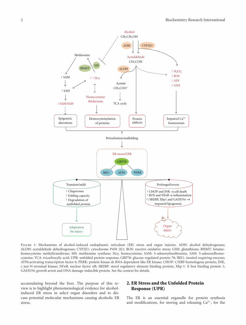
2 Biochemistry Research International
CYP2E1
Alcohol
CH3CH2OH
ADH
Acetaldehyde
CH3COH
Acetate
CH3COO−
TCA cycle
↑H2O2↑ ROS
↓ ATP
↓ GSH
Proteinadducts
↓ SAM/SAH
↓ SAM
↑ SAH
↑ ↑Hcy
Methionine
BHMT
Impaired Ca2+
homeostasis
Epigeneticalterations
Homocysteinethiolactone
Homocysteinylationof proteins
Perturbation/malfolding
ER stress/UPR
GRP78
IRE1
↑ Chaperones
↑ Folding capacity↑ Degradation ofmalfolded protein
↑CHOP and JNK cell death↑ROS and NFκB inflammation↑ SREBP, Xbp1 and GADD34
impaired lipogenesis
Adaptationo injury
Organinjury
MSALDH
PERKATF6
Transient/mild Prolonged/severe
N
↑
Figure 1: Mechanisms of alcohol-induced endoplasmic reticulum (ER) stress and organ injuries. ADH: alcohol dehydrogenase;ALDH: acetaldehyde dehydrogenase; CYP2E1: cytochrome P450 2E1; ROS: reactive oxidative stress; GSH: glutathione; BHMT: betaine-homocysteine methyltransferase; MS: methionine synthase; Hcy, homocysteine; SAM: S-adenosylmethionine, SAH: S-adenosylhomo-cysteine; TCA: tricarboxylic acid; UPR: unfolded protein response; GRP78: glucose-regulated protein 78; IRE1: inositol requiring enzyme;ATF6:activating transcription factor 6; PERK: protein kinase ds RNA-dependent-like ER kinase; CHOP: C/EBP-homologous protein; JNK,c-jun-N-terminal kinase; NFκB, nuclear factor κB; SREBP: sterol regulatory element binding protein; Xbp-1: X box binding protein 1;GADD34: growth arrest and DNA damage-inducible protein. See the context for details.
accumulating beyond the liver. The purpose of this re-view is to highlight phenomenological evidence for alcohol-induced ER stress in select organ disorders and to dis-cuss potential molecular mechanisms causing alcoholic ERstress.
2. ER Stress and the Unfolded ProteinResponse (UPR)
The ER is an essential organelle for protein synthesisand modifications, for storing and releasing Ca2+, for the

Biochemistry Research International 3
biosynthesis of lipids and sterols, and for detoxification ofcertain drugs. ER stress is a condition under which unfoldedor malfolded proteins accumulate in the ER (reviewed in[18–21]). ER stress results from perturbations in ER home-ostasis such as calcium depletion, inhibition of glycosylation,alterations of the redox state, or lipid overloading. ER stresstriggers the unfolded protein response (UPR), which consti-tutes a series of ER-to-nucleus signaling mediated by threeER resident transmembrane sensor proteins, inositol requir-ing protein 1 (IRE1), ds-RNA-activated protein kinase (PKR)like ER kinase (PERK), and activating transcription factor 6(ATF6) (Figure 1). The three sensors are activated upon dis-sociation from their inhibitory binding with the chaperoneGRP78/BiP. IRE1, which has kinase and endoribonucleaseactivities, is activated by transautophosphorylation. The acti-vated IRE1 processes the transcription factor X-box bindingprotein-1 (XBP1) mRNA via the unconventional splicing toform transcriptionally active spliced XBP1 (sXBP1). sXBP1activates UPR target genes, including chaperones and ER-associated degradation (ERAD) pathway genes. The secondsensor PERK phosphorylates the eukaryotic initiation factor2α-subunit (eIF2α), leading to an inhibition of the initiationof translation and a global attenuation in protein translation.Phosphorylation of eIF2α selectively activates activatingtranscription factor 4 (ATF4), which regulates ER chaperonegenes, ERAD pathway genes, amino acid metabolism genes,and the transcription factor C/EBP homologous protein(CHOP) [19–21]. The third sensor ATF6 is cleaved inthe Golgi to form a transcriptionally active fragment thattraffics to the nucleus to activate UPR target genes. Ingeneral the UPR results in reduced synthesis of nascentproteins, increased unloading of unfolded proteins, andincreased capacity of folding, which lead to restoration of ERhomeostasis.
However, prolonged or severe UPR provokes a complexnetwork of interacting and parallel responses contributingto pathological consequences such as apoptosis, inflam-mation, and fat accumulation [19–24]. The ER stress-induced apoptosis is mediated by a few factors. CHOPregulates growth arrest and DNA damage-inducible protein(GADD34). GADD34 binds protein phosphatase-1 andenhances eIF2α dephosphorylation, leading to prematurerestoration of translation and enhanced ER stress. CHOP canalso regulate expression of the TRAIL receptor DR5, pro-and antiapoptotic Bcl-2 family protein Bim, Bax and Bcl-2modulating cell death [19–21]. Sustained activation of IRE1recruits the adaptor protein TRAF2 and activates JNK andNF-κB, both of which mediate apoptosis [23]. In addition,alterations in ER calcium homeostasis, upregulation of ERoxidase 1 (ERO1) by CHOP, activation of caspase 12, andactivation of GSK3β by tribbles 3 (TRB3) and AKT are othermechanisms underlying ER stress-induced inflammationand apoptosis [21, 23, 25]. Lipid accumulation is also a mainpathological feature of prolonged ER stress, and each of thethree ER sensor pathways has direct molecular effects onlipid synthesis. The IRE1α-XBP1 branch regulates C/EBPαand C/EBPβ that control directly the expression of genesinvolved in de novo fatty acid biosynthesis [26]. The ATF6branch is involved in phospholipid biosynthesis as well as
in fatty acid oxidation and lipoprotein secretion [27, 28].The PERK-eIF2α branch influences expression of C/EBPfamily and PPARγ transcription factors via the eIF2α-specificphosphatase GADD34 and regulates SREBP1-related de novolipid synthesis and accumulation [18–24, 29, 30].
3. ER Stress in Alcoholic Organ Injuries
3.1. Liver. Alcohol is mainly metabolized in the liver, andliver cells are rich in ER which assumes synthesis of alarge amount of secretory and membrane proteins [19,20, 29]. Partial role of ER in alcohol metabolism wasinitially realized decades ago as NADH from the hepaticoxidation of ethanol to acetaldehyde by ADH was foundto support also microsomal ethanol oxidations [14, 15].The inducible microsomal ethanol oxidizing system (MEOS)is associated with proliferation of the ER and a con-comitant induction of cytochrome P4502E1 (CYP2E1) inrats and in humans. Free radical release as a consequenceof CYP2E1 function in the ER and subsequent oxidativestress and lipid peroxidation generally contribute to ALD[14, 15]. However, alcohol-induced ER stress response wasnot recognized until recently. Molecular evidence for animpaired UPR was first found in the intragastric alcohol-fed mice using microarray gene expression profiling [18].The alterations of selected ER stress markers were associatedwith severe steatosis, scattered apoptosis, and necroin-flammatory foci. Moderate upregulation of expression ofSREBP-1c and SREBP-2 and their responsive genes wasdetected by immuoblotting [18]. SREBP-1c knockout micewere protected against triglyceride accumulation [30–32].Knocking out CHOP resulted in minimal alcohol-inducedapoptosis in mouse liver [32–34]. In a setting of alcoholinfusion and moderate obesity, there are synergistic effects ofaccentuated ER and mitochondrial stress, nitrosative stressmediated by M1 macrophage activation, and adiponectinresistance on hepatic necroinflammation and steatohepatitis[35]. In micropigs fed alcohol, liver steatosis and apoptosiswere shown to be accompanied by increased mRNA levelsof CYP2E1, GRP78 and SREBP-1c, and protein levels ofCYP2E1, GRP78, activated SREBP and caspase 12 [36].In addition, the ER stress response was correlated withelevated transcripts of lipogenic enzymes such as fattyacid synthase (FAS), acetyl-CoA carboxylase (ACC), andstearoyl-CoA desaturase (SCD). Further, alcohol-inducedlipopolysaccharide (LPS) is linked to impaired UPR andadvanced hepatic injury [37–39]. In cirrhotic rat livers, onlyeIF2αwas activated in the basal state. After LPS challenge, fullUPR as indicated by activation of IRE1α, ATF-6, and eIF2αwas detected [37]. However, LPS-induced accumulation ofNF-κB-dependent antiapoptotic proteins was not observed,suggesting that the UPR sensitized the cirrhotic livers toLPS/TNFα-mediated apoptosis. Alcohol-induced hepatic ERstress response not only occurs in rodents but also in livers ofbaboon and human patients [40, 41]. In baboon fed alcoholorally, upregulation of calpain 2, calpain p94, and ERD21 anddownregulation of eIF2α were among the genes of alteredexpression that was revealed by using cDNA array analysis

4 Biochemistry Research International
[41]. Gene expression profiling of cirrhotic liver samplesfrom human alcoholics also revealed alterations of calpainand calreticulin that are indicative of ER malfunction.
3.2. Pancreas. The pancreas is one of the important digestiveorgans adversely affected by alcohol abuse. Pancreatitis isamong the most common alcohol-related hospital diagnosisin USA [11]. The underlying mechanisms for alcohol-induced pancreatitis are not well understood. Similar to theliver, the pancreas has the capacity to metabolize alcoholvia both the oxidative and nonoxidative pathways yieldingtoxic metabolites such as acetaldehyde and lipid esters. Fattyacid ethyl and cholesteryl esters are known to accumulatein the acinar cell after chronic alcohol consumption whichdecreases the stability of the membranes of zymogen gran-ules and lysosomes [42, 43], which cause a premature activa-tion of intracellular digestive enzyme and may predispose thegland to autodigestive inflammation and injury. In respectto the role of organelles in alcoholic pancreatic injury, theER has been considered as the acinar cell has the highestrate of protein synthesis among all tissues in adult organism.In fact, perturbations of ER homeostasis are found in acutepancreatitis [44, 45], and all the three ER stress/UPR trans-ducers (i.e., IRE1, ATF6, and PERK) and their downstreampathways are activated. However, chronic alcohol feedingalone causes minimal pancreatic tissue injury in animalmodels [45, 46]. Further studies demonstrate that alcoholfeeding activates the UPR in pancreas with upregulationof the transcription factor XBP1 in the intragastric alcoholinfusion model [47, 48]. This suggests that alcohol induces aphysiologic adaptive UPR that may prevent pathophysiologicpancreatitis responses. Indeed, heterozygous deletion ofthe XBP1 gene prevents XBP1 upregulation and results inpathologic changes including extensive dilation of the ERwith occasional dense luminal inclusions, hallmarks of ERstress, and significant accumulation of autophagic vacuolesin acinar cells [48]. Thus, impaired UPR in the pancreas canpotentiate alcohol-induced toxicity and aggravate pancreaticdamages.
3.3. Brain. Alcohol exposure during development has devas-tating effects on the loss of neurons in selected brain areas,which leads to profound damages to the central nervoussystem (CNS). Alcohol consumption during pregnancycauses fetal alcohol spectrum disorders (FASDs) [1, 49].Microcephaly, abnormal cortical thickness, reduced cere-bral white matter volume, ventriculomegaly, and cerebellarhypoplasia are the prominent CNS abnormalities in FASDs.Children with (FASD) have a variety of cognitive, behavioral,and neurological impairments [49]. What cause ethanol-induced neurodegeneration are not clear. Considering thatER stress plays a role in the pathogenesis of several popularneurological diseases such as Huntington’s disease, brainischemia, Alzheimer’s disease, and Parkinson’s disease [50–53], an involvement of ER stress in alcohol-induced neurontoxicity has been hypothesized [54]. Recent evidence fromboth in vitro and in vivo tests appears to support the assump-tion. Exposure of SH-SY5Y neuroblastoma cells or primary
cerebellar granule neurons to ethanol alone had little effecton the expression of ER stress markers [54]; however, ethanolmarkedly increased the expression of GRP78, CHOP, ATF4,ATF6, and phosphorylated PERK and eIF2α in the presenceof tunicamycin or thapsigargin, which was accompaniedwith increased cell death. Acute exposure of seven-day-oldmice to ethanol by subcutaneous injection at a dose of5 g/kg significantly increased ER stress response. Increase ofATF6, CHOP, GRP78, and mesencephalic astrocyte-derivedneurotrophic factor as well as the phosphorylation of IRE1,eIF2α, PERK, and PKR were detected within 24 hoursafter the ethanol exposure. Further, the ethanol-inducedincrease in phosphorylated eIF2α, caspase-12 and CHOP wasdistributed in neurons of specific areas of the cerebral cortex,hippocampus, and thalamus. Since the age of the animalsused in this experiment is equivalent to the third trimesterof pregnancy in humans, the above evidence suggests thatethanol directly induce ER stress in the developing brain.
3.4. Heart. It is well documented that chronic heavy alcoholdrinking is a risk factor for cardiovascular disorders includ-ing cardiac hypertrophy, myofibrillar disruption, reducedcontractility, and decreased ejection fraction [55]. Alcoholmay change the circulatory hemodynamics resulting in stresson the heart. The stressed heart demands more cardiacoutput which leads to compensative hypertrophic responsessuch as neurohormonal activation and increased growthfactors and cytokines, resulting in enlarged cardiomyocytesand increased sarcomere assembly. ER stress may playa critical role in regulating protein synthesis in cardiacmyocytes, and thereby produce cell enlargement and cardiachypertrophy. Chronic alcohol consumption by FVB (Friendvirus-B type) albino mice at 4% of diet for 12 weeks resultedin increased heart weight and heart-to-body weight ratio[56]. In the myocardium of the FVB mice chronicallyfed alcohol, GRP78, CHOP, and IRE1a protein expres-sion levels were increased, indicative of the UPR. Class Ialcohol dehydrogenase efficiently oxidizes alcohol resultingin increased production of acetaldehyde. Overexpressingalcohol dehydrogenase in the FVB mice during chronicethanol treatment resulted in a greater UPR upregulation[56]. The finding indicates that acetaldehyde from alcoholmetabolism may induce ER stress. Furthermore, overex-pressing of the antioxidant protein metallothionein in FVBmice significantly reduced peak shortening and maximalshortening velocity of cardiac myocytes by LPS, which isoften elevated in alcoholics [13–15, 39, 40]. In parallel, thetransgenic FVB mice displayed decreased protein levels ofGRP78, CHOP, PERK, and IRE1 whereas the wild type FVBdisplayed a significant increase in the protein levels of PERK,phospho-JNK, and phospho-p38 in the myocardium inresponse to LPS [56, 57].
4. Mechanisms of Alcohol-Induced ER Stress
4.1. Acetaldehyde Adducts and ER Stress. Alcohol-derivedacetaldehyde is highly reactive [58–62]. At physiologicaltemperature and pH, acetaldehyde reacts with nucleophilic

Biochemistry Research International 5
groups in proteins, such as α-amino groups of internal lysineresidues and the ε-amino group on the N-terminal aminoacid of unblocked proteins forming unstable Schiff baseacetaldehyde adducts. In addition, ethanol abuse may alsolead to the formation of other types of protein adducts, suchas malondialdehyde-acetaldehyde hybrids and α-hydroxy-ethyl protein-adducts. The acetaldehyde adducts initiate im-munogenic reactions, cause conformational changes andinactivation of the adducted targets, or trigger aberrantprotein degradation, which contribute to the developmentof alcoholic organ diseases (Figure 1). Malondialdehyde–acetaldehyde adduct is found to be the dominant epi-tope after malondialdehyde modification of proteins inatherosclerosis [63]. Antibodies to the aldehyde adducts havebeen detected in the serum of patients with atheroscleroticlesions and correlate with the progression of atherosclerosis.It is known that atherosclerosis develops as a result of pro-tein unfolding and modification of protein and/or macro-molecular complex function at the cellular level [63]. Insupporting this, evidence for ER stress response was foundin transgenic mice with cardiac overexpression of ADH thatincreased acetaldehyde exposure [56, 57]. The ADH trans-gene increased induction of IRE1, eIF-2α, GRP78, andCHOP and exacerbated chronic alcohol ingestion-inducedmyocardial dysfunction and hypertrophy. Further, in amouse model of acute ethanol intoxication, inhibition ofADH causes downregulation of GRP78 mRNA levels[64]. This suggests a causal relationship between ethanolmetabolism and ER stress response. Acetaldehyde adductsalso affect ER Ca2+ handling in rat ventricular myocytes[65, 66], which may disturb ER calcium homeostasis playinga critical role in stress-mediated cellular injury [67]. Inresponse to alcohol dosing in vivo, the actin in Type I andType II fibre predominant muscles of rats was found to formstable covalent adducts with acetaldehyde [68]. Histochem-ical analysis showed that unreduced-acetaldehyde-proteinadducts were located within the sarcolemmal (i.e., musclemembrane) and subsarcolemmal regions, which perturbedthe membranes and increased protein and enzyme activityof sarcoplasmic-ER Ca2+-ATPase, resulting in muscle celldeath and alcoholic myopathy. In addition, acetaldehydeadducts are found in the central nervous system which maybe responsible for alcoholic ER stress response. In the brainof a heavy drinker who had died suddenly while drinkingcontinuously, acetaldehyde adducts were immunologicallyidentified [69]. In a mouse model administered with theLieber-DeCarli liquid diet and alcohol, acetaldehyde adductswere readily detected in degenerated neurons in the cerebralcortex [70]. The neural region that alcoholic ER stressresponse occurred colocalized with the acetaldehyde adducts.In young mice, ethanol-induced increase in ER stress proteinmarkers was found to be distributed in the immatureneurons of specific areas of the cerebral cortex, hippocampusand thalamus [54]. Thus, while most organs of the bodycan be affected by alcohol-derived acetaldehyde, cardiac andskeletal muscle cells and neurons appear to be particularlysusceptible to acetaldehyde adducts that cause ER stress andinjury.
4.2. Homocysteine Toxicity and ER Stress. Homocysteine(Hcy) is a normal intermediate involved in the metabolismof the essential amino acid-methionine (Figure 1). ExcessiveHcy is toxic to cells. An abnormally elevated level of Hcy inthe blood, a medical condition termed hyperhomocysteine-mia (HHcy), is an independent risk factor in cardiovascular,neurodegenerative diseases, diabetes, obesity, and hepaticsteatosis [32, 71–73]. It is generally accepted that aminoacylthioester homocysteine thiolactone (HTL) derived from Hcyediting during protein synthesis contributes to the most ofHcy toxicity [74, 75]. HTL undergoes not only nucleophilic,which can be facilitated in the presence of acetaldehyde,but also electrophilic reactions to form protein adducts orhomocysteinylation of protein lysine side chains and/or otherfree amine groups [75]. These reactions cause malfoldingof proteins and trigger ER stress response. Evidence linkingHHcy to ER stress and alcoholic liver injury has well beenestablished in cell and animal models [16, 18–20, 32].The intragastric alcohol feeding exhibited a greater than 5-fold increase in mouse plasma Hcy [18, 34, 35]. Hcy ismetabolized normally by remethylation to methionine whichis catalyzed by methionine synthase (MS) using folate asa methyl donor and by betaine-homocysteine methyltrans-ferase (BHMT) using betaine as a methyl donor. Chronicalcohol-induced disturbance of methionine metabolismappears to contribute to the alcoholic HHcy. Alcohol inhibitsenzyme activity of MS in mice and rats and reduces mRNAexpression of BHMT and MS in mice [16, 17, 34, 76–79].Simultaneous betaine feeding in the intragastric alcohol-fed mice decreased alcoholic HHcy and abrogated ER stressresponse in parallel with decreased ALT and ameliorationof alcohol-induced necroinflammation, apoptosis, and fattyliver [18]. In cultured HepG2 cells, BHMT overexpressioninhibited Hcy-induced ER stress response, lipid accumula-tion, and cell death [77]. In primary mouse hepatocytes,suppression of BHMT by RNA interference potentiated Hcy-induced but not tunicamycin-induced ER stress response andcell injury [77]. Transgenic mice expressing human BHMTin organs peripheral to the liver are resistant to alcohol ora high methionine and low folate diet induced HHcy andfatty liver [78]. In intragastric alcohol-fed rats, BHMT isinduced, which minimizes the effect of inhibited MS on Hcylevels and subsequent ER stress response and injury [79]. Ina survey using 14 mouse strains, Ivan Rusyn has found thatthe alcoholic HHcy is correlated with alcohol-induced liverjury (personal communication, 2011). Therefore, the aboveseveral lines of evidence support Hcy toxicity as a pathogenicfactor contributing to alcohol-induced disorders.
4.3. SAM/SAH Ratio, Epigenetic Alterations and ER Stress.There are two types of important epigenetic regulations ofgene expression: DNA methylation of cytosines within CpGdinucleotides and histone modifications [80, 81]. Aberrantepigenetic changes are involved in the etiology of a growingnumber of disorders such as alcohol dependence. Both globalhypomethylation of DNA in liver and hypermethylationof DNA from peripheral blood cells have been reportedin animal models and in human subjects with alcohol
6 Biochemistry Research International
dependence [82–86]. This is because DNA methylation ingeneral depends on the methyl donor S-adenosylmethionine(SAM) and is inhibited by S-adenosylhomocysteine (SAH).Both SAM and SAH are involved in methionine metabolism[87, 88]. Inside the cell, SAM is demethylated to SAH,which is readily converted to Hcy which is remethylated tomethionine. Plasma Hcy is not metabolized and representsthe cumulative export of Hcy from liver and other tissues.Alcohol consumption decreases levels of SAM and increaseslevels of SAH and/or Hcy resulting in a decrease in SAMto SAH ratio (Figure 1) [76, 78, 87–92]. Thus, alcoholhas a marked impact on the hepatic methylation capacity.Evidence demonstrating epigenetic effects on alcoholic ERstress is emerging [17, 82]. In 66 male alcoholic patientswith alcohol dependence, chronically elevated Hcy levels areassociated with increased DNA methylation in the promoterregion of homocysteine-inducible ER protein (HERP) anddecreased expression of HERP mRNA in the blood [93, 94].The decrease in HERP levels is followed by a lethal ER stress,mitochondrial dysfunction, and cell death in neurons ofthe developing and adult brain [94]. Thus it is conceivablethat alcoholic Hcy regulates HERP and causes ER stress andinjury through an epigenetic mechanism. In respect to theepigenetic modifications of histone, it is reported that alcoholcauses a dose- and time-dependent selective acetylation ofhistone H3-K9 in cultured hepatocytes [95, 96]. Intragastricadministration of ethanol increases the levels of acetylatedH3-K9 by 2-3 folds in the liver of mice after 12 h [97].Further analysis indicates that the increased acetylation istissue specific as it is noted in liver, lung, and spleen but notin tissues from other organs tested. Thus, while other stresspathways such as the MAPK signaling may be involved, thealcoholic epigenetic effects on the ER stress pathways canbe more relevant. For instance, in both cystathionine betasynthase heterozygous (CBS+/−) and wild type (WT) micefed ethanol diets by intragastric infusion for 4 weeks, steato-hepatitis, reduction in liver SAM, elevation in liver SAH, andreduction in the SAM/SAH ratio were observed [17]. HepaticER stress markers including GRP78, ATF4, CHOP, caspase12, and SREBP-1c were upregulated and negative correlatedwith the SAM/SAH ratio in response to alcohol. Further,trimethylated histone H3 lysine-9 (3meH3K9) protein levelsin centrilobular regions revealed by immunohistochemistrywere reduced in ethanol-fed mice. The levels of 3meH3K9in the promoter regions of GRP78, SREBP-1c, and CHOPrevealed specifically by a chromatin immunoprecipitationassay were decreased only in CBS+/− mice fed alcohol. SinceCBS is involved in transsulfuration of Hcy, the findingsimply that interactions of CBS ablation and alcohol feedingimpair methionine metabolism, which leads to epigeneticmodifications of ER stress signaling pathways. In addition,the key modulator of UPR, sXBP1 has recently been foundto be a nonhistone protein target of acetylation mediated byp300 and deacetylation mediated by the NAD+-dependentclass III deacetylase SIRT1 (sirtuin 1) [98, 99]. SIRT1 isdemonstrated to be one of the major targets of alcohol actionwhich influences TNF-α production in macrophages andalters glucose and lipid metabolism in the liver leading tohepatic steatosis and inflammation [100–102]. SIRT1 may
also play a role in alcohol-induced ER stress response andinjury through an epigenetic mechanism.
4.4. Oxidative Stress and Disruption of Ca2+ or Iron Home-ostasis and ER Stress. In the ER, proteins undergo oxidativeprotein folding. PDI is a critical oxoreductase that catalyzesdisulfide bond formation with consequent generation ofreactive oxygen species (ROS) during the oxidative proteinfolding [19, 103]. ROS is normally under control due tocellular glutathione that sustains PDI ability to regenerateand form disulfide bridges repeatedly [103–105]. However,chronic ethanol consumption increases the production ofa variety of ROS, including superoxide, H2O2, lipid per-oxides, and peroxynitrite [1, 13–15]. Alcoholic ROS reduceglutathione level and increase oxidized glutathione, whichbreaks the redox status of the ER (Figure 1). This loss ofredox homeostasis perturbs the oxidative folding and makesPDI ineffective in the catalytic redox cycles leading to moreutilization of reduced glutathione. Depletion of glutathionegenerates excessive ROS which triggers ER stress. Antioxidanttreatment, CHOP deletion, or translation attenuation hasbeen shown to reduce oxidative stress and preserve ERfunction [19–23]. Ethanol rapidly caused oxidative stress incultured neuronal cells and antioxidants blocked alcoholicpotentiation of ER stress and cell death [54]. An associationof ER stress response with increased oxidized glutathione wasfound in the pancreatic acinar cell of the ethanol-fed rats[47]. In HepG2 cells, acetaldehyde impaired mitochondrialglutathione transport and stimulated mitochondrial choles-terol content, the latter of which was preceded by increasedlevels of CHOP and SREBP1 [106]. Chronic exposure of ani-mals to alcohol or overexpression of cytochrome CYP2E1 inhepatocytes increases the expression of superoxide dismutase(SOD) and activates nuclear factor erythroid 2-related factor2 (Nrf2), which is an ER stress responsive factor [14, 107–109]. These lines of evidence suggest an intimate relation-ship between ER stress and ROS production. Furthermore,alcoholic oxidative stress plays a critical role in possibleinterplays between ER stress and mitochondrial stress, whichcan be mediated either by intracellular calcium or iron.Alcohol or Hcy induces alterations of lipid compositionin the ER and affected ratio of phosphatidylcholine (PC)to phosphatidylethanolamine (PE) [20, 78]. Alterations ofthe PC/PE ratio disrupt ER calcium homeostasis causingER stress [110]. Under ER stress, abnormal release ofintracellular Ca2+ from the ER via inositol 1,4,5-triphosphatereceptor (IP3R) channels leads to excessive mitochondrialCa2+ uptake, which in turn promotes ROS production andapoptosis via multiple effects on the mitochondria [67, 111,112]. Elevated serum iron indices (transferrin saturation,ferritin) and hepatic iron overloading are often observed inpatients with alcoholic liver disease [113–117]. Excessive irondamages mitochondrial iron–sulfur clusters that generatedefects in heme-containing cytochrome c and cytochromeoxidase leading to excess mitochondrial ROS [118]. Ironhomeostasis is regulated by hepcidin, a circulatory antimi-crobial peptide synthesized in hepatocytes [119]. Critical-ly, ER stress response can regulate expression of hepcidin
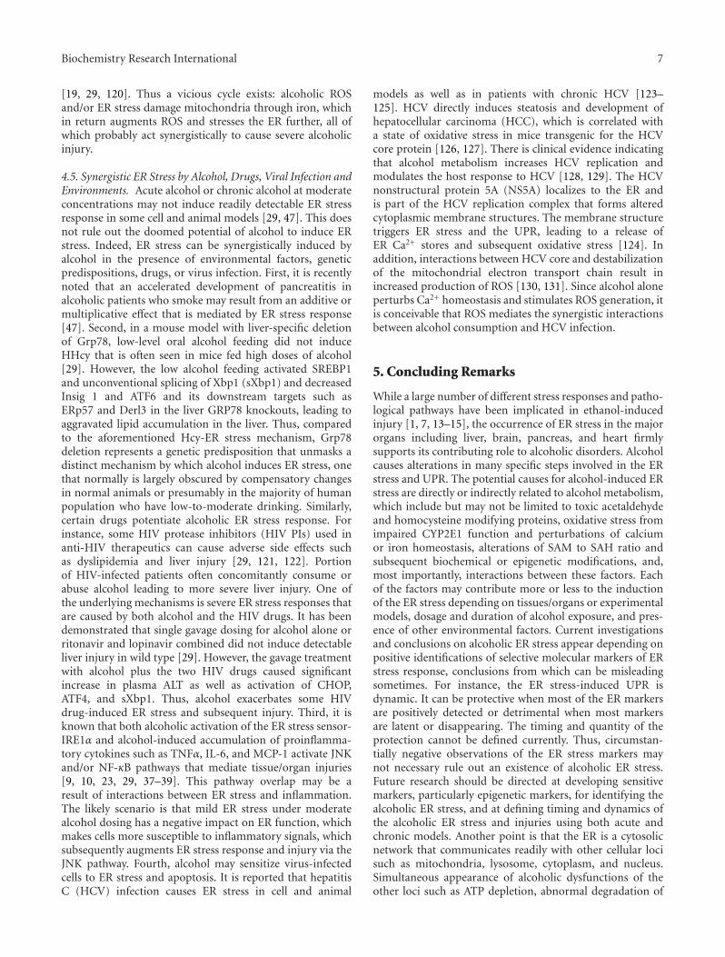
Biochemistry Research International 7
[19, 29, 120]. Thus a vicious cycle exists: alcoholic ROSand/or ER stress damage mitochondria through iron, whichin return augments ROS and stresses the ER further, all ofwhich probably act synergistically to cause severe alcoholicinjury.
4.5. Synergistic ER Stress by Alcohol, Drugs, Viral Infection andEnvironments. Acute alcohol or chronic alcohol at moderateconcentrations may not induce readily detectable ER stressresponse in some cell and animal models [29, 47]. This doesnot rule out the doomed potential of alcohol to induce ERstress. Indeed, ER stress can be synergistically induced byalcohol in the presence of environmental factors, geneticpredispositions, drugs, or virus infection. First, it is recentlynoted that an accelerated development of pancreatitis inalcoholic patients who smoke may result from an additive ormultiplicative effect that is mediated by ER stress response[47]. Second, in a mouse model with liver-specific deletionof Grp78, low-level oral alcohol feeding did not induceHHcy that is often seen in mice fed high doses of alcohol[29]. However, the low alcohol feeding activated SREBP1and unconventional splicing of Xbp1 (sXbp1) and decreasedInsig 1 and ATF6 and its downstream targets such asERp57 and Derl3 in the liver GRP78 knockouts, leading toaggravated lipid accumulation in the liver. Thus, comparedto the aforementioned Hcy-ER stress mechanism, Grp78deletion represents a genetic predisposition that unmasks adistinct mechanism by which alcohol induces ER stress, onethat normally is largely obscured by compensatory changesin normal animals or presumably in the majority of humanpopulation who have low-to-moderate drinking. Similarly,certain drugs potentiate alcoholic ER stress response. Forinstance, some HIV protease inhibitors (HIV PIs) used inanti-HIV therapeutics can cause adverse side effects suchas dyslipidemia and liver injury [29, 121, 122]. Portionof HIV-infected patients often concomitantly consume orabuse alcohol leading to more severe liver injury. One ofthe underlying mechanisms is severe ER stress responses thatare caused by both alcohol and the HIV drugs. It has beendemonstrated that single gavage dosing for alcohol alone orritonavir and lopinavir combined did not induce detectableliver injury in wild type [29]. However, the gavage treatmentwith alcohol plus the two HIV drugs caused significantincrease in plasma ALT as well as activation of CHOP,ATF4, and sXbp1. Thus, alcohol exacerbates some HIVdrug-induced ER stress and subsequent injury. Third, it isknown that both alcoholic activation of the ER stress sensor-IRE1α and alcohol-induced accumulation of proinflamma-tory cytokines such as TNFα, IL-6, and MCP-1 activate JNKand/or NF-κB pathways that mediate tissue/organ injuries[9, 10, 23, 29, 37–39]. This pathway overlap may be aresult of interactions between ER stress and inflammation.The likely scenario is that mild ER stress under moderatealcohol dosing has a negative impact on ER function, whichmakes cells more susceptible to inflammatory signals, whichsubsequently augments ER stress response and injury via theJNK pathway. Fourth, alcohol may sensitize virus-infectedcells to ER stress and apoptosis. It is reported that hepatitisC (HCV) infection causes ER stress in cell and animal
models as well as in patients with chronic HCV [123–125]. HCV directly induces steatosis and development ofhepatocellular carcinoma (HCC), which is correlated witha state of oxidative stress in mice transgenic for the HCVcore protein [126, 127]. There is clinical evidence indicatingthat alcohol metabolism increases HCV replication andmodulates the host response to HCV [128, 129]. The HCVnonstructural protein 5A (NS5A) localizes to the ER andis part of the HCV replication complex that forms alteredcytoplasmic membrane structures. The membrane structuretriggers ER stress and the UPR, leading to a release ofER Ca2+ stores and subsequent oxidative stress [124]. Inaddition, interactions between HCV core and destabilizationof the mitochondrial electron transport chain result inincreased production of ROS [130, 131]. Since alcohol aloneperturbs Ca2+ homeostasis and stimulates ROS generation, itis conceivable that ROS mediates the synergistic interactionsbetween alcohol consumption and HCV infection.
5. Concluding Remarks
While a large number of different stress responses and patho-logical pathways have been implicated in ethanol-inducedinjury [1, 7, 13–15], the occurrence of ER stress in the majororgans including liver, brain, pancreas, and heart firmlysupports its contributing role to alcoholic disorders. Alcoholcauses alterations in many specific steps involved in the ERstress and UPR. The potential causes for alcohol-induced ERstress are directly or indirectly related to alcohol metabolism,which include but may not be limited to toxic acetaldehydeand homocysteine modifying proteins, oxidative stress fromimpaired CYP2E1 function and perturbations of calciumor iron homeostasis, alterations of SAM to SAH ratio andsubsequent biochemical or epigenetic modifications, and,most importantly, interactions between these factors. Eachof the factors may contribute more or less to the inductionof the ER stress depending on tissues/organs or experimentalmodels, dosage and duration of alcohol exposure, and pres-ence of other environmental factors. Current investigationsand conclusions on alcoholic ER stress appear depending onpositive identifications of selective molecular markers of ERstress response, conclusions from which can be misleadingsometimes. For instance, the ER stress-induced UPR isdynamic. It can be protective when most of the ER markersare positively detected or detrimental when most markersare latent or disappearing. The timing and quantity of theprotection cannot be defined currently. Thus, circumstan-tially negative observations of the ER stress markers maynot necessary rule out an existence of alcoholic ER stress.Future research should be directed at developing sensitivemarkers, particularly epigenetic markers, for identifying thealcoholic ER stress, and at defining timing and dynamics ofthe alcoholic ER stress and injuries using both acute andchronic models. Another point is that the ER is a cytosolicnetwork that communicates readily with other cellular locisuch as mitochondria, lysosome, cytoplasm, and nucleus.Simultaneous appearance of alcoholic dysfunctions of theother loci such as ATP depletion, abnormal degradation of

8 Biochemistry Research International
the inside materials, oxidative stress, and numerous otherstress responses could overshadow the role of ER stress inalcoholic diseases. Thus, the role of alcoholic ER stress inorgan disorders can be defined precisely by studying complexinterplays among the organelles and loci in disease patho-genesis, which could provide better therapeutic strategiestargeting the ER. Finally, with respect to the therapeuticinterventions at alcoholic ER stress, possible approachesinclude lowering homocysteine and raising SAM by nutri-tional support with betaine or folate [16, 20, 32], improvingprotein folding by using chemical chaperone PBA (sodium4-phenylbutyrate) and TUDCA [19, 20, 29], blocking eIF2αdephosphorylation by using salubrinal [132], and ameliorat-ing ROS production from the oxidative protein folding byusing antioxidants. However, results of clinical trials are notavailable. Each of the individual approaches alone may not bea simple or universal cure as alcohol-induced pathogenesisis very complex. It is anticipated that properly combinedtreatments with all the beneficial agents can be effective.
Acknowledgments
This work has been supported by NIH Grants R01AA018846,R01AA018612, and R01AA014428 and by the USC ResearchCenter for Liver Disease (P30 DK48522) and the SouthernCalifornia Research Center for ALPD and Cirrhosis (P50AA11999). The author thanks Dr. N. Kaplowitz and thegraduate students and fellows who contributed to thestudies.
References
[1] L. Gunzerath, B. G. Hewitt, T. K. Li, and K. R. Warren,“Alcohol research: past, present, and future,” Annals of theNew York Academy of Sciences, vol. 1216, no. 6648, pp. 1–23,2011.
[2] M. P. Heron, D. L. Hoyert, S. L. Murphy, J. Q. Xu, K. D.Kochanek, and B. Tejada-Vera, “Deaths: final data for 2006,”National Vital Statistics Reports, vol. 57, no. 14, pp. 1–134,2009.
[3] J. Rehm, C. Mathers, S. Popova, M. Thavorncharoensap, Y.Teerawattananon, and J. Patra, “Global burden of disease andinjury and economic cost attributable to alcohol use andalcohol-use disorders,” The Lancet, vol. 373, no. 9682, pp.2223–2233, 2009.
[4] S. Zakhari, “Overview: how is alcohol metabolized by thebody?” Alcohol Research and Health, vol. 29, no. 4, pp. 245–254, 2006.
[5] J. Neiman, “Alcohol as a risk factor for brain damage:neurologic aspects,” Alcoholism: Clinical and ExperimentalResearch, vol. 22, no. 7, pp. 346–351, 1998.
[6] M. Epstein, “Alcohol’s impact on kidney function,” AlcoholHealth and Research World, vol. 21, no. 1, pp. 84–92, 1997.
[7] S. W. French, “The mechanism of organ injury in alcoholics:implications for therapy,” Alcohol and Alcoholism Supplement,vol. 1, pp. 57–63, 1991.
[8] C. Bode and J. C. Bode, “Alcohol’s role in gastrointestinaltract disorders,” Alcohol Health and Research World, vol. 21,no. 1, pp. 76–83, 1997.
[9] G. Szabo, J. R. Wands, A. Eken et al., “Alcohol and hepatitisC virus-interactions in immune dysfunctions and liver
damage,” Alcoholism: Clinical and Experimental Research, vol.34, no. 10, pp. 1675–1686, 2010.
[10] B. Gao, E. Seki, D. A. Brenner et al., “Innate immunity inalcoholic liver disease,” American Journal of Physiology—Gastrointestinal and Liver Physiology, vol. 300, no. 4, pp. 516–525, 2011.
[11] A. L. Yang, S. Vadhavkar, G. Singh, and M. B. Omary, “Epi-demiology of alcohol-related liver and pancreatic disease inthe United States,” Archives of Internal Medicine, vol. 168, no.6, pp. 649–656, 2008.
[12] A. George and V. M. Figueredo, “Alcohol and arrhythmias:a comprehensive review,” Journal of Cardiovascular Medicine,vol. 11, no. 4, pp. 221–228, 2010.
[13] T. H. Frazier, A. M. Stocker, N. A. Kershner, L. S. Marsano,and C. J. McClain, “Treatment of alcoholic liver disease,”Therapeutic Advances in Gastroenterology, vol. 4, no. 1, pp.63–81, 2011.
[14] A. I. Cederbaum, Y. Lu, and D. Wu, “Role of oxidative stressin alcohol-induced liver injury,” Archives of Toxicology, vol.83, no. 6, pp. 519–548, 2009.
[15] R. G. Thurman, S. Ji, and J. J. Lemasters, “Alcohol-inducedliver injury. The role of oxygen,” Recent Developments inAlcoholism, vol. 2, pp. 103–117, 1984.
[16] C. H. Halsted, “Nutrition and alcoholic liver disease,” Sem-inars in Liver Disease, vol. 24, no. 3, pp. 289–304, 2004.
[17] F. Esfandiari, V. Medici, D. H. Wong et al., “Epigenetic regula-tion of hepatic endoplasmic reticulum stress pathways in theethanol-fed cystathionine β synthase-deficient mouse,” Hep-atology, vol. 51, no. 3, pp. 932–941, 2010.
[18] C. Ji and N. Kaplowitz, “Betaine decreases hyperhomocys-teinemia, endoplasmic reticulum stress, and liver injury inalcohol-fed mice,” Gastroenterology, vol. 124, no. 5, pp. 1488–1499, 2003.
[19] H. Malhi and R. J. Kaufman, “Endoplasmic reticulum stressin liver disease,” Journal of Hepatology, vol. 54, no. 4, pp. 795–809, 2011.
[20] C. Ji, “Dissection of endoplasmic reticulum stress signalingin alcoholic and non-alcoholic liver injury,” Journal ofGastroenterology and Hepatology, vol. 23, no. 1, pp. S16–S24,2008.
[21] D. Ron and S. R. Hubbard, “How IRE1 Reacts to ER Stress,”Cell, vol. 132, no. 1, pp. 24–26, 2008.
[22] S. M. Colgan, A. A. Hashimi, and R. C. Austi, “Endoplasmicreticulum stressand lipid dysregulation,” Expert Reviews inMolecular Medicine, vol. 13, p. e4, 2011.
[23] M. Kitamura, “Control of NF-κB and inflammation by theunfolded protein response,” International Reviews of Im-munology, vol. 30, no. 1, pp. 4–15, 2011.
[24] A. H. Lee, E. F. Scapa, D. E. Cohen, and L. H. Glimcher,“Regulation of hepatic lipogenesis by the transcription factorXBP1,” Science, vol. 320, no. 5882, pp. 1492–1496, 2008.
[25] H. Bommiasamy, S. H. Back, P. Fagone et al., “ATF6α inducesXBP1-independent expansion of the endoplasmic reticu-lum,” Journal of Cell Science, vol. 122, no. 10, pp. 1626–1636,2009.
[26] D. T. Rutkowski, J. Wu, S. H. Back et al., “UPR pathwayscombine to prevent hepatic steatosis caused by ER stress-mediated suppression of transcriptional master regulators,”Developmental Cell, vol. 15, no. 6, pp. 829–840, 2008.
[27] S. Oyadomari, H. P. Harding, Y. Zhang, M. Oyadomari, andD. Ron, “Dephosphorylation of translation initiation factor2α enhances glucose tolerance and attenuates hepatosteatosisin mice,” Cell Metabolism, vol. 7, no. 6, pp. 520–532, 2008.

Biochemistry Research International 9
[28] E. Bobrovnikova-Marjon, G. Hatzivassiliou, C. Grigoriadouet al., “PERK-dependent regulation of lipogenesis duringmouse mammary gland development and adipocyte differ-entiation,” Proceedings of the National Academy of Sciences ofthe United States of America, vol. 105, no. 42, pp. 16314–16319, 2008.
[29] C. Ji, N. Kaplowitz, M. Y. Lau, E. Kao, L. M. Petrovic, andA. S. Lee, “Liver-specific loss of glucose-regulated protein78 perturbs the unfolded protein response andexacerbates aspectrum of liver diseases in mice,” Hepatology, vol. 54, no. 1,pp. 229–239, 2011.
[30] C. Ji, C. Chan, and N. Kaplowitz, “Predominant role of sterolresponse element binding proteins (SREBP) lipogenic path-ways in hepatic steatosis in the murine intragastric ethanolfeeding model,” Journal of Hepatology, vol. 45, no. 5, pp. 717–724, 2006.
[31] C. Ji and N. Kaplowitz, “ER stress: can the liver cope?” Journalof Hepatology, vol. 45, no. 2, pp. 321–333, 2006.
[32] L. Dara, C. Ji, and N. Kaplowitz, “The contribution of endo-plasmic reticulum stress to liver diseases,” Hepatology, vol.53, no. 5, pp. 1752–1763, 2011.
[33] C. Ji, R. Mehrian-Shai, C. Chan, Y. H. Hsu, and N. Kaplowitz,“Role of CHOP in hepatic apoptosis in the murine modelof intragastric ethanol feeding,” Alcoholism: Clinical and Ex-perimental Research, vol. 29, no. 8, pp. 1496–1503, 2005.
[34] C. Ji, Q. Deng, and N. Kaplowitz, “Role of TNF-α in ethanol-induced hyperhomocysteinemia and murine alcoholic liverinjury,” Hepatology, vol. 40, no. 2, pp. 442–451, 2004.
[35] J. Xu, K. K. Lai, A. Verlinsky et al., “Synergistic steatohepatitisby moderate obesity and alcohol in mice despite increasedadiponectin and p-AMPK,” Journal of Hepatology, vol. 55, no.3, pp. 673–682, 2011.
[36] F. Esfandiari, J. A. Villanueva, D. H. Wong, S. W. French, andC. H. Halsted, “Chronic ethanol feeding and folate deficiencyactivate hepatic endoplasmic reticulum stress pathway inmicropigs,” American Journal of Physiology—Gastrointestinaland Liver Physiology, vol. 289, no. 1, pp. G54–G63, 2005.
[37] K. A. Tazi, I. Bieche, V. Paradis et al., “In vivo altered unfoldedprotein response and apoptosis in livers from lipopoly-saccharide-challenged cirrhotic rats,” Journal of Hepatology,vol. 46, no. 6, pp. 1075–1088, 2007.
[38] H. A. Jarvelainen, T. Oinonen, and K. O. Lindros, “Alcohol-induced expression of the CD14 endotoxin receptor proteinin rat Kupffer cells,” Alcoholism: Clinical and ExperimentalResearch, vol. 21, no. 8, pp. 1547–1551, 1997.
[39] G. L. Su, A. Rahemtulla, P. Thomas, R. D. Klein, S. C. Wang,and A. A. Nanji, “CD14 and lipopolysaccharide bindingprotein expression in a rat model of alcoholic liver disease,”American Journal of Pathology, vol. 152, no. 3, pp. 841–849,1998.
[40] L. He, J. C. Marecki, G. Serrero, F. A. Simmen, M. J. Ronis,and T. M. Badger, “Dose-dependent effects of alcohol oninsulin signaling: partial explanation for biphasic alcoholimpact on human health,” Molecular Endocrinology, vol. 21,no. 10, pp. 2541–2550, 2007.
[41] D. Seth, M. A. Leo, P. H. McGuinness et al., “Gene ex-pression profiling of alcoholic liver disease in the baboon(Papiohamadryas) and human liver,” American Journal ofPathology, vol. 163, no. 6, pp. 2303–2317, 2003.
[42] S. J. Pandol, S. Periskic, I. Gukovsky et al., “Ethanol diet in-creases the sensitivity of rats to pancreatitis induced bycholecystokinin octapeptide,” Gastroenterology, vol. 117, no.3, pp. 706–716, 1999.
[43] A. S. Gukovskaya, O. A. Mareninova, I. V. Odinokova etal., “Cell death in pancreatitis: effects of alcohol,” Journal ofGastroenterology and Hepatology, vol. 21, no. 3, pp. S10–S13,2006.
[44] C. H. Kubisch, M. D. Sans, T. Arumugam, S. A. Ernst, J.A. Williams, and C. D. Logsdon, “Early activation of endo-plasmic reticulum stress is associated with arginine-inducedacute pancrea-titis,” American Journal of Physiology—Gas-trointestinal and Liver Physiology, vol. 291, no. 2, pp. G238–G245, 2006.
[45] I. Gukovsky, A. Lugea, M. Shahsahebi et al., “A rat modelreproducing key pathological responses of alcoholic chronicpancreatitis,” American Journal of Physiology—Gastrointes-tinal and Liver Physiology, vol. 294, no. 1, pp. G68–G79, 2007.
[46] M. Singh, M. M. LaSure, and D. E. Bockman, “Pancreaticacinar cell function and morphology in rats chronically fedon ethanol diet,” Gastroenterology, vol. 82, no. 3, pp. 425–434,1982.
[47] S. J. Pandol, F. S. Gorelick, A. Gerloff, and A. Lugea, “Alcoholabuse, endoplasmic reticulum stress and pancreatitis,” Diges-tive Diseases, vol. 28, no. 6, pp. 776–782, 2010.
[48] A. Lugea, D. Tischler, J. Nguyen et al., “Adaptive unfoldedprotein response attenuates alcohol-induced pancreatic dam-age,” Gastroenterology, vol. 140, no. 3, pp. 987–997, 2011.
[49] E. P. Riley, M. A. Infante, and K. R. Warren, “Fetal alcoholspectrum disorders: an overview,” Neuropsychology Review,vol. 21, no. 2, pp. 73–80, 2011.
[50] S. Tajiri, S. Oyadomari, S. Yano et al., “Ischemia-inducedneuronal cell death is mediated by the endoplasmic reticulumstress pathway involving CHOP,” Cell Death and Differentia-tion, vol. 11, no. 4, pp. 403–415, 2004.
[51] T. Katayama, K. Imaizumi, T. Manabe, J. Hitomi, T. Kudo,and M. Tohyama, “Induction of neuronal death by ER stressin Alzheimer’s disease,” Journal of Chemical Neuroanatomy,vol. 28, no. 1-2, pp. 67–78, 2004.
[52] R. M. Silva, V. Ries, T. F. Oo et al., “CHOP/GADD153 is amediator of apoptotic death in substantia nigra dopamineneurons in an in vivo neurotoxin model of parkinsonism,”Journal of Neurochemistry, vol. 95, no. 4, pp. 974–986, 2005.
[53] W. Scheper and J. J. Hoozemans, “Endoplasmic reticulumprotein quality control in neurodegenerative disease: thegood, the bad and the therapy,” Current Medicinal Chemistry,vol. 16, no. 5, pp. 615–626, 2009.
[54] Z. Ke, X. Wang, Y. Liu et al., “Ethanol induces endoplasmicreticulum stress in the developing brain,” Alcoholism: Clinicaland Experimental Research, vol. 35, no. 9, pp. 1574–1583,2011.
[55] C. D. Spies, M. Sander, K. Stangl et al., “Effects of alcohol onthe heart,” Current Opinion in Critical Care, vol. 7, no. 5, pp.337–343, 2001.
[56] S. Y. Li and J. Ren, “Cardiac overexpression of alcohol dehy-drogenase exacerbates chronic ethanol ingestion-inducedmyocardial dysfunction and hypertrophy: role of insulinsignaling and ER stress,” Journal of Molecular and CellularCardiology, vol. 44, no. 6, pp. 992–1001, 2008.
[57] S. Y. Li, S. A. Gilbert, Q. Li, and J. Ren, “Aldehyde dehy-drogenase-2 (ALDH2) ameliorates chronic alcohol inges-tion-induced myocardial insulin resistance and endoplasmicreticulum stress,” Journal of Molecular and Cellular Cardiol-ogy, vol. 47, no. 2, pp. 247–255, 2009.
[58] M. Setshedi, J. R. Wands, and S. M. Monte, “Acetaldehydeadducts in alcoholic liver disease,” Oxidative Medicine andCellular Longevity, vol. 3, no. 3, pp. 178–185, 2010.
10 Biochemistry Research International
[59] T. M. Donohue, D. J. Tuma, and M. F. Sorrell, “Binding ofmetabolically derived acetaldehyde to hepatic proteins invitro,” Laboratory Investigation, vol. 49, no. 2, pp. 226–231,1983.
[60] V. J. Stevens, W. J. Fanil, C. B. Newman, R. V. Sims, A.Cerami, and C. M. Peterson, “Acetaldehyde adduct withhemoglobin,” Journal of Clinical Investigation, vol. 67, no. 2,pp. 361–369, 1981.
[61] M. Hoerner, U. J. Behrens, T. Worner, and C. S. Lieber,“Humoral immune response to acetaldehyde adducts inalcoholic patients,” Research Communications in ChemicalPathology and Pharmacology, vol. 54, no. 1, pp. 3–12, 1986.
[62] Y. Israel, E. Hurwitz, O. Niemela, and R. Arnon, “Monoclonaland polyclonal antibodies against acetaldehyde-containingepitopes in acetaldehyde-protein adducts,” Proceedings of theNational Academy of Sciences of the United States of America,vol. 83, no. 20, pp. 7923–7927, 1986.
[63] F. Ursini, K. J. Davies, M. Maiorino, T. Parasassi, and A.Sevanian, “Atherosclerosis: another protein misfolding dis-ease?” Trends in Molecular Medicine, vol. 8, no. 8, pp. 370–374, 2002.
[64] Y. Nishitani and H. Matsumoto, “Ethanol rapidly causes acti-vation of JNK associated with ER stress under inhibition ofADH,” FEBS Letters, vol. 580, no. 1, pp. 9–14, 2006.
[65] K. R. Mills, K. Ward, F. Martin, and T. J. Peters, “Peripheralneuropathy and myopathy in chronic alcoholism,” Alcoholand Alcoholism, vol. 21, no. 4, pp. 357–362, 1986.
[66] Y. H. Sun, Y. Q. Li, S. L. Feng et al., “Calcium-sensing receptoractivation contributed to apoptosis stimulates TRPC6 chan-nel in rat neonatal ventricular myocytes,” Biochemical andBiophysical Research Communications, vol. 394, no. 4, pp.955–961, 2010.
[67] D. Mekahli, G. Bultynck, J. B. Parys, H. de Smedt, and L.Missiaen, “Endoplasmic-reticulum calcium depletion anddisease,” Cold Spring Harbor Perspectives in Biology, vol. 3, no.6, 2011.
[68] S. Worrall, O. Niemela, S. Parkkila, T. J. Peters, and V. R.Preedy, “Protein adducts in type I and type II fibre predom-inant muscles of the ethanol-fed rat: preferential localisationin the sarcolemmal and subsarcolemmal region,” EuropeanJournal of Clinical Investigation, vol. 31, no. 8, pp. 723–730, 2001.
[69] K. Nakamura, K. Iwahashi, A. Furukawa et al., “Acetaldehydeadducts in the brain of alcoholics,” Archives of Toxicology, vol.77, no. 10, pp. 591–593, 2003.
[70] K. Nakamura, K. Iwahashi, M. Itoh et al., “Immunohisto-chemical study on acetaldehyde adducts in alcohol-fed mice,”Alcoholism: Clinical and Experimental Research, vol. 24, no. 4,pp. 93–96, 2000.
[71] S. R. Lentz, “Mechanisms of homocysteine-induced athero-thrombosis,” Journal of Thrombosis and Haemostasis, vol. 3,no. 8, pp. 1646–1654, 2005.
[72] S. Seshadri, A. Beiser, J. Selhub et al., “Plasma homocysteineas a risk factor for dementia and Alzheimer’s disease,” NewEngland Journal of Medicine, vol. 346, no. 7, pp. 476–483,2002.
[73] C. Ji and N. Kaplowitz, “Hyperhomocysteinemia, endo-plasmic reticulum stress, and alcoholic liver injury,” WorldJournal of Gastroenterology, vol. 10, no. 12, pp. 1699–1708,2004.
[74] H. Jakubowski, “Mechanism of the condensation of homo-cysteine thiolactone with aldehydes,” Chemistry, vol. 12, no.31, pp. 8039–8043, 2006.
[75] H. Jakubowski, “Molecular basis of homocysteine toxicity inhumans,” Cellular and Molecular Life Sciences, vol. 61, no. 4,pp. 470–487, 2004.
[76] S. H. Kenyon, A. Nicolaou, and W. A. Gibbons, “The effectof ethanol and its metabolites upon methionine synthaseactivity in vitro,” Alcohol, vol. 15, no. 4, pp. 305–309, 1998.
[77] C. Ji, M. Shinohara, J. Kuhlenkamp, C. Chan, and N.Kaplowitz, “Mechanisms of protection by the betaine-homocysteine methyltransferase/betaine system in HepG2cells and primary mouse hepatocytes,” Hepatology, vol. 46,no. 5, pp. 1586–1596, 2007.
[78] C. Ji, M. Shinohara, D. Vance et al., “Effect of transgenicextrahepatic expression of betaine-homocysteine methyl-transferase on alcohol or homocysteine-induced fatty liver,”Alcoholism: Clinical and Experimental Research, vol. 32, no. 6,pp. 1049–1058, 2008.
[79] M. Shinohara, C. Ji, and N. Kaplowitz, “Differences in be-taine-homocysteine methyltransferase expression, endoplas-mic reticulum stress response, and liver injury betweenalcohol-fed mice and rats,” Hepatology, vol. 51, no. 3, pp.796–805, 2010.
[80] R. Brown and G. Strathdee, “Epigenomics and epigenetictherapy of cancer,” Trends in Molecular Medicine, vol. 8, no.4, pp. S43–S48, 2002.
[81] W. Fischle, Y. Wang, and C. D. Allis, “Histone and chromatincross-talk,” Current Opinion in Cell Biology, vol. 15, no. 2, pp.172–178, 2003.
[82] J. Oliva, J. Dedes, J. Li, S. W. French, and F. Bardag-Gorce,“Epigenetics of proteasome inhibition in the liver of rats fedethanol chronically,” World Journal of Gastroenterology, vol.15, no. 6, pp. 705–712, 2009.
[83] A. J. Garro, N. Espina, D. McBeth, S. L. Wang, and C. Y. Wu-Wang, “Effects of alcohol consumption on DNA methylationreactions and gene expression: implications for increasedcancer risk,” European Journal of Cancer Prevention, vol. 1,no. 3, pp. 19–23, 1992.
[84] A. J. Garro, D. L. McBeth, V. Lima, and C. S. Lieber, “Ethanolconsumption inhibits fetal DNA methylation in mice: impli-cations for the fetal alcohol syndrome,” Alcoholism: Clinicaland Experimental Research, vol. 15, no. 3, pp. 395–398, 1991.
[85] S. W. Choi, F. Stickel, H. W. Baik, Y. I. Kim, H. K. Seitz, and J.B. Mason, “Chronic alcohol consumption induces genomicbut not p53-specific DNA hypomethylation in rat colon,”Journal of Nutrition, vol. 129, no. 11, pp. 1945–1950, 1999.
[86] S. Bleich, B. Lenz, M. Ziegenbein et al., “Epigenetic DNAhypermethylation of the HERP gene promoter inducesdown-regulation of its mRNA expression in patients withalcohol dependence,” Alcoholism: Clinical and ExperimentalResearch, vol. 30, no. 4, pp. 587–591, 2006.
[87] S. C. Lu and J. M. Mato, “Role of methionine adenosyltrans-ferase and S-adenosylmethionine in alcohol-associated livercancer,” Alcohol, vol. 35, no. 3, pp. 227–234, 2005.
[88] J. D. Finkelstein, “Methionine metabolism in mammals,”Journal of Nutritional Biochemistry, vol. 1, no. 5, pp. 228–237,1990.
[89] K. K. Kharbanda, D. D. Rogers II, M. E. Mailliard et al.,“A comparison of the effects of betaine and S-adeno-sylmethionine on ethanol-induced changes in methioninemetabolism and steatosis in rat hepatocytes,” Journal ofNutrition, vol. 135, no. 3, pp. 519–524, 2005.
[90] A. J. Barak, H. C. Beckenhauer, M. E. Mailliard, K. K.Kharbanda, and D. J. Tuma, “Betaine lowers elevated

Biochemistry Research International 11
S-adenosylhomocysteine levels in hepatocytes from ethanol-fed rats,” Journal of Nutrition, vol. 133, no. 9, pp. 2845–2848,2003.
[91] K. C. Trimble, A. M. Molloy, J. M. Scott, and D. G. Weir, “Theeffect of ethanol on one-carbon metabolism: increasedmethionine catabolism and lipotrope methyl-group wast-age,” Hepatology, vol. 18, no. 4, pp. 984–989, 1993.
[92] E. Giovannucci, J. Chen, S. A. Smith-Warner et al.,“Methylenetetrahydrofolate reductase, alcohol dehydroge-nase, diet, and risk of colorectal adenomas,” Cancer Epidemi-ology Biomarkers and Prevention, vol. 12, no. 10, pp. 970–979,2003.
[93] B. Lenz, S. Bleich, S. Beutler et al., “Homocysteine regulatesexpression of herp by DNA methylation involving the AAREand CREB binding sites,” Experimental Cell Research, vol.312, no. 20, pp. 4049–4055, 2006.
[94] Y. Ma and L. M. Hendershot, “Herp is dually regulated byboth the endoplasmic reticulum stress-specific branch ofthe unfolded protein response and a branch that is sharedwith other cellular stress pathways,” Journal of BiologicalChemistry, vol. 279, no. 14, pp. 13792–13799, 2004.
[95] P. H. Park, R. Miller, and S. D. Shukla, “Acetylation of histoneH3 at lysine 9 by ethanol in rat hepatocytes,” Biochemicaland Biophysical Research Communications, vol. 306, no. 2, pp.501–504, 2003.
[96] P. H. Park, R. W. Lim, and S. D. Shukla, “Involvement of his-tone acetyltransferase (HAT) in ethanol-induced acetylationof histone H3 in hepatocytes: potential mechanism for geneexpression,” American Journal of Physiology—Gastrointestinaland Liver Physiology, vol. 289, no. 6, pp. G1124–G1136, 2005.
[97] J. S. Kim and S. D. Shukla, “Acute in vivo effect of ethanol(binge drinking) on histone H3 modifications in rat tissues,”Alcohol and Alcoholism, vol. 41, no. 2, pp. 126–132, 2006.
[98] F. M. Wang, Y. J. Chen, and H. J. Ouyang, “Regulation ofunfolded protein response modulator XBP1s by acetylationand deacetylation,” Biochemical Journal, vol. 433, no. 1, pp.245–252, 2011.
[99] Y. Li, S. Xu, A. Giles et al., “Hepatic overexpression of SIRT1in mice attenuates endoplasmic reticulum stress and insulinresistance in the liver,” FASEB Journal, vol. 25, no. 5, pp.1664–1679, 2011.
[100] C. S. Lieber, M. A. Leo, X. Wang, and L. M. DeCarli, “Effectof chronic alcohol consumption on Hepatic SIRT1 andPGC-1α in rats,” Biochemical and Biophysical Research Com-munications, vol. 370, no. 1, pp. 44–48, 2008.
[101] Z. Shen, J. M. Ajmo, C. Q. Rogers et al., “Role of SIRT1 inregulation of LPS- or two ethanol metabolites-induced TNF-α production in cultured macrophage cell lines,” AmericanJournal of Physiology—Gastrointestinal and Liver Physiology,vol. 296, no. 5, pp. G1047–G1053, 2009.
[102] A. Purushotham, T. T. Schug, Q. Xu, S. Surapureddi, X.Guo, and X. Li, “Hepatocyte-specific deletion of SIRT1 altersfatty acid metabolism and results in hepatic steatosis andinflammation,” Cell Metabolism, vol. 9, no. 4, pp. 327–338,2009.
[103] Y. Shimizu and L. M. Hendershot, “Oxidative folding: cellularstrategies for dealing with the resultant equimolar produc-tion of reactive oxygen species,” Antioxidants and RedoxSignaling, vol. 11, no. 9, pp. 2317–2331, 2009.
[104] J. D. Malhotra and R. J. Kaufman, “Endoplasmic reticulumstress and oxidative stress: a vicious cycle or a double-edgedsword?” Antioxidants and Redox Signaling, vol. 9, no. 12, pp.2277–2293, 2007.
[105] A. Gorlach, P. Klappa, and T. Kietzmann, “The endoplasmicreticulum: folding, calcium homeostasis, signaling, andredox control,” Antioxidants and Redox Signaling, vol. 8, no.9-10, pp. 1391–1418, 2006.
[106] J. M. Lluis, A. Colell, C. Garcıa-Ruiz, N. Kaplowitz, and J.C. Fernandez-Checa, “Acetaldehyde impairs mitochondrialglutathione transport in HepG2 cells through endoplasmicreticulum stress,” Gastroenterology, vol. 124, no. 3, pp. 708–724, 2003.
[107] S. H. Bae, S. H. Sung, E. J. Cho et al., “Concerted actionof sulfiredoxin and peroxiredoxin I protects against alcohol-induced oxidative injury in mouse liver,” Hepatology, vol. 53,no. 3, pp. 945–953, 2011.
[108] O. R. Koch, G. Pani, S. Borrello et al., “Oxidative stressand antioxidant defenses in ethanol-induced cell injury,”Molecular Aspects of Medicine, vol. 25, no. 1-2, pp. 191–198,2004.
[109] P. Gong and A. I. Cederbaum, “Nrf2 is increased by CYP2E1in rodent liver and HepG2 cells and protects against oxidativestress caused by CYP2E1,” Hepatology, vol. 43, no. 1, pp. 144–153, 2006.
[110] S. Fu, L. Yang, P. Li et al., “Aberrant lipid metabolism disruptscalcium homeostasis causing liver endoplasmic reticulumstress in obesity,” Nature, vol. 473, no. 7348, pp. 528–531,2011.
[111] A. Deniaud, O. S. El Dein, E. Maillier et al., “Endoplasmicreticulum stress induces calcium-dependent permeability-transition, mitochondrial outer membranepermeabilizationand apoptosis,” Oncogene, vol. 27, no. 3, pp. 285–299, 2008.
[112] T. I. Peng and M. J. Jou, “Oxidative stress caused bymitochondrial calcium overload,” Annals of the New YorkAcademy of Sciences, vol. 1201, pp. 183–188, 2010.
[113] R. W. Chapman, M. Y. Morgan, M. Laulicht, A. V. Hoffbrand,and S. Sherlock, “Hepatic iron stores and markers ofiron overload in alcoholics and patients with idiopathichemochromatosis,” Digestive Diseases and Sciences, vol. 27,no. 10, pp. 909–916, 1982.
[114] B. J. Potter, R. W. Chapman, R. M. Nunes, D. Sorrentino,and S. Sherlock, “Transferrin metabolism in alcoholic liverdisease,” Hepatology, vol. 5, no. 5, pp. 714–721, 1985.
[115] M. G. Irving, J. W. Halliday, and L. W. Powell, “Associationbetween alcoholism and increased hepatic iron stores,”Alcoholism: Clinical and Experimental Research, vol. 12, no.1, pp. 7–13, 1988.
[116] T. M. de Feo, S. Fargion, L. Duca et al., “Non-transferrin-bound iron in alcohol abusers,” Alcoholism: Clinical andExperimental Research, vol. 25, no. 10, pp. 1494–1499, 2001.
[117] J. B. Whitfield, G. Zhu, A. C. Heath, L. W. Powell, and N.G. Martin, “Effects of alcohol consumption on indices ofiron stores and of iron stores on alcohol intake markers,”Alcoholism: Clinical and Experimental Research, vol. 25, no.7, pp. 1037–1045, 2001.
[118] E. Napoli, F. Taroni, and G. A. Cortopassi, “Frataxin, iron-sulfur clusters, heme, ROS, and aging,” Antioxidants andRedox Signaling, vol. 8, no. 3-4, pp. 506–516, 2006.
[119] C. Pigeon, G. Ilyin, B. Courselaud, P. Leroyer, B. Turlin, and P.Brissot, “A new mouse liver-specific gene, encoding a proteinhomologous to humanantimicrobial peptide hepcidin, isoverexpressed during iron overload,” Journal of BiologicalChemistry, vol. 276, no. 11, pp. 7811–7819, 2001.
[120] C. Vecchi, G. Montosi, K. Zhang et al., “ER stress controlsiron metabolism through induction of hepcidin,” Science,vol. 325, no. 5942, pp. 877–880, 2009.
12 Biochemistry Research International
[121] N. Bertholet, D. M. Cheng, J. H. Samet, E. Quinn, and R.Saitz, “Alcohol consumption patterns in HIV-infected adultswith alcohol problems,” Drug and Alcohol Dependence, vol.112, no. 1-2, pp. 160–163, 2010.
[122] F. H. Galvan, E. G. Bing, J. A. Fleishman et al., “The preva-lence of alcohol consumption and heavy drinking amongpeople with HIV in the United States: results from the HIVcost and services utilization study,” Journal of Studies onAlcohol, vol. 63, no. 2, pp. 179–186, 2002.
[123] T. Asselah, I. Bieche, A. Mansouri et al., “In vivo hepaticendoplasmic reticulum stress in patients with chronic hep-atitis C,” Journal of Pathology, vol. 221, no. 3, pp. 264–274,2010.
[124] M. A. Joyce, K. A. Walters, S. E. Lamb et al., “HCV inducesoxidative and ER stress, and sensitizes infected cells toapoptosis in SCID/Alb-uPA mice,” PLoS Pathogens, vol. 5, no.2, p. e1000291, v2009.
[125] K. D. Tardif, G. Waris, and A. Siddiqui, “Hepatitis C virus,ER stress, and oxidative stress,” Trends in Microbiology, vol.13, no. 4, pp. 159–163, 2005.
[126] H. Lerat, H. L. Kammoun, I. Hainault et al., “Hepatitis Cvirus proteins induce lipogenesis and defective triglyceridesecretion in transgenic mice,” Journal of Biological Chemistry,vol. 284, no. 48, pp. 33466–33474, 2009.
[127] H. Lerat, M. Honda, M. R. Beard et al., “Steatosis andliver cancer in transgenic mice expressing the structural andnonstructural proteins of hepatitis C virus,” Gastroenterology,vol. 122, no. 2, pp. 352–365, 2002.
[128] K. Safdar and E. R. Schiff, “Alcohol and hepatitis C,” Seminarsin Liver Disease, vol. 24, no. 3, pp. 305–315, 2004.
[129] F. Pessione, F. Degos, P. Marcellin et al., “Effect of alcoholconsumption on serum hepatitis C virus RNA and histologi-cal lesions in chronic hepatitis C,” Hepatology, vol. 27, no. 6,pp. 1717–1722, 1998.
[130] K. Otani, M. Korenaga, M. R. Beard et al., “Hepatitis Cvirus core protein, cytochrome P450 2E1, and alcohol pro-duce combined mitochondrial injury and cytotoxicity inhepatoma cells,” Gastroenterology, vol. 128, no. 1, pp. 96–107,2005.
[131] M. Korenaga, M. Okuda, K. Otani, T. Wang, Y. Li, and S.A. Weinman, “Mitochondrial dysfunction in hepatitis C,”Journal of Clinical Gastroenterology, vol. 39, no. 4, pp. S162–S166, 2005.
[132] M. Boyce, K. F. Bryant, C. Jousse et al., “A selective inhibitorof elF2α dephosphorylation protects cells from ER stress,”Science, vol. 307, no. 5711, pp. 935–939, 2005.


![Endoplasmic Reticulum Stress and Lipid Metabolism · 2019. 8. 7. · endoplasmic reticulum [8], which is responsible for the syn-thesis and folding of proteins, as well as calcium](https://static.documents.pub/doc/80x56/60f7de19756675294413c02d/endoplasmic-reticulum-stress-and-lipid-metabolism-2019-8-7-endoplasmic-reticulum.jpg)


