See discussions, stats, and author profiles for this publication at: https://www.researchgate.net/publication/7943890 Endoscope-assisted Inguinal Hernia Repair Article in JSLS: Journal of the Society of Laparoendoscopic Surgeons / Society of Laparoendoscopic Surgeons · January 2005 Source: PubMed CITATIONS 5 READS 72 5 authors, including: Saurabh Chawla Emory University 40 PUBLICATIONS 130 CITATIONS SEE PROFILE Pawan Lal Maulana Azad Medical College 9 PUBLICATIONS 165 CITATIONS SEE PROFILE Niladhar S Hadke Maulana Azad Medical College 39 PUBLICATIONS 297 CITATIONS SEE PROFILE All content following this page was uploaded by Saurabh Chawla on 21 May 2014. The user has requested enhancement of the downloaded file.
Transcript
See discussions, stats, and author profiles for this publication at: https://www.researchgate.net/publication/7943890
Endoscope-assisted Inguinal Hernia Repair
Article in JSLS: Journal of the Society of Laparoendoscopic Surgeons / Society of Laparoendoscopic Surgeons · January 2005
Source: PubMed
CITATIONS
5
READS
72
5 authors, including:
Saurabh Chawla
Emory University
40 PUBLICATIONS 130 CITATIONS
SEE PROFILE
Pawan Lal
Maulana Azad Medical College
9 PUBLICATIONS 165 CITATIONS
SEE PROFILE
Niladhar S Hadke
Maulana Azad Medical College
39 PUBLICATIONS 297 CITATIONS
SEE PROFILE
All content following this page was uploaded by Saurabh Chawla on 21 May 2014.
The user has requested enhancement of the downloaded file.
Saurabh Chawla, MBBS, MS, Pawan Lal, MBBS, MS, DNB, P. K. Ganguly, MBBS, MS,M. P. Arora, MBBS, MS, N. S. Hadke, MBBS, MS
ABSTRACT
Background: Since the advent of laparoscopic inguinalhernia repair, the procedure has invited numerous con-troversies, and although the procedure has some defini-tive advantages, no definitive indications for its use havebeen formulated. The objective of this study was to inves-tigate a novel method for inguinal hernia repair (througha small 2 cm to 2.5 cm) single skin incision that combinesthe time-tested fundamentals of Lichtenstein’s tension-freerepair with the advantages of laparoscopic assistance.
Methods: The study was conducted as a randomized,controlled trial over a 1-year period and included 50patients. Only patients with simple reducible hernias with-out associated comorbid conditions were included. Thepatients were randomized into 2 groups of 25 patientseach. One group underwent conventional tension-freemeshplasty, while the other group underwent the repairthrough a single 2-cm to 2.5-cm skin incision with lapa-roscopic assistance. This repair was carried out with thehelp of an indigenously designed steel retractor, 10-mmlaparoscope, and conventional instruments; the mesh wasfixed with the help of endotacks. Univariate analysis ofvariance techniques using SPSS 7.5 software was used fordata analysis.
Results: Two groups were compared for time taken forthe procedure, size of skin incision, postoperative pain,complications, return to work, and cosmetic appearance.The results showed a significant decrease in postoperativepain and an earlier return to work, along with muchimproved cosmesis for the new procedure.
Conclusions: Although the study was conducted with alimited number of patients and a very short follow-up, it is
worth considering this method over laparoscopic andconventional techniques, especially in reducible hernias.
Key Words: Endoscope, Hernia repair.
INTRODUCTION
Inguinal hernia repair is one of the commonest operationsperformed by the general surgeon.1 Traditional repairs byMcVay, Bassini, and Shouldice involve suturing togethertissues that are not normally in apposition, resulting in thereported recurrence rates of up to 21% for primary repairand lengthy, painful postoperative recovery.2 Modern her-niologists like Lichtenstein advocate no tension repairusing prosthetics like plastic polymer meshes, which hasbeen accepted widely as the gold standard today becauseof low recurrence rates (�1%) and fewer complications.3
With the recent introduction of laparoscopic techniques tothe armamentarium of surgeons, minimally invasive pro-cedures have received increased attention from surgeonsaround the world.
Although laparoscopy generally is a safe procedure and itsapplication to inguinal hernia repair has demonstratedgood short-term results, it is technically demanding andhas introduced a number of potentially serious problems.4
Thus, despite some good early reports and favorablesmall-scale studies, the prudence of this technique is stillin question.5
We propose a new endoscope-assisted technique for in-guinal hernia repair using a 2.5-cm skin incision, whichcombines the scientific basis of open hernia repairs withthe advantages of the laparoscopic repair. A prospectiverandomized study was undertaken to evaluate this novelprocedure, in comparison with the conventional Lichten-stein hernia repair with respect to pain, complications,cosmesis, postoperative mobilization, and recurrence.
METHODS
The study was conducted in the Department of Surgery,Lady Hardinge Medical College, New Delhi, India over an
Department of Surgery, Lady Hardinge Medical College and Associated Hospitals,New Delhi, India (Drs Chawla, Arora, Hadke).
Maulana Azad Medical College, and Associated Lok Nayak Hospitals, New Delhi,India (Dr Lal).
Dr. Ram Manohar Lohia Hospital, New Delhi, India (Dr Ganguly).
Address reprint requests to: Saurabh Chawla, C/O Pawan Lal, C –63, Preet Vihar,Delhi –110 092, India. E-mail: [email protected]
1-year period. Fifty male patients with simple reduciblehernias were included in the study. Regional anaesthesiawas used in all patients. Patients were given a single doseof third-generation cephalosporin (cefotaxime, 1gm intra-venously) in the perioperative period. The exclusion cri-teria were patients �60 years of age, patients with acomplicated inguinal hernia, and patients with coexistingmedical conditions like chronic respiratory and cardiovas-cular diseases, which might have adversely affected theresults in our study.
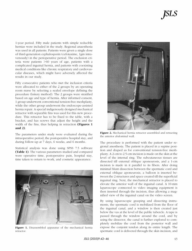
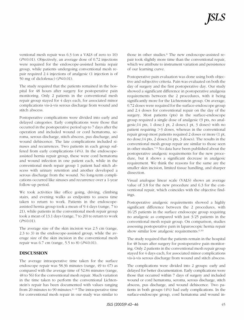
Fifty consecutive patients who met the inclusion criteriawere allocated to either of the 2 groups by an operatingroom nurse by selecting a sealed envelope defining theprocedure (lottery method). The 2 groups were stratifiedbased on age and type of hernia. After informed consent,1 group underwent conventional tension-free meshplasty,while the other group underwent the endoscope-assistedhernia repair. A special indigenously designed mechanicalretractor with separable fins was used for this new proce-dure. This retractor has to be fixed to the table, with abracket, and has screws that adjust the height and thewidth of the fins, thus helping in retraction (Figures 1and 2).
The parameters under study were evaluated during theintraoperative period, the postoperative hospital stay, andduring follow-up at 7 days, 6 weeks, and 6 months.
Statistical analysis was done using SPSS 7.5 software(Table 1). The various parameters studied and comparedwere operative time, postoperative pain, hospital stay,time taken to return to work, and cosmetic appearance.
The procedure is performed with the patient under re-gional anesthesia. The patient is placed in a supine posi-tion and draped as for conventional tension-free mesh-plasty. A 2-cm to 2.5-cm incision is made on the skin at thelevel of the internal ring. The subcutaneous tissues aredissected till external oblique aponeurosis, and a 1-cmincision is made in it parallel to its fibers. After doingminimal blunt dissection between the spermatic cord andexternal oblique aponeurosis, a balloon is inserted be-tween the 2 structures and space created till the superficialinguinal ring. Next, the mechanical retractor is placed toelevate the anterior wall of the inguinal canal. A 10-mmlaparoscope connected to video imaging equipment isthen inserted through the incision, thus allowing a mag-nified view of the inguinal canal on the video screen.
By using laparoscopic grasping and dissecting instru-ments, the spermatic cord is mobilized from the floor ofthe inguinal canal, and a window is then created justbelow the vas at the level of the public tubercle. A tape ispassed through the window around the cord, and byusing the dissector, the canal is further explored to com-pletely mobilize the cord from the posterior wall andexpose the conjoint tendon along its entire length. Thespermatic cord is delivered through the skin incision, and
Figure 1. Disassembled apparatus of the mechanical herniaretractor.
an incision is made in the spermatic fascia. The sac isidentified and dissected from the cord.
The retractors are then reinserted below the externaloblique aponeurosis and spermatic cord for providingexposure to the posterior wall and floor of the inguinalcanal. A sheet of monofilament polypropylene mesh mea-suring 3 in x 6 in (7.6 cm x 15 cm) is inserted into the canalunder laparoscopic guidance and anchored under directvision to public tubercle by endotacks, thus securing themesh medially. A slit is made in the mesh at the level ofinternal ring, which allows emergence of cord and creates2 tails that are crossed over and fixed to each other byendotacks, thereby creating a new internal ring. The ex-ternal oblique and skin are closed in layers.
RESULTS
The mean age of patients in our study was 43.5 years(range, 31 to 60). The mean age of patients in conven-
tional mesh repair was 43.44 years, while the mean age ofpatients in endoscope-assisted hernia repair was 43.56years.
Sixty percent of patients in our study had indirect hernias;the ratio of direct to indirect hernia was 2:3. Fifty-eightpercent of hernias were right sided.
The average time for conventional mesh repair was 52.84minutes (range, 48 to 56). The endoscope-assisted repairtook an average time of 58.36 minutes (range, 49 to 67)
The postoperative pain was measured by both subjectiveand objective parameters. Subjectively, the pain wasscored by each patient using a visual analogue scale ofzero to 10 where zero signified no pain and 10 maximumpain. Objective assessment of pain was done on zero andfirst postoperative days according to the amount of anal-gesic requirement. Average subjective pain score for en-doscope-assisted hernia repair was 3.8 and that of con-
Table 1.Mean Observations of Study Parameters and Their Statistical Significance
Endoscope-assisted Inguinal Hernia Repair, Chawla S et al.
JSLS (2005)9:42–4644
ventional mesh repair was 6.3 (on a VALS of zero to 10)(P�0.01). Objectively, an average dose of 0.72 injectionswere required for the endoscope-assisted hernia repairgroup, while patients undergoing conventional mesh re-pair required 2.4 injections of analgesic (1 injection is of50 mg of diclofenac) (P�0.01).
The study required that the patients remained in the hos-pital for 48 hours after surgery for postoperative painmonitoring. Only 2 patients in the conventional meshrepair group stayed for 4 days each, for associated minorcomplications vis-a-vis serous discharge from wound andstitch abscess.
Postoperative complications were divided into early anddelayed categories. Early complications were those thatoccurred in the postoperative period up to 7 days after theoperation and included wound or cord hematoma, se-roma, serous discharge, stitch abscess, pus discharge, andwound dehiscence. The late complications included si-nuses and recurrences. Two patients in each group suf-fered from early complications (4%). In the endoscope-assisted hernia repair group, these were cord hematomaand wound infection in one patient each, while in theconventional mesh repair group 1 patient had stitch ab-scess with urinary retention and another developed aserous discharge from the wound. No long-term compli-cations occurred like sinuses and recurrence over a 1-yearfollow-up period.
We took activities like office going, driving, climbingstairs, and evening walks as endpoints to assess timetaken to return to work. Patients in the endoscope-assisted hernia group took a mean of 9.4 days (range, 7 to21), while patients in the conventional mesh repair grouptook a mean of 13.1 days (range, 7 to 20) to return to work(P�0.01).
The average size of the skin incision was 2.5 cm (range,2.3 to 3) in the endoscope-assisted group, while the av-erage size of the skin incision in the conventional meshrepair was 6.7 cm (range, 5.5 to 8) (P�0.01).
DISCUSSION
The average intraoperative time taken for the surfaceendoscope repair was 58.36 minutes (range, 49 to 67) ascompared with the average time of 52.84 minutes (range,48 to 56) for the conventional mesh repair. Much variationin the time taken to perform the conventional Lichten-stein’s repair has been documented with values rangingfrom 20 minutes to 90 minutes.6–8 The intraoperative timefor conventional mesh repair in our study was similar to
those in other studies.6 The new endoscope-assisted re-pair took slightly more time than the conventional repair,which we attribute to instrument variation and persistenceof our learning curve.
Postoperative pain evaluation was done using both objec-tive and subjective criteria. Pain was evaluated on both theday of surgery and the first postoperative day. Our studyshowed a significant difference in postoperative analgesiarequirements between the 2 procedures, with it beingsignificantly more for the Lichtenstein group. On average,0.72 doses were required for the surface-endoscope groupand 2.4 doses for conventional repair on the day of thesurgery. Most patients (pts) in the surface-endoscopegroup required a single dose of analgesic (9 pts, no anal-gesic;14 pts, 1 dose;1 pt, 2 doses;1 pt, 3 doses) with nopatient requiring �3 doses, whereas in the conventionalrepair group most patients required 2 doses or more (1 pt,no dose;14 pts, 2 doses;14 pts, 3 doses). The results in theconventional mesh group repair are similar to those seenin other studies.7,9 No data have been published about thepostoperative analgesic requirements for the new proce-dure, but it shows a significant decrease in analgesicrequirement. We think the reasons for the same are thesmaller skin incision, limited tissue handling, and sharperdissection.
Visual analogue linear scale (VALS) shows an averagevalue of 3.8 for the new procedure and 6.3 for the con-ventional repair, which coincides with the objective find-ings.
Postoperative analgesic requirements showed a highlysignificant difference between the 2 procedures, with16/25 patients in the surface endoscope group requiringno analgesic as compared with just 3/25 patients in theconventional mesh repair group. On comparison, studiesassessing postoperative pain in laparoscopic hernia repairshow similar low analgesic requirements.6,10
The study required that the patients remain in the hospitalfor 48 hours after surgery for postoperative pain monitor-ing. Only 2 patients in the conventional mesh repair groupstayed for 4 days each, for associated minor complicationsvis-a-vis serous discharge from wound and stitch abscess.
The complications were divided into 2 groups: early anddelayed for better documentation. Early complications werethose that occurred within 7 days of surgery and includedwound or cord hematoma, seroma, serous discharge, stitchabscess, pus discharge, and wound dehiscence. Two pa-tients in both groups (4%) had early complications. In thesurface-endoscope group, cord hematoma and wound in-
JSLS (2005)9:42–46 45
fection were noted in 1 patient each. In the open herniagroup, 1 patient had serous discharge from the wound whileanother had stitch abscess with urinary retention. Serousdischarge represents exudates, most commonly resultingfrom trauma from the scalpel, scissors, cautery, and foreignbodies (sutures and prosthesis). Since the introduction of theprosthesis, the incidence of this complication has increasedfrom 0% to 17.6%,;11 our study has an incidence of 2%. In ourstudy, 1 patient in each group had infection-related compli-cations. This suggests that the procedure did not influencethe wound infection.
The late complications included sinuses and recurrences.No late complications were documented in the study. Thiswas perhaps because the study sample was small and thefollow-up was limited to less than a year; however, inlong-term studies using mesh, recurrences have beennoted in 0% to 1.7% patients.12
Studies assessing postoperative patient rehabilitation haveused various endpoints to measure return to work.13 Con-sidering our patient profile, we took time taken to resumenormal activities like office going, driving, climbing stairs,and evening walks as endpoints. The patients in the surface-endoscope group took a mean of 9.4 days to return to work,while patients in the conventional hernia group took 13.1days. This difference was highly significant. These results areremarkably similar to those published in other series, withsurface endoscope hernia repair results matching those oflaparoscopic hernia surgery, 10 days versus 14 days,9 7.5days versus 11.8 days,6 9 days versus 17 days.8
The cosmetic appearance was to be assessed indepen-dently by the patient, staff nurse, and doctor. Most of thepatients were satisfied by the surgery as they were inter-ested in relief of the symptoms and the inconvenience. Asa consensus, the new procedure was significantly bettergraded than the open procedure by both the staff nurseand the doctor, as in laparoscopic hernias.9
The new technique however is still in its infancy, andlong-term studies and follow-up are required for evaluat-ing recurrence.
CONCLUSION
Although the study was conducted with a limited numberof patients and a very short follow-up, it is worth consid-
ering this method over laparoscopic and conventionaltechniques, especially with reducible hernias.
2. Abrahamson J. Hernias. In: Zinner MJ, Schwartz SI, Ellis H,eds. Maingot’s Abdominal Operations. 10th ed. Stamford, CT:Appleton and Lange;1997:479–580.
3. Lichenstein IL, Shulman AG, Amid PK, et al. The tension freehernioplasty. Am J Surg. 1989;157:188–193.
4. Nduka C, Darzi A. Other methods of laparoendoscopic her-nia repair: mini hernia –inguinal hernia repair through a 2 cmincision. Semin Laparosc Surg. 1998;5(4):248–252.
6. Brooks DC. A prospective trial of laparoscopic and tensionfree inguinal herniorrhaphy. Arch Surg. 1994;129:361–366.
7. Millikan KW, Doolas A. A prospective comparison of trans-abdominal preperitoneal laparoscopic hernia repair vs. tradi-tional repair in a university setting. Surg Laparosc Endosc. 1994;44:247–253.
8. Payne JH Jr., Griniger LM, Izawa MT, et al. Laparoscopic oropen inguinal herniorrhaphy? A randomized prospective trial.Arch Surg. 1994;129:973–979.
10. Filipi CJ, Gaston-Johnson F, McBrie PJ, et al. An assessmentof pain and return to normal activity: laparoscopic herniorrha-phy vs. open tension free Lichtenstein repair. Surg Endosc.1996;10:983–986.