biomolecules Review Engineering Extracellular Vesicles as Nanotherapeutics for Regenerative Medicine Lalithasri Ramasubramanian 1,2 , Priyadarsini Kumar 1,3 and Aijun Wang 1,2,3, * 1 Surgical Bioengineering Laboratory, Department of Surgery, School of Medicine, University of California–Davis, Sacramento, CA 95817, USA; [email protected] (L.R.); [email protected] (P.K.) 2 Department of Biomedical Engineering, University of California–Davis, Davis, CA 95616, USA 3 Institute for Pediatric Regenerative Medicine, Shriners Hospitals for Children–Northern California, Sacramento, CA 95817, USA * Correspondence: [email protected]Received: 10 December 2019; Accepted: 26 December 2019; Published: 28 December 2019 Abstract: Long thought of to be vesicles that primarily recycled waste biomolecules from cells, extracellular vesicles (EVs) have now emerged as a new class of nanotherapeutics for regenerative medicine. Recent studies have proven their potential as mediators of cell proliferation, immunomodulation, extracellular matrix organization and angiogenesis, and are currently being used as treatments for a variety of diseases and injuries. They are now being used in combination with a variety of more traditional biomaterials and tissue engineering strategies to stimulate tissue repair and wound healing. However, the clinical translation of EVs has been greatly slowed due to difficulties in EV isolation and purification, as well as their limited yields and functional heterogeneity. Thus, a field of EV engineering has emerged in order to augment the natural properties of EVs and to recapitulate their function in semi-synthetic and synthetic EVs. Here, we have reviewed current technologies and techniques in this growing field of EV engineering while highlighting possible future applications for regenerative medicine. Keywords: extracellular vesicles; regenerative medicine; biomaterials; stem cells 1. Introduction Regenerative medicine has been a pivotal area of research aimed at healing or replacing damaged tissue. Traditional regenerative strategies have generally seen the use of stem cells and biomaterials as building materials to either replace the lost tissue or promote the regeneration of new tissue [1–3]. Recently, a new type of the therapeutic known as exosomes have emerged as a strategy for regenerative medicine [4–6]. Exosomes are nanovesicles about 50–150 nm in diameter that are released from almost every type of cell [4,5,7]. They are derived from membrane lipids of parent cells through the fusion of multivesicular bodies with the membrane [7,8]. Exosomes are released to mediate critical cell-to-cell communication by delivering cargo such as proteins, lipids and effector molecules to target cells. In fact, this paracrine cell signaling has been a key area of interest to researchers. Exosomes have been identified to play an important role in several major cell and tissue functions, including cell proliferation and senescence [9–11], angiogenesis [12–15], extracellular matrix support and reorganization [16–18] and immunomodulation [19–21]. Unsurprisingly, these properties have made exosomes a very attractive therapeutic option for regenerative medicine. 1.1. Exosome Biogenesis Membranous vesicles secreted by cells are collectively termed extracellular vesicles (EVs), of which there are three main subtypes: exosomes, microvesicles and apoptotic bodies [8]. These EVs are Biomolecules 2020, 10, 48; doi:10.3390/biom10010048 www.mdpi.com/journal/biomolecules
Transcript
biomolecules
Review
Engineering Extracellular Vesicles asNanotherapeutics for Regenerative Medicine
Lalithasri Ramasubramanian 1,2, Priyadarsini Kumar 1,3 and Aijun Wang 1,2,3,*1 Surgical Bioengineering Laboratory, Department of Surgery, School of Medicine, University of
California–Davis, Sacramento, CA 95817, USA; [email protected] (L.R.); [email protected] (P.K.)2 Department of Biomedical Engineering, University of California–Davis, Davis, CA 95616, USA3 Institute for Pediatric Regenerative Medicine, Shriners Hospitals for Children–Northern California,
Received: 10 December 2019; Accepted: 26 December 2019; Published: 28 December 2019 �����������������
Abstract: Long thought of to be vesicles that primarily recycled waste biomolecules fromcells, extracellular vesicles (EVs) have now emerged as a new class of nanotherapeutics forregenerative medicine. Recent studies have proven their potential as mediators of cell proliferation,immunomodulation, extracellular matrix organization and angiogenesis, and are currently beingused as treatments for a variety of diseases and injuries. They are now being used in combinationwith a variety of more traditional biomaterials and tissue engineering strategies to stimulate tissuerepair and wound healing. However, the clinical translation of EVs has been greatly slowed due todifficulties in EV isolation and purification, as well as their limited yields and functional heterogeneity.Thus, a field of EV engineering has emerged in order to augment the natural properties of EVs andto recapitulate their function in semi-synthetic and synthetic EVs. Here, we have reviewed currenttechnologies and techniques in this growing field of EV engineering while highlighting possiblefuture applications for regenerative medicine.
Regenerative medicine has been a pivotal area of research aimed at healing or replacing damagedtissue. Traditional regenerative strategies have generally seen the use of stem cells and biomaterialsas building materials to either replace the lost tissue or promote the regeneration of new tissue [1–3].Recently, a new type of the therapeutic known as exosomes have emerged as a strategy for regenerativemedicine [4–6]. Exosomes are nanovesicles about 50–150 nm in diameter that are released from almostevery type of cell [4,5,7]. They are derived from membrane lipids of parent cells through the fusion ofmultivesicular bodies with the membrane [7,8]. Exosomes are released to mediate critical cell-to-cellcommunication by delivering cargo such as proteins, lipids and effector molecules to target cells. In fact,this paracrine cell signaling has been a key area of interest to researchers. Exosomes have been identifiedto play an important role in several major cell and tissue functions, including cell proliferation andsenescence [9–11], angiogenesis [12–15], extracellular matrix support and reorganization [16–18] andimmunomodulation [19–21]. Unsurprisingly, these properties have made exosomes a very attractivetherapeutic option for regenerative medicine.
1.1. Exosome Biogenesis
Membranous vesicles secreted by cells are collectively termed extracellular vesicles (EVs), of whichthere are three main subtypes: exosomes, microvesicles and apoptotic bodies [8]. These EVs are
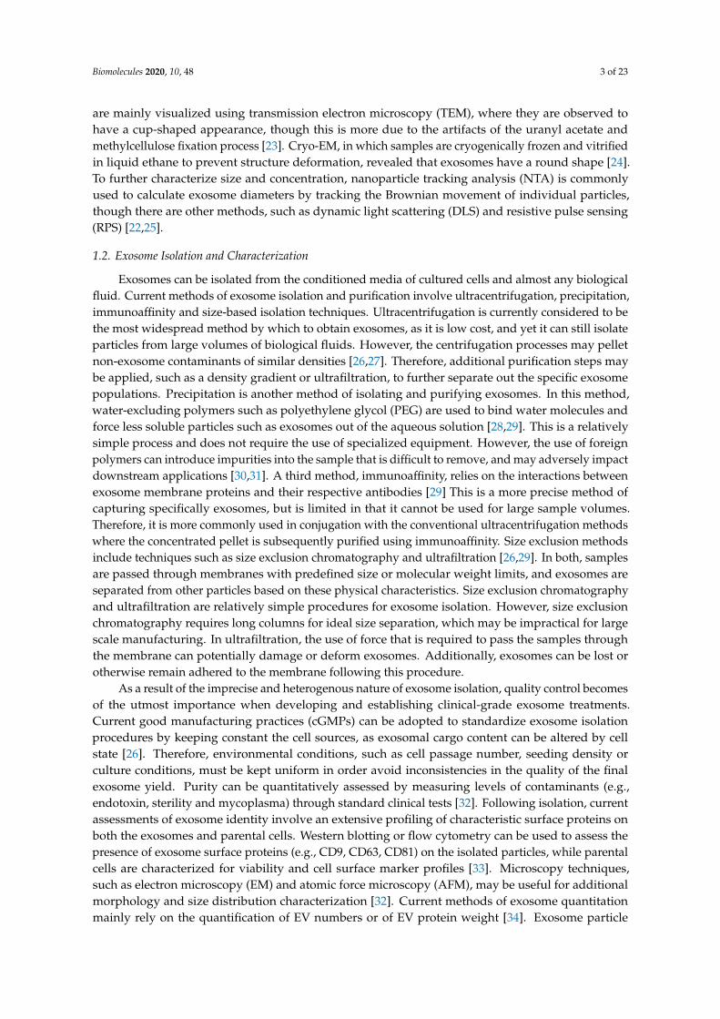
secreted by most cell types and are ubiquitous in all types of biological fluids, including blood, urine,amniotic fluid, saliva and cerebrospinal fluid [22,23]. Exosomes are the smallest type of EVs (50–150 nm)and, unlike the microvesicles and apoptotic bodies which are directly shed from the plasma membrane,exosomes are released following the fusion of late endosomes and multivesicular bodies with theplasma membrane [22]. Exosome release follows a highly dynamic endocytic pathway (Figure 1).The first step involves the accumulation of intraluminal vesicles (ILVs) as early endosomes mature intolate endosomes. These ILVs sort and entrap proteins, lipids and cytosol within these late endosomes,leading to morphological changes that result in multivesicular bodies (MVBs) [7,8,23]. Though in mostcases, MVBs fuse with lysosomes for the degradation and recycling of their contents, certain MVBsare decorated with specific proteins and markers that instead ensures their fusion with the plasmamembrane and allows the release of their content to the extracellular space and become known asexosomes. This sorting is facilitated by the endosomal sorting complex required for transport (ESCRT),a mechanism which involves about 30 different proteins that help sequester specific biomolecules inthe MVBs and guide their release through the plasma membrane as exosomes [8,22,23].
Biomolecules 2019, 9, x FOR PEER REVIEW 2 of 24
1.1. Exosome Biogenesis
Membranous vesicles secreted by cells are collectively termed extracellular vesicles (EVs), of which there are three main subtypes: exosomes, microvesicles and apoptotic bodies [8]. These EVs are secreted by most cell types and are ubiquitous in all types of biological fluids, including blood, urine, amniotic fluid, saliva and cerebrospinal fluid [22,23]. Exosomes are the smallest type of EVs (50–150 nm) and, unlike the microvesicles and apoptotic bodies which are directly shed from the plasma membrane, exosomes are released following the fusion of late endosomes and multivesicular bodies with the plasma membrane [22]. Exosome release follows a highly dynamic endocytic pathway (Figure 1). The first step involves the accumulation of intraluminal vesicles (ILVs) as early endosomes mature into late endosomes. These ILVs sort and entrap proteins, lipids and cytosol within these late endosomes, leading to morphological changes that result in multivesicular bodies (MVBs) [7,8,23]. Though in most cases, MVBs fuse with lysosomes for the degradation and recycling of their contents, certain MVBs are decorated with specific proteins and markers that instead ensures their fusion with the plasma membrane and allows the release of their content to the extracellular space and become known as exosomes. This sorting is facilitated by the endosomal sorting complex required for transport (ESCRT), a mechanism which involves about 30 different proteins that help sequester specific biomolecules in the MVBs and guide their release through the plasma membrane as exosomes [8,22,23].
Figure 1. Biogenesis of extracellular vesicles. Microvesicles and apoptotic bodies originate directly from the plasma membrane, while exosomes are derived from the endosomal compartments. intraluminal vesicles (ILVs) accumulate in the multivesicular bodies (MVBs) after early endosome maturation. Proteins, lipids, nucleic acids and other cargo are sequestered within the ILVs through an endosomal sorting complex required for transport (ESCRT)-dependent pathway. Eventually, MVBs fuse with the plasma membrane and release the ILVs into the extracellular space as exosomes.
Figure 1. Biogenesis of extracellular vesicles. Microvesicles and apoptotic bodies originate directly fromthe plasma membrane, while exosomes are derived from the endosomal compartments. intraluminalvesicles (ILVs) accumulate in the multivesicular bodies (MVBs) after early endosome maturation.Proteins, lipids, nucleic acids and other cargo are sequestered within the ILVs through an endosomalsorting complex required for transport (ESCRT)-dependent pathway. Eventually, MVBs fuse with theplasma membrane and release the ILVs into the extracellular space as exosomes.
Following release, exosomes are mainly distinguished by the presence of several specific surfacemarkers, namely tetraspanins (CD63, CD9, CD81), which are involved in the sorting of different cargo,the tumor susceptibility gene 101 (TSG) 101 and the apoptosis-linked-gene-2 interacting protein X(ALIX) (proteins associated in ESCRT) [23]. They also express parent cell-specific surface markers asa result of the fusion with the cell plasma membrane [23]. Due to their sub-150nm size, exosomes
Biomolecules 2020, 10, 48 3 of 23
are mainly visualized using transmission electron microscopy (TEM), where they are observed tohave a cup-shaped appearance, though this is more due to the artifacts of the uranyl acetate andmethylcellulose fixation process [23]. Cryo-EM, in which samples are cryogenically frozen and vitrifiedin liquid ethane to prevent structure deformation, revealed that exosomes have a round shape [24].To further characterize size and concentration, nanoparticle tracking analysis (NTA) is commonlyused to calculate exosome diameters by tracking the Brownian movement of individual particles,though there are other methods, such as dynamic light scattering (DLS) and resistive pulse sensing(RPS) [22,25].
1.2. Exosome Isolation and Characterization
Exosomes can be isolated from the conditioned media of cultured cells and almost any biologicalfluid. Current methods of exosome isolation and purification involve ultracentrifugation, precipitation,immunoaffinity and size-based isolation techniques. Ultracentrifugation is currently considered to bethe most widespread method by which to obtain exosomes, as it is low cost, and yet it can still isolateparticles from large volumes of biological fluids. However, the centrifugation processes may pelletnon-exosome contaminants of similar densities [26,27]. Therefore, additional purification steps maybe applied, such as a density gradient or ultrafiltration, to further separate out the specific exosomepopulations. Precipitation is another method of isolating and purifying exosomes. In this method,water-excluding polymers such as polyethylene glycol (PEG) are used to bind water molecules andforce less soluble particles such as exosomes out of the aqueous solution [28,29]. This is a relativelysimple process and does not require the use of specialized equipment. However, the use of foreignpolymers can introduce impurities into the sample that is difficult to remove, and may adversely impactdownstream applications [30,31]. A third method, immunoaffinity, relies on the interactions betweenexosome membrane proteins and their respective antibodies [29] This is a more precise method ofcapturing specifically exosomes, but is limited in that it cannot be used for large sample volumes.Therefore, it is more commonly used in conjugation with the conventional ultracentrifugation methodswhere the concentrated pellet is subsequently purified using immunoaffinity. Size exclusion methodsinclude techniques such as size exclusion chromatography and ultrafiltration [26,29]. In both, samplesare passed through membranes with predefined size or molecular weight limits, and exosomes areseparated from other particles based on these physical characteristics. Size exclusion chromatographyand ultrafiltration are relatively simple procedures for exosome isolation. However, size exclusionchromatography requires long columns for ideal size separation, which may be impractical for largescale manufacturing. In ultrafiltration, the use of force that is required to pass the samples throughthe membrane can potentially damage or deform exosomes. Additionally, exosomes can be lost orotherwise remain adhered to the membrane following this procedure.
As a result of the imprecise and heterogenous nature of exosome isolation, quality control becomesof the utmost importance when developing and establishing clinical-grade exosome treatments.Current good manufacturing practices (cGMPs) can be adopted to standardize exosome isolationprocedures by keeping constant the cell sources, as exosomal cargo content can be altered by cellstate [26]. Therefore, environmental conditions, such as cell passage number, seeding density orculture conditions, must be kept uniform in order avoid inconsistencies in the quality of the finalexosome yield. Purity can be quantitatively assessed by measuring levels of contaminants (e.g.,endotoxin, sterility and mycoplasma) through standard clinical tests [32]. Following isolation, currentassessments of exosome identity involve an extensive profiling of characteristic surface proteins onboth the exosomes and parental cells. Western blotting or flow cytometry can be used to assess thepresence of exosome surface proteins (e.g., CD9, CD63, CD81) on the isolated particles, while parentalcells are characterized for viability and cell surface marker profiles [33]. Microscopy techniques,such as electron microscopy (EM) and atomic force microscopy (AFM), may be useful for additionalmorphology and size distribution characterization [32]. Current methods of exosome quantitationmainly rely on the quantification of EV numbers or of EV protein weight [34]. Exosome particle
Biomolecules 2020, 10, 48 4 of 23
numbers have been commonly calculated through the NTA or other similar concentration measuringinstruments and technologies. Exosome total protein content has also been used as a parameter forquantification [32,34]. These quantitative measurements are often used when calculating dosages.Most approaches currently involve administering EVs that are cell equivalent in number (compared toa control treatment group consisting of cells only), as calculated from concentration measurements [31].Doses can also be administered based on total protein content, though this may be a less than accuratemethod due to the variation in protein content between individual exosomes [31,35]. Exact clinicaldoses are dependent on the disease at hand as well as the potency of the exosomes. To assess potency,functional assays can be conducted as a downstream validation process. Otherwise, proteomic ortranscriptomic studies can be conducted to directly assess exosomal contents as a way to standardizethe bioactive cargo between different batches [32].
1.3. Physiological Functions of Exosomes and Implications for Regenerative Medicine
Exosomes were initially thought to be the “trash bags” of the cells, as it was believed that theirprimary function is to carry waste molecules out of the cells. However, it was soon realized thatexosomes carry many important biomolecules, including proteins, nucleic acids and lipids to recipientcells as a way to mediate cell-to-cell communication [6,23,36]. Exosome targeting is still an unclearmechanism, as it is a highly complex process that is dependent upon exosome origin and recipient celltype. Nevertheless, the general mode of targeting relies on receptor-ligand interactions between thesurfaces of the target cell and the exosome, respectively. Different types of molecules are involved inthese targeting interactions, such as integrins, lipids, lectins, proteoglycans and extracellular matrixcomponents [7,23,24]. Once the exosome is bound to the cell surface, it can affect cell function by either(1) remaining on the cell surface and triggering intracellular signaling pathways or (2) be internalizedby the cell through endocytosis, of which both clathrin- and caveolae-dependent and -independentmechanisms have been implicated [24,37,38]. In both cases, exosome contents can be released viafusion and carry out their specific effector functions.
Interestingly, exosome functional properties and the mechanisms by which they mediate cellularprocesses are also heavily dependent on their cellular origin [5,36,39]. Exosomes derived from differentcell types are seen to transfer specific tissue-remodeling molecules and mediate different metabolicpathways to promote the healing and regeneration of varying injured tissue. Mesenchymal stromalcells (MSCs)-derived exosomes, for example, have been shown to have remarkable effects on promotingregeneration [40,41]. Placenta-derived MSCs have been shown to secrete exosomes that play a keyrole in neuroprotection and immunomodulation [42,43], while exosomes from bone marrow-derivedMSCs have been implicated in aiding in bone regeneration [44]. Meanwhile, exosomes derivedfrom endothelial progenitor cells (EPCs) have been shown to deliver microRNAs to promote potentangiogenic effects to induce the vascularization of damaged tissue [12–14]. As potent nanotherapeutics,exosomes have the potential to be used in different areas of regenerative medicine, and can be combinedwith novel engineering strategies for improved functionality and efficiency. However, because ofthe difficulty in obtaining pure exosome fractions and to consider the possible presence of othervesicular structures, exosomes will henceforth be referred to broadly as extracellular vesicles or EVsin upcoming discussions. This review will focus on the current use of EVs in various regenerativetherapies and highlight emerging technologies aimed at augmenting their natural properties withinnovative biomedical strategies and systems.
2. Biological Stem Cell Derived EVs
2.1. Injectable Treatments
The use of EVs is advantageous over cells themselves for multiple reasons. First, cell therapies oftensuffer from poor post-transplantation cell viability due to the shearing effects when the cells are ejectedfrom a syringe needle [45], and as EVs are not constrained by viability, there is no danger of “death”.
Biomolecules 2020, 10, 48 5 of 23
Additionally, cell therapies come with a risk of developing tumors or malignant transformationsshould the transplanted cells become cancerous. EV treatments avoid this disadvantage, as they donot replicate, and instead are recycled after their cargo is delivered. Unwanted host immunogenicresponses are additional major hinderances for current cell therapies due to the expression of foreignmajor histocompatibility complex (MHC) markers that activate host immune responses and mayeventually lead to transplant rejection. EVs, however, have immunomodulatory properties that reducethe risk of adverse immune responses while still maintaining regenerative functional properties [46].EVs also have no need for engraftment, another necessity that has long hindered the effectiveness ofprevious cell therapies.
Due to their paracrine mechanism of action, EVs have seen great success as injectable therapeuticsfor neuroprotection, cardioprotection and renoprotection. In a swine model of traumatic brain injuryand hemorrhagic shock, animals that received bone marrow MSC-derived EVs via intravenous injectionshowed significantly lower neurologic impairment and had a faster full neurological recovery tobaseline functions [47]. Similarly, intravenous administration of EVs from human embryonic stemcell-derived neural stem cells improved the functional and physical outcomes after thromboembolicstroke in middle-aged mice [48]. Mice that received EV treatment had greater improvements in motorfunctions and episodic memory while MRI analysis showed significantly less tissue loss. In a parallelporcine model of ischemic stroke, the same neural stem cell-derived EVs showed that intravenousadministration of EVs led to significant tissue and functional improvements compared to sham controlanimals [49]. Pigs that received treatment exhibited normal exploratory behavior and motor activity,suggesting faster recovery, while long term analysis showed a reduced loss of cerebral tissue, decreasedbrain swelling, and preserved white matter integrity.
The functional properties of EVs are believed to be mainly propagated by their cargo, such asmicroRNAs. MicroRNAs, or miRNAs, are short strands of noncoding RNA that play crucial rolesin mediating gene expression in target cells. EVs carrying specific miRNAs have been preferentiallyselected for and applied as regenerative therapies. EVs derived from cardiac progenitor cells (CPCs)are highly enriched in miR-210, miR-132 and miR-146a-3p [50]. When injected into infarcted hearts in arat model, CPC EVs reduce cardiomyocyte apoptosis and enable vascular remodeling, thus exhibitingsignificant cardioprotective properties through the upregulation or downregulation of miRNA-specifictarget genes. Proangiogenic properties of MSC-derived EVs are also similarly mediated throughthe transfer of miRNAs, specifically miR-30b, miR-miR-30c, miR-424 and miR-let-7f, to endothelialcells [15]. Neural regeneration is also seen with the MSC-mediated transfer of miRNA-133b, whichregulates neurite outgrowth by interacting with parenchymal cells [51]. Similarly, the delivery ofmiR-125b by EVs from differentiating neuronal cells could induce the differentiation of human MSCsinto neurons [52]. This regulation could have significant regenerative impact in the central nervoussystem, especially in the aftermath of degenerative or otherwise damaging neurological diseases.
2.2. Engineered Scaffolds and Surfaces
Tissue-engineered constructs (i.e., scaffolds) have had great success in recreating structural andfunctional microenvironments to allow for the regeneration of tissues [53]. As a result, many studieshave focused on the use of EVs in combination with engineered scaffolds as a method to improvehealing and regeneration. To date, MSC-derived EVs have been widely used to functionalize a varietyof scaffold structures due to their proangiogenic properties. Xie et al. coated decalcified bone matrix(DBM) with bone marrow-derived MSC EVs using fibronectin as the immobilizing agent [54].
The implanted EV-functionalized scaffolds promoted bone regeneration in a murine model.Significant new bone matrix was observed with twice the average number of CD31-positive vesselswithin the matrix compared to unmodified scaffolds, confirming the ability of the EVs to promotebone formation and vascularization. EVs can be immobilized on a variety of polymer-based scaffolds,as another study functionalized β-TCP scaffolds with human-induced pluripotent stem cell-derivedMSC (hIPS-MSC) EVs [55]. No additional reagent was required to immobilize the EVs on the scaffold;
Biomolecules 2020, 10, 48 6 of 23
instead, the EVs were simply added to the constructs and were naturally absorbed into the scaffoldafter incubation. The hIPS-MSC EV-modified scaffolds stimulated osteogenesis in a rat model of acritical-sized calvarial bone defect. The MSC-derived EVs promote osteogenic activity, in that theyupregulate the PI3K/Akt signaling pathway by altering over 117 genes. Though MSC-derived EVs havebeen primarily used due to the natural paracrine signaling that characterize MSC cell function, EVs fromother stem cell sources have seen similar success when combined with scaffolds. Poly(lactic-co-glycolicacid) (PLGA/pDA) scaffolds were modified with human adipose-derived stem cell (hASC) EVs usinga polydopamine coating to immobilize them on the scaffold [56]. In vivo studies using a murinemodel of critical-sized calvarial bone defects showed significantly greater bone tissue and maturecollagen formation in hASC EV-modified grafts compared to the unmodified grafts. The healing waspartially attributed to the ability of the hASC-EVs to recruit host MSCs, which in turn, promotedosteoinductive functions.
Aside from scaffolds, EVs have also been combined with other therapeutic surfaces for woundhealing and tissue regeneration. EV-modified sponges and patches have shown to be a promisingmethod of treatment. Shi et al. successfully loaded gingival MSCs (GMSCs), isolated from the gingivallamina propria, onto a chitosan/silk hydrogel sponge as a strategy to promote wound healing indiabetic patients [57]. Much like with scaffolds, EVs could be easily resuspended in phosphate bufferedsaline (PBS) and added to the hydrogel sponge. The EV-modified sponges were then used as dressingsto cover surgically-induced skin wounds in diabetic mice. Quantification of wound size showed thatthe EV-modified sponges had a significantly greater wound closing effect while immunohistochemicalanalysis showed higher levels CD34-positive cells, indicating angiogenesis, and greater collagendeposition. EV-modified hydrogel patches have also emerged as a therapeutic for cardiac recoveryand regeneration. EVs secreted from induced pluripotent stem cell-derived cardiomyocytes wereencapsulated in hydrogel patches [58]. Secreted EVs were enriched in cardiac-specific miRNAs, andwhen encapsulated in a collagen gelfoam mesh, were sustainably released over 21 days in vitro.Application to rat myocardium revealed similar release profiles and found that EVs promotedarrhythmic recovery, decreased cardiomyocyte apoptosis after infarction, and reduced infarct size andcell hypertrophy. EVs have also been successfully used to modify scaffold-free surfaces to be treatedonto monolayer cell sheets. CXCR4-enriched bone marrow–MSC EVs were used to treat MSC cellsheets, which were subsequently implanted into the infarcted area of a rat myocardium [59]. The EVsdemonstrated cardioprotective properties in an ischemic injury rat model by upregulating the Aktsignaling pathway.
3. Semi-Synthetic EVs
With the current surge in EV research and new findings supporting their importance in regenerativemedicine, researchers have turned their attention to engineering EVs in order to augment their innateproperties to allow for increased therapeutic function.
3.1. Drug Delivery
The current gold standard in drug delivery is that of liposomes, synthetic lipid vesicles, which, dueto their amphiphilic nature, can encapsulate both hydrophobic and hydrophilic molecules. They arehighly biocompatible and can be readily modified for precision medicine, making them a highlypopular drug delivery system in current medicine. However, liposomes are quickly cleared from thereticuloendothelial system (RES), making targeted delivery and prolonged treatment difficult.
Therefore, considering the nanometer-range size, biocompatibility and their natural targetingspecificity, EVs have unsurprisingly become a new class of natural nanotherapeutics. The differentcargo and cargo loading mechanisms are summarized in Table 1.
Biomolecules 2020, 10, 48 7 of 23
3.1.1. Small Molecules
Initial studies focused on the use of EVs, mostly derived from mature cell types, as traditionaldrug delivery vehicles where drugs or other synthetic therapeutic agents are the cargo of choice.These therapies have met with wide success, as small molecule-loaded EVs have been seen tohave great potential in treating inflammation and proinflammatory diseases [60,61], cancer [62–64],neurodegenerative diseases [61,65] and other diseases with relevant pathologies. Research has yetto turn to loading small molecules in stem cell-derived EVs, specifically for tissue engineering andregenerative medicine, but several studies have nonetheless demonstrated the possible appeal andfeasibility of using stem cell-derived EVs as a drug delivery vehicle. Much of small molecule loadinghas used passive methods of encapsulation, with active loading methods being less common techniques.Cell preconditioning or priming, for example, has been an effective form of loading drugs withinEVs. MSC-derived EVs, in particular, have been used in cancer therapy due to the ability of MSCs topreferentially target tumor sites. Paclitaxel, a popular and effective anticancer drug, can be uptakenin significant amounts by bone-marrow MSCs in culture and then be subsequently released withinEVs, as a form of passive encapsulation [64,66]. Isolated EVs collected from the primed MSCs showedsignificant anti-proliferative effects on a human pancreatic cell line, suggesting successful encapsulationof paclitaxel. The ability of cells to uptake and release small molecule drugs via EVs is not only uniqueto bone-marrow MSCs, but are also found in other stromal cells isolated from adipose tissue [67] andthe derma [68]. Incubation and simple mixing have been shown to be another successful methodby which small molecules have been loaded into EVs. EL-4-derived EVs were mixed with curcuminat 22 ◦C, with a loading efficiency of 2.9 g of curcumin to 1 g of EVs [60]. The anti-inflammatoryproperties of the curcumin-EVs were assessed in a septic shock murine model, where the curcumin-EVssignificantly reduced levels of IL-6 and TNF-α and increased mice survival.
3.1.2. Nucleic Acids
Throughout the field, the loading of nuclei acids, such as mRNAs, microRNAs (miRNAs) andsmall interfering RNA (siRNA), within EVs for tissue regeneration has been heavily favored, possiblydue to their natural presence as EV cargo. Though much of current EV engineering research forthe active encapsulation of nucleic acids has been mainly applied for cancer therapies, these studieshave nonetheless set a precedent for the similar use of engineered EVs for regenerative medicine.B-cell-derived EVs were loaded with exogenous miRNA-155 inhibitors and delivered to targetmacrophages as a gene therapy strategy to modulate inflammatory activation [69]. Functional miR-155mimics were electroporated into the EVs and delivered intravenously to miR-155 knockout mice, wherethere was found to be a statistically significant increase in miR-155 levels in primary hepatocytes inthe liver. These miR-155 inhibitors were similarly electroporated into EVs and cultured in vitro withRAW 264.7 macrophages. After stimulation with lipopolysaccharide, levels of TNF-α, a potent markerof inflammation, were significantly diminished as well as the expression of endogenous miRNA-155.SOCS1 mRNA, a cytokine suppressor gene, was significantly increased, indicating the ability of theengineered EVs to regulate expression of anti-inflammatory genes. EVs can also be engineered fornucleic acid delivery at a cellular level using preconditioning or exogenous transfection to induce theenrichment of natural miRNAs in EVs. Cell preconditioning has also been validated as a method ofenriching key miRNAs. Cardiac progenitor cells were stress-treated via H2O2 incubation to producemiR-21 enriched EVs that were used to protect cardiomyocytes from damage in ischemic conditions [70].Hypoxic preconditioning of cells has also shown to be a successful method of engineering miRNAexpression in EVs. Next generation sequencing revealed that EVs originating from human neural stemcells cultured under hypoxic conditions have increased levels of 53 miRNAs and decreased levels of26 miRNAs [71].
Microarray analysis of EVs secreted by hypoxic cardiac progenitor cells showed that 11 miRNAswere upregulated at least 2-fold compared to EVs secreted from normoxic cells [72]. However,preconditioning offers limited control in the specific types of miRNA that is enriched, as only
Biomolecules 2020, 10, 48 8 of 23
naturally-occurring cargo can be overexpressed. Additionally, standardization remains a problem,as different conditions can lead to different types and levels of cargo enrichment. Transfection ortransduction, though still an indirect method of cargo loading compared to electroporation or otheractive methods for exogenous cargo loading, offers more control than preconditioning. Umbilicalcord MSCs were transfected with miR-675 mimic, leading to the secretion of EVs that showedincreased expression of miR-675 [73]. When tested in a murine model for aging-induced vasculardysfunction, the miR-675-enriched EVs, delivered in silk fibroin hydrogels, successfully promotedblood perfusion in ischemic hindlimbs and reduced the expression of proinflammatory molecules.Similarly, adipose tissue-derived MSCs were transfected with plasmids expressing miR-122, thusproducing EVs enriched in miR122 that were then used successfully as a chemotherapeutic agentfor hepatocellular carcinoma [74]. Lentiviral transduction has also been successful in geneticallyengineering MSCs to overexpress miR-let7c and secrete EVs enriched in miR-let7c [75]. These enrichedEVs were seen to mediate the improved transfer of miR-let7c to target the kidneys and attenuaterenal fibrosis.
siRNAs are another class of small RNAs which have been exogenously loaded into EVs.siRNA loading is similar to that of miRNA loading, with electroporation being the most commonmethod of active loading [76]. But unlike miRNAs, siRNAs have also been loaded via conjugation ofsiRNA molecules to cholesterol tags. In a study conducted by Stremersch et al., siRNA was conjugatedto cholesterol anchors (chol-siRNA), which enabled the self-insertion into the lipid outer vesicularmembrane of B17F10 melanoma- and JAWSII monocyte-derived EVs [77]. Binding of the siRNA tothe EV outer membrane was successful, with about 73 chol-siRNA molecules per vesicle, and wasable to be successfully trafficked to target cells. However, subsequent functional studies revealed thatthere was no significant knockdown effect of target gene expression even though chol-siRNA onlywithout the nanocarriers did demonstrate a knockdown effect. The functional impairment may be dueto the immobilization of the siRNA to the EV surface, which may hinder siRNA activation of RNAimechanisms. However, O’Loughlin et al. showed that the cholesterol-siRNA (cc-siRNA) could beinduced to be loaded within the vesicle under optimal parameters (15 molecules of cc-siRNA per EV at37 ◦C for 1 h in 100 µL) [78]. Incubating at higher temperature allows for greater membrane fluidity,thus allowing for the greater interaction between EVs and the cc-siRNA, and ultimately leading togreater internalization and encapsulation. This temperature dependency may explain why Stremerschet al. observed chol-siRNA conjugation on the membrane surface and no functional effects afterincubation at room temperature.
3.1.3. Proteins
The loading of exogenous proteins into EVs has been less studied compared to its nucleic acidand small molecule counterparts, but is nevertheless a viable area of study. EVs are known to shuttle avariety of functional proteins to target cells as part of their mechanism of action. Proteins can be loadedin EVs using a WW domain tag, which is known to bind Ndfip1, an ubiquitin ligase adaptor protein,that has been confirmed to be exported from the cell in EVs [79]. Therefore, by modifying a protein ofinterest, in this case a recombinase protein known as Cre, with the WW tag, the protein can be boundto the Ndfip1, which in turn can guide the modified target protein into EVs. This EV engineeringwas indirect, as the parent cell was modified rather than the EVs directly. A plasmid was coded forthe WW–Cre fusion protein and transfected into LN18 and JEK293T cells [80]. Loading into EVs wasconfirmed in the presence of Ndfip1 using a WW-mCherry fluorescent reporter, while no loadingoccurred without Ndfip1. Targeting of the plasma membrane anchors has been similarly provento be a strategy for preferentially sorting proteins into EVs before vesicle budding. Engineering amyristoylation tag or targeting phosphatidylinositol-(4,5)-bisphosphate (PIP2), all showed highlysuccessful budding of a model TyA-GFP protein in EVs [81]. Using a prenylation/palmitoylation tag ortargeting the CD43 or phosphatidylinositol-(3,4,5)-trisphosphate-binding domain were less efficient infacilitating TyA-GFP budding, but nevertheless induced some level of budding [81].
Biomolecules 2020, 10, 48 9 of 23
Haney et al. demonstrated more direct ways of loading proteins into EVs using a variety ofmethods: coincubation, chemical permeabilization of the membrane, freeze –thaw cycles, sonicationand extrusion [65]. Using catalase as the loading protein, extrusion and sonication proved to be themost efficient loading techniques, followed by freeze–thaw cycles, and then simple incubation at roomtemperature. However, the loading efficiency for even the most successful sonication and extrusiontechniques was about 26% and 22%, respectively, still a relatively low loading capacity. Nevertheless,the catalase function was importantly conserved following EV loading, shown by neuroprotectiveeffects in the in vitro and in vivo murine models of Parkinson’s disease. In comparison, free catalasewas deactivated and lost its enzymatic functions in the same media conditions.
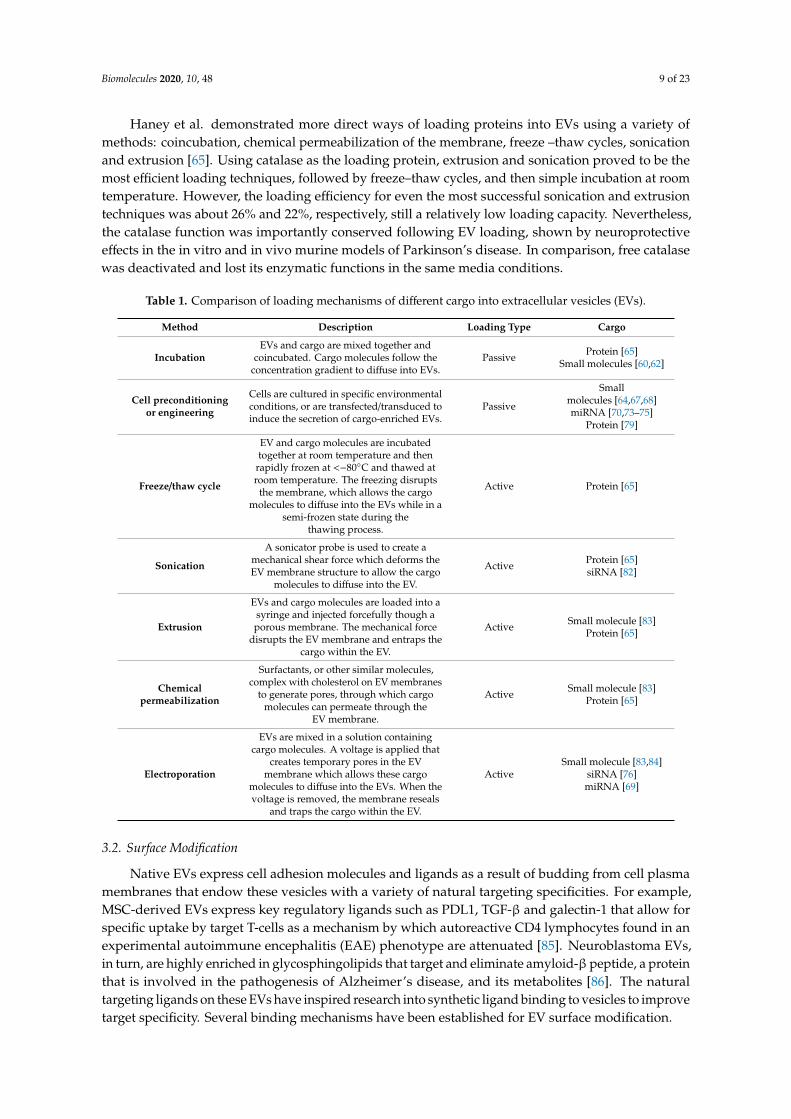
Table 1. Comparison of loading mechanisms of different cargo into extracellular vesicles (EVs).
Method Description Loading Type Cargo
IncubationEVs and cargo are mixed together and
coincubated. Cargo molecules follow theconcentration gradient to diffuse into EVs.
Passive Protein [65]Small molecules [60,62]
Cell preconditioningor engineering
Cells are cultured in specific environmentalconditions, or are transfected/transduced toinduce the secretion of cargo-enriched EVs.
Passive
Smallmolecules [64,67,68]miRNA [70,73–75]
Protein [79]
Freeze/thaw cycle
EV and cargo molecules are incubatedtogether at room temperature and then
rapidly frozen at <−80◦C and thawed atroom temperature. The freezing disrupts
the membrane, which allows the cargomolecules to diffuse into the EVs while in a
semi-frozen state during thethawing process.
Active Protein [65]
Sonication
A sonicator probe is used to create amechanical shear force which deforms theEV membrane structure to allow the cargo
molecules to diffuse into the EV.
Active Protein [65]siRNA [82]
Extrusion
EVs and cargo molecules are loaded into asyringe and injected forcefully though aporous membrane. The mechanical force
disrupts the EV membrane and entraps thecargo within the EV.
Active Small molecule [83]Protein [65]
Chemicalpermeabilization
Surfactants, or other similar molecules,complex with cholesterol on EV membranes
to generate pores, through which cargomolecules can permeate through the
EV membrane.
Active Small molecule [83]Protein [65]
Electroporation
EVs are mixed in a solution containingcargo molecules. A voltage is applied that
creates temporary pores in the EVmembrane which allows these cargo
molecules to diffuse into the EVs. When thevoltage is removed, the membrane reseals
and traps the cargo within the EV.
ActiveSmall molecule [83,84]
siRNA [76]miRNA [69]
3.2. Surface Modification
Native EVs express cell adhesion molecules and ligands as a result of budding from cell plasmamembranes that endow these vesicles with a variety of natural targeting specificities. For example,MSC-derived EVs express key regulatory ligands such as PDL1, TGF-β and galectin-1 that allow forspecific uptake by target T-cells as a mechanism by which autoreactive CD4 lymphocytes found in anexperimental autoimmune encephalitis (EAE) phenotype are attenuated [85]. Neuroblastoma EVs,in turn, are highly enriched in glycosphingolipids that target and eliminate amyloid-β peptide, a proteinthat is involved in the pathogenesis of Alzheimer’s disease, and its metabolites [86]. The naturaltargeting ligands on these EVs have inspired research into synthetic ligand binding to vesicles to improvetarget specificity. Several binding mechanisms have been established for EV surface modification.
Biomolecules 2020, 10, 48 10 of 23
3.2.1. Click Chemistry
Click chemistry, also known as an azide alkyne cycloaddition, is a method by which moleculescan be directly attached to EV surfaces via covalent bonds [87,88]. In this reaction, an alkyne moiety isreacted with an azide group to form a stable triazole linkage. This reaction is greatly accelerated withthe use of a copper catalyst [87,89], but several studies have also demonstrated successful binding usingcopper-free click chemistry [90]. Click chemistry has emerged as a popular method of bioconjugationdue to its relatively mild experimental conditions which generally involve a variety of solvents suchas water, alcohols and dimethyl sulfoxide (DMSO), as well as its high yield and simplicity [88].Additionally, the azide functional group is absent in natural biomacromolecules, thus eliminatingthe possibility of off-target binding [88]. Use of Click chemistry on EVs was validated in a study bySmyth et al., who reported that an azide-flour 545 could be successfully conjugated onto EV surfacesusing Click chemistry [91]. EVs were first modified with alkynes using n-hydroxysuccinimide (NHS)and pentynoic acid and underwent Click chemistry when reacted with azide-fluor 545 in the presenceof copper and salts. The triazole linkage that resulted finally facilitates the azide-fluor 545 bindingto the EV surface. This conjugation was found to have no impact on EV size and did not hinder orotherwise negatively impact target cell uptake. However, the critical alkyne modification of the EVsurface most likely occurs on EV proteins rather than the amines on the membrane phospholipids.As such, this introduces the possibility that the EV protein function may be partially or fully inhibitedby this modification. Nevertheless, the successful binding introduces a proof-of-concept that allowsfor functional peptide binding to EV surfaces.
This possibility was further realized when Jia et al. used Click chemistry to conjugatethe neuropilin-1 (NRP-1)-targeted peptide, RGE, to the surface of macrophage-derived EVs [92].EV membranes were modified with sulfo-NHS and then reacted with azide-modified RGE peptideusing salts and copper as catalysts. Successful conjugation was validated by the use of fluoresceinisothiocyanate (FITC) azido-RGE using fluorescence microscopy.
3.2.2. Integrin-Binding
Following their budding off from the plasma membranes, EVs retain many membrane proteins,some of which are EV-specific, that offer a potentially new method for EV surface modification. Lamp2b,a membrane protein found on EVs, has been a popular target for fusing targeting peptides. In oneinstance, cardiac-targeting peptide (CTP) was expressed on the surface of human embryonic kidneycell (HEK)-derived EVs by fusing the CTP peptide to the N-terminus of the Lamp2b via plasmids [93].The resulting CTP-EVs were seen to preferentially target cardiac cells in vitro (16% increase) andin vivo in a mouse model (15% increase) when compared to control scrambled-peptide-modifiedEVs. Similar methods with the Lamp2b protein were used to fuse the RVG and MSP peptides totarget murine neuronal and muscle cells, respectively [76]. However, the efficiency of Lamp2b proteintagging was greatly diminished due to lysosome-mediated proteolytic degradation [94]. As a way toprotect peptide degradation and improve function, glycosylation motifs were engineered onto the EVsurfaces by using plasmid transfections to fuse N-glycosylation sequences to the N-terminus of theLamp2b, which, in turn, has already been bound with the targeting peptide [94]. The glycosylationtags protected the targeting peptide from proteolysis without inhibiting the targeting capabilities, andthis further enhanced EV uptake by recipient cells.
More typical transmembrane glycoproteins (e.g., α3β1, α4β1 and αvβ3) have also been atarget for peptide binding. Carney et al. used the one-bead one-compound (OBOC) combinatoriallibrary approach to identify the LXY30 ligand which was shown to specifically bind to the α3β1integrin [95]. This specificity allowed conjugation to EVs derived from ovarian tumor cells, whichhave a high expression of α3β1. Flow cytometry revealed a high binding of LXY30 to the EVs,while scrambled-LXY30 did not measurably bind to EVs, indicating a high specificity binding affinity.Different ligands also identified through OBOC technology have similar binding affinities to otherintegrins, such as LXW7 for αvβ3 [96] and LLP2A for α4β1 [97]. Though these ligands have not been
Biomolecules 2020, 10, 48 11 of 23
reported to confer any specific targeting properties to EVs, they can nonetheless be used as a diagnostictool to identify EV types or be used as linkers to associate more functional molecules to EVs.
3.2.3. Phospholipid-Domain Binding
EVs are enriched in phosphatidylserine (PS), which allows for significant opportunity for surfacemodification. Kooijmans et al. fused PS-targeting protein C1C2 with ligands for epidermal growth factorreceptors (EGFR) [98] to generate recombinant fusion protein-encoded vectors that were transfectedinto HEK293 cells. The produced protein was isolated and successfully self-associated with PS on theouter membrane of red blood cell-derived EVs. Uptake studies showed a decreased uptake of modifiedEVs by non-targeted cells, while significant uptake was seen in EGFR-expressing cells, suggestingminimal off-target delivery. A similar approach was used to functionalize L929-derived EVs with aproaptotic peptide KLA and low-density lipoprotein (LDL) using L-4F, an ApoA-I mimetic peptide [99].L-4F preferentially binds to the phospholipid-rich regions of EVs, therefore acting as a linker toconjugate the functional peptides to the EV. Successful conjugation and subsequent functionality wereconfirmed with the increased permeation of EVs through the blood–brain barrier and penetration intoglioma spheroids. The phospholipid-binding method has also been shown to be successful using amore passive approach. Dual ligands, biotin and avidin, were conjugated to the plasma membrane ofparent cells using a distearoyl-sn-glycero-3-phosphoethanolamine-poly(ethylene glycol) (DSPE-PEG)linker, where the DSPE end self-inserts into the phospholipid membrane and the PEG end binds theligand [100]. When the engineered cells secreted EVs through fusion of the plasma membrane, the EVswere found to have retained the ligands on their membrane surface.
3.3. EV Hybrids
Hybridization of EVs with their synthetic counterparts, liposomes, has been another approach usedto optimize and augment the properties of natural EVs. The lipid composition of EV membranes hasmade it an excellent candidate for membrane fusion with artificial liposomes. This method improves thecolloidal stability of EVs, thus increasing its half-life in blood and decreasing its immunogenicity [101].Sato et al. synthesized hybrid EV/liposomes using a freeze–thaw method, where EV/liposome mixturesare frozen in liquid nitrogen and then thawed at room temperature [101]. Fluorescence resonanceenergy transfer (FRET) quantitatively verified the high efficiency of EV and liposome fusion, whiletransmission electron microscopy (TEM) could not find any apparent membrane morphologicaldifferences after membrane fusion. Interestingly, the membrane fusion efficiency varies among EVtypes, as those derived from CMS7-HE (CMS7 cells overexpressing the HER2 receptor) have a relativelyhigher fusion efficiency than those isolated from RAW 264.7 cells. This may be due to the variabilityin membrane lipid composition and protein content between EVs, thus impacting interaction withliposomes, and in turn, fusion efficiency.
Lipid composition is also seen to impact cellular uptake. EVs hybridized with cationic lipids havelower cellular uptake efficiencies while neutral and anionic lipids do not have any observable effects.However, the addition of molecular chains such as polyethylene glycol (PEG) onto the engineered EVsurfaces greatly increases cellular uptake, possibly due to the PEG reducing the electrostatic repulsionsbetween the EV membrane and the target cell membrane. Hybridization of EVs additionally increasesthe size of the vesicles [101,102]. The magnitude of the size increase, however, may be techniquedependent. The freeze–thaw method is seen to lead to anywhere between a 40–100 nm increasein size [101], while the passive incubation of EVs and liposomes after 12 h at 37 ◦C causes over a400 nm increase in average size [102]. While the size increase may decrease the in vivo retention ofthe nanoparticles [103], it is advantageous in that it can improve the drug encapsulation efficiency.Native EVs are limited in their ability to encapsulate large nucleic acids due to their small size, whilethe larger hybridized EVs can carry larger cargo, such as plasmids for CRISPR/Cas9 systems [102].
Biomolecules 2020, 10, 48 12 of 23
4. Synthetic EVs
Despite their great therapeutic potential, EVs unfortunately come with a variety of problems thatmake clinical translation quite difficult. One main problem is the difficulty in isolating a large EVyield. The current gold standard for isolating EVs involves long culture times to obtain hundredsof millions of cells and intensive ultracentrifugation steps, all of which are expensive and require agreat deal of time. Additionally, EV properties can vary between cell batches and passage number,making standardized therapies difficult to manufacture. While quality control and good manufacturingpractices can improve EV standardization, there still remains a degree of heterogeneity that is difficultto eliminate. As a potential solution to improve the clinical translation of EVs, another side of researchhas focused solely on the synthesis of EV-mimics, made of natural or artificial materials, to recapitulatethe functions of native EVs while eliminating their inherent disadvantages. This would improve theclinical translation of EVs, as engineered vesicles are relatively easier to synthesize on a larger scaleand with more homogeneity due to greater control and optimization over the synthesis procedures.The major techniques involving the synthesis of artificial EVs can be classified into either the top-downor bottom-up approaches. These different techniques have been summarized in Table 2.
4.1. Top-Down Technique
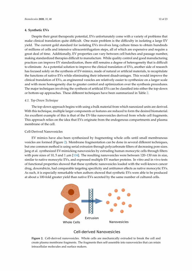
The top-down approach begins with using a bulk material from which nanosized units are derived.With this technique, multiple larger components or features are reduced to form the desired biomaterial.An excellent example of this is that of the EV-like nanovesicles derived from whole cell fragments.This approach relies on the idea that EVs originate from the endogenous compartments and plasmamembrane of the cell.
Cell-Derived Nanovesicles
EV mimics have also been synthesized by fragmenting whole cells until small membranousvesicles are formed (Figure 2). Membrane fragmentation can be done in several different techniques,but one common method is using serial extrusion through polycarbonate filters of decreasing pore sizes.Jang et al. synthesized EV-mimicking nanovesicles by extruding human monocytic cells through filterswith pore sizes of 10, 5 and 1 µm [104]. The resulting nanovesicles were between 120–130 nm in size,similar to native monocytic EVs, and expressed multiple EV marker proteins. In vitro and in vivo testsof functional properties showed that these synthetic nanovesicles loaded with the well-known cancerdrug, doxorubicin, had comparable targeting specificity and antitumor effects as native monocytic EVs.As such, it is especially remarkable when authors showed that synthetic EVs were able to be producedat about a 100-fold greater yield than native EVs secreted by the same number of cultured cells.
Biomolecules 2019, 9, x FOR PEER REVIEW 13 of 24
4.1. Top-Down Technique
The top-down approach begins with using a bulk material from which nanosized units are derived. With this technique, multiple larger components or features are reduced to form the desired biomaterial. An excellent example of this is that of the EV-like nanovesicles derived from whole cell fragments. This approach relies on the idea that EVs originate from the endogenous compartments and plasma membrane of the cell.
Cell-Derived Nanovesicles
EV mimics have also been synthesized by fragmenting whole cells until small membranous vesicles are formed (Figure 2). Membrane fragmentation can be done in several different techniques, but one common method is using serial extrusion through polycarbonate filters of decreasing pore sizes. Jang et al. synthesized EV-mimicking nanovesicles by extruding human monocytic cells through filters with pore sizes of 10, 5 and 1 μm [104]. The resulting nanovesicles were between 120–130 nm in size, similar to native monocytic EVs, and expressed multiple EV marker proteins. In vitro and in vivo tests of functional properties showed that these synthetic nanovesicles loaded with the well-known cancer drug, doxorubicin, had comparable targeting specificity and antitumor effects as native monocytic EVs. As such, it is especially remarkable when authors showed that synthetic EVs were able to be produced at about a 100-fold greater yield than native EVs secreted by the same number of cultured cells.
Figure 2. Cell-derived nanovesicles. Whole cells are mechanically extruded to break the cell and create plasma membrane fragments. The fragments then self-assemble into nanovesicles that can retain intracellular molecules and surface makers.
Similar methods were used to synthesize EV-mimicking nanovesicles from pancreatic β-cells in which cells were harvested and serially extruded through 10, 5 and 1 μm pore-sized polycarbonate membrane filters [105]. Assessment of the physical properties of nanovesicles revealed an average size of 203 nm, and it has been found that protein contents similar to donor cells though insulin and Pdx1, a transcription factor that regulates β-cell development, were not detected. Regardless, functional studies showed that the nanovesicles directed bone marrow cell differentiation into insulin-producing cells in vitro, that when transplanted into diabetic mice, were capable of controlling blood glucose levels for 60 days. Though no control comparison was made to native EVs derived from the same β-cell line, a yield of 8.2 × 10⁸ particles per 1 μg of total protein was reported, confirming that generally a high number of vesicles can be made from this method [105]. A similar
Figure 2. Cell-derived nanovesicles. Whole cells are mechanically extruded to break the cell andcreate plasma membrane fragments. The fragments then self-assemble into nanovesicles that can retainintracellular molecules and surface makers.
Biomolecules 2020, 10, 48 13 of 23
Similar methods were used to synthesize EV-mimicking nanovesicles from pancreatic β-cells inwhich cells were harvested and serially extruded through 10, 5 and 1 µm pore-sized polycarbonatemembrane filters [105]. Assessment of the physical properties of nanovesicles revealed an average sizeof 203 nm, and it has been found that protein contents similar to donor cells though insulin and Pdx1,a transcription factor that regulates β-cell development, were not detected. Regardless, functionalstudies showed that the nanovesicles directed bone marrow cell differentiation into insulin-producingcells in vitro, that when transplanted into diabetic mice, were capable of controlling blood glucoselevels for 60 days. Though no control comparison was made to native EVs derived from the sameβ-cell line, a yield of 8.2 × 10⁸ particles per 1 µg of total protein was reported, confirming that generallya high number of vesicles can be made from this method [105]. A similar protocol was used to produceEV mimics from human breast epithelial cells [106]. A significantly higher yield of mimics was seen,with 379.3 µg of protein and 3.0 × 10¹¹ of mimics produced in comparison to 2.5 µg of protein and2.6 × 109 of EVs from the same number of cells (1.0 × 10⁷ cells). EV-specific surface markers wereconserved in the mimics, while functional assessments revealed that the mimics did not stimulate anyimmune response in mice, and exhibited excellent targeting capabilities to tumor cells.
Large-scale generation of EV-mimicking nanovesicles can also be made by centrifuging cellsuspensions through a microporous polycarbonate membrane enclosed in a larger polycarbonatestructure designed to fit in centrifuge bucket holders [107]. The highest yield rate was found to beat ~16,000 kPa (2000 rpm) using a 1 × 10⁸ murine embryonic stem cell (ES) suspension, which led toabout 250 times more nanovesicles than the EVs produced from an equal number of ES cells. Westernblotting and a reverse transcription polymerase chain reaction (RT-PCR) of both nanovesicles and EVsshowed remarkable similarities in protein expression and RNA profiles while size characterizationrevealed similar ~100 nm diameters [107].
Another method of generating nanovesicles involves using a microfluidic system. Live murineembryonic stem cells were extruded through microchannels lined with an array of 500 nm-thick siliconnitride blades that were used to slice pieces of plasma membrane from the cells [108]. The plasmamembrane pieces spontaneously self-assembled into nanovesicles that retained cellular contents(proteins, intracellular RNAs) or was loaded with exogenous molecules. Resulting nanovesicles were100–300 nm in size and had a yield of ~150 × 108 from 1 × 10⁶ cells.
4.2. Bottom-Up Technique
The bottom-up process is much more specific in that the nanoscale components are carefullychosen and combined to form the larger and more complex structure. The bottom-up design allowsfor the creation of tailored structures that can address a specific purpose. However, they can bemore difficult to synthesize, as piecing together different components may involve complex bindingtechniques and create new challenges.
4.2.1. Liposomes
Liposomes are a well-studied drug delivery system that has had tremendous success for avariety of clinical applications, from cancer treatments (i.e., Doxil®, Myocet®, Marqibo®) to viralvaccines (i.e., Expaxa®, Inflexal® V) to fungal diseases (i.e., Abelcet®, Ambisome®, Amphotec®) [109].Indeed, considering their characteristics, which namely are that of a lipid bilayer with a wide range ofcargo-carrying possibilities, and their targeting potential, liposomes can be considered the artificialcounterparts to EVs. Their lipid bilayer composition makes liposomes an excellent mimic, as EVsare similarly composed of a variety of lipids, specifically phospholipids, such as phosphatidylcholine(PC), phosphatidylethanolamine (PE) and sphingomyelin [110]. Lu et al. synthesized EV-mimickingliposomes using a lipid formulation comparable to the EV lipidomic profile [111]. Liposomes werecreated using a DOPC/SM/Chol/DOPS/DOPE mixture and loaded with VEGF siRNA to model EV cargo.Compared to conventional PC/Chol and DOTAP liposomes, the EV-mimicking liposomes enhancedcellular uptake, serum stability and siRNA silencing, though there was lower encapsulation efficiency.
Biomolecules 2020, 10, 48 14 of 23
Analogous phospholipid-based liposomes have been synthesized, to great effect, for regenerativeapplications [112].
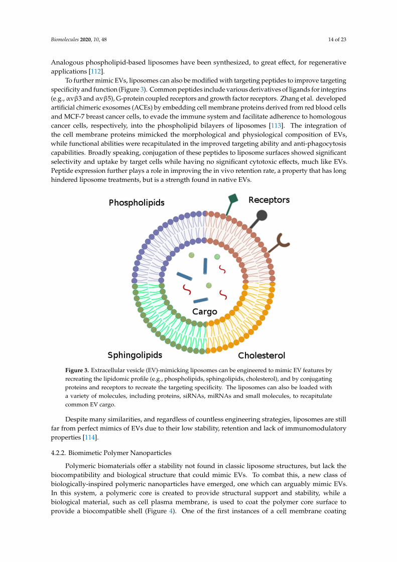
To further mimic EVs, liposomes can also be modified with targeting peptides to improve targetingspecificity and function (Figure 3). Common peptides include various derivatives of ligands for integrins(e.g., αvβ3 and αvβ5), G-protein coupled receptors and growth factor receptors. Zhang et al. developedartificial chimeric exosomes (ACEs) by embedding cell membrane proteins derived from red blood cellsand MCF-7 breast cancer cells, to evade the immune system and facilitate adherence to homologouscancer cells, respectively, into the phospholipid bilayers of liposomes [113]. The integration ofthe cell membrane proteins mimicked the morphological and physiological composition of EVs,while functional abilities were recapitulated in the improved targeting ability and anti-phagocytosiscapabilities. Broadly speaking, conjugation of these peptides to liposome surfaces showed significantselectivity and uptake by target cells while having no significant cytotoxic effects, much like EVs.Peptide expression further plays a role in improving the in vivo retention rate, a property that has longhindered liposome treatments, but is a strength found in native EVs.
Biomolecules 2019, 9, x FOR PEER REVIEW 15 of 24
surfaces showed significant selectivity and uptake by target cells while having no significant cytotoxic effects, much like EVs. Peptide expression further plays a role in improving the in vivo retention rate, a property that has long hindered liposome treatments, but is a strength found in native EVs.
Despite many similarities, and regardless of countless engineering strategies, liposomes are still far from perfect mimics of EVs due to their low stability, retention and lack of immunomodulatory properties [114].
Figure 3. Extracellular vesicle (EV)-mimicking liposomes can be engineered to mimic EV features by recreating the lipidomic profile (e.g., phospholipids, sphingolipids, cholesterol), and by conjugating proteins and receptors to recreate the targeting specificity. The liposomes can also be loaded with a variety of molecules, including proteins, siRNAs, miRNAs and small molecules, to recapitulate common EV cargo.
4.2.2. Biomimetic Polymer Nanoparticles
Polymeric biomaterials offer a stability not found in classic liposome structures, but lack the biocompatibility and biological structure that could mimic EVs. To combat this, a new class of biologically-inspired polymeric nanoparticles have emerged, one which can arguably mimic EVs. In this system, a polymeric core is created to provide structural support and stability, while a biological material, such as cell plasma membrane, is used to coat the polymer core surface to provide a biocompatible shell (Figure 4). One of the first instances of a cell membrane coating on a polymer core is by Hu et al., who coated sub-100 nm poly(lactic-co-glycolic acid) (PLGA) nanoparticles with
Figure 3. Extracellular vesicle (EV)-mimicking liposomes can be engineered to mimic EV features byrecreating the lipidomic profile (e.g., phospholipids, sphingolipids, cholesterol), and by conjugatingproteins and receptors to recreate the targeting specificity. The liposomes can also be loaded witha variety of molecules, including proteins, siRNAs, miRNAs and small molecules, to recapitulatecommon EV cargo.
Despite many similarities, and regardless of countless engineering strategies, liposomes are stillfar from perfect mimics of EVs due to their low stability, retention and lack of immunomodulatoryproperties [114].
4.2.2. Biomimetic Polymer Nanoparticles
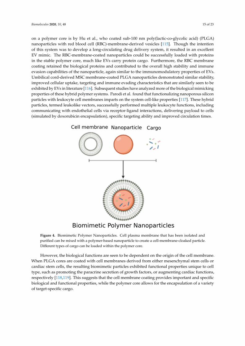
Polymeric biomaterials offer a stability not found in classic liposome structures, but lack thebiocompatibility and biological structure that could mimic EVs. To combat this, a new class ofbiologically-inspired polymeric nanoparticles have emerged, one which can arguably mimic EVs.In this system, a polymeric core is created to provide structural support and stability, while abiological material, such as cell plasma membrane, is used to coat the polymer core surface toprovide a biocompatible shell (Figure 4). One of the first instances of a cell membrane coating
Biomolecules 2020, 10, 48 15 of 23
on a polymer core is by Hu et al., who coated sub-100 nm poly(lactic-co-glycolic acid) (PLGA)nanoparticles with red blood cell (RBC)-membrane-derived vesicles [115]. Though the intentionof this system was to develop a long-circulating drug delivery system, it resulted in an excellentEV mimic. The RBC-membrane-coated nanoparticles could be successfully loaded with proteinsin the stable polymer core, much like EVs carry protein cargo. Furthermore, the RBC membranecoating retained the biological proteins and contributed to the overall high stability and immuneevasion capabilities of the nanoparticle, again similar to the immunomodulatory properties of EVs.Umbilical cord-derived MSC membrane-coated PLGA nanoparticles demonstrated similar stability,improved cellular uptake, targeting and immune evading characteristics that are similarly seen to beexhibited by EVs in literature [116]. Subsequent studies have analyzed more of the biological mimickingproperties of these hybrid polymer systems. Parodi et al. found that functionalizing nanoporous siliconparticles with leukocyte cell membranes imparts on the system cell-like properties [117]. These hybridparticles, termed leukolike vectors, successfully performed multiple leukocyte functions, includingcommunicating with endothelial cells via receptor-ligand interactions, delivering payload to cells(simulated by doxorubicin encapsulation), specific targeting ability and improved circulation times.
Biomolecules 2019, 9, x FOR PEER REVIEW 16 of 24
red blood cell (RBC)-membrane-derived vesicles [115]. Though the intention of this system was to develop a long-circulating drug delivery system, it resulted in an excellent EV mimic. The RBC-membrane-coated nanoparticles could be successfully loaded with proteins in the stable polymer core, much like EVs carry protein cargo. Furthermore, the RBC membrane coating retained the biological proteins and contributed to the overall high stability and immune evasion capabilities of the nanoparticle, again similar to the immunomodulatory properties of EVs. Umbilical cord-derived MSC membrane-coated PLGA nanoparticles demonstrated similar stability, improved cellular uptake, targeting and immune evading characteristics that are similarly seen to be exhibited by EVs in literature [116]. Subsequent studies have analyzed more of the biological mimicking properties of these hybrid polymer systems. Parodi et al. found that functionalizing nanoporous silicon particles with leukocyte cell membranes imparts on the system cell-like properties [117]. These hybrid particles, termed leukolike vectors, successfully performed multiple leukocyte functions, including communicating with endothelial cells via receptor-ligand interactions, delivering payload to cells (simulated by doxorubicin encapsulation), specific targeting ability and improved circulation times.
Figure 4. Biomimetic Polymer Nanoparticles. Cell plasma membrane that has been isolated and purified can be mixed with a polymer-based nanoparticle to create a cell-membrane-cloaked particle. Different types of cargo can be loaded within the polymer core.
However, the biological functions are seen to be dependent on the origin of the cell membrane. When PLGA cores are coated with cell membranes derived from either mesenchymal stem cells or cardiac stem cells, the resulting biomimetic particles exhibited functional properties unique to cell type, such as promoting the paracrine secretion of growth factors, or augmenting cardiac functions, respectively [118,119]. This suggests that the cell membrane coating provides important and specific biological and functional properties, while the polymer core allows for the encapsulation of a variety of target-specific cargo.
Table 2. Summary of semi-synthetic and synthetic EV-mimic types.
Synthetic EV Type Description Method Drug loading [60–64,75]
Figure 4. Biomimetic Polymer Nanoparticles. Cell plasma membrane that has been isolated andpurified can be mixed with a polymer-based nanoparticle to create a cell-membrane-cloaked particle.Different types of cargo can be loaded within the polymer core.
However, the biological functions are seen to be dependent on the origin of the cell membrane.When PLGA cores are coated with cell membranes derived from either mesenchymal stem cells orcardiac stem cells, the resulting biomimetic particles exhibited functional properties unique to celltype, such as promoting the paracrine secretion of growth factors, or augmenting cardiac functions,respectively [118,119]. This suggests that the cell membrane coating provides important and specificbiological and functional properties, while the polymer core allows for the encapsulation of a varietyof target-specific cargo.
Biomolecules 2020, 10, 48 16 of 23
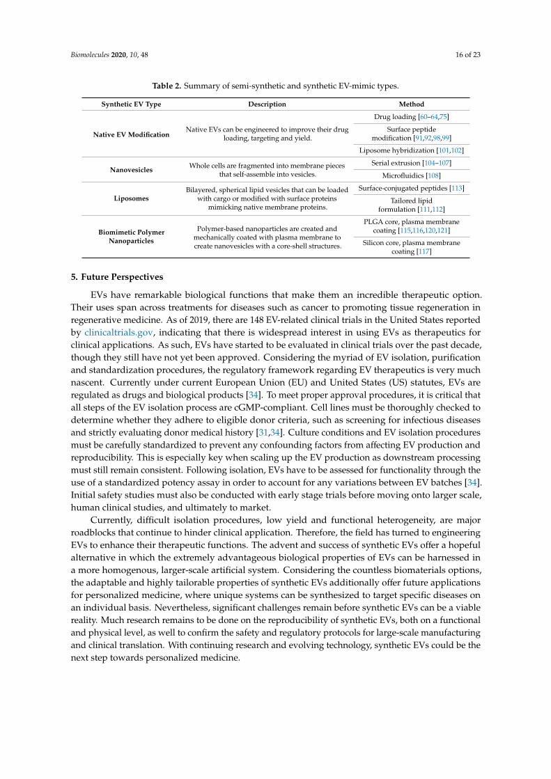
Table 2. Summary of semi-synthetic and synthetic EV-mimic types.
Synthetic EV Type Description Method
Native EV ModificationNative EVs can be engineered to improve their drug
loading, targeting and yield.
Drug loading [60–64,75]
Surface peptidemodification [91,92,98,99]
Liposome hybridization [101,102]
Nanovesicles Whole cells are fragmented into membrane piecesthat self-assemble into vesicles.
Serial extrusion [104–107]
Microfluidics [108]
LiposomesBilayered, spherical lipid vesicles that can be loaded
with cargo or modified with surface proteinsmimicking native membrane proteins.
Surface-conjugated peptides [113]
Tailored lipidformulation [111,112]
Biomimetic PolymerNanoparticles
Polymer-based nanoparticles are created andmechanically coated with plasma membrane tocreate nanovesicles with a core-shell structures.
EVs have remarkable biological functions that make them an incredible therapeutic option.Their uses span across treatments for diseases such as cancer to promoting tissue regeneration inregenerative medicine. As of 2019, there are 148 EV-related clinical trials in the United States reportedby clinicaltrials.gov, indicating that there is widespread interest in using EVs as therapeutics forclinical applications. As such, EVs have started to be evaluated in clinical trials over the past decade,though they still have not yet been approved. Considering the myriad of EV isolation, purificationand standardization procedures, the regulatory framework regarding EV therapeutics is very muchnascent. Currently under current European Union (EU) and United States (US) statutes, EVs areregulated as drugs and biological products [34]. To meet proper approval procedures, it is critical thatall steps of the EV isolation process are cGMP-compliant. Cell lines must be thoroughly checked todetermine whether they adhere to eligible donor criteria, such as screening for infectious diseasesand strictly evaluating donor medical history [31,34]. Culture conditions and EV isolation proceduresmust be carefully standardized to prevent any confounding factors from affecting EV production andreproducibility. This is especially key when scaling up the EV production as downstream processingmust still remain consistent. Following isolation, EVs have to be assessed for functionality through theuse of a standardized potency assay in order to account for any variations between EV batches [34].Initial safety studies must also be conducted with early stage trials before moving onto larger scale,human clinical studies, and ultimately to market.
Currently, difficult isolation procedures, low yield and functional heterogeneity, are majorroadblocks that continue to hinder clinical application. Therefore, the field has turned to engineeringEVs to enhance their therapeutic functions. The advent and success of synthetic EVs offer a hopefulalternative in which the extremely advantageous biological properties of EVs can be harnessed ina more homogenous, larger-scale artificial system. Considering the countless biomaterials options,the adaptable and highly tailorable properties of synthetic EVs additionally offer future applicationsfor personalized medicine, where unique systems can be synthesized to target specific diseases onan individual basis. Nevertheless, significant challenges remain before synthetic EVs can be a viablereality. Much research remains to be done on the reproducibility of synthetic EVs, both on a functionaland physical level, as well to confirm the safety and regulatory protocols for large-scale manufacturingand clinical translation. With continuing research and evolving technology, synthetic EVs could be thenext step towards personalized medicine.
Author Contributions: L.R. performed the literature search and drafted the manuscript. L.R., P.K., and A.W.provided critical revisions to the finalized manuscript. All authors have read and agreed to the published versionof the manuscript.
Funding: This research was funded by the UC Davis School of Medicine Dean’s Fellowship (to A.W.) awards,National Heart, Lung, and Blood Institute of the National Institutes of Health (NIH) under Award NumberU54HL119893, and by the National Center for Advancing Translational Sciences through grant number UL1TR001860, NIH grants 5R01NS100761-02 and R03HD091601-01, Shriners Hospitals for Children research grants(85119-NCA-18, 87200-NCA-19, 85108-NCA-19), and the March of Dimes Foundation Basil O’Connor StarterScholar Research Award (5FY1682).
Acknowledgments: The authors acknowledge Alexandra Iavorovschi (University of California–Davis) for herhelp with manuscript editing.
Conflicts of Interest: The authors declare no conflict of interest.
References
1. Mao, A.S.; Mooney, D.J. Regenerative medicine: Current therapies and future directions. Proc. Natl. Acad.Sci. USA 2015, 112, 14452–14459. [CrossRef] [PubMed]
2. Sampogna, G.; Guraya, S.Y.; Forgione, A. Regenerative medicine: Historical roots and potential strategies inmodern medicine. J. Microsc. Ultrastruct. 2015, 3, 101–107. [CrossRef] [PubMed]
3. Gurtner, G.C.; Callaghan, M.J.; Longaker, M.T. Progress and potential for regenerative medicine. Annu. Rev.Med. 2007, 58, 299–312. [CrossRef] [PubMed]
4. De Jong, O.G.; Van Balkom, B.W.M.; Schiffelers, R.M.; Bouten, C.V.C.; Verhaar, M.C. Extracellular Vesicles:Potential Roles in Regenerative Medicine. Front. Immunol. 2014, 5. [CrossRef] [PubMed]
5. Fatima, F.; Nawaz, M. Stem cell-derived exosomes: Roles in stromal remodeling, tumor progression, andcancer immunotherapy. Chin. J. Cancer 2015, 34. [CrossRef] [PubMed]
6. Lamichhane, T.N.; Sokic, S.; Schardt, J.S.; Raiker, R.S.; Lin, J.W.; Jay, S.M. Emerging Roles for ExtracellularVesicles in Tissue Engineering and Regenerative Medicine. Tissue Eng. Part B Rev. 2015, 21, 45–54. [CrossRef]
7. Théry, C.; Zitvogel, L.; Amigorena, S. Exosomes: Composition, biogenesis and function. Nat. Rev. Immunol.2002, 2, 569–579. [CrossRef]
8. Hessvik, N.P.; Llorente, A. Current knowledge on exosome biogenesis and release. Cell Mol. Life Sci. 2018,75, 193–208. [CrossRef]
9. Takasugi, M.; Okada, R.; Takahashi, A.; Virya Chen, D.; Watanabe, S.; Hara, E. Small extracellular vesiclessecreted from senescent cells promote cancer cell proliferation through EphA2. Nat. Commun. 2017, 8, 15729.[CrossRef]
10. Khan, M.; Nickoloff, E.; Abramova, T.; Johnson, J.; Verma, S.K.; Krishnamurthy, P.; Mackie, A.R.; Vaughan, E.;Garikipati, V.N.S.; Benedict, C.; et al. Embryonic stem cell-derived exosomes promote endogenous repairmechanisms and enhance cardiac function following myocardial infarction. Circ. Res. 2015, 117, 52–64.[CrossRef]
11. Ibrahim, A.G.-E.; Cheng, K.; Marbán, E. Exosomes as critical agents of cardiac regeneration triggered by celltherapy. Stem Cell Rep. 2014, 2, 606–619. [CrossRef] [PubMed]
12. Li, X.; Chen, C.; Wei, L.; Li, Q.; Niu, X.; Xu, Y.; Wang, Y.; Zhao, J. Exosomes derived from endothelialprogenitor cells attenuate vascular repair and accelerate reendothelialization by enhancing endothelialfunction. Cytotherapy 2016, 18, 253–262. [CrossRef] [PubMed]
13. Cantaluppi, V.; Biancone, L.; Figliolini, F.; Beltramo, S.; Medica, D.; Deregibus, M.C.; Galimi, F.; Romagnoli, R.;Salizzoni, M.; Tetta, C.; et al. Microvesicles derived from endothelial progenitor cells enhance neoangiogenesisof human pancreatic islets. Cell Transpl. 2012, 21, 1305–1320. [CrossRef] [PubMed]
14. Deregibus, M.C.; Cantaluppi, V.; Calogero, R.; Lo Iacono, M.; Tetta, C.; Biancone, L.; Bruno, S.; Bussolati, B.;Camussi, G. Endothelial progenitor cell derived microvesicles activate an angiogenic program in endothelialcells by a horizontal transfer of mRNA. Blood 2007, 110, 2440–2448. [CrossRef] [PubMed]
15. Gong, M.; Yu, B.; Wang, J.; Wang, Y.; Liu, M.; Paul, C.; Millard, R.W.; Xiao, D.-S.; Ashraf, M.; Xu, M.Mesenchymal stem cells release exosomes that transfer miRNAs to endothelial cells and promote angiogenesis.Oncotarget 2017, 8, 45200–45212. [CrossRef] [PubMed]
16. Mu, W.; Rana, S.; Zöller, M. Host Matrix Modulation by Tumor Exosomes Promotes Motility and Invasiveness.Neoplasia 2013, 15, 875–887. [CrossRef] [PubMed]
17. Albacete-Albacete, L.; Navarro-Lerida, I.; Lopez, J.A.; Martin-Padura, I.; Astudillo, A.M.; Van-Der-Heyden, M.;Balsinde, J.; Orend, G.; Vazquez, J.; del Pozo, M.A. ECM deposition is driven by caveolin1-dependentregulation of exosomal biogenesis and cargo sorting. BioRxiv 2018. [CrossRef]
18. Sung, B.H.; Ketova, T.; Hoshino, D.; Zijlstra, A.; Weaver, A.M. Directional cell movement through tissues iscontrolled by exosome secretion. Nat. Commun. 2015, 6, 7164. [CrossRef]
19. Robbins, P.D.; Morelli, A.E. Regulation of immune responses by extracellular vesicles. Nature Rev. Immunol.2014, 14, 195–208. [CrossRef]
20. Théry, C.; Ostrowski, M.; Segura, E. Membrane vesicles as conveyors of immune responses. Nat. Rev.Immunol. 2009, 9, 581–593. [CrossRef]
21. Shenoda, B.B.; Ajit, S.K. Modulation of Immune Responses by Exosomes Derived from Antigen-PresentingCells. Clin. Med. Insights Pathol. 2016, 9, 1–8. [CrossRef] [PubMed]
22. van Niel, G.; D’Angelo, G.; Raposo, G. Shedding light on the cell biology of extracellular vesicles. Nat. Rev.Mol. Cell Biol. 2018, 19, 213–228. [CrossRef] [PubMed]
23. Colombo, M.; Raposo, G.; Théry, C. Biogenesis, Secretion, and Intercellular Interactions of Exosomes andOther Extracellular Vesicles. Annu. Rev. Cell Dev. Biol. 2014, 30, 255–289. [CrossRef] [PubMed]
24. Raposo, G.; Stoorvogel, W. Extracellular vesicles: Exosomes, microvesicles, and friends. J. Cell Biol. 2013, 200,373–383. [CrossRef] [PubMed]
25. Erdbrügger, U.; Lannigan, J. Analytical challenges of extracellular vesicle detection: A comparison of differenttechniques. Cytom. Part A 2016, 89, 123–134. [CrossRef] [PubMed]
26. Phan, J.; Kumar, P.; Hao, D.; Gao, K.; Farmer, D.; Wang, A. Engineering mesenchymal stem cells to improvetheir exosome efficacy and yield for cell-free therapy. J. Extracell. Vesicles 2018, 7. [CrossRef] [PubMed]
27. Momen-Heravi, F.; Balaj, L.; Alian, S.; Mantel, P.-Y.; Halleck, A.E.; Trachtenberg, A.J.; Soria, C.E.; Oquin, S.;Bonebreak, C.M.; Saracoglu, E.; et al. Current methods for the isolation of extracellular vesicles. Biol. Chem.2013, 394, 1253–1262. [CrossRef]
44. Zhao, P.; Xiao, L.; Peng, J.; Qian, Y.-Q.; Huang, C.-C. Exosomes derived from bone marrow mesenchymalstem cells improve osteoporosis through promoting osteoblast proliferation via MAPK pathway. Eur. Rev.Med. Pharmacol. Sci. 2018, 22, 3962–3970. [PubMed]
45. Aguado, B.A.; Mulyasasmita, W.; Su, J.; Lampe, K.J.; Heilshorn, S.C. Improving viability of stem cells duringsyringe needle flow through the design of hydrogel cell carriers. Tissue Eng. Part A 2012, 18, 806–815.[CrossRef]
46. Zhu, X.; Badawi, M.; Pomeroy, S.; Sutaria, D.S.; Xie, Z.; Baek, A.; Jiang, J.; Elgamal, O.A.; Mo, X.; Perle, K.L.;et al. Comprehensive toxicity and immunogenicity studies reveal minimal effects in mice following sustaineddosing of extracellular vesicles derived from HEK293T cells. J. Extracell. Vesicles 2017, 6. [CrossRef]
47. Williams, A.M.; Dennahy, I.S.; Bhatti, U.F.; Halaweish, I.; Xiong, Y.; Chang, P.; Nikolian, V.C.; Chtraklin, K.;Brown, J.; Zhang, Y.; et al. Mesenchymal Stem Cell-Derived Exosomes Provide Neuroprotection and ImproveLong-Term Neurologic Outcomes in a Swine Model of Traumatic Brain Injury and Hemorrhagic Shock. J.Neurotrauma 2019, 36, 54–60. [CrossRef]
48. Webb, R.L.; Kaiser, E.E.; Scoville, S.L.; Thompson, T.A.; Fatima, S.; Pandya, C.; Sriram, K.; Swetenburg, R.L.;Vaibhav, K.; Arbab, A.S.; et al. Human Neural Stem Cell Extracellular Vesicles Improve Tissue and FunctionalRecovery in the Murine Thromboembolic Stroke Model. Transl. Stroke Res. 2018, 9, 530–539. [CrossRef]
49. Webb, R.L.; Kaiser, E.E.; Jurgielewicz, B.J.; Spellicy, S.; Scoville, S.L.; Thompson, T.A.; Swetenburg, R.L.;Hess, D.C.; West, F.D.; Stice, S.L. Human Neural Stem Cell Extracellular Vesicles Improve Recovery in aPorcine Model of Ischemic Stroke. Stroke 2018, 49, 1248–1256. [CrossRef]
50. Barile, L.; Lionetti, V.; Cervio, E.; Matteucci, M.; Gherghiceanu, M.; Popescu, L.M.; Torre, T.; Siclari, F.;Moccetti, T.; Vassalli, G. Extracellular vesicles from human cardiac progenitor cells inhibit cardiomyocyteapoptosis and improve cardiac function after myocardial infarction. Cardiovasc. Res 2014, 103, 530–541.[CrossRef]
51. Xin, H.; Li, Y.; Buller, B.; Katakowski, M.; Zhang, Y.; Wang, X.; Shang, X.; Zhang, Z.G.; Chopp, M.Exosome-Mediated Transfer of miR-133b from Multipotent Mesenchymal Stromal Cells to Neural CellsContributes to Neurite Outgrowth. Stem Cells 2012, 30, 1556–1564. [CrossRef] [PubMed]
52. Takeda, Y.S.; Xu, Q. Neuronal Differentiation of Human Mesenchymal Stem Cells Using Exosomes Derivedfrom Differentiating Neuronal Cells. PLoS ONE 2015, 10, e0135111. [CrossRef] [PubMed]
57. Shi, Q.; Qian, Z.; Liu, D.; Sun, J.; Wang, X.; Liu, H.; Xu, J.; Guo, X. GMSC-Derived Exosomes Combinedwith a Chitosan/Silk Hydrogel Sponge Accelerates Wound Healing in a Diabetic Rat Skin Defect Model.Front. Physiol. 2017, 8. [CrossRef]
58. Liu, B.; Lee, B.W.; Nakanishi, K.; Villasante, A.; Williamson, R.; Metz, J.; Kim, J.; Kanai, M.; Bi, L.; Brown, K.;et al. Cardiac recovery via extended cell-free delivery of extracellular vesicles secreted by cardiomyocytesderived from induced pluripotent stem cells. Nat. Biomed. Eng. 2018, 2, 293. [CrossRef]
59. Kang, K.; Ma, R.; Cai, W.; Huang, W.; Paul, C.; Liang, J.; Wang, Y.; Zhao, T.; Kim, H.W.; Xu, M.; et al. ExosomesSecreted from CXCR4 Overexpressing Mesenchymal Stem Cells Promote Cardioprotection via Akt SignalingPathway following Myocardial Infarction. Stem Cells Int. 2015, 2015, 659890. [CrossRef]
60. Sun, D.; Zhuang, X.; Xiang, X.; Liu, Y.; Zhang, S.; Liu, C.; Barnes, S.; Grizzle, W.; Miller, D.; Zhang, H.-G.A novel nanoparticle drug delivery system: The anti-inflammatory activity of curcumin is enhanced whenencapsulated in exosomes. Mol. Ther. 2010, 18, 1606–1614. [CrossRef]
61. Zhuang, X.; Xiang, X.; Grizzle, W.; Sun, D.; Zhang, S.; Axtell, R.C.; Ju, S.; Mu, J.; Zhang, L.; Steinman, L.; et al.Treatment of Brain Inflammatory Diseases by Delivering Exosome Encapsulated Anti-inflammatory DrugsFrom the Nasal Region to the Brain. Mol. Ther. 2011, 19, 1769–1779. [CrossRef] [PubMed]
62. Munagala, R.; Aqil, F.; Jeyabalan, J.; Gupta, R.C. Bovine milk-derived exosomes for drug delivery. Cancer Lett.2016, 371, 48–61. [CrossRef] [PubMed]
63. Kim, M.S.; Haney, M.J.; Zhao, Y.; Mahajan, V.; Deygen, I.; Klyachko, N.L.; Inskoe, E.; Piroyan, A.; Sokolsky, M.;Okolie, O.; et al. Development of exosome-encapsulated paclitaxel to overcome MDR in cancer cells.Nanomedicine 2016, 12, 655–664. [CrossRef] [PubMed]
64. Pascucci, L.; Coccè, V.; Bonomi, A.; Ami, D.; Ceccarelli, P.; Ciusani, E.; Viganò, L.; Locatelli, A.; Sisto, F.;Doglia, S.M.; et al. Paclitaxel is incorporated by mesenchymal stromal cells and released in exosomes thatinhibit in vitro tumor growth: A new approach for drug delivery. J. Control. Release 2014, 192, 262–270.[CrossRef] [PubMed]
65. Haney, M.J.; Klyachko, N.L.; Zhao, Y.; Gupta, R.; Plotnikova, E.G.; He, Z.; Patel, T.; Piroyan, A.; Sokolsky, M.;Kabanov, A.V.; et al. Exosomes as Drug Delivery Vehicles for Parkinson’s Disease Therapy. J. Control. Release2015, 207, 18–30. [CrossRef]
66. Pessina, A.; Bonomi, A.; Coccè, V.; Invernici, G.; Navone, S.; Cavicchini, L.; Sisto, F.; Ferrari, M.; Viganò, L.;Locatelli, A.; et al. Mesenchymal Stromal Cells Primed with Paclitaxel Provide a New Approach for CancerTherapy. PLoS ONE 2011, 6, e28321. [CrossRef]
67. Bonomi, A.; Coccè, V.; Cavicchini, L.; Sisto, F.; Dossena, M.; Balzarini, P.; Portolani, N.; Ciusani, E.; Parati, E.;Alessandri, G.; et al. Adipose tissue-derived stromal cells primed in vitro with paclitaxel acquire anti-tumoractivity. Int. J. Immunopathol. Pharmacol. 2013, 26, 33–41. [CrossRef]
68. Pessina, A.; Coccè, V.; Bonomi, A.; Cavicchini, L.; Sisto, F.; Ferrari, M.; Ciusani, E.; Navone, S.; Marfia, G.;Parati, E.; et al. Human skin-derived fibroblasts acquire in vitro anti-tumor potential after priming withPaclitaxel. Anticancer Agents Med. Chem. 2013, 13, 523–530.
69. Momen-Heravi, F.; Bala, S.; Bukong, T.; Szabo, G. Exosome-mediated delivery of functionally activemiRNA-155 inhibitor to macrophages. Nanomedicine 2014, 10, 1517–1527. [CrossRef]
76. Alvarez-Erviti, L.; Seow, Y.; Yin, H.; Betts, C.; Lakhal, S.; Wood, M.J.A. Delivery of siRNA to the mouse brainby systemic injection of targeted exosomes. Nat. Biotechnol. 2011, 29, 341–345. [CrossRef]
77. Stremersch, S.; Vandenbroucke, R.E.; Van Wonterghem, E.; Hendrix, A.; De Smedt, S.C.; Raemdonck, K.Comparing exosome-like vesicles with liposomes for the functional cellular delivery of small RNAs. J. Control.Release 2016, 232, 51–61. [CrossRef]
78. O’Loughlin, A.J.; Mäger, I.; de Jong, O.G.; Varela, M.A.; Schiffelers, R.M.; El Andaloussi, S.; Wood, M.J.A.;Vader, P. Functional Delivery of Lipid-Conjugated siRNA by Extracellular Vesicles. Mol. Ther. 2017, 25,1580–1587. [CrossRef]
79. Putz, U.; Howitt, J.; Lackovic, J.; Foot, N.; Kumar, S.; Silke, J.; Tan, S.-S. Nedd4 family-interacting protein 1(Ndfip1) is required for the exosomal secretion of Nedd4 family proteins. J. Biol. Chem. 2008, 283, 32621–32627.[CrossRef]
80. Sterzenbach, U.; Putz, U.; Low, L.-H.; Silke, J.; Tan, S.-S.; Howitt, J. Engineered Exosomes as Vehicles forBiologically Active Proteins. Mol. Ther. 2017, 25, 1269–1278. [CrossRef]
81. Shen, B.; Wu, N.; Yang, J.-M.; Gould, S.J. Protein Targeting to Exosomes/Microvesicles by Plasma MembraneAnchors. J. Biol. Chem. 2011, 286, 14383–14395. [CrossRef]
82. Lamichhane, T.N.; Jeyaram, A.; Patel, D.B.; Parajuli, B.; Livingston, N.K.; Arumugasaamy, N.; Schardt, J.S.;Jay, S.M. Oncogene Knockdown via Active Loading of Small RNAs into Extracellular Vesicles by Sonication.Cell. Mol. Bioeng. 2016, 9, 315–324. [CrossRef] [PubMed]
83. Fuhrmann, G.; Serio, A.; Mazo, M.; Nair, R.; Stevens, M.M. Active loading into extracellular vesiclessignificantly improves the cellular uptake and photodynamic effect of porphyrins. J. Control. Release 2015,205, 35–44. [CrossRef] [PubMed]
84. Tian, Y.; Li, S.; Song, J.; Ji, T.; Zhu, M.; Anderson, G.J.; Wei, J.; Nie, G. A doxorubicin delivery platform usingengineered natural membrane vesicle exosomes for targeted tumor therapy. Biomaterials 2014, 35, 2383–2390.[CrossRef] [PubMed]
85. Mokarizadeh, A.; Delirezh, N.; Morshedi, A.; Mosayebi, G.; Farshid, A.-A.; Dalir-Naghadeh, B. Phenotypicmodulation of auto-reactive cells by insertion of tolerogenic molecules via MSC-derived exosomes. Vet. Res.Forum 2012, 3, 257–261. [PubMed]
86. Yuyama, K.; Sun, H.; Sakai, S.; Mitsutake, S.; Okada, M.; Tahara, H.; Furukawa, J.; Fujitani, N.; Shinohara, Y.;Igarashi, Y. Decreased Amyloid-β Pathologies by Intracerebral Loading of Glycosphingolipid-enrichedExosomes in Alzheimer Model Mice. J. Biol. Chem. 2014, 289, 24488–24498. [CrossRef] [PubMed]
96. Hao, D.; Xiao, W.; Liu, R.; Kumar, P.; Li, Y.; Zhou, P.; Guo, F.; Farmer, D.L.; Lam, K.S.; Wang, F.; et al. Discoveryand Characterization of a Potent and Specific Peptide Ligand Targeting Endothelial Progenitor Cells andEndothelial Cells for Tissue Regeneration. ACS Chem. Biol. 2017, 12, 1075–1086. [CrossRef]
97. Peng, L.; Liu, R.; Marik, J.; Wang, X.; Takada, Y.; Lam, K.S. Combinatorial chemistry identifies high-affinitypeptidomimetics against α 4 β 1 integrin for in vivo tumor imaging. Nat. Chem. Biol. 2006, 2, 381. [CrossRef][PubMed]
98. Kooijmans, S.A.A.; Gitz-Francois, J.J.J.M.; Schiffelers, R.M.; Vader, P. Recombinant phosphatidylserine-bindingnanobodies for targeting of extracellular vesicles to tumor cells: A plug-and-play approach †Electronicsupplementary information (ESI) available. Nanoscale 2018, 10, 2413–2426. [CrossRef]
99. Ye, Z.; Zhang, T.; He, W.; Jin, H.; Liu, C.; Yang, Z.; Ren, J. Methotrexate-Loaded Extracellular VesiclesFunctionalized with Therapeutic and Targeted Peptides for the Treatment of Glioblastoma Multiforme.ACS Appl. Mater. Interfaces 2018, 10, 12341–12350. [CrossRef]
100. Wang, J.; Li, W.; Zhang, L.; Ban, L.; Chen, P.; Du, W.; Feng, X.; Liu, B.-F. Chemically Edited Exosomes withDual Ligand Purified by Microfluidic Device for Active Targeted Drug Delivery to Tumor Cells. ACS Appl.Mater. Interfaces 2017, 9, 27441–27452. [CrossRef]
101. Sato, Y.T.; Umezaki, K.; Sawada, S.; Mukai, S.; Sasaki, Y.; Harada, N.; Shiku, H.; Akiyoshi, K. Engineeringhybrid exosomes by membrane fusion with liposomes. Sci. Rep. 2016, 6, 21933. [CrossRef]
102. Lin, Y.; Wu, J.; Gu, W.; Huang, Y.; Tong, Z.; Huang, L.; Tan, J. Exosome–Liposome Hybrid NanoparticlesDeliver CRISPR/Cas9 System in MSCs. Adv. Sci. (Weinh) 2018, 5. [CrossRef]
103. Hoshyar, N.; Gray, S.; Han, H.; Bao, G. The effect of nanoparticle size on in vivo pharmacokinetics andcellular interaction. Nanomedicine 2016, 11, 673–692. [CrossRef] [PubMed]
104. Jang, S.C.; Kim, O.Y.; Yoon, C.M.; Choi, D.-S.; Roh, T.-Y.; Park, J.; Nilsson, J.; Lötvall, J.; Kim, Y.-K.; Gho, Y.S.Bioinspired Exosome-Mimetic Nanovesicles for Targeted Delivery of Chemotherapeutics to MalignantTumors. ACS Nano. 2013, 7, 7698–7710. [CrossRef] [PubMed]
105. Oh, K.; Kim, S.R.; Kim, D.-K.; Seo, M.W.; Lee, C.; Lee, H.M.; Oh, J.-E.; Choi, E.Y.; Lee, D.-S.; Gho, Y.S.; et al.In Vivo Differentiation of Therapeutic Insulin-Producing Cells from Bone Marrow Cells via ExtracellularVesicle-Mimetic Nanovesicles. ACS Nano. 2015, 9, 11718–11727. [CrossRef] [PubMed]
106. Yang, Z.; Xie, J.; Zhu, J.; Kang, C.; Chiang, C.; Wang, X.; Wang, X.; Kuang, T.; Chen, F.; Chen, Z.; et al.Functional exosome-mimic for delivery of siRNA to cancer: In vitro and in vivo evaluation. J. Control. Release2016, 243, 160–171. [CrossRef] [PubMed]
107. Jo, W.; Kim, J.; Yoon, J.; Jeong, D.; Cho, S.; Jeong, H.; Yoon, Y.J.; Kim, S.C.; Gho, Y.S.; Park, J. Large-scalegeneration of cell-derived nanovesicles. Nanoscale 2014, 6, 12056–12064. [CrossRef] [PubMed]
108. Yoon, J.; Jo, W.; Jeong, D.; Kim, J.; Jeong, H.; Park, J. Generation of nanovesicles with sliced cellular membranefragments for exogenous material delivery. Biomaterials 2015, 59, 12–20. [CrossRef]
109. Bulbake, U.; Doppalapudi, S.; Kommineni, N.; Khan, W. Liposomal Formulations in Clinical Use: AnUpdated Review. Pharmaceutics 2017, 9, 12. [CrossRef]
110. Subra, C.; Laulagnier, K.; Perret, B.; Record, M. Exosome lipidomics unravels lipid sorting at the level ofmultivesicular bodies. Biochimie. 2007, 89, 205–212. [CrossRef]
111. Lu, M.; Zhao, X.; Xing, H.; Xun, Z.; Zhu, S.; Lang, L.; Yang, T.; Cai, C.; Wang, D.; Ding, P. Comparison ofexosome-mimicking liposomes with conventional liposomes for intracellular delivery of siRNA. Int. J. Pharm.2018, 550, 100–113. [CrossRef]
112. Monteiro, N.; Martins, A.; Reis, R.L.; Neves, N.M. Liposomes in tissue engineering and regenerative medicine.J. R. Soc. Interface 2014, 11, 20140459. [CrossRef]