Eur. J. Biochem. 162, 333 -338 (1987) 0 FEBS 1987 Enhancer-mediated activation of a growth-regulated promoter Graham MOORE and Moshe YANIV Somatic Cell Laboratory, Imperial Cancer Research Fund, Lincoln’s Inn Fields, London Department of Molecular Biology, Pasteur Institute, Pans (Received April l/August 25, 1986) - EJB 86 0326 We have demonstrated that the collagen a2 type 1 promoter inserted in an expression vector, behaves as a growth-regulated promoter, which is consistent with previous observations that collagen synthesis is growth- regulated in vivo. In contrast, the activity of the H2-K or the simian virus 40 early promoters does not seem to be affected by the rate of cell proliferation. The insertion of a polyoma enhancer 5‘ or 3’ to the collagen transcription unit activates the collagen a2 type 1 promoter, by a threefold greater factor in slowly growing cells compared to cells growing exponentially. These results show that enhancers can also function in slowly pro- liferating cells and activate the normally low activity of a promoter in these cells. Recent studies on the regulation of gene expression have concentrated on DNA sequences found upstream of transcrip- tion initiation sites of messenger RNA in DNA tumour viruses (simian virus 40, SV40; polyoma; adenovirus) and several cellular genes [l-31. Sequences found more than 100 base pairs (bp) upsteam of the cap site are essential for the synthesis of the viral early RNA of both SV40 and polyoma virus [4- 71. These sequences have been termed enhancers since they .are capable of activating or enhancing transcription from both viral and cellular promoters when placed in either the 5‘ or 3’ position of the gene [8]: in the case of polyoma virus a fragment of 244 bp was defined as the entire polyoma enhancer [9]. Enhancer sequences, similar to those required for expression of early viral functions of polyoma have been found in SV40 virus [8], in the long terminal repeat (LTR) elements of some retroviruses [lo], in the genome of bovine papillomavirus [I I], upstream of the Ela coding sequences in adenovirus 2 and 5 [12,13], between the J and C coding blocks of the heavy-chain domain and between the constant domain and J element of K light chains of immunoglobulins [14- 181. Recent studies, based on assays using expression vectors exogeneously introduced into differentiated and undiffer- entiated mouse cells, suggest that the polyoma enhancer can be further subdivided into two elements: the A element, in- cluding a sequence homologous to a repeated core sequence of adenovirus Ela enhancer [12], and a second element B, which contains the consensus sequence of Weiher et al. [19] found in SV40 enhancer. Element A is more active in fibroblasts (3T6) than element B, although both have the same activity in mouse embryonal carcinoma cells (EC) [20]. It has been postulated that enhancers have a role in cell differentiation and transformation [21, 221. However, pre- vious investigations of promoter or enhancer functions have Correspondence to M. Yaniv, Department of Molecular Biology, Abbreviations. bp, base pair; CAT, chloramphenicol acetyltrans- Pasteur Institute, 25, rue du Docteur Roux, F-75015 Paris, France ferase; SV40, simian virus 40. not taken into account the integral rate of cell proliferation. The majority of cells in a eukaryotic organism are not replicat- ing and, if they are, it is only at a slow rate. If enhancers are physiologically important then they must be able to function in a non-proliferating or slowly proliferating cell. The rate of cell proliferation can be reduced by decreasing the concentra- tion of serum in the medium in which cells are growing [23]. The rate of protein synthesis is also reduced [24, 251. This study investigates the activities of various promoters cloned in expression vectors in the presence or absence of an enhancer and introduced into either exponentially growing cells or cells growing at a reduced rate because of serum starvation. We have demonstrated that the activity of the collagen ct2 type 1 promoter is growth-regulated and has a lower activity in slowly proliferating cells. The activity of the promoter may be increased in these cells by the insertion of an enhancer in the 5‘ or 3’ position by a greater factor than that observed in exponentially growing cells. EXPERIMENTAL PROCEDURE Construction of expression vectors An expression vector containing the chicken a2 type 1 collagen promoter fragment (-280 to +110 relative to the cap site), cloned 5‘ to the chloramphenicol acetyltransferase (CAT) coding sequences, has been described previously [20]. The vector has been designated pCaCAT and its promoter pCa (Fig. 1). A larger chicken a2 type 1 collagen promoter fragment (- 1100 to + 110, relative to the cap site), designated pCOL CAT, was assayed in the same construction, as de- scribed previously [20]. Two SV40 promoters, with and without the enhancer sequences inserted 5’ to CAT, were previously described [20, 261. The expression vector, contain- ing SV40 342-bp PvuII-Hind111 fragment including the two 72-bp tandem repeats, the 21-bp repeats and TATA box, is similar to pSVECAT of Gorman et al. [26], except that the pBR322 portion was replaced with the EcoRI-SaZI fragment
Transcript
Eur. J. Biochem. 162, 333 -338 (1987) 0 FEBS 1987
Enhancer-mediated activation of a growth-regulated promoter Graham MOORE and Moshe YANIV
Somatic Cell Laboratory, Imperial Cancer Research Fund, Lincoln’s Inn Fields, London Department of Molecular Biology, Pasteur Institute, Pans
(Received April l/August 25, 1986) - EJB 86 0326
We have demonstrated that the collagen a2 type 1 promoter inserted in an expression vector, behaves as a growth-regulated promoter, which is consistent with previous observations that collagen synthesis is growth- regulated in vivo. In contrast, the activity of the H2-K or the simian virus 40 early promoters does not seem to be affected by the rate of cell proliferation. The insertion of a polyoma enhancer 5‘ or 3’ to the collagen transcription unit activates the collagen a2 type 1 promoter, by a threefold greater factor in slowly growing cells compared to cells growing exponentially. These results show that enhancers can also function in slowly pro- liferating cells and activate the normally low activity of a promoter in these cells.
Recent studies on the regulation of gene expression have concentrated on DNA sequences found upstream of transcrip- tion initiation sites of messenger RNA in DNA tumour viruses (simian virus 40, SV40; polyoma; adenovirus) and several cellular genes [l-31. Sequences found more than 100 base pairs (bp) upsteam of the cap site are essential for the synthesis of the viral early RNA of both SV40 and polyoma virus [4- 71. These sequences have been termed enhancers since they .are capable of activating or enhancing transcription from both viral and cellular promoters when placed in either the 5‘ or 3’ position of the gene [8]: in the case of polyoma virus a fragment of 244 bp was defined as the entire polyoma enhancer [9]. Enhancer sequences, similar to those required for expression of early viral functions of polyoma have been found in SV40 virus [8], in the long terminal repeat (LTR) elements of some retroviruses [lo], in the genome of bovine papillomavirus [I I], upstream of the Ela coding sequences in adenovirus 2 and 5 [12,13], between the J and C coding blocks of the heavy-chain domain and between the constant domain and J element of K light chains of immunoglobulins [14- 181. Recent studies, based on assays using expression vectors exogeneously introduced into differentiated and undiffer- entiated mouse cells, suggest that the polyoma enhancer can be further subdivided into two elements: the A element, in- cluding a sequence homologous to a repeated core sequence of adenovirus Ela enhancer [12], and a second element B, which contains the consensus sequence of Weiher et al. [19] found in SV40 enhancer. Element A is more active in fibroblasts (3T6) than element B, although both have the same activity in mouse embryonal carcinoma cells (EC) [20].
It has been postulated that enhancers have a role in cell differentiation and transformation [21, 221. However, pre- vious investigations of promoter or enhancer functions have
Correspondence to M. Yaniv, Department of Molecular Biology,
Abbreviations. bp, base pair; CAT, chloramphenicol acetyltrans- Pasteur Institute, 25, rue du Docteur Roux, F-75015 Paris, France
ferase; SV40, simian virus 40.
not taken into account the integral rate of cell proliferation. The majority of cells in a eukaryotic organism are not replicat- ing and, if they are, it is only at a slow rate. If enhancers are physiologically important then they must be able to function in a non-proliferating or slowly proliferating cell. The rate of cell proliferation can be reduced by decreasing the concentra- tion of serum in the medium in which cells are growing [23]. The rate of protein synthesis is also reduced [24, 251. This study investigates the activities of various promoters cloned in expression vectors in the presence or absence of an enhancer and introduced into either exponentially growing cells or cells growing at a reduced rate because of serum starvation. We have demonstrated that the activity of the collagen ct2 type 1 promoter is growth-regulated and has a lower activity in slowly proliferating cells. The activity of the promoter may be increased in these cells by the insertion of an enhancer in the 5‘ or 3’ position by a greater factor than that observed in exponentially growing cells.
EXPERIMENTAL PROCEDURE
Construction of expression vectors
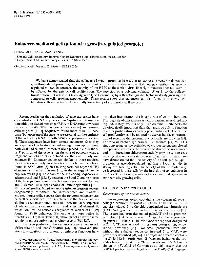
An expression vector containing the chicken a2 type 1 collagen promoter fragment (-280 to +110 relative to the cap site), cloned 5‘ to the chloramphenicol acetyltransferase (CAT) coding sequences, has been described previously [20]. The vector has been designated pCaCAT and its promoter pCa (Fig. 1). A larger chicken a2 type 1 collagen promoter fragment (- 1100 to + 110, relative to the cap site), designated pCOL CAT, was assayed in the same construction, as de- scribed previously [20]. Two SV40 promoters, with and without the enhancer sequences inserted 5’ to CAT, were previously described [20, 261. The expression vector, contain- ing SV40 342-bp PvuII-Hind111 fragment including the two 72-bp tandem repeats, the 21-bp repeats and TATA box, is similar to pSVECAT of Gorman et al. [26], except that the pBR322 portion was replaced with the EcoRI-SaZI fragment
334
AmpR EcoRI
. . P v u S
AmpR
(bp)
Fig. 1. Plasmids used in this study. The histocompatibility class 1 H2K gene promoter (2024 bp), the chicken 1x2 type 1 collagen promoter pCu (390 bp), and SV40 early promoter PvuI-Hind111 fragment (342 bp) were inserted 5’ to the coding region of the bacterial CAT gene, which has at its 3’ end two SV40 fragments providing an intron and a polyadenylation signal. pBR322 sequences from its deleted variant (pML2 of Lusky and Botchan [27]), supply the ampicillin resistance gene and the origin of replication required for propagation of the plasmids in E. coli
of pML2 [27]. The second SV40 promoter construction, which Assay of CAT activity contains the 21-bp repeats and the TATA box in a pML2CAT background, but only 22 bp of one of the 72 bp, was desig- nated pdSVCAT. The histocompatibility antigen class 1 H-2K” promoter [28] HindIII-NruI fragment (- 2021 to + 3, relative to the cap site), was cloned into theHindIII site of pSBl [20], after repair of the NruI site and the CAT proximal site of the vector with T4 polymerase. The final construct was designated pH2KCAT.
Transfection of the cells The day before transfection, 10-cm dishes were seeded
with either 3T6 or CV1 cells at a density of 1 x lo6 or 2 x 10⁶ per dish respectively, and grown in medium containing 7% fetal calf serum. The medium was changed the following morning with medium containing either 7% or 1% fetal calf serum. The two-day serum-starved cells were maintained from then on in medium containing 1% fetal calf serum. 4 h later the cells were transfected with 10 pg of the particular ex- pression vector under study and 2.5 pg of either pCHllO (291 or pML2 [27] after coprecipitation of the DNA with calcium phosphate [30]. The following morning the calcium phos- phate/DNA precipitates were removed and the cells were then maintained for a further 24 h in each medium with 7% serum for exponentially growing cells and 1% serum for starved cells, until harvesting. The cells, which were serum-starved for only 1 day, had the serum in the medium reduced to 1% at this time.
The preparation of the lysates and the assay for CAT activity have been described previously by Herbomel et al. [20, 311.
RESULTS
The expression of the collagen promoter is regulated by cell growth
To study the activity of promoters with and without enhancer elements under a variety of growth conditions, we used the CAT (chloramphenicol acetyltransferase) system of Gorman et al. [26]. In this system the expression of the bac- terial CAT gene and production of the CAT enzyme is depen- dent on the presence of eukaryotic transcription control signals. The enzymatic activity can be assayed in cells extracts 24 - 72 h after DNA transfection . Several reports have shown that the level of enzymatic activity is directly proportional to the amount of specific mRNA synthesized after transfection [20, 26, 321. Since in this system, only one type of mRNA is produced, whether enhancer elements are present or not, any problems due to different RNA processing or translational efficiency are eliminated.
The promoters used in this study were the chicken a2 type 1 collagen promoter [33], mouse histocompatibility pro- moter H2-K“ [28] and the SV40 early promoter, with and without its enhancer sequence. The shortened chicken a2 type
335
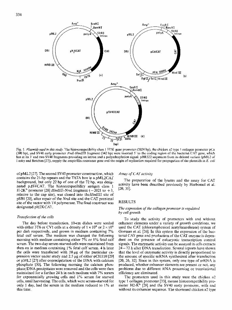
Fig. 2. CAT activity in 3T6 mousefibroblasts. The CAT activity in extracts of transfected exponentially growing and serum-starved 3T6 cells was done according to Gorman et al. [26] with the modifications mentioned by Herbomel et al. [20]. Chloramphenicol is converted into its monoacetylated derivatives Acl and Ac3 by the CAT-containing cells extracts. The extracts from pCaCAT (pCa) and pH2KCAT (H2K) were incubated for 3 h and pSV40 CAT (SVE) for 0.5 h at 37°C. 1 and 2 stand for one or two days of serum starvation
Table 1 . Effect of serum starvation on promoter activity in 3T6 cells DNA transfection of cells, preparation of extracts and the assay of CAT enzymatic activity were done as described in Experimental Procedure. Activities of the different constructions are expressed rela- tive to that of pCaCAT in exponentially growing cells (arbitrarily defined as 1). Cells starved for one day were kept in 1 70 serum between 20 h and 44 h post-transfection, the time of harvest. Cells starved for two days were kept in 1 % serum form 4 h before the transfection until the time of harvest 44 h after the addition of DNA. Values reflect the mean plus standard error of duplicate transfection experiments
Promoter Activity of
exponentially 1 day of serum 2 days of serum growing cells starvation starvation
CCC 1 f0 .2 0.3 + 0.06 0.2 f 0.05 H2K 6.8 f 0.7 6.2 f 0.4 4.6 0.8 svE 48 +8 38 + 2 44 + 5
1 promoter fragment (390 bp) was used in most of these stud- ies, to allow the insertion of the polyoma A enhancer element closer to the transcriptional unit, as described previously [20]. The different expression vectors are described in Fig. 1 ; their constructions detailed in the experimental section.
Each construction was transfected into either 3T6 or CV1 cells maintained in 7% fetal calf serum or in 1% fetal calf serum for 1 or 2 days after transfection. Culturing 3T6 cells in 1% serum slowed their overall rate of proliferation and reduced the number of cells by 50% after one day and 70% after two days respectively, as compared to exponentially growing cells, at the time that they were harvested to assay the transient expression of the constructions. The enzymatic
activity obtained after transfection with pCaCAT was threefold lower in cells serum-starved for 1 day compared to exponentially growing cells. This further decreased to fivefold lower after 2 days of serum starvation (Fig. 2 and Table 1). In comparison, the expression of pH2KCAT fell only slightly, while pSVECAT expression remained roughly constant, suggesting that the efficiency of expression of these promoters was not affected by the rate of cell proliferation. Since these expression vectors are unable to replicate in mammalian cells, the total number of copies of the vector in the cell population remains constant throughout each transfection study and the number of copies per cell therefore increases in direct propor- tion to any fall in cell number. The number of cells after serum starvation decreased by 50% after one day and 70% after two days, respectively, when compared to exponentially growing cells, leading to a correspondingly higher copy number per cell. This leads to a higher calculated activity per cell of H2K and SVE promoter because of the increased copy number. However, when calculating the overall activity of the promoter there is no need to correct for reduction in cell number as the increased copy number per cell balances it out exactly. These results show that the activity of the collagen a2 type 1 pro- moter used in these studies was sensitive to the proliferative state of the cells or to growth factors present in the serum. It therefore constituted a suitable system in which to study the effect of enhancers on growth regulated genes.
The polyoma A enhancer element was inserted 5‘ to the collagen promoter (pCaCAT 5’) in the early orientation (i.e. the early coding strand of the insert was in the same sense as the CAT coding strand), and also at the 3’ of the CAT coding sequences (PCcrCAT 3’) in the late orientation [20]. The activi- ties of pCaCAT 5‘ and pCaCAT 3’, when cotransfected with pML2 [27] as a carrier plasmid into exponentially growing
336
Table 2. Effect of cotransfected plasmids and of serum starvation on enhancer strength in 3T6 cells CAT activities expressed relative to pCaCAT in exponentially growing cells. Values in parenthesis give the enhancement factor by the polyoma A enhancer element inserted 5’ or 3’ to the collagen CAT transcription unit. Values reflect the mean plus standard deviations of four or more experiments
Plasmid Acitivity of
exponentially cells kept exponentially growing cells in I % serum growing cells cotransfected for 1 day cotransfected
with pCHllO
pCaCAT l (1 ) 0.35 (1) 0.35 (1) pCaCAT 5’ 11 (11 f 1.5) 11.7 (31 f 3.5) 4.6 (13 f 1) pCaCAT 3’ 2.8 (2.8 & 1) 5 (14.5 1.5) 3 (8.5 +_ 1.6)
cells, were compared to the level of expression of pCaCAT, to evaluate the degree of promoter activation. Element A activated the collagen a2 type 1 promoter 11-fold when in- serted 5’, and threefold when inserted 3’, in exponentially growing cells. On cotransfection with pML2 into one-day serum-starved cells, the activation by element A, inserted in either the 5‘ or 3’ position to the promoter, was three to four times greater than that observed in exponentially growing cells (Table 2). Hence element A inserted in the 5’ or 3’ position maintained the level of activity of the pCa promoter, even in 1 -day serum-starved cells under conditions which reduce the level of activity of the collagen a2 type 1 promoter threefold in plasmids lacking enhancer (Table 2) . To check whether the 390-bp chicken collagen a2 type 1 promoter behaved differently from the intact promoter, a 1210-bp chicken collagen a2 type 1 promoter fragment, cloned 5’ to the CAT sequence (pCOL CAT) [20], was transfected into exponentially growing and serum-starved 3T6 cells with and without polyoma A enhancer in the 3‘ position. Although the basal level of pCOL CAT activity in 3T6 cells was slightly lower than that of pCaCAT, the activity of pCOL CAT fell 2.5-fold in serum-starved cells compared to exponentially growing cells, and was increased threefold in the presence of enhancer in exponentially growing cells and 6.5-fold in serum-starved cells. This demonstrates that the polyoma A enhancer rescues collagen promoter activity in serum-starved cells. These effects are comparable to those observed with the shortened collagen a2 type 1 promoter (Table 2).
EJfect of competing plasmid
In the previous section we have shown that the activity of the collagen a2 type 1 promoter was markedly decreased in serum-starved cells when the plasmid did not contain a viral enhancer element. This decrease in activity may be due to competition by stronger cellular promoters for a limited pool of transcription factors and/or RNA polymerase existing in these cells. To test this hypothesis we cotransfected pCaCAT into exponentially growing cells with pCHllO [29] instead of pML2. This plasmid codes for a /3-galactosidase fusion pro- tein, expressed in mammalian cells under the control of the SV40 early promoter, and includes the SV40 enhancer se- quence. With a ratio of pCH110 to pCaCAT of one to four, the CAT activity decreased threefold as compared with pML2 as carrier (Table 2) . Transfections were performed to study the effect of the polyoma A enhancer element on the activity
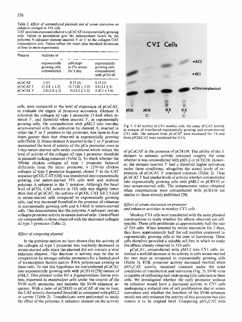
Fig. 3. CAT activity in CVI monkey cells. An assay of CAT activity in extracts of transfected exponentially growing and serum-starved CV1 cells. The extracts from pCaCAT were incubated for 3 h and from pH2KCAT were incubated for 0.5 h
of pCaCAT in the presence of pCHl10. The ability of the 5’ element to enhance activity remained roughly the same whether it was cotransfected with pML2 or pCH110. Howev- er, the element inserted 3’ had a threefold higher activation under these conditions; altogether the actual levels of ex- pression of pCaCAT 3’ remained constant (Table 2). Thus pCaCAT 3’ had similar levels of activity whether cotransfected into exponentially growing cells with pML2 or pCHllO or into serum-starved cells. The enhancement ratios obtained when constructions were cotransfected with pCHl10 are similar to those already published [20].
Effect of serum starvation on promoter and enhancer activities in monkey CVI cells
Monkey CV1 cells were transfected with the same plasmid constructions to study whether the effects observed are cell- specific. These cells proliferate at approximately half the rate of 3T6 cells. When arrested by sverum starvation for 2 days, they have approximately half the cell number compared to exponentially growing cells at the time of harvesting. These cells therefore provided a suitable cell line in which to study the effects already observed in 3’1’6 cells.
pCaCAT, cotransfected with pML2 into CV1 cells, ex- hibited a tenfold decrease in its activity in cells serum-starved for two days as compared to exponentially growing cells (Table 3). H2K promoter activity decreased twofold while pSVECAT activity remained constant under the same conditions of transfection and starvation (Fig. 3). SV40 virus is capable of replicating and undergoing lytic infection in these cells. We investigated whether the early promoter without its enhancer would have a decreased activity in CV1 cells undergoing a reduced rate of cell proliferation due to serum starvation and whether the insertion of the SV40 enhancer would not only enhance the activity of this promoter but also restore it to its original level. Comparing pSVECAT with
337
Table 3. Effect ofserum starvation on promoter activity in CV1 monkey cells CAT activities expressed relative to pCuCAT in growing cells. Numbers in parenthesis give the enhancement factor of the SV40 early promoter by its homologous enhancer placed in its original 5’ position. Values reflect the mean and standard error of duplicate transfection experiments ~~~~ ~
Promoter Activity of
exponentially growing cells for 2 days
cells kept in 1 % serum
H2K 2.9 f 0.2 1.4 & 0.4 PCN 1 k0.25 0.1 * 0.01 ASV 0.3 & 0.03 (1) 0.2f0.04 (1) SVE 3.2 & 0.3 (10.4) 3.4 f 0.6 (1 6.4)
Table 4. Effect of serum starvation on polyoma enhancer function in CVl cells CAT activities expressed relative to that of pCaCAT in exponentially growing cells (mean and standard error of two transfection experi- ments). Values in parenthesis give enhancement factors
enhancer and p ASVECAT without enhancer, the SV40 enhancer augmented the activity of the basic SV40 promoter tenfold in exponentially growing cells (the same value found by Gorman et al. [26]) and 16-fold in serum-starved cells, a result which indicates that there was little increase in the level of enhancement of the SV40 promoter in serum-starved cells (Table 3).
The level of enhancement of pCaCAT by the polyoma enhancer element A inserted in the 5‘ position was reduced threefold in CV1 cells relative to 3T6 mouse cells, whereas when placed at the 3‘ position its activity was similar in both cell types (Tables 2, 4). Serum starvation decreased the level of activity of the collagen promoter with the polyoma A enhancer at either the 5‘ or 3’ position only threefold (Table 4) relative to the tenfold decrease of the constructions lacking enhancer, so that there was an effective threefold net increase in enhancement of the pCa promoter in two-day serum-starved cells compared to exponentially growing cells (Table 4).
DISCUSSION Mouse 3T6 cells and monkey CV1 cells provided two
suitable cell lines in which to study the effects of growth inhibition of cells by serum starvation on promoters, since they could be transfected with expression plasmids by the calcium phosphate coprecipitation technique while other serum-starved cell lines were virtually refractory. The effect of the proliferative state of the cells on the activity of different promoters placed before the bacterial gene CAT was studied in these cell lines. The SV40 early promoter (WE) activity
was insensitive to the growth state of the cells; however, the collagen-promoter(pCa)-dependent CAT activity decreased threefold and tenfold respectively in serum-starved 3T6 and CV1 cells. The mouse H2K promoter and the enhancer-less SV40 early promoter (AWE) showed an intermediate pattern of behaviour : a decrease in activity of up to 50%.
The characteristics of the collagen a2 type 1 promoter in our two cell lines are consistent with changes in collagen synthesis found during growth in vivo of human skin fibroblasts [36, 371 guinea-pig fibroblasts 1361 and monkey and rabbit smooth muscle cells [34, 351. In all these cells the rate of collagen synthesis decreases in conditions of reduced cell proliferation, in a proportion similar to that of non- collagen protein synthesis [34]. When growth ceases, the de- cline in the synthesis of collagens type I and type 111 differs: the synthesis of the first declines more rapidly than that of the second. In another study on human fetal lung cells, in which cells growing to high cell density were compared with ex- ponentially growing cells [38], there was no decline in collagen type I or mRNA synthesis, although it is not clear exactly what effect growth to high cell density was having on this type of cell.
The observation that both collagen a2 type 1 promoter fragments (i. e. pCclCAT and pCOL CAT) behave in a similar manner, in that their activities decrease in serum-starved cells and can be rescued by element A in the 3’ position, demonstrates that the -280 to +110 promoter fragment contains the upstream regulatory sequences for this collagen gene. Since a study on the effect of viral transformation on the 1210-bp chicken collagen a2 type 1 promoter fragment (pCOL CAT) demonstrated that it behaved in a similar manner to the endogeneous mouse collagen a2 type 1 pro- moter, this suggests that the transcriptional regulatory se- quences are contained within these promoter fragments [39].
We inserted the major polyoma enhancer element A into either the 3’ or the 5‘ position of the pCaCAT transcription unit in order to investigate the effect of viral enhancers on the activity of a growth-sensitive promoter. We have demon- strated that this enhancer prevents the marked decline in the level of activity of this promoter in serum-starved 3T6 cells; it restores the activity of the collagen a2 type 1 promoter to at least a level identical to that measured in the presence of enhancer in exponentially growing cells. Consequently the net enhancement of the collagen a2 type 1 promoter by the polyoma A element is three to fivefold greater in starved cells relative to normally growing cells. The situation is similar in CV1 cells. The enhancement is greater in starved than in growing cells although the absolute level of CAT activity obtained in starved cells is still lower than in exponentially growing cells. This result is compatible with previous observa- tions that showed that the polyoma enhancer is less active in monkey than in mouse cells [40]. The activity of the enhancer- less ASV40 promoter is increased by its homologous enhancer in exponentially growing and starved CV1 cells by factors of 10 an 16 respectively, which reflects the minimal change in SV40 promoter activity seen in serum-starved cells.
Cotransfection of pCaCAT with another chimeric plasmid containing a strong promoter, SVE, results in a similar decrease in activity of the collagen promoter as seen after serum starvation. Here again the addition of the polyoma A enhancer element 3’ or 5’ to the pCaCAT transcription unit restores totally or partially the activity of this transcription unit relative to the same constructions cotransfected with a carrier plasmid not carrying an eukaryotic promoter. This demonstrates that the polyoma A enhancer can effectively
338
compete with the strong enhancer found upstream of SV40 early promoter, for cellular factors.
In all the experiments conducted in this study the activity of the promoters was determined by an enzymatic assay of chloramphenicol acetyltransferase. This assumes that the ac- tivity of the promoter in the different constructs is directly proportional to the enzymatic activity and that no differences in translational efficiency of CAT mRNA or in RNA stability have occurred under our experimental conditions. Such differences are unlikely in respect to the collagen promoter. Firstly, the RNA is initiated correctly with or without polyoma enhancer elements in the presence of p-galactosidase plasmid [20], and the level of CAT mRNA corresponds to the enzymatic activities measured. Secondly, if the CAT mRNA were three to five times more unstable in serum-starved cells than in exponentially growing cells, then the insertion of an enhancer element either 5' or 3' to the promoter should stabilise the mRNA, which is unlikely because no enhancer sequences are present in mature CAT mRNA. Furthermore, since the enhancer increases promoter activity in serum- starved cells by a greater amount than in exponentially growing cells, if the reduced activity in serum starved cells were due to a decrease in mRNA stability the enhancer ele- ment would have to be activating the promoter by an amount to keep the level of production of mRNA three times higher than in exponentially growing cells, in order to maintain the apparent equal level of activity of the promoter under the two growth conditions, a possibility which is highly unlikely.
Previous studies have shown that the magnitude of the effect seen by enhancers can depend on their distance [20,41] and sometimes orientation [20, 42, 431 relative to the test promoter. Furthermore different promoters respond differ- ently to the same set of enhancers [44]. Here we show that the strength of an enhancer can depend on the physiological state of the cells used in transfection assays and on the presence of competing plasmids.
In conclusion these results clearly show that enhancers can function either 5' or 3' to a gene in slowly growing cells to activate the low expression of a growth-dependent promoter like that of the collagen a2 type 1 gene. The intriguing possibility exists that a growth-regulated gene controlling some steps in cell proliferation may be activated in normally resting or slowly growing cells by insertion of a viral enhancer element next to that gene. This event could result in an increase in the growth rate of a previously non-proliferating cell.
We are grateful to P. Herbomel for making available the pCctCAT constructs and P. Kourilsky and S. Kvist for the gift of H2K plasmids. We thank G. Ringold for providing us with plasmid pCH110. We are also indebted to J. Ars and Dr C. Laroche for their help with the preparation of this manuscript. G. Moore held a Royal Society European Fellowship. This work was supported by grants from the Centre National de la Recherche Scientqique LA 270, the Institut National de la Santt et de la Recherche Mkdicale PRC 11 801 1, the Ministry of Industry and Research (82 V1290 82 V1388, 83 V0297), the Fondation pour la Recherche Medicale Francaise and the Association pour la Recherche sur le Cancer.
REFERENCES 1. Mc Knight, S. L. & Kingsbury, R. (1982) Science (Wash. DC)
2. Dierks, P., Van Ooyen, A,, Cochran, M. D., Dobkin, C., Reiser, 217,316-324.
J. & Weissman, C. (1983) Cell 32, 695-706.
3. Yaniv, M. (1984) Biol. Cell. 50, 203-216. 4. Benoist, C. & Chambon, P. (1981) Nature (Lond.) 290, 304-
5. Gruss, P., Dhar, R. & Khoury, P. (1981) Proc. Nut1 Acad. Sci. 310.
6. 7.
8. 9.
10.
11.
12. 13.
14. 15.
16. 17.
18. 19.
20.
21. 22. 23.
24. 25. 26.
27. 28.
29.
, ,
USA 78,943 - 947. Fromm, M. & Berg, P. (1982) J. Mol. Appl. Genet. I , 451 -481. Tyndall, C., La Mantia, G., Thacker, C. M., Favarolo, J. &
Banerji, J., Rusconi, S. & Schaffner, W. (1981) Cell 27,299-308. DeVilliers, J. & Schaffner, W. (1981) Nucleic Acids Res. 9,6251 -
Levinson, B., Khoury, G., Van de Woude, G. & Gruss, P. (1983)
Lusky, M., Berg, L., Weiher, H. ,& Botchan, M. (1983) Mol. Cell.
Hearing, P. & Shenk, T. (1983) CeU33,695-703. Hen, R., Borelli, E., Sassone-Corsi, P. & Chambon, P. (1983)
Banerji, J., Olson, L. & Schaffner, W. (1983) Cell 33, 729-740. Gillies, S. D., Morrison, S. L., Oi, V. T. & Tonegawa, S . (1983)
Queen, C. & Baltimore, D. (1983) Cell 33, 741 -748. Emorine, L., Kuehl, M., Weir, L., Leder, P. &Wax, E. E. (1983)
Picard, D. & Schaffner, W. (1984) Nature (Lond.) 307, 80-82. Weiher, H., Konig, M. & Gruss, P. (1982) Science (Wash. D C )
Herbomel, P., Bourachot, B. & 'Yaniv, M. (1984) CeN 39, 653-
Yaniv, M. (1982) Nature (Lond.) 297, 17-18. Khoury, G. & Gruss, P. (1983) Cell 33, 313-314. Pardee, A. B., Dubrow, R., Hamelin, J. L. & Kletzien, R. F.
Austin, S . A. & Kay, J. E. (1982) Essays Biochem. 18, 79- 120. Austin, S. A. & Clemens, M. J. (1981) Biosci. Rep. 1 , 35-44. Gorman, C. M., Moffat, L. F. &Howard, B. H. (1982) Mol. Cell.
Lusky, M. & Botchan, M. (1981:) Nature (Lond.) 293, 79-82. Kimura, A,, Israel, A,, Le Bail, 0. & Kourilsky, P. (1986) Cell
Hall, C., Jacob, E., Ringold, G. & Lee, F. (1983) J . Mol. Appl.
Kamen, R. (1981) Nucleic Acids Res. 9, 6231 -6250.
6264.
Nature (Lond.) 295, 568 - 572.
Biol. 3, 1108 - 1122.
Nucleic Acids Res. 11, 8747 - 8760.
Cell 33, 729 - 740.
Nature (Lond.) 304,442 - 449.
210,626-631.
662.
(1978) Annu. Rev. Biochem. 47, 715-730.
Biol. 2,1044-1051.
44,261 -212.
Genet. 2, 101 - 109. 30. Graham, F. L. &Van der Eb, A. J. (1973) Virology 52,456-467. 31. Herbomel, P., de Crombrugghe, B. & Yaniv, M. (1983) Cold
Spring Harbor. Con$ 10,285-294. 32. Bohnlein, E., Chowdhury, K. & Gruss, P. (1985) Nucleic Acids
Res. 13,4789-4809. 33. Vogeli, G., Ohkubo, H., Sobel, M. E., Yamada, Y., Pastan, I . &
de Crombrugghe, B. (1981) Proc. Natl Acad. Sci. USA 78,5334. 34. Burke, J. M. & Ross, R. (1977) Exp. Cell. Res. 107, 387-395. 35. Holderbaum, 0. & Ehrhart, A. L. (1984) Exp. Cell Res. 153,16-
37. Aumailley, M., Krieg, T., Razaka, G., Mueller, P. & Bricaud, H.
38. Tolstoshev, P., Berg, R., Rennard, S., Bradley, K., Trapnell, B. &
39. Schmidt, A., Setoyama, C. & de Crombrugghe, B. (1 985) Nature
40. De Villiers, J., Olson, L., Tyndall, C. & Schaffner, W. (1982)
41. Wasylylk, B., Wasylylk, C., Augerean, P. & Chambon, P. (1983)
42. Fromm, M. & Berg, P. (1983) Mol. Cell Biol. 3, 991 -999. 43. Linney, E. & Donerley, S. (1983) Cell 35, 693-699. 44. Gluzman, Y. & Shenk, T. (1983) Current communications in mo-
lecular biology, Cold Spring Harbor Laboratory.
24.
(Lond.) 279,442-444.
(1982) Biochem. J. 206, 505-510.
Crystal, R. (1981) J . Biol. Chem. 256, 3135-3140.