17
Enterprise | Interest None

Enterprise | Interest
None

CASE REPORTCytology and histology of a potential life-threatening airway injury
Vanessa HenriquesLucília Monteiro

Clinical History
• 90 years-old male
• Bedridden
• Previous thoracic trauma with lung perforation
Presented to the ER with:
Dyspnea, thoracalgia, vomiting.
Lymphocytosis
Elevated RCP Chest X-ray on admission: Right hilum and lower lobe opacities.

Chest X-ray on 10th day after admission:Right hilum and lower lobe hypotransparency.
Chest CT Scan: Right lower lobecollapse and pleural effusion.
Day 10 of Antibiotics

Right main bronchus: slightly elevated, yellow and friable with easy bleeding.
Bronchoscopy
Right bronchus branching: yellowish and necrotic aspect.
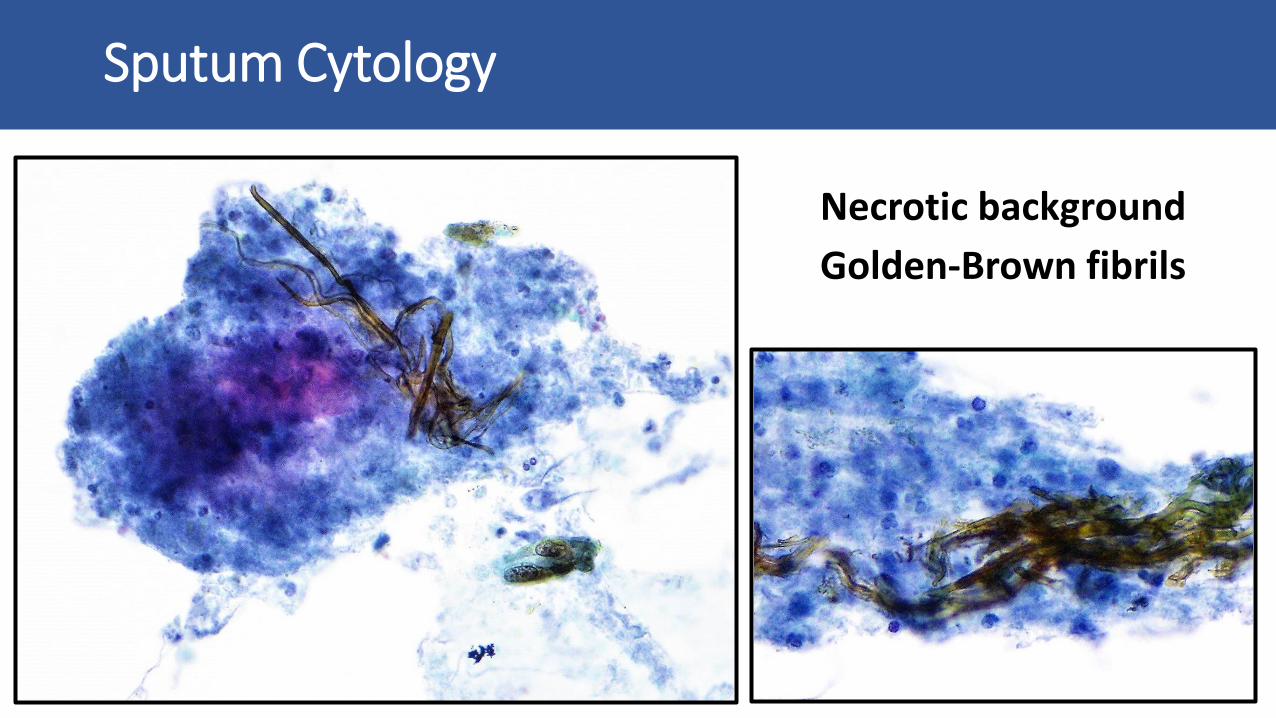
Necrotic background
Golden-Brown fibrils
Sputum Cytology

Squamous cells with reactive features.
Sputum Cytology

Yeasts and
Hyphae
Sputum Cytology

Necrosis
Golden-brown fragments
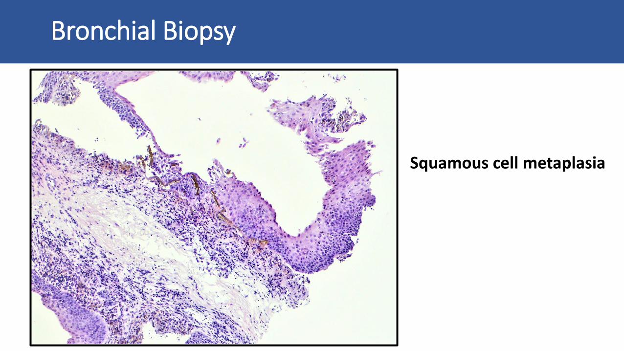
Bronchial Biopsy

Fibrillary and
Birefringent deposits
Bronchial Biopsy
Hyphae morphologicallysuggestive of Candidaspp.
Bronchial Biopsy
Squamous cell metaplasia

Residual iron fragments under squamous cell metaplasia
Perls Stain
Bronchial Biopsy

Patient medication history?
Oral iron supplementation was prescribed during admission.

DIAGNOSIS
Iron pill Aspiration Bronchitis

• 32 cases published (mean age of 68 y-o)
• > 50% patients did not realise the aspiration event
• Clinical presentation commonly non-specific: 8 haemoptysis 2 fatal
• Diagnosis relies on biopsy and medication history
• No foreign body identification Chemical burn!
Literature Review
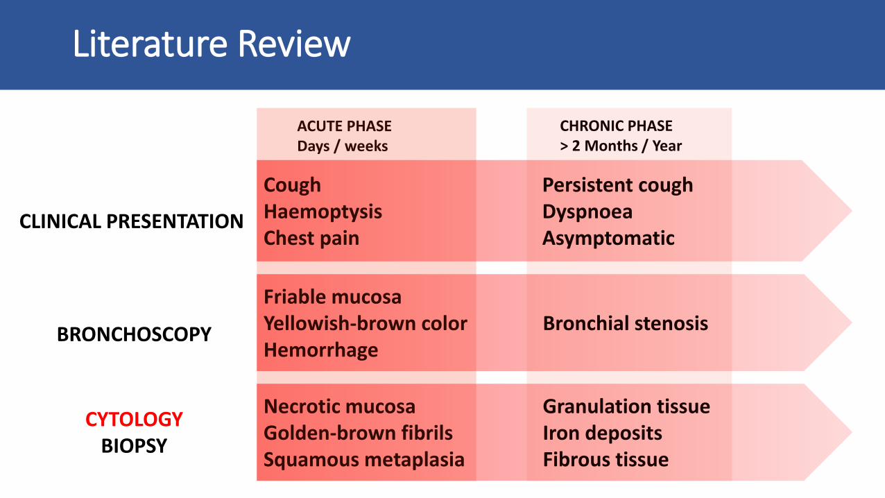
CoughHaemoptysisChest pain
Persistent coughDyspnoeaAsymptomatic
Friable mucosaYellowish-brown colorHemorrhage
Bronchial stenosis
CLINICAL PRESENTATION
BRONCHOSCOPY
CYTOLOGYBIOPSY
Necrotic mucosaGolden-brown fibrils Squamous metaplasia
Granulation tissueIron depositsFibrous tissue
Literature Review
ACUTE PHASEDays / weeks
CHRONIC PHASE> 2 Months / Year

Thank you for your attention!
Lisbon, Portugal


