163
ENTRAPMENT SYNDROMES DR.GURU PRASAD (DNB ORTHO)
| Date post: | 15-Jul-2015 |
| Category: |
Education |
| Upload: | sguruprasad311286 |
| View: | 546 times |
| Download: | 4 times |
ENTRAPMENT SYNDROMES
DR.GURU PRASAD (DNB ORTHO)

INTRODUCTION AND
GENERAL FEATURES
Definition
• Focal neuropathy due to restriction or mechanical distortion of nerve within the fibrous or fibro-osseous tunnel
• The nerve is injured by 1. chronic direct compression,
-external-internal
2. angulations
3. stretching forces
causing mechanical damage to the nerve.
IN GENERAL
• All entrapments may have one of the basic structurea)Fibro-osseous tunnel
- Carpal tunnel - Tarsal tunnel- Suprascapular nerve tunnel
b)Fibrotendinous archade-supinator (archade of frohse)-pyriformis-peroneal nerve entrapment-interosseous nerve entrapment
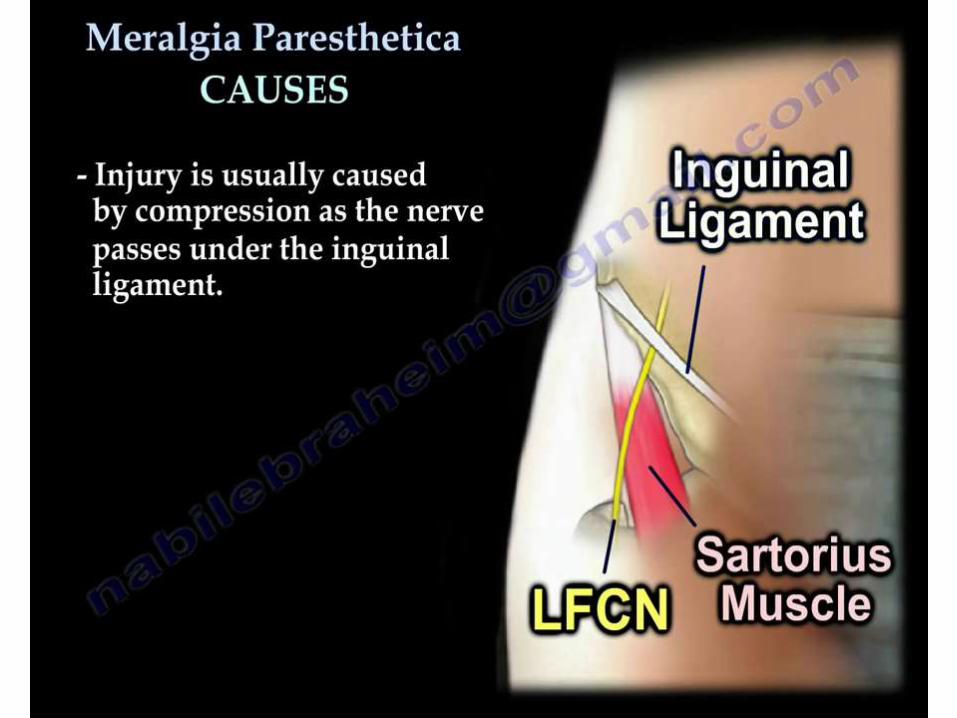
c)Abnormal bands causing compression-Thoracic outlet syndrome -meralgia parasthetica
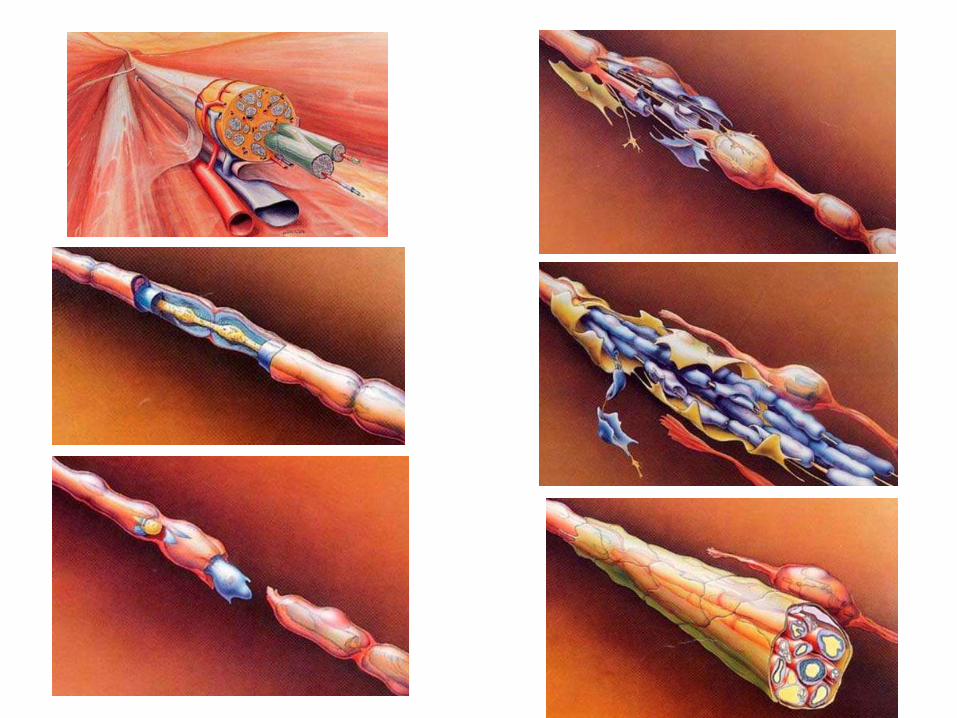
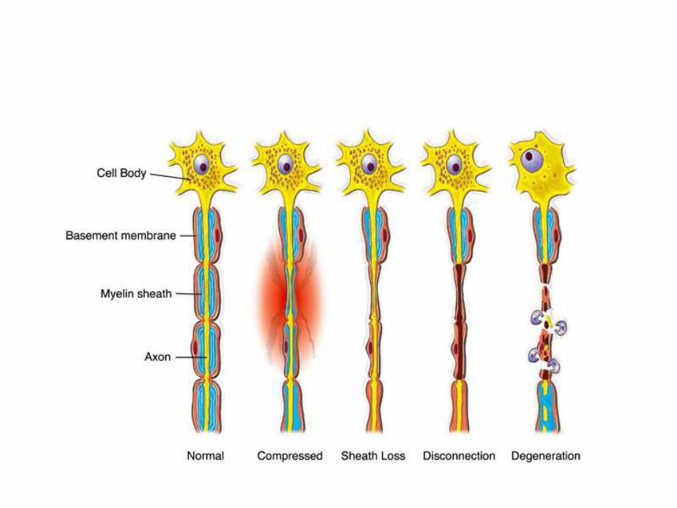
Pathophysiology
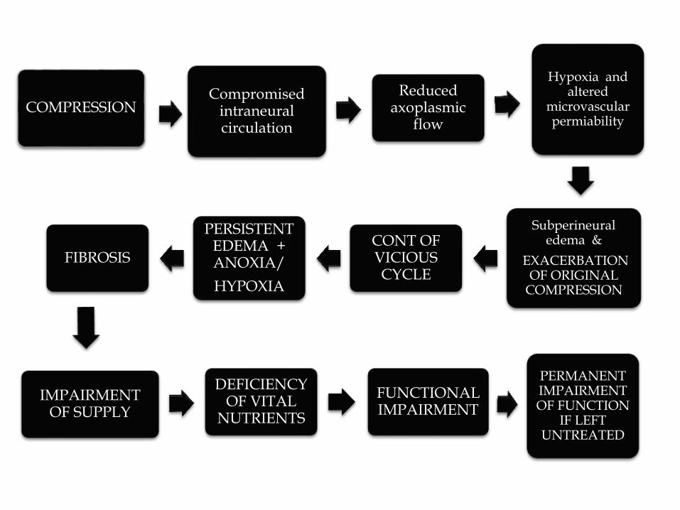
COMPRESSIONCompromised
intraneuralcirculation
Reduced axoplasmic
flow
Hypoxia and altered
microvascularpermiability
Subperineuraledema &
EXACERBATION OF ORIGINAL
COMPRESSION
CONT OF VICIOUS CYCLE
PERSISTENT EDEMA + ANOXIA/
HYPOXIA
FIBROSIS
IMPAIRMENT OF SUPPLY
DEFICIENCY OF VITAL
NUTRIENTS
FUNCTIONAL IMPAIRMENT
PERMANENT IMPAIRMENT OF FUNCTION
IF LEFT UNTREATED

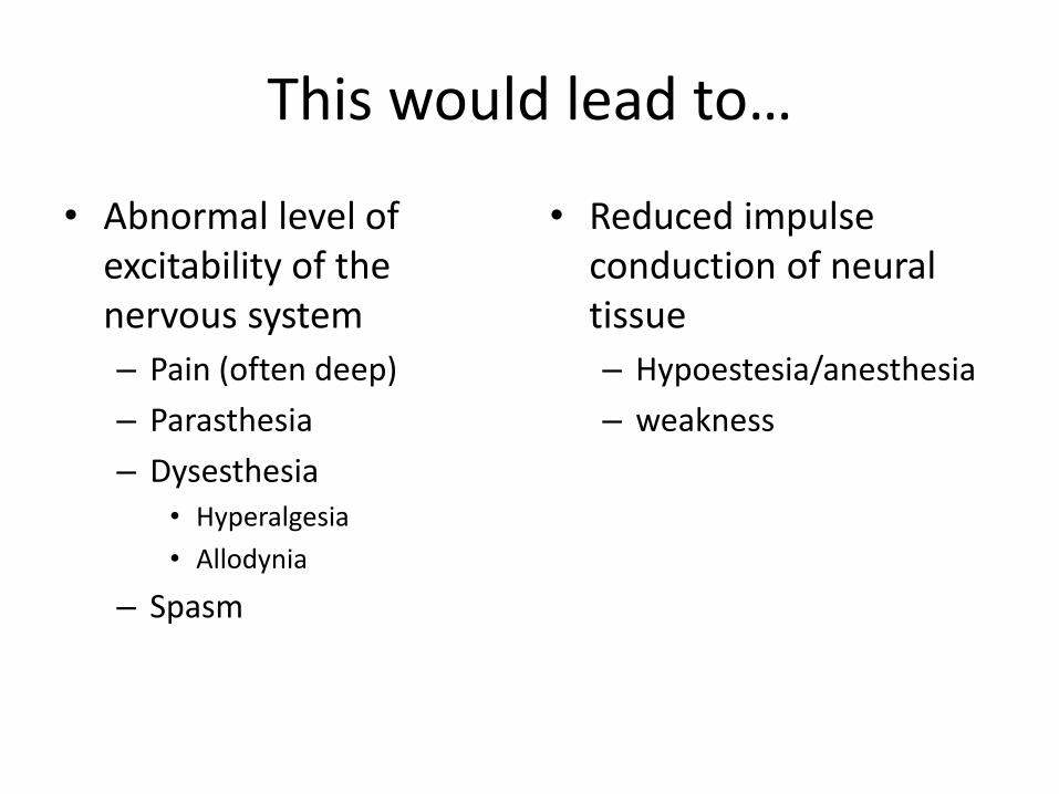
This would lead to…
• Abnormal level of excitability of the nervous system
– Pain (often deep)
– Parasthesia
– Dysesthesia• Hyperalgesia
• Allodynia
– Spasm
• Reduced impulse conduction of neural tissue
– Hypoestesia/anesthesia
– weakness
CLINICAL SCENARIO
Either or all
• Pain
• Numbness
• Tingling
• Burning
• Weakness
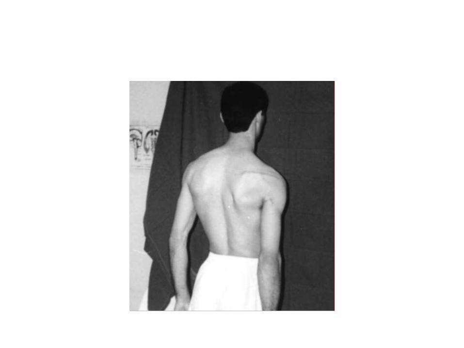
• Muscle wasting(severe cases)
in respective anatomical areas
Evalution
• History
• Physical examinations
• Investigations
General conditions associated that lead to neuropathy
• Systemic
• Guillain-Barre syndrome
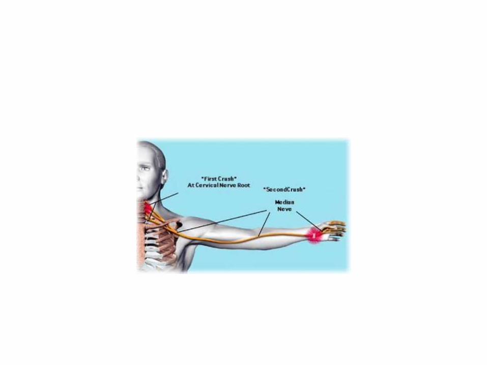
• Double crush syndrome
- A proximal level of nerve compression could cause
more distal sites to be susceptible to compression.
Physical examination
• Motor changes • -deformity• -loss of movements• -lagging• Sensory changes• - areas of loss of sensation• Autonomous • -vasomotor• -pilomotor• -tropic
• Provocative tests
• Special tests
• Tinels sign
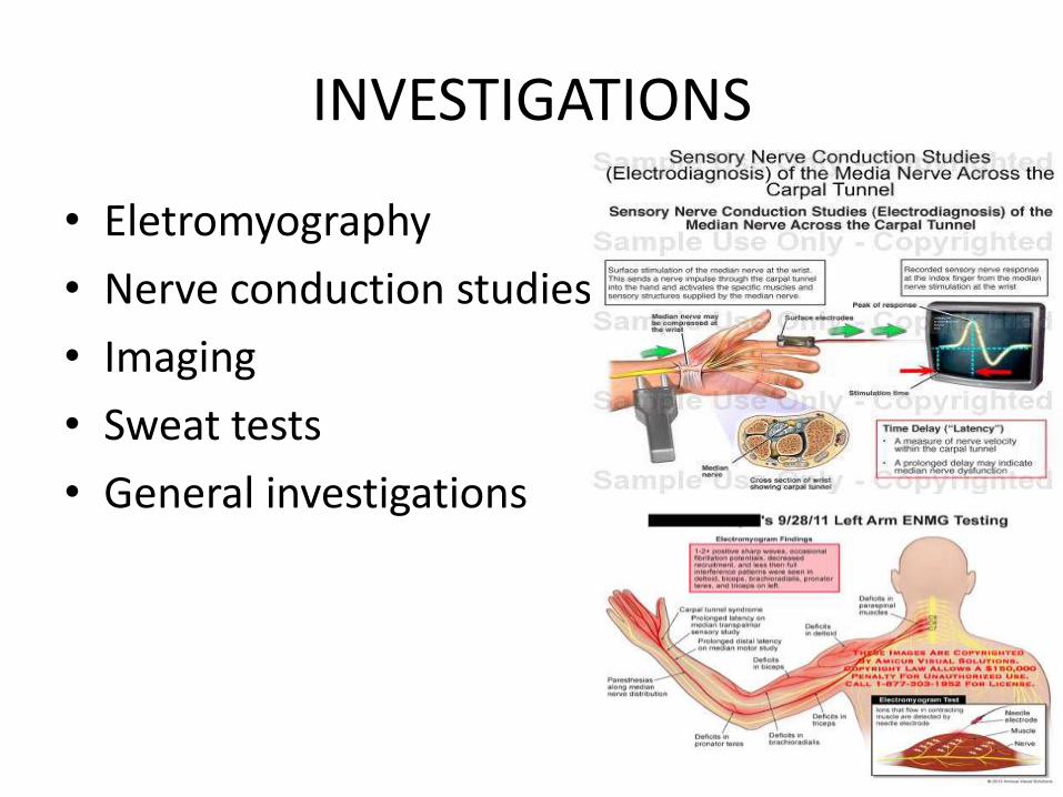
INVESTIGATIONS
• Eletromyography
• Nerve conduction studies
• Imaging
• Sweat tests
• General investigations
Treatment
• Conservative
• surgical
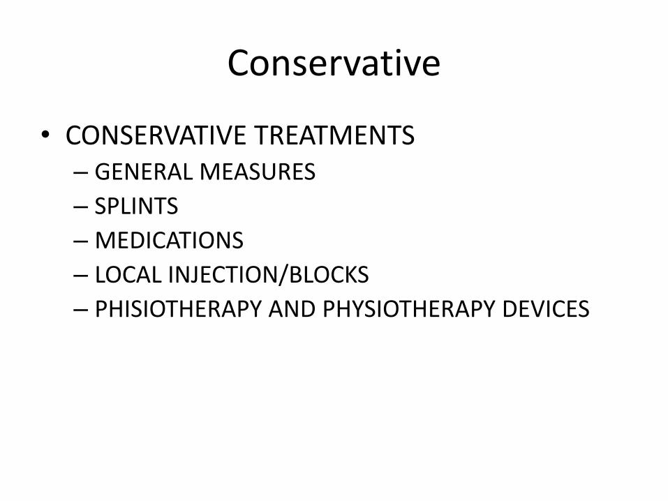
Conservative
• CONSERVATIVE TREATMENTS– GENERAL MEASURES
– SPLINTS
– MEDICATIONS
– LOCAL INJECTION/BLOCKS
– PHISIOTHERAPY AND PHYSIOTHERAPY DEVICES
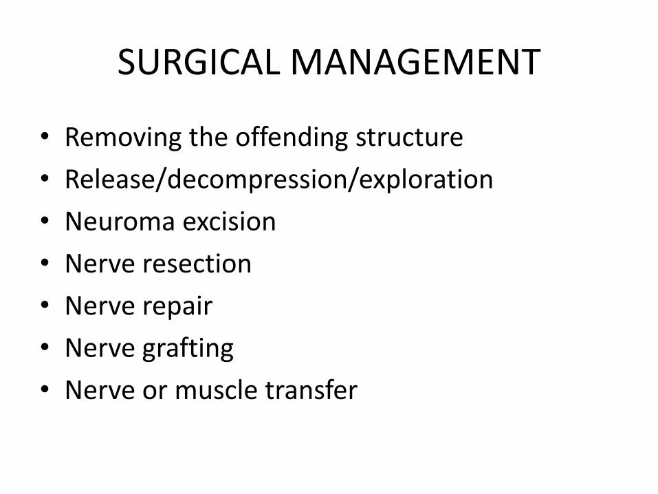
SURGICAL MANAGEMENT
• Removing the offending structure
• Release/decompression/exploration
• Neuroma excision
• Nerve resection
• Nerve repair
• Nerve grafting
• Nerve or muscle transfer
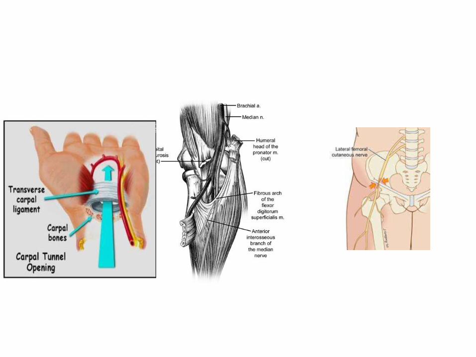
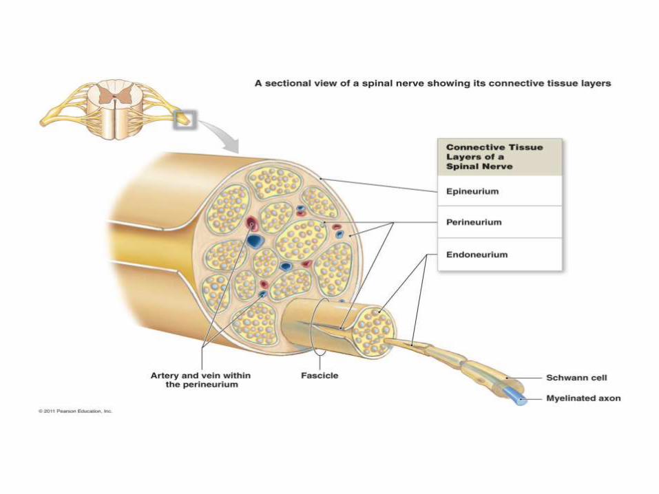
General anatomy
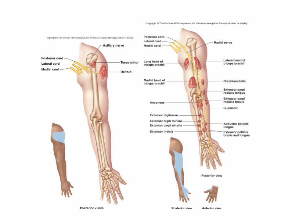

SPECIFIC ENTRAPMENT SYNDROMES
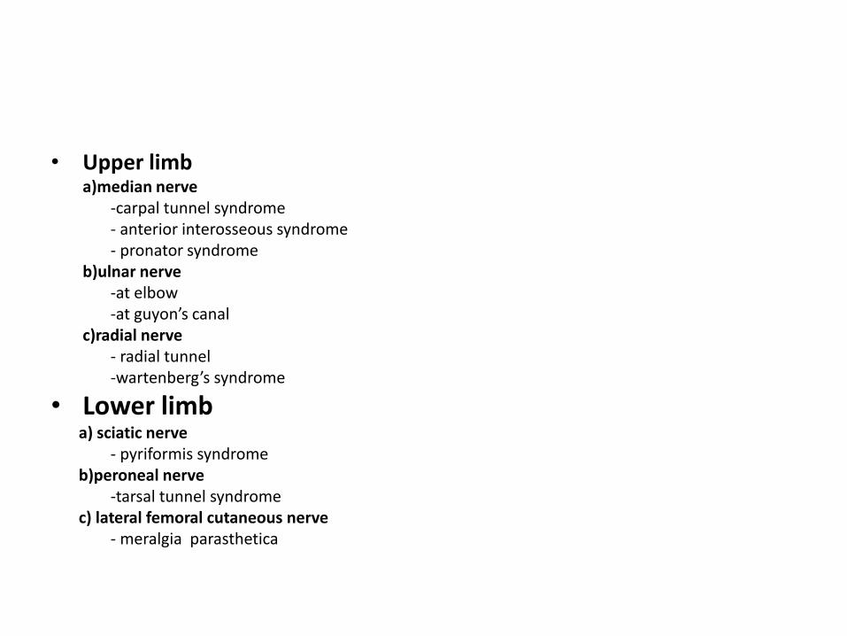
• Upper limba)median nerve
-carpal tunnel syndrome- anterior interosseous syndrome- pronator syndrome
b)ulnar nerve-at elbow-at guyon’s canal
c)radial nerve- radial tunnel-wartenberg’s syndrome
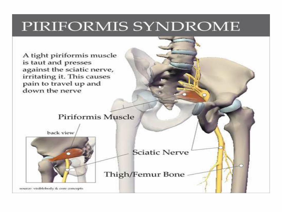
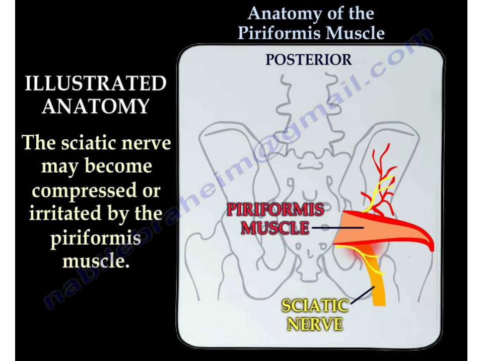
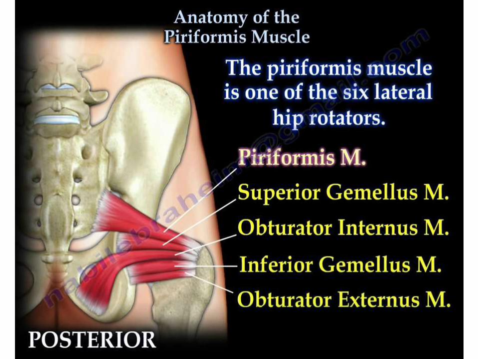
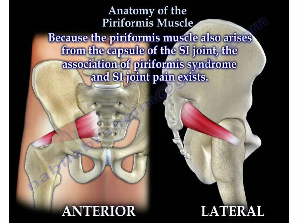
• Lower limba) sciatic nerve
- pyriformis syndromeb)peroneal nerve
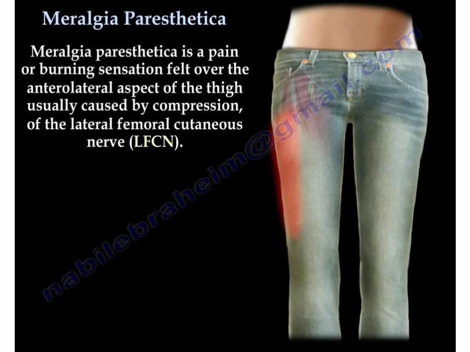
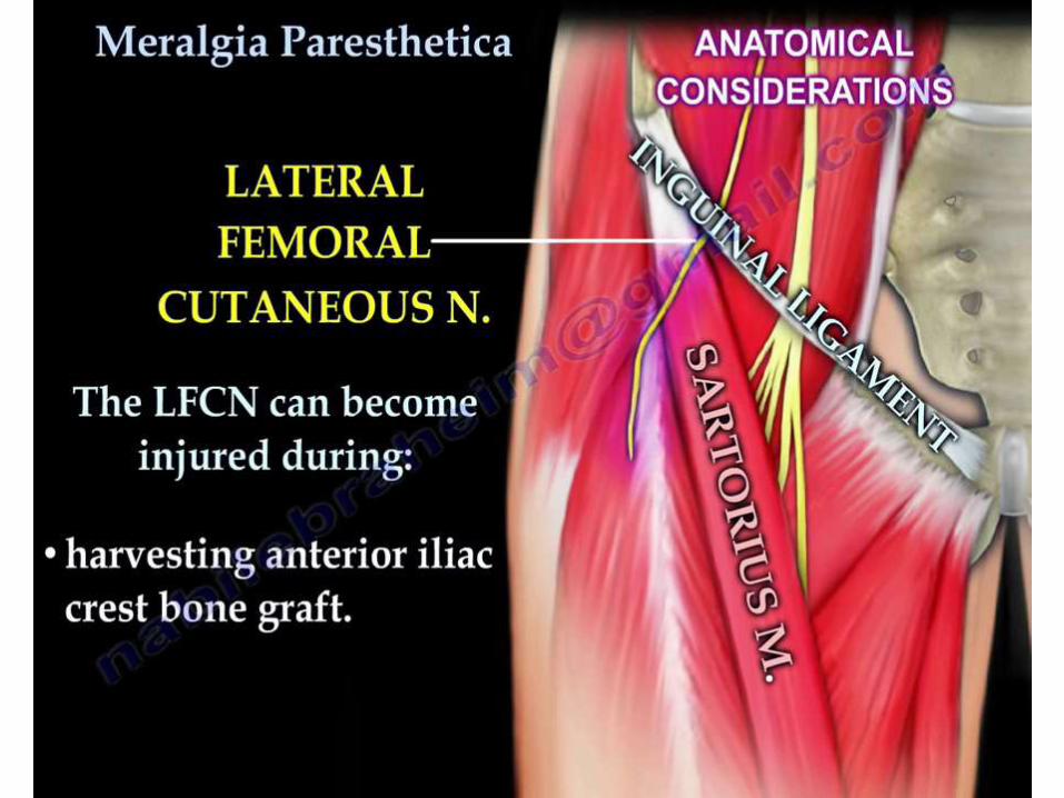
-tarsal tunnel syndromec) lateral femoral cutaneous nerve
- meralgia parasthetica
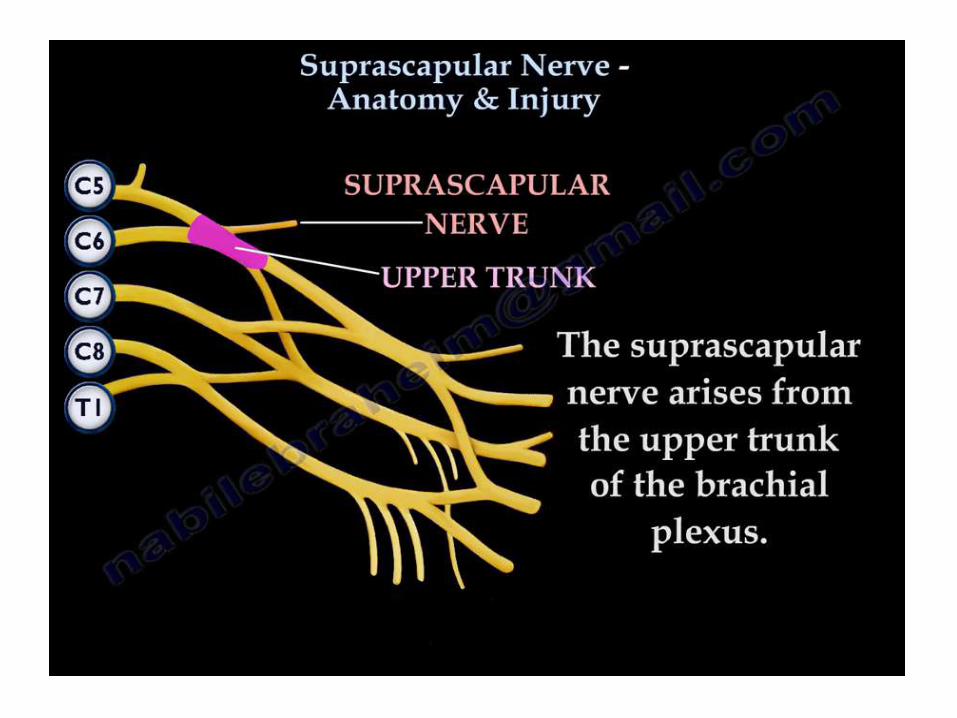
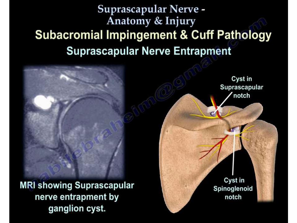
SUPRASCAPULAR NERVE ENTRAPMENT
SUPRASCAPULAR NERVE ENTRAPMENT
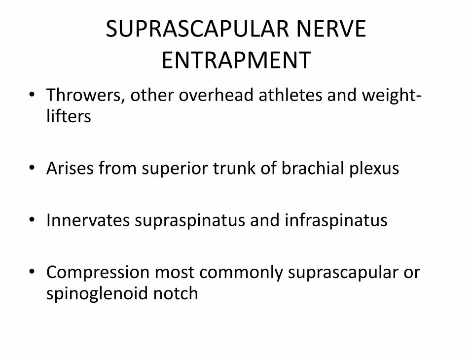
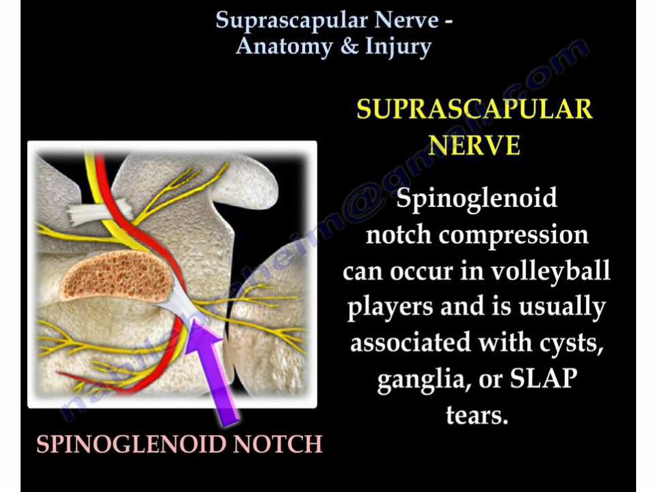
• Throwers, other overhead athletes and weight-lifters
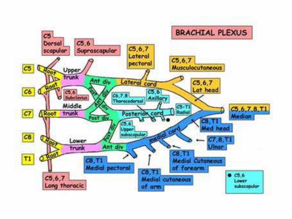
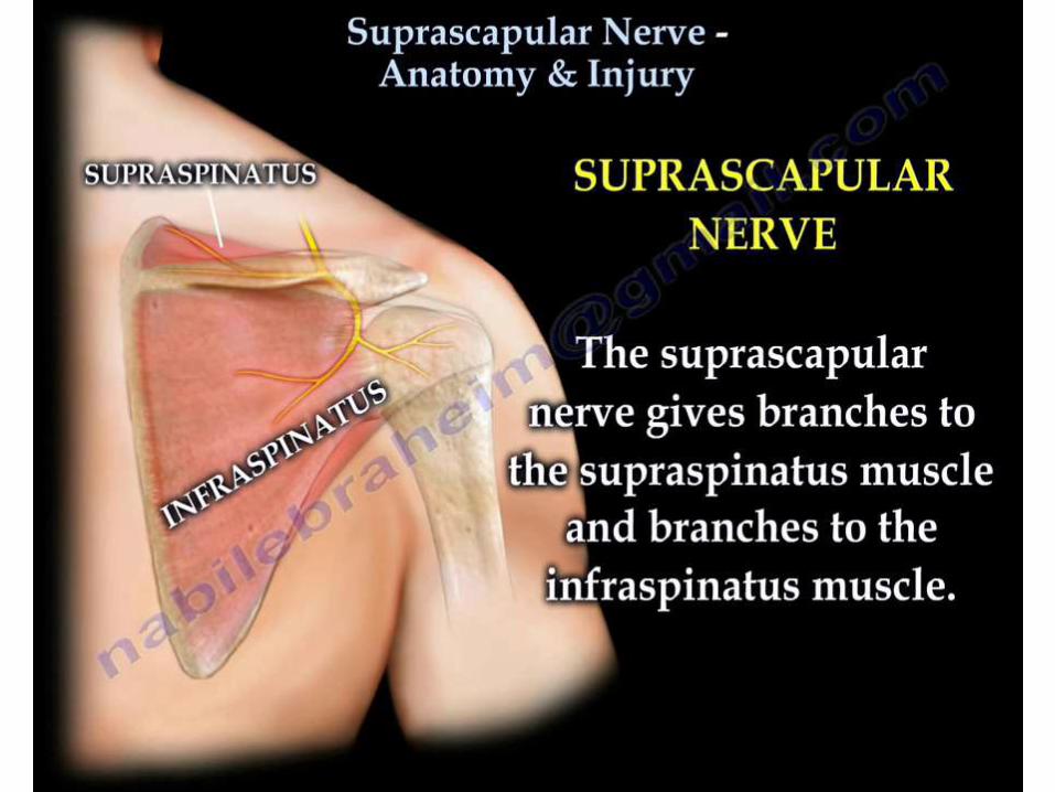
• Arises from superior trunk of brachial plexus
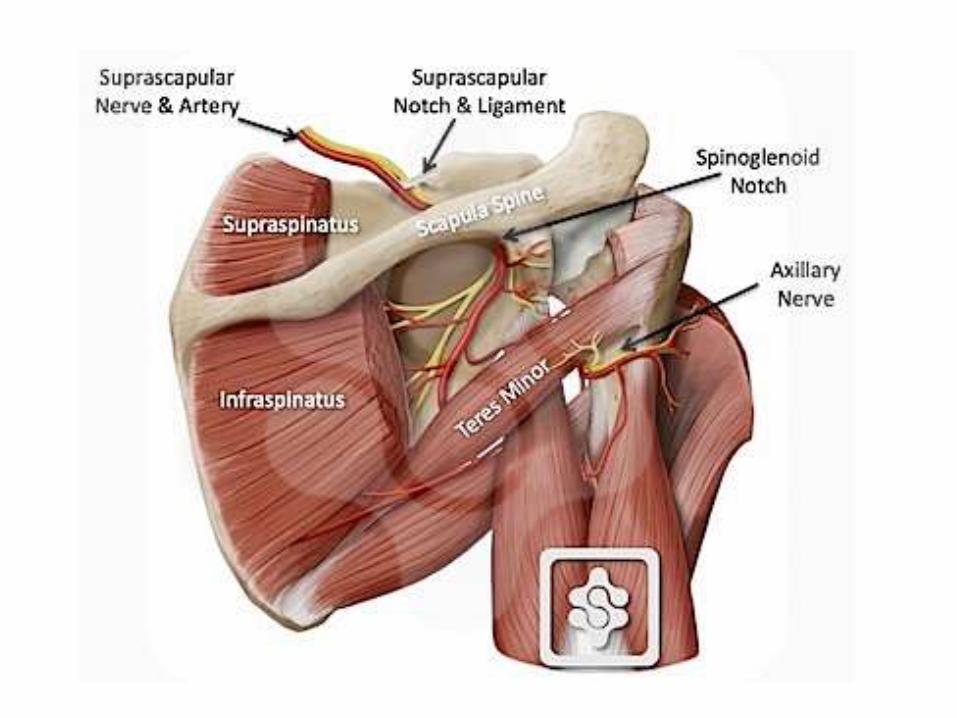
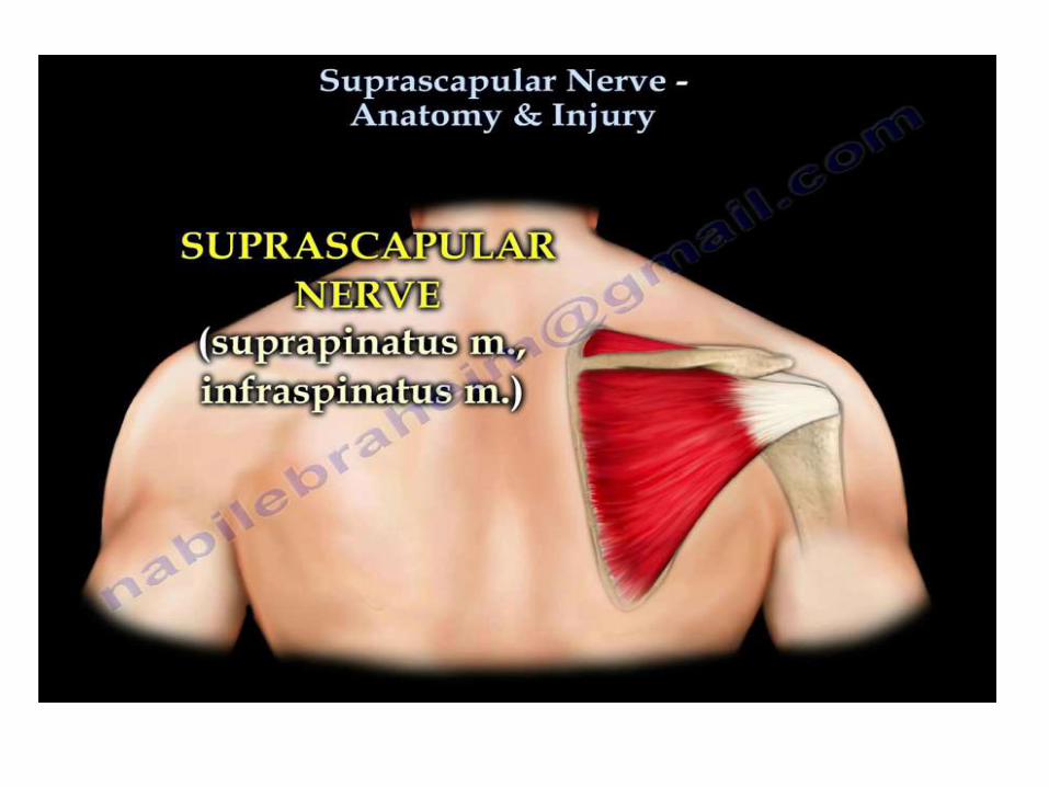
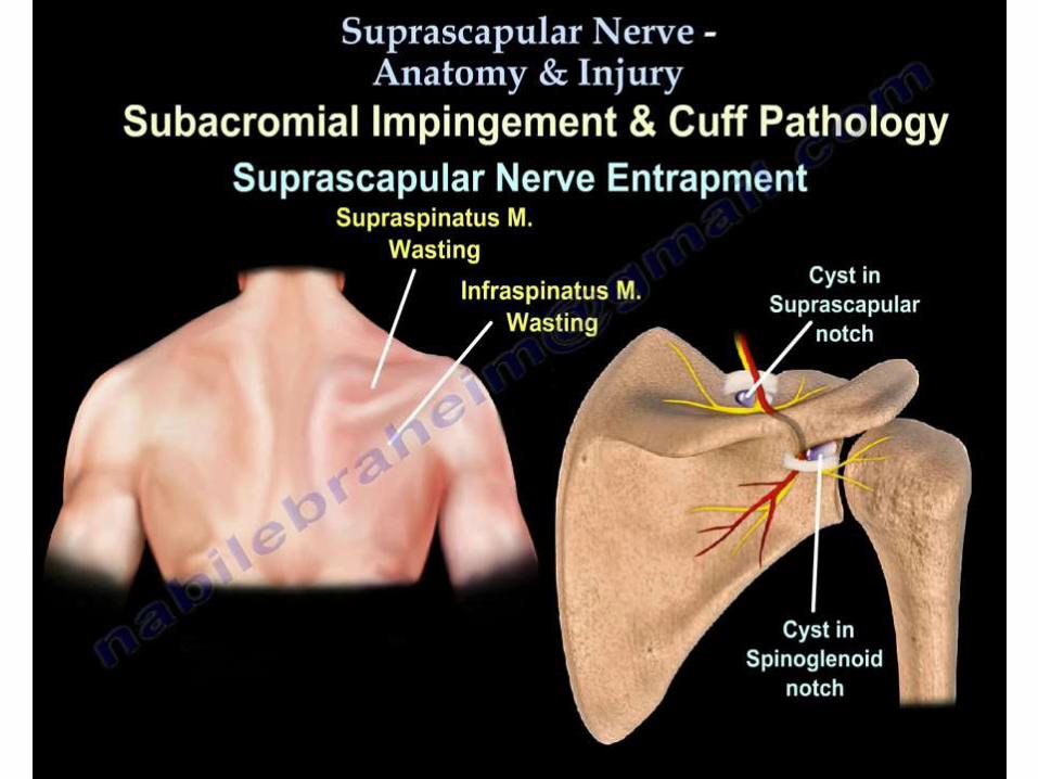
• Innervates supraspinatus and infraspinatus
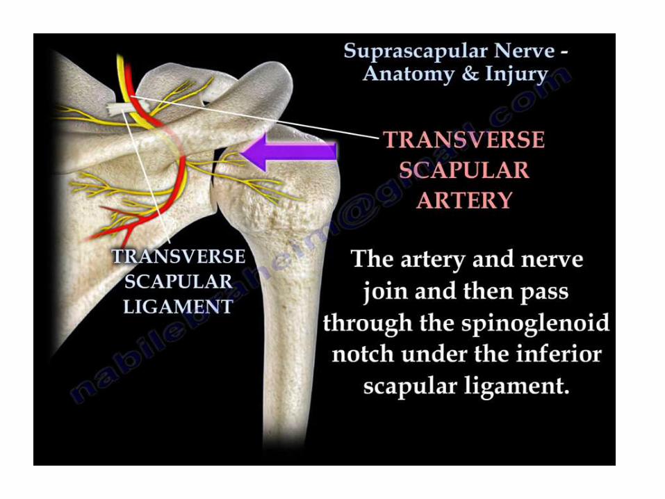
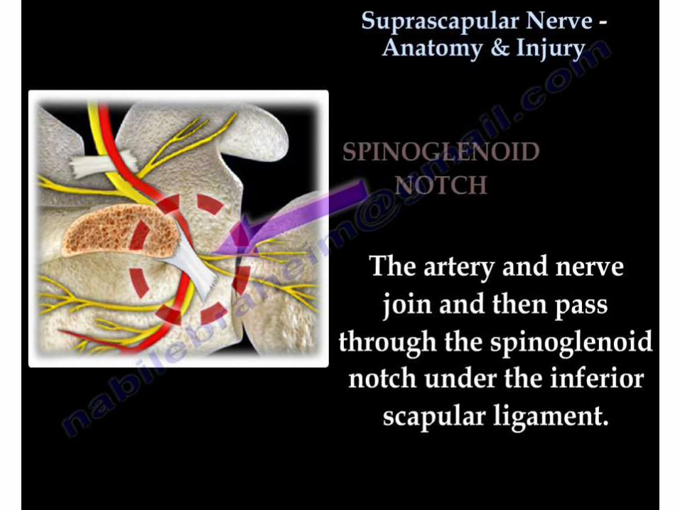
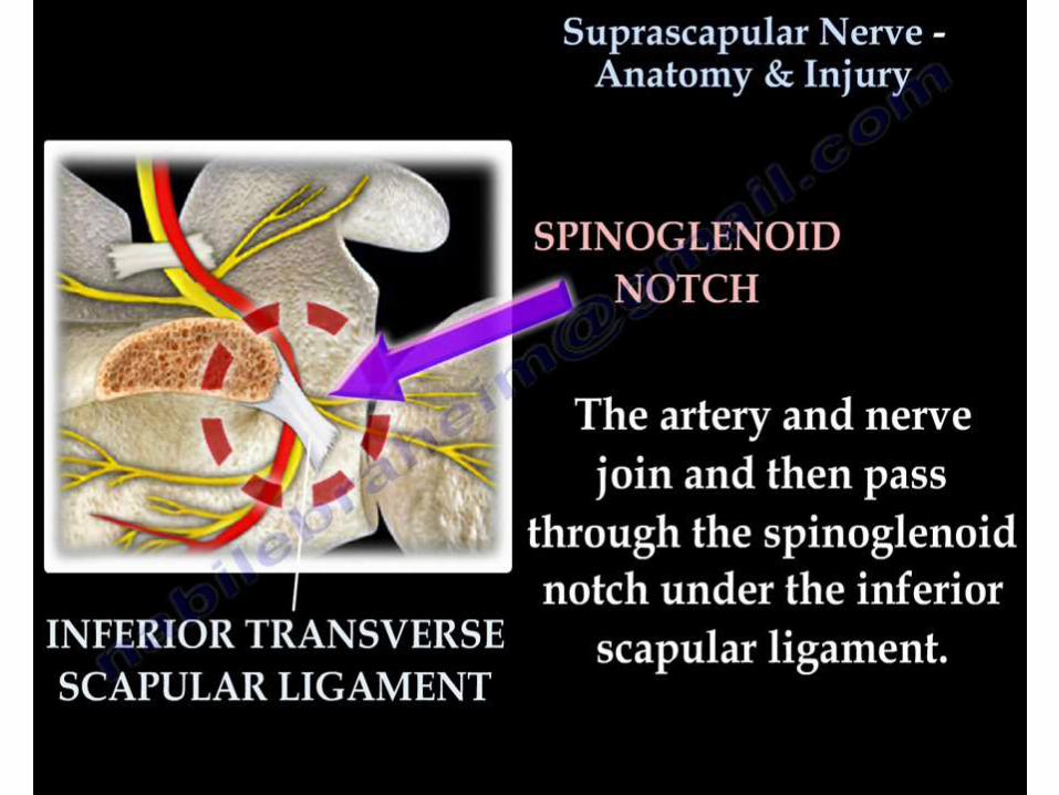
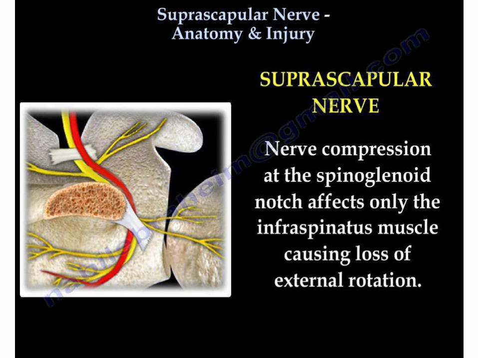
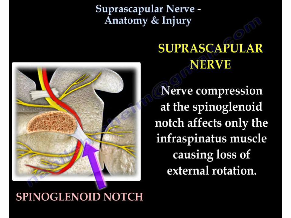
• Compression most commonly suprascapular or spinoglenoid notch
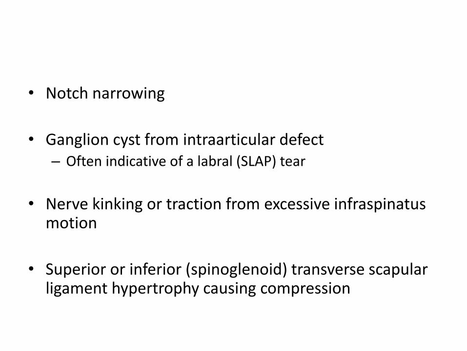
• Notch narrowing
• Ganglion cyst from intraarticular defect– Often indicative of a labral (SLAP) tear
• Nerve kinking or traction from excessive infraspinatus motion
• Superior or inferior (spinoglenoid) transverse scapular ligament hypertrophy causing compression
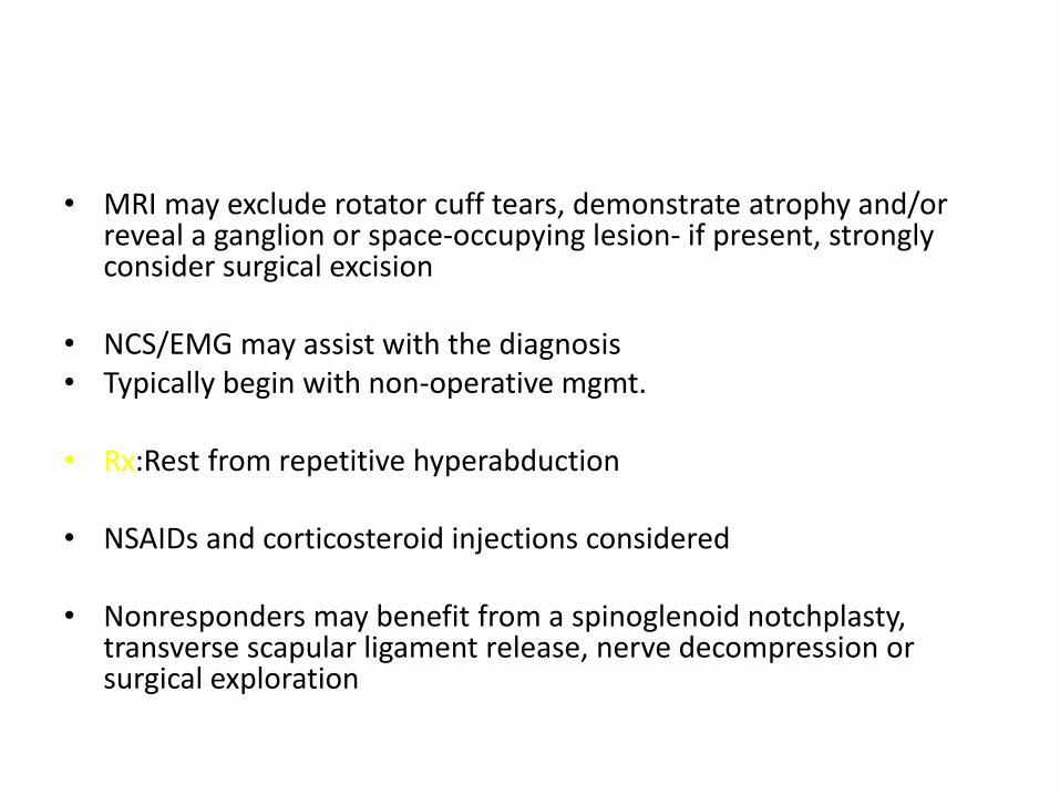
• MRI may exclude rotator cuff tears, demonstrate atrophy and/or reveal a ganglion or space-occupying lesion- if present, strongly consider surgical excision
• NCS/EMG may assist with the diagnosis• Typically begin with non-operative mgmt.
• Rx:Rest from repetitive hyperabduction
• NSAIDs and corticosteroid injections considered
• Nonresponders may benefit from a spinoglenoid notchplasty, transverse scapular ligament release, nerve decompression or surgical exploration
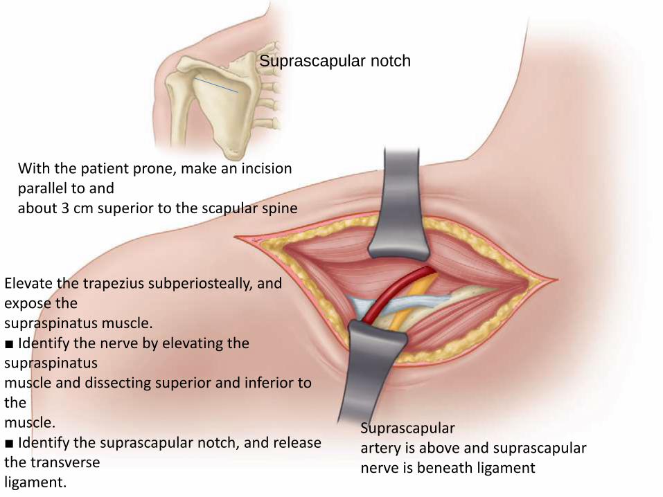
Suprascapular notch
With the patient prone, make an incision parallel to andabout 3 cm superior to the scapular spine
Suprascapularartery is above and suprascapularnerve is beneath ligament
Elevate the trapezius subperiosteally, and expose thesupraspinatus muscle.■ Identify the nerve by elevating the supraspinatusmuscle and dissecting superior and inferior to themuscle.■ Identify the suprascapular notch, and release the transverseligament.
UPPER LIMB
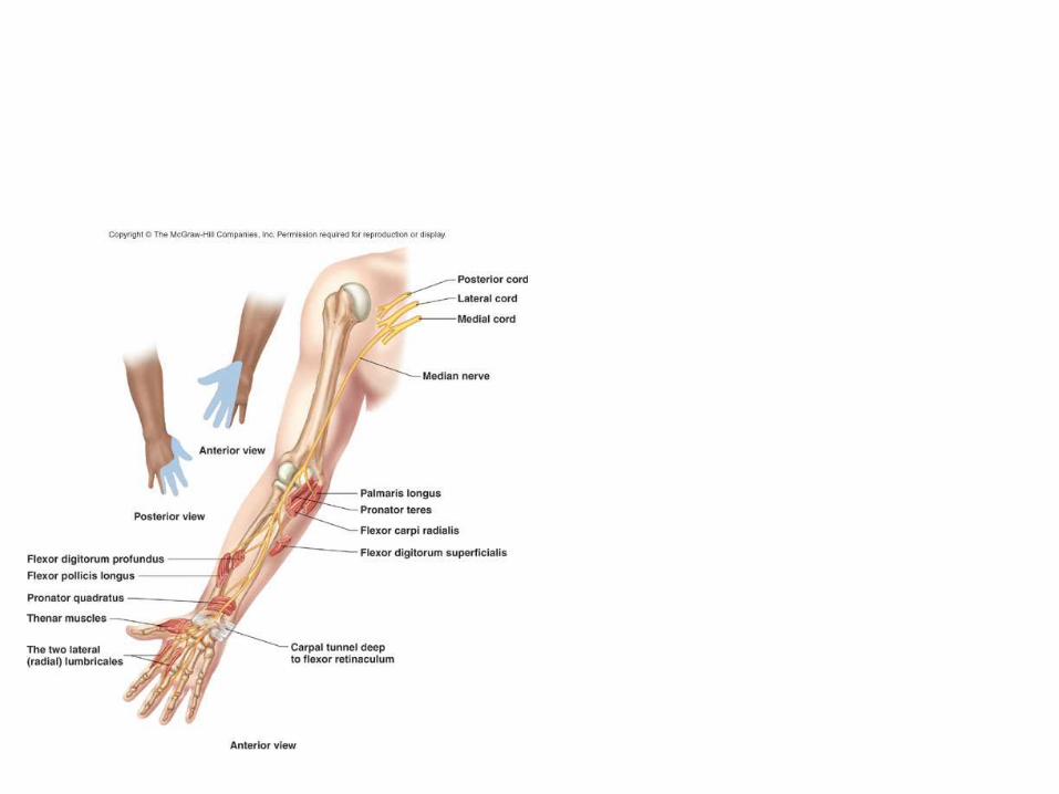
MEDIAN NERVE ENTRAPMENTS
CARPAL TUNNEL SYNDROME
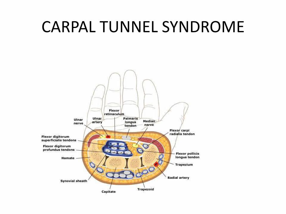
CARPAL TUNNEL SYNDROME
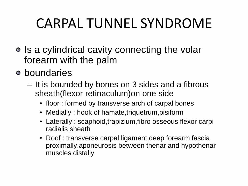
Is a cylindrical cavity connecting the volar forearm with the palm
boundaries– It is bounded by bones on 3 sides and a fibrous
sheath(flexor retinaculum)on one side• floor : formed by transverse arch of carpal bones
• Medially : hook of hamate,triquetrum,pisiform
• Laterally : scaphoid,trapizium,fibro osseous flexor carpi radialis sheath
• Roof : transverse carpal ligament,deep forearm fascia proximally,aponeurosis between thenar and hypothenarmuscles distally
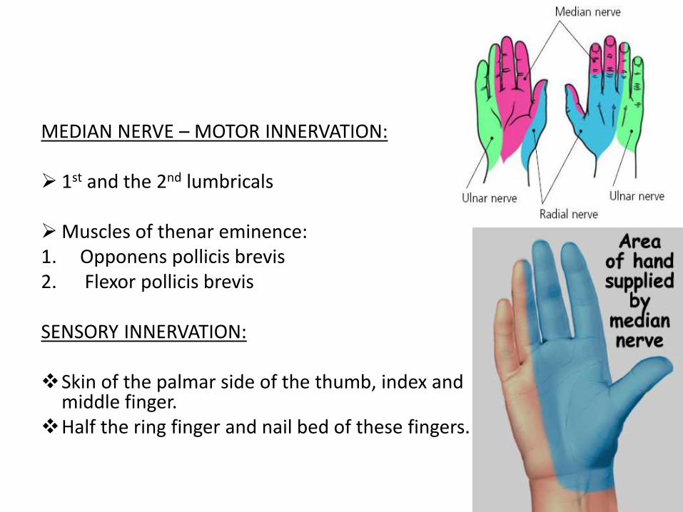
MEDIAN NERVE – MOTOR INNERVATION:
1st and the 2nd lumbricals
Muscles of thenar eminence: 1. Opponens pollicis brevis2. Flexor pollicis brevis
SENSORY INNERVATION:
Skin of the palmar side of the thumb, index and middle finger.
Half the ring finger and nail bed of these fingers.
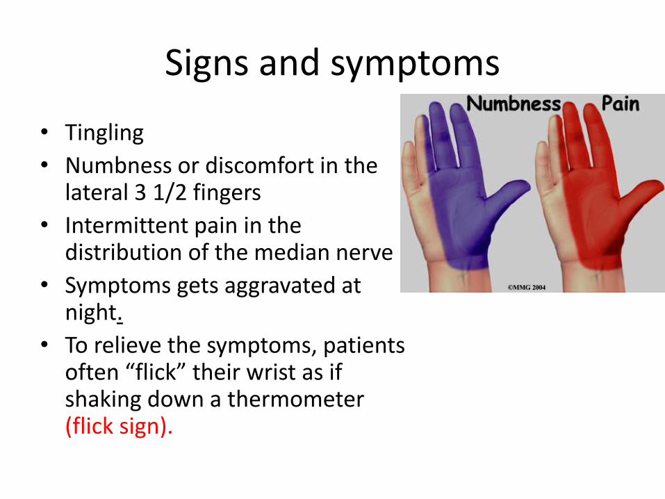
Signs and symptoms
• Tingling
• Numbness or discomfort in the lateral 3 1/2 fingers
• Intermittent pain in the distribution of the median nerve
• Symptoms gets aggravated at night.
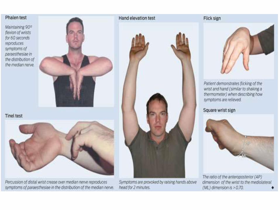
• To relieve the symptoms, patients often “flick” their wrist as if shaking down a thermometer (flick sign).
MOTOR CHANGES:
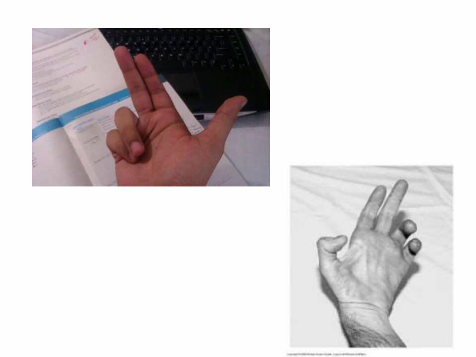
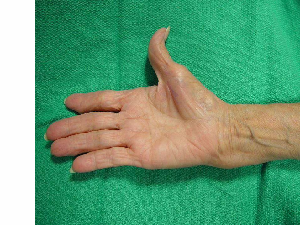
Apelike thumb deformity
Loss of opposition of thumb
Index and middle finger lag behind when making the fist.
SENSORY CHANGES:
Loss of sensation of lateral 3 1/2 digits including the nail bed and distal phalanges ondorsum of hand(An important point to remember for Carpal tunnel syndrome is that there is no sensory loss overthe thenar eminence in Carpal tunnel syndrome because the branch of median nerve thatinnervates it (palmar cutaneous branch) passes superficial to Carpal tunnel and not through it).
VASOMOTOR CHANGES:
• Skin area with sensory loss is warmer• Dry skin
TROPHIC CHANGES:
• Long standing cases leads to dry and scaly skin• Nail crack easily• Atrophy of the pulp of the fingers.

Physical Assessment Tests: • Less sensitivity to pain where the median nerve runs to
the fingers• Thumb weakness• Inability to tell the difference between one and two
sharp points on the fingertips• Flick Signal. The patient is asked, "What do you do
when your symptoms are worse?"If the patient responds with a motion that resembles
shaking a thermometer, the doctor can strongly suspect carpal tunnel.
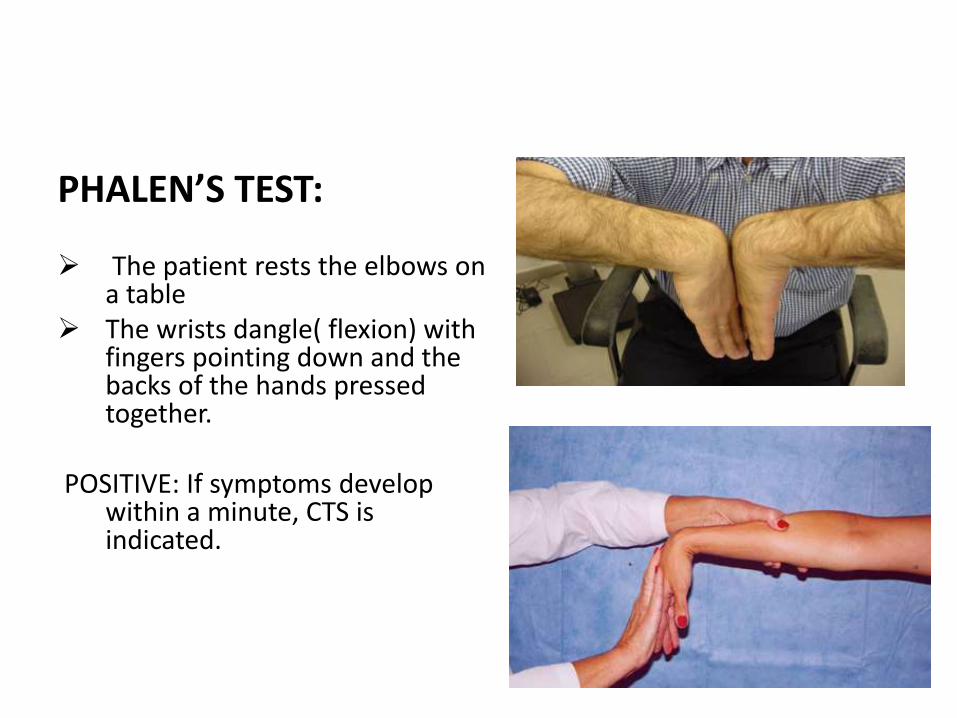
PHALEN’S TEST:
The patient rests the elbows on a table
The wrists dangle( flexion) with fingers pointing down and the backs of the hands pressed together.
POSITIVE: If symptoms develop within a minute, CTS is indicated.
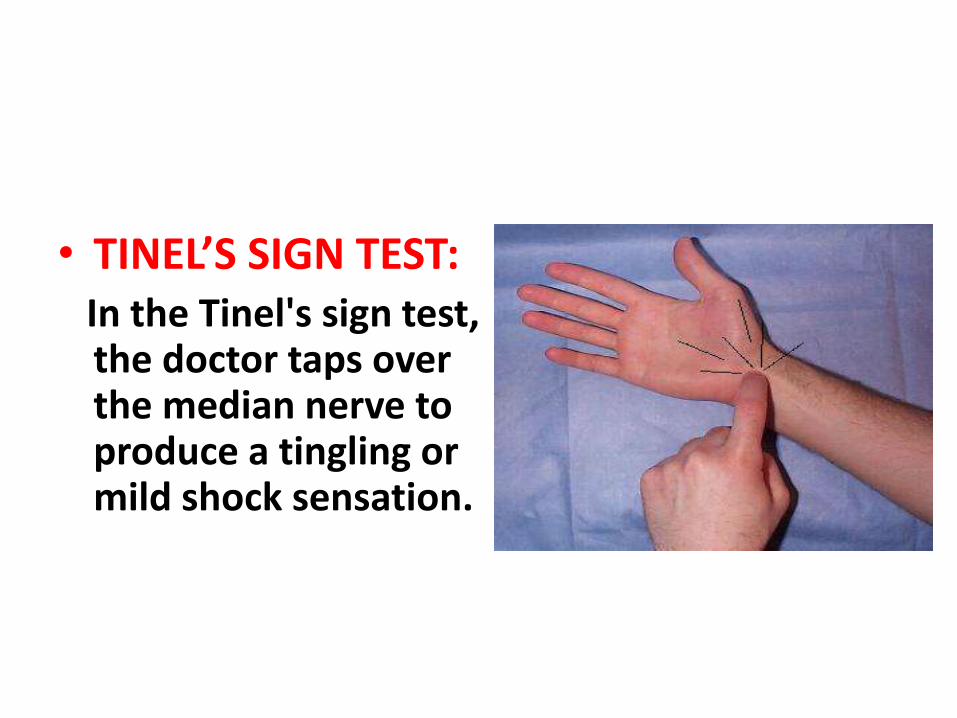
• TINEL’S SIGN TEST:In the Tinel's sign test, the doctor taps over the median nerve to produce a tingling or mild shock sensation.
o DURKAN TEST:
The doctor presses over the carpal tunnel for 30 seconds to produce tingling or shock in the median nerve.
o HAND ELEVATION TEST:
The patient raises his or her hand overhead for 2 minutes to produce symptoms of CTS.
• Torniquet test:
Torniquet inflated above systolic for one minute intensifies the symptoms
• Carpal compression test:
Pressure with both the thumbs to the median nerve in the carpal tunnel for 30 sec will aggravate the symptoms
• Tests for sensations :
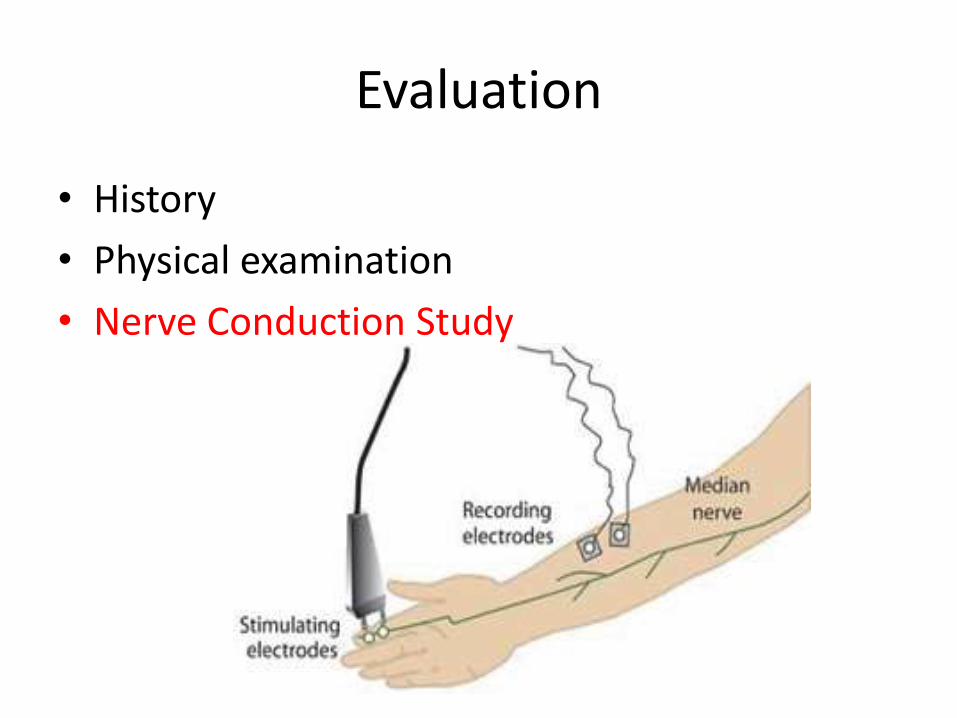
Evaluation
• History
• Physical examination
• Nerve Conduction Study
• CONSERVATIVE TREATMENTS– GENERAL MEASURES
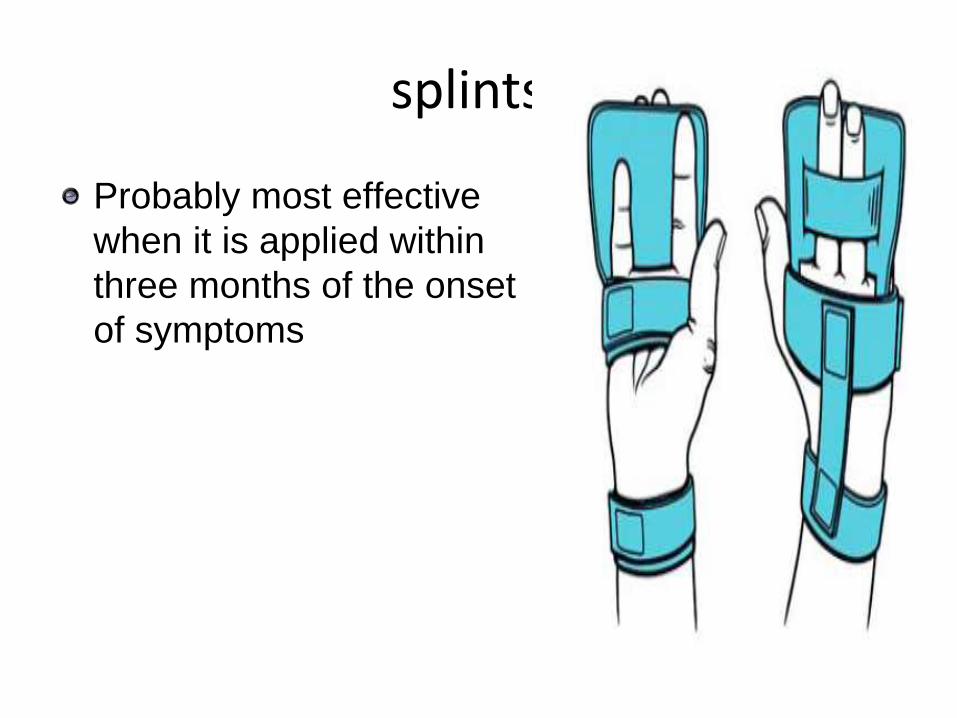
– WRIST SPLINTS
– ORAL MEDICATIONS
– LOCAL INJECTION
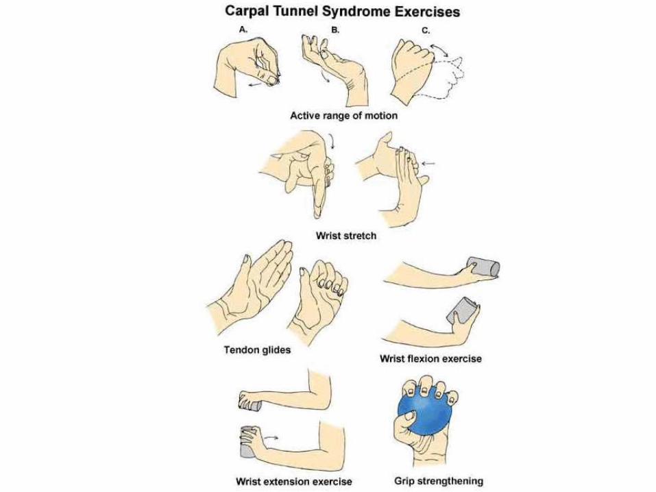
– ULTRASOUND THERAPY
– Predicting the Outcome of Conservative Treatment
• SURGERY
• Avoid repetitive wrist and hand motions that may exacerbate symptoms or make symptom relief difficult to achieve.
• Not use vibratory tools
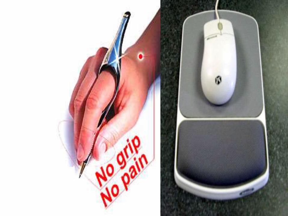
• Ergonomic measures to relieve symptoms depending on the motion that needs to be minimized
splintss
Probably most effective
when it is applied within
three months of the onset
of symptoms
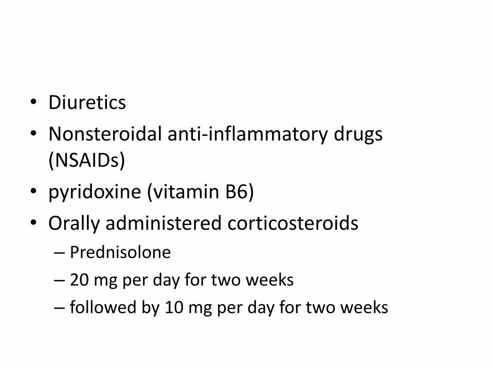
• Diuretics
• Nonsteroidal anti-inflammatory drugs (NSAIDs)
• pyridoxine (vitamin B6)
• Orally administered corticosteroids
– Prednisolone
– 20 mg per day for two weeks
– followed by 10 mg per day for two weeks
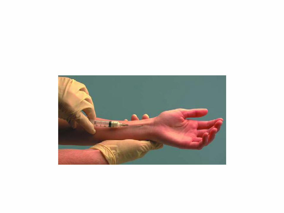
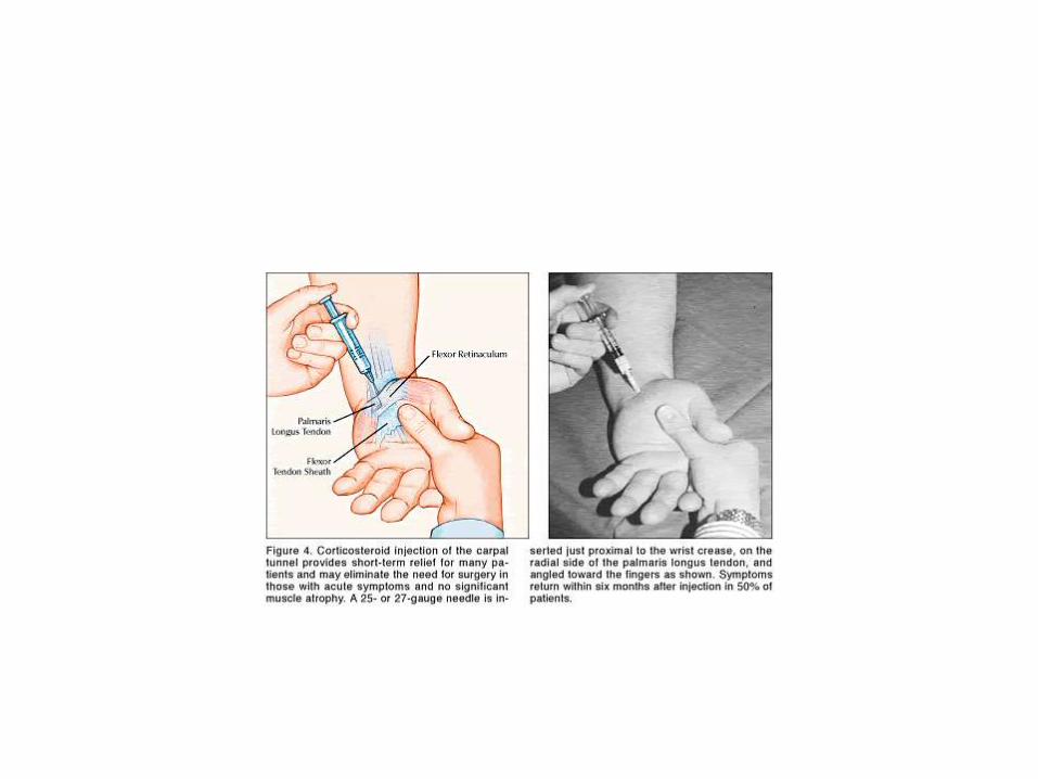
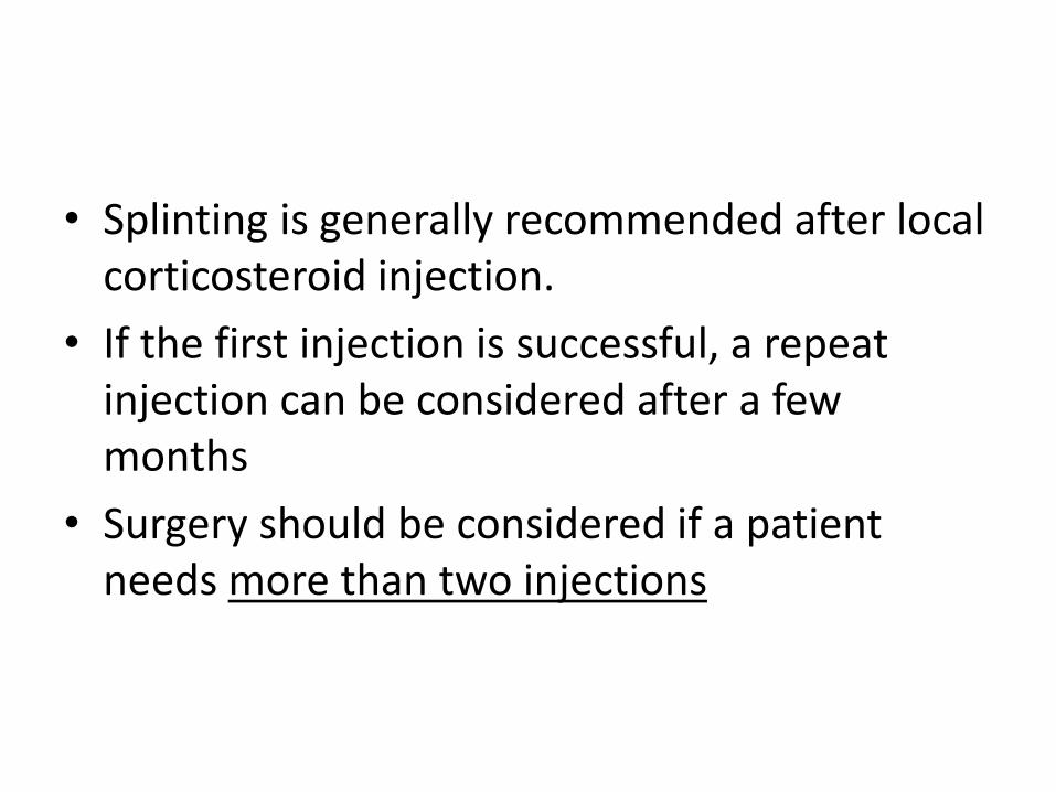
• Splinting is generally recommended after local corticosteroid injection.
• If the first injection is successful, a repeat injection can be considered after a few months
• Surgery should be considered if a patient needs more than two injections
• ULTRASOUND THERAPY
Surgical management
• Should be considered in patients with symptoms that do not respond to conservative measures and in patients with severe nerve entrapment as evidenced by nerve conduction studies,thenar atrophy, or motor weakness.
• It is important to note that surgery may be effective even if a patient has normal nerve conduction studies
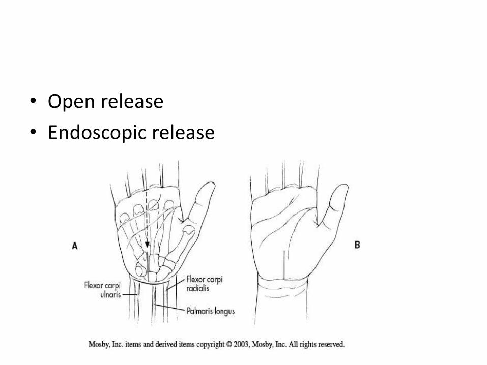
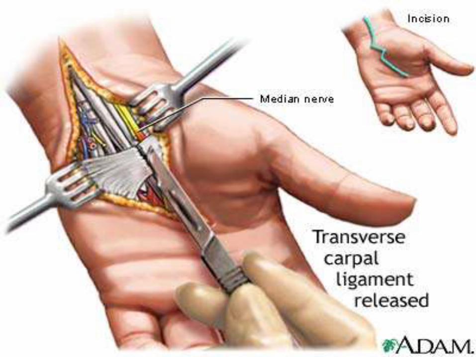
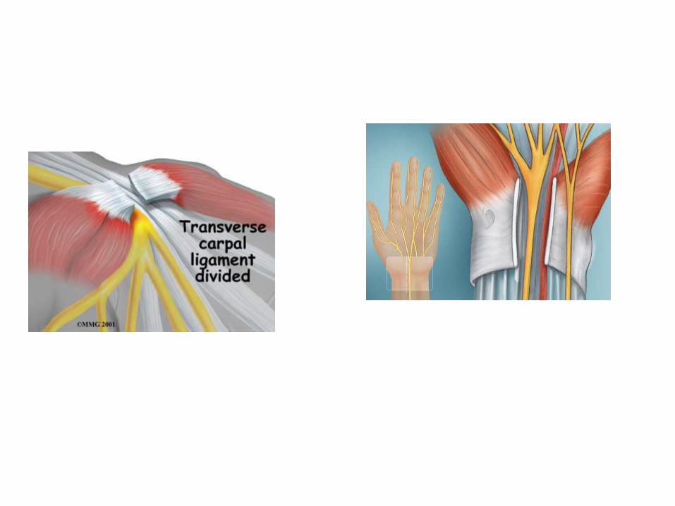
• Open release
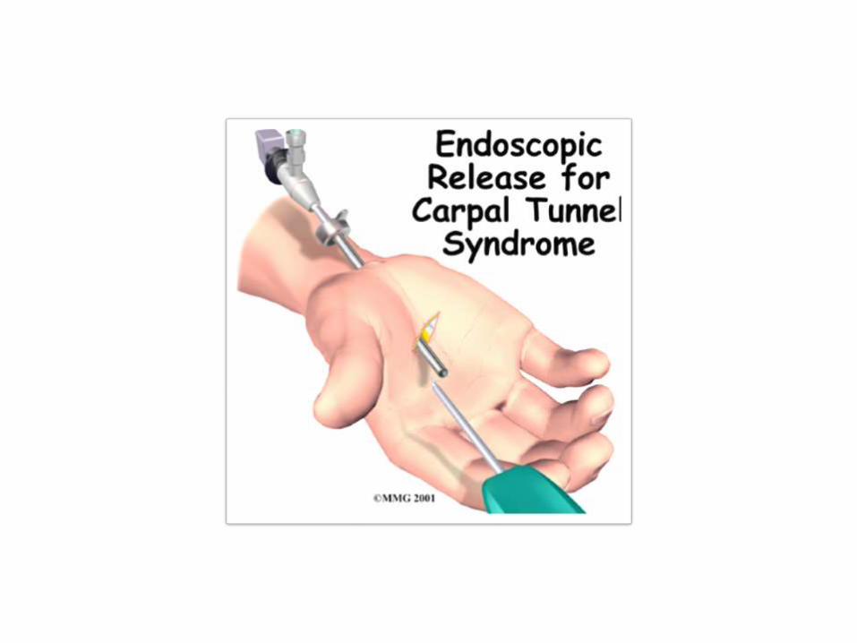
• Endoscopic release
• Transverse incision proximal to the anterior wrist crease between flexor carpi ulnaris and flexor carpi radialis tendons. Distal longitudinal incision made between proximal palmar crease and 1 cm distal to hamate hook in line with radial border of ring finger.

ANTERIOR INTEROSSEOUS SYNDROME&
PRONATOR SYNDROME
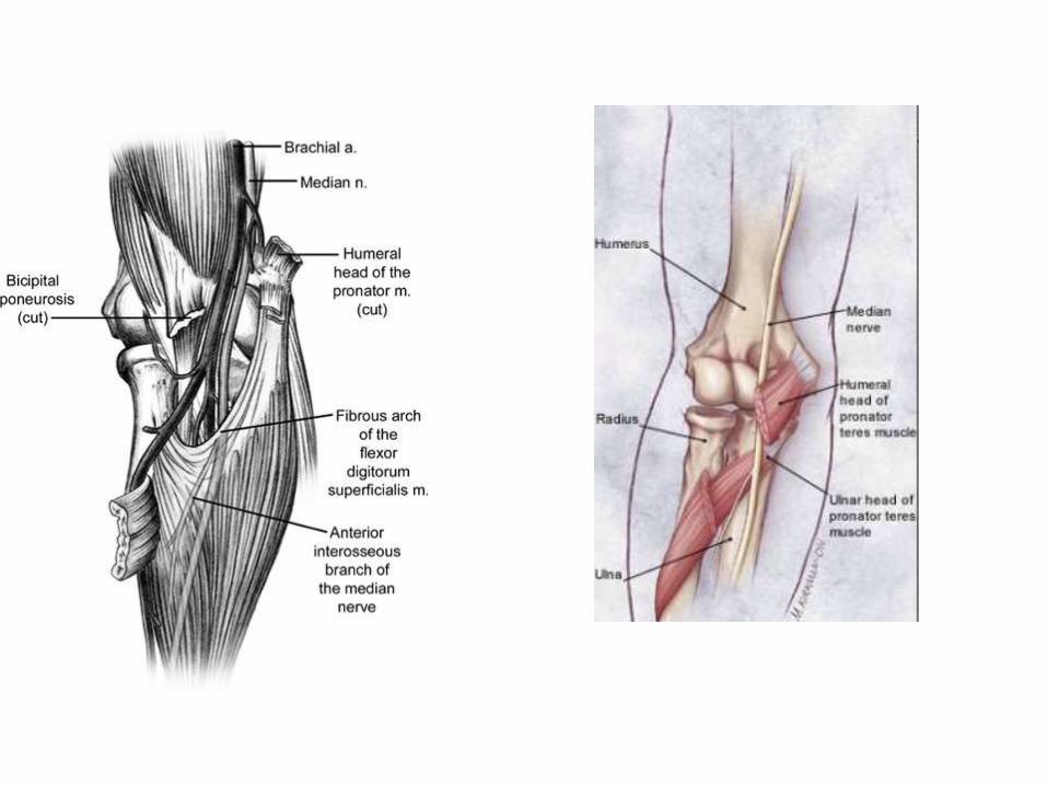
• Site of compression essentially same for both Pronator syndrome(PS) and Ant. Int. nerve
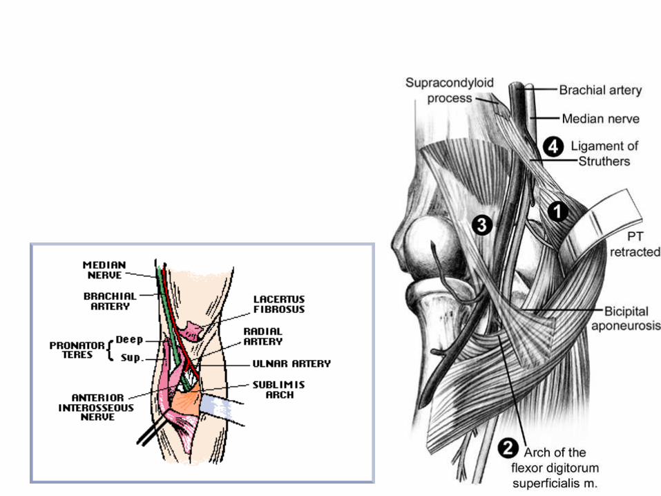
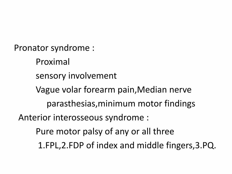
Pronator syndrome :
Proximal
sensory involvement
Vague volar forearm pain,Median nerve
parasthesias,minimum motor findings
Anterior interosseous syndrome :
Pure motor palsy of any or all three
1.FPL,2.FDP of index and middle fingers,3.PQ.
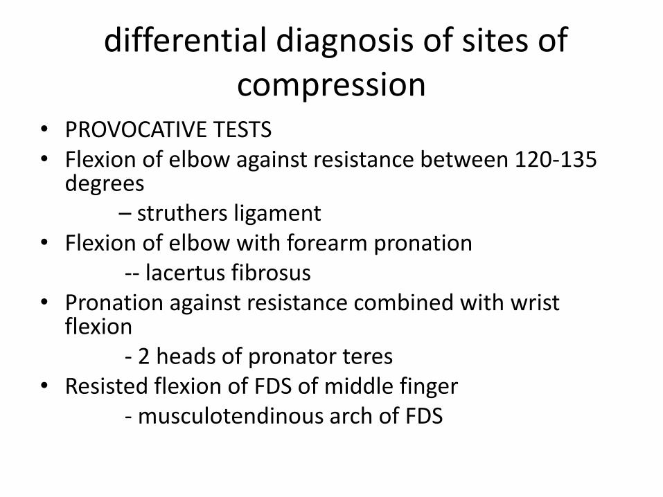
differential diagnosis of sites of compression
• PROVOCATIVE TESTS• Flexion of elbow against resistance between 120-135
degrees – struthers ligament
• Flexion of elbow with forearm pronation-- lacertus fibrosus
• Pronation against resistance combined with wrist flexion
- 2 heads of pronator teres• Resisted flexion of FDS of middle finger
- musculotendinous arch of FDS
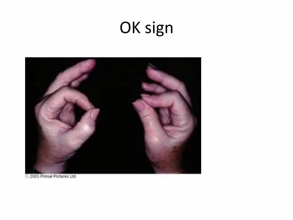
OK sign
TREATMENT
• INITIALLY: CONSERVATIVE
• SURGICAL: INDICATIONS
No resolution of symptoms
Severe symptoms
• SURGICAL EXPLORATION: Identification & division of the offending structure.
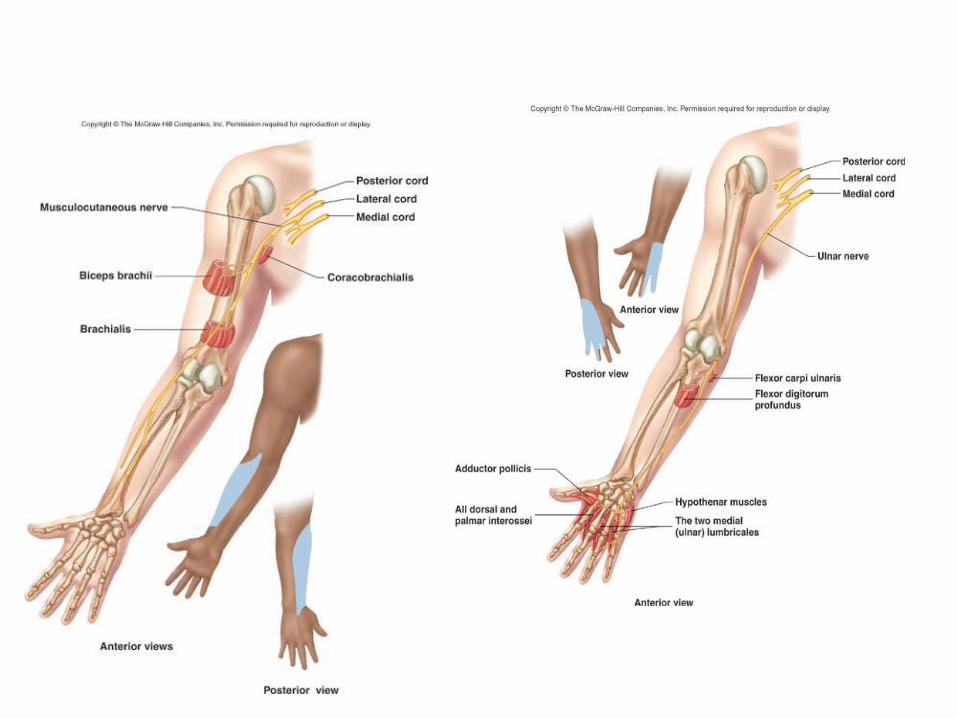
ULNAR NERVE ENTRAPMENT
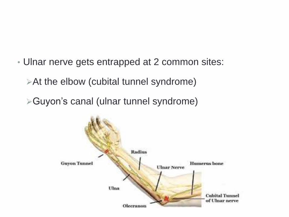
• Ulnar nerve gets entrapped at 2 common sites:
At the elbow (cubital tunnel syndrome)
Guyon’s canal (ulnar tunnel syndrome)
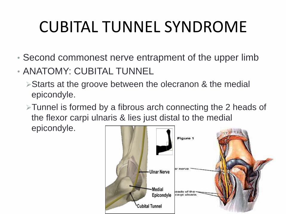
CUBITAL TUNNEL SYNDROME
• Second commonest nerve entrapment of the upper limb
• ANATOMY: CUBITAL TUNNEL
Starts at the groove between the olecranon & the medial
epicondyle.
Tunnel is formed by a fibrous arch connecting the 2 heads of
the flexor carpi ulnaris & lies just distal to the medial
epicondyle.
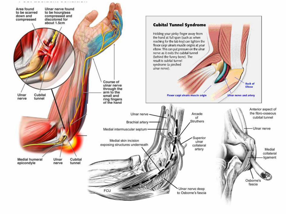
CAUSES OF ENTRAPMENT
• ARCADE OF STRUTHER’S: Formed by superficial muscle
fibres of the medial head of triceps attaching to the medial
epicondyle ridge by a thickened condensation of fascia.
• Tight fascial band over the cubital tunnel.
• Medial head of triceps
• Aponeurosis of flexor carpi ulnaris
• Recurrent subluxation of ulnar nerve, results in neuritis.
• Osteophytic spurs
• Cubitus valgus following supra condylar fracture.
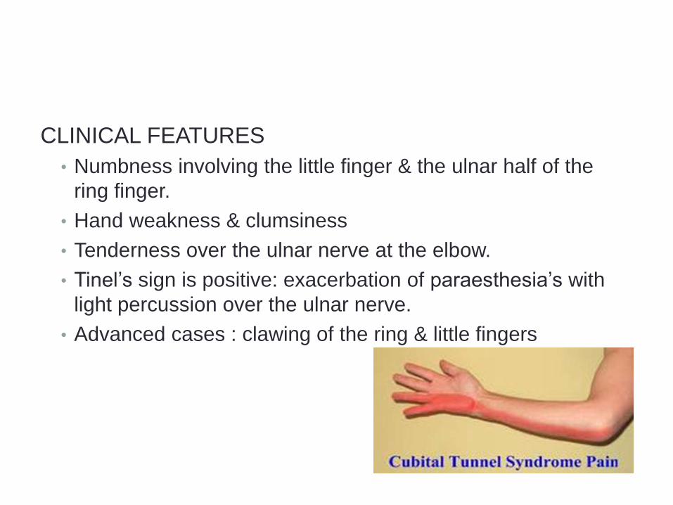
CLINICAL FEATURES
• Numbness involving the little finger & the ulnar half of the
ring finger.
• Hand weakness & clumsiness
• Tenderness over the ulnar nerve at the elbow.
• Tinel’s sign is positive: exacerbation of paraesthesia’s with
light percussion over the ulnar nerve.
• Advanced cases : clawing of the ring & little fingers
TREATMENT
• NON OPERATIVE: Early stages
Activity modification
Immobilization of the elbow in 30 degrees of extension, followed by periods of
mobilization with elbow padding.
• SURGICAL:
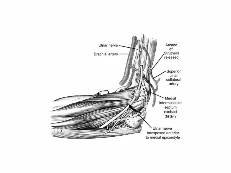
Decompression of the nerve by dividing of the basic offending structure.
Anterior transposition of the ulnar nerve
Medial epicondylectomy
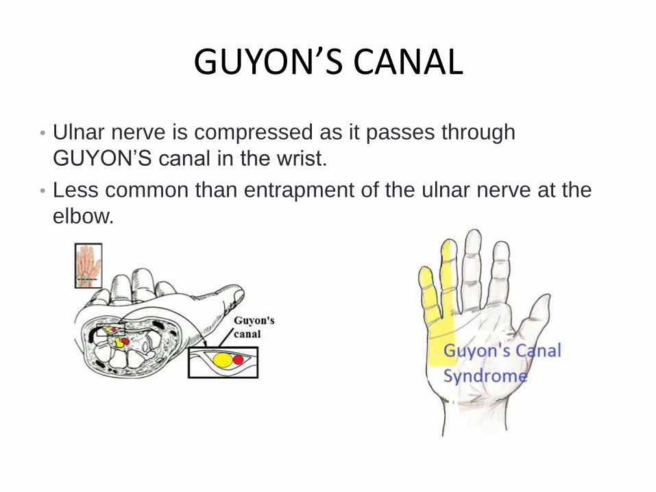
GUYON’S CANAL
• Ulnar nerve is compressed as it passes through
GUYON’S canal in the wrist.
• Less common than entrapment of the ulnar nerve at the
elbow.
ANATOMY:GUYON’S CANAL
– ROOF: composed of palmar carpal ligament blending into the FCU tendon attaching to the pisiform & the pisiohamateligaments.
– Medial wall : pisiform & pisiohamate ligament.
– Lateral wall: hook of hamate & some fibres of the transverse carpal ligament.
– Ulnar nerve enters guyon’s canal accompanied by ulnar A & Ulnar V.
– Guyon’s canal lies in the space between flexor retinaculum & volar carpal ligaments
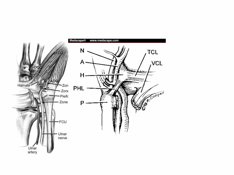
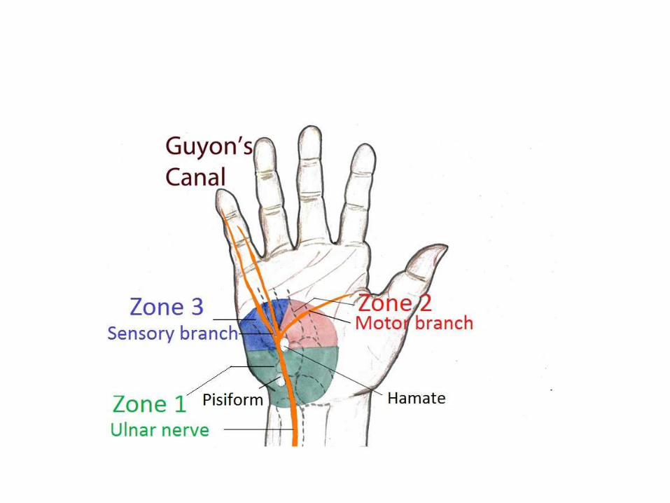
• The anatomy of distal ulnar tunnel is divided into 3 zones.
• Zone 1:proximal to the bifurcation of the ulnar nerve &
consists of both sensory & motor fibres of the nerve.
• Zone 2: represents the motor branch of the ulnar N distal
to the bifurcation.
• Zone 3: represents the sensory branches of the ulnar
nerve beyond its bifurcation
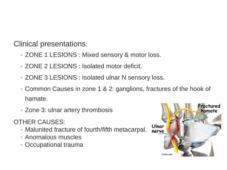
Clinical presentations:
• ZONE 1 LESIONS : Mixed sensory & motor loss.
• ZONE 2 LESIONS : Isolated motor deficit.
• ZONE 3 LESIONS : Isolated ulnar N sensory loss.
• Common Causes in zone 1 & 2: ganglions, fractures of the hook of
hamate.
• Zone 3: ulnar artery thrombosis
OTHER CAUSES:
• Malunited fracture of fourth/fifth metacarpal.
• Anomalous muscles
• Occupational trauma
INVESTIGATIONS
• X RAY : Oblique/carpal tunnel views
Delineate bony anatomy to diagnose hook of hamate fractures.
• MRI: Ganglia, space occupying lesions
TREATMENT
• Operative release of the canal by reflecting the FCU, pisiform & pisiohamate ligament ulnarly.
• Distal deep fascia of the forearm below the wrist crease should be released.
• Resection of any space occupying lesion
• Treatment of hook of hamate fractures.

RADIAL NERVE ENTRAPMENTS
• POSTERIOR INTEROSSEOUS NERVE SYNDROME
• RADIAL TUNNEL SYNDROME
• WARTENBERG’S SYNDROME
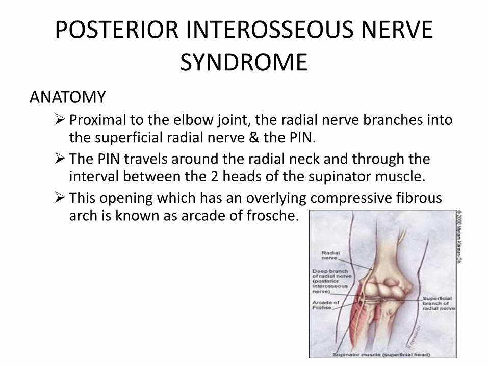
POSTERIOR INTEROSSEOUS NERVE SYNDROME
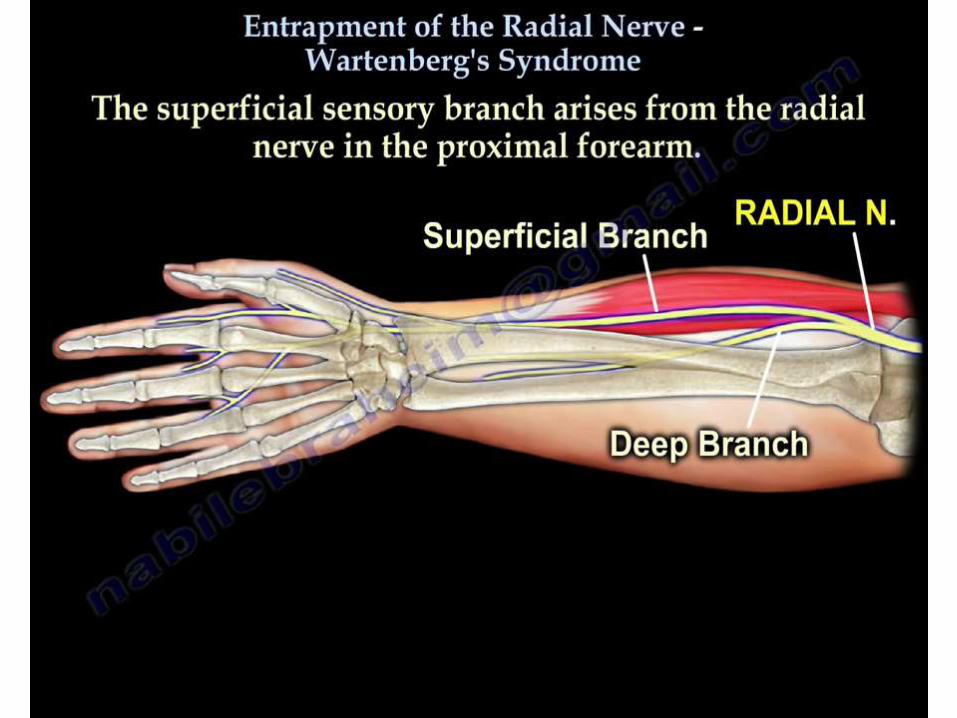
ANATOMYProximal to the elbow joint, the radial nerve branches into
the superficial radial nerve & the PIN.
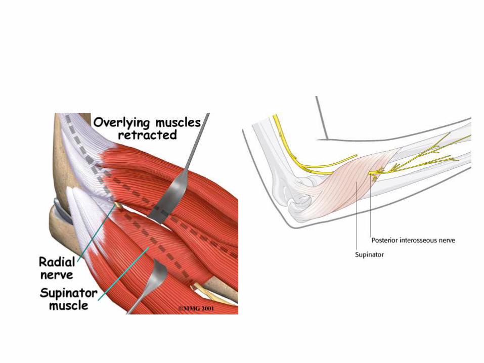
The PIN travels around the radial neck and through the interval between the 2 heads of the supinator muscle.
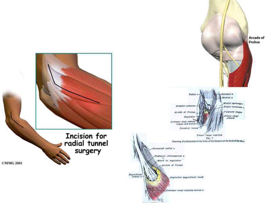
This opening which has an overlying compressive fibrous arch is known as arcade of frosche.
Clinical features:– Initially, presents with a dull ache in the proximal forearm.– Later, there is difficulty in extending the fingers & the thumb.
Etiology: Ganglion cyst Proliferative synovitis (rheumatoid arthritis)
• Electro diagnostic testing may localize the site of compression.
• Initially : observation & non operative treatment.• Operative methods: exploration & appropriate division
of compressing structures.
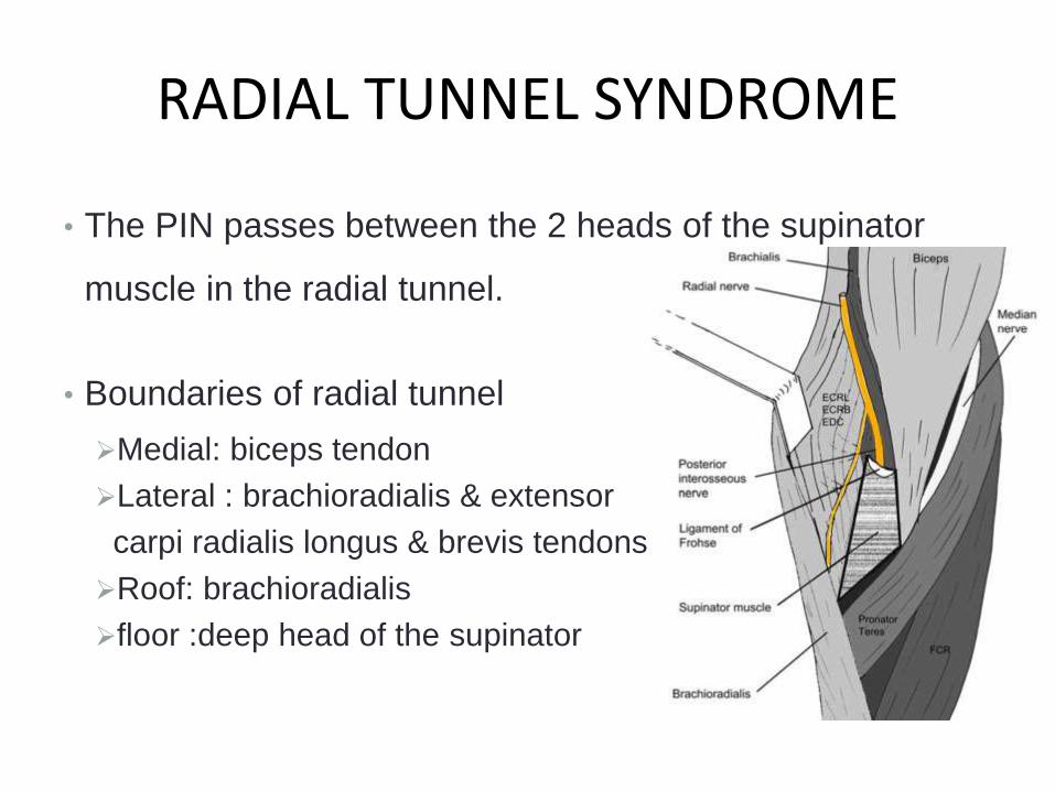
RADIAL TUNNEL SYNDROME
• The PIN passes between the 2 heads of the supinator
muscle in the radial tunnel.
• Boundaries of radial tunnel
Medial: biceps tendon
Lateral : brachioradialis & extensor
carpi radialis longus & brevis tendons
Roof: brachioradialis
floor :deep head of the supinator
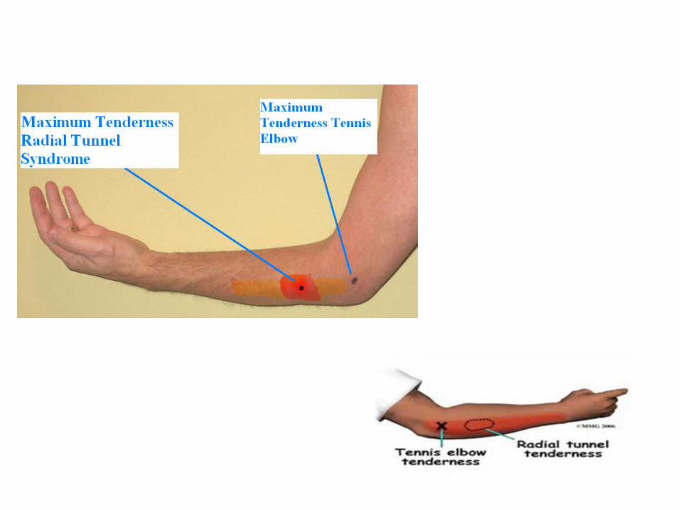
• Pain is often acute & can mimic tennis elbow.
• Electrophysiological studies shows no abnormality.
• Treatment: non-operative: Activity modification, splinting,
NSAID’S & rest.
• Surgical decompression is often combined with lateral
epicondyle release.
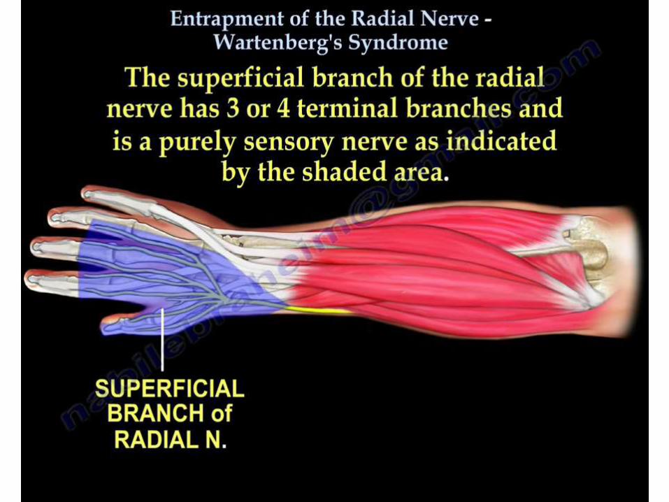
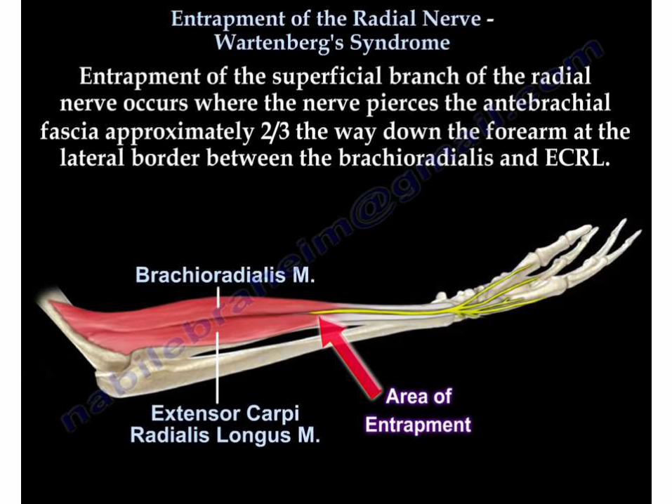
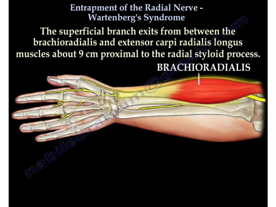
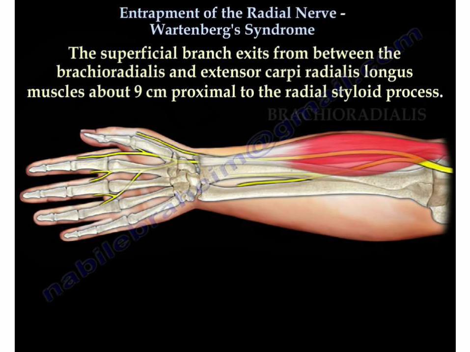
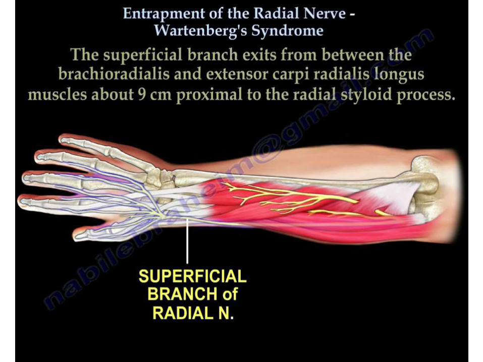
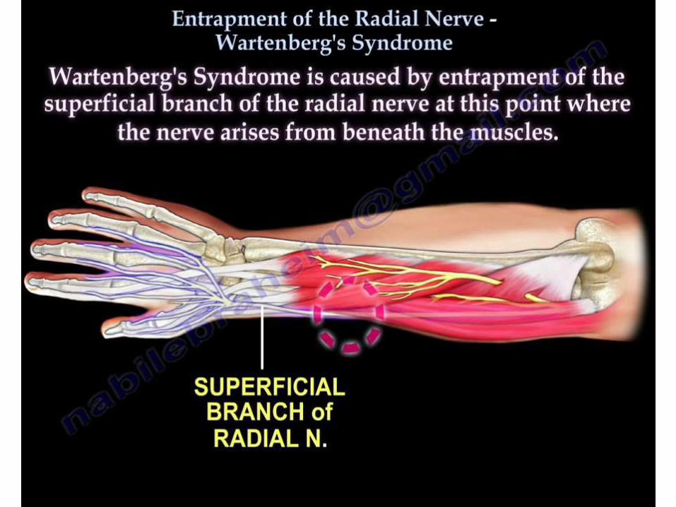
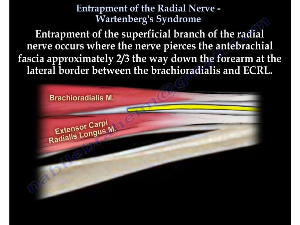
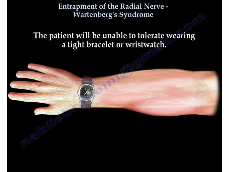
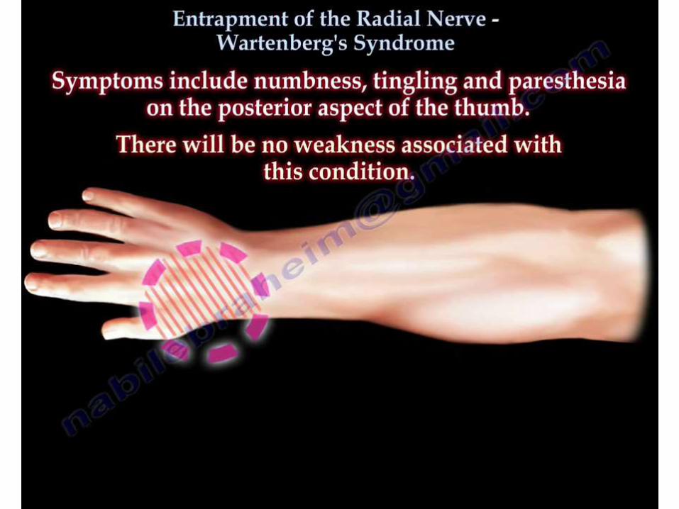
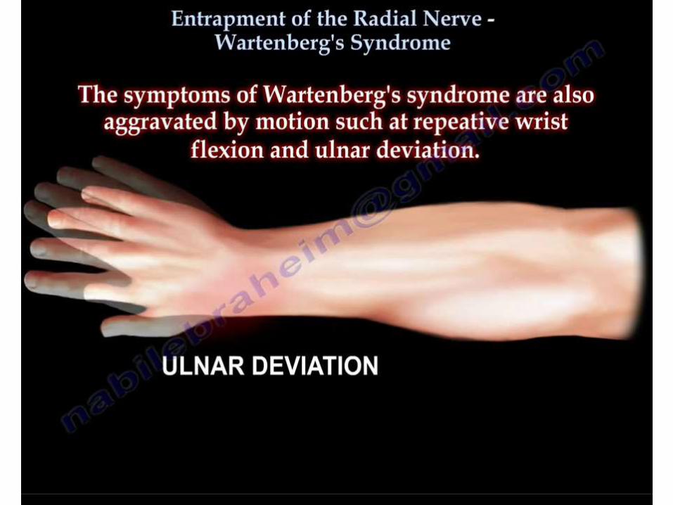
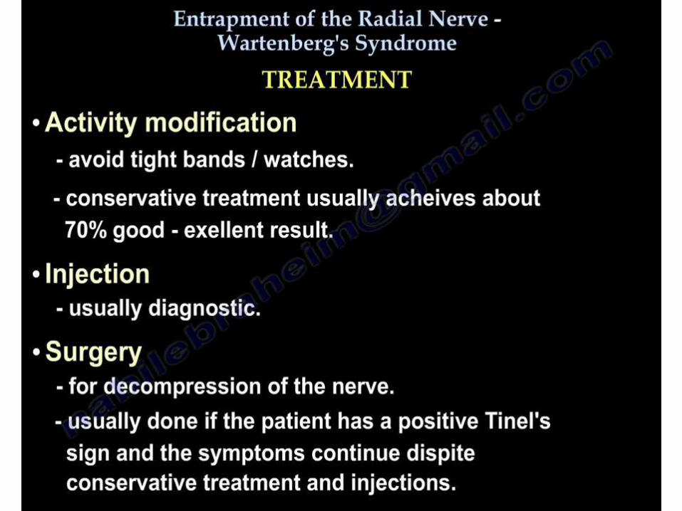
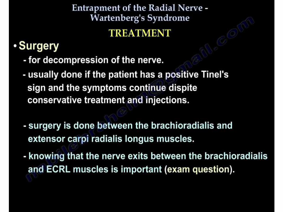
WARTENBERG’S SYNDROME
• Compression of the superficial branch of the radial nerve
can occur most commonly as it exits from beneath the
brachioradialis in the forearm.
• Nerve can get trapped b/w the ECRL & the
brachioradialis, especially with pronation in the forearm.
LOWER LIMB
SCIATIC NERVE
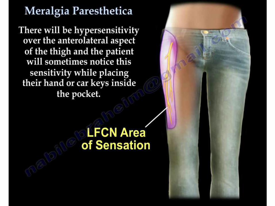
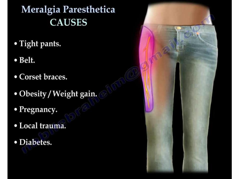
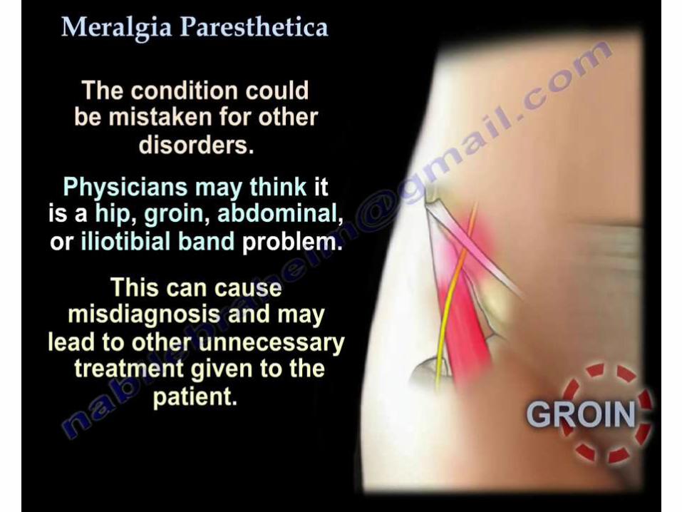
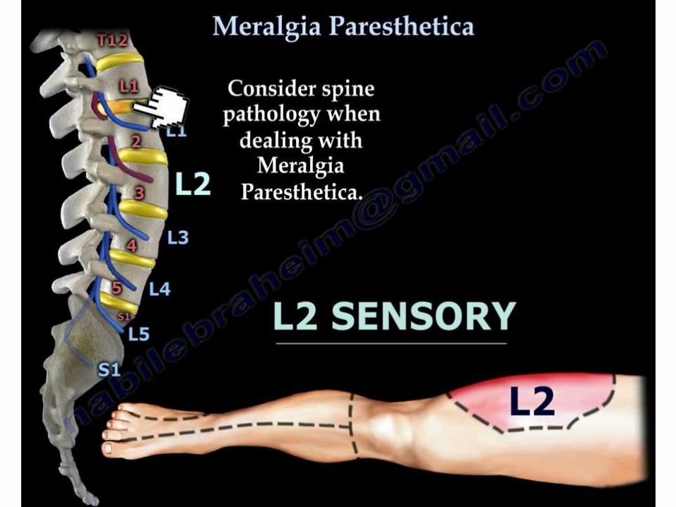
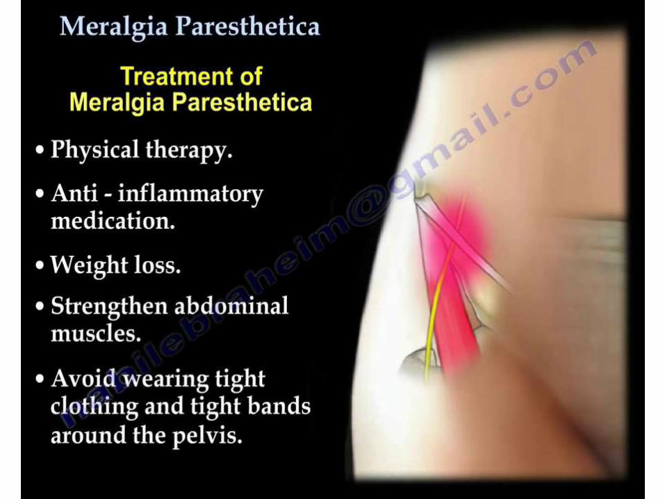
LATERAL FEMORAL CUTANEOUS NERVE
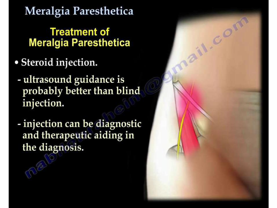
PERONEAL NEUROPATHY
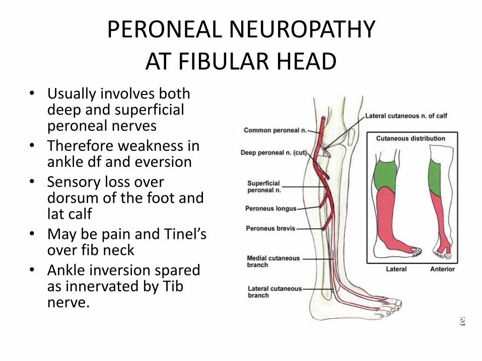
PERONEAL NEUROPATHY AT FIBULAR HEAD
• Usually involves both deep and superficial peroneal nerves
• Therefore weakness in ankle df and eversion
• Sensory loss over dorsum of the foot and lat calf
• May be pain and Tinel’sover fib neck
• Ankle inversion spared as innervated by Tibnerve.

Causes
• Habitual leg crossing
• Repetitive stretch from squatting
• Thin pt’s
• Ganglions etc
• Associated to ankle inversion injury including # fib
– Traction to nerve
– Prolonged immobilisation (especially sedated pt’s)
• Differential diagnosis
• Sciatic neuropathy
• L5 radiculopathy
• Investigations
• EMG and NCS
• MRI’s in slowly progressing to check for masses
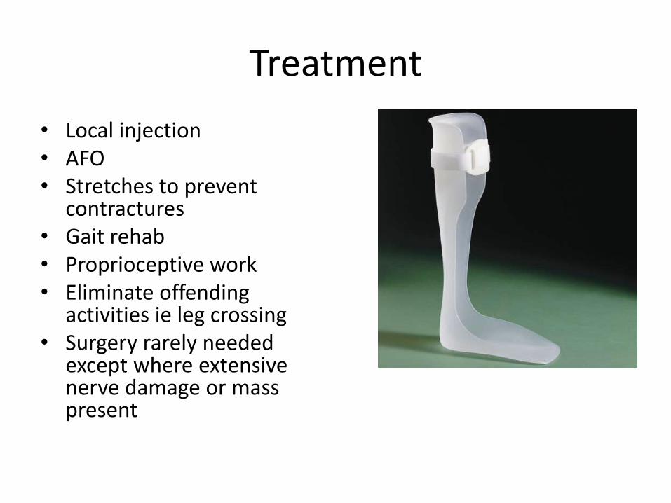
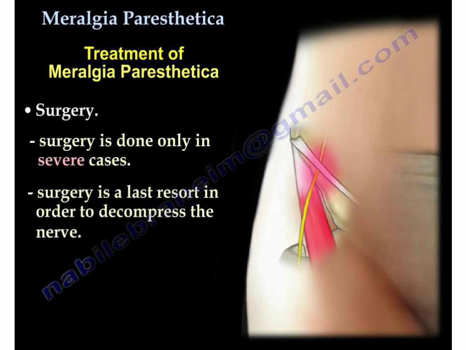
Treatment
• Local injection• AFO• Stretches to prevent
contractures• Gait rehab• Proprioceptive work• Eliminate offending
activities ie leg crossing• Surgery rarely needed
except where extensive nerve damage or mass present
TARSAL TUNNEL SYNDROME
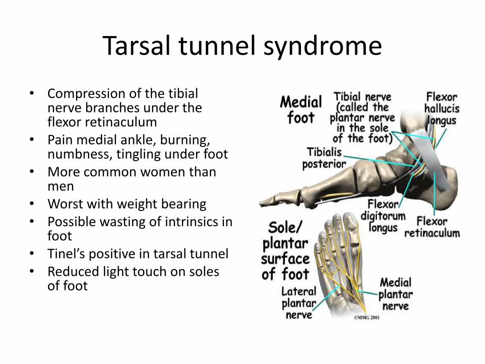
Tarsal tunnel syndrome
• Compression of the tibialnerve branches under the flexor retinaculum
• Pain medial ankle, burning, numbness, tingling under foot
• More common women than men
• Worst with weight bearing• Possible wasting of intrinsics in
foot• Tinel’s positive in tarsal tunnel• Reduced light touch on soles
of foot
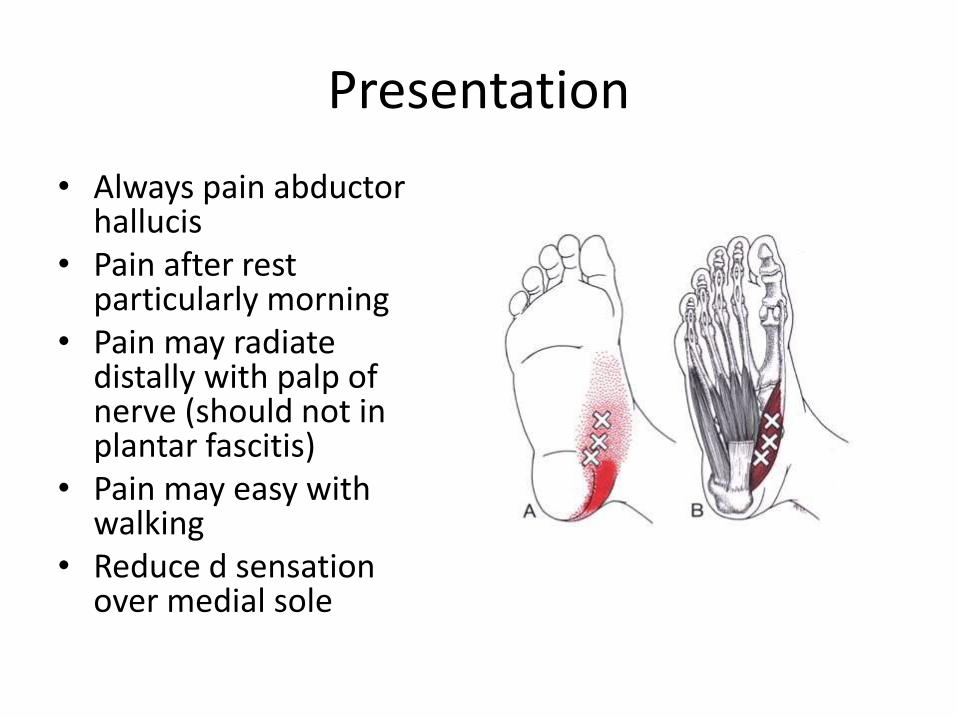
Presentation
• Always pain abductor hallucis
• Pain after rest particularly morning
• Pain may radiate distally with palp of nerve (should not in plantar fascitis)
• Pain may easy with walking
• Reduce d sensation over medial sole
Differential diagnosis
• Plantar Fascitis– DF with eversion then SLR
– Tinel’s not +ve in pf
– EMG/NCS
– High resolution US
• Fat pad atrophy– More pain over fat pad
– Visible loss of fat pad

• Good evidence very limited
• Rest
• NSAID’s
• Steroid Injections
• Heel pads
• Orthoses
• Stretching exercises for PF and calf
• Surgical intervention
MORTONS NEUROMA
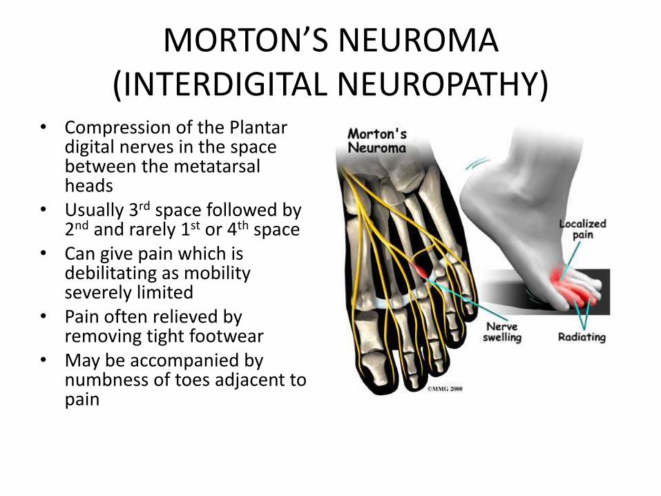
MORTON’S NEUROMA(INTERDIGITAL NEUROPATHY)
• Compression of the Plantar digital nerves in the space between the metatarsal heads
• Usually 3rd space followed by 2nd and rarely 1st or 4th space
• Can give pain which is debilitating as mobility severely limited
• Pain often relieved by removing tight footwear
• May be accompanied by numbness of toes adjacent to pain
Differential diagnosis
• TTS
– Can be very difficult to differentiate
• Plantar fascitis
• TMT OA
– Both of these will have no neurological S&S
– Also compression of the MT heads should not be exquisitely painful
Treatment
• No good evidence
• Conservative treatment helps 50%– Insoles
– Stop offending activities
– Steroid inj
– Alcohol inj x4
– Physio (not specified)
• Surgery neurectomy/neurolysis (variable outcomes)

THANK YOU


