Enzyme Prodrug Therapy Achieves Site-Specific, PersonalizedPhysiological Responses to the Locally Produced Nitric OxideAnna K. Winther,†,¶ Betina Fejerskov,†,¶ Marja ter Meer,‡ Najah B.S. Jensen,† Ross Dillion,§
Jeremy E. Schaffer,§ Rona Chandrawati,∥,∇ Molly M. Stevens,∥ Leo J. Schultze Kool,‡ Ulf Simonsen,⊥
and Alexander N. Zelikin*,†,#
†Department of Chemistry, ⊥Department of Biomedicine, and #iNano Interdisciplinary Nanoscience Centre, Aarhus University,Aarhus 8000, Denmark‡Department of Radiology and Nuclear Medicine 766, Radboud University Medical Center, Nijmegen 6525, The Netherlands§Fort Wayne Metals, Research and Development, Fort Wayne 46809, Indiana, United States∥Department of Materials, Department of Bioengineering, and Institute of Biomedical Engineering, Imperial College London, LondonSW7 2AZ, U.K.
ABSTRACT: Nitric oxide (NO) is a highly potent but short-livedendogenous radical with a wide spectrum of physiological activities. In thiswork, we developed an enzymatic approach to the site-specific synthesis ofNO mediated by biocatalytic surface coatings. Multilayered polyelectrolytefilms were optimized as host compartments for the immobilized β-galactosidase (β-Gal) enzyme through a screen of eight polycations andeight polyanions. The lead composition was used to achieve localizedproduction of NO through the addition of β-Gal−NONOate, a prodrug thatreleases NO following enzymatic bioconversion. The resulting coatingsafforded physiologically relevant flux of NO matching that of the healthyhuman endothelium. The antiproliferative effect due to the synthesized NOin cell culture was site-specific: within a multiwell dish with freely sharedmedia and nutrients, a 10-fold inhibition of cell growth was achieved on topof the biocatalytic coatings compared to the immediately adjacent enzyme-free microwells. The physiological effect of NO produced via the enzyme prodrug therapy was validated ex vivo in isolatedarteries through the measurement of vasodilation. Biocatalytic coatings were deposited on wires produced using alloys used inclinical practice and successfully mediated a NONOate concentration-dependent vasodilation in the small arteries of rats. Theresults of this study present an exciting opportunity to manufacture implantable biomaterials with physiological responsescontrolled to the desired level for personalized treatment.
Nitric oxide (NO) is a molecule with an incredibly broadspectrum of physiological activity.1−4 This small but a highlypotent molecule is implicated in the progression of andtherapies for inflammation,5 cancer,6 and viral pathologies,7
among others. It was dubbed the “guardian of cardiovasculargrafts”8 because of its proproliferative activity on theendothelium as well as antiadhesion and antiaggregationsignaling to platelets in the circulating blood. Being a radicalspecies, the lifetime of NO in human blood is very shortapproximately 1 s, over which time it has the capacity to rapidlydiffuse over a distance of approximately 100 μm, which is thelength scale of the adjacent interacting cells. Because of theimportant biological functions of NO and its highly tissue-specific activity, strategies for site-specific delivery of thismolecule for human therapy are highly desirable.4,9,10 Currentmethods for the site-specific delivery of NO rely on the releaseof the drug from its adduct depots.9,10 Although powerful in
their own right, these methods are limited in their capacity to(a) engineer a constant, zero-order release of NO, (b) engineerNO depots into the existing biomaterials used for vasculartissue engineering, and (c) control the dosage of the drugwithin the biomaterial.9,10 The latter aspect is particularlyimportant in that controlling the drug feed upon implantationof a therapeutic device is the necessary step towardpersonalized medical care such that the drug levels can betuned to a desired level established individually for each patient.Inspired by nature, we hypothesized that localized synthesis
is the most appropriate approach toward the generation andsite-specific delivery of controlled amounts of NO. We envisionthat this can be accomplished with the use of substrate-mediated enzyme prodrug therapy (SMEPT).11 In our past
Received: January 30, 2018Accepted: March 15, 2018Published: March 23, 2018
This is an open access article published under a Creative Commons Attribution (CC-BY)License, which permits unrestricted use, distribution and reproduction in any medium,provided the author and source are cited.
studies, we engineered biocatalytic (enzyme-containing) hydro-gel matrices12,13 and multilayered polymer coatings14,15 toillustrate the fundamental advantages of SMEPT over theconventional implant-mediated drug delivery. Specifically,SMEPT affords an on-demand drug delivery15 becauselocalized drug synthesis relies on externally administeredbenign prodrugs. The same biomaterial can be tuned to releasethe desired amount of the drug in unit time simply through thechoice of concentration of the administered prodrug.12 UsingSMEPT, the same biomaterial is capable of synthesizing a rangeof therapeutic molecules at their nominated concentration andtime of administrationtaken individually, in sequence, or incombination.13 Within the lifetime of the enzyme, drug releaseis sustained and follows a highly beneficial zero-order, linearrelease pattern.14 These features make SMEPT well-suited toaccomplish the localized synthesis needed to deliver NO.Several research groups have recently and independently
developed SMEPT-like systems for localized synthesis of NO.The original report from Cha and Meyerhoff revealed thatselenium-containing organic compounds mimic selenium-containing enzyme, glutathione peroxidase, and efficientlymediate the release of NO from S-nitrosothiols (RSNO).16
On the basis of this finding, several groups have performedsubstrate-mediated synthesis of NO achieved by thebiomaterials that are surface-modified with selenocystamineand/or diselenodipropionic acid17,18 and organotelluriumcompounds.19 These systems produced highly favorable resultsupon in vivo validation and provided inspiration for a broaderdevelopment of this approach. An impressive achievement wasreported by Yang et al.,20 whereby NO was generated usingendogenous donors of NO (natural RSNO), thus avoidingreliance on the external administration of prodrugs.Being highly important in their own right, enzyme mimics
are limited in that, as described, these catalysts can onlymediate the synthesis of NO, that is, they are only suited tomediate a monotherapy. In contrast, natural enzymes arecapable of converting a wide range of substrates, and within thesame family of prodrugs (e.g., glucuronides, phosphates), oneenzyme performs bioconversion to synthesize multiple drugsproviding for the flexibility of drug choice and to mediatecombination therapy, as is highly desired for a range of drugdelivery applications.13 Of the enzymes that are typically usedfor enzyme−prodrug therapies,21 only β-galactosidase (β-Gal)has a readily available corresponding prodrug for the synthesisof NO, namely, β-Gal−NONOate.22,23 On the basis of theabove discussion, we chose β-Gal as the enzyme and β-Gal−NONOate as the prodrug.The overall goal of the work presented herein is to engineer
SMEPT onto the surface of the metallic substrates used for theproduction of diverse medicinal implants such as to achieve anenzyme-mediated localized synthesis of NO. To engineerSMEPT onto the surface of the metallic wires, we used thesequential polymer deposition technique (also known as “layer-by-layer” deposition, LbL) to form biocatalytic β-Gal-containing multilayered films (Figure 1). The prime advantageof this surface modification method is that it is an all-aqueous,solution-based approach. This technique accommodatesmodification of any substrate with no restriction on surfacegeometry and topography. Multilayered thin films havepreviously been deposited onto the surface of cardiovascularstents for surface-mediated gene delivery.24 For the productionof NO, multilayered films have previously been assembled tocontain arginine, a natural precursor for the synthesis of NO by
NO synthase,25 and to contain selenium-based enzymemimics.26 However, to our knowledge, there are no priorreports of multilayered thin films for the enzyme-mediatedsynthesis of NO. A prime consideration for the biocatalyticperformance of such coatings is the choice of the polyelec-trolyte pair used to assemble the multilayered thin film. Theexisting examples of enzyme-containing LbL films27 do notprovide predictive power to nominate an optimal coating forthe catalytic output of the immobilized enzyme. Therefore, thefirst objective of this work was to conduct a broad screen ofpolyelectrolyte multilayered coatings, focusing on the catalyticoutput of the film as a criterion of selection. The secondobjective of this work was to establish control over thesynthesis of NO by the biocatalytic coatings and to validate if asurface-mediated approach to the delivery of NO is site-specific.Finally, the ultimate goal of this study was to providebiomedical characterization of the physiological activity ofNO as produced via localized enzymatic biocatalytic con-version. We envisioned that an ex vivo wire myograph modelpresents a favorable setting for this test, in that it usesmammalian tissue and records native physiological responsesand it is readily suitable for the systematic variation ofexperimental conditions (such as recording dose−responsecurves). In doing so, ex vivo tissue-based studies minimize theuse of laboratory animals, yet provide the sought-aftervalidation of physiological effects mediated by implantablebiomaterials.
■ MATERIALS AND METHODSMaterials and Instruments. Unless stated otherwise, all materials
were purchased from Sigma-Aldrich. Pyrogallol and the enzyme β-Gal(derived from Escherichia coli, 465 kDa) were purchased from Merck,NO gas was obtained from Air Liquide Danmark AS, and β-Gal−NONOate was obtained from Cayman Chemical. Fluoresceindiacetate (FDA) and propidium iodide (PI) were used as live/deadstains. PrestoBlue cell viability reagent and Quant-iT PicoGreendsDNA assay kit were both obtained from Life Technologies. Roundmetal wires with a 200 μm diameter (alloys 35N LT, 316L, and L605)were manufactured by Fort Wayne Metals and processed as reportedelsewhere.28
Figure 1. Enzymatic synthesis of NO is engineered in this work intomultilayered polyelectrolyte coatings. When used as substrates for cellculture, these biocatalytic coatings provide localized synthesis of NOfor localized delivery to the adhering cells.
ACS Applied Materials & Interfaces Research Article
Quartz crystal microbalance (QCM) experiments were conductedon QSense E4 (Biolin Scientific). Quantitative absorbance andfluorescence measurements were conducted using an EnSpirePerkinElmer multilabel plate reader and a Tecan infinite M200 PROmultimode reader. Imaging was performed on a Zeiss Axio ObserverZ1 microscope. Ultrapure water (MQ) with a resistivity of 18.2 MΩcm−1 obtained from a Milli Q direct 8 system (Millipore) was used forall solutions.Atomic force microscopy (AFM) characterization of the samples
was carried out in a tapping mode (MultiMode VIII, Bruker, USA). Acantilever (ScanAsyst-Air, Bruker, USA) with a sharp tip (nominal tipheight 2.5−8 μm and nominal tip radius 2 nm) and triangulargeometry (nominal resonant frequency 70 kHz, spring constant 0.4 N/m, length 115 μm, and width 25 μm) was used to conduct theexperiment. Operational parameters such as tapping amplitude andgains were adjusted to optimize the resolution and prevent the tip−sample damage. The scan rate was set to 1 Hz. The AFM experimentwas conducted in air at 21 °C and 54% humidity. Raw data wereprocessed using open-source software Gwyddion for tilt correction andadequate polynomial leveling. To calculate the root-mean-square(rms) roughness, images were acquired from five different locations atvarious scan sizes.Polymers used in this work were poly(ethyleneimine) (PEI, ∼25
000 Da, branched), poly(sodium-4-styrene sulfonate) (PSS, ∼70 000Da), poly(methacrylic acid) (PMA, ∼18 500 Da), poly(acrylic acid)(PAA, ∼28 300 Da), dextran sulfate (200 000 Da), chrondoitin sulfate(5−100 kDa), hyaluronic acid (HA, low: 15 000−30 000 Da, high:1.5−1.8 × 10⁶ Da), alginate (ALG, 12 000−40 000 Da), DNA (fromherring testes), poly(diallyldimethylammonium chloride) (100 000−200 000 Da), poly-L-lysine (30 000−70 000 Da), poly-L-argininehydrochloride (PLA, 15 000−70 000 Da), poly-L-histidine (PLH,5000−25 000 Da), poly(allylamine hydrochloride) (PAH, ∼15 000and 17 500 Da), chitosan (Chi, 190 000−310 000 Da), biodegradablepolyamidoester29 (∼8300 Da), and protamine sulfate (PRT, ∼5100Da). Phosphate-buffered saline (PBS) and 4-(2-hydroxyethyl)-piperazine-1-ethane-sulfonic acid (HEPES) of 10 mM containing150 mM NaCl in MQ with pH 7.4 were used as buffers.Polymer Screen. For the assembly of polyelectrolyte multilayered
coatings, the polymers were dissolved in PBS to 0.1 g/L. Chi wasdissolved in acetic acid to 10 g/L and diluted in PBS to 0.1 g/L,whereas PLA and PLH were dissolved in 0.1 M HCl to 10 g/L anddiluted to 0.1 g/L with PBS. All multilayered films were fabricated instandard tissue culture polystyrene (TCPS) 96-well plates, unlessstated otherwise. To ensure film buildup, a primary layer of PEI wasapplied, followed by the alternating layers of polyanions andpolycations (100 μL per well). Each layer was left to adsorb for 5min, followed by a washing step two times with MQ. The enzyme β-Gal was added at a concentration of 20 mg/L (100 μL), unless statedotherwise, and allowed to incubate for 1 h prior to a single washingstep. Additional film buildup was performed as described above,resulting in a final architecture of PEI−(polyanion/polycation)3−β-Gal−(polycation/polyanion)2.5. For quantitative evaluations, fluoro-genic prodrug resorufin galactopyranoside was added together withfresh media yielding 5 mg/L and allowed to incubate for 30 min priorto readout.Film Assembly Using PSS/PAH. The multilayered films of PSS
and PAH were prepared with the polymers dissolved in HEPES bufferto a concentration of 0.1 g/L. To ensure film buildup, a primary layerof PEI was applied, followed by the alternating layers of PSS and PAH,as described previously. The resulting multilayered films had the finalarchitecture of PEI−(PSS/PAH)3−β-Gal−(PAH/PSS)2.5. The multi-layered films were then incubated in a 100 μL HEPES buffer at 4 °Cuntil usage.Multilayer Film Assembly on Metal Wires. The films deposited
on metal wires consisted of the polymers PEI/PSS/PAH dissolved inPBS to a concentration of 1 g/L. PEI was dissolved in MQ water andleft to adsorb for 30 min, all PSS/PAH layers were left to adsorb for 10min, and the enzyme β-Gal was allowed to incubate for 2 h. Prior tofilm assembly, the wires were cleaned by consecutive immersions in 20w/v % citric acid, demineralized water, and 70% ethanol using
sonication. The resulting multilayered films had the final architectureof PEI−(PSS/PAH)3−β-Gal−(PAH/PSS)1.5. The durability of theenzyme activity deposited onto the wires was evaluated after 7, 14, 21,and 28 days of incubation in PBS at 37 °C using resorufin-β-D-galactopyranoside at a concentration of 5 mg/L, which was allowed toincubate for 30 min prior to readout.
Quantification of NO. Deoxygenated water was prepared usingargon gas. The latter was first led through a vial containing 10 mMpyrogallol (to remove traces of oxygen) and then bubbled throughwater for 1 h to undergo deoxygenation. NO gas was led through 10mM NaOH in water to remove traces of higher nitrogen oxides andthen through deoxygenated water to prepare a saturated solution ofNO (with a concentration of 2 mM, ref 30). Serial dilutions of NOwere then made using deoxygenated water using a gastight Hamiltonsyringe. The solutions of NO were mixed with DAF-FM in a black 96-well plate to the final concentration of the dye of 8 μM. Thefluorescence of the solutions was recorded for 40 min (at λex/λem 495/515 nm) during which time the fluorescence readings reached aconstant level. The final attained values of fluorescence were used toplot the f luorescence intensity versus NO concentration calibration curve,which resulted in a linear fit with the rms correlation coefficient of0.92. The multilayered PSS/PAH coatings containing β-Gal wereassembled in the wells of the black 96-well plates as described above.The wells were filled with fresh physiological saline solution containing8 μM DAF-FM and varied concentrations of β-Gal−NONOate (5, 10,15, and 20 μM). The fluorescence of the solutions was recorded over30 min on a plate reader (λex/λem 495/515 nm). All experiments werecarried out thrice in triplicates.
Cell Culture. The mouse myoblast cell line C2C12 was cultured inDulbecco’s modified Eagle medium supplemented with 10 v/v % fetalbovine serum, 1 v/v % penicillin−streptomycin, and 1 mM sodiumpyruvate. A 1/10 cell splitting was performed before reaching 70−80%confluence.
Myoblasts on Multilayered Coatings. The multilayered filmsproduced as described above with an architecture of PEI−(PSS/PAH)3−β-Gal−(PAH/PSS)2.5 were UV-sterilized for 10 min prior tocell seeding. C2C12 myoblasts were seeded out at a starting density of500 cells per well in 100 μL media and allowed to adhere overnight.NONOate (100−0 μM) was added together with fresh media, and thecells were left to incubate for 24 h at 37 °C and 5% CO2. Forincubation of 48 and 72 h, the cells were administered fresh mediawith the respective (pro)drug every 24 h. The viability of the C2C12myoblasts was evaluated using the PrestoBlue viability reagent,whereas quantitative DNA measurements were performed withQuant-iT PicoGreen.
Cell Imaging. C2C12 myoblasts were seeded out in 12-well tissueculture plates on 16 mm glass slides coated with multilayered filmswith or without the enzyme. The initial cell seeding density was 5000cells per well in 1 mL media. Cells were allowed to adhere overnight,followed by the addition of 100 μM NONOate in fresh media. Thesamples were incubated for 24, 48, and 72 h, with fresh media andNONOate added every 24 h. Fresh media containing the LIVE/DEADstains of FDA (5 mg/L) and PI (4 mg/L) were added to the samplesand incubated for 5 min in the dark. After 2× washing with PBS, thecells were visualized.
Local Delivery Using Coculture μ-Slides. For the demon-stration of local delivery, myoblast cells were seeded out into cocultureμ-slides allowing nine individual subcultures in one major well (IbidiGmbH). The designated wells were precoated with biocatalyticcoatings as described above. The starting density of the cell was 700cells in 50 μL media per minor well. The cells were allowed to adherefor 3−4 h before replenishing with 1 mL fresh media and incubatedovernight. 100 μM solution of NONOate was subsequentlyadministered in fresh media and replenished after 24 h. After a totalof 48 h of incubation with NONOate, the samples were evaluatedusing LIVE/DEAD stain as described.
Ex Vivo Wire Myograph Study. Ethics Statement. All animalexperiments in this study were approved by the Danish AnimalExperiments Inspectorate (permission 2011/561-2011), and recom-mendations described in the Guide for the Care and Use of Laboratory
ACS Applied Materials & Interfaces Research Article
Animals of the U.S. National Institutes of Health and the ARRIVEGuidelines were followed. Animals were housed in the animal facilityin Universal Euro III type long with cages with standard wood beddingand space for two rats. There was a 12 h shift between light anddarkness, and the animals had free access to food and drinking water.Tissue. Male Wistar rats (9−11 weeks) with a weight of
approximately 450−550 g were euthanized by cervical dislocationfollowed by exsanguination. The mesenteric bed was removed andplaced in cold physiological saline solution (4.7 mM KCl, 1.17 mMMgSO4·7H2O, 119 mM NaCl, 25 mM NaHCO3, 1.18 mM KH2PO4,0.026 mM ethylenediaminetetraacetic acid, 5.5 mM glucose, and 1.6mM CaCl2). The first or second branch arteries with a diameter ofaround 300−450 μm were dissected using microforceps (Dumont no.5) and a microsurgery scissor.Mounting and Normalization. Arteries with a length of 1.5−2 mm
were mounted on a dual wire myograph (model 410 A, Danish MyoTechnology A/S, Denmark) using a 40 μm steel wire and coldphysiological saline solution. The arteries were left to equilibrate at 37°C while bubbled with a bioair (21% O2, 5% CO2, and 74% N2). Toensure comparable results, the arteries were normalized to an internalcircumference corresponding to 90% of the internal circumference of afully relaxed artery at a transmural pressure of 100 mmHg.Norepinephrine (NE) was used for contraction of the smallmesenteric arteries. Iberiotoxin (IbTX) and 1H-[1,2,4]oxa-diazolo-[4,3-a]quinoxalin-1-one (ODQ), both from Tocris Bioscience, wereused as inhibitors of the NO-mediated vasodilation.Experimental Protocol. Before experimentation, the viability of the
smooth muscle cells (SMCs) was tested by contracting with 10 μMNE. Only the arteries with a contraction corresponding to a transmuralpressure above 75 mmHg were included in these studies. Stent wireswith a diameter of 100 μm and a length of 9 mm were placed in thelumen of the arteries before testing the viability of the SMCs. Toinvestigate if the prodrug NONOate could produce NO in thepresence of the LbL-coated wire, the arteries were contracted with 3μM NE, and when stabilized, NONOate was added in a cumulativemanner to produce a concentration−response curve (CRC) rangingfrom 0.5 nM to 15 μM. The arteries were discarded if they developed acontraction of less than 60% of the maximum contraction.Furthermore, controls were made both with and without the presenceof the uncoated wire. For inhibition studies, the arteries wereincubated for 30 min with 3 μM ODQ and 0.1 μM IbTX beforeadding NONOate, as described above.Data Analysis. Unless stated otherwise, the numerical data are
presented as mean ± SD and calculated based on at least threeindependent experiments. All the data were analyzed using MicrosoftExcel 2010 and plotted in OriginPro 8 or GraphPad Prism 7. Ex vivo
wire myograph data were collected by the LabChart 5 softwareprogram (ADInstruments Ltd, Oxfordshire, UK), presented as mean ±SEM, and calculated based on at least five experiments. The statisticswere conducted using Student’s t-test or one-way analysis of variance(ANOVA) followed by Tukey’s multiple comparison test in Excel orGraphPad Prism 7. For the myograph experiments, two-way ANOVAwas used. Statistical significance was defined as P < 0.05 (*), P < 0.01(**), and P < 0.001 (***).
■ RESULTS AND DISCUSSION
The optimization of the architecture of the multilayeredpolyelectrolyte films was conducted toward selection ofcompositions that favored high catalytic output of the coating.A total of eight polyanions and eight polycations (Figure 2)were used to assemble the multilayered thin films in the wells ofthe standard 96-well plates. Polyelectrolyte assembly at theinterface may proceed “linearly” and afford relatively densecoatings (e.g., PSS/PAH, refs 31 and 32) or “exponentially” andafford hydrogel-like films (HA- and ALG-containing films andpolypeptide-containing films, ref 33). For polymers with pH-dependent ionization (e.g., PAA, PMA, and PAH), thethickness and density will also depend on the coating assemblyconditions.34 Furthermore, the polymers differ in their capacityto support protein adsorption,35 and it is also important thatthe enzyme is not displaced upon the deposition of subsequentpolymer layers. These combined effects define the catalyticperformance of the assembled coating. Although in-depthanalysis of these factors individually fell beyond the scope ofthis study, we aimed to identify the composition(s) that satisfyeach of these conditions and in doing so afford the coatingssuitable for subsequent applications in SMEPT.Multilayered polyelectrolyte coatings were assembled,
starting with a priming layer of PEI followed by a total offive polyelectrolyte bilayers. Enzyme immobilization wasconducted through the exposure of the three bilayer coatings(with a polycation surface layer) followed by the subsequentdeposition of 2.5 more bilayers (polyanion top layer). Theresulting coatings were evaluated in terms of their catalyticoutput using the fluorogenic galactosidase substrate, resorufinβ-D-galactopyranoside. To probe the spontaneous release of theprotein from the coatings, bioconversion was also tested in the
Figure 2. Chemical structure and abbreviations of polyanions and polycations used in this study (DNA and PRT not shown).
ACS Applied Materials & Interfaces Research Article
supernatants aspirated from the coatings immediately prior tothe evaluation of the coating.The prime conclusion from this screen is that the overall
majority of coatings supported well the activity of theimmobilized β-Gal (Figure 3). Linearly growing multilayers
based on synthetic polyelectrolytes (e.g., PSS/PAH)31,32 wereactive, rather similar to the exponentially growing coatingsbased on polysaccharides,33 revealing that the density of thecoating may not be decisive for the activity of the immobilizedenzyme. This includes coatings based on polypeptides with thepotential to make gradually degradable coatings36 and coatingsbased on nondegradable polymers such as to assemblepermanent material surface modification.37 With a view towardlong-lasting coatings, subsequent work relied on the PSS/PAHmultilayers.We quantified the buildup of these multilayers and more
importantly the amount of the enzyme immobilized within thecoating. The analyses were performed using QCM (Figure 4A).This experiment revealed that under chosen conditions (20mg/L enzyme concentration in HEPES buffer, pH 7.4),enzyme immobilization proceeds rather fast and is completewithin minutes affording a protein coverage of 198 ± 20 ng/cm2. This enzyme coverage is within the same order ofmagnitude as what has previously been reported for PSS/PAH
and immobilization of immunoglobulin38 or albumin39 andadsorption of other proteins onto the multilayered polyelec-trolyte surface coatings.35 The resulting multilayered coatingswere imaged using AFM (Figure 4B), illustrating a typicalmorphology of LbL coatings and an rms roughness of 5 ± 2 nmalso typical for these coatings.40
Next, we aimed to characterize the coatings in detail in termsof their biocatalytic output and specifically in the production ofNO. To achieve this, we established a fluorescence-basedreadout to quantify the production of NO. DAF-FM is a dyethat reacts with NO and upon doing so becomes fluorescent.Because of this property, it has been previously used to, forexample, visualize the delivery of NO to cells.41 The calibrationcurve for fluorescence versus concentration of NO wasobtained using NO gas and serial dilution of its saturatedsolution (2 mM, ref 30) in deoxygenated water. Thisexperiment afforded a linear calibration curve correlatingfluorescence of DAF-FM and feed of NO in solution, thusproviding a facile method to quantify the production of NO bythe biocatalytic coatings (for details, see Materials andMethods).To quantify the production of NO using biocatalytic
coatings, the latter were prepared as discussed above in thewells of black 96-well plates and incubated with the β-Gal−NONOate prodrug in the presence of DAF-FM. Thefluorescence of the solutions was continuously recorded on aplate reader (Figure 5A). These data demonstrate that in theabsence of β-Gal in the surface coatings, no production of NOwas observed. In contrast, enzyme-containing coatings affordeda steady evolution of fluorescence, that is, steady production ofNO over at least 30 min. The values of fluorescence intensitywere then converted into the concentrations of NO and NOflux, that is, the rate of production of NO by the biocatalyticsurface coatings in unit time (Figure 5B). This analysis affords ahighly important conclusion that under SMEPT conditions, theassembled coatings afford a flux of NO, which matches withthat reported for healthy endothelium (0.05−0.4 nmol min−1
cm−2).42,43 Furthermore, prodrug concentration is a facile toolto fine-tune NO flux to the desired level, a unique opportunityfor personalized medicine.From a different perspective, we hypothesized that NO flux
can also be optimized through the variation of the conditions ofassembly of biocatalytic coatings, leading to a variation ofenzyme content in the multilayered polyelectrolyte film. To testthis, PSS/PAH coatings were assembled as discussed above,except that the enzyme immobilization step was performedusing protein solutions with varied concentrations. QCMmeasurements revealed that with 20 mg/L feed, the enzymesurface immobilization was 2.0 ± 0.2 mg/m2 (Figure 4). With adecreased enzyme feed, the surface immobilization decreasedaccordingly, and at 2 mg/L, the feed was 0.38 ± 0.12 mg/m2,and at 0.2 mg/L, the feed was 0.10 ± 0.04 mg/m2. Theresulting biocatalytic coatings were incubated with β-Gal−NONOate in the presence of DAF-FM with continuousrecording of the fluorescence of the solution. With an excessprodrug, the biocatalytic production affords a highly desiredlinear profile of the evolution of fluorescence (Figure 6A).Furthermore, variation of the enzyme feed in the assemblysolution affords a facile means of control over the biocatalyticoutput of the coating in a wide range of physiologically relevantflux of NO (Figure 6B). Over several days of analysis,assembled coatings revealed a minor decrease in the biocatalyticperformance, indicating loss of enzyme activity; however, NO
Figure 3. Results of quantification of the catalytic output for themultilayered coatings composed of different polyanion/polycationcombinations and equipped with β-Gal: (A) multilayered coatings withPAH or Chi as polycations and a variation of polyanions; (B)multilayers with PSS or HA as polyanions and a variation ofpolycations. HA with high and low molar mass is denoted as HAH andHAL, respectively. Enzymatic catalysis was evaluated using afluorogenic enzyme substrate, resorufin β-D-galactopyranoside.
ACS Applied Materials & Interfaces Research Article
flux remained well within the physiologically relevant range42,43
(Figure 6C).Initial cell culture characterization of the biocatalytic coatings
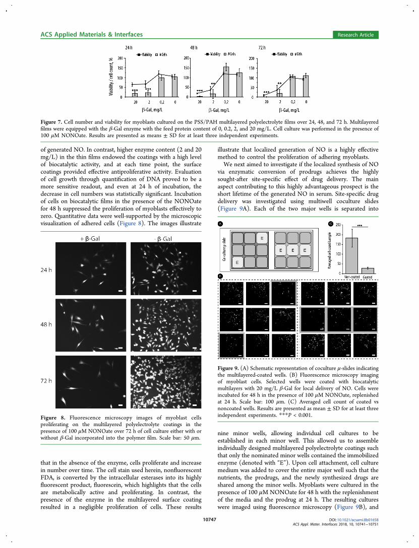
was carried out using myoblast cells. In the context ofatherosclerosis and cardiovascular stenting, the proliferationof muscle cells is a highly undesirable event that can lead to therestenosis cascade.8 Current stents on the market are designedto gradually release cytotoxins such as paclitaxel specifically toprevent proliferation of muscle cells.44 NO in high concen-trations is also known to elicit an antiproliferative activity onmuscle cells,45 providing a convenient reporter system for theinitial evaluation of surface coatings, releasing controlledquantities of NO. Multilayered surface coatings were assembledwithin the wells of the standard 96-well cell culture plate.Myoblasts were seeded and cultured directly on top of thecoatings over 72 h. To quantify cell growth, two assays wereperformed, namely, viability screen using the standardcommercially available viability kit (PrestoBlue) as well asdirect quantification of DNA, the latter being proportional tothe number of cells in the well. The effects of NO werequantified at 24, 48, and 72 h time points (Figure 7). Thin filmsprepared using 0.2 mg/L enzyme feed solution revealed nochange in cell proliferationreadily explained by the low levels
Figure 4. (A) QCM monitoring of the assembly of multilayered surface coatings based on PSS and PAH (PEI priming layer) and immobilization ofβ-Gal. Quantification of protein coverage is based on three independent experiments. For experimental details, see the Materials and Methods. (B)AFM image of the PSS/PAH coating with immobilized β-Gal. Scale bars: 300 nm (black, XY dimension) and 0−6 nm (Z-direction).
Figure 5. (A) Kinetic curves illustrating the evolution of fluorescenceresulting from the biocatalytic production of NO by the multilayeredsurface coatings at varied concentrations of β-Gal−NONOate andsubsequent conversion of DAF-FM into its fluorescent product. (B)Flux of NO afforded by the biocatalytic surface coatings at variedconcentrations of β-Gal−NONOate (calculated from the linear part ofthe data curves in panel (A)).
Figure 6. (A) Kinetic curves illustrating the evolution of fluorescence resulting from the reaction of DAF-FM with NO produced by the biocatalyticsurface coatings. Coatings were assembled using enzyme feed solutions, with the protein content from 0.2 to 20 mg/L; 100 μM β-Gal−NONOate.(B) NO flux sustained by the biocatalytic coatings (calculated from the linear part of the curves in panel (A)). (C) NO flux sustained by thebiocatalytic surface coatings assembled using 20 mg/L enzyme feed solution in the presence of 100 μM β-Gal−NONOate as measured at the timepoints from 1 to 4 days.
ACS Applied Materials & Interfaces Research Article
of generated NO. In contrast, higher enzyme content (2 and 20mg/L) in the thin films endowed the coatings with a high levelof biocatalytic activity, and at each time point, the surfacecoatings provided effective antiproliferative activity. Evaluationof cell growth through quantification of DNA proved to be amore sensitive readout, and even at 24 h of incubation, thedecrease in cell numbers was statistically significant. Incubationof cells on biocatalytic films in the presence of the NONOatefor 48 h suppressed the proliferation of myoblasts effectively tozero. Quantitative data were well-supported by the microscopicvisualization of adhered cells (Figure 8). The images illustrate
that in the absence of the enzyme, cells proliferate and increasein number over time. The cell stain used herein, nonfluorescentFDA, is converted by the intracellular esterases into its highlyfluorescent product, fluorescein, which highlights that the cellsare metabolically active and proliferating. In contrast, thepresence of the enzyme in the multilayered surface coatingresulted in a negligible proliferation of cells. These results
illustrate that localized generation of NO is a highly effectivemethod to control the proliferation of adhering myoblasts.We next aimed to investigate if the localized synthesis of NO
via enzymatic conversion of prodrugs achieves the highlysought-after site-specific effect of drug delivery. The mainaspect contributing to this highly advantageous prospect is theshort lifetime of the generated NO in serum. Site-specific drugdelivery was investigated using multiwell coculture slides(Figure 9A). Each of the two major wells is separated into
nine minor wells, allowing individual cell cultures to beestablished in each minor well. This allowed us to assembleindividually designed multilayered polyelectrolyte coatings suchthat only the nominated minor wells contained the immobilizedenzyme (denoted with “E”). Upon cell attachment, cell culturemedium was added to cover the entire major well such that thenutrients, the prodrugs, and the newly synthesized drugs areshared among the minor wells. Myoblasts were cultured in thepresence of 100 μM NONOate for 48 h with the replenishmentof the media and the prodrug at 24 h. The resulting cultureswere imaged using fluorescence microscopy (Figure 9B), and
Figure 7. Cell number and viability for myoblasts cultured on the PSS/PAH multilayered polyelectrolyte films over 24, 48, and 72 h. Multilayeredfilms were equipped with the β-Gal enzyme with the feed protein content of 0, 0.2, 2, and 20 mg/L. Cell culture was performed in the presence of100 μM NONOate. Results are presented as means ± SD for at least three independent experiments.
Figure 8. Fluorescence microscopy images of myoblast cellsproliferating on the multilayered polyelectrolyte coatings in thepresence of 100 μM NONOate over 72 h of cell culture either with orwithout β-Gal incorporated into the polymer film. Scale bar: 50 μm.
Figure 9. (A) Schematic representation of coculture μ-slides indicatingthe multilayered-coated wells. (B) Fluorescence microscopy imagingof myoblast cells. Selected wells were coated with biocatalyticmultilayers with 20 mg/L β-Gal for local delivery of NO. Cells wereincubated for 48 h in the presence of 100 μM NONOate, replenishedat 24 h. Scale bar: 100 μm. (C) Averaged cell count of coated vsnoncoated wells. Results are presented as mean ± SD for at least threeindependent experiments. ***P < 0.001.
ACS Applied Materials & Interfaces Research Article
the cell density was quantified through a direct cell count(Figure 9C). The microscopy images demonstrate a clearnegative correlation between the presence of the enzyme in theunderlying surface coating and the resulting cell density in theminor wellattributable to the localized enzymatic productionof NO. This conclusion is supported by the cell count thatillustrates a statistically significant, almost 10-fold decrease inthe number of cells in the minor wells with the immobilizedenzyme. Despite the cell culture medium being shared amongthe nine minor wells, the therapeutic effect due to thesynthesized NO is only observed locally in the well whereNO is produced. This provides a direct evidence of the site-specific nature of this mode of delivery of NO.Encouraged by the successful design and implementation of
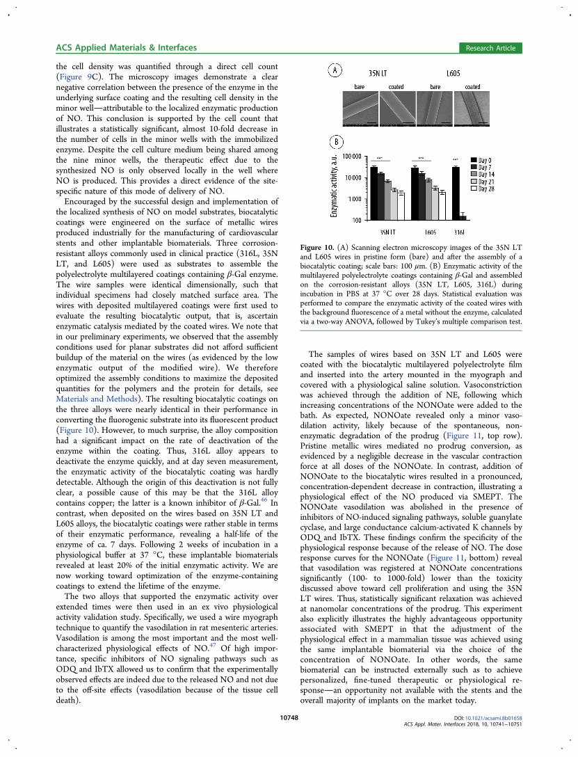
the localized synthesis of NO on model substrates, biocatalyticcoatings were engineered on the surface of metallic wiresproduced industrially for the manufacturing of cardiovascularstents and other implantable biomaterials. Three corrosion-resistant alloys commonly used in clinical practice (316L, 35NLT, and L605) were used as substrates to assemble thepolyelectrolyte multilayered coatings containing β-Gal enzyme.The wire samples were identical dimensionally, such thatindividual specimens had closely matched surface area. Thewires with deposited multilayered coatings were first used toevaluate the resulting biocatalytic output, that is, ascertainenzymatic catalysis mediated by the coated wires. We note thatin our preliminary experiments, we observed that the assemblyconditions used for planar substrates did not afford sufficientbuildup of the material on the wires (as evidenced by the lowenzymatic output of the modified wire). We thereforeoptimized the assembly conditions to maximize the depositedquantities for the polymers and the protein for details, seeMaterials and Methods). The resulting biocatalytic coatings onthe three alloys were nearly identical in their performance inconverting the fluorogenic substrate into its fluorescent product(Figure 10). However, to much surprise, the alloy compositionhad a significant impact on the rate of deactivation of theenzyme within the coating. Thus, 316L alloy appears todeactivate the enzyme quickly, and at day seven measurement,the enzymatic activity of the biocatalytic coating was hardlydetectable. Although the origin of this deactivation is not fullyclear, a possible cause of this may be that the 316L alloycontains copper; the latter is a known inhibitor of β-Gal.46 Incontrast, when deposited on the wires based on 35N LT andL605 alloys, the biocatalytic coatings were rather stable in termsof their enzymatic performance, revealing a half-life of theenzyme of ca. 7 days. Following 2 weeks of incubation in aphysiological buffer at 37 °C, these implantable biomaterialsrevealed at least 20% of the initial enzymatic activity. We arenow working toward optimization of the enzyme-containingcoatings to extend the lifetime of the enzyme.The two alloys that supported the enzymatic activity over
extended times were then used in an ex vivo physiologicalactivity validation study. Specifically, we used a wire myographtechnique to quantify the vasodilation in rat mesenteric arteries.Vasodilation is among the most important and the most well-characterized physiological effects of NO.47 Of high impor-tance, specific inhibitors of NO signaling pathways such asODQ and IbTX allowed us to confirm that the experimentallyobserved effects are indeed due to the released NO and not dueto the off-site effects (vasodilation because of the tissue celldeath).
The samples of wires based on 35N LT and L605 werecoated with the biocatalytic multilayered polyelectrolyte filmand inserted into the artery mounted in the myograph andcovered with a physiological saline solution. Vasoconstrictionwas achieved through the addition of NE, following whichincreasing concentrations of the NONOate were added to thebath. As expected, NONOate revealed only a minor vaso-dilation activity, likely because of the spontaneous, non-enzymatic degradation of the prodrug (Figure 11, top row).Pristine metallic wires mediated no prodrug conversion, asevidenced by a negligible decrease in the vascular contractionforce at all doses of the NONOate. In contrast, addition ofNONOate to the biocatalytic wires resulted in a pronounced,concentration-dependent decrease in contraction, illustrating aphysiological effect of the NO produced via SMEPT. TheNONOate vasodilation was abolished in the presence ofinhibitors of NO-induced signaling pathways, soluble guanylatecyclase, and large conductance calcium-activated K channels byODQ and IbTX. These findings confirm the specificity of thephysiological response because of the release of NO. The doseresponse curves for the NONOate (Figure 11, bottom) revealthat vasodilation was registered at NONOate concentrationssignificantly (100- to 1000-fold) lower than the toxicitydiscussed above toward cell proliferation and using the 35NLT wires. Thus, statistically significant relaxation was achievedat nanomolar concentrations of the prodrug. This experimentalso explicitly illustrates the highly advantageous opportunityassociated with SMEPT in that the adjustment of thephysiological effect in a mammalian tissue was achieved usingthe same implantable biomaterial via the choice of theconcentration of NONOate. In other words, the samebiomaterial can be instructed externally such as to achievepersonalized, fine-tuned therapeutic or physiological re-sponsean opportunity not available with the stents and theoverall majority of implants on the market today.
Figure 10. (A) Scanning electron microscopy images of the 35N LTand L605 wires in pristine form (bare) and after the assembly of abiocatalytic coating; scale bars: 100 μm. (B) Enzymatic activity of themultilayered polyelectrolyte coatings containing β-Gal and assembledon the corrosion-resistant alloys (35N LT, L605, 316L) duringincubation in PBS at 37 °C over 28 days. Statistical evaluation wasperformed to compare the enzymatic activity of the coated wires withthe background fluorescence of a metal without the enzyme, calculatedvia a two-way ANOVA, followed by Tukey’s multiple comparison test.
ACS Applied Materials & Interfaces Research Article
In this work, we engineered an enzyme−prodrug therapy ontothe surface of metallic wires based on the alloys commonly usedin clinical practice. The resulting coatings performed localizedbioconversion of prodrugs and produced the physiologicalmessenger molecule NO with a flux similar to the levelproduced by the healthy human endothelium. We successfullydemonstrated physiological responses to the locally producedNO, in an ex vivo wire myograph model. The NO-mediatedvasorelaxation was instructed by the concentration of theadministered NONOate, thus illustrating that the physiologicalresponse itself is not engineered into the implantablebiomaterial. Rather, it is the capacity to respond that wassuccessfully incorporated and preserved after biomaterialprocessing and delivery to tissue. This design paradigm canbe used toward personalized treatments using therapeuticimplants.
ORCIDMolly M. Stevens: 0000-0002-7335-266XAlexander N. Zelikin: 0000-0002-9864-321XPresent Address∇School of Chemical and Biomolecular Engineering, TheUniversity of Sydney, Sydney, NSW 2006, Australia.
Author Contributions¶A.K.W. and B.F. contributed equally.NotesThe authors declare no competing financial interest.
■ ACKNOWLEDGMENTSThe authors wish to acknowledge Dr. Aslan Husnu and RikkeMeyer (Aarhus University) for AFM imaging and EssiTaipaleenmaki and Dr. Brigitte Stadler (Aarhus University)for SEM imaging of the biocatalytic coatings. The authors wishto acknowledge the financial support from the EuropeanResearch Council Consolidator grant (A.N.Z., ERC-2013-CoG617336 BTVI), the ERC Seventh Framework ProgrammeConsolidator grant “Naturale CG” under grant agreement no.616417 (M.M.S.), a Wellcome Trust Senior Investigator Award(M.M.S., 098411/Z/12/Z), and the Novo Nordisk Foundation(US, NNF16OC0023284).
■ REFERENCES(1) Bredt, D. S.; Snyder, S. H. Nitric Oxide: A Physiologic MessengerMolecule. Annu. Rev. Biochem. 1994, 63, 175−195.(2) Garthwaite, J.; Boulton, C. L. Nitric Oxide Signaling in theCentral Nervous System. Annu. Rev. Physiol. 1995, 57, 683−706.(3) Anggard, E. Nitric oxide: mediator, murderer, and medicine.Lancet 1994, 343, 1199−1206.(4) Carpenter, A. W.; Schoenfisch, M. H. Nitric oxide release: Part II.Therapeutic applications. Chem. Soc. Rev. 2012, 41, 3742−3752.(5) MacMicking, J.; Xie, Q.-w.; Nathan, C. Nitric oxide andmacrophage function. Annu. Rev. Immunol. 1997, 15, 323−350.
Figure 11. Ex vivo wire myograph quantification of the contraction force exerted ex vivo by the rat mesenteric arteries (A,B) and calculated degree ofvasorelaxation (C,D) in the presence of NONOate (0.5 nM to 15 μM) and the wires based on 35N LT and L605 alloys coated with the biocatalyticmultilayered polyelectrolyte coatings (denoted as wire + Enz + NONOate). Control experiments include administering the NONOate in theabsence of wires (denoted as NONOate), using the wires and multilayered coatings with no incorporated enzyme (denoted wire + NONOate), andusing the samples identical to the experimental group and also containing specific inhibitors of the NO-mediated signaling pathways (denoted as wire+ Enz + NONOate + ODQ/IbTX). Data are presented as mean ± SEM, n = 5 or greater. Statistics is shown for comparing the effects mediated bythe biocatalytic coatings with those mediated by the NONOate (¤), the coatings with no enzyme (#), and the biocatalytic coatings in the presence ofinhibitors (*) and calculated via a two-way ANOVA followed by Tukey’s multiple comparison test.
ACS Applied Materials & Interfaces Research Article
(6) Felley-Bosco, E. Role of nitric oxide in genotoxicity: Implicationfor carcinogenesis. Cancer Metastasis Rev. 1998, 17, 25−37.(7) Akaike, T.; Maeda, H. Nitric oxide and virus infection.Immunology 2000, 101, 300−308.(8) de Mel, A.; Murad, F.; Seifalian, A. M. Nitric Oxide: A Guardianfor Vascular Grafts? Chem. Rev. 2011, 111, 5742−5767.(9) Jen, M. C.; Serrano, M. C.; van Lith, R.; Ameer, G. A. Polymer-Based Nitric Oxide Therapies: Recent Insights for BiomedicalApplications. Adv. Funct. Mater. 2012, 22, 239−260.(10) Riccio, D. A.; Schoenfisch, M. H. Nitric oxide release: Part I.Macromolecular scaffolds. Chem. Soc. Rev. 2012, 41, 3731−3741.(11) Fejerskov, B.; Olesen, M. T. J.; Zelikin, A. N. Substrate mediatedenzyme prodrug therapy. Adv. Drug Delivery Rev. 2017, 118, 24−34.(12) Fejerskov, B.; Zelikin, A. N. Substrate Mediated EnzymeProdrug Therapy. PLoS One 2012, 7, No. e49619.(13) Mendes, A. C.; Zelikin, A. N. Enzyme Prodrug TherapyEngineered into Biomaterials. Adv. Funct. Mater. 2014, 24, 5202−5210.(14) Andreasen, S. Ø.; Fejerskov, B.; Zelikin, A. N. Biocatalyticpolymer thin films: optimization of the multilayered architecturetowards in situ synthesis of anti-proliferative drugs. Nanoscale 2014, 6,4131−4140.(15) Fejerskov, B.; Jensen, N. B. S.; Teo, B. M.; Stadler, B.; Zelikin,A. N. Biocatalytic Polymer Coatings: On-Demand Drug Synthesis andLocalized Therapeutic Effect under Dynamic Cell Culture Conditions.Small 2014, 10, 1314−1324.(16) Cha, W.; Meyerhoff, M. E. Catalytic generation of nitric oxidefrom S-nitrosothiols using immobilized organoselenium species.Biomaterials 2007, 28, 19−27.(17) Fan, Y.; Pan, X.; Wang, K.; Wu, S.; Han, H.; Yang, P.; Luo, R.;Wang, H.; Huang, N.; Tan, W.; Weng, Y. Influence of chirality oncatalytic generation of nitric oxide and platelet behavior onselenocystine immobilized TiO2 films. Colloids Surf., B 2016, 145,122−129.(18) Weng, Y.; Song, Q.; Zhou, Y.; Zhang, L.; Wang, J.; Chen, J.;Leng, Y.; Li, S.; Huang, N. Immobilization of selenocystamine onTiO2 surfaces for in situ catalytic generation of nitric oxide andpotential application in intravascular stents. Biomaterials 2011, 32,1253−1263.(19) Hwang, S.; Meyerhoff, M. E. Organoditelluride-tetheredpolymers that spontaneously generate nitric oxide when in contactwith fresh blood. J. Mater. Chem. 2008, 18, 1784−1791.(20) Yang, Z.; Yang, Y.; Xiong, K.; Li, X.; Qi, P.; Tu, Q.; Jing, F.;Weng, Y.; Wang, J.; Huang, N. Nitric oxide producing coatingmimicking endothelium function for multifunctional vascular stents.Biomaterials 2015, 63, 80−92.(21) Walther, R.; Rautio, J.; Zelikin, A. N. Prodrugs in medicinalchemistry and enzyme prodrug therapies. Adv. Drug Delivery Rev.2017, 118, 65−77.(22) Chandrawati, R.; Chang, J. Y. H.; Reina-Torres, E.; Jumeaux, C.;Sherwood, J. M.; Stamer, W. D.; Zelikin, A. N.; Overby, D. R.; Stevens,M. M. Localized and Controlled Delivery of Nitric Oxide to theConventional Outflow Pathway via Enzyme Biocatalysis: TowardTherapy for Glaucoma. Adv. Mater. 2017, 29, 1604932.(23) Wang, Z.; Lu, Y.; Qin, K.; Wu, Y.; Tian, Y.; Wang, J.; Zhang, J.;Hou, J.; Cui, Y.; Wang, K.; Shen, J.; Xu, Q.; Kong, D.; Zhao, Q.Enzyme-functionalized vascular grafts catalyze in-situ release of nitricoxide from exogenous NO prodrug. J. Controlled Release 2015, 210,179−188.(24) Jewell, C. M.; Zhang, J.; Fredin, N. J.; Wolff, M. R.; Hacker, T.A.; Lynn, D. M. Release of Plasmid DNA from Intravascular StentsCoated with Ultrathin Multilayered Polyelectrolyte Films. Biomacro-molecules 2006, 7, 2483−2491.(25) Thierry, B.; Winnik, F. M.; Merhi, Y.; Tabrizian, M.Nanocoatings onto arteries via layer-by-layer deposition: Toward thein vivo repair of damaged blood vessels. J. Am. Chem. Soc. 2003, 125,7494−7495.(26) Yang, J.; Welby, J. L.; Meyerhoff, M. E. Generic nitric oxide(NO) generating surface by immobilizing organoselenium species viaLayer-by-Layer assembly. Langmuir 2008, 24, 10265−10272.
(27) Zelikin, A. N. Drug Releasing Polymer Thin Films: New Era ofSurface-Mediated Drug Delivery. ACS Nano 2010, 4, 2494−2509.(28) Schaffer, J. E.; Nauman, E. A.; Stanciu, L. A. Cold-DrawnBioabsorbable Ferrous and Ferrous Composite Wires: An Evaluationof Mechanical Strength and Fatigue Durability. Metall. Mater. Trans. B2012, 43, 984−994.(29) Appadoo, V.; Carter, M. C. D.; Lynn, D. M. Controlling thesurface-mediated release of DNA using “mixed multilayers”. Bioeng.Transl. Med. 2016, 1, 181−192.(30) Simonsen, U.; Wadsworth, R. M.; Buus, N. H.; Mulvany, M. J.In vitro simultaneous measurements of relaxation and nitric oxideconcentration in rat superior mesenteric artery. J. Physiol. 1999, 516,271−282.(31) Ruths, J.; Essler, F.; Decher, G.; Riegler, H. Polyelectrolytes I:Polyanion/Polycation Multilayers at the Air/Monolayer/Water Inter-face as Elements for Quantitative Polymer Adsorption Studies andPreparation of Hetero-superlattices on Solid Surfaces. Langmuir 2000,16, 8871−8878.(32) Gong, H.; Garcia-Turiel, J.; Vasilev, K.; Vinogradova, O. I.Interaction and Adhesion Properties of Polyelectrolyte Multilayers.Langmuir 2005, 21, 7545−7550.(33) Picart, C.; Mutterer, J.; Richert, L.; Luo, Y.; Prestwich, G. D.;Schaaf, P.; Voegel, J.-C.; Lavalle, P. Molecular basis for the explanationof the exponential growth of polyelectrolyte multilayers. Proc. Natl.Acad. Sci. U.S.A. 2002, 99, 12531−12535.(34) Mendelsohn, J. D.; Yang, S. Y.; Hiller, J.; Hochbaum, A. I.;Rubner, M. F. Rational Design of Cytophilic and CytophobicPolyelectrolyte Multilayer Thin Films. Biomacromolecules 2003, 4,96−106.(35) Salloum, D. S.; Schlenoff, J. B. Protein Adsorption Modalities onPolyelectrolyte Multilayers. Biomacromolecules 2004, 5, 1089−1096.(36) Dimitrova, M.; Affolter, C.; Meyer, F.; Nguyen, I.; Richard, D.G.; Schuster, C.; Bartenschlager, R.; Voegel, J.-C.; Ogier, J.; Baumert,T. F. Sustained delivery of siRNAs targeting viral infection by cell-degradable multilayered polyelectrolyte films. Proc. Natl. Acad. Sci.U.S.A. 2008, 105, 16320−16325.(37) Kerdjoudj, H.; Boura, C.; Marchal, L.; Dumas, D.; Schaff, P.;Voegel, J. C.; Stoltz, J. F.; Menu, P. Decellularized umbilical arterytreated with thin polyelectrolyte multilayer films: potential use invascular engineering. Biomed. Mater. Eng. 2006, 16, S123−S129.(38) Feldoto, Z.; Lundin, M.; Braesch-Andersen, S.; Blomberg, E.Adsorption of IgG on/in a PAH/PSS multilayer film: Layer structureand cell response. J. Colloid Interface Sci. 2011, 354, 31−37.(39) Cortez, C.; Quinn, J. F.; Hao, X.; Gudipati, C. S.; Stenzel, M. H.;Davis, T. P.; Caruso, F. Multilayer Buildup and Biofouling Character-istics of PSS-b-PEG Containing Films. Langmuir 2010, 26, 9720−9727.(40) Smith, R. N.; McCormick, M.; Barrett, C. J.; Reven, L.; Spiess,H. W. NMR Studies of PAH/PSS Polyelectrolyte MultilayersAdsorbed onto Silica. Macromolecules 2004, 37, 4830−4838.(41) Duong, H. T. T.; Kamarudin, Z. M.; Erlich, R. B.; Li, Y.; Jones,M. W.; Kavallaris, M.; Boyer, C.; Davis, T. P. Intracellular nitric oxidedelivery from stable NO-polymeric nanoparticle carriers. Chem.Commun. 2013, 49, 4190−4192.(42) Radomski, M. W.; Palmer, R. M. J.; Moncada, S. The role ofnitric oxide and cGMP in platelet adhesion to vascular endothelium.Biochem. Biophys. Res. Commun. 1987, 148, 1482−1489.(43) Vaughn, M. W.; Kuo, L.; Liao, J. C. Estimation of nitric oxideproduction and reaction rates in tissue by use of a mathematicalmodel. Am. J. Physiol. 1998, 274, H2163−H2176.(44) de Winter, R. J.; Katagiri, Y.; Asano, T.; Milewski, K. P.; Lurz, P.;Buszman, P.; Jessurun, G. A. J.; Koch, K. T.; Troquay, R. P. T.; Hamer,B. J. B.; Ophuis, T. O.; Wohrle, J.; Wyderka, R.; Cayla, G.; Hofma, S.H.; Levesque, S.; Zurakowski, A.; Fischer, D.; Kosmider, M.; Goube,P.; Arkenbout, E. K.; Noutsias, M.; Ferrari, M. W.; Onuma, Y.; Wijns,W.; Serruys, P. W. A sirolimus-eluting bioabsorbable polymer-coatedstent (MiStent) versus an everolimus-eluting durable polymer stent(Xience) after percutaneous coronary intervention (DESSOLVE III):
ACS Applied Materials & Interfaces Research Article