Page 1
RESEARCH ARTICLE
EP4 receptor stimulation down-regulates humaneosinophil function
Petra Luschnig-Schratl • Eva M. Sturm • Viktoria Konya • Sonia Philipose •
Gunther Marsche • Eleonore Frohlich • Claudia Samberger • Doris Lang-Loidolt •
Stefan Gattenlohner • Irmgard Th. Lippe • Bernhard A. Peskar •
Rufina Schuligoi • Akos Heinemann
Received: 3 May 2010 / Revised: 7 February 2011 / Accepted: 15 February 2011 / Published online: 2 March 2011
� The Author(s) 2011. This article is published with open access at Springerlink.com
Abstract Accumulation of eosinophils in tissue is a
hallmark of allergic inflammation. Here we observed that a
selective agonist of the PGE2 receptor EP4, ONO AE1-
329, potently attenuated the chemotaxis of human periph-
eral blood eosinophils, upregulation of the adhesion
molecule CD11b and the production of reactive oxygen
species. These effects were accompanied by the inhibition
of cytoskeletal rearrangement and Ca2? mobilization. The
involvement of the EP4 receptor was substantiated by a
selective EP4 antagonist, which reversed the inhibitory
effects of PGE2 and the EP4 agonist. Selective kinase
inhibitors revealed that the inhibitory effect of EP4 stim-
ulation on eosinophil migration depended upon activation
of PI 3-kinase and PKC, but not cAMP. Finally, we found
that EP4 receptors are expressed by human eosinophils,
and are also present on infiltrating leukocytes in inflamed
human nasal mucosa. These data indicate that EP4 ago-
nists might be a novel therapeutic option in eosinophilic
diseases.
Keywords Eosinophils � Prostaglandins � Receptors �Chemotaxis � Reactive oxygen species � Degranulation
Abbreviations
BSA Bovine serum albumin
cAMP Cyclic adenosine monophosphate
COX Cyclooxygenase
CREB cAMP response element binding protein
EP E-type prostanoid receptor
ERK Extracellular signal-regulated kinase
FITC Fluorescein isothiocyanate
IL Interleukin
IP I-type prostanoid receptor
LT Leukotriene
PE Phycoerythrine
PG Prostaglandin
PI3K Phosphatidylinositol 3-kinase
PKA Protein kinase A
PKC Protein kinase C
PMA Phorbol 12-myristate 13-acetate
TP Thromboxane-type prostanoid receptor
Introduction
Eosinophils play a major role in late-phase reactions by
releasing bronchoconstrictor mediators such as leukotriene
(LT) C4 and other chemoattractants that cause further
influx of inflammatory cells into the tissue, and immuno-
regulatory type-2 cytokines, interleukin (IL)-4, IL-5, IL-10,
Electronic supplementary material The online version of thisarticle (doi:10.1007/s00018-011-0642-5) contains supplementarymaterial, which is available to authorized users.
P. Luschnig-Schratl � E. M. Sturm � V. Konya � S. Philipose �G. Marsche � I. Th. Lippe � B. A. Peskar � R. Schuligoi �A. Heinemann (&)
Institute of Experimental and Clinical Pharmacology, Medical
University of Graz, Universitaetsplatz 4, 8010 Graz, Austria
e-mail: [email protected]
E. Frohlich � C. Samberger
Center for Medical Research, Medical University of Graz,
Graz, Austria
D. Lang-Loidolt
Department of Otorhinolaryngology, Medical University
of Graz, Graz, Austria
S. Gattenlohner
Institute of Pathology, Medical University of Graz, Graz, Austria
Cell. Mol. Life Sci. (2011) 68:3573–3587
DOI 10.1007/s00018-011-0642-5 Cellular and Molecular Life Sciences
123
Page 2
and IL-13 [1]. Mucosal damage in chronic asthma is
associated with cytotoxic mediators that are released by
activated eosinophils, including matrix metalloproteases,
major basic protein, eosinophil cationic protein, eosinophil
peroxidase and eosinophil-derived neurotoxin, leading to
airway remodeling and angiogenesis in chronically
inflamed tissue [2, 3]. Importantly, it was shown that
asthmatic patients who receive treatment based on eosin-
ophil counts in sputum have significantly fewer severe
asthma exacerbations than patients treated according to
standard management therapy [4]. Therefore, eosinophils
are currently considered a major therapeutic target in
allergic diseases and asthma [5], eosinophilic esophagitis
[6], colitis ulcerosa [7], or hypereosinophilic syndrome [8].
PGE2 is the predominant cyclooxygenase (COX)
product of airway macrophages, epithelial cells, and
smooth muscle cells, and is regarded as a potent inflam-
matory mediator due to its effects on vasodilation, vascular
permeability, and nociception. However, the role of PGE2
in allergic inflammation is less clear. In the asthmatic lung,
PGE2 affects both airway smooth muscle and the inflam-
matory process: PGE2 causes bronchial relaxation [9] and
inhibits allergen-induced bronchoconstriction [10], but it
may also provoke bronchoconstrictor responses and cough
in some individuals [11, 12] because of activation of
C-fibers and reflex cholinergic pathways [13]. In rats and
humans, PGE2 reduces allergen-induced airway eosino-
philia [14, 15], attenuates anaphylactic mediator release
from guinea-pig perfused lungs [16], abrogates eosinophil
accumulation after passive cutaneous anaphylaxis in gui-
nea pigs [17], and protects against bleomycin-induced
pulmonary fibrosis in mice [18]. Conversely, eosinophil
influx is exaggerated in COX-1 or COX-2 knockout mice
[19, 20] and also in mice treated with selective COX-1 or
COX-2 inhibitors [21]. At the cellular level, PGE2 has
been found to attenuate immunoglobulin-dependent
degranulation and LTC4 biosynthesis of eosinophils [22],
and agonist-induced CD11b upregulation and L-selectin
shedding in eosinophils and neutrophils [23]. In contrast,
PGE2 is anti-apoptotic for eosinophils [24], while its
analogue misoprostol inhibits eosinophil survival in vitro
[25].
The biological effects of PGE2 are mediated through
four different G protein-coupled heptahelical receptors,
termed EP1, EP2, EP3, and EP4 [26]. Each of these
receptors has a distinct pharmacological signature based on
its selectivity towards synthetic PGE2 analogs and intra-
cellular signal transduction. Stimulation of the EP1
receptor results in Gq-mediated activation of phospholipase
C and phosphatidylinositol hydrolysis, elevation of the
intracellular Ca2? level, and causes the activation of pro-
tein kinase C (PKC) [27]. The EP3 receptor exists as a
number of splice variants displaying various degrees of
constitutive activity. EP3 signals through activation of a Gi
protein to inhibit adenylyl cyclase leading to reduction of
intracellular cyclic adenosine monophosphate (cAMP)
generation and elevation of intracellular-free Ca2? levels.
However, isoforms of EP3 also have the capacity to
enhance cAMP formation by coupling to Gs protein [28]. In
contrast, stimulation of EP2 and EP4 receptors usually
increases intracellular cAMP levels and activates protein
kinase A (PKA) through Gs protein. Interestingly, EP2
receptor stimulation can also trigger Ca2? currents in a
cAMP-dependent/PKA-independent manner [29].
We recently described that stimulation of EP2 receptors
attenuates eosinophil trafficking [30], and we also obtained
some preliminary information on a potential role of EP4
receptors in the regulation of eosinophil function. There-
fore, in this study we investigated the expression, function,
and signaling of EP4 receptors in eosinophils in detail and
suggest that EP4 receptors are even more substantially
involved in the regulation of eosinophil effector functions
as compared to EP2 receptors. EP4 agonists might hence be
novel therapeutic options for the treatment of eosinophilic
diseases.
Materials and methods
Chemicals
All laboratory reagents were from Sigma (Vienna, Austria),
unless specified. Assay buffer used in all experiments was
made from Dulbecco’s modified phosphate-buffered saline
(PBS; with 0.9 mM Ca2? and 0.5 mM Mg2?; Invitrogen,
Vienna, Austria), 0.1% bovine serum albumin (BSA);
10 mM HEPES and 10 mM glucose, pH 7.4. CellFix and
FACS-Flow were from Becton-Dickinson (Vienna, Aus-
tria). Human eotaxin and IL-8 were from Peprotech
(London, UK). PGD2, PGE2, the EP2 receptor agonist
butaprost, the EP1/EP3 receptor agonist sulprostone, the
EP4 receptor antagonist GW627368X and the EP1/I-type
prostanoid (IP) receptor agonist iloprost were from Cay-
man Chemicals (Ann Arbor, MI, USA). Polyclonal rabbit
anti-human EP4 receptor antibodies directed against the C
terminus of the receptor were provided by Sigma and
Cayman. Goat antibody against eosinophil peroxidase was
from Santa Cruz Biotechnology (Heidelberg, Germany).
Mouse anti-eosinophil peroxidase antibody was supplied
by Becton-Dickinson. Donkey anti-mouse Alexa Fluor 555
and donkey anti-rabbit Alexa Fluor 488 antibodies were
obtained from Invitrogen. Rabbit and goat control IgG was
from Linaris (Wertheim-Bettingen, Germany). The aden-
ylyl cyclase inhibitor SQ22536, the PI3K inhibitor
LY294002 and the PKC inhibitor chelerythrine were sup-
plied from Biomol (Hamburg, Germany). The EP4 agonist
3574 P. Luschnig-Schratl et al.
123
Page 3
ONO AE1-329 (2-[3-[(1R,2S,3R)-3-hydroxy-2-[(E,3S)-3-
hydroxy-5-[2-(methoxymethyl)phenyl]pent-1-enyl]-5-oxo-
cyclopentyl]sulfanylpropylsulfanyl]acetic acid) and the EP4
antagonist ONO AE3-208 (2-[[2-[2-(2-methylnaphthalen-
1-yl)propanoylamino]phenyl]methyl]benzoic acid) were a
kind gift from ONO Pharmaceutical (Osaka, Japan). ONO
AE1-329 has been shown in competitive radioligand binding
assays to selectively bind to EP4 receptors (Ki = 9.7 nM)
relative to the EP1, EP2, and EP3 receptors (Ki =
[ 10,000, [ 2,000 and [ 1,000 nM, respectively) [31].
GW627368X (4-(4,9-diethoxy-1,3-dihydro-1-oxo-2H-benz
[f]isoindol-2-yl)-N-(phenyl sulfonyl)-benzeneacetamide)
exhibits a Ki value of 100 nM towards EP4 and 158 nM to
thromboxane-type prostanoid (TP) receptors, with Ki values
above 10,000 nM for all other prostanoid receptors [32]. The
Ki values of ONO AE3-208 are 1.3, 30, 790, 2,400 nM
for EP4, EP3, F-type prostanoid (FP) receptor, and TP,
respectively, and more than 10,000 nM for other prostanoid
receptors [33].
Preparation of human leukocytes
This study was approved by the Ethics Committee of
the Medical University of Graz. Prior to blood sampling
from healthy non-atopic volunteers, all donors signed an
informed-consent form. Platelet-rich plasma was removed
by centrifugation of citrated whole blood. Erythrocytes
were removed by dextran sedimentation. High-density
polymorphonuclear leukocytes (PMNL, containing neu-
trophils and eosinophils) were isolated by Histopaque
gradient centrifugation. Any erythrocyte contamination of
the PMNL pellet was removed by hypotonic shock lysis as
described previously [34]. Purified eosinophil preparations
were obtained by negative magnetic selection using anti-
body cocktails (CD2, CD14, CD16, CD19, CD56, and
glycophorin A) and colloidal magnetic particles from
StemCell Technologies (Vancouver, Canada). Resulting
purity and viability was typically [97%.
Chemotaxis
Migration of eosinophils was determined in 48-well
microBoyden chemotaxis chambers. Purified eosinophils
were resuspended in assay buffer at 2 9 106 cells/ml and
50 ll of the cell-suspension were loaded into the top wells
of the chamber which were separated from the bottom wells
by a 5-lm pore-size polyvinylpyrrolidone-free polycar-
bonate filter. Thirty microliters of assay buffer or agonists
were placed into the bottom wells of the chamber. Baseline
migration was determined in wells containing only assay
buffer. The chamber was incubated at 37�C for 1 h in a
humidified incubator. The membrane was subsequently
removed and migrated cells were enumerated by a
FACSCalibur flow cytometer (Becton-Dickinson, Moun-
tain View, CA, USA) [35].
Leukocyte shape change assay
Preparations of polymorphonuclear leukocytes (PMNL;
containing eosinophils and neutrophils) or purified eosin-
ophils were resuspended in assay buffer and aliquots of the
cell-suspension were mixed with agonists at a final volume
of 100 ll and stimulated for 4 min at 37�C. Cells were
transferred to ice and 250 ll of ice-cold fixative solution
was added to terminate the reaction. Changes in the cell
shape were estimated immediately by the increase of for-
ward scatter using a FACSCalibur flow cytometer (Becton-
Dickinson, Mountain View, CA, USA). Eosinophils were
distinguished from neutrophils according to granularity
(side scatter) and by their autofluorescence.
Respiratory burst
Purified eosinophils (5 9 105 cells/ml) were stimulated
with agonists in the presence of 1 lM dihydrorhodamine-
123 for 20 min at 37�C and then fixed with 150 ll of ice-
cold 2.5% Cellfix. Respiratory burst of eosinophils was
immediately quantified by flow cytometry as the increase
of fluorescence in the FL-1 channel due to the oxidiza-
tion by reactive oxygen species of the non-fluorescent
dye dihydrorhodamine-123 into fluorescent rhodamine-123
[36]. Responses were expressed as percent changes from a
control sample incubated with buffer alone.
Upregulation of eosinophil CD11b expression
Polymorphonuclear leukocyte preparations were incubated
with agonists for 30 min at 37�C and then stained with
anti-CD11b (FITC) and anti-CD16 (PE-Cy5) antibodies.
CD11b expression on CD16-negative eosinophils was
quantified by flow cytometry and expressed as percent of
the maximal control response (i.e., in the absence of a
prostanoid).
Calcium ion flux
Intracellular Ca2? levels in eosinophils were analyzed by
flow cytometry as described previously [37]. Polymor-
phonuclear leukocyte preparations were treated with 2 lM
of the acetoxymethyl ester of Fluo-3 in the presence of
0.02% pluronic F-127 for 60 min at room temperature
before being washed with PBS without Ca2? and Mg2?.
Cells were then stained with anti-CD16 (PE) and resus-
pended in assay buffer at 3 9 106 cells/ml. Changes in
intracellular Ca2? levels were detected by flow cytometry
as the increase of the fluorescence of the Ca2? sensitive dye
EP4 receptors and eosinophil function 3575
123
Page 4
Fluo-3 in the FL1-channel. Eosinophils were identified as
CD16-negative cells.
Flow cytometric analysis of EP receptor expression
Expression of EP1, EP2, EP3, and EP4 receptors on human
peripheral blood eosinophils was quantified by indirect
immunofluorescence flow cytometry. As the EP antibodies
had been raised against the intracellular C terminus of the EP
receptors, aliquots of isolated eosinophils were first per-
meabilized with Fix&Perm solution (ADG Bio Research;
Kaumberg, Austria) for 15 min at room temperature. Sam-
ples were then treated with the following reagents for
30 min each on ice with appropriate washing steps in
between: Ultra V Block (Labvision, Westinghouse, CA,
USA) to block Fc receptors; 20 lg/ml polyclonal EP1, EP2,
EP3 or EP4 antibody or 20 lg/ml polyclonal rabbit isotype
control antibody; and 4 lg/ml anti-rabbit IgG secondary
antibody conjugated with Alexa Fluor-488. After adding the
fixative solution the cells were analyzed on a FACSCalibur
flow cytometer (BD Biosciences).
Western blot of EP4 receptors
Purified eosinophils were lysed in a buffer containing
50 mM Tris–HCl, 25 mM KCl, 5 mM MgCl2, and 0.2%
Nonidet P-40 supplemented with protease inhibitors
(Roche; Basel, Switzerland) and centrifuged at 10,000 rpm
for 10 min at 4�C. Bradford protein assay (Bio-Rad;
Vienna, Austria) was performed to determine the protein
content in the supernatants. Protein samples (50 lg) were
separated by SDS-polyacrylamide gel electrophoresis on a
gradient gel (4–20%) and protein bands were blotted onto
polyvinylidenefluoride (Bio-Rad) membrane. Target pro-
teins were immunochemically detected using a polyclonal
rabbit anti-human EP4 antibody (20 lg/ml). Bands were
visualized with horseradish peroxidase-conjugated goat
anti-rabbit IgG (4 lg/ml; Pierce, Rockford, IL, USA) and
Amersham ECL Plus detection reagents (GE Healthcare;
Vienna, Austria).
Immunohistochemistry of EP4 receptors
Paraffin blocks of nasal polyps that had been previously
classified by a pathologist to contain eosinophil infiltrates
were obtained from the Tissue Bank of the Medical Uni-
versity of Graz as approved by the local ethics committee.
Five-micrometer sections were deparaffinized, antigen
retrieval was performed in DakoCytomation Target Retrie-
val Solution (Glostrup, Denmark) for 10 min at 120�C, and
sections were blocked with 1% BSA and 0.05% Triton
X-100 in PBS for 30 min at room temperature. The samples
were incubated overnight with a rabbit anti-human EP4
antibody (1.7 lg/ml, Sigma) or rabbit control IgG antibody.
After washing, the bound antibody was detected using the
Liquid DAB ? Substrate Chromogen System (Dako Cyto-
mation). Staining of eosinophil peroxidase-positive cells
was performed with a polyclonal goat antibody against
eosinophil peroxidase (20 lg/ml) or control antibody for
30 min at room temperature. After washing, the bound
antibody was then visualized with the EnVision Permanent
Red (Dako Cytomation). Slides were finally counterstained
with Mayer’s hematoxylin. Sections were visually examined
with a Zeiss Axiophot microscope (Zeiss, Oberkochen,
Germany) and a Zeiss Plan-Neofluar 920/05 lens. Photo-
graphs were taken with a CoolSNAP camera (1392 9 1040
pixels; Photometrics, Tucson, AZ, USA) and a RGB Tun-
able Micro*color filter (CRI, Woburn, MA, USA). Further
processing of images was performed with MCID-M2 soft-
ware (Imaging Research Inc., Brock University, St.
Catharines, Ontario, Canada) for additional white balance,
contrast, and brightness adjustments.
Double immunofluorescence stainings of eosinophil
peroxidase and EP4 were performed as described [38] with
incubation times of 10 and 50 min, respectively, using the
following antibodies: mouse-anti-human eosinophil perox-
idase antibody; rabbit anti-human EP4 antibody (12.5 lg/
ml, Sigma); and donkey anti-mouse Alexa Fluor 555 or
donkey anti-rabbit Alexa Fluor 488 (both 0.5 lg/ml).
Images were taken with a confocal laser scanning micro-
scope (Leica TCS SP2; Leica, Bensheim, Germany) using
Leica Confocal Software version 2.61 Build 1537.
Statistical analyses
Data are shown as mean ± SEM for n observations.
Comparisons of groups were performed using one-way
ANOVA or two-way ANOVA for repeated measurements
followed by Holm-Sidak post-hoc test to determine the
levels of significance for each group. Probability values of
p \ 0.05 were considered as statistically significant.
Results
Involvement of EP4 receptors in the PGE2-induced
attenuation of eosinophil migration
We showed recently that PGE2 and the EP2 agonist buta-
prost attenuate the migratory responsiveness of human
eosinophil granulocytes [30]. Here we investigated the
potential role of EP4 receptors in eosinophil function. For
that purpose, we pretreated purified human eosinophils with
the EP4 receptor antagonists GW627368X (1 or 10 lM) or
ONO AE3-208 (100 nM) or their vehicle for 15 min at
37�C, and then mixed them with various concentrations of
3576 P. Luschnig-Schratl et al.
123
Page 5
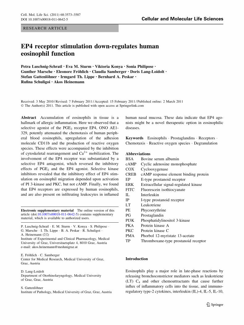
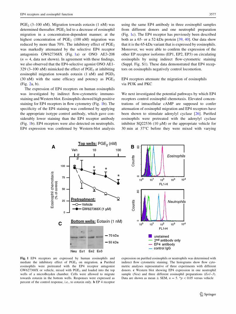
PGE2 (3–100 nM). Migration towards eotaxin (1 nM) was
determined thereafter. PGE2 led to a decrease of eosinophil
migration in a concentration-dependent manner; at the
highest concentration of PGE2 (100 nM) migration was
reduced by more than 70%. The inhibitory effect of PGE2
was markedly attenuated by the selective EP4 receptor
antagonists GW627368X (Fig. 1a) or ONO AE3-208
(n = 4, data not shown). In agreement with these findings,
we also observed that the EP4-selective agonist ONO AE1-
329 (3–100 nM) mimicked the effect of PGE2 at inhibiting
eosinophil migration towards eotaxin (1 nM) and PGD2
(30 nM) with the same efficacy and potency as PGE2
(Fig. 2a, b).
The expression of EP4 receptors on human eosinophils
was investigated by indirect flow-cytometric immuno-
staining and Western blot. Eosinophils showed high positive
staining for EP4 receptors in flow cytometry (Fig. 1b). The
specificity of the EP4 staining was confirmed by applying
the appropriate isotype control antibody, which gave con-
siderably lower staining than the EP4 receptor antibody
(Fig. 1b). EP4 receptors were also detected on neutrophils.
EP4 expression was confirmed by Western-blot analysis
using the same EP4 antibody in three eosinophil samples
from different donors and one neutrophil preparation
(Fig. 1c). The EP4 receptor has previously been described
both as a 65- or a 52-kDa protein [39, 40]. Our data show
that it is the 65-kDa variant that is expressed by eosinophils.
Moreover, we were able to confirm the expression of the
other EP receptor isoforms (EP1, EP2, EP3) on circulating
eosinophils by using indirect flow-cytometric staining
(Suppl. Fig. S1). These data demonstrated that EP4 recep-
tors on eosinophils negatively control locomotion.
EP4 receptors attenuate the migration of eosinophils
via PI3K and PKC
We next investigated the potential pathways by which EP4
receptors control eosinophil chemotaxis. Elevated concen-
trations of intracellular cAMP are supposed to confer
attenuation of eosinophil migration and EP4 receptors have
been shown to stimulate adenylyl cyclase [26]. Purified
eosinophils were pretreated with the adenylyl cyclase
inhibitor SQ22536 (10 lM) or the appropriate vehicle for
30 min at 37�C before they were mixed with varying
Fig. 1 EP4 receptors are expressed by human eosinophils and
mediate the inhibitory effect of PGE2 on migration. a Purified
eosinophils were pretreated with the EP4 receptor antagonist
GW627368X or vehicle, mixed with PGE2 and loaded into the top
wells of a microBoyden chamber. Cells were allowed to migrate
towards eotaxin in the bottom wells. Responses were expressed as
percent of the control response, i.e., to eotaxin only. b EP 4 receptor
expression on purified eosinophils or neutrophils was determined with
indirect flow cytometric staining. The histograms show flow cyto-
metric analyses representative of three experiments with different
donors. c Western blot showing EP4 expression in one neutrophil
sample (Neu) and three different eosinophil preparations (Eo1–3).
Data are shown as mean ± SEM, n = 5. *p \ 0.05 versus vehicle
EP4 receptors and eosinophil function 3577
123
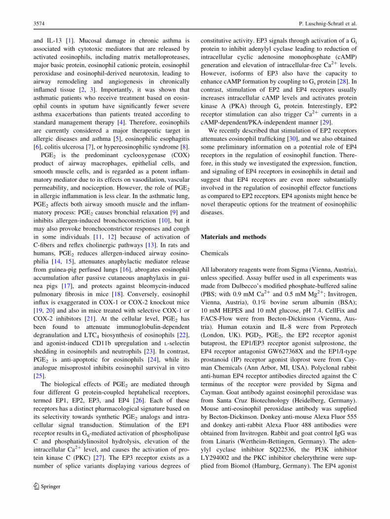
Page 6
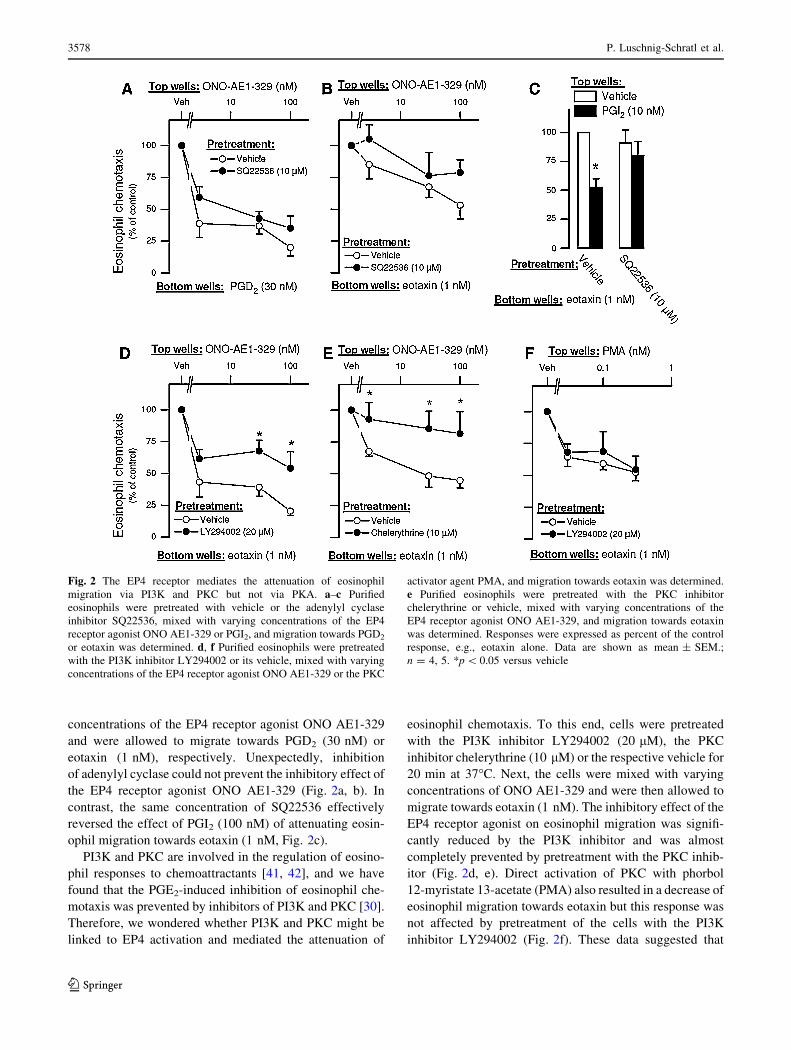
concentrations of the EP4 receptor agonist ONO AE1-329
and were allowed to migrate towards PGD2 (30 nM) or
eotaxin (1 nM), respectively. Unexpectedly, inhibition
of adenylyl cyclase could not prevent the inhibitory effect of
the EP4 receptor agonist ONO AE1-329 (Fig. 2a, b). In
contrast, the same concentration of SQ22536 effectively
reversed the effect of PGI2 (100 nM) of attenuating eosin-
ophil migration towards eotaxin (1 nM, Fig. 2c).
PI3K and PKC are involved in the regulation of eosino-
phil responses to chemoattractants [41, 42], and we have
found that the PGE2-induced inhibition of eosinophil che-
motaxis was prevented by inhibitors of PI3K and PKC [30].
Therefore, we wondered whether PI3K and PKC might be
linked to EP4 activation and mediated the attenuation of
eosinophil chemotaxis. To this end, cells were pretreated
with the PI3K inhibitor LY294002 (20 lM), the PKC
inhibitor chelerythrine (10 lM) or the respective vehicle for
20 min at 37�C. Next, the cells were mixed with varying
concentrations of ONO AE1-329 and were then allowed to
migrate towards eotaxin (1 nM). The inhibitory effect of the
EP4 receptor agonist on eosinophil migration was signifi-
cantly reduced by the PI3K inhibitor and was almost
completely prevented by pretreatment with the PKC inhib-
itor (Fig. 2d, e). Direct activation of PKC with phorbol
12-myristate 13-acetate (PMA) also resulted in a decrease of
eosinophil migration towards eotaxin but this response was
not affected by pretreatment of the cells with the PI3K
inhibitor LY294002 (Fig. 2f). These data suggested that
Fig. 2 The EP4 receptor mediates the attenuation of eosinophil
migration via PI3K and PKC but not via PKA. a–c Purified
eosinophils were pretreated with vehicle or the adenylyl cyclase
inhibitor SQ22536, mixed with varying concentrations of the EP4
receptor agonist ONO AE1-329 or PGI2, and migration towards PGD2
or eotaxin was determined. d, f Purified eosinophils were pretreated
with the PI3K inhibitor LY294002 or its vehicle, mixed with varying
concentrations of the EP4 receptor agonist ONO AE1-329 or the PKC
activator agent PMA, and migration towards eotaxin was determined.
e Purified eosinophils were pretreated with the PKC inhibitor
chelerythrine or vehicle, mixed with varying concentrations of the
EP4 receptor agonist ONO AE1-329, and migration towards eotaxin
was determined. Responses were expressed as percent of the control
response, e.g., eotaxin alone. Data are shown as mean ± SEM.;
n = 4, 5. *p \ 0.05 versus vehicle
3578 P. Luschnig-Schratl et al.
123
Page 7
both PI3K and PKC, but not the adenylyl cyclase/cAMP
pathway, are involved in the signaling of the EP4-mediated
attenuation of eosinophil migration.
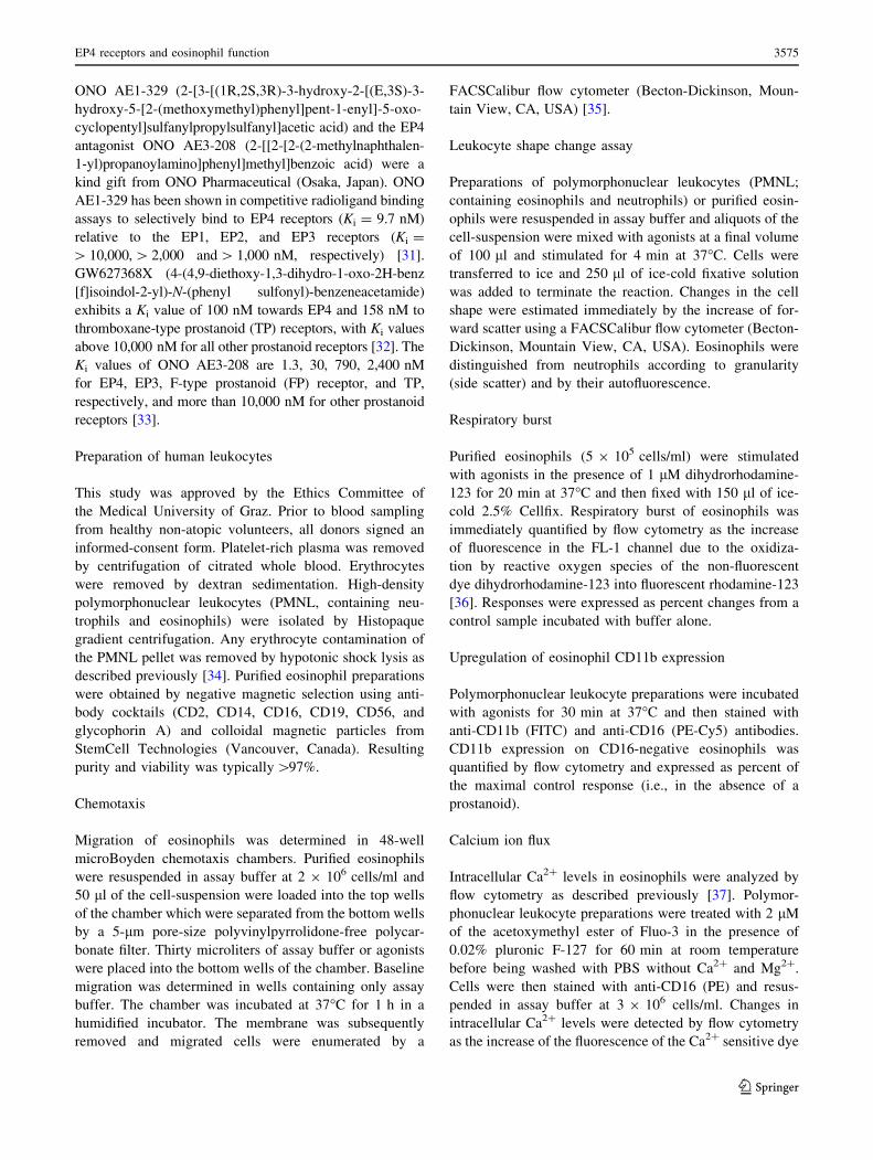
PGE2 inhibits Ca2? flux and shape change
of eosinophils via EP4 receptors
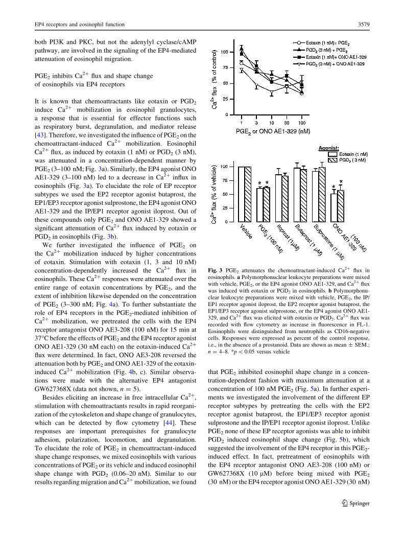
It is known that chemoattractants like eotaxin or PGD2
induce Ca2? mobilization in eosinophil granulocytes,
a response that is essential for effector functions such
as respiratory burst, degranulation, and mediator release
[43]. Therefore, we investigated the influence of PGE2 on the
chemoattractant-induced Ca2? mobilization. Eosinophil
Ca2? flux, as induced by eotaxin (1 nM) or PGD2 (3 nM),
was attenuated in a concentration-dependent manner by
PGE2 (3–100 nM; Fig. 3a). Similarly, the EP4 agonist ONO
AE1-329 (3–100 nM) led to a decrease in Ca2? influx in
eosinophils (Fig. 3a). To elucidate the role of EP receptor
subtypes we used the EP2 receptor agonist butaprost, the
EP1/EP3 receptor agonist sulprostone, the EP4 agonist ONO
AE1-329 and the IP/EP1 receptor agonist iloprost. Out of
these compounds only PGE2 and ONO AE1-329 showed a
significant attenuation of Ca2? flux induced by eotaxin or
PGD2 in eosinophils (Fig. 3b).
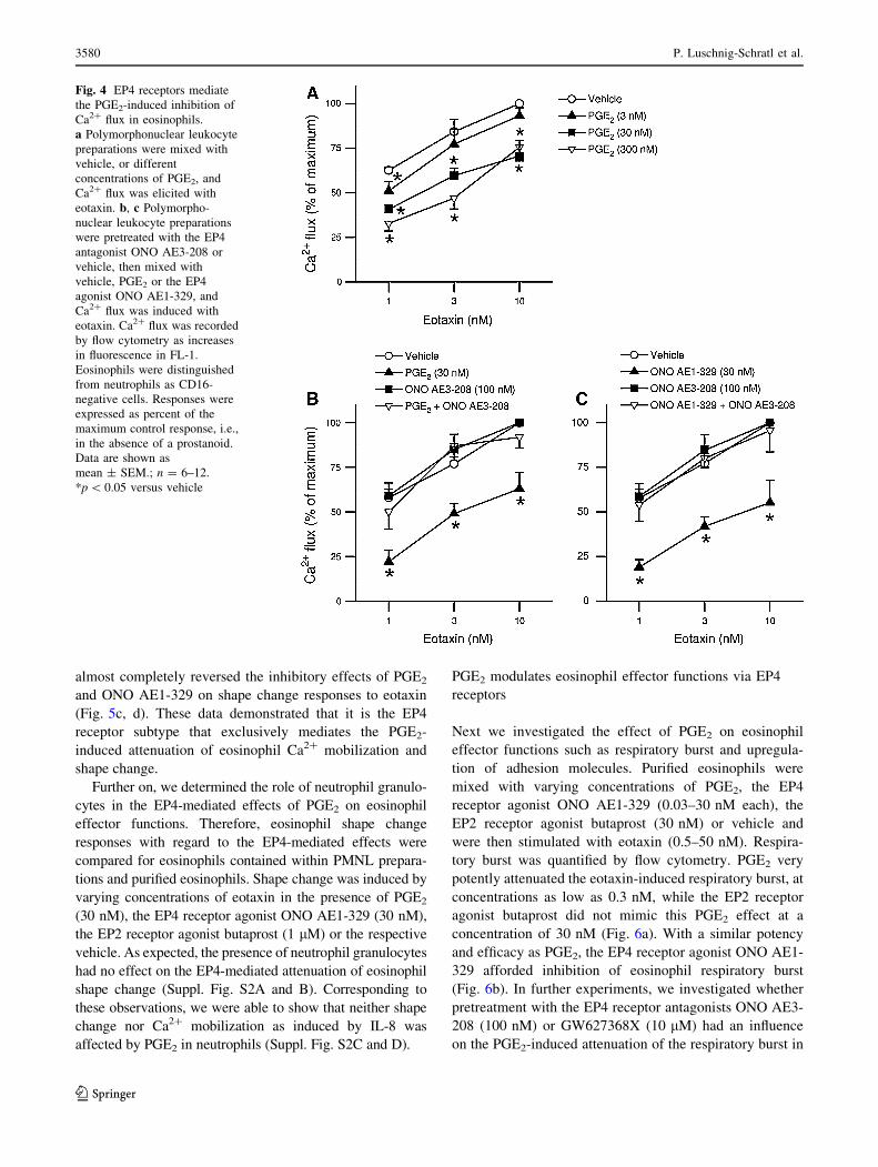
We further investigated the influence of PGE2 on
the Ca2? mobilization induced by higher concentrations
of eotaxin. Stimulation with eotaxin (1, 3 and 10 nM)
concentration-dependently increased the Ca2? flux in
eosinophils. These Ca2? responses were attenuated over the
entire range of eotaxin concentrations by PGE2, and the
extent of inhibition likewise depended on the concentration
of PGE2 (3–300 nM; Fig. 4a). To further substantiate the
role of EP4 receptors in the PGE2-mediated inhibition of
Ca2? mobilization, we pretreated the cells with the EP4
receptor antagonist ONO AE3-208 (100 nM) for 15 min at
37�C before the effects of PGE2 and the EP4 receptor agonist
ONO AE1-329 (30 nM each) on the eotaxin-induced Ca2?
flux were determined. In fact, ONO AE3-208 reversed the
attenuation both by PGE2 and ONO AE1-329 of the eotaxin-
induced Ca2? mobilization (Fig. 4b, c). Similar observa-
tions were made with the alternative EP4 antagonist
GW627368X (data not shown, n = 5).
Besides eliciting an increase in free intracellular Ca2?,
stimulation with chemoattractants results in rapid reorgani-
zation of the cytoskeleton and shape change of granulocytes,
which can be detected by flow cytometry [44]. These
responses are important prerequisites for granulocyte
adhesion, polarization, locomotion, and degranulation.
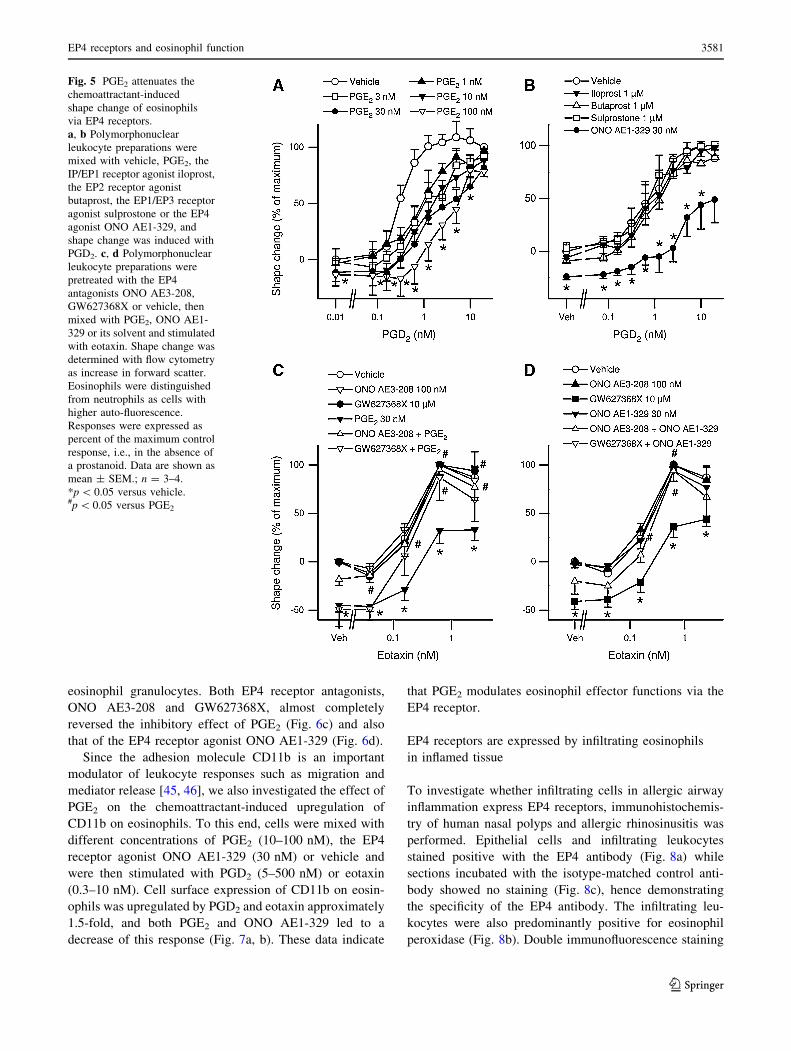
To elucidate the role of PGE2 in chemoattractant-induced
shape change responses, we mixed eosinophils with various
concentrations of PGE2 or its vehicle and induced eosinophil
shape change with PGD2 (0.06–20 nM). Similar to our
results regarding migration and Ca2? mobilization, we found
that PGE2 inhibited eosinophil shape change in a concen-
tration-dependent fashion with maximum attenuation at a
concentration of 100 nM PGE2 (Fig. 5a). In further experi-
ments we investigated the involvement of the different EP
receptor subtypes by pretreating the cells with the EP2
receptor agonist butaprost, the EP1/EP3 receptor agonist
sulprostone and the IP/EP1 receptor agonist iloprost. Unlike
PGE2 none of these EP receptor agonists was able to inhibit
PGD2 induced eosinophil shape change (Fig. 5b), which
suggested the involvement of the EP4 receptor in this PGE2-
induced effect. In fact, pretreatment of eosinophils with
the EP4 receptor antagonist ONO AE3-208 (100 nM) or
GW627368X (10 lM) before being mixed with PGE2
(30 nM) or the EP4 receptor agonist ONO AE1-329 (30 nM)
Fig. 3 PGE2 attenuates the chemoattractant-induced Ca2? flux in
eosinophils. a Polymorphonuclear leukocyte preparations were mixed
with vehicle, PGE2, or the EP4 agonist ONO AE1-329, and Ca2? flux
was induced with eotaxin or PGD2 in eosinophils. b Polymorphonu-
clear leukocyte preparations were mixed with vehicle, PGE2, the IP/
EP1 receptor agonist iloprost, the EP2 receptor agonist butaprost, the
EP1/EP3 receptor agonist sulprostone, or the EP4 agonist ONO AE1-
329, and Ca2? flux was elicited with eotaxin or PGD2. Ca2? flux was
recorded with flow cytometry as increase in fluorescence in FL-1.
Eosinophils were distinguished from neutrophils as CD16-negative
cells. Responses were expressed as percent of the control response,
i.e., in the absence of a prostanoid. Data are shown as mean ± SEM.;
n = 4–8. *p \ 0.05 versus vehicle
EP4 receptors and eosinophil function 3579
123
Page 8
almost completely reversed the inhibitory effects of PGE2
and ONO AE1-329 on shape change responses to eotaxin
(Fig. 5c, d). These data demonstrated that it is the EP4
receptor subtype that exclusively mediates the PGE2-
induced attenuation of eosinophil Ca2? mobilization and
shape change.
Further on, we determined the role of neutrophil granulo-
cytes in the EP4-mediated effects of PGE2 on eosinophil
effector functions. Therefore, eosinophil shape change
responses with regard to the EP4-mediated effects were
compared for eosinophils contained within PMNL prepara-
tions and purified eosinophils. Shape change was induced by
varying concentrations of eotaxin in the presence of PGE2
(30 nM), the EP4 receptor agonist ONO AE1-329 (30 nM),
the EP2 receptor agonist butaprost (1 lM) or the respective
vehicle. As expected, the presence of neutrophil granulocytes
had no effect on the EP4-mediated attenuation of eosinophil
shape change (Suppl. Fig. S2A and B). Corresponding to
these observations, we were able to show that neither shape
change nor Ca2? mobilization as induced by IL-8 was
affected by PGE2 in neutrophils (Suppl. Fig. S2C and D).
PGE2 modulates eosinophil effector functions via EP4
receptors
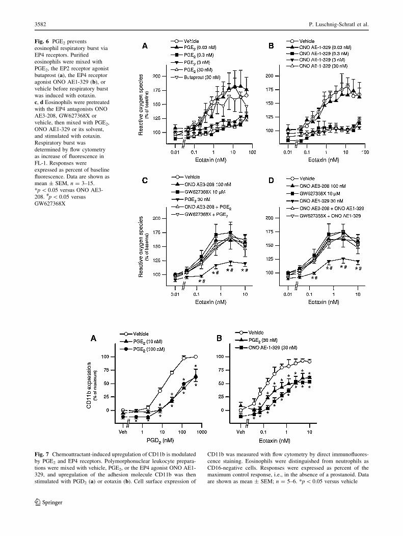
Next we investigated the effect of PGE2 on eosinophil
effector functions such as respiratory burst and upregula-
tion of adhesion molecules. Purified eosinophils were
mixed with varying concentrations of PGE2, the EP4
receptor agonist ONO AE1-329 (0.03–30 nM each), the
EP2 receptor agonist butaprost (30 nM) or vehicle and
were then stimulated with eotaxin (0.5–50 nM). Respira-
tory burst was quantified by flow cytometry. PGE2 very
potently attenuated the eotaxin-induced respiratory burst, at
concentrations as low as 0.3 nM, while the EP2 receptor
agonist butaprost did not mimic this PGE2 effect at a
concentration of 30 nM (Fig. 6a). With a similar potency
and efficacy as PGE2, the EP4 receptor agonist ONO AE1-
329 afforded inhibition of eosinophil respiratory burst
(Fig. 6b). In further experiments, we investigated whether
pretreatment with the EP4 receptor antagonists ONO AE3-
208 (100 nM) or GW627368X (10 lM) had an influence
on the PGE2-induced attenuation of the respiratory burst in
Fig. 4 EP4 receptors mediate
the PGE2-induced inhibition of
Ca2? flux in eosinophils.
a Polymorphonuclear leukocyte
preparations were mixed with
vehicle, or different
concentrations of PGE2, and
Ca2? flux was elicited with
eotaxin. b, c Polymorpho-
nuclear leukocyte preparations
were pretreated with the EP4
antagonist ONO AE3-208 or
vehicle, then mixed with
vehicle, PGE2 or the EP4
agonist ONO AE1-329, and
Ca2? flux was induced with
eotaxin. Ca2? flux was recorded
by flow cytometry as increases
in fluorescence in FL-1.
Eosinophils were distinguished
from neutrophils as CD16-
negative cells. Responses were
expressed as percent of the
maximum control response, i.e.,
in the absence of a prostanoid.
Data are shown as
mean ± SEM.; n = 6–12.
*p \ 0.05 versus vehicle
3580 P. Luschnig-Schratl et al.
123
Page 9
eosinophil granulocytes. Both EP4 receptor antagonists,
ONO AE3-208 and GW627368X, almost completely
reversed the inhibitory effect of PGE2 (Fig. 6c) and also
that of the EP4 receptor agonist ONO AE1-329 (Fig. 6d).
Since the adhesion molecule CD11b is an important
modulator of leukocyte responses such as migration and
mediator release [45, 46], we also investigated the effect of
PGE2 on the chemoattractant-induced upregulation of
CD11b on eosinophils. To this end, cells were mixed with
different concentrations of PGE2 (10–100 nM), the EP4
receptor agonist ONO AE1-329 (30 nM) or vehicle and
were then stimulated with PGD2 (5–500 nM) or eotaxin
(0.3–10 nM). Cell surface expression of CD11b on eosin-
ophils was upregulated by PGD2 and eotaxin approximately
1.5-fold, and both PGE2 and ONO AE1-329 led to a
decrease of this response (Fig. 7a, b). These data indicate
that PGE2 modulates eosinophil effector functions via the
EP4 receptor.
EP4 receptors are expressed by infiltrating eosinophils
in inflamed tissue
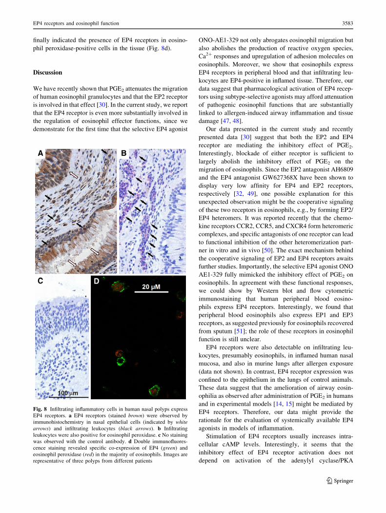
To investigate whether infiltrating cells in allergic airway
inflammation express EP4 receptors, immunohistochemis-
try of human nasal polyps and allergic rhinosinusitis was
performed. Epithelial cells and infiltrating leukocytes
stained positive with the EP4 antibody (Fig. 8a) while
sections incubated with the isotype-matched control anti-
body showed no staining (Fig. 8c), hence demonstrating
the specificity of the EP4 antibody. The infiltrating leu-
kocytes were also predominantly positive for eosinophil
peroxidase (Fig. 8b). Double immunofluorescence staining
Fig. 5 PGE2 attenuates the
chemoattractant-induced
shape change of eosinophils
via EP4 receptors.
a, b Polymorphonuclear
leukocyte preparations were
mixed with vehicle, PGE2, the
IP/EP1 receptor agonist iloprost,
the EP2 receptor agonist
butaprost, the EP1/EP3 receptor
agonist sulprostone or the EP4
agonist ONO AE1-329, and
shape change was induced with
PGD2. c, d Polymorphonuclear
leukocyte preparations were
pretreated with the EP4
antagonists ONO AE3-208,
GW627368X or vehicle, then
mixed with PGE2, ONO AE1-
329 or its solvent and stimulated
with eotaxin. Shape change was
determined with flow cytometry
as increase in forward scatter.
Eosinophils were distinguished
from neutrophils as cells with
higher auto-fluorescence.
Responses were expressed as
percent of the maximum control
response, i.e., in the absence of
a prostanoid. Data are shown as
mean ± SEM.; n = 3–4.
*p \ 0.05 versus vehicle.#p \ 0.05 versus PGE2
EP4 receptors and eosinophil function 3581
123
Page 10
Fig. 6 PGE2 prevents
eosinophil respiratory burst via
EP4 receptors. Purified
eosinophils were mixed with
PGE2, the EP2 receptor agonist
butaprost (a), the EP4 receptor
agonist ONO AE1-329 (b), or
vehicle before respiratory burst
was induced with eotaxin.
c, d Eosinophils were pretreated
with the EP4 antagonists ONO
AE3-208, GW627368X or
vehicle, then mixed with PGE2,
ONO AE1-329 or its solvent,
and stimulated with eotaxin.
Respiratory burst was
determined by flow cytometry
as increase of fluorescence in
FL-1. Responses were
expressed as percent of baseline
fluorescence. Data are shown as
mean ± SEM, n = 3–15.
*p \ 0.05 versus ONO AE3-
208. #p \ 0.05 versus
GW627368X
Fig. 7 Chemoattractant-induced upregulation of CD11b is modulated
by PGE2 and EP4 receptors. Polymorphonuclear leukocyte prepara-
tions were mixed with vehicle, PGE2, or the EP4 agonist ONO AE1-
329, and upregulation of the adhesion molecule CD11b was then
stimulated with PGD2 (a) or eotaxin (b). Cell surface expression of
CD11b was measured with flow cytometry by direct immunofluores-
cence staining. Eosinophils were distinguished from neutrophils as
CD16-negative cells. Responses were expressed as percent of the
maximum control response, i.e., in the absence of a prostanoid. Data
are shown as mean ± SEM; n = 5–6. *p \ 0.05 versus vehicle
3582 P. Luschnig-Schratl et al.
123
Page 11
finally indicated the presence of EP4 receptors in eosino-
phil peroxidase-positive cells in the tissue (Fig. 8d).
Discussion
We have recently shown that PGE2 attenuates the migration
of human eosinophil granulocytes and that the EP2 receptor
is involved in that effect [30]. In the current study, we report
that the EP4 receptor is even more substantially involved in
the regulation of eosinophil effector functions, since we
demonstrate for the first time that the selective EP4 agonist
ONO-AE1-329 not only abrogates eosinophil migration but
also abolishes the production of reactive oxygen species,
Ca2? responses and upregulation of adhesion molecules on
eosinophils. Moreover, we show that eosinophils express
EP4 receptors in peripheral blood and that infiltrating leu-
kocytes are EP4-positive in inflamed tissue. Therefore, our
data suggest that pharmacological activation of EP4 recep-
tors using subtype-selective agonists may afford attenuation
of pathogenic eosinophil functions that are substantially
linked to allergen-induced airway inflammation and tissue
damage [47, 48].
Our data presented in the current study and recently
presented data [30] suggest that both the EP2 and EP4
receptor are mediating the inhibitory effect of PGE2.
Interestingly, blockade of either receptor is sufficient to
largely abolish the inhibitory effect of PGE2 on the
migration of eosinophils. Since the EP2 antagonist AH6809
and the EP4 antagonist GW627368X have been shown to
display very low affinity for EP4 and EP2 receptors,
respectively [32, 49], one possible explanation for this
unexpected observation might be the cooperative signaling
of these two receptors in eosinophils, e.g., by forming EP2/
EP4 heteromers. It was reported recently that the chemo-
kine receptors CCR2, CCR5, and CXCR4 form heteromeric
complexes, and specific antagonists of one receptor can lead
to functional inhibition of the other heteromerization part-
ner in vitro and in vivo [50]. The exact mechanism behind
the cooperative signaling of EP2 and EP4 receptors awaits
further studies. Importantly, the selective EP4 agonist ONO
AE1-329 fully mimicked the inhibitory effect of PGE2 on
eosinophils. In agreement with these functional responses,
we could show by Western blot and flow cytometric
immunostaining that human peripheral blood eosino-
phils express EP4 receptors. Interestingly, we found that
peripheral blood eosinophils also express EP1 and EP3
receptors, as suggested previously for eosinophils recovered
from sputum [51]; the role of these receptors in eosinophil
function is still unclear.
EP4 receptors were also detectable on infiltrating leu-
kocytes, presumably eosinophils, in inflamed human nasal
mucosa, and also in murine lungs after allergen exposure
(data not shown). In contrast, EP4 receptor expression was
confined to the epithelium in the lungs of control animals.
These data suggest that the amelioration of airway eosin-
ophilia as observed after administration of PGE2 in humans
and in experimental models [14, 15] might be mediated by
EP4 receptors. Therefore, our data might provide the
rationale for the evaluation of systemically available EP4
agonists in models of inflammation.
Stimulation of EP4 receptors usually increases intra-
cellular cAMP levels. Interestingly, it seems that the
inhibitory effect of EP4 receptor activation does not
depend on activation of the adenylyl cyclase/PKA
Fig. 8 Infiltrating inflammatory cells in human nasal polyps express
EP4 receptors. a EP4 receptors (stained brown) were observed by
immunohistochemistry in nasal epithelial cells (indicated by whitearrows) and infiltrating leukocytes (black arrows). b Infiltrating
leukocytes were also positive for eosinophil peroxidase. c No staining
was observed with the control antibody. d Double immunofluores-
cence staining revealed specific co-expression of EP4 (green) and
eosinophil peroxidase (red) in the majority of eosinophils. Images are
representative of three polyps from different patients
EP4 receptors and eosinophil function 3583
123
Page 12
pathway, as the adenylyl cyclase inhibitor SQ22536 did not
reverse the inhibitory effect of ONO AE1-329 on eosino-
phil migration. The effectiveness of SQ22536 as an
adenylyl cyclase inhibitor was demonstrated by the fact
that SQ22536 prevented the attenuation of eosinophil
migration induced by PGI2, a known stimulator of adenylyl
cyclase [52]. Stimulation of EP4 has been shown to cause
phosphorylation of extracellular signal-regulated kinases
(ERKs) through a PI3K-dependent mechanism [53].
Additionally, the existence of an alternative EP2/EP4 sig-
naling pathway, linked to PKC activation has been
postulated [54]. Therefore, we investigated the effect of the
PI3K inhibitor LY294002 and the selective PKC inhibitor
chelerythrine on eosinophil migration in the presence of the
EP4 receptor agonist ONO AE1-329. In fact, inhibition of
PI3K prevented the inhibition of eosinophil migration
induced by ONO AE1-329. Moreover, exposure of eosin-
ophils to the selective PKC inhibitor chelerythrine prior to
stimulation of the EP4 receptor with ONO AE1-329
resulted in almost complete reversal of the attenuation of
eosinophil chemotaxis. Since these data implicated PKC as
a negative regulator of eosinophil migration, we investi-
gated the effect of the PKC activator PMA on eosinophil
migration. As expected, PMA very potently mimicked the
inhibitory effect of PGE2 and the EP4 receptor agonist on
eosinophil migration. The inhibitory effect of PMA, how-
ever, could not be prevented by the PI3K inhibitor
LY294002. Therefore, our data indicate that both PI3K and
PKC are involved in the attenuation of eosinophil migra-
tion upon EP4 receptor activation and confirm previous
data that modulation of PI3K mediates the inhibitory effect
of PGE2 in neutrophils [55].
Chemoattractants like PGD2 or eotaxin are known to
elevate intracellular Ca2? concentrations in eosinophils,
which is an essential requirement for effector functions like
degranulation and respiratory burst [43]. Hence, we
investigated the influence of PGE2 on the Ca2? mobiliza-
tion induced by these chemoattractants. Our data show that
PGE2 inhibits chemoattractant-induced Ca2? influx in
eosinophils, and what is more, that this effect is mediated
via EP4 receptors. This conclusion was based on the
observations that the selective EP4 agonist ONO AE1-329
likewise inhibited eosinophil Ca2? responses and that this
effect was reversed in the presence of the selective EP4
receptor antagonist ONO AE3-208. In contrast, the PGE2-
induced attenuation of Ca2? flux in eosinophils was not
mimicked by the IP/EP1 receptor agonist iloprost, the EP2
receptor agonist butaprost, or the EP1/EP3 receptor agonist
sulprostone. Another important prerequisite for chemoat-
tractant-induced eosinophil migration, oxidative burst, and
degranulation is the rapid reorganization of the cytoskele-
ton, which results in a shape change of the cells [43].
Similar to our observation with Ca2? mobilization we
found that PGE2 inhibits shape change of eosinophils
solely by EP4 receptors, since inhibition of shape change
could also be evoked with ONO AE1-329 but not with
iloprost, butaprost, or sulprostone. This notion was sup-
ported by the fact that inhibition of eosinophil shape
change induced by PGE2 or ONO AE1-329 was reversed
by the EP4 receptor antagonists ONO AE3-208 and
GW627368X in a similar fashion.
Interestingly, neither chelerythrine nor LY294002 was
able to reverse the inhibitory effect of the EP4 agonist on
Ca2? flux and shape change (unpublished observation),
suggesting that the roles of PI3K and PKC are largely
restricted to EP4-mediated inhibition of chemotaxis. This
notion complements our finding that both EP2 and EP4
activation curtails eosinophil chemotaxis, while only EP4
controls shape change, Ca2? flux, and production of reac-
tive oxygen species. Therefore, a detailed analysis of EP4-
related signaling needs to be carried out for each eosinophil
function separately in future studies.
Since Ca2? mobilization and shape change are important
requirements for eosinophil adhesion, respiratory burst, and
mediator release, we investigated the consequences of
reduced Ca2? responses by PGE2 with regard to these
eosinophil effector functions. Indeed we found that the
eotaxin- and PGD2-induced stimulation of CD11b expres-
sion was negatively modulated in the presence of PGE2 in
eosinophils, and that this effect could be mimicked by the
EP4 receptor agonist ONO AE1-329. In contrast to other
adhesion molecules, cell surface expression of CD11b is
rapidly increased on leukocytes after stimulation [56]. The
b2-integrin CD11b/CD18 (also referred to as CR3, Mac-1,
or amb2) is an important complement receptor that binds
multiple ligands, including C3bi, ICAM-1, fibrinogen, and
b-glucan. Apart from adhesion, CD11b/CD18 is an impor-
tant modulator of further leukocyte responses, including
migration [45], respiratory burst [57], degranulation [58],
and apoptosis [59]. CD11b expression on circulating
eosinophils is significantly elevated in various allergic
disorders, including atopic dermatitis and bronchial asthma
[60, 61]. Therefore, upregulation of surface CD11b closely
reflects eosinophil activation and might be a prerequisite of
leukocyte recruitment to sites of inflammation, by mediat-
ing the release of eosinophils from bone marrow [62] and
their migration [63]. These observations suggest that the
reduced CD11b response in the presence of PGE2 might
have a profound impact on eosinophil functions, such as
migration to sites of allergic reactions, degranulation, and
oxidative burst. In agreement with this notion, we were able
to show that PGE2 and the EP4 receptor agonist ONO AE1-
329 attenuate the formation of reactive oxygen species with
remarkably high potency, while the EP2 agonist butaprost
had no effect. Therefore, it appears that the PGE2-induced
attenuation of eosinophil oxidative burst is also mediated
3584 P. Luschnig-Schratl et al.
123
Page 13
via the EP4 receptor rather than the EP2 receptor. This
notion was further asserted by the EP4 receptor antagonist
ONO AE3-208 and GW627368X, which reversed the
inhibition of reactive oxygen species production by PGE2
and ONO AE1-329.
In conclusion, the present study shows that eosinophils
express EP4 receptors and activation of EP4 receptors
negatively modulates eosinophil migration and respiratory
burst. Furthermore, we were able to show that PI3K and
PKC are involved in the inhibitory effect of EP4 receptors.
In previous studies it was shown that PGE2 has bron-
choprotective and anti-inflammatory properties but the
usefulness of PGE2 as a therapeutic agent is limited because
of its various side-effects such as acute bronchoconstriction,
retrosternal soreness, transient cough, and flu-like symptoms
[10, 11, 14]. While the latter effects of PGE2 are thought to
arise from EP1 or EP3 receptor stimulation, selective EP4
agonists might have more favorable pharmacological
profiles. Therefore, EP4 agonists might be a useful novel
approach in the treatment of eosinophilic diseases as
they not only attenuate chemoattractant-induced eosinophil
migration and degranulation, as EP2 receptor agonists do,
but also abolish other eosinophil effector functions such as
upregulation of adhesion molecules or production of reac-
tive oxygen species.
Acknowledgments V. K. and S. P. were funded by the Ph. D. Pro-
gram Molecular Medicine of the Medical University of Graz. This
work was supported by the Jubilaumsfonds of the Austrian National
Bank (Grants 11967 and 13487), the Austrian Science Fund FWF
(Grants P19424-B05, P22521-B18 and P21004-B02), and the Franz
Lanyar Foundation (Grants 315, 316, and 343). A. H. has received
research support and consultancy fees from AstraZeneca and Almirall.
Open Access This article is distributed under the terms of the
Creative Commons Attribution Noncommercial License which per-
mits any noncommercial use, distribution, and reproduction in any
medium, provided the original author(s) and source are credited.
References
1. Woerly G, Roger N, Loiseau S, Capron M (1999) Expression of
Th1 and Th2 immunoregulatory cytokines by human eosinophils.
Int Arch Allergy Immunol 118:95–97
2. Navarro S, Aleu J, Jimenez M, Boix E, Cuchillo CM, Nogues MV
(2008) The cytotoxicity of eosinophil cationic protein/ribonu-
clease 3 on eukaryotic cell lines takes place through its
aggregation on the cell membrane. Cell Mol Life Sci 65:324–337
3. Aceves SS, Broide DH (2008) Airway fibrosis and angiogenesis
due to eosinophil trafficking in chronic asthma. Curr Mol Med
8:350–358
4. Green RH, Brightling CE, McKenna S, Hargadon B, Parker D,
Bradding P, Wardlaw AJ, Pavord ID (2002) Asthma exacerba-
tions and sputum eosinophil counts: a randomised controlled trial.
Lancet 360:1715–1721
5. Foster PS, Rosenberg HF, Asquith KL, Kumar RK (2008)
Targeting eosinophils in asthma. Curr Mol Med 8:585–590
6. Straumann A, Simon HU (2005) Eosinophilic esophagitis: esca-
lating epidemiology? J Allergy Clin Immunol 115:418–419
7. Wedemeyer J, Vosskuhl K (2008) Role of gastrointestinal
eosinophils in inflammatory bowel disease and intestinal
tumours. Best Pract Res Clin Gastroenterol 22:537–549
8. Simon D, Simon HU (2007) Eosinophilic disorders. J Allergy
Clin Immunol 119:1291–1300
9. Smith AP, Cuthbert MF, Dunlop LS (1975) Effects of inhaled
prostaglandins E1, E2, and F2alpha on the airway resistance of
healthy and asthmatic man. Clin Sci Mol Med 48:421–430
10. Pavord ID, Wong CS, Williams J, Tattersfield AE (1993) Effect
of inhaled prostaglandin E2 on allergen-induced asthma. Am Rev
Respir Dis 148:87–90
11. Mathe AA, Hedqvist P (1975) Effect of prostaglandins F2 alpha
and E2 on airway conductance in healthy subjects and asthmatic
patients. Am Rev Respir Dis 111:313–320
12. Costello JF, Dunlop LS, Gardiner PJ (1985) Characteristics of
prostaglandin induced cough in man. Br J Clin Pharmacol 20:
355–359
13. Roberts AM, Schultz HD, Green JF, Armstrong DJ, Kaufman
MP, Coleridge HM, Coleridge JC (1985) Reflex tracheal con-
traction evoked in dogs by bronchodilator prostaglandins E2 and
I2. J Appl Physiol 58:1823–1831
14. Gauvreau GM, Watson RM, O’Byrne PM (1999) Protec-
tive effects of inhaled PGE2 on allergen-induced airway
responses and airway inflammation. Am J Respir Crit Care Med
159:31–36
15. Martin JG, Suzuki M, Maghni K, Pantano R, Ramos-Barbon D,
Ihaku D, Nantel F, Denis D, Hamid Q, Powell WS (2002) The
immunomodulatory actions of prostaglandin E2 on allergic air-
way responses in the rat. J Immunol 169:3963–3969
16. Adcock JJ, Garland LG, Moncada S, Salmon JA (1978) Enhance-
ment of anaphylactic mediator release from guinea-pig perfused
lungs by fatty acid hydroperoxides. Prostaglandins 16:163–177
17. Teixeira MM, Williams TJ, Hellewell PG (1993) E-type pros-
taglandins enhance local oedema formation and neutrophil
accumulation but suppress eosinophil accumulation in guinea-pig
skin. Br J Pharmacol 110:416–422
18. Moore BB, Ballinger MN, White ES, Green ME, Herrygers AB,
Wilke CA, Toews GB, Peters-Golden M (2005) Bleomycin-
induced E prostanoid receptor changes alter fibroblast responses
to prostaglandin E2. J Immunol 174:5644–5649
19. Gavett SH, Madison SL, Chulada PC, Scarborough PE, Qu W,
Boyle JE, Tiano HF, Lee CA, Langenbach R, Roggli VL, Zeldin
DC (1999) Allergic lung responses are increased in prostaglandin
H synthase-deficient mice. J Clin Invest 104:721–732
20. Nakata J, Kondo M, Tamaoki J, Takemiya T, Nohara M,
Yamagata K, Nagai A (2005) Augmentation of allergic inflam-
mation in the airways of cyclooxygenase-2-deficient mice.
Respirology 10:149–156
21. Peebles RS Jr, Hashimoto K, Morrow JD, Dworski R, Collins RD,
Hashimoto Y, Christman JW, Kang KH, Jarzecka K, Furlong J,
Mitchell DB, Talati M, Graham BS, Sheller JR (2002) Selective
cyclooxygenase-1 and -2 inhibitors each increase allergic inflam-
mation and airway hyperresponsiveness in mice. Am J Respir Crit
Care Med 165:1154–1160
22. Kita H, Abu-Ghazaleh RI, Gleich GJ, Abraham RT (1991)
Regulation of Ig-induced eosinophil degranulation by adenosine
30,50-cyclic monophosphate. J Immunol 146:2712–2718
23. Berends C, Dijkhuizen B, de Monchy JG, Dubois AE, Gerritsen J,
Kauffman HF (1997) Inhibition of PAF-induced expression of
CD11b and shedding of L-selectin on human neutrophils and
eosinophils by the type IV selective PDE inhibitor, rolipram. Eur
Respir J 10:1000–1007
EP4 receptors and eosinophil function 3585
123
Page 14
24. Peacock CD, Misso NL, Watkins DN, Thompson PJ (1999) PGE
2 and dibutyryl cyclic adenosine monophosphate prolong eosin-
ophil survival in vitro. J Allergy Clin Immunol 104:153–162
25. Alam R, Dejarnatt A, Stafford S, Forsythe PA, Kumar D, Grant
JA (1993) Selective inhibition of the cutaneous late but not
immediate allergic response to antigens by misoprostol, a PGE
analog. Results of a double-blind, placebo-controlled randomized
study. Am Rev Respir Dis 148:1066–1070
26. Sugimoto Y, Narumiya S (2007) Prostaglandin E receptors. J Biol
Chem 282:11613–11617
27. Sheller JR, Mitchell D, Meyrick B, Oates J, Breyer R (2000)
EP(2) receptor mediates bronchodilation by PGE(2) in mice.
J Appl Physiol 88:2214–2218
28. Negishi M, Hasegawa H, Ichikawa A (1996) Prostaglandin E
receptor EP3gamma isoform, with mostly full constitutive Gi
activity and agonist-dependent Gs activity. FEBS Lett 386:
165–168
29. Di Cesare A, Del Piccolo P, Zacchetti D, Grohovaz F (2006) EP2
receptor stimulation promotes calcium responses in astrocytes via
activation of the adenylyl cyclase pathway. Cell Mol Life Sci
63:2546–2553
30. Sturm EM, Schratl P, Schuligoi R, Konya V, Sturm GJ, Lippe IT,
Peskar BA, Heinemann A (2008) Prostaglandin E2 inhibits
eosinophil trafficking through E-prostanoid 2 receptors. J Immu-
nol 181:7273–7283
31. Suzawa T, Miyaura C, Inada M, Maruyama T, Sugimoto Y,
Ushikubi F, Ichikawa A, Narumiya S, Suda T (2000) The role of
prostaglandin E receptor subtypes (EP1, EP2, EP3, and EP4) in
bone resorption: an analysis using specific agonists for the
respective EPs. Endocrinology 141:1554–1559
32. Wilson RJ, Giblin GM, Roomans S, Rhodes SA, Cartwright KA,
Shield VJ, Brown J, Wise A, Chowdhury J, Pritchard S, Coote J,
Noel LS, Kenakin T, Burns-Kurtis CL, Morrison V, Gray DW,
Giles H (2006) GW627368X ((N-{2-[4-(4, 9-diethoxy-1-oxo-1,
3-dihydro-2H-benzo[f]isoindol-2-yl)phenyl] acetyl} benzene
sulphonamide): a novel, potent and selective prostanoid EP4
receptor antagonist. Br J Pharmacol 148:326–339
33. Kabashima K, Saji T, Murata T, Nagamachi M, Matsuoka T, Segi
E, Tsuboi K, Sugimoto Y, Kobayashi T, Miyachi Y, Ichikawa A,
Narumiya S (2002) The prostaglandin receptor EP4 suppresses
colitis, mucosal damage and CD4 cell activation in the gut. J Clin
Invest 109:883–893
34. Hartnell A, Heinemann A, Conroy DM, Wait R, Sturm GJ,
Caversaccio M, Jose PJ, Williams TJ (2004) Identification of
selective basophil chemoattractants in human nasal polyps as
insulin-like growth factor-1 and insulin-like growth factor-2.
J Immunol 173:6448–6457
35. Schuligoi R, Schmidt R, Geisslinger G, Kollroser M, Peskar BA,
Heinemann A (2007) PGD2 metabolism in plasma: kinetics and
relationship with bioactivity on DP1 and CRTH2 receptors.
Biochem Pharmacol 74:107–117
36. Royer JF, Schratl P, Lorenz S, Kostenis E, Ulven T, Schuligoi R,
Peskar BA, Heinemann A (2007) A novel antagonist of CRTH2
blocks eosinophil release from bone marrow, chemotaxis and
respiratory burst. Allergy 62:1401–1409
37. Heinemann A, Ofner M, Amann R, Peskar BA (2003) A
novel assay to measure the calcium flux in human basophils:
effects of chemokines and nerve growth factor. Pharmacology
67:49–54
38. Steinbrunn T, Stuhmer T, Gattenlohner S, Rosenwald A, Mottok
A, Unzicker C, Einsele H, Chatterjee M, Bargou RC (2010)
Mutated RAS and constitutively activated Akt delineate distinct
oncogenic pathways, which independently contribute to multiple
myeloma cell survival. Blood 117(6):1998–2004
39. Han C, Wu T (2005) Cyclooxygenase-2-derived prostaglandin E2
promotes human cholangiocarcinoma cell growth and invasion
through EP1 receptor-mediated activation of the epidermal
growth factor receptor and Akt. J Biol Chem 280:24053–24063
40. Caristi S, Piraino G, Cucinotta M, Valenti A, Loddo S, Teti D
(2005) Prostaglandin E2 induces interleukin-8 gene transcription
by activating C/EBP homologous protein in human T lympho-
cytes. J Biol Chem 280:14433–14442
41. Stubbs VE, Schratl P, Hartnell A, Williams TJ, Peskar BA,
Heinemann A, Sabroe I (2002) Indomethacin causes prostaglandin
D(2)-like and eotaxin-like selective responses in eosinophils and
basophils. J Biol Chem 277:26012–26020
42. Schratl P, Sturm EM, Royer JF, Sturm GJ, Lippe IT, Peskar BA,
Heinemann A (2006) Hierarchy of eosinophil chemoattractants:
role of p38 mitogen-activated protein kinase. Eur J Immunol
36:2401–2409
43. Schratl P, Heinemann A (2009) Differential involvement of Ca2?
and actin filament in leukocyte shape change. Pharmacology
83:131–140
44. Heinemann A, Schuligoi R, Sabroe I, Hartnell A, Peskar BA
(2003) Delta12-prostaglandin J2, a plasma metabolite of prosta-
glandin D2, causes eosinophil mobilization from the bone
marrow and primes eosinophils for chemotaxis. J Immunol 170:
4752–4758
45. Rainger GE, Buckley C, Simmons DL, Nash GB (1997) Cross-
talk between cell adhesion molecules regulates the migration
velocity of neutrophils. Curr Biol 7:316–325
46. Couturier C, Haeffner-Cavaillon N, Weiss L, Fischer E,
Kazatchkine MD (1990) Induction of cell-associated interleukin 1
through stimulation of the adhesion-promoting proteins LFA-1
(CD11a/CD18) and CR3 (CD11b/CD18) of human monocytes.
Eur J Immunol 20:999–1005
47. Bousquet J, Chanez P, Lacoste JY, Barneon G, Ghavanian N,
Enander I, Venge P, Ahlstedt S, Simony-Lafontaine J, Godard P
et al (1990) Eosinophilic inflammation in asthma. N Engl J Med
323:1033–1039
48. Jacobsen EA, Taranova AG, Lee NA, Lee JJ (2007) Eosinophils:
singularly destructive effector cells or purveyors of immunoreg-
ulation? J Allergy Clin Immunol 119:1313–1320
49. Abramovitz M, Adam M, Boie Y, Carriere M, Denis D, Godbout C,
Lamontagne S, Rochette C, Sawyer N, Tremblay NM, Belley M,
Gallant M, Dufresne C, Gareau Y, Ruel R, Juteau H, Labelle M,
Ouimet N, Metters KM (2000) The utilization of recombinant
prostanoid receptors to determine the affinities and selectivities of
prostaglandins and related analogs. Biochim Biophys Acta 1483:
285–293
50. Sohy D, Yano H, de Nadai P, Urizar E, Guillabert A, Javitch JA,
Parmentier M, Springael JY (2009) Hetero-oligomerization of
CCR2, CCR5, and CXCR4 and the protean effects of ‘‘selective’’
antagonists. J Biol Chem 284:31270–31279
51. Ying S, O’Connor BJ, Meng Q, Woodman N, Greenaway S,
Wong H, Mallett K, Lee TH, Corrigan C (2004) Expression of
prostaglandin E(2) receptor subtypes on cells in sputum from
patients with asthma and controls: effect of allergen inhalational
challenge. J Allergy Clin Immunol 114:1309–1316
52. Sugama K, Tanaka T, Yokohama H, Negishi M, Hayashi H, Ito S,
Hayaishi O (1989) Stimulation of cAMP formation by prosta-
glandin D2 in primary cultures of bovine adrenal medullary cells:
identification of the responsive population as fibroblasts. Biochim
Biophys Acta 1011:75–80
53. Fujino H, Xu W, Regan JW (2003) Prostaglandin E2-induced
functional expression of early growth response factor-1 by EP4,
but not EP2, prostanoid receptors via the phosphatidylinositol
3-kinase and extracellular signal-regulated kinases. J Biol Chem
278:12151–12156
54. Reno F, Cannas M (2005) Effect of prostaglandin E2 on PMA-
induced macrophage differentiation. Prostaglandins Other Lipid
Mediat 75:13–24
3586 P. Luschnig-Schratl et al.
123
Page 15
55. Burelout C, Thibault N, Levasseur S, Simard S, Naccache PH,
Bourgoin SG (2004) Prostaglandin E2 inhibits the phospholipase
D pathway stimulated by formyl-methionyl-leucyl-phenylalanine
in human neutrophils. Involvement of EP2 receptors and phos-
phatidylinositol 3-kinase gamma. Mol Pharmacol 66:293–301
56. Walker C, Rihs S, Braun RK, Betz S, Bruijnzeel PL (1993)
Increased expression of CD11b and functional changes in
eosinophils after migration across endothelial cell monolayers.
J Immunol 150:4061–4071
57. Nathan C, Srimal S, Farber C, Sanchez E, Kabbash L, Asch A,
Gailit J, Wright SD (1989) Cytokine-induced respiratory burst of
human neutrophils: dependence on extracellular matrix proteins
and CD11/CD18 integrins. J Cell Biol 109:1341–1349
58. Richter J, Ng-Sikorski J, Olsson I, Andersson T (1990) Tumor
necrosis factor-induced degranulation in adherent human neutro-
phils is dependent on CD11b/CD18-integrin-triggered oscillations
of cytosolic free Ca2?. Proc Natl Acad Sci USA 87:9472–9476
59. Rubel C, Gomez S, Fernandez GC, Isturiz MA, Caamano J,
Palermo MS (2003) Fibrinogen-CD11b/CD18 interaction
activates the NF-kappa B pathway and delays apoptosis in human
neutrophils. Eur J Immunol 33:1429–1438
60. Barthel SR, Jarjour NN, Mosher DF, Johansson MW (2006)
Dissection of the hyperadhesive phenotype of airway eosinophils
in asthma. Am J Respir Cell Mol Biol 35:378–386
61. Yamada H, Kurashimo S, Chihara J, Matsukura M, Yudate T,
Tezuka T (1999) Overexpression of CD11b on eosinophils in atopic
dermatitis: downregulation by cyclosporin A and upregulation by
interleukin 5. Int Arch Allergy Immunol 120(Suppl 1):100–103
62. Palframan RT, Collins PD, Severs NJ, Rothery S, Williams TJ,
Rankin SM (1998) Mechanisms of acute eosinophil mobilization
from the bone marrow stimulated by interleukin 5: the role of
specific adhesion molecules and phosphatidylinositol 3-kinase.
J Exp Med 188:1621–1632
63. Ebisawa M, Bochner BS, Georas SN, Schleimer RP (1992)
Eosinophil transendothelial migration induced by cytokines.
I. Role of endothelial and eosinophil adhesion molecules in IL-1
beta-induced transendothelial migration. J Immunol 149:4021–
4028
EP4 receptors and eosinophil function 3587
123