Eph/ephrin signaling: networksDina Arvanitis and Alice Davy1
Université de Toulouse, Centre de Biologie du Développement, 31062 Toulouse cedex 9, France; Centre National de laRecherche Scientifique (CNRS), UMR 5547, 31062 Toulouse, France
Bidirectional signaling has emerged as an important sig-nature by which Ephs and ephrins control biologicalfunctions. Eph/ephrin signaling participates in a widespectrum of developmental processes, and cross-regula-tion with other communication pathways lies at theheart of the complexity underlying their function invivo. Here, we review in vitro and in vivo data describingmolecular, functional, and genetic interactions betweenEph/ephrin and other cell surface signaling pathways.The complexity of Eph/ephrin function is discussed interms of the pathways that regulate Eph/ephrin signalingand also the pathways that are regulated by Eph/ephrinsignaling.
Since the cloning of the first eph gene 20 years ago (Hiraiet al. 1987) and the identification of ligands for Eph re-ceptors (ephrins) a few years later (Bartley et al. 1994;Beckmann et al. 1994; Cheng and Flanagan 1994), muchwork has been done in trying to understand the functionof this receptor/ligand pair. Due to a great number ofstudies related to Eph/ephrin signaling, we are beginningto grasp the full range of action of this signaling cascade.Eph receptors form the largest subfamily of receptor ty-rosine kinases (RTKs). They interact with cell surface-bound ligands that are also part of a family of relatedproteins. Structural differences distinguish two classesof ephrins: Ephrins-A (A1–A6) are tethered to the plasmamembrane via a glycosyl phosphatidyl inositol moiety,while ephrins-B (B1–B3) span the plasma membrane andpossess a short cytoplasmic tail (Fig. 1). Eph receptorsand ephrins are also grouped into class A and class Bbased on their degree of sequence similarity. One of theunique features of Eph/ephrin signaling is the fact thatboth receptors and ligands are competent to transduce asignaling cascade upon interaction. Eph-activated signal-ing is termed forward, and ephrin-activated signaling istermed reverse. Another level of complexity stems fromthe fact that interactions between Eph receptors and eph-rins can happen in trans (between two opposing cells) orin cis (within the same cell). It is commonly assumed
that trans interactions are activating while cis interac-tions are inhibiting (Fig. 1).
Eph receptors and ephrins are expressed in virtually alltissues of a developing embryo, and they are involved ina wide array of developmental processes such as cardio-vascular and skeletal development, axon guidance, andtissue patterning (Palmer and Klein 2003). In many de-velopmental processes, the biological function of Eph/ephrin signaling boils down to the modulation of celladhesion: growth cone retraction in axon guidance, cellsorting in embryo patterning, cell migration and fusionin craniofacial development, and platelet aggregation,among others. Although Eph/ephrins have been studiedclassically in a developmental context, their physiologi-cal functions in the adult are coming to light. They havebeen implicated recently in learning and memory (Gerlai2002), in bone homeostasis (Zhao et al. 2006), and ininsulin secretion (Konstantinova et al. 2007). Alterationsof Eph/ephrin signaling in humans leads to congenitaldiseases and cancer (Pasquale 2005).
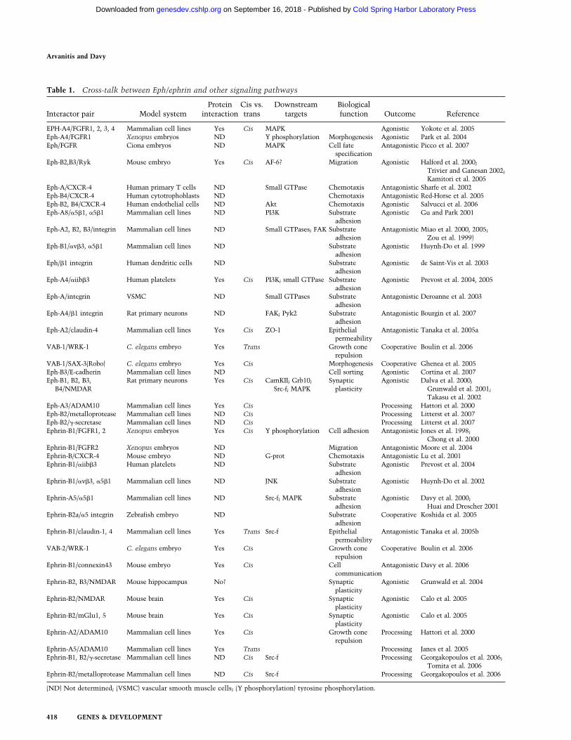
As our understanding of Eph/ephrin signaling im-proves, so does our reckoning that Eph/ephrins do notact in isolation, but are part of a complex network ofregulatory pathways that must act in concert to controlappropriate biological responses (Table 1). This reviewpresents genetic, biochemical, and functional evidencefor cross-talks between Eph/ephrin and other signalingpathways.
Cell surface receptors
Fibroblast growth factor receptor (FGFR)
The notion of cross-talks between Eph/ephrins and otherRTKs was proposed in one of the original papers describ-ing bidirectional signaling. It was shown in this studythat ephrin-Bs could be phosphorylated on tyrosine inresponse to Platelet-Derived Growth Factor receptor ac-tivation (Bruckner et al. 1997). Since then, a number ofstudies have reported interactions between Eph/ephrinand another family of RTK, FGFR. Jones et al. (1998)reported that injection of ephrin-B1 in both blastomeresof a two-cell stage Xenopus embryo resulted in blasto-mere dissociation at the mid-blastula stage, and that thephenotype could be rescued by culturing the injectedembryos in the presence of basic FGF. In addition, thisactivity was associated with the cytoplasmic and trans-
[Keywords: Cell surface receptors; development; proteases; synaptic plas-ticity; cell adhesion]1Corresponding author.E-MAIL [email protected]; FAX 33-5-61556507.Article is online at http://www.genesdev.org/cgi/doi/10.1101/gad.1630408.
membrane portions of the ephrin-B1 protein, and the ex-tracellular domain was not required for the induceddeadhesion. In a follow-up paper (Chong et al. 2000), thesame laboratory elucidated the mechanisms by whichFGF signaling could rescue the ephrin-induced cell dis-sociation. Activated FGFR bound directly to ephrin-B1in cis and induced its phosphorylation on tyrosine,which in turn inhibited the ability of ephrin-B1 to induceblastomere dissociation (Chong et al. 2000).
The relevance of this interaction was subsequently il-lustrated in the context of eye field formation in Xeno-pus. Retinal specification is a multistep process in whichcellular movements during gastrulation and neurulationare critical to allow retinal progenitors to populate theeye field. Moore et al. (2004) showed that activatedFGFR2 repressed eye field formation by restrictingmovements of retinal progenitors, therefore limitingtheir access to the eye field. Ectopic expression of ephrin-B1 rescued that phenotype, and ephrin-B1 knockdownphenocopied the repression associated with activatedFGFR2. Moody (2004) concluded from their studies thatthese two signaling pathways coordinately regulate ac-cess to the eye field by modulating cell movement. Al-together, these studies point to antagonistic interactionbetween FGF and ephrin signaling pathways.
Other studies have reported a direct agonistic interac-tion between FGFR and Eph/ephrin signaling pathways.Indeed, in mammalian cells, FGFR and Eph-A4 couldtrans-phosphorylate each other and activate commondownstream signaling pathways. Moreover, costimula-tion of both receptors resulted in the potentiation of mi-togen-activated protein kinase (MAPK) stimulation (Yo-
kote et al. 2005). No functional significance for the Eph-A4/FGFR potentiation was reported in that study(Yokote et al. 2005); however, an earlier study (Park et al.2004) in Xenopus had also revealed an agonistic interac-tion between Eph-A4 and FGF signaling. Overexpressionof Eph-A4 (like ephrin-B1) induced cell dissociation inearly Xenopus embryos. As development proceeded, em-bryos recovered from the loss of cell adhesion; however,overexpression of Eph-A4 induced ectopic formation ofposterior protrusions (Park et al. 2004) These tail-likestructures are multicellular protrusions that express anumber of posterior markers. They are not a direct con-sequence of the early loss of cell adhesion, but ratherreflect the role of Eph/ephrin signaling in directing mor-phogenetic movements during development. Similar ec-topic structures were induced following activation ofFGFR1 in Xenopus embryos, and FGF8 knockdown res-cued the Eph-A4-induced protrusions, indicating thatFGF signaling could be involved in the Eph-A4-inducedphenotype. Moreover, coinjection of both Eph-A4 andFGFR1 receptors increased the incidence of these struc-tures (Park et al. 2004), which is consistent with thepotentiating effect described in vitro (Yokote et al. 2005).While the sites of interaction between FGFR3 and Eph-A4 were mapped to the juxtamembrane domain inFGFR3 and the N-terminal portion of the tyrosine kinasedomain in Eph-A4 (Yokote et al. 2005), the binding sitefor ephrin-B1 on FGFRs has not been reported (Fig. 2).
More recently, the interplay between FGF and Eph/ephrin signaling has been involved in regulating asym-metric cell division and cell fate determination in Cionaembryos (Picco et al. 2007). In these chordate embryos,notochord and neural precursors share a commonmother cell, and the binary choice between both fates isdetermined by differential extracellular signal-regulatedkinase (ERK) activation. Picco et al. (2007) have shownthat FGF signaling and Eph/ephrin signaling act in coor-dination to differentially regulate ERK and have pro-posed that local inhibition of ERK by an activated Ephreceptor polarizes the mother cell and initiates asym-metric cell division leading to the acquisition of distinctcell fates. Again, this study illustrates how the antago-nistic relationship between FGF signaling and Eph/eph-rin signaling controls key developmental processes.Picco et al. (2007) inferred that the cross-talk betweenboth pathways was downstream from the receptors, atthe level of ERK activation; however, it does not neces-sarily preclude direct binding of FGFR and Eph receptor.Because MAPK is a cytoplasmic effector common to anumber of receptor/ligand pairs, modulation of MAPKactivity could be a mechanism by which Eph/ephrin im-pinge on a number of signaling cascades.
Ryk
RYK is an atypical RTK that contains a catalytically in-active tyrosine kinase domain. Genetic studies in Dro-sophila and the mouse have implicated RYK in regulat-ing axon guidance and craniofacial development, twodevelopmental processes that involve cell migration
Figure 1. Main features of Eph/ephrin signaling. (A) Bothclasses of Eph receptors and ephrins activate bidirectional sig-naling. Interaction between Eph receptors and ephrins leads toactivation of forward and reverse signaling in neighboring cells.(B) Eph receptors and ephrins expressed in opposing cells inter-act in trans and activate bidirectional signaling. Eph receptorsand ephrins coexpressed in the same cell interact in cis. Cisinteraction has been shown to inhibit trans interaction and/orsignaling.
Eph/ephrin signaling integrated
GENES & DEVELOPMENT 417
Cold Spring Harbor Laboratory Press on September 16, 2018 - Published by genesdev.cshlp.orgDownloaded from
Eph-A3/ADAM10 Mammalian cell lines Yes Cis Processing Hattori et al. 2000Eph-B2/metalloprotease Mammalian cell lines ND Cis Processing Litterst et al. 2007Eph-B2/�-secretase Mammalian cell lines ND Cis Processing Litterst et al. 2007Ephrin-B1/FGFR1, 2 Xenopus embryos Yes Cis Y phosphorylation Cell adhesion Antagonistic Jones et al. 1998;
Chong et al. 2000Ephrin-B1/FGFR2 Xenopus embryos ND Migration Antagonistic Moore et al. 2004Ephrin-B/CXCR-4 Mouse embryo ND G-prot Chemotaxis Antagonistic Lu et al. 2001Ephrin-B1/�iib�3 Human platelets ND Substrate
(Halford and Stacker 2001). Because these biologicalfunctions overlap with those attributed to Eph/ephrinsignaling, a potential interaction between these path-ways has been sought. In the mouse, homozygous dele-tion of Ryk resulted in craniofacial defects similar todefects observed in Eph-B2/Eph-B3-deficient embryos. Inaddition, RYK directly interacted with Eph receptors,and activated Eph receptors could phosphorylate RYK ontyrosines (Halford et al. 2000). The molecular mecha-nisms by which RYK influences Eph/ephrin signaling arestill unclear; however, the authors (Halford et al. 2000;Halford and Stacker 2001) have proposed that RYK couldfacilitate the recruitment of AF-6, a cell junction-associ-ated protein, to Eph receptors, therefore facilitating ac-tivation of downstream signaling events such as cell mi-gration. Surprisingly, human RYK also interacted withEph-B2 and Eph-B3, but this interaction did not lead totyrosine phosphorylation of RYK, and no interactioncould be detected between human RYK and AF-6 (Trivierand Ganessan 2002). Direct interaction between RYKand Eph receptors has also been observed in a rat model.The site of interaction between rat RYK and Eph-B3 wasmapped to the extracellular domain of RYK (Kamitori etal. 2005; Fig. 2).
Interaction between RYK and Eph receptors is also in-volved in regulating the migration of neuroprogenitors.Indeed, overexpression of full-length RYK, but not a mu-tant form that does not bind to Eph-B3, inhibited radialmigration of cortical neuroprogenitors in the rat cortex.Conversely, a mutant form of RYK lacking the kinasedomain (including six out of nine tyrosines) had no effecton radial cell migration and still bound Eph-B3, suggest-ing that the function of RYK in cortical cell migration isindependent of tyrosine phosphorylation but correlateswith Eph-B3 binding (Kamitori et al. 2005). Altogether,these results suggest that RYK and Eph receptors act asagonists in regulating cortical cell migration during de-velopment (Fig. 2).
The latter study, however, must be interpreted in lightof recent findings showing that RYK is an alternate re-ceptor for Wnt proteins (Inoue et al. 2004; Lu et al. 2004)and that the Wnt/�-catenin pathway is also involved incortical cell migration (Machon et al. 2003). To compli-cate matters further, a recent study uncovered an antago-nistic relationship between Wnt/RYK and Eph/ephrinpathways in controlling retinotectal topographic map-ping (Schmitt et al. 2006). Topographic mapping is a pro-
cess ensuring that axons project to their appropriate tar-get in the brain. Eph/ephrin signaling has been involvedin positioning retinotectal projections along the medial–lateral axis; however, it cannot alone account for correctmapping in the tectum. In their very elegant study,Schmitt et al. (2006) demonstrate that the Wnt3/RYKpathway acts as a lateral mapping force to counterbal-ance the Eph-B/ephrin-B medial mapping force in thetectum. Although Schmitt et al. (2006) favor the idea oftwo independent pathways, their results do not rule outthe possibility that both pathways directly modulateeach other.
Chemokine receptors
Chemokines and their cell surface G-protein-coupled re-ceptors are major regulators of cell trafficking. Amongthe wide number of chemokines and chemokine recep-tors (Zlotnik and Yoshie 2000), the CXCR4/SDF-1 pairstands out since the function of the CXCR4/SDF-1 sig-naling pathway is not limited to cell trafficking but alsoencompasses important roles in organogenesis and em-bryonic development (Kucia et al. 2004). The CXCR4/SDF-1 pathway has been shown to regulate multiple cel-lular processes such as locomotion, adhesion, secretion,and potentially survival and proliferation (Kucia et al.2004). In 2001, Lu et al. (2001) demonstrated that acti-vation of ephrin-B reverse signaling inhibited SDF-1-in-duced chemotaxis of cerebellar granule cells. Themechanistic basis for this inhibition was partly eluci-dated with the identification of PDZ-RGS3, a proteinthat binds the cytoplasmic domain of ephrin-Bs and isable to inactivate G-protein signaling via its GAP activ-ity (Fig. 2; Lu et al. 2001). Similar results were obtainedin T cells, as it was shown that activation of Eph-A re-ceptors inhibited SDF-1-induced chemotaxis by alteringthe balance of small GTPases activity in these cells (Fig.2; Sharfe et al. 2002). Down-regulation of chemokine-induced migration by Eph/ephrin signaling was alsoshown to be important to regulate trophoblast move-ment involved in arteriole remodeling during humanplacentation (Red-Horse et al. 2005). Unlike the studiesabove, which demonstrated an antagonistic relationshipbetween Eph/ephrin and CXCR4/SDF-1 signaling, Sal-vucci et al. (2006) reported that an agonistic interactionbetween these pathways regulates endothelial move-ment and morphogenesis of blood vessels (Fig. 2). Stimu-
Figure 2. Interactions with cell surface receptors. (A)Eph receptors interact with FGFR, Ryk, and chemokinereceptors. Direct interactions are indicated by dashedgreen lines. Arrows represent agonistic interaction,while blunted lines indicate antagonistic regulation ofdownstream effectors or biological processes. Tyrosinephosphorylation events are shown in red. (B) Ephrinsinteract with FGFR and chemokine receptors.
Eph/ephrin signaling integrated
GENES & DEVELOPMENT 419
Cold Spring Harbor Laboratory Press on September 16, 2018 - Published by genesdev.cshlp.orgDownloaded from
lation of the Eph/ephrin signaling cascade enhancedSDF-1-induced chemotaxis in endothelial cells, and bothpathways synergized to activate/phosphorylate AKT(Salvucci et al. 2006). Therefore, these studies clearlydemonstrate that Eph/ephrin signaling and CXCR4/SDF-1 signaling cooperate to regulate a number of bio-logical processes involving cell chemotaxis. Althoughthe data so far point to a cross-talk at the level of down-stream effectors, it would be interesting to test for directinteractions between these proteins, especially in lightof the fact that both pathways localize in lipid rafts(Gauthier and Robbins 2003; Wysoczynski et al. 2005;Giri et al. 2007).
Together, these studies show that Eph/ephrin signal-ing interacts with a number of cell surface receptors andthat these interactions can be agonistic or antagonistic(Table 1).
Adhesion molecules
Integrins
Due to the prominent role of Eph/ephrin signaling in cellmigration, a relationship with integrin signaling waspostulated early on. Despite numerous studies showingcross-talks between Eph/ephrin signaling and integrinsignaling, there is still a surprising degree of uncertaintywith respect to the outcome of the interaction betweenboth pathways: While a number of reports show thatEph/ephrin signaling increased integrin-mediated celladhesion (Huynh-Do et al. 1999, 2002; Davy and Robbins2000; Gu and Park 2001; Huai and Drescher 2001; deSaint-Vis et al. 2003; Prévost et al. 2004, 2005), othersdemonstrate a counter-effect of Eph/ephrin on integrin-mediated cell adhesion (Zou et al. 1999; Miao et al. 2000,2005; Deroanne et al. 2003; Bourgin et al. 2007). Theseoutcomes are not linked to a specific class of Eph/ephrinpair, nor does it seem linked to either forward or reversesignaling. It is important to note, however, that all thestudies focusing on reverse signaling have reported anincreased integrin-mediated adhesion following activa-tion of class A and class B ephrins (Davy and Robbins2000; Huai and Drescher 2001; Huynh-Do et al. 2002;Prévost et al. 2004). Common sense now attributes theopposite effects of Eph/ephrin signaling on integrin func-tion to distinct cellular contexts, since all the above-
mentioned studies use different cell types (primary,transformed, or nontransformed cell lines) and differentmodes of expression of the proteins of interest (endog-enous vs. ectopic) (Table 1). Nevertheless, the conclu-sion from these studies is that Eph/ephrin signaling im-pinges on integrin signaling (Fig. 3). In fact, cooperationbetween Eph/ephrin and integrin signaling is supportedby genetic data: itga5 (integrin �5) and fn (fibronectin)are required for somite development in zebrafish, andreducing Eph/ephrin signaling in the context of a mutantfn or itga5 background worsened the somite phenotype(Koshida et al. 2005). The point of convergence betweenboth pathways appears to be at the level of cytoplasmickinases (FAK, PI3K, MAPK) and/or small GTPases (Rac,Rho, Ras, Rap1). Only one study reports a direct inter-action between an Eph receptor and an integrin (Prévostet al. 2005).
Immunoglobulin superfamily (IgSF) proteins
Like Eph/ephrin, IgSF proteins are implicated in manyaspects of the nervous system development, includingaxon pathfinding, target recognition, and synapse forma-tion (Rougon and Hobert 2003). It was reported recentlythat the Caenorhabditis elegans IgSF protein, WRK-1,interacts with Eph/ephrin signaling to provide a midlineguidepost function (Boulin et al. 2006). Midline guide-posts are necessary for developing axons to decidewhether to cross the midline or not, a decision that un-derlies the ability of the nervous system to coordinateevents occurring on each side of the body. WRK-1 is ex-pressed in embryonic midline motoneurons (eMNs), andwrk-1 loss-of-function mutations caused nonautono-mous midline axon guidance defects in the worm similarto those observed in mutants for Eph/ephrin signaling.Epistasis experiments indicated that these genes act in asingle pathway. Interestingly, the cell autonomy ofvab-1 (coding for an Eph receptor) and wrk-1was inves-tigated, and although these genes act in the same path-way, they are required in different cells. The proposedmodel is that vab-1 and wrk-1 are expressed in opposingand presumably interacting neurons (Boulin et al. 2006).The authors further demonstrate a direct interaction be-tween WRK-1, VAB-1, and VAB-2 (an ephrin), leading toa proposed model in which WRK-1 and VAB-2 are coex-pressed in eMNs, where they interact with each other
Figure 3. Regulation of adhesion proteins. Eph/ephrinsignaling regulates cell–cell adhesion and cell–matrix ad-hesion by impinging on formation/stability of tight, ad-herens, and gap junctions, as well as on integrin function.In Eph-expressing cells (blue), activation of forward sig-naling induces the redistribution of E-cadherin to the cellsurface while destabilizing claudins. In ephrin-expressingcells (orange), activation of reverse signaling leads to in-hibition of GJC, while interaction with claudins destabi-lizes tight junctions. Both forward and reverse signalingact on integrin-mediated adhesion. Together, these cas-cades participate in Eph/ephrin-induced cell sorting.
Arvanitis and Davy
420 GENES & DEVELOPMENT
Cold Spring Harbor Laboratory Press on September 16, 2018 - Published by genesdev.cshlp.orgDownloaded from
and provide a repulsive signal to VAB-1-expressing axonsin trans (Fig. 5, below; Table 1; Boulin et al. 2006).
Robo proteins are members of the IgSF family also in-volved in midline axon guidance (Dickson and Gilestro2006). Ghenea et al. (2005) identified sax-3 as a candidategene functioning with vab-1 during embryogenesis. Us-ing genetic and molecular approaches, Ghenea et al.(2005) demonstrated that SAX-3/Robo functions withVAB-1 to regulate embryonic morphogenesis and axonguidance in C. elegans. sax-3 mutants displayed defectsthat are similar to vab-1 mutants, and analysis of a com-bination of double mutants revealed a gene-dosage sen-sitivity between these genes. In addition, direct interac-tion between VAB-1 and SAX-3 was demonstrated usingtwo-hybrid and GST-pull-down assays (Ghenea et al.2005). Since the domain of interaction was mapped tothe juxtamembrane domain of SAX-3, and both proteinswere coexpressed in a subset of neuroblasts, Ghenea etal. (2005) proposed that these two receptors form a com-plex in cis and act together during embryogenesis (Fig. 5,below). Given the well-known role of Robo proteins asmidline guideposts and the data described above (Boulinet al. 2006), it would be interesting to assess midlinecrossing of ventral axons in double sax-3/vab-1 mutants.In vertebrates, evidence for interplay between Eph/eph-rin and IgSF proteins is limited to one member of thefamily. Zisch et al. (1997) showed that L1 is a substratefor Eph-B2 tyrosine kinase, while Suh et al. demon-strated that growth cones stimulated with L1 lost theirresponsiveness to Eph-B (Suh et al. 2004). Together,these studies show the importance of the interplay be-tween Eph/ephrin and IgSF proteins and highlight thecomplexity of interactions between cell surface proteins,which can happen either in cis or in trans.
Cadherins
In addition to the regulation of cell migration and axonnavigation, one of the prominent biological outcomes ofEph/ephrin signaling is the regulation of cell sorting, aprocess by which populations of cells physically segre-gate from each other to generate distinct tissues or com-partments (Xu et al. 1999; Poliakov et al. 2004). The cel-lular and molecular mechanisms by which Eph/ephrinsignaling control cell sorting behaviors are still not wellcharacterized; however, because homotypic interactionsvia cadherins play an important role in cell sorting (Te-pass et al. 2002), it was postulated that these two path-ways might cooperate to regulate cell adhesion and seg-regation. Surprisingly, there is little evidence supportingthis hypothesis. In Xenopus embryos, ectopic expressionof an Eph receptor or an ephrin leads to cell dissociation,a phenotype that could be rescued by overexpressing C-cadherin (Winning et al. 1996; Jones et al. 1998). How-ever, the fact that no alteration of the cadherin/�-catenininteraction could be detected following ectopic expres-sion of Eph or ephrin suggests that overexpression ofC-cadherin might not specifically rescue Eph/ephrin-in-duced dissociation but might serve as indiscriminateSuperGlue. In mammalian epithelial cells, it was shown
that Eph-A2 localizes to sites of cell–cell contact andthat this subcellular localization was dependent on E-cadherin. In addition, ectopic expression of E-cadherinexpression in breast cancer cells that lack endogenousE-cadherin increased tyrosine phosphorylation of Eph-A2 and led to a decreased cell adhesion to the extracel-lular matrix (Zantek et al. 1999). Zantek et al. (1999)concluded that Eph-A2 function is dependent on E-cad-herin; however, they also report that neither coprecipi-tation nor coclustering between the two proteins couldbe detected using their system. This led them to arguethat E-cadherin could primarily serve to stabilize cell–cell contacts and thereby promote interactions betweenEph-A2 and its ligands (Zantek et al. 1999).
The most convincing data showing a direct role of E-cadherin in Eph/ephrin-induced cell sorting was reportedby the Batlle group (Cortina et al. 2007). Expression ofEph-B3 and ephrin-B1 in colorectal cancer cells (CRC)induced cell sorting and stimulation of Eph-expressingCRC resulted in cell clustering and redistribution of E-cadherin to the plasma membrane. Importantly, down-regulation of E-cadherin expression prevented Eph-in-duced clustering and sorting between Eph- and ephrin-expressing cells (Cortina et al. 2007).
Claudins
In contrast to cadherins, claudins have clearly beenshown to directly interact with Eph/ephrin proteins inepithelial cells. Claudins are components of tight junc-tions located in the subapical region of the lateral mem-branes. Tight junctions serve as paracellular barriers re-stricting movements of molecules across epithelial bar-riers (Hartsock and Nelson 2007). Direct interactionbetween Eph-A2 and claudin-4 was reported and mappedto their extracellular domains. This interaction led tothe phosphorylation on tyrosine of claudin-4, whichthen reduced its integration in tight junctions, thus in-creasing paracellular permeability (Fig. 3; Tanaka et al.2005a). Interestingly, claudin-4 also binds to ephrin-B1,and the interaction between these proteins, which wasmapped to their extracellular domain, led to tyrosinephosphorylation of ephrin-B1 that in turn affected inter-cellular adhesion (Fig. 3; Tanaka et al. 2005b). Tyrosinephosphorylation of claudin-4 in this context was not dis-cussed. An interesting bit of data was that interactionbetween claudin-4 and ephrin-B1 happened in trans,lending support to the notion that ephrins could haveEph-independent functions (Tanaka et al. 2005b).
These studies reveal the interplay between Eph/eph-rins and integrins, cadherins, and claudins, which areinvolved in the regulation of intercellular permeabilityand cell adhesion and probably participate in cell sorting(Fig. 3; Table 1).
Channels and pores
Connexins/innexins
As mentioned above, one of the outcomes of Eph/ephrinsignaling is cell sorting. Cell sorting during embryo de-
Eph/ephrin signaling integrated
GENES & DEVELOPMENT 421
Cold Spring Harbor Laboratory Press on September 16, 2018 - Published by genesdev.cshlp.orgDownloaded from
velopment permits the formation of distinct compart-ments with distinct developmental fates. This process isnecessary from early embryo patterning to organ forma-tion. An important characteristic of developmental com-partments is the fact that all cells in the compartmentexchange information by direct coupling of their cyto-plasms. On the contrary, cells of a given compartmentare not directly coupled to cells of an adjacent compart-ment, and this break in coupling forms a developmentalboundary. The structures that allow for direct couplingof cytoplasms and transfer of small molecules are calledgap junctions. Connexins are the main structural sub-units of gap junctions in vertebrates (Laird 1996).
Wilkinson and colleagues (Mellitzer et al. 1999) haveshown that in addition to their role in cell sorting, Eph/ephrins are also involved in negatively regulating gapjunction communication (GJC). Eph/ephrin bidirectionalsignaling was required to restrict cell intermingling, butunidirectional signaling via Eph receptors or ephrins wassufficient to inhibit GJC between Eph- and ephrin-ex-pressing cells. These results clearly demonstrated thatinhibition of GJC is not a mere consequence of an Eph/ephrin-induced deadhesion, since inhibition was ob-served even in conditions in which cells intermingled(Mellitzer et al. 1999). We showed more recently thatinhibition of GJC by Eph/ephrin signaling has adverseconsequences on skeletal development in the mouse em-bryo (Davy et al. 2006). Gap junctions play a critical rolein nearly all aspects of skeletal development, from limbbud patterning to differentiation of osteoblasts; however,the mechanisms underlying this function remain un-clear (Stains and Civitelli 2005). As a result of the link-age of the Efnb1 gene (coding for ephrin-B1) to the X-chromosome, heterozygous females that carry one copyof a loss-of-function Efnb1 allele are mosaic with respectto ephrin-B1 expression. This mosaicism leads to the for-mation of ectopic Eph/ephrin boundaries in ephrin-B1heterozygous females and correlates with the appearanceof skeletal phenotypes that are never seen in ephrin-B1-null animals (Compagni et al. 2003; Davy et al. 2004).This is similar to what has been observed in patientscarrying a mutation in the Efnb1 gene (Twigg et al. 2004;Wieland et al. 2004). Importantly, the skeletal defectsobserved in ephrin-B1 heterozygotes could be partiallyrescued by overexpression of connexin 43 (Cx43) (Davyet al. 2006), suggesting that inhibition of GJC at ectopicEph/ephrin boundaries is the underlying cause of theskeletal defects in ephrin-B1 heterozygotes. More re-cently, interaction between GJC and Eph/ephrin signal-ing has also been shown to be involved in insulin secre-tion (Konstantinova et al. 2007).
The mechanism by which Eph/ephrin signaling inhib-its GJC is still unclear; however, we reported a biochemi-cal interaction between ephrin-B1 and Cx43, raising thepossibility that Eph/ephrin directly regulates GJC. Onepossible mechanism could be via internalization of gapjunctions. Our results also suggested that interaction be-tween ephrin-B1 and Cx43 could be involved in the pro-cess of cell sorting itself (Fig. 3). A recent publication onthe role of gap junction in regulating radial migration in
the developing cortex lends support to this idea since itdemonstrates that gap junction proteins are involved inproviding adhesive contacts necessary for radial migra-tion, independently of cell–cell communication (Elias etal. 2007).
An overlap between Eph/ephrin signaling and GJC hasbeen uncovered in C. elegans. In the adult hermaphro-dite gonad, oocytes are arrested in meiotic prophase andresume maturation in the presence of a sperm signal,MSP. Eph/ephrins are doubly involved in the process ofoocyte maturation: First, Eph/ephrin signaling is re-quired to block maturation of oocytes in the absence ofsperm; second, VAB-1/Eph is one of the MSP receptorsresponsible for lifting the maturation block in the pres-ence of sperm (Miller et al. 2003). In addition to Eph/ephrin signaling, oocyte maturation is also inhibited by aparallel pathway involving CEH-18 in sheath somaticcells. Binding of MSP to an unknown receptor on sheathcells lifts this inhibition (Miller et al. 2003). Interest-ingly, breakdown of gap junctions was observed betweenoocytes and sheath cells in ceh-18 mutants, correlatingwith an increased rate of oocyte maturation. These ob-servations indicated that gap junctions could be involveddownstream from CEH-18 to provide an inhibitory sig-nal to the oocyte. Two recent studies have confirmedthat gap junctions are indeed involved in regulating oo-cyte maturation (Govindan et al. 2006; Whitten andMiller 2007). Mutants in innexins (the proteins forminggap junction pores in invertebrates) exhibited higherthan normal rates of oocyte maturation, which is similarto the Eph/ephrin mutants. Based on these results, thecurrent model is that gap junction pores between sheathcells and oocytes allow for the transfer of a signal inhib-iting oocyte maturation. In presence of sperm, the MSP-induced cascades in the oocyte (which involve VAB-1/Eph) and in the sheath cells lead to destabilization of gapjunctions, blocking the transfer of information to theoocyte. In this model based on genetic data, Eph/ephrinsignaling runs parallel to GJC, while MSP/VAB-1 antago-nizes GJC. Interestingly, this switch from negative topositive regulation of oocyte maturation by VAB-1 in-volves NMR1, another type of cell surface receptor (li-gand-gated ion channels, further discussed below) (Fig. 4;Corrigan et al. 2005). Although there is currently no evi-dence for a direct interaction between Eph/ephrin andGJC in this system, these studies clearly highlight thedelicate intricacies between Eph/ephrin signaling andGJC.
NMDA receptor (NMDAR)
A number of molecular signals control aspects of syn-apse development, including secreted factors that affectthe competence of neurons to generate synapses, cell–cell adhesion proteins that locally drive the organizationand maturation of synaptic specializations, and ligand-or voltage-gated ion channels that respond to neuronalactivity (Li and Sheng 2003; Scheiffele 2003; Craig et al.2006). There is a growing body of evidence for a tightcooperation between Eph/ephrins and ion channels in
Arvanitis and Davy
422 GENES & DEVELOPMENT
Cold Spring Harbor Laboratory Press on September 16, 2018 - Published by genesdev.cshlp.orgDownloaded from
regulating excitatory neurotransmission and synapticplasticity.
Initial studies showed a direct association between theN-terminal domains of Eph-B2 and the NMDA receptor1 (NR1) subunit of the NMDARs at the post-synapticmembrane (Dalva et al. 2000). This interaction was en-hanced by the presence of ephrin-B acting in trans (Fig.4). In parallel, activation of Eph-B by soluble ephrin-B, invitro, induced Eph-B kinase-dependent formation of den-dritic spines, suggesting that the interaction betweenEph-B/ephrin signaling and NMDAR could contribute tolocal changes influencing spinogenesis (Penzes et al.2003). In these early studies, Eph-B signaling was impli-cated in post-synaptic differentiation, where ephrin-Btreatment increased the density of synaptic release sites(Dalva et al. 2000) and of synaptic markers apposed tospines (Penzes et al. 2003). Studies using cultured corti-cal neurons further revealed that the activation of Eph-Bby exogenous addition of ephrin-B2 could potentiateNMDAR clustering and enhanced NMDAR-dependentCa2+ flux, suggesting a mechanism whereby activity-de-pendent and -independent signals converge in the regu-lation of synaptic plasticity (Takasu et al. 2002).
In agreement with these findings, animal models lack-ing Eph-B2 display abnormal NMDAR-dependent synap-tic plasticity and a reduction in synapse-associatedNMDARs (Grunwald et al. 2001; Henderson et al. 2001).Analysis of Eph B1, B2, and B3 triple-knockout miceshow few dendritic NMDAR clusters and, interestingly,contained a reduced AMPA receptor (AMPAR) density(Henkemeyer et al. 2003), thereby indicating that Eph-Bis more broadly involved in post-synaptic development.Further examination of the triple-knockout model re-vealed that Eph-B preferentially regulates the develop-ment and maturation of functional excitatory synapticcontacts between neurons, at pre- and post-synapticmembranes in vitro (Kayser et al. 2006). Using culturedneurons, Kayser et al. (2006) demonstrate that Eph-B2colocalizes with AMPARs in cortical neurons, and thatthe PDZ-binding domains but not the kinase domain ofEph-B2 are required for this colocalization. They further
demonstrate that the disruption of the Eph-B2–AMPARassociation, by mutating the Eph-B2 PDZ-binding do-main, did not result in a decrease in dendritic spines butcould still regulate dendritic spine development in vitro.Therefore, Kayser et al. (2006) put forward two potentialscenarios for the function of Eph-B in synaptogenesis:first, that Eph-B might initiate de novo formation of ex-citatory synapses with the ephrin-B/Eph-B/NMDARcomplex serving to converge protein–protein interaction,or, alternatively, that Eph-B may traffic to pre-existingNMDAR-containing synaptic sites organized by othersynaptogenic molecules and act to recruit AMPAR, in-duce spine formation, and modulate presynaptic func-tions via trans-synaptic signaling.
Recent studies conducted by Tolias et al. (2007) havefurther unraveled how Eph-B receptors may complexwith NMDAR and positively regulate their function.Here the authors show that Tiam1, a large multidomainprotein that is necessary for proper spine and synapsedevelopment (Tolias et al. 2005), specifically interactswith Eph-B2 (Tolias et al. 2007). The activation of Eph-Bby ephrin-B induced the phosphorylation and recruit-ment of Tiam1 to Eph-B complexes containing NMDAR,which in turn leads to Rac-dependent actin remodelingrequired for spine formation (Fig. 4). It is worth notingthat besides Tiam1, ephexin and intersectin, which arealso guanine nucleotide exchange factors (GEFs), havebeen involved in regulating dendritic spine formationdownstream from Eph signaling (Nishimura et al. 2006;Fu et al. 2007).
While most studies to date focused on the actions ofEph-B forward signaling at the pre- and/or post-synapticmembranes, there is emerging evidence describing howephrin-B reverse signaling functions in synaptogenesisand spine formation. Post-synaptic Eph-B trans-activatesephrin-reverse signaling to regulate presynaptic differen-tiation (Grunwald et al. 2004; Segura et al. 2007). In CA1hippocampal neurons, ephrin-Bs are localized to thepost-synaptic membrane where they regulate NMDAR-dependent long-term plasticity (Grunwald et al. 2004;Fig. 4). An interaction between ephrin-B2 and me-
Figure 4. Interactions with synaptic proteins. (A) Thefigure shows a holistic view of Eph/ephrin interactions atsites of synapse formation/regulation. Eph-A4-inducedforward signaling inhibits �1-integrin, which inducesspine remodeling. Eph-B receptors and ephrins-B interactwith NMDAR and potentiates its activity. Recruitmentof Tiam1 to the Eph-B/NMDAR complex and activationof small GTPases facilitates spine formation. Activationof NMDAR induces processing of Eph receptors byMMPs and PS. (B) Interaction between Eph/VAB-1 andNMDAR participates in oocyte maturation, which alsoinvolves down-regulation of GJC.
Eph/ephrin signaling integrated
GENES & DEVELOPMENT 423
Cold Spring Harbor Laboratory Press on September 16, 2018 - Published by genesdev.cshlp.orgDownloaded from
tabotropic Glutamate receptors group 1 (mGlu1) wasshown (Calo et al. 2005). mGlu1 receptors interact withNMDARs and are involved in the regulation of synapticplasticity during development and in adulthood (Spoorenet al. 2003). Although the functional significance ofthis interaction between ephrin-B2 and mGlu1 is un-known, Calo et al. (2005) speculate that it may facilitateNMDAR activation, or directly amplify mGlu1 re-sponses. Together, these studies demonstrate that Eph-B/ephrin-B signaling positively regulates the activity of anumber of ligand-gated ion channels, which underliestheir role in regulating dendritic spine formation andsynaptic plasticity (Fig. 4).
Eph-A4, which belongs to the other class of Eph recep-tors, has been shown to regulate synaptic plasticity(Grunwald et al. 2004) and spine remodeling (Murai et al.2003; Bourgin et al. 2007) by distinct mechanisms. Eph-A4 indirectly regulates synaptic plasticity by activatingreverse signaling post-synaptically (Grunwald et al.2004). In addition, Eph-A4-induced forward signaling ac-tivates a number of cytosolic effectors that then inhibit�1-integrin function and induce dendritic spine remod-eling (Fig. 4; Murai et al. 2003; Bourgin et al. 2007).
Cell surface proteases
ADAM
Cognate Eph and ephrin typically bind at nanomolar af-finity (Zimmer et al. 2003), where after the initial bind-ing event, the ligand–receptor pairs oligomerize to formlarge signaling aggregates. How this tight binding of amembrane-bound receptor to a ligand tethered to an op-posing cell is reconciled with the typically repulsive ac-tivity of the Eph/Ephrin binding began to emerge in thelast few years. The initial studies demonstrated that eph-rin-A2 forms a stable complex with the metalloprotein-ase Kuzbanian (KUZ), the Drosophila homolog ofADAM 10 (Hattori et al. 2000). These studies also dem-onstrated that upon formation of the Eph-A3/ephrin-A2signaling complex, KUZ catalyzed the proteolytic shed-ding of ephrin-A2 from its membrane tether. ADAMs,which can process or remove the extracellular domainsof cell surface proteins, are particularly intriguing in thatthey contain both cell adhesion and proteolytic domains(Kaushal and Shah 2000; Primakoff and Myles 2000). Byfinding that ephrin-A2 is a metalloprotease (MMP) sub-strate, Hattori et al. (2000) showed that cell surfaceMMPs not only modulate the strength of axon guidancesignals, but are also intimately involved in defining theiroutcomes.
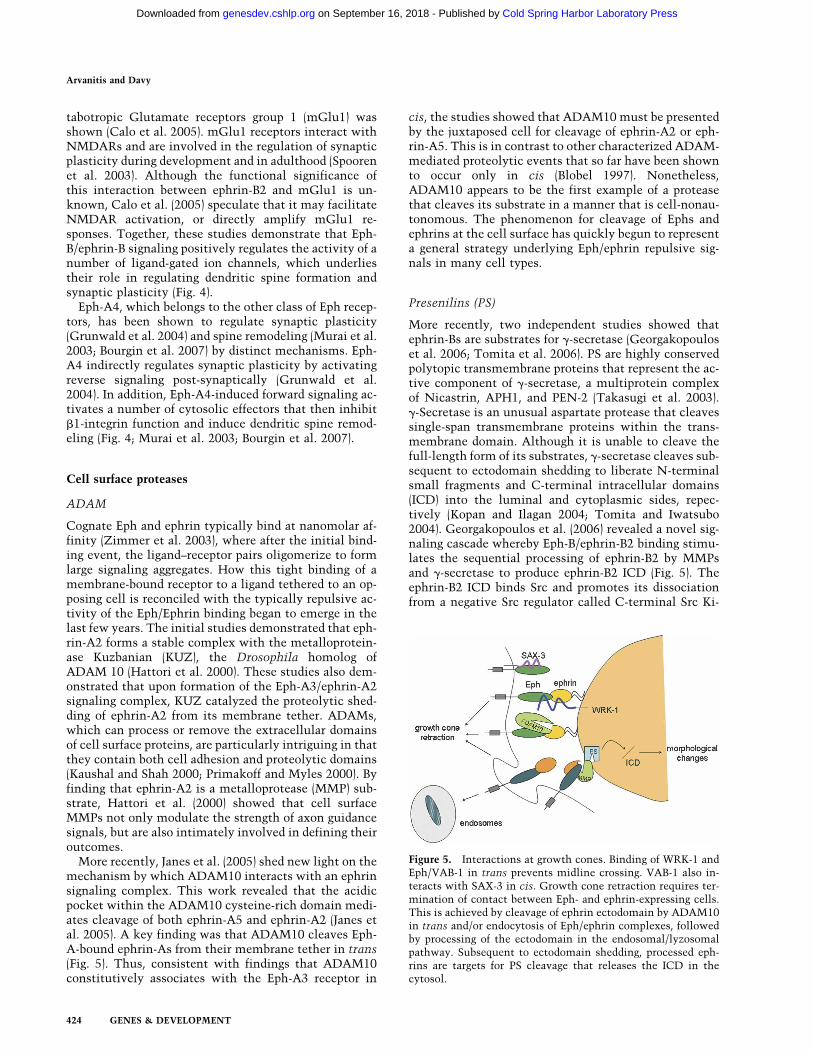
More recently, Janes et al. (2005) shed new light on themechanism by which ADAM10 interacts with an ephrinsignaling complex. This work revealed that the acidicpocket within the ADAM10 cysteine-rich domain medi-ates cleavage of both ephrin-A5 and ephrin-A2 (Janes etal. 2005). A key finding was that ADAM10 cleaves Eph-A-bound ephrin-As from their membrane tether in trans(Fig. 5). Thus, consistent with findings that ADAM10constitutively associates with the Eph-A3 receptor in
cis, the studies showed that ADAM10 must be presentedby the juxtaposed cell for cleavage of ephrin-A2 or eph-rin-A5. This is in contrast to other characterized ADAM-mediated proteolytic events that so far have been shownto occur only in cis (Blobel 1997). Nonetheless,ADAM10 appears to be the first example of a proteasethat cleaves its substrate in a manner that is cell-nonau-tonomous. The phenomenon for cleavage of Ephs andephrins at the cell surface has quickly begun to representa general strategy underlying Eph/ephrin repulsive sig-nals in many cell types.
Presenilins (PS)
More recently, two independent studies showed thatephrin-Bs are substrates for �-secretase (Georgakopouloset al. 2006; Tomita et al. 2006). PS are highly conservedpolytopic transmembrane proteins that represent the ac-tive component of �-secretase, a multiprotein complexof Nicastrin, APH1, and PEN-2 (Takasugi et al. 2003).�-Secretase is an unusual aspartate protease that cleavessingle-span transmembrane proteins within the trans-membrane domain. Although it is unable to cleave thefull-length form of its substrates, �-secretase cleaves sub-sequent to ectodomain shedding to liberate N-terminalsmall fragments and C-terminal intracellular domains(ICD) into the luminal and cytoplasmic sides, repec-tively (Kopan and Ilagan 2004; Tomita and Iwatsubo2004). Georgakopoulos et al. (2006) revealed a novel sig-naling cascade whereby Eph-B/ephrin-B2 binding stimu-lates the sequential processing of ephrin-B2 by MMPsand �-secretase to produce ephrin-B2 ICD (Fig. 5). Theephrin-B2 ICD binds Src and promotes its dissociationfrom a negative Src regulator called C-terminal Src Ki-
Figure 5. Interactions at growth cones. Binding of WRK-1 andEph/VAB-1 in trans prevents midline crossing. VAB-1 also in-teracts with SAX-3 in cis. Growth cone retraction requires ter-mination of contact between Eph- and ephrin-expressing cells.This is achieved by cleavage of ephrin ectodomain by ADAM10in trans and/or endocytosis of Eph/ephrin complexes, followedby processing of the ectodomain in the endosomal/lyzosomalpathway. Subsequent to ectodomain shedding, processed eph-rins are targets for PS cleavage that releases the ICD in thecytosol.
Arvanitis and Davy
424 GENES & DEVELOPMENT
Cold Spring Harbor Laboratory Press on September 16, 2018 - Published by genesdev.cshlp.orgDownloaded from
nase (Csk), thus allowing for the autophosphorylationand activation of Src. Likewise, degradation of the eph-rin-B2 ICD results in Src dephosphorylation and deacti-vation. Georgakopoulos et al. (2006) extrapolate theirfindings to previously established consequences of Eph-B/ephrin-B signaling that lead to the recruitment of cel-lular factors to cytoplasmic ephrin-B and to the rear-rangement of the actin cytoskeleton (for a review on eph-rin-induced reverse signaling, see Palmer and Klein 2003).
Parallel studies reported that Eph-B/ephrin-B1 bindingmediates ephrin-B1 ectodomain shedding, and the mem-brane-tethered fragment is sequentially cleaved by�-secretase to release the ICD (Tomita et al. 2006). Thesestudies further showed that the overexpression of themembrane-tethered ephrin-B1 led to the formation ofcellular protrusions consisting of F-actin, which wasnegatively regulated by �-secretase activity. Moreover,overexpression of the ephrin-B1 ICD and inhibition ofthe proteasome resulted in the nuclear localization ofthe ephrin-B1 ICD. To date, the functions of �-secretase-generated ICDs as transcriptional activators (i.e., Notch,APP, CD44) or repressors (Jagged, N-Cadherin) withinthe nucleus have been reported (for review, see Kopanand Ilagan 2004); however, functional analysis of thenuclear ephrin-B1 ICD awaits further study.
While ephrin-Bs are shown to constitutively undergoectomembrane shedding and sequential cleavage by �-secretase to release the ephrin-B ICDs, new findings sup-port an analogous process for the Eph-B2 receptor (Lit-terst et al. 2007). To date, reports have indicated that themechanism by which adhesive and signaling interac-tions between Eph receptors and ephrin ligands are ter-minated include endocytosis of the cell surface Eph/eph-rin complexes and cleavage of the ectodomain of ephrinligands (Hattori et al. 2000; Marston et al. 2003; Zimmeret al. 2003). Recent work shows that two distinct path-ways regulate the proteolytic processing of Eph-B2 recep-tor and its complexes: one stimulated by calcium influxand the other by ephrin-B2 ligand binding (Litterst et al.2007). Eph-B2 processing was stimulated by N-methyl-D-aspartic acid (NMDA) treatment and calcium influxand was sensitive to a broad spectrum of MMP inhibi-tors, particularly the inhibition of ADAM10. Therefore,these data show an additional physiological consequenceof the interaction between Eph-B2 and NMDAR (Fig. 4),and identified ADAM10 as the protease involved in cal-cium-induced processing of the Eph-B2 receptor.ADAM10 processing of Eph-B2 did not require endocy-tosis but rather resulted in the rapid shedding of the ex-tracellular domain of Eph-B2, suggesting that the ectodo-main shedding and �-secretase cleavage involved in thispathway occur at or near the plasma membrane. In ad-dition to this first pathway, a ligand-induced processingof Eph-B2 was demonstrated in the endosomal/lysosom-al pathway. Eph-B2/ephrin-B2 binding resulted in in-creased ectodomain shedding followed by �-secretaseprocessing and rapid degradation of Eph-B2 ectodomainfollowing ubiquitination (Fig. 5).
Taken together, the findings demonstrate that bothEph receptors and Ephrin ligands are processed by MMPs
and/or �-secretase, a catalytic event necessary for someof their functions (Table 1; Ethell and Ethell 2007).
Conclusions and perspectives
Our understanding of the Eph/ephrin pathway has im-proved tremendously over the last 10 years—we havediscovered that it is involved in many developmentalprocesses, we know it affects a number of cellular func-tions, and we partly characterized its mechanistic modeof action. The challenge we face now is to consider Eph/ephrin signaling not in isolation but as part of a networkof information. Every single cell constantly receives nu-merous signals that have to be instantaneously inte-grated and translated into coherent cellular responses.Direct or indirect regulatory interactions between path-ways serve as a mechanism to simplify the interpreta-tion of the many environmental factors confronting cellsat every decisional key point, since upstream cross-regu-lation eliminates the requirement of having a specificsignaling cascade for each extracellular cue.
One of the obvious difficulties in assembling a modelfrom the studies presented above is the fact that Eph/ephrin signaling clearly impinges on multiple pathwayssimultaneously to achieve its biological function. A sec-ond difficulty lies in the fact that the outcome (agonisticor antagonistic) of a given cross-talk seems to be highlydependent on cellular context. Resolution of these ap-parent discrepancies awaits the characterization of allplayers in the various signaling cascades. Indeed, onecould hypothesize that the presence or absence of (as yet)unknown partners could switch outcomes from agonis-tic to antagonistic. Another possibility that has beenlittle explored is that different Eph/ephrin pairs couldregulate different signaling cascades, therefore leading toopposite outcomes.
For many years, Eph/ephrin signaling has been studiedin a developmental context; however, recent publica-tions clearly highlighted its involvement in organ func-tion and in disease in the adult. Although regulation ofcell adhesion, migration, and morphology underlies mostof the developmental roles of Eph/ephrin signaling, theseproteins seem to regulate a different set of biological out-comes in the adult. In the future, it will be interesting tocompare signaling cascades and signal integration in thecontext of the roles of Eph/ephrins in the adult.
The discovery that Eph/ephrins are substrates for cellsurface proteases was satisfying, as it resolved one of theparadoxes of this signaling pair: how the initial Eph/eph-rin adhesion turned into cell repulsion. However, cleav-age of Eph receptors and ephrins and release of theirectodomain in the extracellular milieu raises the mind-boggling possibility that the action of these proteinsmight not be limited to short-range cell–cell interac-tions, but might also encompass long-range paracrine in-teractions.
Acknowledgments
We apologize to the authors whose work we were not able tocite due to space constraints. We thank Phil Soriano, David
Eph/ephrin signaling integrated
GENES & DEVELOPMENT 425
Cold Spring Harbor Laboratory Press on September 16, 2018 - Published by genesdev.cshlp.orgDownloaded from
Wilkinson, and our laboratory colleagues for critical reading ofthe manuscript, and Michael Miller for his insightful com-ments. D.A. is supported by the Centre National de la Recher-che Scientifique. A.D. is supported by the Centre National de laRecherche Scientifique. Work from our laboratory is supportedby grants from the Centre National de la Recherche Scienti-fique, from the Association Française contre les Myopathies,and from the Human Frontier Science Program Organization.
References
Bartley, T.D., Hunt, R.W., Welcher, A.A., Boyle, W.J., Parker,V.P., Lindberg, R.A., Lu, H.S., Colombero, A.M., Elliott,R.L., Guthrie, B.A., et al. 1994. B61 is a ligand for the ECKreceptor protein-tyrosine kinase. Nature 368: 558–560.
Beckmann, M.P., Cerretti, D.P., Baum, P., Vanden Bos, T.,James, L., Farrah, T., Kozlosky, C., Hollingsworth, T., Shil-ling, H., Maraskovsky, E., et al. 1994. Molecular character-ization of a family of ligands for eph-related tyrosine kinasereceptors. EMBO J. 13: 3757–3762.
Blobel, C.P. 1997. Metalloprotease-disintegrins: Links to celladhesion and cleavage of TNF� and Notch. Cell 90: 589–592.
Boulin, T., Pocock, R., and Hobert, O. 2006. A novel Eph recep-tor-interacting IgSF protein provides C. elegans motoneu-rons with midline guidepost function. Curr. Biol. 16: 1871–1883.
Bourgin, C., Murai, K.K., Richter, M., and Pasquale, E.B. 2007.The EphA4 receptor regulates dendritic spine remodeling byaffecting �1-integrin signaling pathways. J. Cell Biol. 178:1295–1307.
Bruckner, K., Pasquale, E.B., and Klein, R. 1997. Tyrosine phos-phorylation of transmembrane ligands for Eph receptors. Sci-ence 275: 1640–1643.
Calo, L., Bruno, V., Spinsanti, P., Molinari, G., Korkhov, V.,Esposito, Z., Patane, M., Melchiorri, D., Freissmuth, M., andNicoletti, F. 2005. Interactions between ephrin-B andmetabotropic glutamate 1 receptors in brain tissue and cul-tured neurons. J. Neurosci. 25: 2245–2254.
Cheng, H.J. and Flanagan, J.G. 1994. Identification and cloningof ELF-1, a developmentally expressed ligand for the Mek4and Sek receptor tyrosine kinases. Cell 79: 157–168.
Chong, L.D., Park, E.K., Latimer, E., Friesel, R., and Daar, I.O.2000. Fibroblast growth factor receptor-mediated rescue ofx-ephrin B1-induced cell dissociation in Xenopus embryos.Mol. Cell. Biol. 20: 724–734.
Compagni, A., Logan, M., Klein, R., and Adams, R.H. 2003.Control of skeletal patterning by ephrinB1–EphB interac-tions. Dev. Cell 5: 217–230.
Corrigan, C., Subramanian, R., and Miller, M.A. 2005. Eph andNMDA receptors control Ca2+/calmodulin-dependent pro-tein kinase II activation during C. elegans oocyte meioticmaturation. Development 132: 5225–5237.
Cortina, C., Palomo-Ponce, S., Iglesias, M., Fernández-Masip,J.L., Vivancos, A., Whissell, G., Humà, M., Peiro, N., Gal-lego, L., Jonkheer, S., et al. 2007. EphB–ephrinB interactionssuppress colorectal cancer progression by compartmentaliz-ing tumor cells. Nat. Genet. 39: 1376–1383.
Craig, A.M., Graf, E.R., and Linhoff, M.W. 2006. How to build acentral synapse: Clues from cell culture. Trends Neurosci.29: 8–20.
Dalva, M.B., Takasu, M.A., Lin, M.Z., Shamah, S.M., Hu, L.,Gale, N.W., and Greenberg, M.E. 2000. EphB receptors inter-act with NMDA receptors and regulate excitatory synapseformation. Cell 103: 945–956.
Davy, A. and Robbins, S.M. 2000. Ephrin-A5 modulates cell
adhesion and morphology in an integrin-dependent manner.EMBO J. 19: 5396–5405.
Davy, A., Aubin, J., and Soriano, P. 2004. EphrinB1 forward andreverse signaling are required during mouse development.Genes & Dev. 18: 572–583.
Davy, A., Bush, J.O., and Soriano, P. 2006. Inhibition of gapjunction communication at ectopic ephrin boundaries un-derlies craniofrontonasal syndrome. PLoS Biol. 4: 1763–1776.
de Saint-Vis, B., Bouchet, C., Gautier, G., Valladeau, J., Caux,C., and Garrone, P. 2003. Human dendritic cells express neu-ronal Eph receptor tyrosine kinases: Role of EphA2 in regu-lating adhesion to fibronectin. Blood 102: 4431–4440.
Deroanne, C., Vouret-Craviari, V., Wang, B., and Pouysségur, J.2003. EphrinA1 inactivates integrin-mediated vascularsmooth muscle cell spreading via the Rac/PAK pathway. J.Cell Sci. 116: 1367–1376.
Dickson, B.J. and Gilestro, G.F. 2006. Regulation of commis-sural axon pathfinding by slit and its Robo receptors. Annu.Rev. Cell Dev. Biol. 22: 651–675.
Elias, L.A., Wang, D.D., and Kriegstein, A.R. 2007. Gap junctionadhesion is necessary for radial migration in the neocortex.Nature 448: 901–907.
Ethell, I.M. and Ethell, D.W. 2007. Matrix metalloproteinases inbrain development and remodeling: Synaptic functions andtargets. J. Neurosci. Res. 85: 2813–2823.
Gauthier, L.R. and Robbins, S. 2003. Ephrin signaling: One raftto rule them all? One raft to sort them? One raft to spreadtheir call and in signaling bind them? Life Sci. 74: 207–216.
Georgakopoulos, A., Litterst, C., Ghersi, E., Baki, L., Xu, C.,Serban, G., and Robakis, N.K. 2006. Metalloproteinase/Pre-senilin1 processing of ephrinB regulates EphB-induced Srcphosphorylation and signaling. EMBO J. 25: 1242–1252.
Gerlai, R. 2002. EphB and NMDA receptors: Components ofsynaptic plasticity coming together. Trends Neurosci. 25:180–181.
Ghenea, S., Boudreau, J.R., Lague, N.P., and Chin-Sang, I.D.2005. The VAB-1 Eph receptor tyrosine kinase and SAX-3/Robo neuronal receptors function together during C. elegansembryonic morphogenesis. Development 132: 3679–3690.
Giri, B., Dixit, V.D., Ghosh, M.C., Collins, G.D., Khan, I.U.,Madara, K., Weeraratna, A.T., and Taub, D.D. 2007.CXCL12-induced partitioning of flotillin-1 with lipid raftsplays a role in CXCR4 function. Eur. J. Immunol. 37: 2104–2116.
Govindan, J.A., Cheng, H., Harris, J.E., and Greenstein, D. 2006.G�o/i and G�s signaling function in parallel with the MSP/Eph receptor to control meiotic diapause in C. elegans. Curr.Biol. 16: 1257–1268.
Grunwald, I.C., Korte, M., Wolfer, D., Wilkinson, G.A., Un-sicker, K., Lipp, H.P., Bonhoeffer, T., and Klein, R. 2001.Kinase-independent requirement of EphB2 receptors in hip-pocampal synaptic plasticity. Neuron 32: 1027–1040.
Grunwald, I.C., Korte, M., Adelmann, G., Plueck, A., Kullander,K., Adams, R.H., Frotscher, M., Bonhoeffer, T., and Klein, R.2004. Hippocampal plasticity requires postsynaptic eph-rinBs. Nat. Neurosci. 7: 33–40.
Gu, C. and Park, S. 2001. The EphA8 receptor regulates integrinactivity through p110� phosphatidylinositol-3 kinase in atyrosine kinase activity-independent manner. Mol. Cell.Biol. 21: 4579–4597.
Arvanitis and Davy
426 GENES & DEVELOPMENT
Cold Spring Harbor Laboratory Press on September 16, 2018 - Published by genesdev.cshlp.orgDownloaded from
Hartsock, A. and Nelson, W.J. 2007. Adherens and tight junc-tions: Structure, function and connections to the actin cy-toskeleton. Biochim. Biophys. Acta. doi: 10.1016/j.b-bamem.2007.07.012.
Hattori, M., Osterfield, M., and Flanagan, J.G. 2000. Regulatedcleavage of a contact-mediated axon repellent. Science 289:1360–1365.
Henderson, J.T., Georgiou, J., Jia, Z., Robertson, J., Elowe, S.,Roder, J.C., and Pawson, T. 2001. The receptor tyrosine ki-nase EphB2 regulates NMDA-dependent synaptic function.Neuron 32: 1041–1056.
Henkemeyer, M., Itkis, O.S., Ngo, M., Hickmott, P.W., andEthell, I.M. 2003. Multiple EphB receptor tyrosine kinasesshape dendritic spines in the hippocampus. J. Cell Biol. 163:1313–1326.
Hirai, H., Maru, Y., Hagiwara, K., Nishida, J., and Takaku, F.1987. A novel putative tyrosine kinase receptor encoded bythe Eph gene. Science 238: 1717–1720.
Huai, J. and Drescher, U. 2001. An ephrin-A-dependent signal-ing pathway controls integrin function and is linked to thetyrosine phosphorylation of a 120-kDa protein. J. Biol.Chem. 276: 6689–6694.
Huynh-Do, U., Stein, E., Lane, A.A., Liu, H., Cerreti, D.P., andDaniel, T.O. 1999. Surface densities of ephrin-B1 determineEphB1-coupled activation of cell attachment through �v�3
and �v�1 integrins. EMBO J. 18: 2165–2173.Huynh-Do, U., Vindis, C., Liu, H., Cerretti, D.P., McGrew, J.T.,
Enriquez, M., Chen, J., and Daniel, T.O. 2002. Ephrin-B1transduces signals to activate integrin-mediated migration,attachment and angiogenesis. J. Cell Sci. 115: 3073–3081.
Inoue, T., Oz, H.S., Wiland, D., Gharib, S., Deshpande, R., Hill,R.J., Katz, W.S., and Sternberg, P.W. 2004. C. elegans LIN-18is a Ryk ortholog and functions in parallel to LIN-17/Frizzledin Wnt signaling. Cell 118: 795–806.
Janes, P.W., Saha, N., Barton, W.A., Kolev, M.V., Wimmer-Klei-kamp, S.H., Nievergall, E., Blobel, C.P., Himanen, J.P., Lack-mann, M., and Nikolov, D.B. 2005. Adam meets Eph: AnADAM substrate recognition module acts as a molecularswitch for ephrin cleavage in trans. Cell 123: 291–304.
Jones, T.L., Chong, L.D., Kim, J., Xu, R.-H., Kung, H.-F., andDaar, I.O. 1998. Loss of cell adhesion in Xenopus laevis em-bryo mediated by the cytoplasmic domain of XLerk, an Ephligand. Proc. Natl. Acad. Sci. 95: 576–581.
Kamitori, K., Tanaka, M., Okuno-Hirasawa, T., and Kohsaka, S.2005. Receptor related to tyrosine kinase RYK regulates cellmigration during cortical development. Biochem. Biophys.Res. Commun. 330: 446–453.
Kaushal, G.P. and Shah, S.V. 2000. The new kids on the block:ADAMTSs, potentially multifunctional metalloproteinasesof the ADAM family. J. Clin. Invest. 105: 1335–1337.
Kayser, M.S., McClelland, A.C., Hughes, E.G., and Dalva, M.B.2006. Intracellular and trans-synaptic regulation of glutama-tergic synaptogenesis by EphB receptors. J. Neurosci. 26:12152–12164.
Konstantinova, I., Nikolova, G., Ohara-Imaizumi, M., Meda, P.,Kucera, T., Zarbalis, K., Wurst, W., Nagamatsu, S., and Lam-mert, E. 2007. EphA–Ephrin-A-mediated � cell communica-tion regulates insulin secretion from pancreatic islets. Cell129: 359–370.
Kopan, R. and Ilagan, M.X. 2004. �-Secretase: Proteasome of themembrane? Nat. Rev. Mol. Cell Biol. 5: 499–504.
Koshida, S., Kishimoto, Y., Ustumi, H., Shimizu, T., Furutani-Seiki, M., Kondoh, H., and Takada, S. 2005. Integrin�5-de-pendent fibronectin accumulation for maintenance of so-mite boundaries in zebrafish embryos. Dev. Cell 8: 587–598.
Kucia, M., Jankowski, K., Reca, R., Wysoczynski, M., Bandura,L., Allendorf, D.J., Zhang, J., Ratajczak, J., and Ratajczak,M.Z. 2004. CXCR4–SDF-1 signalling, locomotion, chemo-taxis and adhesion. J. Mol. Histol. 35: 233–245.
Laird, D.W. 1996. The life cycle of a connexin: Gap junctionformation, removal, and degradation. J. Bioenerg. Bio-membr. 28: 311–318.
Li, Z. and Sheng, M. 2003. Some assembly required: The devel-opment of neuronal synapses. Nat. Rev. Mol. Cell Biol. 4:833–841.
Litterst, C., Georgakopoulos, A., Shioi, J., Ghersi, E.,Wisniewski, T., Wang, R., Ludwig, A., and Robakis, N.K.2007. Ligand binding and calcium influx induce distinctectodomain/�-secretase-processing pathways of EphB2 re-ceptor. J. Biol. Chem. 282: 16155–16163.
Lu, Q., Sun, E., Klein, R., and Flanagan, J. 2001. Ephrin-B reversesignaling is mediated by a novel PDZ-RGS protein and se-lectively inhibits G-protein-coupled chemoattraction. Cell105: 69–79.
Lu, W., Yamamoto, V., Ortega, B., and Baltimore, D. 2004.Mammalian Ryk is a Wnt coreceptor required for stimula-tion of neurite outgrowth. Cell 119: 97–108.
Machon, O., van den Bout, C.J., Backman, M., Kemler, R., andKrauss, S. 2003. Role of �-catenin in the developing corticaland hippocampal neuroepithelium. Neuroscience 122: 129–143.
Marston, D.J., Dickinson, S., and Nobes, C.D. 2003. Rac-depen-dent trans-endocytosis of ephrinBs regulates Eph–ephrincontact repulsion. Nat. Cell Biol. 5: 879–888.
Mellitzer, G., Xu, Q., and Wilkinson, D.G. 1999. Eph receptorsand ephrins restrict cell intermingling and communication.Nature 400: 77–81.
Miao, H., Burnett, E., Kinch, M., Simon, E., and Wang, B. 2000.Activation of EphA2 kinase suppresses integrin function andcauses focal-adhesion-kinase dephosphorylation. Nat. CellBiol. 2: 62–69.
Miao, H., Strebhardt, K., Pasquale, E.B., Shen, T.L., Guan, J.L.,and Wang, B. 2005. Inhibition of integrin-mediated cell ad-hesion but not directional cell migration requires catalyticactivity of EphB3 receptor tyrosine kinase. Role of Rho fam-ily small GTPases. J. Biol. Chem. 280: 923–932.
Miller, M.A., Ruest, P.J., Kosinski, M., Hanks, S.K., and Green-stein, D. 2003. An Eph receptor sperm-sensing controlmechanism for oocyte meiotic maturation in Caenorhabdi-tis elegans. Genes & Dev. 17: 187–200.
Moody, S.A. 2004. To differentiate or not to differentiate: Regu-lation of cell fate decision by being in the right place at theright time. Cell Cycle 3: 564–566.
Moore, K.B., Mood, K., Daar, I.O., and Moody, S.A. 2004. Mor-phogenetic movements underlying eye field formation re-quire interactions between the FGF and ephrinB1 signalingpathways. Dev. Cell 6: 55–67.
Murai, K.K., Nguyen, L.N., Irie, F., Yamaguchi, Y., andPasquale, E.B. 2003. Control of hippocampal dendritic spinemorphology through ephrin-A3/Eph-A4 signaling. Nat. Neu-rosci. 6: 153–160.
Nishimura, T., Yamaguchi, T., Tokunaga, A., Hara, A.,Hamaguchi, T., Kato, K., Iwamatsu, A., Okano, H., and Kai-buchi, K. 2006. Role of numb in dendritic spine developmentwith a Cdc42 GEF intersectin and EphB2. Mol. Biol. Cell 17:
Eph/ephrin signaling integrated
GENES & DEVELOPMENT 427
Cold Spring Harbor Laboratory Press on September 16, 2018 - Published by genesdev.cshlp.orgDownloaded from
1273–1285.Palmer, A. and Klein, R. 2003. Multiple roles of ephrins in mor-
phogenesis, neuronal networking, and brain function. Genes& Dev. 17: 1429–1450.
Park, E.K., Warner, N., Bong, Y.S., Stapleton, D., Maeda, R.,Pawson, T., and Daar, I.O. 2004. Ectopic EphA4 receptorinduces posterior protrusions via FGF signaling in Xenopusembryos. Mol. Biol. Cell 15: 1647–1655.
Pasquale, E.B. 2005. Eph receptor signalling casts a wide net oncell behaviour. Nat. Rev. Mol. Cell Biol. 6: 462–475.
Penzes, P., Beeser, A., Chernoff, J., Schiller, M.R., Eipper, B.A.,Mains, R.E., and Huganir, R.L. 2003. Rapid induction of den-dritic spine morphogenesis by trans-synaptic ephrinB–EphBreceptor activation of the Rho-GEF kalirin. Neuron 37: 263–274.
Picco, V., Hudson, C., and Yasuo, H. 2007. Ephrin–Eph signal-ling drives the asymmetric division of notochord/neural pre-cursors in Ciona embryos. Development 134: 1491–1497.
Poliakov, A., Cotrina, M., and Wilkinson, D.G. 2004. Diverseroles of Eph receptors and ephrins in the regulation of cellmigration and tissue assembly. Dev. Cell 7: 465–480.
Prévost, N., Woulfe, D.S., Tognolini, M., Tanaka, T., Jian, W.,Fortna, R.R., Jiang, H., and Brass, L.F. 2004. Signaling byephrinB1 and Eph kinases in platelets promotes Rap1 acti-vation, platelet adhesion, and aggregation via effector path-ways that do not require phosphorylation of ephrinB1. Blood103: 1348–1355.
Prévost, N., Woulfe, D.S., Jiang, H., Stalker, T.J., Marchese, P.,Ruggeri, Z.M., and Brass, L.F. 2005. Eph kinases and ephrinssupport thrombus growth and stability by regulating inte-grin outside-in signaling in platelets. Proc. Natl. Acad. Sci.102: 9820–9825.
Primakoff, P. and Myles, D.G. 2000. The ADAM gene family:Surface proteins with adhesion and protease activity. TrendsGenet. 16: 83–87.
Red-Horse, K., Kapidzic, M., Zhou, Y., Feng, K.T., Singh, H., andFisher, S.J. 2005. EPH-B4 regulates chemokine-evoked tro-phoblast responses: A mechanism for incorporating the hu-man placenta into the maternal circulation. Development132: 4097–4106.
Rougon, G. and Hobert, O. 2003. New insights into the diversityand function of neuronal immunoglobulin superfamily mol-ecules. Annu. Rev. Neurosci. 26: 207–238.
Salvucci, O., de la Luz Sierra, M., Martina, J.A., McCormick,P.J., and Tosato, G. 2006. Eph-B2 and Eph-B4 receptors for-ward signaling promotes SDF-1-induced endothelial cellchemotaxis and branching remodeling. Blood 108: 2914–2922.
Scheiffele, P. 2003. Cell–cell signaling during synapse formationin the CNS. Annu. Rev. Neurosci. 26: 485–508.
Segura, I., Essmann, C.L., Weinges, S., and Acker-Palmer, A.2007. Grb4 and GIT1 transduce ephrin-B reverse signalsmodulating spine morphogenesis and synapse formation.Nat. Neurosci. 10: 301–310.
Sharfe, N., Freywald, A., Toro, A., Dadi, H., and Roifman, C.2002. Ephrin stimulation modulates T cell chemotaxis. Eur.J. Immunol. 32: 3745–3755.
Spooren, W., Ballard, T., Gasparini, F., Amalric, M., Mutel, V.,and Schreiber, R. 2003. Insight into the function of Group Iand Group II metabotropic glutamate (mGlu) receptors: Be-havioural characterization and implications for the treat-ment of CNS disorders. Behav. Pharmacol. 14: 257–277.
Stains, J.P. and Civitelli, R. 2005. Gap junctions in skeletal de-
velopment and function. Biochim. Biophys. Acta 1719: 69–81.
Suh, L.H., Oster, S.F., Soehrman, S.S., Grenningloh, G., and Sre-tavan, D.W. 2004. L1/Laminin modulation of growth coneresponse to Eph-B triggers growth pauses and regulates themicrotubule destabilizing protein SCG10. J. Neurosci. 24:1976–1986.
Takasu, M.A., Dalva, M.B., Zigmond, R.E., and Greenberg, M.E.2002. Modulation of NMDA receptor-dependent calcium in-flux and gene expression through Eph-B receptors. Science295: 491–495.
Takasugi, N., Tomita, T., Hayashi, I., Tsuruoka, M., Niimura,M., Takahashi, Y., Thinakaran, G., and Iwatsubo, T. 2003.The role of presenilin cofactors in the �-secretase complex.Nature 422: 438–441.
Tanaka, M., Kamata, R., and Sakai, R. 2005a. EphA2 phosphory-lates the cytoplasmic tail of Claudin-4 and mediates para-cellular permeability. J. Biol. Chem. 280: 42375–42382.
Tanaka, M., Kamata, R., and Sakai, R. 2005b. Phosphorylationof ephrin-B1 via the interaction with claudin following cell–cell contact formation. EMBO J. 24: 3700–3711.
Tepass, U., Godt, D., and Winklbauer, R. 2002. Cell sorting inanimal development: Signalling and adhesive mechanismsin the formation of tissue boundaries. Curr. Opin. Genet.Dev. 12: 572–582.
Tolias, K.F., Bikoff, J.B., Burette, A., Paradis, S., Harrar, D., Ta-vazoie, S., Weinberg, R.J., and Greenberg, M.E. 2005. TheRac1-GEF Tiam1 couples the NMDA receptor to the activ-ity-dependent development of dendritic arbors and spines.Neuron 45: 525–538.
Tomita, T. and Iwatsubo, T. 2004. The inhibition of �-secretaseas a therapeutic approach to Alzheimer’s disease. Drug NewsPerspect. 17: 321–325.
Tomita, T., Tanaka, S., Morohashi, Y., and Iwatsubo, T. 2006.Presenilin-dependent intramembrane cleavage of ephrin-B1.Mol. Neurodegener. 1: 2. doi: 10.1186/1750-1326-1-2.
Trivier, E. and Ganessan, T.S. 2002. RYK, a catalytically inac-tive receptor tyrosine kinase, associates with Eph-B2 andEph-B3 but does not interact with AF-6. J. Biol. Chem. 277:23037–23043.
Twigg, S.R.F., Kan, R., Babbs, C., Bochukova, E.G., Robertson,S.P., Wall, S.A., Moriss-Kay, G.M., and Wilkie, A.O.M. 2004.Mutations of ephrin-B1 (EFNB1), a marker of tissue bound-ary, cause craniofrontonasal syndrome. Proc. Natl. Acad.Sci. 101: 8652–8657.
Whitten, S.J. and Miller, M.A. 2007. The role of gap junctions inCaenorhabditis elegans oocyte maturation and fertilization.Dev. Biol. 301: 432–446.
Wieland, I., Jacubiczka, S., Muschke, P., Cohen, M., Thiele, H.,Gerlach, K.L., Adams, R.H., and Wieacker, P. 2004. Muta-tions of the ephrin-B1 gene cause craniofrontonasal syn-drome. Am. J. Hum. Genet. 74: 1209–1215.
Winning, R.S., Scales, J.B., and Sargent, T.D. 1996. Disruption ofcell adhesion in Xenopus embryos by Pagliaccio, an Eph-class receptor tyrosine kinase. Dev. Biol. 179: 309–319.
Wysoczynski, M., Reca, R., Ratajczak, J., Kucia, M., Shirvaikar,N., Honczarenko, M., Mills, M., Wanzeck, J., Janowska-Wieczorek, A., and Ratajczak, M.Z. 2005. Incorporation ofCXCR4 into membrane lipid rafts primes homing-relatedresponses of hematopoietic stem/progenitor cells to anSDF-1 gradient. Blood 105: 40–48.
Arvanitis and Davy
428 GENES & DEVELOPMENT
Cold Spring Harbor Laboratory Press on September 16, 2018 - Published by genesdev.cshlp.orgDownloaded from
Xu, Q., Mellitzer, G., Robinson, V., and Wilkinson, D.G. 1999.In vivo cell sorting in complementary segmental domainsmediated by Eph receptors and ephrins. Nature 399: 267–271.
Yokote, H., Fujita, K., Jing, X., Sawada, T., Liang, S., Yao, L.,Yan, X., Zhang, Y., Schlessinger, J., and Sakaguchi, K. 2005.Trans-activation of Eph-A4 and FGF receptors mediated bydirect interactions between their cytoplasmic domains.Proc. Natl. Acad. Sci. 102: 18866–18871.
Zantek, N.D., Azimi, M., Fedor-Chaiken, M., Wang, B., Brack-enbury, R., and Kinch, M.S. 1999. E-cadherin regulates thefunction of the EphA2 receptor tyrosine kinase. Cell GrowthDiffer. 10: 629–638.
Zhao, C., Irie, N., Takada, Y., Shimoda, K., Miyamoto, T.,Nishiwaki, T., Suda, T., and Matsuo, K. 2006. BidirectionalephrinB2–EphB4 signaling controls bone homeostasis. CellMetab. 4: 111–121.
Zimmer, M., Palmer, A., Kohler, J., and Klein, R. 2003. EphB–ephrinB bi-directional endocytosis terminates adhesion al-lowing contact mediated repulsion. Nat. Cell Biol. 5: 869–878.
Zisch, A.H., Stallcup, W.B., Chong, L.D., Dahlin-Huppe, K., Vo-shol, J., Schachner, M., and Pasquale, E.B. 1997. Tyrosinephosphorylation of L1 family adhesion molecules: Implica-tion of the Eph kinase Cek5. J. Neurosci. Res. 47: 655–665.
Zlotnik, A. and Yoshie, O. 2000. Chemokines: A new classifi-cation system and their role in immunity. Immunity 12:121–127.
Zou, J.X., Wang, B., Kalo, M.S., Zisch, A.H., Pasquale, E.B., andRuoslahti, E. 1999. An Eph receptor regulates integrin activ-ity through R-Ras. Proc. Natl. Acad. Sci. 96: 13813–13818.
Eph/ephrin signaling integrated
GENES & DEVELOPMENT 429
Cold Spring Harbor Laboratory Press on September 16, 2018 - Published by genesdev.cshlp.orgDownloaded from