CONTENTS Middle East respiratory syndrome coronavirus: a review of the latest situation ............................. pg 60 Outbreak of Vibrio parahaemolyticus food poisoning ........................... pg 70 Disease burden of acute respiratory infections in Singapore, 2010 ................ pg 75 Suggested citation: Ministry of Health, Singapore. [Article title]. Epidemiol News Bull [Year]; [Vol]:[inclusive page numbers] MOH Weekly Infectious Diseases Bulletin http://www.moh.gov.sg/content/moh_web/ home/statistics/infectiousDiseasesStatis- tics/weekly_infectiousdiseasesbulletin.html QUARTERLY ISSN 0218-0103 http://www.moh.gov.sg/content/moh_web/home/Publications/epidemiological_news_bulletin/2015.html Epidemiological News Bulletin JULY - SEPTEMBER 2015 VOL. 41 NO. 3 A PUBLICATION OF THE MINISTRY OF HEALTH, SINGAPORE Middle East respiratory syndrome coronavirus: a review of the latest situation Background Middle East Respiratory Syndrome Coronavirus (MERS-CoV) was first isolated from the sputum of a 60-year-old Saudi man in Jed- dah, Kingdom of Saudi Arabia (KSA), who presented with pneumonia complicated with renal failure, and subsequently died in September 2012. In the same month, the virus was isolated in a 49-year-old male Qatari who developed respiratory symptoms and renal failure, and was transferred to the United Kingdom (UK) for intensive care manage- ment. Genetic sequencing of the virus obtained from the second case was similar to the virus isolated from the first case. Thereafter, sporadic cases and clusters had been reported with epidemiological link to the Middle East. Subsequently, the World Health Organisation (WHO) ret- rospectively identified the virus in specimens from two fatal cases who were part of an earlier cluster of 13 cases linked to a hospital in Zarqha City, Jordan in April 2012. Epidemiology As of 16 August 2015, the WHO reported a total of 1,413 laboratory- confirmed cases of infection with MERS-CoV, including at least 502 deaths (35.6%) (Fig. 1). Most of the cases had occurred in the Middle East, with the KSA reporting close to 76% of the cases (Fig. 2). Of the cases that had been reported outside of the Middle East, all had either recently travelled to the Middle East or had contact with a confirmed case or ill person who had returned from travel in the Middle East. There was a strong predominance of older males with co-morbidities (male : female sex ratio:1.94 : 1; median age: 49 years, age range: 9 months to 99 years) 1 .
Transcript
CONTENTS
Middle East respiratory syndrome coronavirus: a review of the latest situation ............................. pg 60
Outbreak of Vibrio parahaemolyticus food poisoning ........................... pg 70
Disease burden of acute respiratory infections in Singapore, 2010 ................ pg 75
Epidemiological News Bulletin JULY - SEPTEMBER 2015 VOL. 41 NO. 3 A PUBLICATION OF THE MINISTRY OF HEALTH, SINGAPORE
Middle East respiratory syndrome coronavirus: a review of the latest
situation
Background
Middle East Respiratory Syndrome Coronavirus (MERS-CoV) was first isolated from the sputum of a 60-year-old Saudi man in Jed-dah, Kingdom of Saudi Arabia (KSA), who presented with pneumonia complicated with renal failure, and subsequently died in September 2012. In the same month, the virus was isolated in a 49-year-old male Qatari who developed respiratory symptoms and renal failure, and was transferred to the United Kingdom (UK) for intensive care manage-ment. Genetic sequencing of the virus obtained from the second case was similar to the virus isolated from the first case. Thereafter, sporadic cases and clusters had been reported with epidemiological link to the Middle East. Subsequently, the World Health Organisation (WHO) ret-rospectively identified the virus in specimens from two fatal cases who were part of an earlier cluster of 13 cases linked to a hospital in Zarqha City, Jordan in April 2012.
Epidemiology
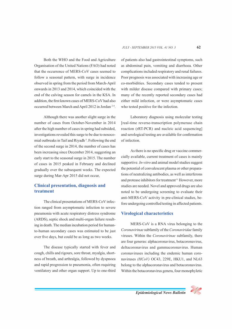
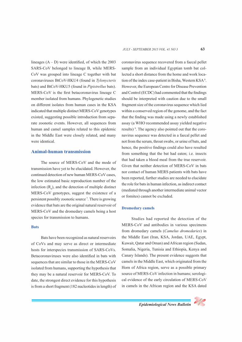
As of 16 August 2015, the WHO reported a total of 1,413 laboratory-confirmed cases of infection with MERS-CoV, including at least 502 deaths (35.6%) (Fig. 1). Most of the cases had occurred in the Middle East, with the KSA reporting close to 76% of the cases (Fig. 2). Of the cases that had been reported outside of the Middle East, all had either recently travelled to the Middle East or had contact with a confirmed case or ill person who had returned from travel in the Middle East. There was a strong predominance of older males with co-morbidities (male : female sex ratio:1.94 : 1; median age: 49 years, age range: 9 months to 99 years) 1.
JULY - SEPTEMBER 2015 VOL. 41 NO 3 61
Epidemiological News Bulletin
Figure 2Geographical distribution of confirmed human cases of MERS-CoV infection
Source: WHO. Middle East respiratory syndrome coronavirus (MERS-CoV). Global map of countries with confirmed cases of MERS-CoV. Available at: http://www.who.int/emergencies/mers-cov/en/
Figure 1Epidemic curve of laboratory-confirmed human cases of MERS-CoV infection reported to WHO as of
14 August 2015 (n = 1413)
Source: WHO. Middle East respiratory syndrome coronavirus (MERS-CoV). Epicurve of confirmed cases and deaths in Republic of Korea, China, Saudi Arabia and other countries. Available at: http://www.who.int/emergencies/mers-cov/new-global-rok-ksa-other-countries-weekly-epicurve22015-08-14.png?ua=1
JULY - SEPTEMBER 2015 VOL. 41 NO. 3 62
Epidemiological News Bulletin
Both the WHO and the Food and Agriculture Organisation of the United Nations (FAO) had noted that the occurrence of MERS-CoV cases seemed to follow a seasonal pattern, with surge in incidence observed in spring from the period from March-April onwards in 2013 and 2014, which coincided with the end of the calving season for camels in the KSA. In addition, the first known cases of MERS-CoV had also occurred between March and April 2012 in Jordan 2-4.
Although there was another slight surge in the number of cases from October-November in 2014 after the high number of cases in spring had subsided, investigations revealed this surge to be due to nosoco-mial outbreaks in Taif and Riyadh 5. Following the end of the second surge in 2014, the number of cases has been increasing since December 2014, suggesting an early start to the seasonal surge in 2015. The number of cases in 2015 peaked in February and declined gradually over the subsequent weeks. The expected surge during Mar-Apr 2015 did not occur,
Clinical presentation, diagnosis and treatment
The clinical presentations of MERS-CoV infec-tion ranged from asymptomatic infection to severe pneumonia with acute respiratory distress syndrome (ARDS), septic shock and multi-organ failure result-ing in death. The median incubation period for human-to-human secondary cases was estimated to be just over five days, but could be as long as two weeks.
The disease typically started with fever and cough, chills and rigours, sore throat, myalgia, short-ness of breath, and arthralgia, followed by dyspnoea and rapid progression to pneumonia, often requiring ventilatory and other organ support. Up to one-third
of patients also had gastrointestinal symptoms, such as abdominal pain, vomiting and diarrhoea. Other complications included respiratory and renal failures. Poor prognosis was associated with increasing age or co-morbidities. Secondary cases tended to present with milder disease compared with primary cases; many of the recently reported secondary cases had either mild infection, or were asymptomatic cases who tested positive for the infection.
Laboratory diagnosis using molecular testing [real-time reverse-transcription polymerase chain reaction (rRT-PCR) and nucleic acid sequencing] and serological testing are available for confirmation of infection.
As there is no specific drug or vaccine commer-cially available, current treatment of cases is mainly supportive. In-vitro and animal model studies suggest the potential of convalescent plasma or other prepara-tions of neutralizing antibodies, as well as interferons and protease inhibitors for treatment 6. However, more studies are needed. Novel and approved drugs are also noted to be undergoing screening to evaluate their anti-MERS-CoV activity in pre-clinical studies, be-fore undergoing controlled testing in affected patients.
Virological characteristics
MERS-CoV is a RNA virus belonging to the Coronavirinae subfamily of the Coronaviridae family viruses. Within the Coronavirinae subfamily, there are four generas: alphacoronavirus, betacoronavirus, deltacoronavirus and gammacoronavirus. Human coronaviruses including the endemic human coro-naviruses (HCoV) OC43, 229E, HKU1, and NL63 belong to the alphacoronavirus and betacoronavirus. Within the betacoronavirus genera, four monophyletic
JULY - SEPTEMBER 2015 VOL. 41 NO 3 63
Epidemiological News Bulletin
lineages (A – D) were identified, of which the 2003 SARS-CoV belonged to lineage B, while MERS-CoV was grouped into lineage C together with bat coronaviruses BtCoV-HKU4 (found in Tylonycteris bats) and BtCoV-HKU5 (found in Pipistrellus bats). MERS-CoV is the first betacoronavirus lineage C member isolated from humans. Phylogenetic studies on different isolates from human cases in the KSA indicated that multiple distinct MERS-CoV genotypes existed, suggesting possible introduction from sepa-rate zoonotic events. However, all sequences from human and camel samples related to this epidemic in the Middle East were closely related, and many were identical.
Animal-human transmission
The source of MERS-CoV and the mode of transmission have yet to be elucidated. However, the continued detection of new human MERS-CoV cases, the low estimated basic reproduction number of the infection (R0), and the detection of multiple distinct MERS-CoV genotypes, suggest the existence of a persistent possibly zoonotic source 7. There is growing evidence that bats are the original natural reservoir of MERS-CoV and the dromedary camels being a host species for transmission to humans.
Bats
Bats have been recognized as natural reservoirs of CoVs and may serve as direct or intermediate hosts for interspecies transmission of SARS-CoVs. Betacoronaviruses were also identified in bats with sequences that are similar to those in the MERS-CoV isolated from humans, supporting the hypothesis that they may be a natural reservoir for MERS-CoV. To date, the strongest direct evidence for this hypothesis is from a short fragment (182 nucleotides in length) of
coronavirus sequence recovered from a faecal pellet sample from an individual Egyptian tomb bat col-lected a short distance from the home and work loca-tion of the index case-patient in Bisha, Western KSA 8. However, the European Centre for Disease Prevention and Control (ECDC) had commented that the findings should be interpreted with caution due to the small fragment size of the coronavirus sequence which lied within a conserved region of the genome, and the fact that the finding was made using a newly established assay (a WHO recommended assay yielded negative results) 9. The agency also pointed out that the coro-navirus sequence was detected in a faecal pellet and not from the serum, throat swabs, or urine of bats, and hence, the positive findings could also have resulted from something that the bat had eaten; i.e. insects that had taken a blood meal from the true reservoir. Given that neither detection of MERS-CoV in bats nor contact of human MERS patients with bats have been reported, further studies are needed to elucidate the role for bats in human infection, as indirect contact (mediated through another intermediate animal vector or fomites) cannot be excluded.
Dromedary camels
Studies had reported the detection of the MERS-CoV and antibodies in various specimens from dromedary camels (Camelus dromedaries) in the Middle East (Iran, KSA, Jordan, UAE, Egypt, Kuwait, Qatar and Oman) and African region (Sudan, Somalia, Nigeria, Tunisia and Ethiopia, Kenya and Canary Islands). The present evidence suggests that camels in the Middle East, which originated from the Horn of Africa region, serve as a possible primary source of MERS-CoV infection in humans; serologi-cal evidence of the early circulation of MERS-CoV in camels in the African region and the KSA dated
JULY - SEPTEMBER 2015 VOL. 41 NO. 3 64
Epidemiological News Bulletin
back to 1983 and 1992, respectively 10, 11. However, no autochthonous MERS-CoV infections in humans had been reported in the KSA till 2012 and in Africa to date, suggesting that there might have been silent transmission between camels and humans in these two regions for the past two decades, and the absence of cases in human could be due to poor surveillance; lack of awareness and diagnostic capability for the disease; or a recent mutation in the virus which facilitated its jump from camels (or other animals) to humans.
MERS-CoV infection in animals appears to be restricted to the dromedary camels in the Middle East and African Region. Studies thus far did not find evidence of MERS-CoV infection (acute and past) in the one-hump dromedary camels in the United States (U.S.), Canada and Australia 12, 13; the two-hump Bac-trian camels (Camelus bactrianus) in Germany and Mongolia 14, 15; and in other animals such as goats, cows, water buffaloes, sheep, horses, donkeys, mules and chickens in the Middle East 11, 12, 16-18.
Several phylogenetic studies and genetic evidence had supported the plausibility of a role for camels in human infection and cross-species trans-mission between camels and humans 19-21. High viral loads had been detected in nasal swabs, conjunctival swabs, rectal swabs, and milk from camels suggesting that droplet contact, fomite and food-borne transmis-sion might be involved. A recent study published by Azhar EI et al. reported the isolation of the virus in an air sample collected in a camel barn implicated in a possible camel-to-human outbreak, highlighting the need for further investigation into possible airborne transmission of MERS-CoV 22. Even though MERS-CoV virus or RNA has not been detected in camel urine to date, the detection of MERS-CoV in urine in human cases suggested that virus shedding in urine
is plausible in camels. This, in turn, could be another potential source of food-borne transmission due to the occasional use of camel urine as a traditional medicine in Arabic culture 23. In a study in Qatar, 13% of lymph node samples taken at a camel slaughterhouse were positive for the virus, suggesting that camel meat might be another source of food-borne transmission 24.
Exposure to dromedary camels was found to be a risk factor in MERS-CoV infection. Serological surveys had found the seroprevalence of MERS-CoV to be higher in healthy camel-exposed individuals such as shepherds and slaughterhouse workers, as compared to the general population in the KSA and individuals without exposure to camels in Qatar 25,
26. It was proposed that there was a risk of camel workers becoming infected with MERS-CoV, often without being diagnosed, and proceeding to introduce the virus to the general population, where the more severe cases would trigger testing for the virus and result in disease recognition. Notwithstanding these study findings, it was observed that only a minority of the primary cases reported from the KSA had docu-mented camel contact; and other studies had shown an absence of MERS-CoV antibodies in camel abattoir workers in Egypt and the KSA 27-30.
Younger camels were postulated to play a particular role in zoonotic transmission since they seemed to be more frequently infected and shed more virus than older ones. In a study conducted in Dubai, United Arab Emirates (UAE), from March - June 2014, serological evidence of MERS-CoV infection was found in >96% of all dromedaries >2 years of age 31. Seroprevalence among dromedaries calves (<1 year of age) was significantly lower but still exceeded 80%. In addition, RT-PCR testing and virus isolation of nasal swab specimens were only
JULY - SEPTEMBER 2015 VOL. 41 NO 3 65
Epidemiological News Bulletin
successful among dromedaries <4 years of age (8.3% and 12.1% respectively), particularly in calves (35.3% and 13.6% respectively); while none of the adult dromedaries (>4 years of age) were found positive for the virus, suggesting increased infectivity of calves. The authors recommended that avoiding camels <2 years of age and postponing separation of the calves from the mother until the calves were older could be effective in preventing or controlling the spread of the MERS-CoV infection to humans.
Human-human transmission
Person-to-person transmission of the virus has been documented in several human clusters as-sociated with healthcare facilities, households and workplace. Nosocomial outbreak is a distinct hallmark in MERS-CoV transmission involving hospitalised patients, healthcare workers and close family con-tacts in healthcare facilities in affected countries in the Middle East and in some countries where the disease had been exported to, the most recent being the Republic of Korea (ROK). Nosocomial transmis-sion due to suboptimal infection control measures had been identified as one of the primary causes of an upsurge of cases in the Middle East during spring 2013 in Al-Ahsa, KSA; and 2014 in Jeddah, KSA and Abu Dhabi, UAE 32. It was also a significant cause of the large outbreak in the ROK in 2015, which had resulted from an imported case in a traveller returning from the Middle East.
As of 31 March 2015, an estimated 23% of the cases in the KSA were healthcare workers 33. Healthcare workers are at a high risk of MERS-CoV infection due to potential exposure to the virus from infected patients. In addition, healthcare workers could develop asymptomatic or mild disease, and unknowingly contribute to the propagation of noso-
comial outbreaks. In the large nosocomial outbreaks in Jeddah, KSA, Abu Dhabi, UAE and ROK, where inadequate infection control was implicated, the pro-portion of infected healthcare workers was as high as 30.4%, more than two-thirds of all the cases, and 21%, respectively 34. Nonetheless, a study to understand the frequency of secondary transmission to healthcare workers in healthcare settings revealed that only 19 out of 1695 (1.12%) healthcare workers contacts of confirmed cases in the KSA were tested positive, indicating a rather small overall risk of transmission to healthcare workers 35.
In the SARS pandemic in 2003, it was observed that there was a variation in the ability of the cases to transmit the coronavirus infection to others with many cases not transmitting the disease at all, and a few cases who transmitted the infection efficiently to several oth-ers, leading to the coining of the term ‘super-spreader’ 36. A documented study on the 2013 hospital outbreak of MERS-CoV in Al-Ahsa which involved four healthcare facilities in the eastern province of the KSA, mapped out the transmission chain of the 23 confirmed cases and 11 probable cases 37. The authors observed that the appar-ent heterogeneity in transmission, with many infected patients not transmitting disease at all and one patient transmitting disease to seven others, was reminiscent of SARS. However, although epidemiological and phylogenetic analyses supported the presence of person-to-person transmission, it was not possible to ascertain if there were single or multiple introductions from the com-munity; the mode of transmission (respiratory droplet or direct/indirect contact); and if transmission occurred during the incubation phase or asymptomatic infection.
Family clusters have been documented with evidence of secondary transmission to family mem-bers (with transmission rates ranging from 1.3% to
JULY - SEPTEMBER 2015 VOL. 41 NO 3 66
Epidemiological News Bulletin
4.3% of household contacts investigated), indicating low rate of transmission within households. Between 1 December 2012 and 1 December 2013, a nationwide serological survey was conducted on serum samples collected from 10,009 healthy people from all 13 provinces in the KSA, of which 15 samples (0.15%) were tested positive for MERS-CoV antibodies, indi-cating low transmission in the community 25.
Thus far, the possibility of transmission from asymptomatic PCR-positive cases cannot be ex-cluded, necessitating the need for close monitoring and investigation of all contacts, including asympto-matic contacts. To date, there have been no reports of transmission between confirmed cases and their co-passengers on board the same airplane. The imported case in ROK had resulted in subsequent transmission of the infection to 185 persons through four genera-tions of transmission. In addition, through intensive investigation of the contacts of index cases, there were four human-to-human transmissions documented outside Middle East UK (2), France (1) and Tunisia (1)) 1, 38-41. Notwithstanding this, other investigations of family contacts and healthcare contacts of index-cases in other non-Middle East countries with strong healthcare systems and established infection control practices such as Germany 42, Netherlands 43 and the U.S. 44, did not reveal any evidence of secondary MERS-CoV infection, supporting the hypothesis that human transmission of the infection can be effectively prevented with appropriate public health and infection control measures.
To date, with the exception of exportation of seven Umrah-associated cases to non-Middle East countries [Algeria (2), Netherlands (2), Tunisia (2) and Malaysia (1)] 40, 43, 45, 46, none of the Hajj pilgrims (symptomatic and asymptomatic) who had travelled to
the KSA and whose samples were collected, had tested positive for MERS-CoV. Majority of the pathogens detected in the respiratory samples of these individu-als were seasonal influenza A(H1N1, H3N2) andB, rhinovirus and Streptococcus pneumoniae, while a minority of them were positive for adenoviruses, human coronavirus OC43/229E, enteroviruses and parainfluenza viruses 47-52.
WHO’s advice on interruption of MERS-CoV transmission
The WHO emphasised on the importance of in-fection prevention and control measures to prevent the possible spread of MERS-CoV in healthcare facilities. Healthcare workers were advised to always apply stand-ard precautions consistently with all patients, regardless of their diagnosis. Extra precautions should be exercised according to the clinical presentations and clinical procedures. Droplet precautions should be added to the standard precautions when providing care to patients with symptoms of acute respiratory infection; contact precautions and eye protection should be added when caring for probable or confirmed cases of MERS-CoV infection; and airborne precautions should be applied when performing aerosol- generating procedures.
High-risk group such as those with diabetes, renal failure and chronic lung disease, and who are immunocompromised should avoid close contact with animals, particularly camels, when visiting farms, markets, or barn areas where the virus is known to be potentially circulating. General hygiene measures, such as regular hand washing before and after touch-ing animals and avoiding contact with sick animals, should be adhered to.
Food hygiene practices should be observed. Drinking raw camel milk or camel urine, or eating
JULY - SEPTEMBER 2015 VOL. 41 NO 3 67
Epidemiological News Bulletin
meat that has not been properly cooked should be avoided, particularly for high-risk individuals. Sick animals should never be slaughtered for consump-tion and dead animals should be safely buried or destroyed. Handling camels is also known to have an increased risk of infec tion with MERS-CoV. Until there is a better under standing of the specific modes of zoonotic transmis sion, it would be prudent for work-ers on farms, in slaughter houses and markets, as well as veterinarians and those handling camels at racing facilities to practice good personal hygiene, including frequent hand washing after touching animals. They should wear facial protec tion where feasible and protective clothing, which should be removed after work and be washed daily 5.
The WHO currently does not advise special screening at points of entry nor recommend travel or trade restrictions to prevent international spread of MERS-CoV.
Risk assessment
According to the WHO’s assessment 53, the current understanding of MERS-CoV is that it is a zoonotic virus, which has entered the human popula-tion in the Middle East on multiple occasions from direct or indirect contact with infected dromedary camels or camel-related products; the evidence link-ing MERS-CoV transmission between camels to humans is irrefutable. Human-to-human transmission
of the virus has been observed, and the majority of cases reported to date have resulted from nosocomial transmission. On the other hand, human-to-human transmission in households had been observed to a limited extent.
Until current gaps in knowledge are addressed, recurring episodes of zoonotic transmission from animals to humans and subsequent propagation to sec-ondary cases, most significantly in hospital settings, followed by exportation of cases to other countries including Singapore can be expected. Current evi-dence indicates that transmission is inefficient and is preventable with good infection control measures.
To date, most of the cases have occurred in the Middle East with a few exported cases among travellers outside that region. Except for the outbreak in ROK, most of the exported cases were followed by limited secondary transmission in household or healthcare settings. In view of the presence of air traffic between Singapore and the affected areas in the Middle East, the importation of the MERS-CoV infection into Singapore is possible. The risk of seri-ous public health impact to Singapore in the event of an imported case of the MERS-CoV infection would be mitigated by a strong healthcare system and established infection control practices in the lo-cal healthcare settings. Thus far, transmission seen in overseas cases is limited and there is no evidence of sustained community spread.
(Contributed by Public Health Intelligence Unit, Epidemiology & Disease Control Division, and Communicable Diseases Division, Ministry of Health)
References
1. WHO. Weekly Epidemiological Record. 15 May 2015, vol. 90, 20 (pp. 245–250). [cited 18 May 2015]; Available from: http://www. who.int/wer/2015/wer9020/en/
2. WHO.Riskassessment:MiddleEastrespiratorysyndromecoronavirus(MERS‐CoV).24Apr2014.[cited23February2015]; Available from: http://www.who.int/csr/disease/coronavirus_infections/MERS_CoV_RA_20140424.pdf
JULY - SEPTEMBER 2015 VOL. 41 NO 3 68
Epidemiological News Bulletin
3. FAO. MERS-CoV: Towards a Better Understanding of the Disease. [cited 28 February 2015]; Available from: http://www.fao.org/ ag/againfo/programmes/en/empres/news_060514.html
4. Hijawi B, Abdallat M, Sayaydeh A, Alqasrawi S, Haddadin A, Jaarour N, et al. Novel coronavirus infections in Jordan, April 2012: epidemiologicalfindingsfromaretrospectiveinvestigation.EastMediterrHealthJ.2013;19Suppl1:S12-8.
5. ECDC. Severe respiratory diseases associated with Middle East respiratory syndrome coronavirus (MERS-CoV). Thirteenth update 15 January 2015. Stockholm: ECDC; 2015. [cited 27 February 2015]; Available from: http://ecdc.europa.eu/en/publications /Publications/RRA-MERS-CoV-thirteenth-update.pdf
6. Public Health England and ISARIC. Treatment of MERS-CoV: Decision Support Tool. Clinical Decision Making Tool for Treatment of MERS-CoV v.1.1, 29 July, 2013. [cited 5 March 2015]; Available from: https://isaric.tghn.org/site_media/media/articles/ Decision_Support_Document_v1_1_20130729.pdf
7. Cauchemez S, Van Kerkhove MD, Riley S, Donnelly CA, Fraser C, Ferguson NM. Transmission scenarios for Middle East Respiratory Syndrome Coronavirus (MERS-CoV) and how to tell them apart. Euro Surveill. 2013;18(24).
8. Memish ZA, Mishra N, Olival KJ, Fagbo SF, Kapoor V, Epstein JH, et al. Middle East respiratory syndrome coronavirus in bats, Saudi Arabia. Emerg Infect Dis. 2013 Nov;19(11):1819-23.
9. Center for Infectious Disease Research and Policy. Questions raised about MERS-CoV bat report. 30 Aug 2013. [cited 20 February 2015]; Available from: http://www.cidrap.umn.edu/news-perspective/2013/08/questions-raised-about-mers-cov-bat-report
10. Muller MA, Corman VM, Jores J, Meyer B, Younan M, Liljander A, et al. MERS coronavirus neutralizing antibodies in camels, Eastern Africa, 1983-1997. Emerg Infect Dis. 2014 Dec;20(12):2093-5.
11. Alagaili AN, Briese T, Mishra N, Kapoor V, Sameroff SC, Burbelo PD, et al. Middle East respiratory syndrome coronavirus infection in dromedary camels in Saudi Arabia. MBio. 2014;5(2):e00884-14.
12. Alexandersen S, Kobinger GP, Soule G, Wernery U. Middle East respiratory syndrome coronavirus antibody reactors among camels in Dubai, United Arab Emirates, in 2005. Transbound Emerg Dis. 2014 Apr;61(2):105-8.
13. Hemida MG, Perera RA, Al Jassim RA, Kayali G, Siu LY, Wang P, et al. Seroepidemiology of Middle East respiratory syndrome (MERS)coronavirusinSaudiArabia(1993)andAustralia(2014)andcharacterisationofassayspecificity.EuroSurveill.2014;19(23).
14. Meyer B, Muller MA, Corman VM, Reusken CB, Ritz D, Godeke GJ, et al. Antibodies against MERS coronavirus in dromedary camels, United Arab Emirates, 2003 and 2013. Emerg Infect Dis. 2014 Apr;20(4):552-9.
15. Chan S, Damdinjav B, Perera R, Chu D, Khishgee B, Enkhbold B, et al. Absence of MERS-coronavirus in Bactrian camels, southern Mongolia, November 2014 [letter]. Emerg Infect Dis 2014 Nov.
16. Hemida MG, Perera RA, Wang P, Alhammadi MA, Siu LY, Li M, et al. Middle East Respiratory Syndrome (MERS) coronavirus seroprevalence in domestic livestock in Saudi Arabia, 2010 to 2013. Euro Surveill. 2013;18(50):20659.
17. Reusken CB, Ababneh M, Raj VS, Meyer B, Eljarah A, Abutarbush S, et al. Middle East Respiratory Syndrome coronavirus (MERS- CoV) serology in major livestock species in an affected region in Jordan, June to September 2013. Euro Surveill. 2013;18(50):20662.
18. Meyer B, Garcia-Bocanegra I, Wernery U, Wernery R, Sieberg A, Muller MA, et al. Serologic assessment of possibility for MERS- CoV infection in equids. Emerg Infect Dis. 2015 Jan;21(1):181-2.
19. Haagmans BL, Al Dhahiry SH, Reusken CB, Raj VS, Galiano M, Myers R, et al. Middle East respiratory syndrome coronavirus in dromedary camels: an outbreak investigation. Lancet Infect Dis. 2014 Feb;14(2):140-5.
20. Azhar EI, El-Kafrawy SA, Farraj SA, Hassan AM, Al-Saeed MS, Hashem AM, et al. Evidence for camel-to-human transmission of MERS coronavirus. N Engl J Med. 2014 Jun 26;370(26):2499-505.
21. MemishZA,CottenM,MeyerB,WatsonSJ,AlsahafiAJ,AlRabeeahAA,etal.HumaninfectionwithMERScoronavirusafter exposure to infected camels, Saudi Arabia, 2013. Emerg Infect Dis. 2014 Jun;20(6):1012-5.
22. Azhar EI, Hashem AM, El-Kafrawy SA, Sohrab SS, Aburizaiza AS, Farraj SA, et al. Detection of the Middle East respiratory syndrome coronavirus genome in an air sample originating from a camel barn owned by an infected patient. MBio. 2014;5(4):e01450-14.
23. Deuraseh N. ‘Chapter: To Treat With the Urine of Camels” ‘ in the Book of Medicine (Kitab al-Tibb) of Sahih al-Bukhari: An Interpretation. Journal of the International Society for the History of Islamic Medicine (JISHIM) 2009-2010, 8-9:26
25. Muller MA, Meyer B, Corman VM, Al-Masri M, Turkestani A, Ritz D, et al. Presence of Middle East respiratory syndrome coronavirus antibodies in Saudi Arabia: a nationwide, cross-sectional, serological study. Lancet Infect Dis. 2015 May;15(5):559-64.
26. Reusken C, Farag E, Haagmans B, Mohran K, Godeke G-J, Raj V, et al. Occupational exposure to dromedaries and risk for MERS- CoV infection, Qatar, 2013–2014. Emerg Infect Dis 2015 Aug.
27. Chu DK, Poon LL, Gomaa MM, Shehata MM, Perera RA, Abu Zeid D, et al. MERS coronaviruses in dromedary camels, Egypt. Emerg Infect Dis. 2014 Jun;20(6):1049-53.
28. Memish ZA, Alsahly A, Masri MA, Heil GL, Anderson BD, Peiris M, et al. Sparse evidence of MERS-CoV infection among animal workerslivinginSouthernSaudiArabiaduring2012.InfluenzaOtherRespirViruses.2015Mar;9(2):64-7.
29. Aburizaiza AS, Mattes FM, Azhar EI, Hassan AM, Memish ZA, Muth D, et al. Investigation of anti-middle East respiratory syndrome antibodies in blood donors and slaughterhouse workers in Jeddah and Makkah, Saudi Arabia, fall 2012. J Infect Dis. 2014 Jan 15;209(2):243-6.
30. Hemida MG, Al-Naeem A, Perera RA, Chin AW, Poon LL, Peiris M. Lack of middle East respiratory syndrome coronavirus transmission from infected camels. Emerg Infect Dis. 2015 Apr;21(4):699-701.
31. Wernery U, Corman V, Wong E, Tsang A, Muth D, Lau S, et al. Acute Middle East respiratory syndrome coronavirus infection in livestock dromedaries, Dubai, 2014. Emerg Infect Dis 2015 Jun
32. WHO. Middle East respiratory syndrome coronavirus (MERS-CoV): Summary of Current Situation, Literature Update and Risk Assessment–as of 5 February 2015. [cited 23 February 2015]; Available from: http://www.who.int/csr/disease/coronavirus_ infections/mers-5-february-2015.pdf
33. WorldHealthOrganisationRegionalOfficeforEasternMediterranean.MERS-CoVSituationUpdate,31March2015.[cited20 April 2015]; Available from: http://www.emro.who.int/images/stories/csr/documents/MERS-CoV_March_2015.pdf
34. WHO. MERS-CoV summary and literature update, as of 9 May 2014. [cited 21 February 2015]; Available from: http://www.who. int/entity/csr/disease/coronavirus_infections/MERS_CoV_Update_09_May_2014.pdf?ua=1
35. MemishZA,Al-TawfiqJA,MakhdoomHQ,Al-RabeeahAA,AssiriA,AlhakeemRF,etal.ScreeningforMiddleEastrespiratory syndrome coronavirus infection in hospital patients and their healthcare worker and family contacts: a prospective descriptive study. Clin Microbiol Infect. 2014 May;20(5):469-74.
36. Sars Investigation Team from DMERI SGH. Strategies adopted and lessons learnt during the severe acute respiratory syndrome crisis in Singapore. Rev Med Virol. 2005 Jan-Feb;15(1):57-70.
37. Assiri A, McGeer A, Perl TM, Price CS, Al Rabeeah AA, Cummings DA, et al. Hospital outbreak of Middle East respiratory syndrome coronavirus. N Engl J Med. 2013 Aug 1;369(5):407-16.
38. Thomas HL, Zhao H, Green HK, Boddington NL, Carvalho CF, Osman HK, et al. Enhanced MERS coronavirus surveillance of travelers from the Middle East to England. Emerg Infect Dis. 2014 Sep;20(9):1562-4.
39. Mailles A, Blanckaert K, Chaud P, van der Werf S, Lina B, Caro V, et al. First cases of Middle East Respiratory Syndrome Coronavirus (MERS-CoV) infections in France, investigations and implications for the prevention of human-to-human transmission, France, May 2013. Euro Surveill. 2013;18(24).
40. Abroug F, Slim A, Ouanes-Besbes L, Hadj Kacem MA, Dachraoui F, Ouanes I, et al. Family cluster of Middle East respiratory syndrome coronavirus infections, Tunisia, 2013. Emerg Infect Dis. 2014 Sep;20(9):1527-30.
41. WHO. Middle East respiratory syndrome coronavirus (MERS-CoV) – Republic of Korea. Disease Outbreak News. 24 May 2015. [cited; Available from: http://www.who.int/csr/don/24-may-2015-mers-korea/en/
42. Reuss A, Litterst A, Drosten C, Seilmaier M, Bohmer M, Graf P, et al. Contact investigation for imported case of Middle East respiratory syndrome, Germany. Emerg Infect Dis. 2014 Apr;20(4):620-5.
43. Kraaij-Dirkzwager M, Timen A, Dirksen K, Gelinck L, Leyten E, Groeneveld P, et al. Middle East respiratory syndrome coronavirus (MERS-CoV) infections in two returning travellers in the Netherlands, May 2014. Euro Surveill. 2014;19(21).
JULY - SEPTEMBER 2015 VOL. 41 NO. 3 70
Epidemiological News Bulletin
44. United States Centres for Disease Control and Prevention. MERS-CoV not spread to household members or health care contacts of the two U.S. cases. 17 Jun 2014. [cited 23 February 2015]; Available from: http://www.cdc.gov/media/releases/2014/p0617- Mers.html
45. WHO. Middle East respiratory syndrome coronavirus (MERS-CoV) – update. Disease Outbreak News. 14 Jun 2014. [cited 1 January 2015]; Available from: http://www.who.int/csr/don/2014_06_14_mers/en/
46. PremilaDeviJ,NorainiW,NorhayatiR,CheeKheongC,BadrulAS,ZainahS,etal.Laboratory-confirmedcaseofMiddleEast respiratory syndrome coronavirus (MERS-CoV) infection in Malaysia: preparedness and response, April 2014. Euro Surveill. 2014;19(18).
47. Memish Z, Assiri A, Almasri M, Alhakeem R, Turkestani A, Al Rabeeah A, et al. Prevalence of MERS-CoV nasal carriage and compliance with the Saudi health recommendations among pilgrims attending the 2013 Hajj. J Infect Dis 2014 Oct 1;210(7):1067-72.
48. Gautret P, Charrel R, Benkouiten S, Belhouchat K, Nougairede A, Drali T, et al. Lack of MERS coronavirus but prevalence of influenzavirusinFrenchpilgrimsafter2013Hajj.EmergInfectDis.2014Apr;20(4):728-30.
50. Barasheed O, Rashid H, Alfelali M, Tashani M, Azeem M, Bokhary H, et al. Viral respiratory infections among Hajj pilgrims in 2013. Virol Sin. 2014 Dec;29(6):364-71.
51. Benkouiten S, Charrel R, Belhouchat K, Drali T, Salez N, Nougairede A, et al. Circulation of respiratory viruses among pilgrims during the 2012 Hajj pilgrimage. Clin Infect Dis. 2013 Oct;57(7):992-1000.
52. Benkouiten S, Charrel R, Belhouchat K, Drali T, Nougairede A, Salez N, et al. Respiratory viruses and bacteria among pilgrims during the 2013 Hajj. Emerg Infect Dis. 2014 Nov;20(11):1821-7.
53. WHO. MERS-CoV: Summary of Current Situation, Literature Update and Risk Assessment – as of 7 July 2015. [cited 8 July 2015]; Available from: http://www.who.int/csr/disease/coronavirus_infections/risk-assessment-7july2015/en/
Notification
On 12 and 13 May 2015, the Ministry of Health (MOH) was alerted by the National Environmental Agency (NEA) of three separate incidents of food poisoning following consumption of food purchased from a food stall. The first notification from a clinic involved five colleagues who worked in a supermarket and another group of seven colleagues who worked in an eatery. These places of work were located in
the vicinity of the implicated food stall. The second notification was anonymous and involved five col-leagues; one of them was reportedly hospitalized. The third notification was from a member of public who revealed that he and two friends had consumed food from the implicated food stall. In addition, one case of V. parahaemolyticus gastroenteritis was notified through the notifiable disease surveillance system on 18 May 2015. This case was subsequently determined to be linked to the other incidents.
Outbreak of Vibrio parahaemolyticus food poisoning
JULY - SEPTEMBER 2015 VOL. 41 NO 3 71
Epidemiological News Bulletin
Epidemiological investigations
Epidemiological investigations were imme-diately conducted by MOH based on established guidelines. A case was defined as a previously well individual who developed diarrhoea (≥2 times in 24 hours) after consuming food served at the implicated stall between 11 and 12 May 2015. Cases were con-tacted and personal and epidemiological data such as date and time of onset of illness, food items consumed in the last 3 days, and medical treatment sought were obtained. Attempts were made to collect stool samples from the cases.
The implicated food premise was inspected and food remnants were collected for microbiological analy-ses. Special attention was made on the whole process of food preparation to determine how contamination could have occurred. Four implicated food handlers were referred to the National University Hospital referral
laboratories, National University Hospital, for medical examination and stool screening of foodborne pathogens.
Findings
Based on the case definition, a total of 20 cases comprising 17 (85.0%) Chinese and 3 (15.0%) Vietnam-ese were identified. There were 15 females (75.0%) and 5 males (25.0%). Based on the reported average of 100 customers per day, the attack rate was 10.0%.
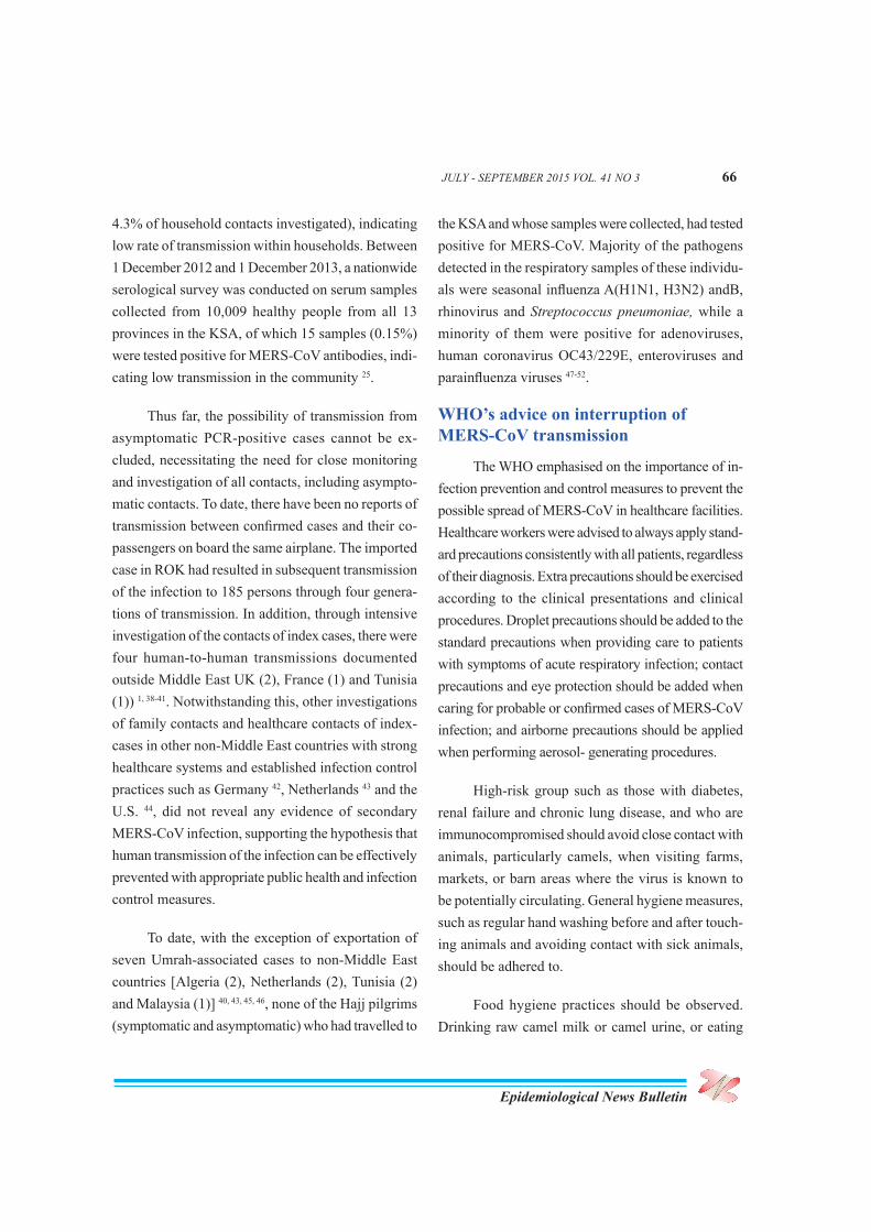
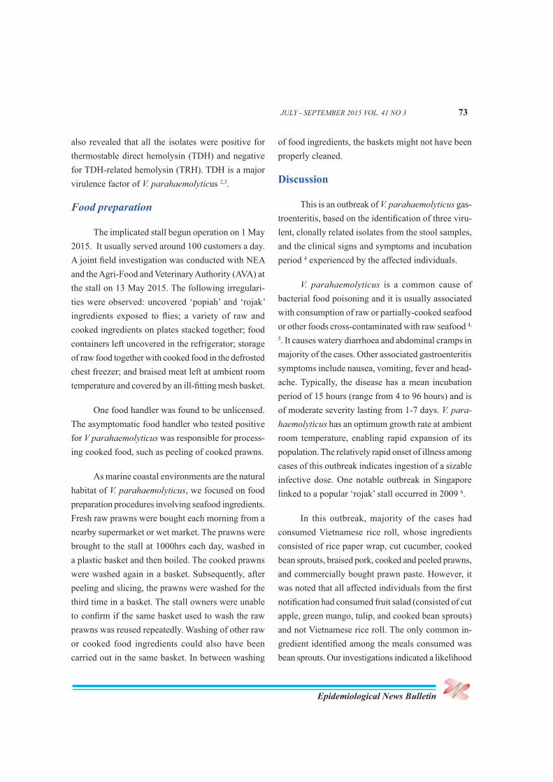
The clinical symptoms reported by the cases were diarrhoea (100.0%), abdominal pain (100.0%), vomiting (40.0%), fever (30.0%), and nausea (10.0%). Three cases (15.0%) were hospitalized, 13 (55.0%) sought outpatient treatment while the remaining four (30.0%) self-medi-cated. The onset of illness was between 11 and 13 May 2015 (Fig. 3). The median incubation period based on the interval between time of consumption and onset of illness was established to be 9.0 hours, while the mean incubation period was 10.9 hours (range 4 to 20 hours).
0
1
2
3
4
5
6
7
8
9
AM PM AM PM AM PM
11-May-15 12-May-15 13-May-15
Num
ber
of c
ases
Date and time of onset
Two main time of consumption around the early afternoon of 11 and 12 May 2015
*Onset of illness of 2 reported cases unknown
Figure 3Onset of illness of 20 reported cases* of food poisoning following consumption of food in a Vietnamese
stall, 11 May -12 May 2015
JULY - SEPTEMBER 2015 VOL. 41 NO. 3 72
Epidemiological News Bulletin
12 of the cases (60.0%) reported consumption of Vietnamese rice roll, while five (25.0%) consumed a dish other than the rice roll. The remaining three cases (15.0%) could not recall the exact food item consumed.
Microbiological findings
V. parahaemolyticus was isolated from the stool samples of two of the three hospitalized cases. One of the four food handlers referred for stool screening was tested positive for V. parahaemolyticus. The remain-ing three food handlers tested negative for bacterial foodborne pathogens, norovirus and rotavirus. No V. parahaemolyticus was detected in the food samples
taken from the stall. These samples were found to be bacteriologically satisfactory.
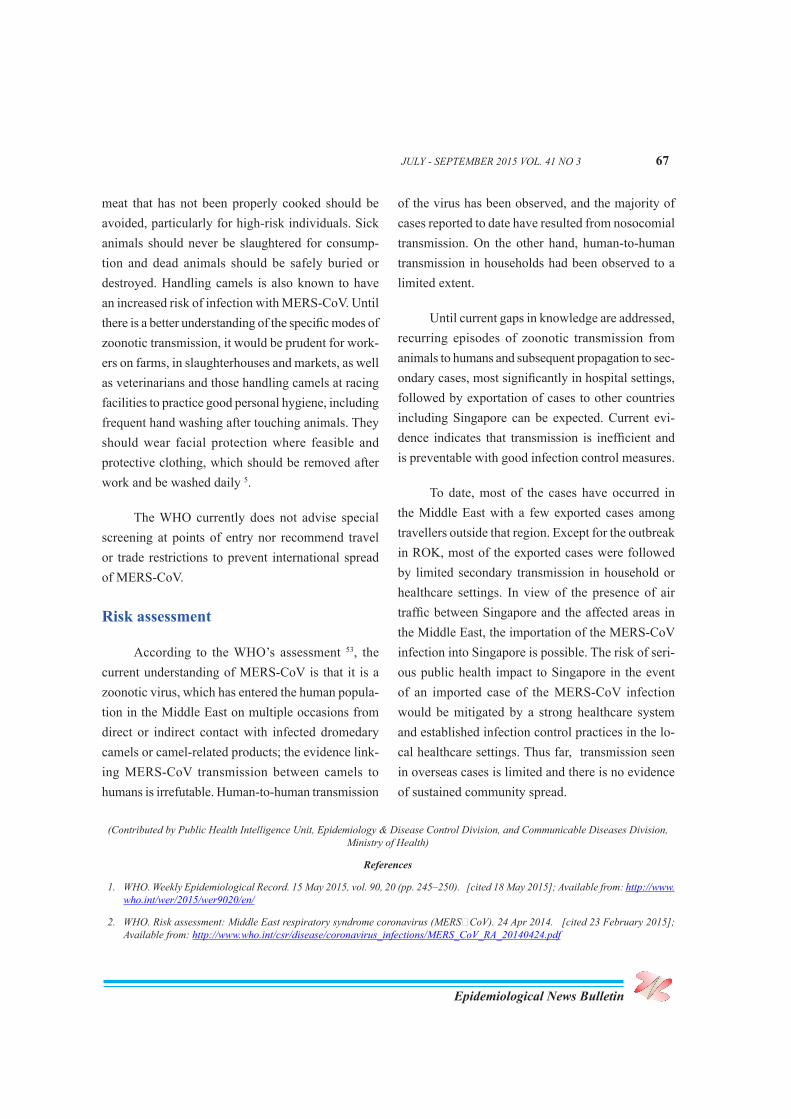
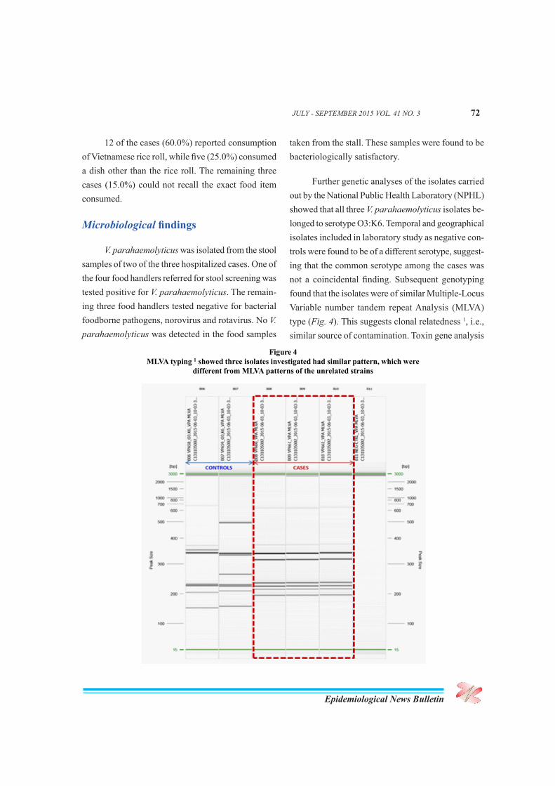
Further genetic analyses of the isolates carried out by the National Public Health Laboratory (NPHL) showed that all three V. parahaemolyticus isolates be-longed to serotype O3:K6. Temporal and geographical isolates included in laboratory study as negative con-trols were found to be of a different serotype, suggest-ing that the common serotype among the cases was not a coincidental finding. Subsequent genotyping found that the isolates were of similar Multiple-Locus Variable number tandem repeat Analysis (MLVA) type (Fig. 4). This suggests clonal relatedness 1, i.e., similar source of contamination. Toxin gene analysis
Figure 4MLVA typing 1 showed three isolates investigated had similar pattern, which were
different from MLVA patterns of the unrelated strains
JULY - SEPTEMBER 2015 VOL. 41 NO 3 73
Epidemiological News Bulletin
also revealed that all the isolates were positive for thermostable direct hemolysin (TDH) and negative for TDH-related hemolysin (TRH). TDH is a major virulence factor of V. parahaemolyticus 2,3.
Food preparation
The implicated stall begun operation on 1 May 2015. It usually served around 100 customers a day. A joint field investigation was conducted with NEA and the Agri-Food and Veterinary Authority (AVA) at the stall on 13 May 2015. The following irregulari-ties were observed: uncovered ‘popiah’ and ‘rojak’ ingredients exposed to flies; a variety of raw and cooked ingredients on plates stacked together; food containers left uncovered in the refrigerator; storage of raw food together with cooked food in the defrosted chest freezer; and braised meat left at ambient room temperature and covered by an ill-fitting mesh basket.
One food handler was found to be unlicensed. The asymptomatic food handler who tested positive for V parahaemolyticus was responsible for process-ing cooked food, such as peeling of cooked prawns.
As marine coastal environments are the natural habitat of V. parahaemolyticus, we focused on food preparation procedures involving seafood ingredients. Fresh raw prawns were bought each morning from a nearby supermarket or wet market. The prawns were brought to the stall at 1000hrs each day, washed in a plastic basket and then boiled. The cooked prawns were washed again in a basket. Subsequently, after peeling and slicing, the prawns were washed for the third time in a basket. The stall owners were unable to confirm if the same basket used to wash the raw prawns was reused repeatedly. Washing of other raw or cooked food ingredients could also have been carried out in the same basket. In between washing
of food ingredients, the baskets might not have been properly cleaned.
Discussion
This is an outbreak of V. parahaemolyticus gas-troenteritis, based on the identification of three viru-lent, clonally related isolates from the stool samples, and the clinical signs and symptoms and incubation period 4 experienced by the affected individuals.
V. parahaemolyticus is a common cause of bacterial food poisoning and it is usually associated with consumption of raw or partially-cooked seafood or other foods cross-contaminated with raw seafood 4,
5. It causes watery diarrhoea and abdominal cramps in majority of the cases. Other associated gastroenteritis symptoms include nausea, vomiting, fever and head-ache. Typically, the disease has a mean incubation period of 15 hours (range from 4 to 96 hours) and is of moderate severity lasting from 1-7 days. V. para-haemolyticus has an optimum growth rate at ambient room temperature, enabling rapid expansion of its population. The relatively rapid onset of illness among cases of this outbreak indicates ingestion of a sizable infective dose. One notable outbreak in Singapore linked to a popular ‘rojak’ stall occurred in 2009 6.
In this outbreak, majority of the cases had consumed Vietnamese rice roll, whose ingredients consisted of rice paper wrap, cut cucumber, cooked bean sprouts, braised pork, cooked and peeled prawns, and commercially bought prawn paste. However, it was noted that all affected individuals from the first notification had consumed fruit salad (consisted of cut apple, green mango, tulip, and cooked bean sprouts) and not Vietnamese rice roll. The only common in-gredient identified among the meals consumed was bean sprouts. Our investigations indicated a likelihood
JULY - SEPTEMBER 2015 VOL. 41 NO. 3 74
Epidemiological News Bulletin
of cross-contamination between ready-to-serve food items and raw seafood ingredients during preparation.
A few possible sources of cross contamination were identified. First, the washing of prawns was carried out in the same sink as the other food items, and general sanitization of the sink was only done at the end of the day. Second, plastic baskets were not differentiated for raw or cooked food, and they might not be properly cleaned after each use. Third, poor storage of food, such as improper stacking in the refrigerator and freezer, with food exposed at ambient room temperature. The cooked bean sprouts could have been cross-contaminated when placed in the same baskets for washing raw prawns, both before and after blanching. The cooked bean sprouts were also left exposed at ambient room temperature, which facilitated the growth of V. parahaemolyticus in contaminated cooked bean sprouts.
Transmission was interrupted following voluntary cessation of sales of the Vietnamese rice roll. Although V. parahaemolyticus isolated from the foodhandler was clonally related to the strains isolated from two of the reported cases, she may not be responsible for the transmission of infection. Person-to-person transmission is uncommon as the
infective dose of V. parahaemolyticus is high 7. It was highly probable that she could have acquired the infection from the implicated stall where she had her daily lunch.
Food establishments should constantly remind their food handlers to take precautions against cross contamination, even when processed frozen seafood products are used. It may be prudent that food establish-ments use frozen prawns instead of fresh prawns, as V. parahaemolyticus does not survive well with freezing 8. A study by the National University of Singapore found many other seafood products, although processed, were contaminated with V. parahaemolyticus 9. This highlights the need for constant vigilance by all food handlers against the compromise of food safety.
Although we were not able to determine the full extent of this outbreak, this investigation underscores the importance of disease reporting by health care providers. Primary care physicians on the ground are the ‘eyes and ears’ of the health authority, helping to safeguard public health by reporting unusual trends and possible common exposures. Credit should be given to the primary care physician for the first noti-fication as it had enabled rapid action in uncovering and controlling this outbreak.
(Contributed by Lee JJ1, Cui L2, Han HK1, Toh HY1, Raj P1, Koh HF1, Lai YQ1, Badaruddin H1 and Ooi PL1, Communicable Diseases Division, Ministry of Health1, National Public Health Laboratory2)
References
1. Kimura B, Sekine Y, Takahashi H, et al. Multiple-locus variable-number of tandem-repeats analysis distinguishes Vibrio parahaemolyticus pandemic O3: K6 strains. Journal Micro Meth, 2008. 72: 313-20.
2. Nishibuchi M, Kaper, JB. Thermostable direct hemolysin gene of Vibrio parahaemolyticus: a virulence gene acquired by a marine bacterium. Infection Immunity, 1995. 63: 2093.
3. Nishibuchi M, Fasano A, Russell RG, et al. Enterotoxigenicity of Vibrio parahaemolyticus with and without genes encoding thermostable direct hemolysin. Infection Immunity, 1992. 60: 3539-3545.
JULY - SEPTEMBER 2015 VOL. 41 NO 3 75
Epidemiological News Bulletin
4. Heymann D. Cholera and other vibrioses. Control of communicable diseases manual, 2008: p. 120.
5. Daniels NA, MacKinnon L, Bishop R, et al., Vibrio parahaemolyticus infections in the United States, 1973–1998. Journal Infect Dis 2000181: 1661-6.
6. Suhana S, Tang ZC, Mak TM, et al. An outbreak of Vibrio parahaemolyticus food poisoning. Epidemiol News Bulletin, 2009. 35: 48
7. Colville JL, Berryhill DL. Vibriosis in Handbook of Zoonoses J.L.C.L. Berryhill( Editor). 2007, Mosby: Saint Louis. p. 211-4.
8. Boonyawantang A, Mahakarnchanakul W, Rachtanapun C, et al., Behavior of pathogenic Vibrio parahaemolyticus in prawn in response to temperature in laboratory and factory. Food Control 2012; 26: 479-85.
9. Huang Y, Ghate V, Phua L, et al., Prevalence of Salmonella and Vibrio spp. in seafood products sold in Singapore. J Food Protect 2012; 75: 1320-3.
Introduction
The most common type of acute infections occurs in the respiratory tract, and the impact on healthcare utilisation at primary and tertiary care level due to acute respiratory infections is considerable.1
In Singapore, upper respiratory tract infections (URTI) has been the top medical condition seen at polyclinics and general practitioner (GP) clinics based on the Primary Care Survey (PCS) conducted by the Ministry of Health (MOH) in 2005 and 2010, and constituted 25% of all diagnoses in both years.2 Seasonal influenza was associated with 8.3 deaths per 100,000 population based on a modelling study which used influenza virological data from 2004 to 2006.3 The overall influenza-associated hospitalisa-tion rate per 100,000 person-years for pneumonia and influenza was 28.3 during 2004–2008 and 29.6 during 2010–2012.4
The objective of this study is to quantify the disease burden of acute respiratory infections (ARI) in the Singapore resident population in 2010 using the disability-adjusted life year (DALY) methodology.
Methods
The DALY model was endorsed by the World Health Organization (WHO) in 1996 as a methodol-ogy to prioritise interventions in the healthcare sector based on their potential to reduce burden of disease.5 In order to guide decisions on health policies and resource allocation, the Singapore Burden of Disease (SBoD) study was conducted once every three years in 20046, 2007 and 20107, which aimed to provide a comprehensive assessment of the health status of Singapore resident population.
DALYs is a measure of the associated burden caused by a disease outcome, and it is calculated as a
Disease burden of acute respiratory infections in Singapore, 2010
JULY - SEPTEMBER 2015 VOL. 41 NO. 3 76
Epidemiological News Bulletin
combination of years of life lost (YLL) due to prema-ture mortality and equivalent years of “healthy” life lost due to ill health or disability (YLD). These indi-cators provide a measure of the gap between current health status and an ideal situation in which everyone lives into old age without any disease or ill health.
ARI in the SBoD study comprises lower respira-tory tract infections (LRTI), URTI and otitis media.7 Estimates were generated using the incidence-based DALY approach based on episodes of LRTI (influenza, acute bronchitis and pneumonia) and URTI (acute nasopharyngitis, acute sinusitis and pharyngitis/tonsil-litis) at polyclinics and GP clinics from the PCS 2010. In view of apparent inconsistencies in diagnosis coding between polyclinics and GP clinics, we redistributed the total attendances for LRTI and URTI recorded in the survey (other than influenza and acute nasopharyn-gitis) to the specific respiratory conditions based on the distribution seen at GP clinics, since this was deemed more plausible than the distribution at polyclinics. We assumed that about 1% of those reported under other URTI were influenza and the remaining was attributed to acute nasopharyngitis. For pneumonia, further adjustment was done to take into account at-tendances seen at the emergency department of public acute hospitals in 2010.
For LRTI and URTI, we used the same assump-tions for disability and durations as in the 2003 Aus-tralian study on burden of disease and injury (ABD)8. Global burden disease (GBD) duration estimates were halved to 3.5 days for acute bronchitis, and remained at one week for influenza and two weeks for pneumo-nia. For acute nasopharyngitis, we applied an average duration of 1.5 days, and for tonsillitis/ pharyngitis and sinusitis, we used the GBD duration of 3.5 days. We applied the Australian derived weights from dis-
ability weight regression models for these conditions: influenza 0.047; acute bronchitis 0.132; pneumonia 0.373; acute nasopharyngitis 0.014; tonsillitis and pharyngitis and sinusitis 0.061.
For otitis media, we modelled the following stages: acute infection, bilateral chronic infection, and life-long deafness. We estimated the overall incidence rate of acute episodes for each gender using data from the PCS 2010 but adopted the age specific incidence pattern observed in the 2003 ABD, as local experts had commented that the age distribution from the survey did not appear plausible.
As we assumed that those with relatively low disability did not seek treatment, treated numbers were used for estimation of YLD. We assumed duration of one week and applied the Australian derived weight from disability weight regression model. We estimated the incidence of bilateral chronic infection using the ratio of acute infections to chronic infection episodes from the PCS 2010. We assumed an average duration of one year and used the Dutch weight for early acquired mild to moderate hearing loss of 0.11. Permanent deaf-ness as a result of otitis media is very rare. Infections resulting in deafness were based on GBD assumptions for established market economies; it was assumed that 5 in 100,000 episodes of acute otitis media among children aged 0-14 years resulted in lifelong deafness. We derived the duration using the Disease Modelling (DisMod) II software package 9, and used the Dutch weight of 0.233 for early acquired severe hearing loss.
Results
In 2010, the total burden of disease and injury resulting from premature mortality and disability was estimated at 399,675 DALYs (106 DALYs lost per
JULY - SEPTEMBER 2015 VOL. 41 NO 3 77
Epidemiological News Bulletin
1,000 resident population). ARI constituted 2.8% of the total DALYs, and 58.0% of DALYs among com-municable diseases burden (including infectious and parasitic diseases) (Table 1).
In term of specific causes, LRTI (primarily pneumonia) was the greatest contributor of disease burden among all communicable diseases. It was ranked as the eighth and fourth leading causes of the
total DALYs (2.8%) and YLL (5.9%), respectively. Among YLL of communicable diseases, LRTI con-stituted the greatest share at 74.7%.
Overall, 91.0% of the disease burden from ARI was due to premature deaths (91.0%) rather than ill-health (9.0%). The proportion of YLL increased with older age (Fig. 5). In persons below 45 years of age, the disease burden was approximately equally
Table 1Disease burden due to ARI (DALYs, YLL and YLD) in 2010
Specific cause DALYs % of total DALYs YLL % of total
Figure 5Distribution (%) of YLL and YLD due to ARI by age group in 2010
0%
10%
20%
30%
40%
50%
60%
70%
80%
90%
100%
<15 15-24 25-44 45-64 65-74 75+Age group (years)
YLL
JULY - SEPTEMBER 2015 VOL. 41 NO. 3 78
Epidemiological News Bulletin
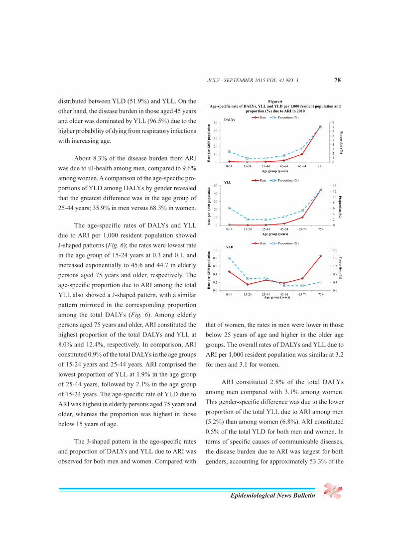
distributed between YLD (51.9%) and YLL. On the other hand, the disease burden in those aged 45 years and older was dominated by YLL (96.5%) due to the higher probability of dying from respiratory infections with increasing age.
About 8.3% of the disease burden from ARI was due to ill-health among men, compared to 9.6% among women. A comparison of the age-specific pro-portions of YLD among DALYs by gender revealed that the greatest difference was in the age group of 25-44 years; 35.9% in men versus 68.3% in women.
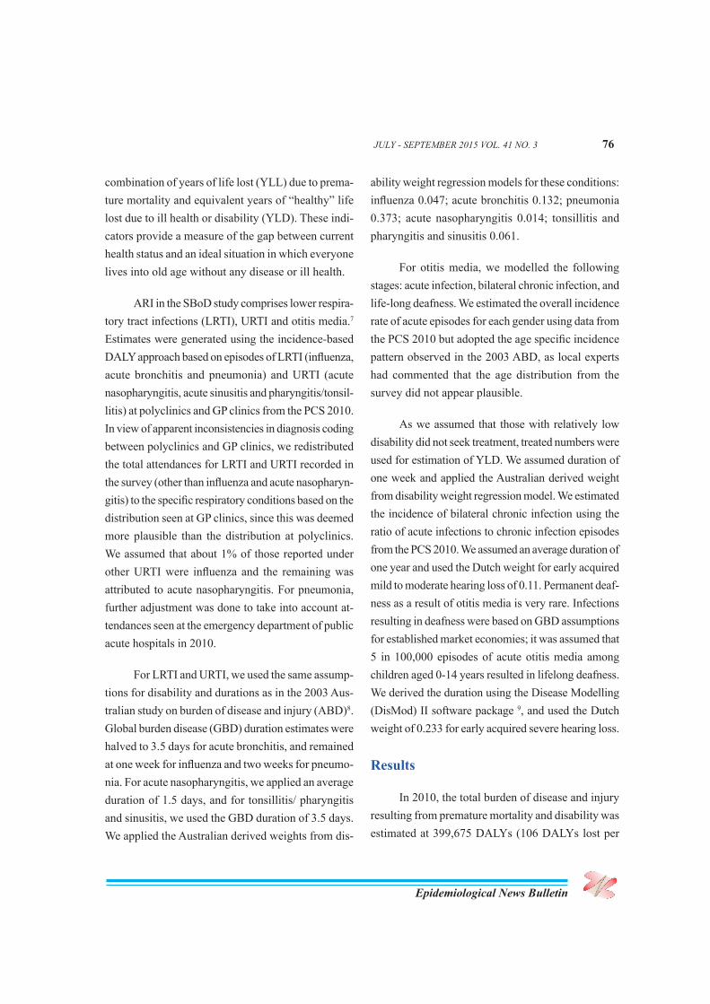
The age-specific rates of DALYs and YLL due to ARI per 1,000 resident population showed J-shaped patterns (Fig. 6); the rates were lowest rate in the age group of 15-24 years at 0.3 and 0.1, and increased exponentially to 45.6 and 44.7 in elderly persons aged 75 years and older, respectively. The age-specific proportion due to ARI among the total YLL also showed a J-shaped pattern, with a similar pattern mirrored in the corresponding proportion among the total DALYs (Fig. 6). Among elderly persons aged 75 years and older, ARI constituted the highest proportion of the total DALYs and YLL at 8.0% and 12.4%, respectively. In comparison, ARI constituted 0.9% of the total DALYs in the age groups of 15-24 years and 25-44 years. ARI comprised the lowest proportion of YLL at 1.9% in the age group of 25-44 years, followed by 2.1% in the age group of 15-24 years. The age-specific rate of YLD due to ARI was highest in elderly persons aged 75 years and older, whereas the proportion was highest in those below 15 years of age.
The J-shaped pattern in the age-specific rates and proportion of DALYs and YLL due to ARI was observed for both men and women. Compared with
0123456789
0
10
20
30
40
50
0-14 15-24 25-44 45-64 65-74 75+
Proportion (%)
Rat
e pe
r 1,
000
popu
latio
n
Age group (years)
Rate Proportion (%)
0
2
4
6
8
10
12
14
0
10
20
30
40
50
0-14 15-24 25-44 45-64 65-74 75+
Proportion (%)
Rat
e pe
r 1,
000
popu
latio
n
Age group (years)
Rate Proportion (%)
0.0
0.4
0.8
1.2
1.6
2.0
0.0
0.2
0.4
0.6
0.8
1.0
0-14 15-24 25-44 45-64 65-74 75+
Proportion (%)
Rat
e pe
r 1,
000
popu
latio
n
Age group (years)
Rate Proportion (%)
DALYs
YLL
YLD
Figure 6Age-specific rate of DALYs, YLL and YLD per 1,000 resident population and
proportion (%) due to ARI in 2010
that of women, the rates in men were lower in those below 25 years of age and higher in the older age groups. The overall rates of DALYs and YLL due to ARI per 1,000 resident population was similar at 3.2 for men and 3.1 for women.
ARI constituted 2.8% of the total DALYs among men compared with 3.1% among women. This gender-specific difference was due to the lower proportion of the total YLL due to ARI among men (5.2%) than among women (6.8%). ARI constituted 0.5% of the total YLD for both men and women. In terms of specific causes of communicable diseases, the disease burden due to ARI was largest for both genders, accounting for approximately 53.3% of the
JULY - SEPTEMBER 2015 VOL. 41 NO. 3 79
Epidemiological News Bulletin
DALYs (9,999 DALYs) among men and 63.6% of DALYs among women (5,945 DALYs).
Comments
The J-shaped pattern observed for age-specific rates of DALYs and YLL due to ARI was also seen in influenza-associated hospitalisations for pneumonia and influenza.4 This reflects the considerable disease burden of ARI among vulnerable groups of elderly persons and young children in Singapore.
The average daily number of polyclinic at-tendances and weekly number of emergency depart-ment attendances due to ARI tend to increase in the beginning and just before the middle of the year.1 These increases in attendances for ARI are usually accompanied by a corresponding rise in influenza positivity rate among outpatients with influenza-like illness in the community.10 Bimodal peaks of influenza activity are typically observed in the beginning and middle of the year based on MOH’s influenza virus surveillance programme.11
Our findings underscore the importance of continuous surveillance for ARI, and provide support-ing evidence to guide public health policy priorities. In Singapore, vaccination against influenza is a key strategy for preventing or reducing respiratory virus infections. The MOH’s Expert Committee on Im-munisation has recommended the use of influenza vaccine to protect vulnerable populations at higher risk for influenza-related complications, including persons aged 65 years and older, adults and children with chronic medical conditions, pregnant women, and children 6 months to below 5 years of age.
The Health Promotion Board regularly rolls out educational campaigns to educate the public on the importance of influenza vaccinations, such as “Know how to fight flu”12 which included the recommenda-tion of vaccination as one of the five ways to protect against influenza infection. Educational pamphlets (i.e. “Fight influenza”) are distributed in polyclinics and other healthcare institutions. These measures would raise awareness among the public and help to increase uptake of influenza vaccine in Singapore.
(Reported by Ang LW, Ma S and James L, Epidemiology & Disease Control Division, Ministry of Health, Singapore)
References
1. Ang LW, Ma S, Cutter JL et al. Impact of acute respiratory infections on healthcare utilisation in Singapore, 2007 – 2011. Epidemiol
The Epidemiological News Bulletin is published quarterly by the Ministry of Health, Singapore
EDITORIAL BOARD
EditorA/Prof Jeffery Cutter
MembersA/Prof Stefan Ma Dr Ooi Peng Lim Dr Joanne Tay
SCIENTIFIC ADVISORY COMMITTEEA/Prof Vincent Chow, Department of Microbiology, National University of SingaporeProf Richard James Coker, Saw Swee Hock School of Public Health, National University of SingaporeProf Leo Yee Sin, Director, Institute of Infectious Diseases and Epidemiology and Clini-cal Director, Communicable Disease Centre, Tan Tock Seng Hospital A/Prof Ng Lee Ching, Director, Environmental Health Institute, National Environment AgencyDr Leong Hon Keong, Director, Risk Assessment and Epidemiology Department, Agri-Food and Veterinary Authority of SingaporeA/Prof Raymond Lin, Head, National Public Health Laboratory, Ministry of Health
EDITORIAL STAFF
Ms Ang Li Wei Mr Chng Meng Hong Mr Han Hwi Kwang Ms Toh Hai Yin Mr Yuske Kita
5. Murray CJL, Lopez AD (eds). Global Burden of Disease: A comprehensive assessment of mortality and disability from diseases,
injuries, and risk factors in 1990 and projected to 2020 (Global Burden of Disease and Injury Series). Boston: Harvard School of
Public Health, 1996.
6. Ministry of Health, Singapore. Singapore Burden of Disease Study 2004. Singapore: Ministry of Health, 2009.
7. Ministry of Health, Singapore. Singapore Burden of Disease Study 2010. Singapore: Ministry of Health, 2014.
8. Begg S, Vos T, Barker B et al. The burden of disease and injury in Australia 2003. Canberra: Australian Institute of Health and
Welfare, 2007.
9. Barendregt JJ, Van Oortmarssen GJ, Vos T et al. A generic model for the assessment of disease epidemiology: the computational