Epidemiology and genetic diversity ofbovine leukemia virusMeripet Polat1,2, Shin-nosuke Takeshima1,2,3 and Yoko Aida1,2,3*
Abstract
Bovine leukemia virus (BLV), an oncogenic member of the Deltaretrovirus genus, is closely related to human T-cellleukemia virus (HTLV-I and II). BLV infects cattle worldwide and causes important economic losses. In this review,we provide a summary of available information about commonly used diagnostic approaches for the detectionof BLV infection, including both serological and viral genome-based methods. We also outline genotyping methods usedfor the phylogenetic analysis of BLV, including PCR restriction length polymorphism and modern DNA sequencing-basedmethods. In addition, detailed epidemiological information on the prevalence of BLV in cattle worldwide is presented.Finally, we summarize the various BLV genotypes identified by the phylogenetic analyses of the whole genome and envgp51 sequences of BLV strains in different countries and discuss the distribution of BLV genotypes worldwide.
BackgroundBovine leukemia virus (BLV) is a retrovirus, anoncogenic member of the Deltaretrovirus genus, and thecausative agent of enzootic bovine leukosis (EBL) [1, 2].The Deltaretrovirus genus also includes human T-celllymphotropic virus types I and II (HTLV-I and -II) andsimian T-cell lymphotropic virus (STLV) [3, 4]. EBL is acontagious lymphoproliferative disease of cattle, charac-terized by B-cell lymphosarcoma, which occurs through-out the world [2, 5]. Although BLV can infect variousimmune cell populations, including CD5+ IgM+ andCD5− IgM+ B-cells; CD2+, CD3+, CD4+, CD8+, and γ/δT-cells; monocytes; and granulocytes in peripheralblood and lymphoid tissues of cattle [6–11], BLV-induced tumors usually arise from the CD5+ IgM+ B-cell subpopulation [12].BLV infection can result in a variety of clinical out-
comes [2]. The majority of BLV-infected cattle areasymptomatic carriers of the virus, neither showing anyclinical signs nor any changes in lymphocyte count;however, a recent study showed that although lympho-cyte counts were not elevated in BLV-infected but
clinically normal cattle, CD5+ IgM+ B-cells wereincreased [11], and there is substantial evidence suggest-ing that BLV-infected but clinically normal cattle mayexhibit a degree of immunological dysregulation leadingto economic losses for various reasons includingreduced milk production [13], a high incidence of infec-tious disease [14], and reproductive inefficiency [15]. Ap-proximately one-third of infected cattle develop a benignform of non-malignant proliferation of untransformedB-lymphocytes, termed persistent lymphocytosis (PL).PL is typically characterized by a permanent and stableincrease in the number of CD5+ IgM+ B-cells circulatingin the peripheral blood. Less than 5% of infected cattledevelop malignant B-cell lymphoma originating frommono- or oligo-clonal accumulation of CD5+ IgM+ B-cells after a relatively long period of latency. This malig-nant form of B-cell lymphoma is predominantly detectedin cattle over 4–5 years old [16]. Such malignanciesinduce disruption of the spleen and remarkable enlarge-ment of the lymph nodes, which can be visible underthe skin. BLV-induced neoplastic cells can penetrate intothe abomasums, right auricle of the heart, intestine, kid-ney, lung, liver, and uterus. The clinical signs of BLV-induced tumors are varied and primarily involve digestivedisturbance, weight loss, weakness, reduced milk produc-tion, loss of appetite, and enlarged lymph nodes [17].
* Correspondence: [email protected] Infectious Diseases Unit, RIKEN, 2-1 Hirosawa, Wako, Saitama 351-0198,Japan2Nano Medical Engineering Laboratory, RIKEN, 2-1 Hirosawa, Wako, Saitama351-0198, JapanFull list of author information is available at the end of the article
BLV genome structureThe BLV genome consists of 8714 nucleotides (nt) [18]including essential structural protein and enzyme codinggenes and a pX region, flanked by two identical long ter-minal repeats (LTRs) (Fig. 1a). The structural proteinand enzyme coding genes, namely, gag, pro, pol, and env,have essential and indispensable roles in the viral life-cycle, viral infectivity, and the production of infectiousvirions [19–24]. The gag gene of BLV is translated as theprecursor, Pr45 Gag, and processed to generate threemature proteins [19, 23]: the matrix protein, p15, whichbinds viral genomic RNA and interacts with the lipid bi-layer of the viral membrane [25]; the capsid protein,p24, which is the major target of the host immuneresponse, with high antibody titers against this moleculefound in the serum of infected animals [26, 27]; and thenucleocapsid protein, p12, which binds to packaged gen-omic RNA [28] (Fig. 1b). The env gene encodes the ma-ture extracellular protein, gp51, and a transmembraneprotein, gp30 [19]. The pX region, which is located be-tween env and the 3′ LTR [2], encodes the regulatoryproteins Tax and Rex, and the accessory proteins R3 andG4 (Fig. 1a). The regulatory proteins are important forregulation of viral transcription, transformation of BLV-induced leukemogenesis, and nuclear export of viralRNA into the cytoplasm [29–36]. The R3 and G4accessory proteins contribute to the maintenance of highviral loads [37, 38]. In addition to the genes describedabove, the BLV genome also contains RNA polymerase-III-encoded viral microRNAs (miRNAs) between the envand pX regions. Viral miRNAs are strongly expressed in
preleukemic and malignant cells, and may have roles intumor onset and progression [39, 40] through theireffects on proviral load and consequently viral replica-tion in the natural host [41]. Besides, Van Driessche etal. revealed the recruitment of positive epigenetic markson BLV miRNA cluster, inducing strong antisense pro-moter activity [42]. They also identified cis-actingelements of an RNAPII-dependent promoter [42].
BLV diagnosisA variety of techniques have been developed for diagno-sis of BLV and implemented worldwide. These diagnosticmethods can be assigned into two main groups, consist-ing of antibody-based serological tests and detection ofthe proviral genome by nucleic acid-based polymerasechain reaction (PCR) assays (summarized in Table 1).
Serological testsFor indirect BLV diagnostic methods, particularlyantibody-based tests, antibodies recognizing the p24 capsidprotein encoded by the gag gene and the extracellular gp51protein encoded by env-gp51 are targeted. This is becauseantibodies against these proteins are produced shortly afterBLV infection, can be detected 2–3 weeks post-infection,and remain detectable for the life of the host animal [43].In addition, the p24 capsid protein is a major target forhost immune responses, inducing high antibody titers [44],and gp51 invokes the expression of massive amounts ofspecific antibodies in infected animals [24, 45, 46]. There-fore, antibodies against these proteins are targeted for BLVdiagnostics using conventional serological techniques such
Fig. 1 Schematic representations of the BLV genome structure (a) and viral particle (b). The structural and enzymatic genes, gag, pro, pol, and env;regulatory genes, tax and rex; accessory genes R3 and G4; and microRNA (miRNA) are indicated in (a). Proteins encoded by structural and enzymaticgenes, including the Env glycoproteins (gp51 and gp30) encoded by the env gene, the Gag proteins (p12, p24, and p15) encoded by the gag gene,reverse transcriptase and integrase (RT-IN) encoded by the pol gene, and protease (Pro) encoded by the pro gene are indicated in (b)
Polat et al. Virology Journal (2017) 14:209 Page 2 of 16
Table 1 Summary of common techniques used for diagnosis of BLV prevalence
Specific, simple, and easy to performLarge scale screening Less expensiveRapid
Less sensitive and inconclusiveCannot evaluate disease statesof infected cattle
Aida et al., 1989[47]
Wang et al., 1991[48]
Montiet al., 2005 [49]
Kurdi et al., 1999[50]
Jimba et al.,2012 [43]
Naif et al., 1990[55]
ELISA Serum MilkBulk milk
Antibodies(p24, gp51)
Specific and sensitive Large scalescreening Time saving
False negatives (cattle in earlyinfection phase) False positive(maternally derived antibodies)Cannot evaluate disease states ofinfected cattle A number ofcontrols and a plate reader requiredResults require interpretation
Naif et al., 1990[55]
Burridge et al.,1982 [56]
Schoepf et al.,1997 [53]
Kurdi et al., 1999[50]
Monti et al.,2005 [49]
Jimba et al.,2012 [43]
Zaghawa et al.,2002 [52]
PHA Virus particle BLVglycoprotein
Sensitive Specific detection of BLVLarge scale titration Less expensiveRapid
Affected by pH and temperatureHemagglutination activity reducedby trypsin, potassium periodate,and neuraminidase
Fukai et al., 1999[51]
RIA Serum Antibodies(p24)
Sensitive Able to detect BLV duringthe early period of infection
Cannot be used for mass screening Levy et al., 1977[54]
Provirus Direct, fast, sensitive A variety ofsamples can be used BLV detectionduring the early phase of infectionor in the presence of colostrumantibodiesCan detect new infections, beforethe development of antibodies toBLV
Unable to detect BLV when theproviral load is too lowCross contamination occurs easilyRequires specific primers Requiresequipment (PCR machine) Falsenegatives in the presence of PCRinhibitory substances in samplesRequires internal control Needsconfirmatory testing, such assequencing
Provirus Direct, fast, sensitive Low risk ofcontamination A variety of samplescan be used Distinguishes EBL fromSBL BLV can be detected during theearly phase of infection or in thepresence of colostrum antibodiesQuantitative measurement ofproviral load
Requires internal control Requirespositive controls of differentconcentrations Requires specificprimers and probes Requireequipment (real-time PCR machine)ExpensiveComplicated sample preparationprocedure
Somura et al.,2014 [68]
Lew et al., 2004[69]
Jimba et al.,2010 [70]
Jimba et al.,2012 [43]
Polat et al. Virology Journal (2017) 14:209 Page 3 of 16
as agar gel immunodiffusion (AGID) [43, 47–50], passivehemagglutination assay (PHA) [43, 51], enzyme-linkedimmunosorbent assay (ELISA) [43, 49, 50, 52, 53], andradio immunoassay (RIA) [54]. Most of these serologicalmethods aim to detect antibodies in bovine serum andmilk, and the supernatants of BLV-infected cell cultures.AGID is relatively inexpensive and can be used to screenmany serum samples simultaneously; however, it is notsufficiently sensitive [55] and it is not suitable for analysisof milk samples. ELISA is a highly sensitive and easilyimplemented procedure, and can be used to analyze bothserum and milk samples; however, it requires a number ofcontrols and produces both false-negative result in serumsamples from cattle in the early phase of infection [55] andfalse-positive results in calves that contain maternally-derived antibodies [56]. PHA aims to detect BLV glycopro-teins, but, PHA test efficiency is sensitive to pH,temperature, and trypsin. RIA is suitable for diagnosingBLV soon after animals are exposed, but not suitable forthe purpose of mass screening [57]. Overall, theseantibody-based detection methods cannot be used to testcalves less than 6 months old, due to the presence ofmaternal antibodies, which may trigger false-positiveresults [58].
Proviral DNA detectionBLV can integrate into dispersed sites within the hostgenome [59] and appears to be transcriptionally silent invivo [60–62] and remain in cellular genomes, even inthe absence of detectable BLV antibodies. Indeed,transcription of the BLV genome in fresh tumor or per-ipheral blood mononuclear cell samples from infectedindividuals is almost undetectable by conventional tech-niques [60, 63]. Interestingly, one copy of the full-lengthproviral genome can be detected in BLV-infected cattlethroughout the course of the disease [64]. Another studyalso demonstrated that BLV-induced tumors and BLV-
infected cells contain provirus, with approximately fourcopies of proviral DNA in each tumor [65]. Hence, inaddition to the routine diagnosis of BLV infection usingthe conventional serological techniques described above,nucleic acid-based PCR methods can greatly acceleratethe detection of BLV prevalence.A variety of PCR methods, including standard PCR
[49, 50], nested PCR [33, 52, 64], real-time quantitativePCR (qPCR) [43, 66–71], and direct blood-based PCR[72, 73], have been extensively applied worldwide forBLV detection (Table 1). A variety of genes in the BLVgenome are targeted for detection of BLV infectionprevalence by direct diagnostic PCR methods, includingthe LTR region [43, 70, 71, 73–77], and the gag [78], pol[69, 79, 80], env [55, 79], and tax [68, 79] genes.Importantly, the BLV provirus copy number is gener-
ally very low compared with that of host genes therefore,the majority of PCR systems designed to detect BLVused a nested design [64, 74, 76]. These nested assaysare extremely sensitive, but also obtain false-positiveresults due to DNA contamination. However, themethod requires expensive real-time PCR machines andreagents and involves difficult sample preparation proto-cols. Recently, a novel blood-based PCR system thatamplifies target DNA regions without a requirement forDNA isolation and purification was developed [72, 73].The assay can detect BLV provirus with high specificityand at low cost, facilitating timely identification of BLV-infected cattle.As discussed above, PCR-based genome screening
methods for diagnosis of BLV broaden the range of sam-ples that can be used, increase testing sensitivity, specifi-city, and efficiency, and are less time consuming. PCRalso allows the detection of BLV infection in cattleseveral weeks before it is possible to detect antibodies[81]; however, PCR-based provirus screening involvescomplicated sample preparation processes, which can
Table 1 Summary of common techniques used for diagnosis of BLV prevalence (Continued)
Blood Provirus Cost-effective No need forDNA purification Low riskof contamination
Unable to detect BLV whenthe proviral load is too lowResults in failure if there aremismatches between the PCRprimers and BLV sequencesRelatively low sensitivity
Nishimori et al.,2016 [72]
Takeshima et al.,2016 [73]
AGID agar gel immunodiffusion, BLV bovine leukemia virus, EBL enzootic bovine leukosis, ELISA enzyme-linked immunosorbent assay, PHA passivehemagglutination assay, RIA radio immunoassay
Polat et al. Virology Journal (2017) 14:209 Page 4 of 16
lead to false-positive results if cross contaminationoccurs. In addition, PCR-based BLV detection methodsrequire specific laboratory facilities, including PCRmachines, and the design of specific primers and probesis also necessary. The CoCoMo algorithm, is a methodused to design degenerate primer sets that amplify allavailable sequences within a target region. Recently, theBLV-CoCoMo-qPCR assay was developed to measurethe BLV proviral load with extremely high sensitivityand to amplify both known and novel BLV variants [43,70, 71]. This assay enabled us to demonstrate that theproviral load correlates not only with BLV infectioncapacity but also with BLV disease progression [43, 82],and identification of risk factor associated with increasedBLV proviral load in infected cattle [82, 83] and detec-tion of BLV provirus in nasal secretion and salivasamples [84].
Other methodsIn addition to the techniques described above, otherBLV diagnostic approaches, including detection of viralproteins by western blotting [21, 31, 33, 85], a syncytiumformation assay [85], and detection of BLV antigens byindirect immunofluorescent assay [47], have also beendescribed.
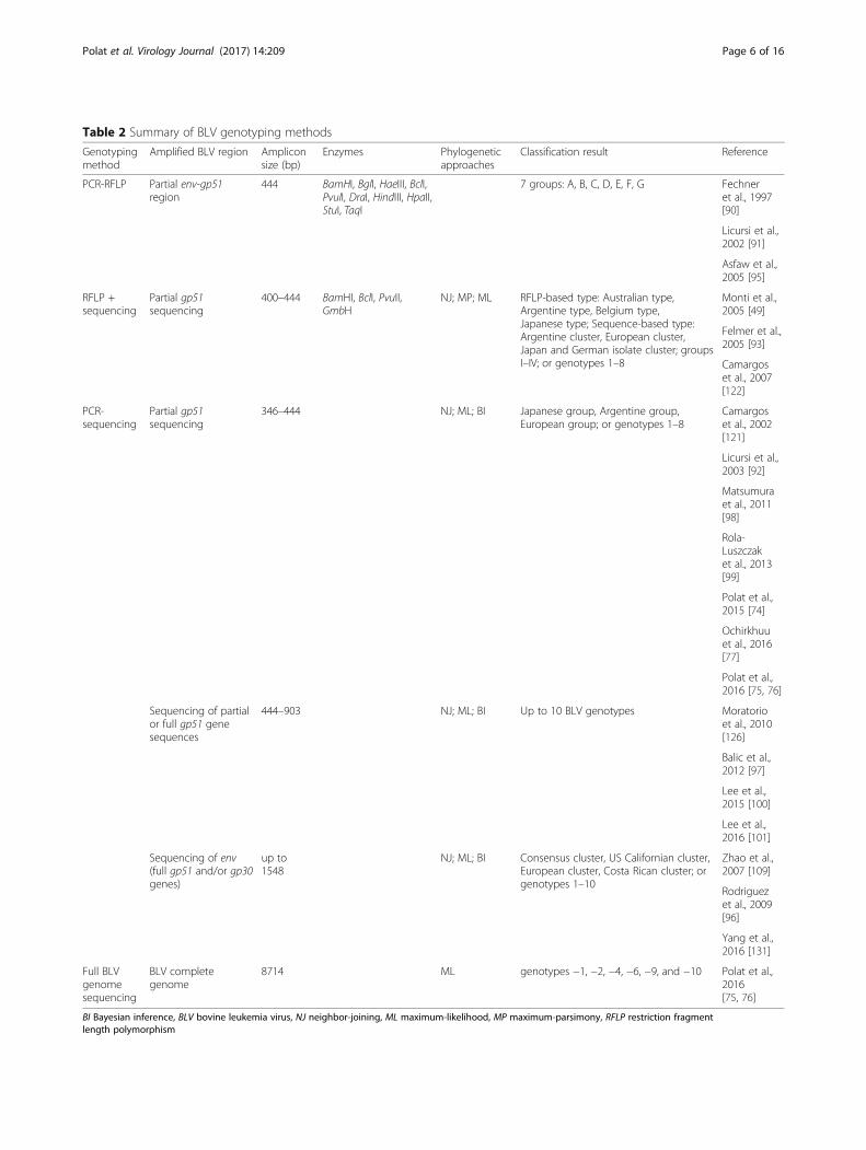
BLV genotyping and identification of ten distinctgenotypesStudies of BLV genotypes for phylogenetic and epi-demiological analyses have primarily focused on the envgene, the env gp51 gene in particular, because of its bio-logical functions. The extracellular gp51 protein has keyroles in the viral lifecycle and is indispensable for viralentry into host cells [20, 86]. In addition, because of thesurface localization of the gp51 glycoprotein, it is alsothe target of neutralizing antibodies [87]. The conform-ational epitopes, F, G, and H, located in the N-terminalhalf of gp51, are important in syncytium formation andviral infectivity [87, 88]. Therefore, the env gp51 se-quence region is frequently used for BLV phylogeneticanalysis.Over the years, a number of methods have been
applied for BLV genotyping, as summarized in Table 2.In the early days of BLV genotyping, researchers clus-tered or genotyped BLV strains from different geograph-ical regions based on restriction fragment lengthpolymorphisms (RFLP) of PCR-products, generatedusing various restriction enzymes [86, 89–96]. BLVclusters and genotypes were named after the geograph-ical region of sample isolation, such as “Argentine type”or “Australian type”, or with reference to phylogeneticclustering (e.g., “cluster one”). A total of seven BLV clus-ters/genotypes were determined by PCR-RFLP [91];
however, PCR-RFLP genotyping studies were notconsistent or comprehensive.In 2007, Rodriguez et al. reported sequencing of the
env gene (all of gp51 and part of gp30) of 28 BLV fieldstrains, performed phylogenetic analysis of thesesequences in comparison with published sequence datarepresentative of established genetic groups by neighbor-joining, maximum likelihood, and Bayesian inferencemethods, and assigned BLV sequences into seven geno-types [97]. Subsequently, a new genotype, genotype-8,was identified in BLV samples from Croatia by Balic etal. [98], who concluded that BLV may be more divergentthan previously thought, speculating that additional ge-notypes might be discovered in the future. Indeed, thepresence of eight BLV genotypes was later confirmed indifferent geographical locations [74, 77, 99–101]. Finally,in 2016, the novel BLV genotypes, genotype-9 and -10,were discovered in Bolivia [75], Thailand [102], andMyanmar [76], a totaling ten BLV genotype clusters(Fig. 2). Previously, almost all phylogenetic studies ofBLV genotypes focused on the partial or entire env gene.However, for the first time in their study [75, 76], Polatet al. successfully concluded the existence of genotypes-1, −2, −4, −6, −9 and −10 among ten BLV genotypes(Fig. 3) by phylogenetic analysis using completesequences of BLV strains newly determined by next gen-eration sequencing and sequencing cloned, overlappingPCR products in their studies, and using complete BLVgenome sequences available in the database (NCBI &DDBJ). These phylogenetic analysis of complete BLVgenomes demonstrated that each BLV genotype encodesspecific amino acid substitutions in both structural andnon-structural gene regions.
BLV prevalenceBLV has spread to all continents via the trade inbreeding animals, and is prevalent in cattle worldwide.BLV infection levels vary between and within coun-tries, as shown in Table 3 (data obtained on March17th, 2017; updated and detailed information is avail-able at http://www.oie.int/wahis_2/public/wahid.php/Diseaseinformation/statuslist) [17, 103]. BLV eradica-tion programs and control measures have been esta-blished in European Community member countriessince the second half of the twentieth century, anderadication programs have been very successful in themajority of western Europe [104–107]; indeed, somecountries, including Denmark, Finland, Switzerland,Estonia, The Netherlands and Poland, are completelyfree of BLV [104, 108–110]. Despite the majority ofcountries in Western Europe being free from disease,EBL still exists in eastern European nations, includingPoland, Ukraine, and Croatia [98, 100, 111–113]. In
Polat et al. Virology Journal (2017) 14:209 Page 5 of 16
7 groups: A, B, C, D, E, F, G Fechneret al., 1997[90]
Licursi et al.,2002 [91]
Asfaw et al.,2005 [95]
RFLP +sequencing
Partial gp51sequencing
400–444 BamHI, BclI, PvuII,GmbH
NJ; MP; ML RFLP-based type: Australian type,Argentine type, Belgium type,Japanese type; Sequence-based type:Argentine cluster, European cluster,Japan and German isolate cluster; groupsI–IV; or genotypes 1–8
Monti et al.,2005 [49]
Felmer et al.,2005 [93]
Camargoset al., 2007[122]
PCR-sequencing
Partial gp51sequencing
346–444 NJ; ML; BI Japanese group, Argentine group,European group; or genotypes 1–8
Camargoset al., 2002[121]
Licursi et al.,2003 [92]
Matsumuraet al., 2011[98]
Rola-Luszczaket al., 2013[99]
Polat et al.,2015 [74]
Ochirkhuuet al., 2016[77]
Polat et al.,2016 [75, 76]
Sequencing of partialor full gp51 genesequences
444–903 NJ; ML; BI Up to 10 BLV genotypes Moratorioet al., 2010[126]
Balic et al.,2012 [97]
Lee et al.,2015 [100]
Lee et al.,2016 [101]
Sequencing of env(full gp51 and/or gp30genes)
up to1548
NJ; ML; BI Consensus cluster, US Californian cluster,European cluster, Costa Rican cluster; orgenotypes 1–10
Zhao et al.,2007 [109]
Rodriguezet al., 2009[96]
Yang et al.,2016 [131]
Full BLVgenomesequencing
BLV completegenome
8714 ML genotypes −1, −2, −4, −6, −9, and −10 Polat et al.,2016[75, 76]
BI Bayesian inference, BLV bovine leukemia virus, NJ neighbor-joining, ML maximum-likelihood, MP maximum-parsimony, RFLP restriction fragmentlength polymorphism
Polat et al. Virology Journal (2017) 14:209 Page 6 of 16
addition, in Italy, Portugal, Belarus, Latvia, Greece,Romania, and Bulgaria, BLV is present, although dis-ease is either absent or limited to specific areas [103].Nationwide BLV eradication and control programs
were introduced in Australia and New Zealand in 1983and 1996, respectively, and 99.7% of Australian dairyherds were declared free from EBL in December 2013,
while those in New Zealand have been free from BLV-induced EBL since 2008 [113, 114].In North America, an epidemiological study of BLV
prevalence in US dairy cattle conducted by the De-partment of Agriculture’s National Animal HealthMonitoring System demonstrated that 83.9% of dairycattle were BLV-positive at herd level and 39% of beef
Fig. 2 Maximum likelihood phylogenetic tree constructed based on partial BLV env sequences identified in geographical locations around the world. Amaximum likelihood (ML) phylogenetic tree was constructed based on sequences from known BLV strains, representing ten different BLV genotypesderived from viruses isolated worldwide. Nucleotide sequences were obtained from the GenBank nucleotide sequence database.Sequences are labeled with their accession numbers and countries of origin. Genotypes are indicated by numbers to the right ofthe figure. One thousand replications were performed to calculate bootstrap values (indicated on the tree). The bar at the bottomof the figure indicates evolutionary distance
Polat et al. Virology Journal (2017) 14:209 Page 7 of 16
herds had at least one BLV-infected animal [115]. InCanada, studies of BLV prevalence revealed that up to37.2% of cows and 89% of herds were BLV-positive[116–118]. BLV is also present in both beef and dairycattle in Mexico [119]; however, disease is either ab-sent or limited to specific areas [17] (accessed on 22Dec 2016).In South America, relatively high levels of BLV preva-
lence have been observed, and BLV-induced leukosis ispresent in the majority of countries. In Brazil, BLVprevalence varies among states, with infection rates ran-ging from 17.1% to 60.8% [120–123]. Individual andherd level BLV prevalence in Argentina are as high as77.4% and 90.9%, respectively [75, 95, 124]. Moreover,individual infection rates between 19.8% and 54.7% havebeen reported in Chile, Bolivia, Peru, Venezuela,Uruguay, Paraguay, and Columbia [75, 94, 125–131].BLV infection is widespread in Chinese dairy farms.
Infection rates are up to 49.1% among individual dairycattle, while 1.6% of beef cattle are BLV-positive [132].Moreover, serological tests revealed that 20.1% of yaks inChina were BLV-positive [133]. Epidemiological studies
in Japan revealed varying levels of BLV prevalencethroughout the country, based on different detectionmethods [83, 134–136], and BLV infection rates of40.9% of dairy and 28.7% of beef cattle, with infectionrates in animals over 2-years-old reaching 78% indairy herds and 69% in beef cattle herds [136]. Lessthan 6% of cattle were infected with BLV in Mongolia(3.9%) [77], Cambodia (5.3%) [137], and Taiwan(5.8%) [48], while a serological survey in Iran revealedthat the prevalence of BLV was between 22.1% and25.4% in that country [138, 139]. Lee et al. [102]demonstrated an average prevalence of BLV of 58.7%in Thailand, reaching maxima of 87.8% and 100% ofcattle when assayed using PCR and ELISA, respect-ively. In Korea, 54.2% of dairy cattle and 86.8% ofdairy herds were BLV-positive, whereas only 0.14% ofbeef cattle were infected with BLV [101]. BLV infec-tion levels in The Philippines ranged from 4.8% to9.7% [74] while it was 9.1% in Myanmar [76]. BLV in-fections in Middle Eastern countries are relativelylow. The prevalence of BLV infection is approximately5% in Israel [140], while in Saudi Arabia, 20.2% of
Fig. 3 Maximum likelihood (ML) phylogenetic tree constructed from complete BLV genomic sequences. The ML phylogenetic tree was constructed usingcomplete BLV genomic sequences from the GenBank nucleotide sequence database. One thousand replications were performed to calculate bootstrapvalues (indicated on the tree). The strains identified in this study are indicated by the sample identification number and country name.Genotypes are indicated by numbers to the right of the figure. The bar at the bottom of the figure indicates evolutionary distance
Polat et al. Virology Journal (2017) 14:209 Page 8 of 16
Table 3 Detailed information on BLV infection levels worldwide
Geographicaldivision
Country Withincountry
BLV prevalencea References
Europe Andorra Nationwide BLV-free, 1994 OIE, 2009 [103]
Poland BLV-free, 2017 EFSA Panel on Animal Health andWelfare, 2017 [110]
Ukraine Present OIE, 2012 [17]; Rola-Luszczak et al.,2013 [100]
Croatia Present OIE, 2012 [17]; Balik et al., 2012
Italy Present OIE, 2009 [103]; Molteni et al., 1996[144]
Portugal Present OIE, 2009 [103]
Belarus Present OIE, 2012 [17]; Rola-Luszczak et al.,2013 [100]
Latvia Present OIE, 2009 [103]
Romania Restricted to certain area OIE, 2009 [103]
Bulgaria Present OIE, 2009 [103]
Greece Present OIE, 2009 [103]
Oceania Australia BLV-free in dairy cattle, 2013 EPAHW, 2015 [113]
NewZealand
BLV-free, 2008 Chethanond, 1999 [114]
North America USA 83.9% dairy cattle; 39% beef cattle, 2007 APHIS, 2008 [115]
Canada Nationwide 89% at herd level APHIS, 2008 [115]
Nationwide 78% at herd level, 1998–2003 Nekouei, 2015 [13]
Saskatchewan 37.2% at individual level, 2001 VanLeeuwen et al., 2001 [116]
Maritime 20.8% at individual and 70.0% at herd level, 1998–1999 VanLeeuwen et al., 2005 [117]
Maritime 30.4% at individual and 90.8% at herd level, 2013 Nekouei, 2015 [118]
Mexico Nationwide 36.1% of dairy and 4.0% of beef cattle, 1983 Suzan et al., 1983 [119]
South America Brazil 17.1% to 60.8%, 1980–1989 and 1992–1995 Sammara et al., 1997 [120] ;D’Angelino et al., 1998 [121]
Argentina Buenos Aires 77.4% at individual and 90.9% at herd level, 2007 Polat et al., 2016 [75]
Multipleregions
32.85% at individual and 84% at herd level, 1998–1999 Trono et al., 2001 [124]
Chile Southernregion
27.9% at individual level, 2009 Polat et al., 2016 [75]
Polat et al. Virology Journal (2017) 14:209 Page 9 of 16
dairy cattle tested as BLV-positive [141]. Compared tothese countries, BLV infection rates in Turkey arehigher, with 48.3% of dairy herds including sero-positive animals [142].
Distribution of BLV genotypes worldwideAs mentioned above, phylogenetic analyses of wholegenome (Fig. 3) and env gp51 sequences (Fig. 2) of BLVstrain showed that BLV can be classified into ten
Table 3 Detailed information on BLV infection levels worldwide (Continued)
Geographicaldivision
Country Withincountry
BLV prevalencea References
Bolivia Multipleregions
30.7% at individual level, 2008 Polat et al., 2016 [75]
Peru Multipleregions
42.3% at individual level, 2008 Polat et al., 2016 [75]
Multipleregions
31.0% at individual level, 1983 Ch, 1983 [125]
Venezuela Nationwide 33.3% at individual level, 1978 Marin et al., 1978 [126]
Uruguay Present Moratorio et al., 2010 [127]
Paraguay Asuncion 54.7% at individual level, 2008 Polat et al., 2016 [75]
Colombia Narino 19.8% at individual level, 2013 Benavides et al., 2013 [131]
Africa SouthAfrica
BLV-free, 2012 OIE, 2012 [17]
Tunisia BLV-free, 2005 OIE, 2009 [103]
Egypt BLV-free, 1997 OIE, 2009 [103]
Asia Kazakhstan BLV-free, 2007 OIE, 2009 [103]
Kyrgyzstan BLV-free, 2008 OIE, 2009 [103]
China 49.1% of dairy and 1.6% of beef cattle, 2013–2014 Yang et al., 2016 [132]
Japan Nationwide 40.9% of dairy and 28.7% of beef cattle, 2009–2011 Murakami et al., 2013 [136]
Nationwide 79.1% of dairy herd, 2007 Kobayashi et al., 2010 [134]
Nationwide 28.6% overall; 34.7% of dairy, 16.3% of beef, and 7.9% offattening beef cattle, 2007
Murakami et al., 2011 [135]
Nationwide 73.3% at individual cattle, 2012–2014 Ohno et al., 2015 [83]
Mongolia 3.9% of dairy cattle, 2014 Ochirkhuu et al., 2016 [77]
Cambodia 5.3% of draught cattle, 2000 Meas et al., 2000 [137]
Taiwan 5.8% of dairy cattle, 1986 Wang et al., 1991 [48]
Iran Nationwide Between 22.1% to 25.4%, 2012–2014 Nekoei et al., 2015 [138]; Mousaviet al., 2014 [139].
KhorasanRazavi
29.8% of dairy cattle, 2009 Mousavi et al., 2014 [139].
KhorasanShomali
1.5% of dairy cattle, 2009 Mousavi et al., 2014 [139].
Thailand 58.7% of cattle, 2013–2014 Lee et al., 2016 [102]
Philippines 4.8% to 9.7% of cattle, 2010–2012 Polat et al., 2015 [74]
Myanmar 9.1% at individual level 2016 Polat et al., 2016 [76]
Korea 54.2% of dairy cattle and 86.8% of dairy herds; 0.14% of beefcattle, 2014
Lee et al., 2015 [101]
Middle East Israeli 5% at individual level Trainin & Brenner, 2005 [140]
SaudiArabia
20.2% of dairy cattle, 1990 Hafez et al., 1990 [141]
Turkey 48.3% of dairy herd Burgu et al., 2005 [142]
BLV prevalence in this table shows BLV infection in certain specific period. Therefore, there might be a change in BLV prevalence in different timesAPHIS Animal and Plant Health Inspection Service, BLV bovine leukemia virus, EFSA European Food Safety Authority, EPAHW European Panal on Animal Health andWelfare, OIE The World Organisation for Animal HealthNote: aBLV prevalence in each sample collection year; however, no information about sample collection year was provided in some cases
Polat et al. Virology Journal (2017) 14:209 Page 10 of 16
genotypes. Three genotypes of BLV, namely genotype-1,genotype-4 and genotype-6, were mainly detected fromacross the world, as shown in Table 4. Genotype-1 is themost dominant genotype of BLV and is distributedacross almost all continents, including Europe, America,Asia, and Australia. In particularly, genotype-1 spread toSouth and North America, and these continents stillhave a high prevalence of BLV infection. In addition,genotype-1 continues to spread worldwide, includingAsian countries. The second most widely distributedgenotype is genotype-4, which is primarily detected inEurope and some American countries. However, it isonly found in Mongolia among Asian nations. Interest-ingly, although genotype-4 used to exist in Europe, it de-creased because of BLV eradication in Europeancountries. Genotype 6 may have come from SouthAmerica and spread to South Asia by animal trading. Ofthe other genotypes, genotype-2 is restricted to SouthAmerican countries and is only found in Japan amongAsian nations, while genotype-8 is restricted to Europe.Genotypes-5 (in Brazil and Costa Rica) and −10 (inThailand and Myanmar) are only observed in geograph-ically proximal areas, where there may be an exchangeof animals across national boundaries [76, 102]. Bycontrast, genotypes-7 is distributed across geographicallydispersed regions [74, 77].In detail, in Europe, a total of five different BLV geno-
types have been detected (genotypes −1, −3, −4, −7, and−8): genotype-4 in Belarus [100] and Belgium [86, 143];genotypes-4, −7, and −8 in Russia and Ukraine [100];genotype-8 in Croatia [98]; genotypes −4 and −7 in Poland[100]; genotypes −3 and −4 in France [86]; genotypes −1and −4 in Germany [91]; and genotype-7 in Italy [144]. InAustralia, only genotype-1 was detected [90]. In NorthAmerica, genotypes −1, −3, and −4 have been detected inthe USA [86, 143, 145], and genotype-1 was reported inthe Caribbean [146]. In Central America, genotypes −1and −5 were detected in Costa Rica [143]. A variety ofBLV genotypes (−1, −2, −4, −5, −7, and −9) were detectedin South America: genotypes −1, −2, −4, and −6 inArgentina [93, 95, 97, 147, 148]; genotypes −1, −2, −5, −6,and −7 in Brazil [122, 123, 127]; genotypes −4 and −7 inChile [94]; genotypes −1, −2, −6, and −9 in Bolivia [75];genotypes −1, −2, and −6 in Peru and Paraguay [75]; andgenotype-1 in Uruguay [126]. In Asia, a total of seven BLVgenotypes have been confirmed (−1, −2, −3, −4, −6, −7,and −10): genotypes −1 and −3 in Korea [101, 149]; geno-types −1, −2, and −3 in Japan [93, 99, 143, 150]; genotypes−1 and −6 in The Philippines [74]; genotypes −1, −6, and−10 in Thailand [102]; genotypes −1, −4, and −7 inMongolia [77]; genotype-10 in Myanmar [76]; and geno-types −1 and −6 in Jordan [151].Based on the European Food Safety Authority panel
on animal health and welfare, BLV-induced EBL may
have originated and spread widely from an area ofMemel in East Prussia (now Klaipeda in Lithuania)[113, 152]. The worldwide distribution of the diseaseoccurred due to the introduction of cattle from Euro-pean countries into herds in other countries free ofthe disease, and also through the international tradeof bred animals [113]. Interestingly, genotype-4existed primarily in East Prussia as shown in Table 4.Then, infected cattle were reintroduced into someEuropean countries; for example, BLV was introducedinto the UK via bred animals from Canada in 1968and 1973 [113]. As detailed in some previous publica-tions, the widespread distribution of BLV genotypeswithin and between distant geographical locationsmay be driven by the spread of virus through themovement of live animal populations, associated withhuman migration and animal domestication, and alsowith viral transmission during close contact betweenindividual animals [97].
Future prospectsIt appears that at least ten different BLV genotypes ofBLV strains are circulating in various geographicallocations worldwide. The completion of whole gen-ome sequencing of these BLV strains has revealedthat BLV genomes contain a number of unique geno-type specific substitutions not only in the env region,but also in the LTR, Gag, Pro, Pol, Tax, Rex, R3, G4,and miRNA encoding regions, distinguishing eachgenotype [75]. However, the BLV genome sequencesof strains from different geographic origins, especiallythe important sites on the regulation of viral replica-tion of BLV, are relatively stable and highly conservedamong BLV strains, assigned to different genotypes.By contrast, several groups recently reported that theexpression or pathogenesis of BLV does not dependon strains, but rather, is related with the specific siteof mutation in their BLV genome [153, 154]. Theseresults clearly demonstrate that BLV strain should bedetermined by full genome sequencing. However,although BLV is present worldwide, BLV genotypingstudies are limited to certain areas, as shown inTable 4. Therefore, the accumulation of the fullgenome sequencing of BLV strains, assigned to differ-ent genotypes worldwide may define the genotype-dependent pathogenesis and association betweengenetic variability in each genotype and its infectivity,and differences in its functions in the future.
ConclusionBLV is the etiologic agent of EBL, which is the mostcommon neoplastic disease in cattle. It infects cattleworldwide, thereby imposing a severe economicburden on the dairy cattle industry. In this review,
Polat et al. Virology Journal (2017) 14:209 Page 11 of 16
we summarized currently available detailed informa-tion on BLV infection worldwide, and indicated thatBLV has spread to most countries except for somecountries which are completely free of BLV bysuccessful BLV eradication. We also outlined at leastten different BLV genotypes circulating in variousgeographical locations worldwide and the distributionof these BLV genotypes worldwide. This should beuseful information to those investigating BLV for thepotential development of diagnostic methods andvaccines, and for reducing the incidence of BLV inherds.
AbbreviationsAGID: Agar gel immunodiffusion; BI: Bayesian inference method; BLV: Bovineleukemia virus; EBL: Enzootic bovine leukosis; ELISA: Enzyme-linkedimmunosorbent assay; EPAHW: European Food Safety Authority panel on
animal health and welfare; HTLV-I &-II: Human T-cell lymphotropic virus typesI and II; LTR: Long terminal repeats; miRNA: microRNA; ML: Maximumlikelihood method; NJ: Neighbor-joining method; PCR: Polymerase chainreaction; PHA: Passive hemagglutination assay; PL: Persistent lymphocytosis;qPCR: Quantitative PCR; RFLP: Restriction fragment length polymorphism;RIA: Radio immunoassay; STLV: Simian T-cell lymphotropic virus
AcknowledgmentsWe thank our collaborators for kindly assisting with the large-scale samplingfrom many farms in the Philippine, Myanmar, South America (Argentina,Peru, Paraguay, Chile and Bolivia) and Japan.
FundingThe studies on BLV were supported by Grants-in-Aid for Scientific Research[A (08021470), A (16H02590), B (10004294), and C (25450405)] from the JapanSociety for the Promotion of Science (JSPS), by a grant from IntegrationResearch for Agriculture and Interdisciplinary Fields in Japan (14538311), andby a grant from the Project of the NARO Bio-oriented Technology ResearchAdvancement Institution (the special scheme project on regional developingstrategy) (Grant No. 16817983)].
Table 4 Worldwide geographical distribution of the ten known BLV genotypes based on env-gp51 sequences
America USA 1 3 4 Derse et al., 1985 [144]; Mamoun et al., 1990 [85]; Zhao & Buehring, 2007 [142]
Caribbean 1 Yang et al., 2016 [145]
Costa Rica 1 5 Zhao & Buehring, 2007 [142]
Argentina 1 2 4 6 Dube et al., 2000 [146]; Licursi et al., 2003 [92]; Monti et al., 2005 [94]; Dube et al.,2009 [147]; Rodriguez et al., 2009 [96]
Brazil 1 2 5 6 7 Camargos et al., 2002 [121]; Camargos et al., 2007 [122]; Moratorio et al., 2010 [126]
Chile 4 7 Felmer et al., 2005 [93]
Bolivia 1 2 6 9 Polat et al., 2016 [75]
Peru 1 2 6 Polat et al., 2016 [75]
Paraguay 1 2 6 Polat et al., 2016 [75]
Uruguay 1 Moratorio et al., 2010 [126]
Asia Korea 1 3 Lim et al., 2009 [148]; Lee et al., 2015 [100]
Japan 1 2 3 Licursi et al., 2003 [92]; Zhao & Buehring, 2007 [142]; Matsumura et al., 2011 [98];Inoue et al., 2011 [149]
Philippines 1 6 Polat et al., 2015 [74]
Thailand 1 6 10 Lee et al., 2016 [101]
Myanmar 10 Polat et al., 2016 [76]
Mongolia 1 4 7 Ochirkhuu et al., 2016 [77]
Jordan 1 6 Ababneh et al., 2016 [150]
Polat et al. Virology Journal (2017) 14:209 Page 12 of 16
Availability of data and materialsNot applicable
Authors’ contributionsYoko Aida designed the concept of the review article, edited and revised themanuscript. Meripet Polat wrote the manuscript, constructed Tables andFigures, and edited and revised the manuscript. Shin-nosuke Takeshimawrote some part and helped with the revision of the manuscript. All authorsread and approved the final manuscript.
Ethics approval and consent to participateNot applicable
Consent for publicationNot applicable
Competing interestsThe authors declare that they have no competing interest.
Publisher’s NoteSpringer Nature remains neutral with regard to jurisdictional claims inpublished maps and institutional affiliations.
References1. Kettmann R, Portetelle D, Mammerickx M, Cleuter Y, Dekegel D, Galoux M,
Ghysdael J, Burny A, Chantrenne H. Bovine leukemia virus: an exogenousRNA oncogenic virus. Proc Natl Acad Sci U S A. 1976;73:1014–8.
2. Aida Y, Murakami H, Takahashi M, Takeshima SN. Mechanisms ofpathogenesis induced by bovine leukemia virus as a model for human T-cell leukemia virus. Front Microbiol. 2013;4:328.
3. Tozser J. Comparative studies on retroviral proteases: substrate specificity.Viruses. 2010;2:147–65.
4. Hajj HE, Nasr R, Kfoury Y, Dassouki Z, Nasser R, Kchour G, Hermine O, DeThe H, Bazarbachi A. Animal models on HTLV-1 and related viruses: whatdid we learn? Front Microbiol. 2012;3:333.
5. Kirkland PD, Rodwell BJ. Enzootic Bovine Leukosis. Australia and NewZealand Standard Diagnostic Procedures. 2005:1–14.
6. Williams DL, Barta O, Amborski GF. Molecular studies of T-lymphocytes fromcattle infected with bovine leukemia virus. Vet Immunol Immunopathol.1988;19:307–23.
7. Stott ML, Thurmond MC, Dunn SJ, Osburn BI, Stott JL. Integrated bovineleukosis proviral DNA in T helper and T cytotoxic/suppressor lymphocytes. JGen Virol. 1991;72(Pt 2):307–15.
8. Schwartz I, Bensaid A, Polack B, Perrin B, Berthelemy M, Levy D. In vivoleukocyte tropism of bovine leukemia virus in sheep and cattle. J Virol.1994;68:4589–96.
9. Mirsky ML, Olmstead CA, Da Y, Lewin HA. The prevalence of proviral bovineleukemia virus in peripheral blood mononuclear cells at two subclinicalstages of infection. J Virol. 1996;70:2178–83.
10. Wu D, Takahashi K, Murakami K, Tani K, Koguchi A, Asahina M, Goryo M,Aida Y, Okada K. B-1a, B-1b and conventional B cell lymphoma fromenzootic bovine leukosis. Vet Immunol Immunopathol. 1996;55:63–72.
11. Panei CJ, Takeshima SN, Omori T, Nunoya T, Davis WC, Ishizaki H, Matoba K,Aida Y. Estimation of bovine leukemia virus (BLV) proviral load harbored bylymphocyte subpopulations in BLV-infected cattle at the subclinical stage ofenzootic bovine leucosis using BLV-CoCoMo-qPCR. BMC Vet Res. 2013;9:95.
12. Aida Y, Okada K, Amanuma H. Phenotype and ontogeny of cells carrying atumor-associated antigen that is expressed on bovine leukemia virus-induced lymphosarcoma. Cancer Res. 1993;53:429–37.
13. Nekouei O, VanLeeuwen J, Stryhn H, Kelton D, Keefe G. Lifetime effects ofinfection with bovine leukemia virus on longevity and milk production ofdairy cows. Prev Vet Med. 2016;133:1–9.
14. Sandev N, Koleva M, Binev R, Ilieva D. Influence of enzootic bovine leukosisvirus upon the incidence of subclinical mastitis in cows at a different stageof infection. Veterinarski Archiv. 2004;76:411–6.
15. Bartlett PC, Norby B, Byrem TM, Parmelee A, Ledergerber JT, Erskine RJ.Bovine leukemia virus and cow longevity in Michigan dairy herds. J DairySci. 2013;96:1591–7.
16. Ferrer JF, Marshak RR, Abt DA, Kenyon SJ. Persistent lymphocytosis in cattle: itscause, nature and relation to lymphosarcoma. Ann Rech Vet. 1978;9:851–7.
17. OIE. Manual of diagnostic tests and vaccines for terrestrial animals: chapter2.4.11. Enzootic Bovine Leukosis. Seventh Edition edn. France: Worldorganization for animal health; 2012.
18. Sagata N, Yasunaga T, Tsuzuku-Kawamura J, Ohishi K, Ogawa Y, Ikawa Y.Complete nucleotide sequence of the genome of bovine leukemia virus: itsevolutionary relationship to other retroviruses. Proc Natl Acad Sci U S A.1985;82:677–81.
19. Sagata N, Yasunaga T, Ohishi K, Tsuzuku-Kawamura J, Onuma M, Ikawa Y.Comparison of the entire genomes of bovine leukemia virus and human T-cell leukemia virus and characterization of their unidentified open readingframes. EMBO J. 1984;3:3231–7.
20. Callebaut I, Voneche V, Mager A, Fumiere O, Krchnak V, Merza M, Zavada J,Mammerickx M, Burny A, Portetelle D. Mapping of B-neutralizing and T-helper cell epitopes on the bovine leukemia virus external glycoproteingp51. J Virol. 1993;67:5321–7.
21. Inabe K, Nishizawa M, Tajima S, Ikuta K, Aida Y. The YXXL sequences ofa transmembrane protein of bovine leukemia virus are required for viralentry and incorporation of viral envelope protein into virions. J Virol.1999;73:1293–301.
22. Jewell NA, Mansky LM. The beginning: genome recognition, RNAencapsidation and the initiation of complex retrovirus assembly. J Gen Virol.2000;81:1889–99.
23. Hamard-Peron E, Muriaux D. Retroviral matrix and lipids, the intimateinteraction. Retrovirology. 2011;8:15.
24. Bai L, Otsuki H, Sato H, Kohara J, Isogai E, Takeshima SN, Aida Y.Identification and characterization of common B cell epitope in bovineleukemia virus via high-throughput peptide screening system in infectedcattle. Retrovirology. 2015;12:106.
25. Copeland TD, Morgan MA, Oroszlan S. Complete amino acid sequenceof the nucleic acid-binding protein of bovine leukemia virus. FEBS Lett.1983;156:37–40.
26. Mager A, Masengo R, Mammerickx M, Letesson JJ. T cell proliferativeresponse to bovine leukaemia virus (BLV): identification of T cell epitopeson the major core protein (p24) in BLV-infected cattle with normalhaematological values. J Gen Virol. 1994;75(Pt 9):2223–31.
27. Willems L, Kerkhofs P, Attenelle L, Burny A, Portetelle D, Kettmann R. Themajor homology region of bovine leukaemia virus p24gag is required forvirus infectivity in vivo. J Gen Virol. 1997;78(Pt 3):637–40.
28. Katoh I, Yasunaga T, Yoshinaka Y. Bovine leukemia virus RNA sequencesinvolved in dimerization and specific gag protein binding: close relationto the packaging sites of avian, murine, and human retroviruses. J Virol.1993;67:1830–9.
29. Willems L, Heremans H, Chen G, Portetelle D, Billiau A, Burny A, Kettmann R.Cooperation between bovine leukaemia virus transactivator protein and ha-ras oncogene product in cellular transformation. EMBO J. 1990;9:1577–81.
30. Willems L, Grimonpont C, Heremans H, Rebeyrotte N, Chen G, Portetelle D,Burny A, Kettmann R. Mutations in the bovine leukemia-virus tax proteincan abrogate the long terminal repeat-directed Transactivating activitywithout concomitant loss of transforming potential. Proc Natl Acad Sci U SA. 1992;89:3957–61.
31. Tajima S, Aida Y. The region between amino acids 245 and 265 of thebovine leukemia virus (BLV) tax protein restricts transactivation not onlyvia the BLV enhancer but also via other retrovirus enhancers. J Virol.2000;74:10939–49.
32. Felber BK, Derse D, Athanassopoulos A, Campbell M, Pavlakis GN. Cross-activation of the Rex proteins of HTLV-I and BLV and of the rev protein ofHIV-1 and nonreciprocal interactions with their RNA responsive elements.New Biol. 1989;1:318–28.
33. Tajima S, Takahashi M, Takeshima SN, Konnai S, Yin SA, Watarai S,Tanaka Y, Onuma M, Okada K, Aida Y. A mutant form of the taxprotein of bovine leukemia virus (BLV), with enhanced transactivationactivity, increases expression and propagation of BLV in vitro but not invivo. J Virol. 2003;77:1894–903.
Polat et al. Virology Journal (2017) 14:209 Page 13 of 16
34. Takahashi M, Tajima S, Takeshima SN, Konnai S, Yin SA, Okada K, Davis WC,Aida Y. Ex vivo survival of peripheral blood mononuclear cells in sheepinduced by bovine leukemia virus (BLV) mainly occurs in CD5- B cells thatexpress BLV. Microbes Infect. 2004;6:584–95.
35. Takahashi M, Tajima S, Okada K, Davis WC, Aida Y. Involvement ofbovine leukemia virus in induction and inhibition of apoptosis.Microbes Infect. 2005;7:19–28.
36. Tajima S, Aida Y. Mutant tax protein from bovine leukemia virus withenhanced ability to activate the expression of c-fos. J Virol. 2002;76:2557–62.
37. Willems L, Kerkhofs P, Dequiedt F, Portetelle D, Mammerickx M, Burny A,Kettmann R. Attenuation of bovine leukemia virus by deletion of R3 and G4open reading frames. Proc Natl Acad Sci U S A. 1994;91:11532–6.
38. Florins A, Gillet N, Boxus M, Kerkhofs P, Kettmann R, Willems L. Evenattenuated bovine leukemia virus proviruses can be pathogenic in sheep. JVirol. 2007;81:10195–200.
39. Kincaid RP, Burke JM, Sullivan CS. RNA virus microRNA that mimics a B-celloncomiR. Proc Natl Acad Sci U S A. 2012;109:3077–82.
40. Rosewick N, Momont M, Durkin K, Takeda H, Caiment F, Cleuter Y, Vernin C,Mortreux F, Wattel E, Burny A, et al. Deep sequencing reveals abundantnoncanonical retroviral microRNAs in B-cell leukemia/lymphoma. Proc NatlAcad Sci U S A. 2013;110:2306–11.
41. Gillet NA, Hamaidia M, de Brogniez A, Gutierrez G, Renotte N, Reichert M,Trono K, Willems L. Bovine leukemia virus small noncoding RNAs arefunctional elements that regulate replication and contribute to Oncogenesisin vivo. PLoS Pathog. 2016;12:e1005588.
42. Van Driessche B, Rodari A, Delacourt N, Fauquenoy S, Vanhulle C, Burny A,Rohr O, Van Lint C. Characterization of new RNA polymerase III and RNApolymerase II transcriptional promoters in the bovine leukemia virusgenome. Sci Rep. 2016;6:31125.
43. Jimba M, Takeshima SN, Murakami H, Kohara J, Kobayashi N, Matsuhashi T,Ohmori T, Nunoya T, Aida Y. BLV-CoCoMo-qPCR: a useful tool for evaluatingbovine leukemia virus infection status. BMC Vet Res. 2012;8:167.
44. Walker PJ, Molloy JB, Rodwell BJ. A protein immunoblot test for detectionof bovine leukemia virus p24 antibody in cattle and experimentally infectedsheep. J Virol Methods. 1987;15:201–11.
45. Portetelle D, Bruck C, Mammerickx M, Burny A. In animals infected bybovine leukemia virus (BLV) antibodies to envelope glycoprotein gp51 aredirected against the carbohydrate moiety. Virology. 1980;105:223–33.
46. Bai L, Takeshima SN, Isogai E, Kohara J, Aida Y. Novel CD8(+) cytotoxic T cellepitopes in bovine leukemia virus with cattle. Vaccine. 2015;33:7194–202.
47. Aida Y, Miyasaka M, Okada K, Onuma M, Kogure S, Suzuki M, Minoprio P, LevyD, Ikawa Y. Further phenotypic characterization of target cells for bovineleukemia virus experimental infection in sheep. Am J Vet Res. 1989;50:1946–51.
48. Wang CT. Bovine leukemia virus infection in Taiwan: epidemiological study.J Vet Med Sci. 1991;53:395–8.
49. Monti GE, Frankena K, Engel B, Buist W, Tarabla HD, de Jong MC. Evaluation of anew antibody-based enzyme-linked immunosorbent assay for the detection ofbovine leukemia virus infection in dairy cattle. J Vet Diagn Investig. 2005;17:451–7.
50. Kurdi A, Blankenstein P, Marquardt O, Ebner D. Serologic and virologicinvestigations on the presence of BLV infection in a dairy herd in Syria. BerlMunch Tierarztl Wochenschr. 1999;112:18–23.
51. Fukai K, Sato M, Kawara M, Hoshi Z, Ueno S, Chyou N, Akashi H. A case ofan embryo transfer calf infected with bovine leukemia virus from therecipient cow. Zentralbl Veterinarmed B. 1999;46:511–5.
52. Zaghawa A, Beier D, Abd El-Rahim IH, Karim I, El-ballal S, Conraths FJ,Marquardt O. An outbreak of enzootic bovine leukosis in upper Egypt:clinical, laboratory and molecular-epidemiological studies. J Vet Med BInfect Dis Vet Public Health. 2002;49:123–9.
53. Schoepf KC, Kapaga AM, Msami HM, Hyera JM. Serological evidence of theoccurrence of enzootic bovine leukosis (EBL) virus infection in cattle inTanzania. Trop Anim Health Prod. 1997;29:15–9.
54. Levy D, Deshayes L, Parodi AL, Levy JP, Stephenson JR, Devare SG, GildenRV. Bovine leukemia virus specific antibodies among French cattle. II.Radioimmunoassay with the major structural protein (BLV p24). Int J Cancer.1977;20:543–50.
55. Naif HM, Brandon RB, Daniel RCW, Lavin MF. Bovine leukemia ProviralDNA detection in cattle using the polymerase chain-reaction. VetMicrobiol. 1990;25:117–29.
56. Burridge MJ, Thurmond MC, Miller JM, Schmerr MJ, Van Der Maaten MJ. Fallin antibody titer to bovine leukemia virus in the periparturient period. Can JComp Med. 1982;46:270–1.
57. Nguyen VK, Maes RF. Evaluation of an enzyme-linked immunosorbent assayfor detection of antibodies to bovine leukemia virus in serum and milk. JClin Microbiol. 1993;31:979–81.
58. Ohshima K, Morimoto N, Kagawa Y, Numakunai S, Hirano T, Kayano HA.Survey for maternal antibodies to bovine leukemia virus (BLV) in calves bornto cows infected with BLV. Nihon Juigaku Zasshi. 1984;46:583–6.
59. Kettmann R, Meunier-Rotival M, Cortadas J, Cuny G, Ghysdael J,Mammerickx M, Burny A, Bernardi G. Integration of bovine leukemia virusDNA in the bovine genome. Proc Natl Acad Sci U S A. 1979;76:4822–6.
60. Kettmann R, Deschamps J, Cleuter Y, Couez D, Burny A, Marbaix G.Leukemogenesis by bovine leukemia virus: proviral DNA integration andlack of RNA expression of viral long terminal repeat and 3′ proximatecellular sequences. Proc Natl Acad Sci U S A. 1982;79:2465–9.
61. Tajima S, Tsukamoto M, Aida Y. Latency of viral expression in vivo is notrelated to CpG methylation in the U3 region and part of the R region ofthe long terminal repeat of bovine leukemia virus. J Virol. 2003;77:4423–30.
62. Tajima S, Aida Y. Induction of expression of bovine leukemia virus (BLV) inblood taken from BLV-infected cows without removal of plasma. MicrobesInfect. 2005;7:1211–6.
63. Kettmann R, Cleuter Y, Mammerickx M, Meunier-Rotival M, Bernardi G, BurnyA, Chantrenne H. Genomic integration of bovine leukemia provirus:comparison of persistent lymphocytosis with lymph node tumor form ofenzootic. Proc Natl Acad Sci U S A. 1980;77:2577–81.
64. Tajima S, Ikawa Y, Aida Y. Complete bovine leukemia virus (BLV) provirus isconserved in BLV-infected cattle throughout the course of B-celllymphosarcoma development. J Virol. 1998;72:7569–76.
65. Burny A, Cleuter Y, Kettmann R, Mammerickx M, Marbaix G, Portetelle D,Vandenbroeke A, Willems L, Thomas R. Bovine leukemia - facts andhypotheses derived from the study of an infectious cancer. Vet Microbiol.1988;17:197–218.
66. Brym P, Rusc A, Kaminski S. Evaluation of reference genes for qRT-PCR geneexpression studies in whole blood samples from healthy and leukemia-virusinfected cattle. Vet Immunol Immunopathol. 2013;153:302–7.
67. Tawfeeq MM, Horiuchi N, Kobayashi Y, Furuoka H, Inokuma H. Evaluation ofgene expression in peripheral blood cells as a potential biomarker forenzootic bovine Leukosis. J Vet Med Sci. 2013;75:1213–7.
68. Somura Y, Sugiyama E, Fujikawa H, Murakami K. Comparison of thecopy numbers of bovine leukemia virus in the lymph nodes of cattlewith enzootic bovine leukosis and cattle with latent infection. ArchVirol. 2014;159:2693–7.
69. Lew AE, Bock RE, Miles J, Cuttell LB, Steer P, Nadin-Davis SA. Sensitive andspecific detection of bovine immunodeficiency virus and bovine syncytialvirus by 5’Taq nuclease assays with fluorescent 3’minor groove binder-DNAprobes. J Virol Methods. 2004;116:1–9.
70. Jimba M, Takeshima SN, Matoba K, Endoh D, Aida Y. BLV-CoCoMo-qPCR:Quantitation of bovine leukemia virus proviral load using the CoCoMoalgorithm. Retrovirology. 2010;7:91.
71. Takeshima SN, Kitamura-Muramatsu Y, Yuan Y, Polat M, Saito S, Aida Y. BLV-CoCoMo-qPCR-2: improvements to the BLV-CoCoMo-qPCR assay for bovineleukemia virus by reducing primer degeneracy and constructing an optimalstandard curve. Arch Virol. 2015;160:1325–32.
72. Nishimori A, Konnai S, Ikebuchi R, Okagawa T, Nakahara A, Murata S, OhashiK. Direct polymerase chain reaction from blood and tissue samples for rapiddiagnosis of bovine leukemia virus infection. J Vet Med Sci. 2016;78:791–6.
73. Takeshima SN, Watanuki S, Ishizaki H, Matoba K, Aida Y. Development of adirect blood-based PCR system to detect BLV provirus using CoCoMoprimers. Arch Virol. 2016;161:1539–46.
74. Polat M, Ohno A, Takeshima SN, Kim J, Kikuya M, Matsumoto Y, Mingala CN,Onuma M, Aida Y. Detection and molecular characterization of bovineleukemia virus in Philippine cattle. Arch Virol. 2015;160:285–96.
75. Polat M, Takeshima SN, Hosomichi K, Kim J, Miyasaka T, Yamada K, AraingaM, Murakami T, Matsumoto Y, de la Barra Diaz V, et al. A new genotype ofbovine leukemia virus in South America identified by NGS-based wholegenome sequencing and molecular evolutionary genetic analysis.Retrovirology. 2016;13:4.
76. Polat M, Moe HH, Shimogiri T, Moe KK, Takeshima SN, Aida Y. The molecularepidemiological study of bovine leukemia virus infection in Myanmar cattle.Arch Virol. 2016.
77. Ochirkhuu N, Konnai S, Odbileg R, Nishimori A, Okagawa T, Murata S,Ohashi K. Detection of bovine leukemia virus and identification of itsgenotype in Mongolian cattle. Arch Virol. 2016;161:985–91.
Polat et al. Virology Journal (2017) 14:209 Page 14 of 16
78. Dus Santos MJ, Trono K, Lager I, Wigdorovitz A. Development of a PCR todiagnose BLV genome in frozen semen samples. Vet Microbiol. 2007;119:10–8.
79. Martin D, Arjona A, Soto I, Barquero N, Viana M, Gomez-Lucia E.Comparative study of PCR as a direct assay and ELISA and AGID as indirectassays for the detection of bovine leukaemia virus. J Vet Med B Infect DisVet Public Health. 2001;48:97–106.
80. Heenemann K, Lapp S, Teifke JP, Fichtner D, Mettenleiter TC, VahlenkampTW. Development of a bovine leukemia virus polymerase gene-based real-time polymerase chain reaction and comparison with an envelope gene-based assay. J Vet Diagn Investig. 2012;24:649–55.
81. Kelly EJ, Jackson MK, Marsolais G, Morrey JD, Callan RJ. Early detection ofbovine leukemia virus in cattle by use of the polymerase chain reaction. AmJ Vet Res. 1993;54:205–9.
82. Miyasaka T, Takeshima SN, Jimba M, Matsumoto Y, Kobayashi N, MatsuhashiT, Sentsui H, Aida Y. Identification of bovine leukocyte antigen class IIhaplotypes associated with variations in bovine leukemia virus proviral loadin Japanese black cattle. Tissue Antigens. 2013;81:72–82.
83. Ohno A, Takeshima SN, Matsumoto Y, Aida Y. Risk factors associated withincreased bovine leukemia virus proviral load in infected cattle in Japanfrom 2012 to 2014. Virus Res. 2015;210:283–90.
84. Yuan Y, Kitamura-Muramatsu Y, Saito S, Ishizaki H, Nakano M, Haga S,Matoba K, Ohno A, Murakami H, Takeshima SN, Aida Y. Detection ofthe BLV provirus from nasal secretion and saliva samples using BLV-CoCoMo-qPCR-2: comparison with blood samples from the same cattle.Virus Res. 2015;210:248–54.
85. Inabe K, Ikuta K, Aida Y. Transmission and propagation in cell culture ofvirus produced by cells transfected with an infectious molecular clone ofbovine leukemia virus. Virology. 1998;245:53–64.
86. Mamoun RZ, Morisson M, Rebeyrotte N, Busetta B, Couez D, Kettmann R,Hospital M, Guillemain B. Sequence variability of bovine leukemia virus envgene and its relevance to the structure and antigenicity of theglycoproteins. J Virol. 1990;64:4180–8.
87. Portetelle D, Couez D, Bruck C, Kettmann R, Mammerickx M, Van derMaaten M, Brasseur R, Burny A. Antigenic variants of bovine leukemia virus(BLV) are defined by amino acid substitutions in the NH2 part of theenvelope glycoprotein gp51. Virology 1989; 169:27-33.
89. Kettmann R, Couez D, Burny A. Restriction endonuclease mapping of linearunintegrated proviral DNA of bovine leukemia virus. J Virol. 1981;38:27–33.
90. Coulston J, Naif H, Brandon R, Kumar S, Khan S, Daniel RC, Lavin MF. Molecularcloning and sequencing of an Australian isolate of proviral bovine leukaemiavirus DNA: comparison with other isolates. J Gen Virol. 1990;71:1737–46.
91. Fechner H, Blankenstein P, Looman AC, Elwert J, Geue L, Albrecht C, Kurg A,Beier D, Marquardt O, Ebner D. Provirus variants of the bovine leukemiavirus and their relation to the serological status of naturally infected cattle.Virology. 1997;237:261–9.
92. Licursi M, Inoshima Y, Wu D, Yokoyama T, Gonzalez ET, Sentsui H. Geneticheterogeneity among bovine leukemia virus genotypes and its relation tohumoral responses in hosts. Virus Res. 2002;86:101–10.
93. Licursi M, Inoshima Y, Wu D, Yokoyama T, Gonzalez ET, Sentsui H. Provirusvariants of bovine leukemia virus in naturally infected cattle from Argentinaand Japan. Vet Microbiol. 2003;96:17–23.
94. Felmer R, Munoz G, Zuniga J, Recabal M. Molecular analysis of a 444 bpfragment of the bovine leukaemia virus gp51 env gene reveals a highfrequency of non-silent point mutations and suggests the presence of twosubgroups of BLV in Chile. Vet Microbiol. 2005;108:39–47.
95. Monti G, Schrijver R, Beier D. Genetic diversity and spread of bovineleukaemia virus isolates in argentine dairy cattle. Arch Virol. 2005;150:443–58.
96. Asfaw Y, Tsuduku S, Konishi M, Murakami K, Tsuboi T, Wu D, Sentsui H.Distribution and superinfection of bovine leukemia virus genotypes inJapan. Arch Virol. 2005;150:493–505.
97. Rodriguez SM, Golemba MD, Campos RH, Trono K, Jones LR. Bovineleukemia virus can be classified into seven genotypes: evidence for theexistence of two novel clades. J Gen Virol. 2009;90:2788–97.
98. Balic D, Lojkic I, Periskic M, Bedekovic T, Jungic A, Lemo N, Roic B, Cac Z,Barbic L, Madic J. Identification of a new genotype of bovine leukemia virus.Arch Virol. 2012;157:1281–90.
99. Matsumura K, Inoue E, Osawa Y, Okazaki K. Molecular epidemiology ofbovine leukemia virus associated with enzootic bovine leukosis in Japan.Virus Res. 2011;155:343–8.
100. Rola-Luszczak M, Pluta A, Olech M, Donnik I, Petropavlovskiy M, GerilovychA, Vinogradova I, Choudhury B, Kuzmak J. The molecular characterization ofbovine leukaemia virus isolates from Eastern Europe and Siberia and itsimpact on phylogeny. PLoS One. 2013;8:e58705.
101. Lee E, Kim EJ, Joung HK, Kim BH, Song JY, Cho IS, Lee KK, Shin YK.Sequencing and phylogenetic analysis of the gp51 gene from Koreanbovine leukemia virus isolates. Virol J. 2015;12:64.
102. Lee E, Kim EJ, Ratthanophart J, Vitoonpong R, Kim BH, Cho IS, Song JY, Lee KK,Shin YK. Molecular epidemiological and serological studies of bovine leukemiavirus (BLV) infection in Thailand cattle. Infect Genet Evol. 2016;41:245–54.
103. OIE. World animal health infromation database-version: 1.4. Paris France:World organisation for animal Health; 2009.
104. Nuotio L, Rusanen H, Sihvonen L, Neuvonen E. Eradication of enzooticbovine leukosis from Finland. Prev Vet Med. 2003;59:43–9.
105. Acaite J, Tamosiunas V, Lukauskas K, Milius J, Pieskus J. The eradicationexperience of enzootic bovine leukosis from Lithuania. Prev Vet Med.2007;82:83–9.
106. Kautzsch S, Schluter H. Prognosis and economic-aspects relating to controlof enzootic bovine Leukosis. Monatsh Veterinarmed 1990; 45:41-45.
107. Maresca C, Costarelli S, Dettori A, Felici A, Iscaro C, Feliziani F. Enzooticbovine leukosis: report of eradication and surveillance measures in Italyover an 8-year period (2005-2012). Prev Vet Med. 2015;119:222–6.
108. Gottschau A, Willeberg P, Franti CE, Flensburg JC. The effect of a controlprogram for enzootic bovine leukosis. Changes in herd prevalence inDenmark, 1969-1978. Am J Epidemiol. 1990;131:356–64.
109. Stark KD. Animal health monitoring and surveillance in Switzerland. Aust VetJ. 1996;73:96–7.
110. EFSA Panel on Animal Health and Welfare, More SJ, Bøtner A, ButterworthA, Calistri P, Depner K, Bicout D. Assessment of listing and categorisationof animal diseases within the framework of the Animal Health Law(Regulation (EU) No 2016/429): enzootic bovine leukosis (EBL). EFSA J.2017;15(8):1–28.
111. Zhao XR, Jimenez C, Sentsui H, Buehring GC. Sequence polymorphisms inthe long terminal repeat of bovine leukemia virus: evidence for selectionpressures in regulatory sequences. Virus Res 2007; 124:113–124.
112. Sandev N, Illieva D, Rusenova N, Marasheva V. Prevalence of enzootic BobivneLeukosis in Bulgaria. Bulletin UASVM Veterinary Medicine. 2015;72:43–6.
113. European Panal on Animal Health and Welfare (EPAHW). Scientific opinionon enzootic bovine leukosis. EFSA J. 2015;13:63.
114. Chethanond U-S. The epidemiology of enzootic bovine leukosis in diarycattle in New Zealand. Massey University; 1999.
115. APHIS. Bovine Leukosis Virus (BLV) on U.S.Dairy Operations, 2007. Unitedstates department of agriculture; 2008.
116. VanLeeuwen JA, Keefe GP, Tremblay R, Power C, Wichtel JJ. Seroprevalenceof infection with Mycobacterium Avium subspecies paratuberculosis, bovineleukemia virus, and bovine viral diarrhea virus in maritime Canada dairycattle. Can Vet J. 2001;42:193–8.
117. VanLeeuwen JA, Forsythe L, Tiwari A, Chartier R. Seroprevalence ofantibodies against bovine leukemia virus, bovine viral diarrhea virus,Mycobacterium Avium subspecies paratuberculosis, and Neospora caninumin dairy cattle in Saskatchewan. Can Vet J. 2005;46:56–8.
118. Nekouei OA. Study of prevalence, risk factors, and lifetime impacts ofinfection with bovine keukemia virus in the Canadian dairy industry.University of Prince Edward Island, Atlanti veterinary college, department of.Health Management. 2015;
119. Suzan VM, Onuma M, Aguilar RE, Murakami Y. Prevalence of bovineHerpesvirus-1, Para-Influenza-3, bovine rotavirus, bovine viral diarrhea,bovine Adenovirus-7, bovine leukemia-virus and bluetongue virus-antibodies in cattle in Mexico. Jap J Vet Res. 1983;31:125–32.
120. Samara SI, Lima EG, Nascimento AA. Monitoring of enzootic bovine leukosisin dairy cattle from the Pitangueiras region in São Paulo, Brazil. Braz J VetRes Anim Sci. 1997;34:349–51.
121. D'Angelino JL, Garcia M, Birgel EH. Epidemiological study of enzooticbovine leukosis in Brazil. Trop Anim Health Prod. 1998;30:13–5.
122. Camargos MF, Stancek D, Rocha MA, Lessa LM, Reis JK, Leite RC. Partialsequencing of env gene of bovine leukaemia virus from Braziliansamples and phylogenetic analysis. J Vet Med B Infect Dis Vet PublicHealth. 2002;49:325–31.
Polat et al. Virology Journal (2017) 14:209 Page 15 of 16
123. Camargos MF, Pereda A, Stancek D, Rocha MA, dos Reis JK, Greiser-Wilke I,Leite RC. Molecular characterization of the env gene from Brazilian fieldisolates of bovine leukemia virus. Virus Genes. 2007;34:343–50.
124. Trono KG, Perez-Filgueira DM, Duffy S, Borca MV, Carrillo C. Seroprevalenceof bovine leukemia virus in dairy cattle in Argentina: comparison ofsensitivity and specificity of different detection methods. Vet Microbiol.2001;83:235–48.
125. Bovine CAH. Leukaemia virus infection in Peru. Trop Anim Health Prod.1983;15:61.
126. Marín C, de López NM, Alvarez L, Lozano O, España W, Castaños H, León A.Epidemiology of bovine leukemia in Venezuela. Ann Rech Vet. 1978;9:4.
127. Moratorio G, Obal G, Dubra A, Correa A, Bianchi S, Buschiazzo A,Cristina J, Pritsch O. Phylogenetic analysis of bovine leukemia virusesisolated in South America reveals diversification in seven distinctgenotypes. Arch Virol. 2010;155:481–9.
128. Rama G, Moratorio G, Greif G, Obal G, Bianchi S, Tomé L, Carrion F, Meikle A,Pritsch O. Development of a real time PCR assay using SYBR Greenchemistry for bovine leukemia virus detection. Retrovirology. 2011: 8.
129. Alfonso R, Almansa JE, Barrera JD. Serological prevalence and evaluation ofrisk factors of enzootic bovine leukosis in the Sabana de Bogota region andthe Ubate and Chiquinquira valleys of Colombia. Revue Scientifique EtTechnique De L Office International Des Epizooties. 1998;17:723–32.
130. Hernández-Herrera DY, Posso-Terranova A, Benavides J, Muñoz-Flórez J,Giovambattista G, Álvarez-Franco L. Bovine leukosis virus detection in CreoleColombian breeds using nested-PCR. ACTA AGRONÓMICA. 2011;60:311–7.
131. Benavides B, Quevedo D, Cruz M. Epidemiological study of bovine leukemiavirus in dairy cows in six herds in the municipality of Pasto Nariño. RevistaLasallista de Investigación. 2013;10:18–26.
132. Yang Y, Fan W, Mao Y, Yang Z, Lu G, Zhang R, Zhang H, Szeto C, Wang C.Bovine leukemia virus infection in cattle of China: association with reducedmilk production and increased somatic cell score. J Dairy Sci. 2016;99:3688–97.
133. Ma JG, Zheng WB, Zhou DH, Qin SY, Yin MY, Zhu XQ, Hu GX. First Report ofBovine Leukemia Virus Infection in Yaks (Bos mutus) in China. Biomed ResInt. 2016:9170167.
134. Kobayashi S, Tsutsui T, Yamamoto T, Hayama Y, Kameyama K, Konishi M,Murakami K. Risk factors associated with within-herd transmission of bovineleukemia virus on dairy farms in Japan. BMC Vet Res. 2010;6:1.
135. Murakami K, Kobayashi S, Konishi M, Kameyama K, Yamamoto T, Tsutsui T.The recent prevalence of bovine leukemia virus (BLV) infection amongJapanese cattle. Vet Microbiol. 2011;148:84–8.
136. Murakami K, Kobayashi S, Konishi M, Kameyama K, Tsutsui T. Nationwidesurvey of bovine leukemia virus infection among dairy and beef breedingcattle in Japan from 2009-2011. J Vet Med Sci. 2013;75:1123–6.
137. Meas S, Ohashi K, Tum S, Chhin M, Te K, Miura K, Sugimoto C, Onuma M.Seroprevalence of bovine immunodeficiency virus and bovine leukemiavirus in draught animals in Cambodia. J Vet Med Sci. 2000;62:779–81.
138. Nekoei S, Hafshejani TT, Doosti A, Khamesipour F. Molecular detection ofbovine leukemia virus in peripheral blood of Iranian cattle, camel andsheep. Pol J Vet Sci. 2015;18:703–7.
139. Mousavi S, Haghparast A, Mohammadi G, Tabatabaeizadeh SE. Prevalence ofbovine leukemia virus (BLV) infection in the northeast of Iran. Vet ResForum. 2014;5:135–9.
140. Trainin Z, Brenner J. The direct and indirect economic impacts of bovineleukemia virus infection on dairy cattle. Israel Journal of Veterinary Medicine.2005;60:90–105.
141. Hafez SM, Sharif M, Al-Sukayran A, Dela-Cruz D. Preliminary studies onenzootic bovine leukosis in Saudi dairy farms. Dtsch Tierarztl Wochenschr.1990;97:61–3.
142. Burgu I, Alkan F, Karaoglu T, Bilge-Dagalp S, Can-Sahna K, Gungor B,Demir B. Control and eradication programme of enzootic bovineleucosis (EBL) from selected dairy herds in Turkey. Dtsch TierarztlWochenschr. 2005;112:271–4.
143. Zhao X, Buehring GC. Natural genetic variations in bovine leukemiavirus envelope gene: possible effects of selection and escape. Virology.2007;366:150–65.
144. Molteni E, Agresti A, Meneveri R, Marozzi A, Malcovati M, Bonizzi L, Poli G,Ginelli E. Molecular characterization of a variant of proviral bovine leukaemiavirus (BLV). Zentralbl Veterinarmed B. 1996;43:201–11.
145. Derse D, Diniak AJ, Casey JW, Deininger PL. Nucleotide sequence andstructure of integrated bovine leukemia virus long terminal repeats.Virology. 1985;141:162–6.
146. Yang Y, Kelly PJ, Bai J, Zhang R, Wang C. First molecularcharacterization of bovine leukemia virus infections in the Caribbean.PLoS One. 2016;11:e0168379.
147. Dube S, Dolcini G, Abbott L, Mehta S, Dube D, Gutierrez S, Ceriani C,Esteban E, Ferrer J, Poiesz B. The complete genomic sequence of a BLVstrain from a Holstein cow from Argentina. Virology. 2000;277:379–86.
148. Dube S, Abbott L, Dube DK, Dolcini G, Gutierrez S, Ceriani C, Juliarena M,Ferrer J, Perzova R, Poiesz BJ. The complete genomic sequence of an invivo low replicating BLV strain. Virol J. 2009;6:120.
149. Lim SI, Jeong W, Tark DS, Yang DK, Kweon CH. Agar gel immunodiffusionanalysis using baculovirus-expressed recombinant bovine leukemia virusenvelope glycoprotein (gp51/gp30(T-)). J Vet Sci. 2009;10:331–6.
150. Inoue E, Matsumura K, Maekawa K, Nagatsuka K, Nobuta M, Hirata M,Minagawa A, Osawa Y, Okazaki K. Genetic heterogeneity among bovineleukemia viruses in Japan and their relationship to leukemogenicity. ArchVirol. 2011;156:1137–41.
151. Ababneh MM, Al-Rukibat RK, Hananeh WM, Nasar AT, Al-Zghoul MB.Detection and molecular characterization of bovine leukemia viruses fromJordan. Arch Virol. 2012;157:2343–8.
152. Rodriguez SM, Florins A, Gillet N, de Brogniez A, Sanchez-Alcaraz MT, BoxusM, Boulanger F, Gutierrez G, Trono K, Alvarez I, et al. Preventive andtherapeutic strategies for bovine leukemia virus: lessons for HTLV. Viruses.2011;3:1210–48.
153. Inoue E, Matsumura K, Soma N, Hirasawa S, Wakimoto M, Arakaki Y,Yoshida T, Osawa Y, Okazaki K. L233P mutation of the tax proteinstrongly correlated with leukemogenicity of bovine leukemia virus. VetMicrobiol. 2013;167:364–71.