Epidemiology of the Cerebral Palsies Eve Blair, PhD This article provides an overview of cerebral palsy (CP). The author discusses definitions of CP, its epidemiology, pathologies, and range of possible clinical descriptions, and briefly touches on management and prevention. CP can no longer be considered a disease of children. For the last 50 years the routine use of antibiotics has protected even the most severely impaired from the previously inevitable early death from pneumonia. Now half of the most severely impaired survive to adulthood, but because this longevity is relatively recent, there is no empirical experience of their life expectancy past middle age. The last 2 decades have seen significant developments in the management of persons with CP, involving specialist services from an increasing number of disciplines that require coor- dination to maximize their effectiveness. Because the role of coordinator seems increasingly to be falling to the general practitioner, a sound under- standing of the concept of CP is becoming mandatory. WHAT IS CEREBRAL PALSY? Many publications attempt to define CP (see for example Refs. 1e3 ). Definition is defined as a precise statement of the essential nature of a thing 4 and the clear determination of the limits of anything. 5 A definition should therefore describe what a thing is and what it is not, precisely and clearly. No publication has yet achieved this, but there is agreement that CP is an “umbrella term” covering a wide variety of clinical conditions that meet 4 criteria: Presence of a disorder of movement or posture Secondary to a cerebral abnormality Arising early in development By the time movement impairment exists, the cerebral abnormality is static. There is no test, genetic, metabolic, immuno- logic, or otherwise, that demonstrates the exis- tence or absence of CP because there is no specified cause, cerebral pathology, or even type of motor impairmentdonly that motor impairment exists resulting from nonprogressive cerebral pathology acquired early in life. It is not a single disease. Even as a clinical description these criteria fail in several aspects to achieve the preci- sion required of a definition, 6,7 such as specifying the age at which development is no longer consid- ered “early.” There is no agreement on this age, but most surveillance systems distinguish cases in which motor impairment is obviously acquired postneonatally, typically following cerebral infec- tion or head trauma. 8,9 Because it is difficult to definitively differentiate between pre- and neona- tally acquired brain damage, all those not postneo- natally acquired are usually considered together. The 4 criteria cannot be addressed until (a) motor development can be clearly recognized as being normal or disordered, and (b) the possibility of progressive cerebral disease can be excluded. Signs suggesting disordered motor control may be recognized very early in life, but accurate Eve Blair is supported by National Health and Medical Research Council of Australia Program grant #003209. Division of Population Sciences, Centre for Child Health Research, University of Western Australia at The Tele- thon Institute for Child Health Research, PO Box 855, West Perth, WA 6872, Australia E-mail address: [email protected]KEYWORDS Cerebral palsy Definition Classification Epidemiology Etiology Management Review Orthop Clin N Am 41 (2010) 441–455 doi:10.1016/j.ocl.2010.06.004 0030-5898/10/$ e see front matter Ó 2010 Elsevier Inc. All rights reserved. orthopedic.theclinics.com
This article provides an overview of cerebral palsy(CP). The author discusses definitions of CP, itsepidemiology, pathologies, and range of possibleclinical descriptions, and briefly touches onmanagement and prevention.
CP can no longer be considered a disease ofchildren. For the last 50 years the routine use ofantibiotics has protected even the most severelyimpaired from the previously inevitable early deathfrom pneumonia. Now half of the most severelyimpaired survive to adulthood, but because thislongevity is relatively recent, there is no empiricalexperience of their life expectancy past middleage. The last 2 decades have seen significantdevelopments in the management of personswith CP, involving specialist services from anincreasing number of disciplines that require coor-dination to maximize their effectiveness. Becausethe role of coordinator seems increasingly to befalling to the general practitioner, a sound under-standing of the concept of CP is becomingmandatory.
WHAT IS CEREBRAL PALSY?
Many publications attempt to define CP (see forexample Refs.1e3). Definition is defined asa precise statement of the essential nature ofa thing4 and the clear determination of the limitsof anything.5 A definition should therefore describewhat a thing is and what it is not, precisely andclearly. No publication has yet achieved this, butthere is agreement that CP is an “umbrella term”
Eve Blair is supported by National Health and Medical RDivision of Population Sciences, Centre for Child Health Rthon Institute for Child Health Research, PO Box 855, WE-mail address: [email protected]
Orthop Clin N Am 41 (2010) 441–455doi:10.1016/j.ocl.2010.06.0040030-5898/10/$ e see front matter � 2010 Elsevier Inc. Al
covering a wide variety of clinical conditions thatmeet 4 criteria:
eseareseaest P
l righ
� Presence of a disorder of movement orposture
� Secondary to a cerebral abnormality� Arising early in development� By the time movement impairment exists,the cerebral abnormality is static.
There is no test, genetic, metabolic, immuno-logic, or otherwise, that demonstrates the exis-tence or absence of CP because there is nospecified cause, cerebral pathology, or even typeof motor impairmentdonly that motor impairmentexists resulting from nonprogressive cerebralpathology acquired early in life. It is not a singledisease. Even as a clinical description thesecriteria fail in several aspects to achieve the preci-sion required of a definition,6,7 such as specifyingthe age at which development is no longer consid-ered “early.” There is no agreement on this age,but most surveillance systems distinguish casesin which motor impairment is obviously acquiredpostneonatally, typically following cerebral infec-tion or head trauma.8,9 Because it is difficult todefinitively differentiate between pre- and neona-tally acquired brain damage, all those not postneo-natally acquired are usually considered together.
The 4 criteria cannot be addressed until (a) motordevelopment can be clearly recognized as beingnormal or disordered, and (b) the possibility ofprogressive cerebral disease can be excluded.Signs suggesting disordered motor control maybe recognized very early in life, but accurate
ch Council of Australia Program grant #003209.rch, University of Western Australia at The Tele-erth, WA 6872, Australia
prediction has only been confirmed by trainedobservers in the small proportion of persons withCP born very preterm.10 Acquisition of the cerebralabnormality may precede recognition of the motordisorder by many months or even years. However,brain-impaired infants, particularly the mostseverely impaired, are at increased risk of dyingbefore reaching an age at which the criteria for CPcan be confirmed. Early death is a competingoutcome. On the other hand, it is difficult to defini-tively exclude the possibility of progression or reso-lution at any age. Even if cerebral pathology isstatic, motor abilities change in all children overtime, even if that development is grossly abnormal,making functional change an unreliable marker forprogressive cerebral pathology. Conversely,a proportion of children described as CP at an earlyage catch up with their normally developing peersat a later age11 and the CP label is withdrawn.Therefore, the choice of an age that must be at-tained before being counted as CP, as well as theage beyond which development is no longer early,is arbitrary and depends on the interest in usingthe CP label. Treating clinicians are more flexiblein applying the CP label, because their primaryconcern is to balance the psychological effects oflabeling a child as having CP with the therapeuticopportunities that the label can afford. This balancecan change with time. For example, the increasingfrequency of children labeled as having CP ofminimal severity inWestern Australia12 is attributedto approval of botulinum toxin therapy for therelease of hypertonia in lower limbs, but only forthose labeled as CP. Before the availability of thistherapy, there was little advantage for a minimallyimpaired toe walker to be labeled as CP.By contrast, those responsible for population-
based CP registers or surveillance systems13
need to know exactly whom to count. Thecompilers adhere strictly to self-imposed limitschosen to facilitate reliability over time andbetween observers contributing to their database.However, different registers face different prob-lems. Registers with a long life span requireprimarily a constant definition over time, and thiswas the guiding principle of the recommendationby Badawi and colleagues14 that conditions histor-ically excluded from CP (not “diagnosed” as CP onaccount of having another diagnosis) continue tobe excluded, even if meeting the criteria for CP.By contrast, reliability between current observersis the guiding principle of the more recent multi-center surveillance system in Europe, which adop-ted a flow chart driven by dichotomousresponses.15 The reality of barriers to achievinginterobserver agreement of classification isdemonstrated by the relatively poor agreement
achieved with this flow chart, even when initialobservations were standardized by presentingclassifiers with written descriptions.16
EPIDEMIOLOGY
The reported population-based prevalences of CPdepend on the definition, ascertainment propor-tion, rates of early mortality, and choice of denom-inator as well as the frequency of underlying brainabnormality. Comparisons of rates cannot beassumed to reflect relative rates of brain abnor-mality without careful scrutiny of the methods ofestimation. However, trends are not similarlydependent. A valid trend constitutes a series offrequencies (each estimated with the samemethods) that vary systematically with anothervariable (typically calendar time). Thus 2 validtrends may be meaningfully compared, even ifgenerated by different methods. By contrast,comparing one CP frequency with another gener-ated by different methods may inform primarily ofthe consequences of the variation in methods. Aserious challenge to valid comparisons is theincreasing demand for registers to obtain consentof the potential registrant/carers before registra-tion is permitted, leading to unquantifiable andundoubtedly biased17 underascertainment thatmay change with time, undermining the value ofsuch registers.18 Passive consent (opt-outsystems)19 would significantly reduce, and statu-tory notification status for CP eliminate, suchunderascertainment, but are rarely used.Population-based prevalences of CP have been
reported from several areas in the developedworld with adequate population-based reportingsystems of birth, death, and impairment. Recentlypublished rates from geographically defined popu-lations (Table 1) show significant differences, dueprimarily to variations in methods.13 Variationswithin a reporting system over time tend to besmall (see Table 1) without any consistent changeover the last 50 years being reported by thelongest standing registers (Fig. 1).With CP prevalences much less than 1%, trends
are susceptible to the statistical uncertainty associ-ated with small numbers, but several trends are re-ported consistently. Males are at higher risk of CP,perhaps because of gender-specific neuronalvulnerabilities recently identified.20 The proportionof children described as CP increases withdecreasing gestational age at birth. The advent ofmechanical ventilation to neonatal intensive carehas allowed survival of increasingly preterm births,creating a new source of high-risk neonates, andperhaps a new cause of brain damage.21 In mostlocations, gestation-specific prevalences of CP
Table 1Recently published rates of CPa from population-based samples
Geographic Area Birth Cohorts No. of Cases Rate per 1000
Western Australia12 1960e1999 2278 2.61960e1964 222 2.61995e1999 352 2.8
USA, 3 areas120 2002 416 3.6
a Includes postneonatally acquired.b All ascertained cases (maximum).c All cases confirmed at 5 years of age (minimum).
Epidemiology of the Cerebral Palsies 443
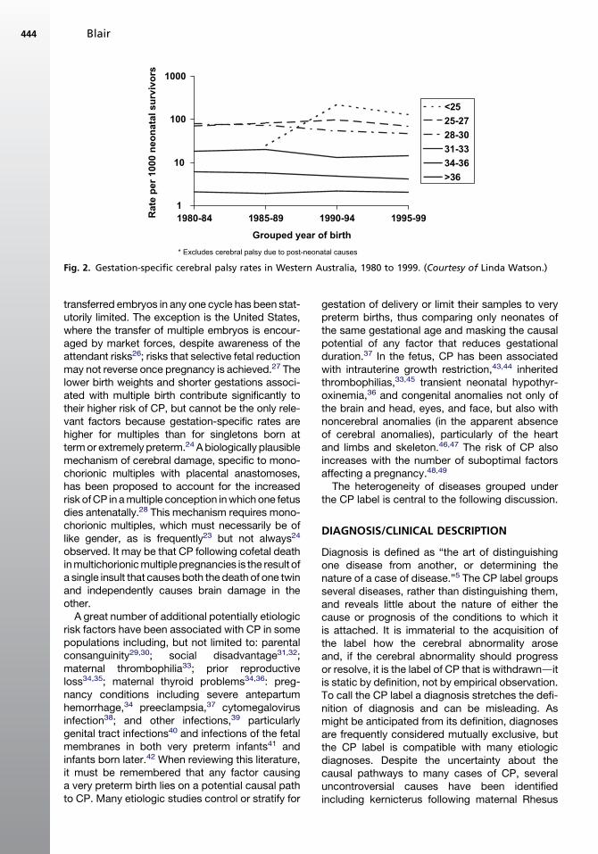
increase as each new gestational survivalboundary is crossed, and then decline as gesta-tionally appropriate neonatal management tech-niques are refined, but remain severalfold higherthan rates observed in term and near-term births(Fig. 2). Rates around 10% of survivors, the iatro-genic nature of very preterm survival, and theinvestment in neonatal intensive care that suchsurvival requires has encouraged much attentionto be devoted to CP in infants born before 32weeks’ gestation. However, because births before32weeks contribute less than2%of neonatal survi-vors, they contribute a minority, approximately
0
0.5
1
1.5
2
2.5
3
1959-62
1963-66
1967-70
1971-74
1975-78
1979-82
Grouped year of b
Ra
te
pe
r 1
00
0 liv
e b
irth
s2
1
2
NE England data 1964-93, grouped years approximate fit: 1964Final period for Swedish data 1995-98.
NE England rates per 1000 neonatal survivors
Fig. 1. Cerebral palsy rates in 3 populations, 1959 to 1999
20% to 25%, of all CP in developed countries(see for example Refs.12,22). Most CP cases areborn at term.
The risks of CP increase fourfold in twins and 18-fold in triplets,23e25 Increasingmaternal age anduseof assisted reproduction technologies (ART) hasincreased the proportion of all births that aremultiple. Concomitantly, their contribution to CPhas risen from 4% in the 1960s to 10% in the1990s.12The increasingproportionof thepopulationof triplets and higher multiples were attributableexclusively to ART. This increase has been haltedin many developed countries, where the number of
1983-86
1987-90
1991-94
1995-99
irth
W Australia
W Sweden
NE England
-68, 1969-73, 1974-78, 1979-83, 1984-88, 1989-93.
. (Courtesy of Linda Watson.)
1
10
100
1000
1980-84 1985-89 1990-94 1995-99
<25
25-27
28-30
31-33
34-36
>36
* Excludes cerebral palsy due to post-neonatal causes
Rate p
er 1000 n
eo
natal su
rvivo
rs
Grouped year of birth
Fig. 2. Gestation-specific cerebral palsy rates in Western Australia, 1980 to 1999. (Courtesy of Linda Watson.)
Blair444
transferred embryos in any one cycle has been stat-utorily limited. The exception is the United States,where the transfer of multiple embryos is encour-aged by market forces, despite awareness of theattendant risks26; risks that selective fetal reductionmay not reverse once pregnancy is achieved.27 Thelower birth weights and shorter gestations associ-ated with multiple birth contribute significantly totheir higher risk of CP, but cannot be the only rele-vant factors because gestation-specific rates arehigher for multiples than for singletons born attermor extremely preterm.24 A biologically plausiblemechanism of cerebral damage, specific to mono-chorionic multiples with placental anastomoses,has been proposed to account for the increasedrisk ofCP inamultiple conception inwhichone fetusdies antenatally.28 This mechanism requires mono-chorionic multiples, which must necessarily be oflike gender, as is frequently23 but not always24
observed. It may be that CP following cofetal deathinmultichorionicmultiplepregnancies is the result ofa single insult that causes both the death of one twinand independently causes brain damage in theother.A great number of additional potentially etiologic
risk factors have been associated with CP in somepopulations including, but not limited to: parentalconsanguinity29,30; social disadvantage31,32;maternal thrombophilia33; prior reproductiveloss34,35; maternal thyroid problems34,36: preg-nancy conditions including severe antepartumhemorrhage,34 preeclampsia,37 cytomegalovirusinfection38; and other infections,39 particularlygenital tract infections40 and infections of the fetalmembranes in both very preterm infants41 andinfants born later.42 When reviewing this literature,it must be remembered that any factor causinga very preterm birth lies on a potential causal pathto CP. Many etiologic studies control or stratify for
gestation of delivery or limit their samples to verypreterm births, thus comparing only neonates ofthe same gestational age and masking the causalpotential of any factor that reduces gestationalduration.37 In the fetus, CP has been associatedwith intrauterine growth restriction,43,44 inheritedthrombophilias,33,45 transient neonatal hypothyr-oxinemia,36 and congenital anomalies not only ofthe brain and head, eyes, and face, but also withnoncerebral anomalies (in the apparent absenceof cerebral anomalies), particularly of the heartand limbs and skeleton.46,47 The risk of CP alsoincreases with the number of suboptimal factorsaffecting a pregnancy.48,49
The heterogeneity of diseases grouped underthe CP label is central to the following discussion.
DIAGNOSIS/CLINICAL DESCRIPTION
Diagnosis is defined as “the art of distinguishingone disease from another, or determining thenature of a case of disease.”5 The CP label groupsseveral diseases, rather than distinguishing them,and reveals little about the nature of either thecause or prognosis of the conditions to which itis attached. It is immaterial to the acquisition ofthe label how the cerebral abnormality aroseand, if the cerebral abnormality should progressor resolve, it is the label of CP that is withdrawnditis static by definition, not by empirical observation.To call the CP label a diagnosis stretches the defi-nition of diagnosis and can be misleading. Asmight be anticipated from its definition, diagnosesare frequently considered mutually exclusive, butthe CP label is compatible with many etiologicdiagnoses. Despite the uncertainty about thecausal pathways to many cases of CP, severaluncontroversial causes have been identifiedincluding kernicterus following maternal Rhesus
Epidemiology of the Cerebral Palsies 445
isoimmunization (now rare in developed countriesbut still a significant cause in less developed coun-tries),29 maternal methyl mercury exposure (Mina-mata disease),50 maternal iodine deficiency(cretinism),51,52 acute hypoxia following a sentinelintrapartum event,53 and Moyamoya disease.14
These and many other diagnoses will persistwhether or not the individual meets criteria forthe CP label. Such diagnoses are informativeabout etiology, pathology, and frequently aboutfinal clinical presentation. These diagnoses tendto be mutually exclusive, whereas for each personcategorized as CP an additional etiologic diag-nosis must exist, whether or not it has beenmade. The author therefore considers that theCP label refers to clinical description rather thana diagnosis.
The range of clinical descriptions covered by theCP label is very wide in terms of the type, severity,and bodily distribution of primary motor impair-ment, of associated nonmotor neurologic andbehavioral impairments, functional deficits, andcerebral pathology. It is seldom productive toconsider the group as a whole. Subcategorizationis sometimes referred to as differential diagnosis,though the term differential description is moreaccurate. However, agreement in categorizingpeople with CP has proved extremely difficult.
Whatever the responsible insult, the extent andseverity of brain impairment can be variable andalthough specific insults can target specific areasof the brain, the area targeted frequently varieswith the gestational age at which the insult occurs.Although some specific syndromes exist,14 theimpairments associated with CP may more gener-ally be described as a series of continua of impair-ment in many dimensions, rather than a discreteset of syndromes.
To be useful, categorization systems must bereliable (people agree which category eachperson belongs to). Reliability requires thata person fit into one and only one category; thisis only possible if categories are defined bya limited number of criteria and all empiricallypossible combinations of criteria are representedwithin the categorization system. In conditionsthat comprise discrete syndromes, clinicalfeatures may be sharply defined and the numberof possible combinations of clinical featureslimited. Such is not the case with CP, so attempt-ing to reliably categorize individuals with CP onthe basis of the sum of their clinical features isdoomed to failure. However, there are severalpossible reasons for categorization and eachtends to focus on a different clinical feature, orlimited set of features. It is therefore likely to beboth more attainable and more useful to aim for
reliable categorization systems for each of theclinical features associated with CP than fora reliable categorization system for individualswith CP.
A little progress has beenmade toward this goal.The shining example is the Gross Motor FunctionClassification System (GMFCS) devised for chil-dren with CP.54 The GMFCS classifies only grossmotor function, primarily ambulatory ability and itsprecursors, into 5 possible categories. It is welldocumented with age-specific criteria, recentlyaugmented by illustrations (Fig. 3). The GMFCShas enormous success firstly because the degreeof assistance required with ambulation is veryimportant and useful for service provision, andsecondly, because the limited aim allows reliable,tessellated categories defined by a single factor.It has been followed by a similar system for upperlimb function, the Manual Abilities ClassificationSystem (MACS),55 and similar systems for commu-nication ability are currently being developed56 ortested for reliability.57
However, the very success of such classificationsystems risks their misuse. The GMFCS is some-times used to classify people with CP rather thantheir gross motor function. Although the severityof different impairments tends to correlate withinthe CP population, such correlations cannot beassumed at the individual level.58 Furthermore,these simple categorization systems are descrip-tive: they enable communication between themembers of the multidisciplinary teams thatmanage individuals with CP59 and may suggestthe types of treatment likely to be required, butthey are inadequate to determine the details ofmanagement60 or to evaluate change in responseto interventions, because the anticipated degreeof change is usually too small to cross these broadcategories.
Functional ability has a significant impact onquality of life and is a universally understoodconcept, so it is unsurprising that it has been thefirst to be successfully classified. Primary impair-ments do not share these advantages. Spasticityis the primary impairment in most persons withC,P but the tools currently used to assess itsseverity cannot measure spasticity as it is usuallydefined, and therefore lack validity as well as reli-ability.61 These shortcomings have been ad-dressed by the Australian Spasticity AssessmentScale, which has promising reliability perfor-mance.62 Moreover, the relative contributions ofprimary impairments to functional performance,participation, or overall quality of life are underdebate, with the suggestion that more emphasisbe placed on improving strength and physicalfitness.63
Fig. 3. Age-specific example of criteria for the Gross Motor Function Classification System (GMFCS). (Courtesy ofKerr Graham, MD, The Royal Children’s Hospital, Melbourne, Australia.)
Blair446
Table 2Frequency of impairments in total populationsamplesa of persons with CP
Impairment % of All CP Affected
Motor 100
Spasticity 77e93
Dyskinesias 2e15
Ataxia 2e8
Isolated hypotoniab 0.7e2.6
GMFCS I 32e51II 17e21III 9e12IV 10e15V 12e19
IQ <70c 17e60
Ongoing epilepsy 31e40
Visual impairment 21e63
Blind 1e7
Hearing impairment 11e13
Deaf 1.7e3
a Populations cited in Table 1 were perused for this table.Data were not available for all impairments from allsources.b Some surveillance systems specifically exclude isolatedhypotonia.c May be estimated if formal assessment is not possible.
Epidemiology of the Cerebral Palsies 447
PATHOPHYSIOLOGYCerebral
By definition, the primary pathology responsiblefor CP is in the brain. Advances in cerebralimaging, using ultrasound, computed tomog-raphy, and now magnetic resonance imaging(MRI) technology have allowed a greater under-standing of the variety of cerebral pathologiesassociated with CP. Because by definition everyperson with CP has some cerebral pathology, itmay be surprising that the pathology remainsunidentified in 10% to 20% of cases investigated,more often those with ataxia or with less severebilateral impairments.64e66
Early prediction is highly desirable in high-riskneonatal survivors, and because cerebral ultra-sonography could be brought into the nursery,ultrasonography was the first imaging modalitybrought to focus on early brain impairment.Ultrasonography has been used extensively invery preterm born infants, in whom periventric-ular white matter injury is common.67 Patternsof white matter injury associated with increasedrisks of CP have been identified (see forexample Ref.68) but although useful, early ultra-sonography has not proved very specific in pre-dicting CP nor is it as sensitive as is sometimesassumed.69 Because the acoustic window islimited to the fontanelles, ultrasonography iswell suited to imaging the central regions ofthe neonatal brain but is less useful for corticaland cerebellar structures and impossible oncethe fontanelles have closed, precluding imagingfinal pathology. MRI provides clearer images,can image all parts of the brain at all ages,has become more accessible and user friendlyin recent years, and can retrospectively suggestboth the very approximate timing of the insult(s)and possible causes.70 There are strong corre-lations between cerebral pathology and clinicaldescription of motor impairment: unilateralpathology is associated with unilateral impair-ment, periventricular white matter damage withlower limb spastic impairment increasinglyaffecting the upper limbs with increasing extentof the damage, and basal ganglia damage withdyskinetic impairment. However, accurateprediction of CP has not proved possible withany imaging modality, even in high-risk verypreterm infants. Moreover, MRI remains expen-sive, usually requires sedation, if not generalanesthesia, to attain the required duration ofimmobility and, although of great interest toetiologic research, may provide little clinicalbenefit to the impaired individual. The possibilityof identifying genetic causes may be of
considerable importance for genetic counselingconcerning subsequent pregnancies to theparents, but applies to only a small fraction ofcases in areas with little parental consanguinity.Cerebral imaging studies therefore tend torecruit convenience samples of persons withCP who have a suitable cerebral MRI study aspart of their clinical workup.64,65 In studies oftotal population samples, the proportion of CPcases with such studies has not exceeded60% to 70%.66,71 Despite the heterogeneity ofpublished samples, studies agree that the cere-bral pathology is varied, no damage is apparentin 10% to 20%, cerebral malformations origi-nating in early pregnancy are seen in approxi-mately 10%, and that damage acquired afterorganogenesis can occur in periventricularwhite matter, cortex, basal ganglia, thalamus,or in several locations. Further comparisonsare hampered by inconsistent methods ofimaging and of describing and classifying thefindings.64,65 Categories of cerebral anomaliesare variously defined by location, by nature orextent of the abnormality, by (presumed) prox-imal cause, and/or by (approximate) timing of
Blair448
the cause, according to the aims of theclassifier.The cerebral damage found in persons with CP
need not be limited to motor centers. Moreextensive brain damage results in additionalneurologic impairments, with frequencies in totalpopulation-based samples shown in Table 2. CPhas also been associated with increased rates ofimpaired proprioception72e74 and tactile appreci-ation,75 but these sensory abilities are notroutinely assessed in population surveillancesystems and their prevalence in CP populationsis unknown. The wide variation in proportions ofdisability (see Table 2) reflect primarily thedifferent severity of impairment criteria requiredfor registration because, in persons with spas-ticity, the number of associated impairmentstends to increase with the severity of motorimpairment. The variation in proportion withdyskinesias reflects that spasticity frequentlycoexists with dyskinesia and that mixed signsare sometimes included with spasticity andsometimes with dyskinesia.
Musculoskeletal
The cerebral pathology responsible for CP is, bydefinition, static after the period of acquisition,but the pathologic effects on the developing childare not. The effects on the musculoskeletal systemvary with the specific types and distributions ofmotor impairments. Sometimes categorized asbilateral or unilateral or by the number of limbsaffected, in fact topographic continua areobserved. It is rare to find spastic hemiplegiawith no signs of impairment on the less affectedside, bilateral spasticity that is perfectlysymmetric, or diplegia with no signs of impairmentin the arms; from a functional perspective thedegrees of involvement of the trunk, neck, andhead are extremely important. Whatever bodyparts are affected, increased muscle tone inhibitsmuscle growth, resulting in a progressive failurefor muscle length to keep pace with bone length.Untreated, this can result in fixed muscle contrac-tures in which the mismatch between bone andmuscle length precludes movement at the joint,leading to joint contracture. Imbalances of muscletone can lead to deformities such as scoliosis orprogressive hip displacement (failure to maintainthe head of the femur in the acetabulum) and ulti-mately, hip dislocation. Paradoxically, hyperto-nicity is frequently accompanied by muscularweakness, which is exacerbated by the difficultiesof engaging in gross motor exercise, particularly inthe nonambulant.
Musculoskeletal pathology develops over timeand can have devastating consequences onquality of life, not only in terms of motor limitationsbut also for a person never destined to achieve anyfunctional motor abilities. Hip dislocation can bepainful and preclude sitting, and hence normal toi-leting, or the possibility of observation from anupright position, thus limiting the possibilities forsocial interaction. Severe contracture canpreclude normal hygiene skin care, rendering theperson vulnerable to skin breakdown andinfection.Dyskinetic motor impairment arises from invol-
untary changes in muscle tone producing athetoidor dystonic movements. Dyskinetic movementscan be frequent and forceful, and may generateconsiderable strength. Violent activity, seenparticularly in athetoid CP, has been associatedwith premature aging of the joints, particularly inthe cervical spine, necessitating surgical interven-tions by middle age.76,77
Maintaining musculoskeletal health hasassumed increasing importance with the survivalto adulthood of those with even the most severeimpairment.78 However, musculoskeletal patholo-gies are secondary, and considerable attention isbeing devoted to devising strategies wherebythey may be minimized or prevented: for example,guidelines for hip surveillance in children with CPhave recently become electronically available.79
VULNERABILITY TO PNEUMONIA ANDMALNUTRITION
Lack of coordination is another source ofsecondary pathology, quite apart from injury dueto falls. Persons with severe CP are frequentlyunable to expectorate effectively and are hencevulnerable to pneumonia, the most common causeof death in persons with CP. For those withoropharyngeal involvement, the possibility of aspi-ration adds to the risk of pneumonia.Difficulties with swallowing can make feeding
both time consuming and unpleasant for subjectand caregiver, and is responsible for the poornutritional status of many persons with CP. Nutri-tional status declines with increasing GMFCScategory,80 even among the ambulatory.81 Inter-vention trials have indicated that extreme under-weight and poor growth predisposes toinfection,82 and may be associated with gastroin-testinal disorders such as reflux and chronic con-stipation and with decreased motor function.83 Inrecent years malnutrition has been addressedwith parenteral feeding via gastrostomy, whichimproves measures of weight for height,80 reducesconstipation, vomiting, and sialorrhea (one of the
Epidemiology of the Cerebral Palsies 449
most distressing sequelae of oromotor dysfunc-tion), and tends to improve micronutrient status.84
However, major complications are possible,84,85
minor complications, especially infection, occurfrequently,84 the risk of reflux requiring fundoplica-tion or medication is increased,86 and it is notuniversally welcomed by caregivers who maybelieve that it removes an important opportunityfor social interaction.87
Furthermore, the weight gained following gas-trostomy in persons with moderate or severe CPwho have low energy requirements is primarily asfat rather than fat-free mass88; this may be desir-able for thermal protection,89 but higher fat massand gastrostomy feeding has been associatedwith a greater risk of bone fracture,90 suggestingthat the formulae used for parenteral feeding inpersons with CP may need further refinement.
The vulnerability to bone fracture91 is a result ofthe osteoporosis frequently seen in persons withsevere CP.92 Nutritional deficiencies and difficultyof achieving weight-bearing exercise are com-pounded by the likelihood of receiving little sunexposure and the possibility of drug exposure.Thus bone mineral acquisition is frequently insuffi-cient for skeletal growth.90
MANAGEMENT
Cure, in the sense that the brain damage is re-paired, is not currently an option, although stemcell therapy hovers on an uncertain horizon.93
Parents find this difficult to accept and are vulner-able to extravagant claims of expensive andunproven therapies. For example, hyperbaricoxygen therapy professes the ability to rescuecompromised neurons, but has not furnished anyconvincing evidence of effectiveness and mayhave undesirable side effects.94
With cure unavailable, the objective of CPmanagement is to improve quality of life andprevent or minimize the secondary pathologies.Beyond this, the means by which quality of lifecan be improved depends on the clinical descrip-tion of impairments, and the aspirations and incli-nations of the person with CP and theircaregivers. For those with mild impairments,optimal management typically involves normaliza-tion of appearance and of performance of activi-ties of daily living, but for the most severelyimpaired primary goals are typically comfort andmaximizing their potential for satisfying pastimes.
The traditional therapies for CP management ofstretching, casting, and orthoses has broadenedconsiderably in recent years, as outlined ina previous Seminar on CP.95 Much research isbeing conducted to optimize protocols, measure
efficacy, identify adverse effects, and identify thedescriptions of CP most likely to benefit fromeach management protocol that includes thenew therapies such as botulinum toxin A,96
intrathecal baclofen,97,98 selective dorsalrhizotomy,99e101 and multilevel surgery.102 Lessinvasive new techniques include constraint-induced therapy for asymmetrical motor impair-ments103,104 and strength training.105
Management of the many problems associatedwith CP must be tailored to the individual andhas become highly specialized, frequentlyrequiring the input of many specialties. As thenumber and severity of impairments increase,such teams may include physiotherapists, occu-pational therapists, orthotists, developmentalpediatricians, neurologists, radiologists, ortho-pedic surgeons, speech pathologists, alternativecommunication and IT specialists, psychologists,ophthalmologists, dentists, social workers, specialeducators, educational aides, and respite carers.These specialist services must be continuouslycoordinated to achieve maximum benefit, andthe role of the general practitioner is particularlyimportant when the person is transferring frompediatric to adult services.
PREVENTION
Rational prevention addresses causes. If CP isdefined by motor impairment resulting froma cerebral abnormality, then the cerebral abnor-mality is the most proximal cause of the motorimpairment. However, by the time the abnor-mality exists, it is too late to enact prevention.Preceding causes must be sought. Any factorthat can interrupt cerebral development is poten-tially a proximal cause of the cerebral abnor-mality. Such factors include genetic conditionsand teratogens (physical, infectious, metabolic,or toxic),106 the effects on cerebral developmentvarying with the gestational age of exposure,genetic susceptibility, and environmental factors.
In practice, prevention is often approached byaddressing proximal causes as they becomeapparent: for example, inducing hypothermiafollowing acute hypoxia,107 exchange transfusionfollowing severe hyperbilirubinemia, mechanicalventilation to prevent hypoxia in very prematurelyborn infants, or emergency cesarean section inthe face of fetal distress. Pharmacologic neuro-protection is being intensively investigated,primarily in animal models relevant to CP in verypreterm births.108e110 Several human trials ofmaternally administered magnesium sulfatewhen very preterm birth is imminent have beensystematically reviewed,111 and are associated
Blair450
with a relative risk for CP of 0.68 (95% confidenceinterval [CI] 0.54e0.87). However, the reviewers’conclusion that “the neuroprotective role for ante-natal magnesium sulfate therapy given to womenat risk of preterm birth for the preterm fetus isnow established” sits uneasily with their reportedrelative risks of 1.07 (CI 0.82e1.4) for major neu-rodisability, 0.94 (CI 0.78e1.12) for combineddeath or CP, and 1.00 (CI 0.91e1.11) for deathor any neurologic impairment. If magnesium hasno effect on these risks of combined pooroutcomes, a reduced risk of CP suggests thatthe risks of other adverse outcomes must beincreased.These late strategies may help those reaching
these unenviable situations, but are reactiverather than proactive. Earlier strategies are pref-erable. For example, recent animal worksuggests that fetal vulnerability to hypoxia maybe decreased by adding naturally occurring nutri-ents such as creatine112 and melatonin113 to thematernal diet by mid pregnancy. Strategiesenacted only once pathology is apparent may,by enabling survival of the already compromised,equally well increase the number of CP asdecrease it. This conundrum is no doubt thereason that, despite great advances in commu-nity health, and obstetric and neonatal technolo-gies over the last 50 years, the rate of CP has notdeclined in parallel with rates of perinatalmortality.To effectively reduce the proportion of persons
with CP (ie, to prevent without simultaneouslycausing), distal causes must be addressed,factors that lie on potential causal paths beforeany pathology is present. Many distal factors arerecognized and addressed by public healthmeasures or standard medical care. Iodine isadded to salt to avoid cretinism, foodstuffs areprotected from contaminants such as mercury,girls are vaccinated against rubella, periconcep-tional folate is advised or added to basic food-stuffs to reduce the risk of neural tube defectsincluding cerebral defects, Rh status of prospec-tive parents is routinely investigated to allowrecognition of Rhesus incompatibility and theneed for anti-D treatment to avoid the possibilityof Rh isoimmunization and kernicterus in futureoffspring, and the dimensions of the fetal headare compared with those of the maternal pelvisto gauge the likelihood of cephalopelvic dispro-portion and need for elective abdominal delivery.In the developed world these preventive activities,which may be enacted years before the pregnancyexists, are routine, and the factors they addressseldom cause CP. If we take to its logical conclu-sion the idea that it is prophylaxis, not rescue, that
is the most effective means of prevention, then wemust consider as potentially preventable causesall genetic and environmental factors known tobe associated with suboptimal pregnancyoutcomes, of which CP is but one.The lifestyle advice pertinent to optimizing preg-
nancy outcome is as follows. Enter the pericon-ceptional period in optimum physical andnutritional condition, without chronic health prob-lems and keeping alcohol intake low, in particularavoiding both binge drinking and drugs, whetherprescribed or recreational. During pregnancymaintain optimal physical, dental, and nutritionalcondition, avoid exposure to infections, psycho-logical stress, or physical trauma. And finally,desist in the face of repeated reproductive failure.These ideals are more easily attainable when preg-nancy is planned and sufficient material andemotional resources are available. Advocatingthis advice in this context may seem an attemptto blame the woman for poor pregnancyoutcomes. This is not the aim: a good pregnancyoutcome can never be assured however muchcare is taken. However, the likelihood of a goodpregnancy outcome can be greatly improved bythe choices a woman makes over the timing ofand self care in pregnancy; the worst choice maybe to fail to choose.The role of the medical profession is not only
to apply standard medical care and encourageuptake of appropriate lifestyle advice, as out-lined in this article, but to desist from enablingand encouraging reproduction (in parents) orsurvival (in neonates) of those who are patentlyunfit. Such advice is not what many prospectiveparents or clinicians want to hear, but it is theadvice most likely to reduce the risk of CP.
SUMMARY
CP is not a diagnosis but an umbrella term formany clinical descriptions. These conditions,which by definition include impairments of move-ment or posture of cerebral origin, are neithercurable nor fatal. Management strategies aimingto improve quality of life have developed consider-ably over the last two decades and are likely toincrease longevity of the most severely impaired,half of whom already reach adulthood. However,the CP label can only be applied to survivors ofan age at which motor impairment can exist, soearly death is a competing outcome. There aremany causal pathways to CP but, although thosethat are understood have been addressed, itsfrequency has not decreased over the past 50years both despite and because of advances in
Epidemiology of the Cerebral Palsies 451
obstetric, neonatal, and early childhood care aswell as decreasing perinatal mortality.
Search Strategy and Selection Criteria
The information in this article is based on mate-rial in peer-reviewed medical journals, the Co-chrane database of systematic reviews,conference proceedings of material not yetotherwise published, textbooks, and reportsfrom registers and surveillance systems of CP.In selecting material when many referencesaddress the same topic preference has beengiven, in order, to more recent publications,systematic reviews, original observations, andreviews. Where possible, contributions repre-senting different geographic areas have beenselected, provided English language abstractswere available. The most recent publicationswere sought using the Medline database,entering cerebral palsy as a search term incombination with keyword(s) describing theassociated concept: for example, <cerebralpalsy> and <gastrostomy>.
ACKNOWLEDGMENTS
I thank Sarah Love for reading the manuscriptand advice, Linda Watson for help with Figs. 1and 2 and running the Western Australian CPRegister, and Professor H. Kerr Graham forpermission to reprint Fig. 3.
REFERENCES
1. Bax MC. Terminology and classification of cerebral
palsy. Dev Med Child Neurol 1964;6:295e7.
2. Mutch LW, Alberman E, Hagberg B, et al. Cerebral
palsy epidemiology: where are we now and where
are we going? Dev Med Child Neurol 1992;34:
547e55.
3. Rosenbaum P, Dan B, Leviton A, et al. Proposed
definition and classification of cerebral palsy, April
2005. Dev Med Child Neurol 2005;47:571e6.
4. Onions CT. The Shorter Oxford Dictionary. 3rd
edition. Oxford: Clarendon Press; 1968. p. 473.
5. Taylor EJ. Dorland’s illlustrated medical dictionary.
27thedition.Philadelphia:WBSaunders;1988.p.438.
6. Blair E, Love S. Commentary on the definition and
classification of cerebral palsy. Dev Med Child
Neurol 2005;47:510.
7. Stanley F, Blair E, Alberman E. ‘What are the cere-
bral palsies?’ Cerebral palsies: epidemiology and
causal pathways. London: MacKeith Press; 2000.
p. 8e13, Chapter 2.
8. Reid S, Lanigan A, Reddihough D. Post-neonatally
acquired cerebral palsy in Victoria, Australia,
1970e1999. J Paediatr Child Health 2006;42(10):
606e11.
9. Stanley F, Blair E, Alberman E. Post neonatally
acquired cerebral palsy: incidence and anteced-
ents. Cerebral Palsies: epidemiology and causal
pathways. London: MacKeith Press; 2000. p.
124e37, Chapter 11.
10. Constantinou J, Adamson-Macedo E, Mirmiran M,
et al. Movement, imaging and neurobehavioural
assessment as predictors of cerebral palsy in
preterm infants. J Perinatol 2007;27(4):225e9.
11. Nelson KB, Ellenberg JH. Children who ‘outgrew’
cerebral palsy. Pediatrics 1982;69:529e36.
12. Watson L, Blair E, Stanley F. Report of the Western
Australian cerebral palsy register to birth year
1999. Perth (Western Australia): Telethon Institute
for Child Health Research; 2006.
13. Smithers-Sheedy H, McIntyre S, Watson L, et al.
Report of the international survey of cerebral palsy
registers and surveillance systems. Sydney
(Australia): CP Institute; 2009. Available at: http://
www.cpinstitute.com.au/publications/index.html.
Accessed July 19, 2010.
14. Badawi N, Watson L, Petterson B, et al. What
constitutes cerebral palsy? Dev Med Child Neurol
1998;40:520e7.
15. SCPE. Surveillance of cerebral palsy in Europe:
a collaboration of cerebral palsy surveys and regis-
ters. Dev Med Child Neurol 2000;42(12):816e24.
16. Gainsborough M, Surman G, Maestri G, et al. Val-
idity and reliability of the guidelines of the Surveil-
lance of Cerebral Palsy in Europe for the
classification of cerebral palsy. Dev Med Child
Neurol 2008;50(11):828e31.
17. Tu J, Willison D, Silver F, et al. Impracticality of
informed consent in the registry of the Canadian
Stroke Network. N Engl J Med 2004;350:1414e21.
18. Ingelfinger J, Drazen J. Registry research and
medical privacy. N Engl J Med 2004;350(14):1452.
19. Littenberg B, MacLean C. Passive consent for clin-
ical research in the age of HIPAA. J Gen Intern Med
2006;21(3):207e11.
20. Johnston M, Hagberg H. Sex and the pathogenesis of
cerebralpalsy.DevMedChildNeurol2007;49(1):74e8.
21. Aly H. Mechanical ventilation and cerebral palsy.
Pediatrics 2005;115(6):1765e6.
22. McManus V, Guillem P, Surman G, et al. SCPE
work, standardization and definitiondan overview
of the activities of SCPE: a collaboration of Euro-
pean CP registers. Zhongguo Dang Dai Er Ke Za
Zhi 2006;8(4):261e5.
23. Stanley F, Blair E, Alberman E. ‘The special case of