Page 1
EPIGENETICS: A NEW THERAPEUTIC APPROACH IN CHRONIC
LYMPHOCYTIC LEUKEMIA
João A. Carvalhoa, Filipa Carvalho
a; André Ribeiro
a; Vera Alves
b; Ana Cristina Gonçalves
c,d;
Ana Bela Sarmento-Ribeiroc,d
a Applied Molecular Biology / University Clinic of Hematology, Faculty of Medicine,
University of Coimbra (FMUC), Portugal; b
Immunology, FMUC, Portugal c
CIMAGO -
Center of Investigation on Environment, Genetics and Oncobiology, FMUC, Portugal; d
CNC
- Center for Neuroscience and Cell Biology, University of Coimbra, Coimbra, Portugal.
Correspondence should be sent to Ana Bela Sarmento-Ribeiro, Applied Molecular Biology /
University Clinic of Hematology, Faculty of Medicine, University of Coimbra, Azinhaga de
Santa Comba, Celas, 3000-548, Coimbra, Portugal. Tel: +35 1239480247, Fax:
+351239480038. E-mail: [email protected]
Page 2
2
Abstract
Background/Aims: Nowadays, epigenetic changes have highlighted a new possible
contribution to the development of Chronic Lymphocytic Leukemia B (CLL-B), namely
by histone deacetylases or DNA metiltransferases. This project aims to study the
potential therapeutic role of hypomethylating agents (HM) and histone deacetylase
inhibitors (HDACis), in monotherapy and in association of both drugs, in a CLL-B cell
line. Materials and Methods: A CLL-B cell line were incubated with the HM agent,
decitabine (DAC), and the HDACi, trichostatin (TSA), at different concentrations;
viability and proliferation studies were performed using rezasurin test; cell death was
analyzed by flow cytometry and optical microscopy; methylation studies were
performed using Methylation specific-PCR (MSP) and Real-Time PCR (qPCR).
Results: Our results show a decrease in cell proliferation and viability in a dose and time
dependent manner, leading to a cell death preferentially by apoptosis. Besides that, a
decrease in the degree of gene methylation was also reached. Discussion: This study
suggests that epigenetic modulation might constitute a new approach to the treatment of
lymphoid malignancies, namely CLL-B.
Keywords: epigenetics, Chronic Lymphocitic Leukemia B, trichostatin A, decitabine,
methylation
Page 3
3
Introduction
Chronic Lymphocytic Leukemia B (CLL-B) is the most common leukemia in the
Western World and consists on a clonal malignancy of mature neoplastic B cells,
characterized by a low proliferation rate and impaired apoptosis [1,2]. These cells
characteristically present low express surface immunoglobulin (slg), are monoclonal
regarding to expression of either κ or λ light chains and show B-cell surface antigens, as
CD19, CD20 and CD23, with the CD5 antigen, in the absence of other pan-T-cell
markers [2-5].
The aetiology of CLL-B is unknown and the natural history is heterogeneous. Unlike
other leukemias, in CLL-B, it is not described a causing translocation, deletion, or
mutation [6]. In recent years, more interest has been focused on epigenetic changes
contributing to the development of CLL-B, by an abnormal DNA methylation pattern
and chromatin histones modification. Although DNA methylation is globally decreased
in this disease, there is an increasing belief that regional hypermethylation of gene
promoters, namely of tumour suppressor genes, leads to gene silencing, unravelling the
role of epigenetic modifications in CLL-B development. So, new epigenetic drugs, such
as DNA-methyltransferases inhibitors (DNMTis) and histone deacetylases inhibitors
(HDACis), may attempt to correct these changes [1,6-8].
DNA methylation level in CLL-B patients is generally lower than in healthy
individuals. However, regional hypermethylation of gene promoters, especially in the
CpG dinucleotides, leads to gene silencing of particularly important genes in cell cycle
and in apoptosis regulation. On the other hand, histones deacethylation could be
associated with cancer due to the resultant chromatin compaction that blocks the access
to transcription factors [6,7].
Page 4
4
Although epigenetic modifications may play an important role in CLL-B, little is known
about the extent of promoter methylation [6]. Laura Rush (2004) has identified many
aberrantly methylated genes and suggested that a portion of these events confers a
selective advantage to the malignant cell [1]. Duhamel et al (2008) and Billot et al
(2011) referred that the deregulated expression of IKZF3 (Aiolos), an important
transcription factor involved in the control of mature B lymphocyte differentiation and
maturation, is due to epigenetic modifications, namely DNA hypomethylation and an
enrichment of euchromatin associated with histone markers, such as the
hypomethylation of the lysine 4 on histone H3 [9,10].
The current use of hypomethylating agents and HDACis may provide a potential
mechanism to change this disordered gene expression in CLL-B. The promising
preclinical activity of these drugs makes it imperative that we continue to investigate the
contribution of epigenetic alterations in these haematological neoplasias, which can
contribute to a better patient risk stratification and to select the subgroup of patients that
will benefit from treatment with this class of drugs.
This work aimed to evaluate the potential therapeutic effect of epigenetic modulators,
hypomethylating agents/DNA methyltransferase inhibitors (DNMTis), and histone
deacetylase inhibitors (HDACis), in a CLL-B cell line in culture, as single agents or in
combination therapy.
Materials and methods
Cell culture: A CLL-B cell line, the EHEB cells, isolated from the peripheral blood of
a 69-year old woman, was provided by German Collection of Microorganisms and Cell
Cultures (DSMZ, Braunschweig, Germany) [11]. The cells were maintained in culture
in RPMI-1640 medium (Sigma-Aldrich, St. Louis, MO, USA) supplemented with 20%
Page 5
5
fetal calf serum (Sigma-Aldrich, St. Louis, MO, USA). Cells were cultured at initial
density of 0,5x10⁶ cells/ml, during 72 hours, in the absence and in the presence of
increasing concentrations of epigenetic modulators, such as the hypomethylant agent 5’-
Aza-2’-deoxycytidine or Decitabine (DAC) (Sigma-Aldrich, St. Louis, MO, USA) and
the HDACi, Trichostatin A (TSA) (Sigma-Aldrich, St. Louis, MO, USA), alone or with
each other.
Cell viability/proliferation analysis: Cell proliferation was accessed each 24 hours,
during 72h, using the resazurin metabolic test (Sigma-Aldrich, St. Louis, MO, USA).
Resazurin was prepared as a stock solution of 100 μg/ml in PBS and this stock solution
was filtered with a sterile 0.20 μm-pore filter and stored at -20°C in the dark. After each
24h of treatment, a final concentration of 10 μg/ml of resazurin was added to cells and
incubated at 37°C during 6 hours [12,13]. Then, absorbance was measured using a
Synergy™ HT Multi-Mode Microplate Reader (BioTek Instruments). The IC50 was
evaluated by a dose response curve.
Cell death analysis: Cell death was analysed by flow cytometry, staining the cells with
annexin V-FITC (AV) and propidium iodide (PI) [14,15]. After 48h, cells, cultured in
the absence or in the presence of the drugs, were collected, washed (centrifuged at 1.000
xg for 5 min) and incubated for 15 min in 100 L AV binding buffer (Immunostep Kit,
Salamanca, Spain) and 5 l FITC-labeled AV and 5 l PI. After incubation time, cells
were diluted in 400 µL of binding buffer and analyzed by flow cytometry. The results
were analysed on a flow cytometer equipped with an argon ion-laser emitting a 488 nm
beam. Green fluorescence of AV was collected with a 525-nm band pass filter and red
fluorescence of PI with a 610 band pass filter. The results are expressed in % of viable,
early apoptotic, late apoptotic and necrotic cells.
Page 6
6
Morphologic studies: Morphologic studies were performed in order to further
characterize the results obtained by flow cytometry. Briefly, after incubation period,
cells were collected and stained for 3 minutes with May-Grünwald solution (Sigma, St.
Louis, MO, USA), diluted in a 1:1 ratio with distilled water, followed by staining with
Giemsa solution (Sigma, St. Louis, MO, USA) diluted 1:8 in distilled water for 15
minutes. After rinsed with distilled water, smears were left to dry at room temperature
and cell morphology was analyzed by light microscopy using a Nikon Eclipse 80i
microscope equipped with a Nikon Digital Camera DXm 1200F.
Methylation specific PCR (MSP): One µg of genomic DNA, obtained from EHEB
cells cultured in the absence and in the presence of DAC, TSA in monotherapy or with
the association of both drugs for 72h, was treated with sodium bisulfite according to the
EpiTect Bisulfite Kit (Qiagen, Chatsworth, USA), allowing the analysis of the
methylation pattern. Methylation-specific PCR’s of p15, p16, p53, death-associated
protein kinase (DAPK) and O6-Methylguanine DNA methyltransferase (MGMT) genes
were carried out as previously described by others [16-18]. PCR products were resolved
on 4% agarose gels, stained with ethidium bromide and visualized under UV
illumination.
Real-Time PCR (qPCR): In order to quantify the amount of methylated DNA, bisulfite
modified DNA (1 μL) was amplified using SsoFast™ EvaGreen® Supermix (Biorad,
Mitry Mory, France) containing a final concentration of 0.25 μM of each p15
methylated primers. All PCR reactions were carried out in duplicate. The amount of
methylated DNA was determined by the threshold cycle number (Ct) for each sample,
compared against a standard curve generated from the dilution of DNA obtained from
untreated cells (control).
Page 7
7
Statistical analysis: Data are expressed as mean SD obtained from independent
determinations, each one performed in duplicate or triplicate. Differences between data
sets were evaluated by performing analysis of variance (ANOVA). A p value <0.05 was
considered as statistically significant.
Results
Cell proliferation analysis: TSA or DAC, in monotherapy or in combination, lead to a
decrease in cell proliferation in a time- and dose-dependent manner. however, TSA was
more effective in monotherapy than DAC, reducing cell viability in approximately
50% at 250 nM (53% ± 9%), after 72h of incubation (Fig.1A). Even higher doses of
DAC couldn’t achieve the same effect. A decrease of about 40% in cell proliferation
was observed in the cells treated with the highest concentration of DAC (100 M)
(Fig.1B). The combination of 5 µM of DAC with 100 nM of TSA enhanced the
antiproliferative effect of these drugs at concentrations lower than those used to achieve
the IC50 in monotherapy. In fact, as a single agent, and after an incubation period of
72h, 100 nM of TSA and 5 µM of DAC induced a reduction in cell proliferation of 24%
and 20%, respectively. When cells were treated with the combination of drugs
administered simultaneously, a decrease of 43% in cell proliferation is achieved
(Fig.1C). However, the administration schedule of drugs didn’t appear to influence its
cytotoxic effect.
0
Page 8
8
Figure 1. Dose response Curves of TSA and DAC in EHEB cells. Cell proliferation/viability was
analysed each 24 h during 72 hours of incubation, with the drug concentrations represented in figure. In
(A), it is represented the effect of TSA, in (B), the effect of DAC and (C) represents the effect of TSA and
DAC combination therapy in EHEB cells. TSA 100 nM and DAC 5 µM were administrated, alone,
simultaneously (TSA 100 nM + DAC 5 µM) or with a gap of 4 hours, TSA 100 nM firstly and 4 hours
later DAC 5 µM (TSA 100 nM* + DAC 5 µM) and vice-versa (TSA 100 nM + DAC 5 µM*). The data is
expressed in percentage (%) normalized to the control and represents the mean± SD of at least 3
independent experiments.
B A
C
Page 9
9
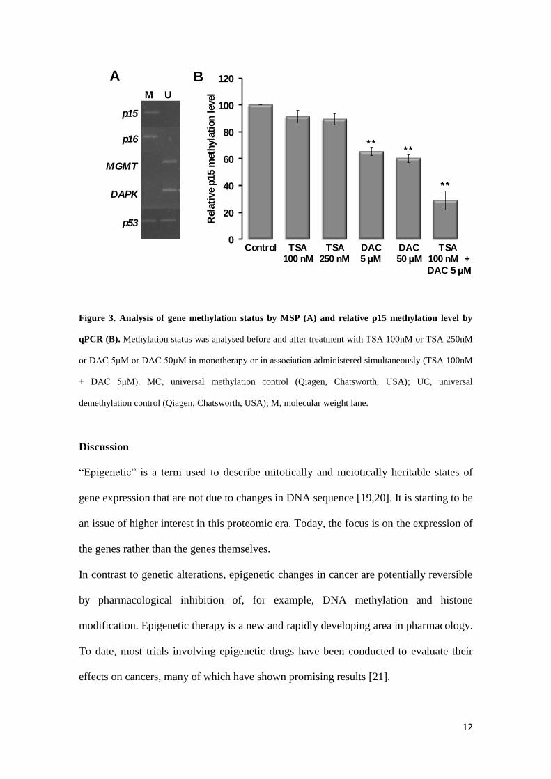
Cell death analysis: The cytotoxic effect was evaluated by flow cytometry using
annexin V/PI incorporation (Fig.2A). The results show, in cells treated with 250 nM of
TSA, a higher decrease in cell viability (about 60%±8%), and an increase in cell death
by early apoptosis (3% ± 3%), late apoptosis/necrosis (23% ± 2%) and necrosis (14% ±
4%). However, in EHEB cells treated with 5 µM and 50 µM of DAC, only a decrease in
cell viability of approximately 10% to 15%, respectively, is observed. Besides that,
when drugs were administered in simultaneous combination, we observed a synergistic
cytotoxic effect compared with those obtained in cells treated with each drug in
monotherapy. In fact, as we can see in Fig.2A, a decrease of 35%±3% in cell viability is
observed when cells are incubated with the combination of 100 nM TSA and 5 µM
DAC compared with the effect obtained with the same dose of drugs administered in
monotherapy (15% with TSA 100 nM and 2% with DAC 5 µM). The decrease in cell
viability is accompanied by an increase in cell death mainly by apoptosis.
To confirm the cytotoxic effect observed in the previous experiments, namely the type
of cell death, we analysed, by optical microscopy, the morphological characteristics of
untreated (control) and treated EHEB cells. The representative cell smears, shown in
Fig.2B, show that EHEB cells treated with TSA and DAC, after a treatment period of
48h, mostly display the morphological characteristics of cell death by apoptosis, such as
cell contraction, nuclear fragmentation, blebbing and apoptotic bodies, although smears
of cells treated with 250 nM of TSA displays also a few cell in necrosis.
Page 10
10
Figure 2. Cell death analysis by flow cytometry and optical morphology. In (A), it is represented the
results obtained by flow cytometry when EHEB cells were incubated in the absence (control) and in the
presence of drugs indicated in the figure legend. In (B), it is represented the morphological aspects of
EHEB cells untreated (control) and treated with 250 nM of TSA, 50 µM of DAC and with the therapeutic
combination of 100 nM of TSA plus 5 µM of DAC administered simultaneously. Cell death was detected
by annexin V and propidium iodide staining and analyzed by flow cytometry and cell smears were stained
with May-Grünwald-Giemsa. A - alive cells; EA - cells in early stage of apoptosis; AN - cells in late
stage of apoptosis or in necrosis; N - necrotic cells. Results are expressed in % SD and represent the
mean± SD of at least 3 independent experiments. Amplification: 500x.
Page 11
11
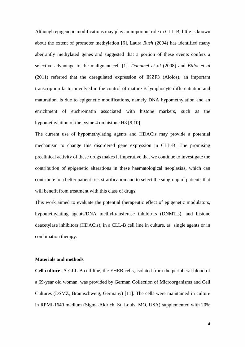
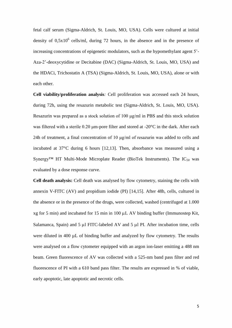
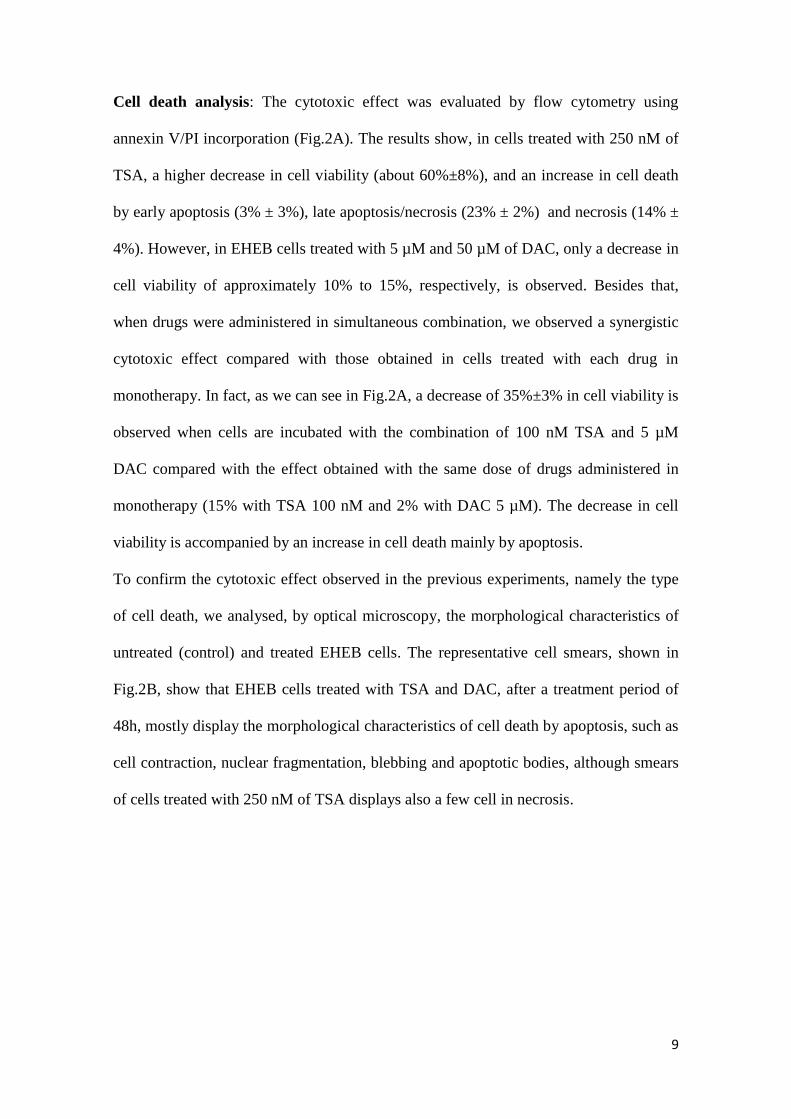
Methylation status and quantification: Methylation status of p15, p16, p53, MGMT
and DAPK genes was analysed in EHEB cells (Fig.3A), since epigenetic modulators
induce changes in the hypermethylation of CpG islands. In this CLL-B cell line, the p15
and p16 tumor suppressor genes were methylated, however, MGMT and DAPK genes
were demethylated. Besides that, p53 promoter gene presented one methylated locus
and the other demethylated. Since p15 is ordinarily found methylated, and was also
methylated in EHEB cells, the hypomethylating effect of both drugs was evaluated in
this gene (Fig.3B). TSA, even in higher doses, induced a limited decrease in the relative
p15 methylation level, approximately 10%. However, as expected, DAC induce a higher
hypomethylating effect, leading to a decrease of about 35% and 40% in the p15
methylation, when EHEB cells were treated with 5 µM and 50 µM of DAC
respectively. This demethylating effect was enhanced when cells were treated with the
simultaneous association of 5 µM of DAC with 100 nM of TSA, leading to a reduction
of about 70% in p15 methylated gene.
Page 12
12
Figure 3. Analysis of gene methylation status by MSP (A) and relative p15 methylation level by
qPCR (B). Methylation status was analysed before and after treatment with TSA 100nM or TSA 250nM
or DAC 5μM or DAC 50μM in monotherapy or in association administered simultaneously (TSA 100nM
+ DAC 5μM). MC, universal methylation control (Qiagen, Chatsworth, USA); UC, universal
demethylation control (Qiagen, Chatsworth, USA); M, molecular weight lane.
Discussion
“Epigenetic” is a term used to describe mitotically and meiotically heritable states of
gene expression that are not due to changes in DNA sequence [19,20]. It is starting to be
an issue of higher interest in this proteomic era. Today, the focus is on the expression of
the genes rather than the genes themselves.
In contrast to genetic alterations, epigenetic changes in cancer are potentially reversible
by pharmacological inhibition of, for example, DNA methylation and histone
modification. Epigenetic therapy is a new and rapidly developing area in pharmacology.
To date, most trials involving epigenetic drugs have been conducted to evaluate their
effects on cancers, many of which have shown promising results [21].
M U
p15
p16
MGMT
DAPK
p53
** **
**
0
20
40
60
80
100
120
Rel
ati
ve
p15 m
eth
yla
tion
lev
el
A B
Control TSA TSA DAC DAC TSA 100 nM 250 nM 5 µM 50 µM 100 nM +
DAC 5 µM
Page 13
13
In experimental settings, 5-azacytidine and DAC, in low doses, were capable of
reactivating tumour suppressor genes silenced by promoter hypermethylation, in
association with regional DNA hypomethylation [1].Conventional anticancer drugs are
often toxic and the relapse may occur. So, epigenetic drugs may prove to be a
significant advance over the conventional anticancer drugs. These drugs have been
already approved as treatments for the myelodysplastic syndrome [22].
Generally, during tumorigenesis, DNA methylation accumulates in the gene promoter
region, which attract methyl-binding proteins such as H3-K9 methylase SET domain
bifurcated-1 (SETDB1). In addition, increased activity of histone deacetylases and
histone methylases leads to loss of active markers such as H3-K4 and H4-K16
acethylation. Changes in the DNA methylation and histone tail modification lead to
silencing and reduced activity of a gene, which in case of a tumour suppressor gene
could be pivotal in carcinogenesis [19].
In CLL-B, DNA is globally hypomethylated, when compared with peripheral blood
mononuclear cells from healthy volunteers. However, expression of tumor suppressor
genes is commonly silenced by DNA hypermethylation. Another important fact is that
expression of histone methyltransferases, methyl-CpG binding proteins, chromatin
associated proteins and their interaction with histone acetylases and deacetylases may
all contribute to CLL-B pathogenesis [6].
In our work, it was shown that either the hypomethylant agent Decitabine or the HDACi
Trichostatin A induces a decrease in cell proliferation. However, TSA was more
effective in monotherapy than DAC. These results are in agreement with our previously
results in Acute Lymphoblastic Leukemia (ALL) cell lines (MOLT-3 and MOLT-4
cells) [7,23] showing that incubation with TSA is more effective in reducing cell
proliferation and viability. Furthermore, the studies of Hollenbach et. al (2010) in Acute
Page 14
14
Myeloid Leukemia (AML) cell lines (THP-1, HL-60, KG-1a and OCI-AML3 cells)
show that DAC induce dose-dependent responses on cell viability, cell cycle, protein
synthesis, DNA-methyltransferase 1 (DNMT1) depletion, DNA hypomethylation,
induction of apoptosis, DNA damage, and in gene expression [24]. In our study, DAC,
in the same range of concentration, did not reduce cell proliferation below 50% at any
concentration used, and this fact can be related to the half-live of DAC in cell culture
(8–12 hours) [24]. We hypotheses that daily treatments with DAC could enhance the
reduction of cell viability, since that will ensure continued exposure to DAC.
However, we observed a synergistic effect in the therapeutic combination of TSA and
DAC, in agreement with other studies in ALL [7,23] and AML [25] cell lines,
respectively in MOLT-3, MOLT-4, HL-60 and KG1a cells.
Epidrugs in general are known to target aberrantly heterochromatic regions, leading to
reactivation of tumor suppressor genes and/or other genes that are crucial for the normal
functioning of cells [19]. But, what is confirmed with this work is that the association of
TSA with DAC could have an enhanced effect than either drug alone and even in lower
doses. This is in agreement with the fact that demethylating agents only have a transient
effect on treated cells and abnormal methylation patterns return with the removal of the
drug [6,19]. Possibly, HDACis prevent the chromatin compaction, opening the DNA
access either for transcription factors or for DNMTis. Both tested drugs are able to
reduce DNA methylation, being the simultaneous combination the best approach.
Further studies must be done to confirm this theory. Studies indicate that the use of
hypomethylating agents could have therapeutic benefit in acute myeloid leukemia [26]
and lymphoid malignancies [13], namely in acute lymphoblastic leukemia [23].
Besides the antiproliferative effect, our results show that TSA and DAC induce cell
death mainly by apoptosis, as confirmed by flow cytometry analysis and optical
Page 15
15
microscopy. Shin et al (2012) have observed the same effect in U937 and HL-60 cells,
where DAC-induced apoptosis was correlated with downregulation of anti-apoptotic
BCL-2, XIAP, cIAP-1 and cIAP-2 protein expression levels, caspases activation and
reduction of mitochondrial membrane potential (MMP). Besides that, they also indicate
that DAC induce production of reactive oxygen species (ROS) in these human leukemia
cells, which are key mediators of MMP collapse, which induces apoptosis followed by
caspase activation [27].
Besides these mechanisms, DAC is a hypomethylant agent that could re-express tumour
suppressor genes as cell cycle regulators, apoptotic modulators and DNA repair
enzymes, as p15, p16, DAPK, p53 and MGMT genes, respectively, that could explain
the results observed. Although TSA is more effective in reducing cell proliferation and
viability, DAC showed a higher efficacy in reducing the methylation status of p15, only
surpassed by the simultaneous association of DAC and TSA. These results are in
agreement to our previously results with ALL cell line [7], where a better reduction of
the methylation status was obtained with the association of both drugs. Although TSA is
mainly a histone deacetylases inhibitor, it induced a slight decrease in the methylation
pattern of p15. However, we didn´t evaluate the deacethylant effect, which is its main
mechanism of action. Stamatopoulos et al. (2010) found that other HDACi, the
suberoylanilide hydroxamic acid, induces apoptosis and down-regulates the CXCR4
chemokine receptor, leading to decreased CLL-B cells migration, being then a
promising therapeutic approach through inhibition of CLL-B cell survival and
potentially in overcoming drug resistance [28]. Our results show the same methylation
pattern in p15 and in p16. These results are in concern with other diseases such as
myelodysplastic syndromes [29], however in contradiction with what was observed in
ALL cell lines [7].
Page 16
16
MGMT is a DNA repair enzyme that leads to the removal of alkylation adducts from the
O6-position of guanine in DNA. So, when MGMT gene is silenced, it has two main
consequences. First, it uncovers a new mutator pathway that causes the accumulation of
G-to-A transition mutations possibly leading to a genomic instability. Second, having
the MGMT promoter gene hypermethylated, it increases tumor sensitivity to alkylating
drugs. Many tumors express a specific MGMT hypermethylation profile gene in several
human cancers, such as gliomas, lymphomas, colon, head and neck and non-small cell
lung carcinomas [30]. In EHEB cells, the in vitro cell model use in this study, the
MGMT promoter gene is unmethylated. Future studies could confirm if the
hypomethylation of this gene in CLL-B patients could be useful as a biomarker of the
response to alkylating drugs and DNMTis.
DAPK encodes an actin-filament-associated, calcium calmodulin-dependent, serine-
threonine kinase that promotes apoptosis. Loss or reduced expression of this protein
underlies cases of heritable predisposition to CLL-B and the majority of sporadic CLL-
B. Epigenetic silencing of DAPK-1 by gene promoter hypermethylation occurs in
almost all sporadic CLL-B cases [31]. DAPK-1 gene is also methylated in other
hematological neoplasias, such as multiple myeloma [17]. However, in the EHEB cells
this gene was unmethylated. On the other hand, in CLL-B, DAPK-1 overexpression
results in upregulation of p53, suggesting a signalling feedback loop in which DAPK-1
and p53 regulate the expression of each other. Besides that, DAPK-1 suppresses cMYC-
and E2F-induced cell transformation by activating p19ARF/p53-dependent apoptosis
and also by blocking tumor metastasis in vivo [31]. This feedback may be presented
here and may explain why p53 is partially hypomethylated in this CLL-B model. P53 is
a tumor suppressor protein that is important in different cellular tasks as cell cycle and
apoptosis regulation and in DNA repair mechanisms. The P53 gene that codes for this
Page 17
17
protein is frequently mutated or silenced by methylation in several cancer types, but
fewer studies are done regarding this epigenetic phenomenon in CLL-B.
The integration of new prognostic markers could lead to refine risk stratification for
individual patients in a wise and timely fashion that incrementally improve the ability to
identify patients that may benefit from these type of drug treatment.
Our study suggests that epigenetic modulation might constitute a new therapeutic
approach to the treatment of Chronic Lymphocytic Leukemia. However, drugs
administration approach may interfere with their therapeutic efficacy. So, the choice of
the optimal schedule of drugs administration may be crucial to the success of the
therapy.
Acknowledgements
This work was supported by a grant from GAPI – Office for Support of Investigational
Projects, FMUC, Portugal, and CIMAGO – Center of Investigation on Environment,
Genetics and Oncobiology, FMUC, Portugal. None of the authors have any actual or
potential conflict of interest including any financial, personal or other relationships with
other people or organizations.
REFERENCES
1. Rush LJ, Raval A, Funchain P, Johnson AJ, Smith L, Lucas DM, et al. Epigenetic
profiling in Chronic Lymphocytic Leukemia Reveals Novel Methylation Targets.
Cancer Research 2004; 64:2424-2433.
2. Palma M, Kokhaei P, Lundin J, Choudhury A, Mellstedt H and Österborg A. The
biology and treatment of chronic lymphocytic leukemia. Annals of Oncology 2006; 17
(Supplement10): x144 - x154.
Page 18
18
3. Allendorf DJ and Davis RS. Unraveling the molecular pathogenesis of chronic
lymphocytic leukemia, dissecting a microRNA regulatory work. JAMA 2011; 305(1):
95-97.
4. Dighiero G. CLL Biology and Prognosis: Hematology/ the Education Program of the
American Society of Hematology 2005; 278-284.
5. Eichhorst B, Dreyling M, Robak T, Montserrat E and Hallek M. Chronic lymphocytic
leukemia: ESMO Clinical Practice Guidelines for diagnosis, treatment and follow-up.
Annals of Oncology 2011; 22 Supplement 6:vi50–54.
6. Yu MK. Epigenetics and Chronic Lymphocytic Leukemia. American Journal of
Hematology 2006; 81:864-869.
7. Costa C. Decitabine and Trichostatin A: a synergistic combination in ALL. MSc
[dissertation]. Faculty of Medicine, University of Coimbra; 2009.
8. Cang S, Ma Yuehua and Liu D. New clinical developments in histone deacetylase
inhibitors for epigenetic therapy of cancer. Journal of Hematology & Oncology 2009;
Vol2 (22):1-11.
9. Duhamel M, Arrouss I, Merle-Béral H and Rebollo A. The Aiolos transcription factor
is up-regulated in chronic lymphocytic leukemia. Blood Journal 2008; 111:3225– 3228.
10. Billot K, Sœur J, Chereau F, Arrouss I, Merle Béral H, Huanq ME et al.
Deregulation of Aiolos expression in chronic lymphocytic leukemia is associated with
epigenetic modifications. Blood Journal 2011; 117: 1917 – 1927.
11. Saltman D, Bansal NS, Ross FM, Ross JA, Turner G and Guy K. Establishment of a
karyotypically normal B-chronic lymphocytic leukemia cell line; Evidence of leukemic
origin by immunoglobulin gene rearrangement. Leukemia Research 1990; 14(4):381-
387.
Page 19
19
12. Al-Nasiry S, Geusens N, Hanssens M, Luyten C and Pijnenborg et al. The use of
Alamar Blue assay for quantitative analysis of viability, migration, and invasion of
choriocarcinoma cells. Human Reproduction 2007; 22(5):1304-1309.
13. O’Brien J, Wilson I, Orton T and Pognan F. Investigation of the Alamar Blue
(resazurin) fluorescent dye for the assessment of mammalian cell cytotoxicity. European
Journal Biochemistry 2000; 267:5421– 5426.
14. Aubry JP, Blaecke A, Lecoanet-Henchoz S, Jeannin P, Herbault N, Caron G et al.
Annexin-V used for measuring apoptosis in the early events of cellular cytotoxicity.
Cytometry 1999; 37:197-204.
15. Gorman AM, Samali A, McGowan AJ and Cotter TG. Use of flow cytometry
techniques in studying mechanisms of apoptosis in leukemic cells. Cytometry 1997; 29:
97-105.
16. Herman JG, Graff JR, Myöhänen S, Nelkin BD and Baylin SB. Methylation-specific
PCR: a novel PCR assay for methylation status of CpG islands. Proceedings of the
National Academy of Sciences of the United States of America 1996; 93:9821-9826.
17. Chim CS, Liang R, Fung TK, Choi CL and Kwong YL. Epigenetic dysregulation of
the death-associated protein kinase/p14/HDM2/p53/Apaf-1 apoptosis pathway in
multiple myeloma. Journal of Clinical Pathology 2007; 60:664 – 669.
18. Esteller M, Toyota M, Sanchez-Cespedes M, Capella G, Peinado MA, Watkins DN
et al. Inactivation of the DNA Repair Gene O6-Methylguanine-DNA methyltransferase
by promoter hypermethylation is associated with G to A Mutations in K-ras in
colorectal tumorigenesis. Cancer Research 2000; 60:2368 – 2371.
19. Yoo CB and Jones PA. Epigenetic therapy of cancer: past, present and future.
Nature Reviews 2006; 5:37-50.
Page 20
20
20. Esteller M. Epigenetics in Cancer. New England Journal of Medicine 2008;
358:1148-59.
21. Peedicayil J. Epigenetic therapy - a new development in pharmacology. Indian
Journal of Medical Research 2006; 123:17-24.
22. Silverman LR, McKenzie DR, Peterson BL, et al. Further analysis of trials with
azacitidine in patients with myelodysplastic syndrome: Studies 8421, 8921, and 9221 by
the Cancer and Leukemia Group B. Journal of Clinical Oncology 2006; 24:3895-3903.
23. Flotho C, Claus R, Batz C, Schneider M, Sandrock I, Ihde S et al. The DNA
methyltransferase inhibitors azacitidine, decitabine and zebularine exert differential
effects on cancer gene expression in acute myeloid leukemia cells. Leukemia 2009; 23:
1019–1028.
24. Hollenbach PW, Nguyen AN, Brady H, Williams M, Nin Y, Richard N et al. A
Comparision of Azacitidine and Decitabine Activities in Acute Myeloid Leukemia Cell
Lines. PLoS One 2010; 5(2):e9001.
25. Shaker S, Bernstein M, Momparler LF, Momparler RL. Preclinical evaluation of
antineoplastic activity of inhibitors of DNA methylation (5-aza-2'-deoxycytidine) and
histone deacetylation (trichostatin A, depsipeptide) in combination against myeloid
leukemic cells. Leukemia Research 2003; 27(5):437-44.
26. Sarmento-Ribeiro A, Costa C, Carmo JF, Oliveira PM, Gonçalves AC, Oliveira A et
al. Epigenetic modulation-A new therapeutic approach to lymphoid malignancies.
Haematologica 2008; 93 (s1):538.
27. Shin DY, Park YS, Yang K, Kim GY, Kim WJ, Han MH et al. Decitabine, a DNA
methyltransferase inhibitor, induces apoptosis in human leukemia cells through
intracellular reactive oxygen species generation. International Journal of Oncology
2012; 41(3):910-8.
Page 21
21
28. Stamatopoulos B, Meuleman N, De Bruyn C, Delforge A, Bron D and Lagneaux L:
The histone deacetylase inhibitor suberoylanilide hydroxamic acid induces apoptosis,
down-regulates the CXCR4 chemokine receptor and impairs migration of chronic
lymphocytic leukemia cells. Haematologica 2010; 95: 1136 – 1143.
29. Cortesão E, Gonçalves AC, Sousa MI, et al. Evaluation of apoptotic molecular
markers and gene methylation status in patients with mielodysplastic syndrome.
Haematologica – The Hematology Journal 2009; 94:239.
30. Jacinto FV and Esteller M. MGMT hypermethylation: a prognostic foe, a predictive
friend. DNA Repair (Amst) 2007; 6(8):1155-1160.
31. Raval A, Tanner SM, Byrd JC, Angerman EB, Perko JD, Chen SS et al:
Downregulation of death-associated protein kinase 1 (DAPK1) in chronic lymphocytic
leukemia. Cell Journal 2007; 129(5): 879-890.