20 E t r e m b l a y pilepsy is one of the oldest, and most well known ailments of the brain. It has afflicted many his- torical figures, and an enormous collection of research has been performed in an effort to understand the many facets of the disease. What causes seizures? What are the mecha- nisms in the process by which one becomes epileptic, known as epileptogenesis? Most of the biological processes underlying this disease have yet to be clearly understood, despite its thousand year or more history. Along the way, neuroscience evolved. A progression has taken place from the world of gross anatomy into the present molecular and cellular approaches. While the epileptic human brain is not entirely intractable, ani- mal models have been developed out of ne- cessity, because one cannot study the brain of a living human being unless a diseased portion is removed. The paradigm has been shifted. From the late 19 th century, the time of Ramon y Cajal (the father of neurosci- ence), until the mid-1990’s, neuroscientists believed that people are born with a fixed set of neurons. As an adult many would be lost, but nothing would replace them. 1 Contrary to this hypothesis, neuroscience is currently exploding with new findings about the birth of new neurons in ancient regions of the brain, namely the olfactory cortex and the hippocampus. Neurogen- esis, as this phenomenon is called, adds yet another dimension to the epileptic brain. Another set of questions has arisen in the ever-growing complex network of molecules and cells. My work addresses whether the level of neurogenesis is altered in animal models of epilepsy, and examine possible causes of such alterations. The work is im- portant in the quest to better understand the processes leading to the development of epilepsy. BRIEF HISTORY AND OVERVIEW OF EPILEPSY The victims of epilepsy included a great many famous individuals in our history in- cluding Socrates, Alexander the Great, Ju- lius Caesar, Joan of Arc, and Dostoyevsky. In the time of Hippocrates it was believed that epilepsy, often referred to as the “fall- ing sickness,” only affected individuals that were possessed by evil spirits. 2 John Hughlings Jackson is credited with being the first scientist to perform neuro- biological analysis of epilepsy in the 1860’s. Jackson was the first to describe a partial seizure, as well as the progression of seizures as they spread through the brain. For this reason the spread of partial seizures became known as the “Jacksonian march.” Treatment of epilepsy began to emerge shortly after Jackson’s work. The first sur- gical treatment of epilepsy, involving the removal of a region of cerebral cortex (the outer surface of the brain) surrounding a skull fracture, was performed by Victor Horsley in 1886. Modern surgical treatment of epilepsy dates back to the 1950’s and the work of two of the best-known neurosur- geons, Wilder Penfield and Herbert Jasper. The first pharmacological treatment of epi- lepsy, a drug known as Phenobarbital, dates back to a 1912 discovery by Hauptmann. In 1937, Houston Merritt and Tracey Putnam discovered one of the most common drugs in treating epilepsy, phenytoin (more com- monly known as Dilantin). Current epidemiological studies indicate that epilepsy occurs in as much as 1% of the US population. It is also estimated that 7-10% of the general population will have at least one seizure in their lifetime. A clas- sification of types of seizures, as well as types of epilepsy, has emerged from our increased understanding of the disorder (Table 1). Seizures can be classified into two basic categories: partial and generalized. Partial seizures are those that originate in a par- ticular region of the brain (usually an area of the cerebral cortex), but may or may not spread to other regions. Partial seizures that spread from the site of origin, or focus, are known as complex partial seizures. Interest- ingly, partial seizures are often preceded by a phenomenon known as an aura, during which the patient has some unusual sensa- tions. Patients report such things as: a sense of fear, the smell of wood burning, or the taste something in their mouth. General- ized seizures are those that involve the entire brain and lead to loss of consciousness, but are not preceded by auras. Generalized sei- zures fall into many categories based on the symptoms of the seizure. Absence seizures, classified as nonconvulsive, are difficult to detect, because one of the only outward manifestations of these brief episodes is eye blinking. Absence seizures were known by outdated terminology as “petit mal”. The visualization that most people have when they think of a seizure is a tonic-clonic, or “grand mal”, seizure. Tonic-clonic seizures begin with the tightening of extremities (tonic phase), and are followed by the re- petitive jerking of extremities (clonic phase) and often involve the loss of both bladder Volume 2 Issue 1 Fall 2003 Epilepsy and Neurogenesis A Profile of Hippocampal Neurogenesis after Induced Temporal Lobe Epileptic Seizures Matthew Tremblay, 2003 Advisor: Shirley Joseph, Ph.D. Department of Clinical and Social Psychology The development of epilepsy can be better understood by determining how new neurons are ‘born’ in this diseased state.
Transcript
20 21
E
tr
em
bl
ay
pilepsy is one of the oldest, and most well known ailments of the brain. It has afflicted many his-
torical figures, and an enormous collection of research has been performed in an effort to understand the many facets of the disease. What causes seizures? What are the mecha-nisms in the process by which one becomes epileptic, known as epileptogenesis? Most of the biological processes underlying this disease have yet to be clearly understood, despite its thousand year or more history. Along the way, neuroscience evolved. A progression has taken place from the world of gross anatomy into the present molecular and cellular approaches. While the epileptic human brain is not entirely intractable, ani-mal models have been developed out of ne-cessity, because one cannot study the brain of a living human being unless a diseased portion is removed. The paradigm has been shifted. From the late 19th century, the time of Ramon y Cajal (the father of neurosci-ence), until the mid-1990’s, neuroscientists believed that people are born with a fixed set of neurons. As an adult many would be lost, but nothing would replace them.1 Contrary to this hypothesis, neuroscience is currently exploding with new findings about the birth of new neurons in ancient regions of the brain, namely the olfactory cortex and the hippocampus. Neurogen-esis, as this phenomenon is called, adds yet another dimension to the epileptic brain. Another set of questions has arisen in the ever-growing complex network of molecules and cells. My work addresses whether the level of neurogenesis is altered in animal models of epilepsy, and examine possible
causes of such alterations. The work is im-portant in the quest to better understand the processes leading to the development of epilepsy.
BRIEF HISTORY AND OVERVIEW OF EPILEPSY
The victims of epilepsy included a great many famous individuals in our history in-cluding Socrates, Alexander the Great, Ju-lius Caesar, Joan of Arc, and Dostoyevsky. In the time of Hippocrates it was believed that epilepsy, often referred to as the “fall-ing sickness,” only affected individuals that were possessed by evil spirits.2
John Hughlings Jackson is credited with being the first scientist to perform neuro-biological analysis of epilepsy in the 1860’s. Jackson was the first to describe a partial seizure, as well as the progression of seizures as they spread through the brain. For this reason the spread of partial seizures became known as the “Jacksonian march.”
Treatment of epilepsy began to emerge shortly after Jackson’s work. The first sur-gical treatment of epilepsy, involving the removal of a region of cerebral cortex (the outer surface of the brain) surrounding a skull fracture, was performed by Victor Horsley in 1886. Modern surgical treatment of epilepsy dates back to the 1950’s and the work of two of the best-known neurosur-geons, Wilder Penfield and Herbert Jasper. The first pharmacological treatment of epi-lepsy, a drug known as Phenobarbital, dates back to a 1912 discovery by Hauptmann. In 1937, Houston Merritt and Tracey Putnam discovered one of the most common drugs in treating epilepsy, phenytoin (more com-monly known as Dilantin).
Current epidemiological studies indicate that epilepsy occurs in as much as 1% of the US population. It is also estimated that 7-10% of the general population will have at least one seizure in their lifetime. A clas-sification of types of seizures, as well as types of epilepsy, has emerged from our increased understanding of the disorder (Table 1). Seizures can be classified into two basic categories: partial and generalized. Partial seizures are those that originate in a par-ticular region of the brain (usually an area of the cerebral cortex), but may or may not spread to other regions. Partial seizures that spread from the site of origin, or focus, are known as complex partial seizures. Interest-ingly, partial seizures are often preceded by a phenomenon known as an aura, during which the patient has some unusual sensa-tions. Patients report such things as: a sense of fear, the smell of wood burning, or the taste something in their mouth. General-ized seizures are those that involve the entire brain and lead to loss of consciousness, but are not preceded by auras. Generalized sei-zures fall into many categories based on the symptoms of the seizure. Absence seizures, classified as nonconvulsive, are difficult to detect, because one of the only outward manifestations of these brief episodes is eye blinking. Absence seizures were known by outdated terminology as “petit mal”. The visualization that most people have when they think of a seizure is a tonic-clonic, or “grand mal”, seizure. Tonic-clonic seizures begin with the tightening of extremities (tonic phase), and are followed by the re-petitive jerking of extremities (clonic phase) and often involve the loss of both bladder
Volume 2 Issue 1 Fal l 2003 jur.rochester.edu
Epilepsy and NeurogenesisA Profile of Hippocampal Neurogenesis afterInduced Temporal Lobe Epileptic Seizures
Matthew Tremblay, 2003Advisor: Shirley Joseph, Ph.D.Department of Clinical and Social Psychology
The development of epilepsy can be better understood by determining how new neurons are ‘born’ in this diseased state.
20 21
ne
ur
os
ci
en
ce
and bowel control. One final distinction in the classification of seizures is the concept of partial seizures evolving into generalized seizures. That is to say that some seizures are preceded by an aura, originate in a single fo-cus, and spread throughout the entire brain, causing loss of consciousness.3
The classification of various epilepsies is not something which I intend to dwell upon. The classification of epilepsies basically breaks down to answering two questions: (1) Are the seizures partial or complex? (2) What is the etiology, or cause of the patient’s epilepsy? Etiology is an interesting aspect of epilepsy, because the origins of the disorder are so disparate. Some experts think of epilepsy not as a disease itself, but rather the symptom or manifestation of some other pathology. The various problems that lead to epilepsy include but are not limited to: brain tumors, head trauma, metabolic dysfunction, infec-tions, vascular disease (diseases involving the circulation or blood flow), and genetic predisposition. Many cases fall into the class of idiopathic origin, which is to say that the cause is unknown. For the purposes of understanding the focus of my research, it is important to understand the classifica-tion of one particularly common type of epilepsy, Temporal Lobe Epilepsy (TLE). As the name implies, TLE is characterized by partial seizures originating in the region of the temporal lobes. The major etiology of TLE is a febrile seizure, a clinical seizure in infancy or childhood resulting from fever, often induced by an infection of the central nervous system (CNS), particularly encephalitis or meningitis. The disorder of-
ten lies dormant, manifesting in adulthood with reoccurring complex partial seizures originating in an important temporal lobe structure known as the hippocampus.
OVERVIEW OF THE HIPPOCAMPUS
The hippocampus, meaning “seahorse” in Latin (for its unique shape in coronal slices) is a structure encased within the temporal lobes of the human brain. The hippocampus plays an important role in cognitive function as a critical structure within what is known as the limbic system. Although modern neuroscience is opposed to such gross simplification, the limbic sys-tem is most notably credited with being the seat of memory and emotion.4
In order to appreciate the critical role of the hippocampus in learning and memory, one has only to consider the interesting case of an epileptic patient made famous under the alias H.M. H.M. had intractable bilat-eral temporal lobe epilepsy, which is to say that his seizures originated in the temporal lobes on both sides of his brain, and were not treatable with standard anticonvulsant medications. In an attempt to treat the 27-year-old man, neurosurgeons removed the hippocampus on both sides of H.M.’s brain. The treatment of H.M.’s epilepsy was successful. However, without either hippo-campus, H.M. was no longer able to form new long-term memories. For example, H.M. met with Dr. Brenda Milner on a regular basis for several years, yet each time they met H.M. reacted as if the two had never met and required introduction. For a Hollywood recreation of the consequences of bilateral hippocampal damage, one need
only rent the film Memento.5 Before discussing my work, I must in-
troduce the circuitry of the hippocampus in order to provide a framework for under-standing the rhyme and reason of the stud-ies. The circuitry of the hippocampus must be dealt with from both an intrinsic and extrinsic perspective. The extrinsic circuitry allows for communication between the hip-pocampus and the rest of the brain, while the intrinsic circuitry provides a network of communication between groups of neurons (the functional cells of the brain) within the hippocampus.
The extrinsic circuitry was first described by James Papez, and later extended by Paul MacLean. The Papez Circuit, as it is called, involves output of the hippocampus via a fiber tract known as the fornix to the mam-millary bodies of the hypothalamus, which then send information to the thalamus, which sends its axons on to the cingulate gyrus, which finishes the circuit by connect-ing to the hippocampus.
The intrinsic circuitry as described, might better be characterized as afferent, or into the hippocampus, although neither is a perfect fit. Information from the sensory systems (e.g., vision, hearing, smell, etc) is sent back and forth to regions of the cortex known as parahippacampal and perirhinal, which in turn sends information back and forth to entorhinal cortex. The entorhinal cortex sends axons into the hippocampus starting the portion best characterized as intrinsic. Information passes from the entorhinal cortex to the dentate gyrus (via the perforant pathway) to the hippocampal CA3 region (via the mossy fiber pathway)
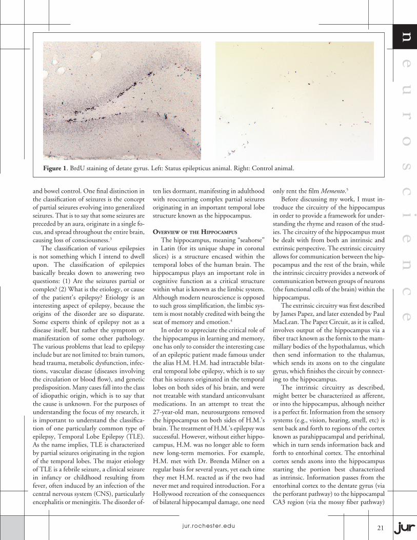
Figure 1. BrdU staining of detate gyrus. Left: Status epilepticus animal. Right: Control animal.
Volume 2 Issue 1 Fal l 2003 jur.rochester.edu
22 23
tr
em
bl
ay to the hippocampal CA1 region (via the
Schaffer-collateral pathway) and back out to the entorhinal cortex.
The primary neurotransmitter system under investigation in Temporal Lobe Epi-lepsy is glutamate. Throughout the circuitry described above, information is passed from one neuron to another by the release of glutamate from one neuron and binding of glutamate to receptors on the next neuron in the circuit, which induces certain changes in the cell’s electrical properties. Fortunately there are very few glutamate receptors that need to be discussed. The three major types of glutamate receptors include the AMPA/Kainate receptors, NMDA receptor (both named for artificial compounds that bind to them), and the metabotropic receptors 16.
The AMPA/Kainate receptors and NMDA receptors basically allow for the flow of ions (charged particles) through a channel in the protein. AMPA/Kainate receptors primarily allow for Na+ to pass
through the channel, making it more posi-tive, known as depolarizing the neuron. NMDA receptors allow both Na+ and Ca2+ to flow into the cell, thus depolarizing it. The NMDA receptor is noted for playing a crucial role in learning as well as cascades leading to neuronal death, both of which are mediated by the amount of free Ca2+ in the cell. Metabotropic glutamate receptors are known to effect long-term changes in the properties of the cell via so-called second messengers. Metabotropic receptors have also been implicated recently as possibly affecting the brain through the other major class of cells in the brain, glia.6
OVERVIEW OF NEUROGENESIS
Most animal cells replicate as a mecha-nism of repair of tissues and replacement of dead cells. Neurons in the Central Nervous System (CNS) have long been considered an exception to the proliferative cycle of animal tissues, and no evidence to date suggests mi-totic division of mature neurons. However, until recently, it was believed that new neu-rons were not produced in adulthood. Pro-duction of new neurons in the adult brain was first demonstrated just over 30 years ago.7,8 It is only recently that the conclusions of this earlier work have become accepted as scientific truth. Research has led to the discovery of proliferative zones where new neurons are being produced in the adult brain. One area of neural progenitors is the dentate gyrus of the hippocampal forma-tion.9,10,11 The production of new neurons in the hippocampus occurs in mammalian species including human and non-human primates.12,13
Research into this new phenomenon, known as adult neurogenesis, has come to be one of the burgeoning subfields of neuroscience research. A great number of questions have arisen since the initial discovery of these neurons produced dur-ing adulthood. A subset of these questions include: From what cell lines do these adult-produced neurons come? What factors in-duce the division of so-called progenitor cells, the multipotent cells responsible for creating neurons? Do these new neurons help or hurt existing neurons and cogni-tive processes? Could these new neurons or their progenitor stem cells be useful in replacement therapy for treating diseases of the nervous system?
Neurogenesis has proven itself to be a contentious field of neuroscience research.
Some members of the scientific commu-nity propose new ideas, while others tear them down for their naivety and technical differences. Elizabeth Gould recently pro-posed the introduction of new neurons in the cerebral cortex of primates, a finding refuted by neurogenesis aficionados Pasko Rakic and David Kornack.14,15 Rakic has proven himself most combative to the crowd of neuroscientists pushing their way into the study of neurogenesis. In a recent review, “Adult Neurogenesis in Mam-mals: An Identity Crisis”, Rakic delivers a deliberate argument positing a need for stringent criteria in identifying new neu-rons.16 Cameron recently expressed dismay at with the technique used by many to study neurogenesis; particularly those scientists she feels are seemingly ignoring a possible role of the blood brain barrier (the brain’s protective shielding from toxic molecules in the blood).17
Where does epilepsy fit into all of this? In 1997, a group of researchers made a discovery that there was a vast increase in the number of new neurons present in the brains (particularly the dentate gyrus) of rats that had been given pilocarpine, a drug known to elicit seizures, as well as rat given seizures by a process known as kindling.18 This finding begs some of the questions presented early, as well as some new ones. Again, one is left wondering what causes neurogenesis, and what, in particular to seizures, would cause such an increase. Also, one is left wondering whether neu-rogenesis is compensating for the death of certain other neurons. Another interesting, yet erroneous, hypothesis is that a charac-teristic pathology of epilepsy, known as mossy fiber sprouting, is caused specifically by the improper projections of axons from the newly born neurons. This conclusion was later proven erroneous when the same research team found that killing the new neurons with radiation did not prevent mossy fiber sprouting.19
NEUROGENESIS IN THE KAINIC ACID MODEL OF TLE
Increased hippocampal neurogenesis has been demonstrated in a number of animal models of TLE. Increased hippocampal neurogenesis has recently been suggested and documented in animal models of TLE paradigms including lithium-pilocarpine, flourothyl, and perforant path kindling, lim-bic kindling, electroconvulsive shock, and
Volume 2 Issue 1 Fal l 2003 jur.rochester.edu
TABLE 1.TYPES OF SEIZURES1. Partial (focal) seizures
3. Epilepsies and syndromes undetermined with respect to 1 or 2
3.1 With both partial and generalized seizures (eg, neonatal seizures) 3.2 Without unequivocal generalized or partial features
4. Special syndromes (eg, febrile convulsions)
22 23
ne
ur
os
ci
en
ce
kainic acid microinjection.20,21,22,23,24,25,26,27 However, use of my model of TLE has yet to be used in investigating the increase in neurogenesis in the hippocampus.
The animal model of TLE used in Dr. Shirley Joseph’s laboratory is the systemic kainic acid-induced status epilepticus mod-el. Kainic acid is the drug for which kainate receptors were named. It acts by binding the kainate receptors in such a way as to allow a greater response to glutamate binding, thus it is an agonist. This increase in glu-tamate transmission induces seizures. The seizures spread from the hippocampus and generalize to the entire brain. In summary, kainic acid elicits a complex partial seizure with so-called secondary generalization, in which a partial seizure progresses to become generalized. It must be noted that the kai-nic acid model described above is distinct from the microinjection technique listed previously. The microinjection involves surgical injection of kainic acid into the desired region (in that case hippocampus CA3 region). This particular model is used more-or-less to create a lesion, or destroy a region of the brain. Our systemic injections (into the gross circulation) of kainic acid allow for smaller levels of the neurotoxin to seep into all of the brain. Status epilepticus (sometimes referred to as epileptic status) is a clinical term used to describe 30 minutes or more of generalized seizure without sig-nificant interruption. For the purposes of my research, animals are injected with kain-ate and allowed to reach status epilepticus for a period of no more than one hour.
How exactly does this animal model have anything to do with the development of epilepsy in human beings? As mentioned earlier, many people with TLE had some form of early insult to the brain (e.g., en-cephalitis, meningitis, or head injury). Oc-casionally, these early insults are enough to induce a period of status epilepticus, which can only be ended by use a phenobarbital or other anticonvulsants, as is also done with our animals. In essence, the use of systemic kainic acid injections to elicit seizure is analogous to the febrile seizure or early in-sult of children who may potentially have adult-onset epilepsy.
Birth-dating of newly born cells is a process worth discussing in order to understand the findings of subsequent studies. Bromodeoxyuridine(BrdU) is commonly used as the chemical of choice in determining the age of new cells. BrdU
is able to incorporate into the DNA of cells that are in the process of dividing, known as mitosis. BrdU can later be stained for us-ing antibodies that specifically bind to the chemical, a technique known as immuno-histochemistry. Using this technique, one is able to specifically determine which cells were born at approximately the time the chemical was given.
In my earliest study, animals were given BrdU on days 5-8 following treatment with either kainic acid or saline (control group). Animals were sacrificed on day 9 in order to analyze 40um brain slices. As expected, there were vastly more cells in the dentate gyrus that stained for BrdU in animals treated with kainate in comparison to those treated with saline. However, this evidence alone neither indicates that the new cells are neurons, nor that the hippocampus is the only brain region in which the number of BrdU-stained cells were increased.
Using immunohistochemistry, it is also possible to stain for other molecules and proteins that may be present. Some of these proteins are present specifically in certain types of cells, thus allowing the investigator to stain only for the particular type of cells in which they are interested. Utilizing this technique, I was able to determine whether the same cells that had stained for BrdU were actually neurons by staining for Neu-ron-specific Nuclear Antigen (NeuN). Sur-prisingly, none of the cells stained for BrdU in the dentate gyrus of the controls nor the seizure animals were stained for NeuN. In fact, none of the BrdU cells anywhere in the brain also stained for NeuN, thus indicating that they were not mature neurons.
In a later studies animals survived 6 weeks, 8 weeks, and 6 months following the same treatments as described in the previous study. The dentate gyrus region of the hippocampus did contain neurons that stained for BrdU. There was also an apparent increase of an order of magnitude in the number of neurons born after seizure compared to control. The newly born cells in the dentate gyrus eventually differentiate into mature neurons and migrate from a re-gion known as the subgranular zone (SGZ) into the granule cells of the dentate.
Interestingly, the number of adult-gener-ated neurons remained relatively stable be-tween the shortest and longest survival time following BrdU injection. The relative sig-nal or strength of BrdU staining, however, did not remain stable. Although the number
of new neurons in either case were not sig-nificantly different. There was a significant reduction in the level of staining present in the long survival time. Hypotheses for this development have been suggested, but ex-perimental evidence lies outside of the scope of neuroscience, within genetics, chemistry, and molecular biology. The hypotheses are (in no particular order): (1) a combination of continued division of progenitor cells and death of new neurons diluted the quantity of BrdU during further replication of DNA, while offsetting the increase via the death of older BrdU-stained cells. (2) degradation of BrdU. (3) Specific excision of BrdU from DNA by enzymes during DNA repair. As a scientist, one must also concede that the possible solution may be a combina-tion of hypotheses, or something not yet mentioned.
Curiously, other regions of the brain, all of them known for experiencing seizure-in-duced neuronal death, illustrated abundant increases in the quantity of BrdU stained cells. These areas include: CA3 and CA1 of the hippocampus, the subventricular zone, amygdala, piriform cortex, and nuclei of the thalamus. None of the newly born cells in these regions of the brain stained with NeuN. These newly generated cells all appear in regions involved in the Papez circuit, both intrinsic and extrinsic areas, as described earlier.
Since the majority of new cells were not neurons, the question became: What type of cells are they? A multitude of antibod-ies were used to stain for a vast array of cell types. Possible cell types investigated included the various types of glia and white blood cells. Our rationale for looking at white blood cells is based on a growing body of evidence indicating a crucial role of the immune system in exacerbating neuronal death. The result of our hunt for the origin of the abundant new cells throughout the Papez circuit concluded that these cells are a combination of glia and immune cells.
Microglia are phagocytic cells within the nervous system. Using an antibody named ED1, specifically created to stain macro-phages (a type of phagocytic white blood cell from which microglia are derived), we were able to determine the origins of the non-neuronal BrdU-stained cells.28 Microglia are known for their response, known as activation, during neurode-generation. Microglial activation occurs during Parkinson’s disease, Alzheimer’s
Volume 2 Issue 1 Fal l 2003 jur.rochester.edu
24 25
tr
em
bl
ay
the drug provided a way of inhibiting two proposed mechanisms of neuronal death simultaneously. In one sense it was prevent-ing immune response by blocking signaling of proinflammatory cytokines, molecules that attract immune cells to damage sites. In another sense FK506 was preventing cell death by limiting NO signaling.
The preliminary results of FK506 pretreatment were encouraging. Animals treated with FK506 prior to seizure exhib-ited similar neuronal loss in CA1 in com-parison to the dextromethorphan animals. However, there was a substantial reduction in the immune response. Far fewer ED1 stained microglia were present in the CA1 region of animals with similar seizure sever-ity. Again, there were no significant differ-ences in the quantity of new neurons pres-ent in the dentate gyrus when compared to animals without pretreatment. These results suggest little or no involvement of the im-mune response in the increased production of new neurons in the dentate gyrus.
A final group of animals was given pre-treatment with both dextromethorphan and FK506. Functioning in concert, the two drugs appear to be very effective in prevent-ing both the neuronal loss and microglial immune response in the CA1 region of the hippocampus. These animals exhibited less neuronal loss and fewer ED1 stained microglia. However, as one would come to expect, there were no significant differences in the number of new neurons in the den-tate gyrus in comparison to animals without pretreatment. This once again supports the lack of an influence of NMDA-mediated signaling and immune cells in the increased production of neurons in the dentate gyrus following status epilepticus.
In essence, the results of my drug pre-treatment experiments disprove two of my hypotheses: (1) that NMDA receptor-medi-ated signaling pathways regulate the increase in the number of adult generated neurons in rats after status epilepticus, (2) that im-mune response may be crucial in initiating increased neurogenesis in animals after status epilepticus. Although not discussed previously, evidence suggests that significant increases in adult generated neurons can be linked to status epilepticus and not merely to kainic acid treatment. Only approxi-mately 60-70 percent of animals receiving kainic acid actually achieve status epilepti-cus. Other animals have low-grade seizures and exhibit a level of neurogenesis similar
disease, multiple sclerosis, AIDS-related dementia, and most notably epilepsy.29 The phenomenon that prompted the birth of cells throughout the Papez circuit was the activation of microglia in response to excessive excitation and presumably death of neurons in the hippocampus, amygdala, and thalamus.
A topic of interest to my investigation is where these microglia came from, and what prompted their rapid proliferation. Given some recent discoveries in the field of neuroscience, there exist a number of factors that may contribute to the explo-sion of microglia in response to seizures. I would like to highlight three findings in particular: (1) astrocytes, a particular type of glial cell, express receptors for glutamate which influence the blood vessels to which their endfeet are attached. This glutamate-induced signaling causes an increase in the circumference of blood vessels, thus dilating them.30 (2) a growth factor, granulocyte/monocyte-colony stimulating factor (GM-CSF) has been found to induce the rapid proliferation and maturation of microglia into their active form.31,32 (3) GM-CSF is capable of crossing the blood brain barrier, as well as being produced in some circum-stances by astrocytes.33,34 Thus, it appears that astrocytes may play a crucial role in the process of epileptogenesis.
I propose that the implications of these three findings in concert provide the neces-sary environment for microglial activation in our kainic-acid model of Temporal Lobe Epilepsy. Administration of kainic acid in-creases excitation through AMPA/Kainate receptors, which also allow for the activation NMDA receptors and increased release of glutamate. Increases in glutamate signaling may then increase activation of signaling pathways of astrocytes including the dila-tion of local blood vessels and increased re-lease of GM-CSF. The same molecules that attract microglia toward sites of neuronal death are likely to attract macrophages and other immune cells from the blood. The combination of these elements are likely to precipitate the immune response to neuro-nal death often described in pathologies of the brain.
One might be wondering what rela-tionship microglial response and NMDA receptor signaling pathways play in affect-ing neuronal death and birth. I approached these questions in a recent study by exploit-ing the wonderful array of pharmacological
agents at the modern scientist’s disposal. The specific intent of my investigation was two-fold: (1) to examine the effects of a drug that would block the detrimental influx of calcium ions through NMDA receptors as alluded to earlier. (2) to examine the ef-fects of suppressing the microglial immune response.
There are a great many pharmacologi-cal agents designed to specifically block the Ca2+ channel of the NMDA receptor. Unfortunately, many of these drugs have undesirable psychotropic effects due to the very fact that they block the NMDA recep-tor. Such substances include phencyclidine (PCP) and ketamine. Use of psychotropic substances or doses to prevent epileptogen-esis is clinically undesirable for obvious rea-sons. A drug known as dextromethorphan was chosen because of a moderate binding affinity to NMDA receptors and evidence of anticonvulsant and protective effects in kainic acid treated hippocampal neurons.35 Dextromethorphan was administered as a pretreatment 30 minutes prior to adminis-tration of kainic acid.
Preliminary results of the dextrometho-rphan study were not as expected. Animals pretreated with dextromethorphan exhibited a greater extent of neuronal loss in the CA1 region of the hippocampus. In conjunction with this neuronal loss, the animals also ex-hibited a more severe microglial immune response in CA1. Surprisingly, there were no significant differences in the number of new neurons in the dentate gyrus between animals that received pretreatment and animals that received only saline prior to seizures. Thus, dextromethorphan neither increased nor decreased the effect of kainic acid-induced status epilepticus on neuro-genesis, suggesting little or no involvement of NMDA-mediated signal pathways in the increased production of new neurons in the dentate gyrus.
There are surprisingly few drugs com-monly used to suppress immune response. For the purposes of studying the effects of blocking microglial immune response, I chose a compound known as FK506, because it was also demonstrated to inhibit certain steps along the NMDA-mediated signaling pathways. Specifically, FK506 inhibits calcineurin, a phosphatase which is responsible for activating nitric oxide syn-thase (NOS), which produces nitric oxide (NO) a powerful signaling molecule known to be deleterious in large quantities.36 Thus,
Volume 2 Issue 1 Fal l 2003 jur.rochester.edu
24 25
Figure 2. From left to right, top to bottom: 1. NeuN (Green) SE animal (9day) dentate gyrus; 2. BrdU (Red) SE animal (9day) dentate; 3. Merged NeuN(Green)/BrdU(Red) SE animal (9day) dentate; 4. NeuN (Green) SE animal (6 weeks) dentate; 5. BrdU (Red) SE animal (6 weeks) dentate; 6. Merged NeuN(Green)/BrdU(Red) SE animal (6 weeks) dentate; 7. NeuN (Green) SE animal (6 month) dentate; 8. BrdU (Red) SE animal (6 month) dentate; 9. Merged NeuN(Green)/BrdU(Red) SE animal (6 month) dentate.
to control animals. Given this information, one can only conclude that there is some trigger for neurogenesis that depends upon continuous high-grade seizures.
FUTURE DIRECTIONS
The next phase in my studies of in-creased neurogenesis in epilepsy will ex-amine the axonal projections of adult born neurons. As illustrated earlier, the correct axonal projections from the dentate gyrus will synapse in hippocampal area CA3, a projection known as the mossy fiber pathway. One of the specific changes that often takes place in an epileptic hippocam-pus, also demonstrated in animal models of epilepsy, is a sprouting of mossy fiber axons back onto the dentate gyrus, known as mossy fiber sprouting. There is inconsis-tent evidence in the literature regarding the contribution of adult generated neurons to
would be useful for determining whether the increase in proliferation in the dentate gyrus were an acute or chronic effect of status epilepticus. It would appear from the literature that the rate of neurogenesis in normal animals is indicative of a turnover in the dentate granule neuron population.42 It may be possible that increased apoptotic death due to continuous seizures is causing an increase in the turnover rate of granule cells in the dentate gyrus, which may in fact explain why there is little discussion in epilepsy literature of neuronal loss in the dentate gyrus.43
Another speculative project of inter-est would investigate the possible role of astrocytes in propagating an increase in neurogenesis in animal models of epilepsy. As indicated earlier, evidence suggests many roles of astrocytes in affecting neurotrans-mission, vascular regulation, and immune response. Of particular interest are the abilities of astrocytes to release GM-CSF and modulate dilation of local blood vessels. Given the increased glutamate neurotrans-mission during seizures, it seems plausible that activation of metabotropic glutamate receptors on astrocytes could lead to an increase in GM-CSF via two mechanisms: (1) direct astrocytic release of the factor, (2) increased local blood flow in concert with blood brain barrier disruption. Although no evidence exists to suggest such a finding, it appears plausible that the coincidence of sei-zure and neurogenesis may in part be due to GM-CSF signaling in the progenitor cells from which adult generated neurons arise. Interestingly, this suggestion was also made in recent literature describing the role of GM-CSF in microglial activation.
As indicated by the many future direc-tions of this work, many of the fundamen-tal questions regarding the relationship between epilepsy and neurogenesis remain unanswered. There is evidence of diverse modulation of neurogenesis related to neurotransmitters, growth factors, cyto-kines, drugs, and experience.44 The idea that such an effect is mediated by a single factor would appear ludicrous given the overwhelming evidence of such interactions in normal animals. However, the tight link between seizures and neurogenesis suggests that epilepsy may be an exceptional case in which the effects of multiple interactions and signaling pathways converge via a com-mon mechanism induced by seizures.
mossy fiber sprouting.37,38 There is recent evidence that adult generated neurons in normal animals contribute to normal mossy fiber connections between the dentate gyrus and CA3.39,40,41 In my experiment I intend to inject a fluorescent substance into CA3 neurons that will be transported backward to the dentate gyrus and forward to CA1, known as retrograde and anterograde re-spectively. Exploiting BrdU for the same purpose as previously, I expect to identify a normal mossy fiber projection from adult generated neurons via fluorescent BrdU staining and the tracer substance in the same neuron.
Another project of interest is a temporal study of the level of proliferation after status epilepticus. By giving BrdU at varying times after status epilepticus it would become pos-sible to establish a clear relationship between the timing of events. In essence, this study
Volume 2 Issue 1 Fal l 2003 jur.rochester.edu
ne
ur
os
ci
en
ce
26 27
tr
em
bl
ay
ing the proliferation of neuronal progenitor cells may one day allow for brain repair. It appears possible that this may be just the purpose neurogenesis is serving in the den-tate gyrus. Better understanding of the role of new neurons in replacement and plastic-ity can only lead to a better understanding of epileptogenesis and plasticity.59 Although it seems unlikely at the present, the con-vergence of stem cell and gene therapy research may one day allow for the correc-tion of various neurological disorders. The technology to regulate neurogenesis and modulate gene expression may afford phy-sicians and scientists the illusion of control over the complexity of our most important organ, the brain.
To underscore the importance of un-derstanding the mechanisms and effects of neurogenesis, one has only to review the literature regarding the possible roles of these adult generated neurons in learning and memory. Animal models have demon-strated a strong relationship between maze learning and hippocampal neurogenesis in rats.45,46,47 Investigators have long known of the relationship between bird song and neurogenesis. Birds that learn song to at-tract a mate display neurogenesis in cen-ters of the brain regulating song learning throughout the process of acquiring song, again implying a crucial role in learning and memory.48,49 Studies have demonstrated an age-related decline in neurogenesis as well, although correlation with memory deficits is not necessarily related.50,51
Recent research into the mechanisms underlying depression and the action of antidepressants has proven relevant to our understanding of neurogenesis. Evidence for antidepressant effects on neurogenesis has been found across a vast array of drugs ranging from classical antidepressants such as selective serotonin reuptake inhibitors
(SSRIs) like Prozac and Zoloft, tricyclic antidepressants, to newer drugs like tianep-tine and drugs the block the neuropeptide substance P.52,53,54 Some investigators have proposed a “neurogenic theory” of depres-sion.55 In conconcordance with evidence of a neurogenic effect of antidepressants is evi-dence that depletion of serotonin decreases neurogenesis in the dentate gyrus.56 Recent evidence that neurogenesis is modulated by cyclic adenosine monophosphate (cAMP) and cAMP Response Element-Binding Pro-tein (CREB) add great support to the role of neurogenesis and depression.57 It is currently believed that these same pathways mediate the mechanism of action of antidepressants. Some scientists in the field speculate that these recent findings may lead to targeting of neurogenesis and the cAMP-CREB sig-naling pathway in our understanding and treatment of major depression.58
Given the implications that neurogenesis may have in diseases and function of the nervous system, the importance of deter-mining the cause and effect relationship between neurogenesis and animal models of epilepsy cannot be overstated. Understand-
Matthew Tremblay recieved a B.S. in Neuroscience from the University of Rochester in May 2003. The article is an abridged version of his senior honors thesis. Matthew is pursuing a M.D./Ph.D. degree in the Medical Scientist Training Program at Albert Einstein College of Medicine.