5 Epstein-Barr Virus Serology in the Detection and Screening of Nasopharyngeal Carcinoma Li-Jen Liao 1,2 and Mei-Shu Lai 2 1 Department of Otolaryngology, Far Eastern Memorial Hospital, New Taipei City, 2 Graduate Institute of Epidemiology and Preventive Medicine, College of Public Health, National Taiwan University, Taipei, Taiwan 1. Introduction Nasopharyngeal carcinoma (NPC) is a common cancer among Southern Chinese with a male dominance of about 3:1. The age-adjusted incidence for both sexes is less than one per 100, 000 population worldwide. The reported incidence of NPC among men and women in Hong Kong is 20–30 per 100, 000 and 15–20 per 100, 000.(Wei and Sham, 2005) The reported incidence of NPC among men and women in Taiwan is 8.3 per 100 000 and 2.8 per 100 000, respectively.(Bureau of health promotion, Taiwan, 2010) It mainly afflicts people in mid-life. There is now compelling evidences to suggest that Epstein-Barr virus (EBV) is associated with the development of NPC and is most likely to be involved in the multi-step and multi- factorial carcinogenesis of NPC. In this chapter, the role of EBV in pathogenesis of NPC is reviewed briefly, and principle applications of EBV antibodies and circulating EBV DNA as markers of NPC are outlined. Based on current knowledge of EBV antibody responses by NPC and taking available testing technologies into account, serologic screening strategy to facilitate efficient early detection of NPC is formulated. 2. EBV related pathogenesis of NPC The pathogenesis of NPC includes multi-stepped process that leads to the development of NPC (Fig. 1.). EBV infection alone cannot drive normal cells towards carcinoma development. It is thought that loss of heterozygosity (LOH on chromosome 3p and 9p, which are the location of some tumor suppressor genes), possibly as a result of inherited traits (Chinese ethnicity) as well as exposure to dietary factors (salted fish) and other environmental cofactors (Formaldehyde), is an early stage event in the pathogenesis of this disease. EBV is infected within these low-grade pre-invasive lesions, subsequent to further genetic and epigenetic alterations. EBV was first suspected to be linked with NPC on the basis of the serological observations by Old and colleagues (Old et al., 1966) in 1966. This link was formally demonstrated later by in situ hybridization of the viral DNA in the nuclei of epithelial cells (zur Hausen et al., 1970). The full length EBV genome is contained in all malignant epithelial cells, but not in most infiltrating lymphocytes. The association with EBV is constant, regardless of the www.intechopen.com
Transcript
5
Epstein-Barr Virus Serology in the Detection and Screening of Nasopharyngeal Carcinoma
Li-Jen Liao1,2 and Mei-Shu Lai2 1Department of Otolaryngology, Far Eastern Memorial Hospital, New Taipei City,
2Graduate Institute of Epidemiology and Preventive Medicine, College of Public Health, National Taiwan University, Taipei,
Taiwan
1. Introduction
Nasopharyngeal carcinoma (NPC) is a common cancer among Southern Chinese with a male dominance of about 3:1. The age-adjusted incidence for both sexes is less than one per 100, 000 population worldwide. The reported incidence of NPC among men and women in Hong Kong is 20–30 per 100, 000 and 15–20 per 100, 000.(Wei and Sham, 2005) The reported incidence of NPC among men and women in Taiwan is 8.3 per 100 000 and 2.8 per 100 000, respectively.(Bureau of health promotion, Taiwan, 2010) It mainly afflicts people in mid-life. There is now compelling evidences to suggest that Epstein-Barr virus (EBV) is associated with the development of NPC and is most likely to be involved in the multi-step and multi-factorial carcinogenesis of NPC. In this chapter, the role of EBV in pathogenesis of NPC is reviewed briefly, and principle applications of EBV antibodies and circulating EBV DNA as markers of NPC are outlined. Based on current knowledge of EBV antibody responses by NPC and taking available testing technologies into account, serologic screening strategy to facilitate efficient early detection of NPC is formulated.
2. EBV related pathogenesis of NPC
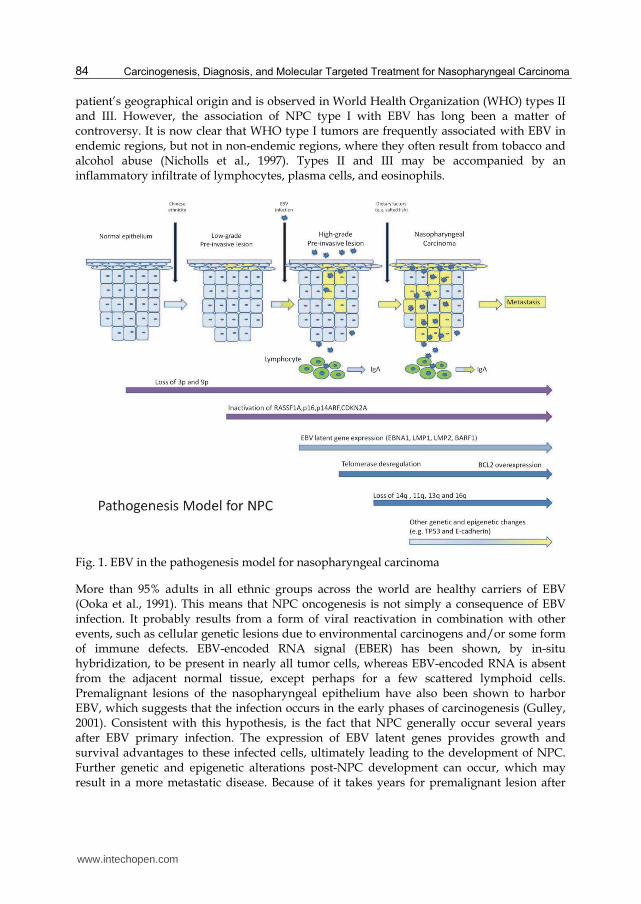
The pathogenesis of NPC includes multi-stepped process that leads to the development of
NPC (Fig. 1.). EBV infection alone cannot drive normal cells towards carcinoma
development. It is thought that loss of heterozygosity (LOH on chromosome 3p and 9p,
which are the location of some tumor suppressor genes), possibly as a result of inherited
traits (Chinese ethnicity) as well as exposure to dietary factors (salted fish) and other
environmental cofactors (Formaldehyde), is an early stage event in the pathogenesis of this
disease. EBV is infected within these low-grade pre-invasive lesions, subsequent to further
genetic and epigenetic alterations.
EBV was first suspected to be linked with NPC on the basis of the serological observations by Old and colleagues (Old et al., 1966) in 1966. This link was formally demonstrated later by in situ hybridization of the viral DNA in the nuclei of epithelial cells (zur Hausen et al., 1970). The full length EBV genome is contained in all malignant epithelial cells, but not in most infiltrating lymphocytes. The association with EBV is constant, regardless of the
www.intechopen.com
Carcinogenesis, Diagnosis, and Molecular Targeted Treatment for Nasopharyngeal Carcinoma
84
patient’s geographical origin and is observed in World Health Organization (WHO) types II and III. However, the association of NPC type I with EBV has long been a matter of controversy. It is now clear that WHO type I tumors are frequently associated with EBV in endemic regions, but not in non-endemic regions, where they often result from tobacco and alcohol abuse (Nicholls et al., 1997). Types II and III may be accompanied by an inflammatory infiltrate of lymphocytes, plasma cells, and eosinophils.
Fig. 1. EBV in the pathogenesis model for nasopharyngeal carcinoma
More than 95% adults in all ethnic groups across the world are healthy carriers of EBV (Ooka et al., 1991). This means that NPC oncogenesis is not simply a consequence of EBV infection. It probably results from a form of viral reactivation in combination with other events, such as cellular genetic lesions due to environmental carcinogens and/or some form of immune defects. EBV-encoded RNA signal (EBER) has been shown, by in-situ hybridization, to be present in nearly all tumor cells, whereas EBV-encoded RNA is absent from the adjacent normal tissue, except perhaps for a few scattered lymphoid cells. Premalignant lesions of the nasopharyngeal epithelium have also been shown to harbor EBV, which suggests that the infection occurs in the early phases of carcinogenesis (Gulley, 2001). Consistent with this hypothesis, is the fact that NPC generally occur several years after EBV primary infection. The expression of EBV latent genes provides growth and survival advantages to these infected cells, ultimately leading to the development of NPC. Further genetic and epigenetic alterations post-NPC development can occur, which may result in a more metastatic disease. Because of it takes years for premalignant lesion after
www.intechopen.com
Epstein-Barr Virus Serology in the Detection and Screening of Nasopharyngeal Carcinoma
85
EBV infection to NPC formation(Choi et al., 2011; Ji et al., 2007; Ng et al., 2010; Ng et al., 2005), the long period of pre-clinical detectable phase (PCDP) offer the opportunity for screening and early diagnosis of NPC.
3. Applications of EBV antibodies and EBV DNA as markers of NPC in population
3.1 Plasma EBV protein in the diagnosis of NPC
Serological studies have shown that the clinical onset of NPC is preceded by the appearance
of a high titer of various EBV antibodies such as viral capsid antigens (VCA) IgA(Li et al.,
2010; Ng et al., 2010; O et al., 2007), anti-EBV DNase(Chien et al., 2001) and combined EBV
EA(early antigen) + EBNA-1 (Nuclear antigen 1) IgA test(Chang et al., 2008)( Table 1.).
Study type Author Marker Results of the utility
Case-control study O et al. (O et al., 2007)
VCA IgA (ELISA)
Sensitivity 90.6% Specificity 93.5%
Ng et al. (2010) (Ng et al., 2010)
VCA IgA (ELISA and IF)
Sensitivity 83.3% Specificity 87.0%
Chang et al. (2008) (Chang et al., 2008)
EA+EBNA1 IgA (ELISA)
Sensitivity 94.2% Specificity 82.6%
Meta-analysis Li et al. (2010) (Li et al., 2010)
VCA IgA (ELISA and IF)
Sensitivity 92% Specificity 98%
Cohort study Chien et al. (2001) (Chien et al., 2001)
VCA IgA(IF) and anti-DNase (enzyme neutralization assay)
Rate ratio VCA IgA 22.0 (7.3–66.9) anti-DNase 3.5 (1.4–8.7)
Table 2. The comparison of the stage distribution between screen-detected and clinically detected based on published data on NPC screening during 1979–1992 (Ng et al., 2010).
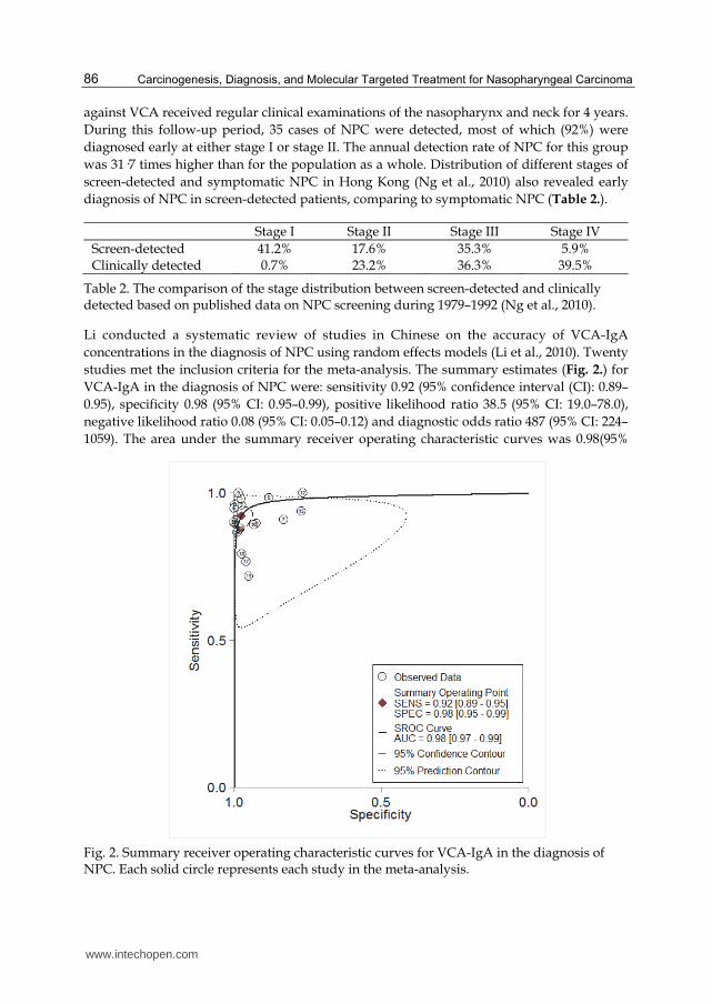
Li conducted a systematic review of studies in Chinese on the accuracy of VCA-IgA
concentrations in the diagnosis of NPC using random effects models (Li et al., 2010). Twenty
studies met the inclusion criteria for the meta-analysis. The summary estimates (Fig. 2.) for
VCA-IgA in the diagnosis of NPC were: sensitivity 0.92 (95% confidence interval (CI): 0.89–
negative likelihood ratio 0.08 (95% CI: 0.05–0.12) and diagnostic odds ratio 487 (95% CI: 224–
1059). The area under the summary receiver operating characteristic curves was 0.98(95%
Fig. 2. Summary receiver operating characteristic curves for VCA-IgA in the diagnosis of NPC. Each solid circle represents each study in the meta-analysis.
www.intechopen.com
Epstein-Barr Virus Serology in the Detection and Screening of Nasopharyngeal Carcinoma
87
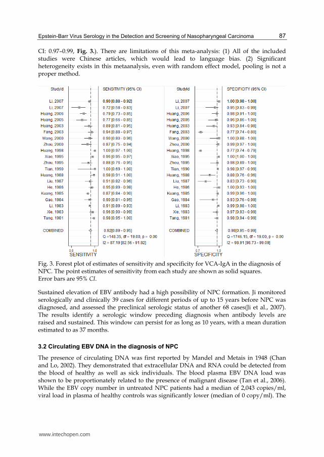
CI: 0.97–0.99, Fig. 3.). There are limitations of this meta-analysis: (1) All of the included studies were Chinese articles, which would lead to language bias. (2) Significant heterogeneity exists in this metaanalysis, even with random effect model, pooling is not a proper method.
Fig. 3. Forest plot of estimates of sensitivity and specificity for VCA-IgA in the diagnosis of NPC. The point estimates of sensitivity from each study are shown as solid squares. Error bars are 95% CI.
Sustained elevation of EBV antibody had a high possibility of NPC formation. Ji monitored serologically and clinically 39 cases for different periods of up to 15 years before NPC was diagnosed, and assessed the preclinical serologic status of another 68 cases(Ji et al., 2007). The results identify a serologic window preceding diagnosis when antibody levels are raised and sustained. This window can persist for as long as 10 years, with a mean duration estimated to as 37 months.
3.2 Circulating EBV DNA in the diagnosis of NPC
The presence of circulating DNA was first reported by Mandel and Metais in 1948 (Chan and Lo, 2002). They demonstrated that extracellular DNA and RNA could be detected from the blood of healthy as well as sick individuals. The blood plasma EBV DNA load was shown to be proportionately related to the presence of malignant disease (Tan et al., 2006). While the EBV copy number in untreated NPC patients had a median of 2,043 copies/ml, viral load in plasma of healthy controls was significantly lower (median of 0 copy/ml). The
www.intechopen.com
Carcinogenesis, Diagnosis, and Molecular Targeted Treatment for Nasopharyngeal Carcinoma
88
demonstration of EBV DNA in the plasma/serum of patients suffering from NPC has provided us with a new tool for NPC detection and monitoring.
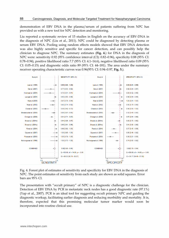
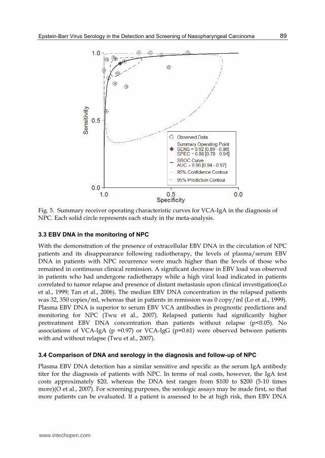
Liu reported a systematic review of 15 studies in English on the accuracy of EBV-DNA in
the diagnosis of NPC (Liu et al., 2011). NPC could be diagnosed by detecting plasma or
serum EBV DNA. Pooling using random effects models showed that EBV DNA detection
was also highly sensitive and specific for cancer detection, and can possibly help the
clinician to diagnose NPC. The summary estimates (Fig. 4.) for DNA in the diagnosis of
0.78–0.94), positive likelihood ratio 7.7 (95% CI: 4.1–14.6), negative likelihood ratio 0.09 (95%
CI: 0.05–0.15) and diagnostic odds ratio 89 (95% CI: 44–181). The area under the summary
receiver operating characteristic curves was 0.96(95% CI: 0.94–0.97, Fig. 5.).
Fig. 4. Forest plot of estimates of sensitivity and specificity for EBV DNA in the diagnosis of NPC. The point estimates of sensitivity from each study are shown as solid squares. Error bars are 95% CI.
The presentation with “occult primary” of NPC is a diagnostic challenge for the clinician.
Detection of EBV DNA by PCR in metastatic neck nodes has a good diagnostic rate (97.1%)
(Yap et al., 2007). PCR is an ideal tool for suggesting occult primary NPC and guiding the
diagnostic workup, facilitating earlier diagnosis and reducing morbidity and mortality. It is,
therefore, expected that this promising molecular tumor marker would soon be
incorporated into routine clinical use.
www.intechopen.com
Epstein-Barr Virus Serology in the Detection and Screening of Nasopharyngeal Carcinoma
89
Fig. 5. Summary receiver operating characteristic curves for VCA-IgA in the diagnosis of NPC. Each solid circle represents each study in the meta-analysis.
3.3 EBV DNA in the monitoring of NPC
With the demonstration of the presence of extracellular EBV DNA in the circulation of NPC patients and its disappearance following radiotherapy, the levels of plasma/serum EBV DNA in patients with NPC recurrence were much higher than the levels of those who remained in continuous clinical remission. A significant decrease in EBV load was observed in patients who had undergone radiotherapy while a high viral load indicated in patients correlated to tumor relapse and presence of distant metastasis upon clinical investigation(Lo et al., 1999; Tan et al., 2006). The median EBV DNA concentration in the relapsed patients was 32, 350 copies/ml, whereas that in patients in remission was 0 copy/ml (Lo et al., 1999). Plasma EBV DNA is superior to serum EBV VCA antibodies in prognostic predictions and monitoring for NPC (Twu et al., 2007). Relapsed patients had significantly higher pretreatment EBV DNA concentration than patients without relapse (p<0.05). No associations of VCA-IgA (p =0.97) or VCA-IgG (p=0.61) were observed between patients with and without relapse (Twu et al., 2007).
3.4 Comparison of DNA and serology in the diagnosis and follow-up of NPC
Plasma EBV DNA detection has a similar sensitive and specific as the serum IgA antibody titer for the diagnosis of patients with NPC. In terms of real costs, however, the IgA test costs approximately $20, whereas the DNA test ranges from $100 to $200 (5-10 times more)(O et al., 2007). For screening purposes, the serologic assays may be made first, so that more patients can be evaluated. If a patient is assessed to be at high risk, then EBV DNA
www.intechopen.com
Carcinogenesis, Diagnosis, and Molecular Targeted Treatment for Nasopharyngeal Carcinoma
90
PCR can be performed in a series fashion (O et al., 2007). If this work-up is positive; then the patient should be referred to a specialist for further work-up including fiberoptic nasopharyngoscopy or MRI examination. The economic cost of providing this screening is reduce by prescreening to exclude the low-risk patients and performing series testing on the at-risk patients.
4. Applications of EBV antibodies and EBV DNA as markers of NPC among individual from high risk NPC families
4.1 Screening in high risk group with positive family history
The incidence of NPC is relative low through most of the world (less 1/100,000). However,
familial clustering of NPC has been widely documented in Chinese population (Chen and
Huang, 1997) and individual with family history are at a high risk of developing the disease.
Both family history and anti-EBV seropositivity are determinants in subsequent NPC
development. Based on mass screening with positive family history conducted in Taiwan
and Hong-Kong (Choi et al., 2011; Hsu et al., 2011; Ng et al., 2010; Yu et al., 2011), subjected
with positive family history will increase risk of subsequent NPC development. Screening
this high risk group significantly shifted to earlier stage (Ng et al., 2010), which mean it is a
screening modality to be worth.
The etiology of NPC is not completely understood now; approaches to primary prevention of NPC remain inconclusive. This situation highlights the need of secondary prevention for early detection, diagnosis, and treatment of NPC. The effectiveness of a screening program rests on several key factors: “the importance of the disease; a well-defined population at risk; the availability of noninvasive, low-cost screening tests that can detect disease at an early stage; and effective treatments resulting in long term survival.”(Choi et al., 2011) Screening for NPC for positive family history with EBV serology meets all of these criteria.
Chen first reported an evaluation of screening for NPC using Markov chain models (Chen et
al., 1999). Average duration of the PCDP is estimated as 3.1 years was estimated; therefore it
is possible for massive screening in NPC. The stage distribution for EBV-IgA antibodies is
higher (68.7%) than in the non-screened (25%) group for stage I and II. Based on these
findings, they suggest design a randomized trial in a high incidence area such as Hong
Kong.
In another cohort study from Taiwan (Hsu et al., 2011) comparing different covariates, the authors compared the long-term risk NPC of male participants in a NPC cohort after adjusting for anti- EBV seromarkers and cigarette smoking. The adjusted hazard ratio was 6.8 (95% CI: 2.3, 20.1) for the multiplex family cohort compared with the community cohort. In the evaluation of anti-EBV VCA IgA and anti-EBV DNase, the adjusted hazard ratios were 2.8 (95% CI: 1.3, 6.0) and 15.1 (95% CI: 4.2, 54.1) for those positive for 1 EBV seromarker and positive for both seromarkers, respectively, compared with those negative for both EBV seromarkers. The adjusted hazard ratio was 31.0 (95% CI: 9.7, 98.7) for participants who reported a family history of NPC and who were anti-EBV-seropositive compared with individuals without such a history who were anti-EBV-seronegative. These findings suggest that both family history of NPC and anti-EBV seropositivity are important determinants of
www.intechopen.com
Epstein-Barr Virus Serology in the Detection and Screening of Nasopharyngeal Carcinoma
91
subsequent NPC development. Screening for high risk multiplex family may be more cost-effective.
Screening in Hong-Kong with positive NPC family history was conducted since 1994 (Lee et al., 2005; Ng et al., 2010; Ng et al., 2005). Participants in this screening program for NPC were all first degree relatives of patients with NPC and they were all 18 years of age or older. Participants were offered annual assessment serological test of EBV and endoscopic examination of the nasopharynx. Between 1994 and 2005, total 1,199 asymptomatic family members of NPC patients were recruited and reported (Ng et al., 2010). Eighteen participants of the screening program developed NPC; 16 of them were detected in the screening. Stage distributions and survival outcomes of the 17 cases were compared with that of 1,185 consecutive symptomatic patients diagnosed in the same period through general referral. It was found that the screening program resulted in early detection of cancer (Table 2), with 59% presenting at early stage (stage I: 41%, stage II: 18%) compared to 24% (stage I: 1%, stage II: 23%) of symptomatic cancers (P=0.001), and a significant improvement in disease-free survival (P = 0.04). The cancer specific survival and overall survival rate at 5-year are also higher (92 vs. 77% and 92 vs. 70%, respectively).
4.2 Screening strategies of NPC among individual from high risk NPC families
The efficacy of any screening strategy should be evaluated before putting it into practice. Based on Markov chain models, Choi simulated and compared the outcomes of 4 screening strategies over a period of 12 years: (A) Annual screening, (B) biennial screening, (C) triennial screening, and (D) triennial screening for participants tested EBV negative and annual screening once the participants are tested EBV positive (Choi et al., 2011) . The result is summarized at Table 3, strategy A (screening annually) yields the maximum disease pick-up rate, but strategy D (triennial screening for participants tested EBV negative and annual screening once the participants are tested EBV positive) offered the highest efficacy for NPC screening of family members of NPC patients.
Strategy A Strategy B Strategy C Strategy D No screening
Total screens 77,652 41,837 29,898 44,618 - Positive EBV test 14,962 8,071 5,772 11,413 - Screen detected case 47 42 38 47 - Disease pick up rate 88.2% 78.6% 70.8% 87.4% - Reduction in disease pick up rate*
* Relative to strategy A Strategy A. Annual screening Strategy B. Biennial screening Strategy C. Triennial screening Strategy D. Triennial screening for participants tested EBV negative and annual screening once the participants are tested EBV positive
Table 3. Simulated screening result with the four strategies based on an imaginary population of 6,000 participants follow up for 12 years with family history of NPC (Choi et al., 2011).
www.intechopen.com
Carcinogenesis, Diagnosis, and Molecular Targeted Treatment for Nasopharyngeal Carcinoma
92
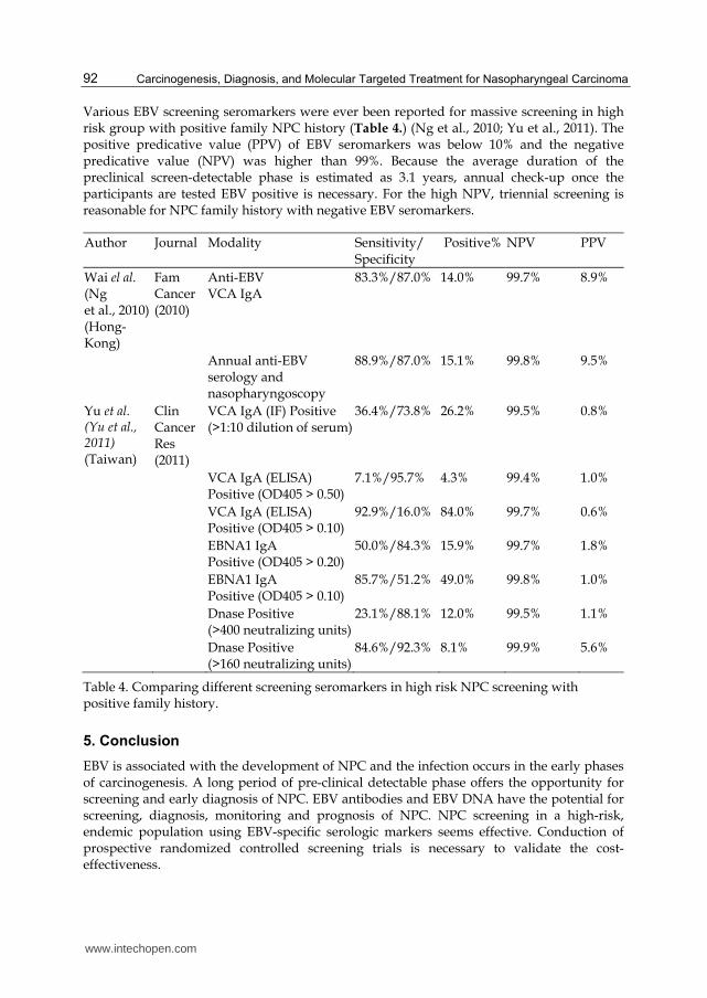
Various EBV screening seromarkers were ever been reported for massive screening in high risk group with positive family NPC history (Table 4.) (Ng et al., 2010; Yu et al., 2011). The positive predicative value (PPV) of EBV seromarkers was below 10% and the negative predicative value (NPV) was higher than 99%. Because the average duration of the preclinical screen-detectable phase is estimated as 3.1 years, annual check-up once the participants are tested EBV positive is necessary. For the high NPV, triennial screening is reasonable for NPC family history with negative EBV seromarkers.
Author Journal Modality Sensitivity/ Specificity
Positive% NPV PPV
Wai el al. (Ng et al., 2010) (Hong-Kong)
Fam Cancer (2010)
Anti-EBV VCA IgA
83.3%/87.0% 14.0% 99.7% 8.9%
Annual anti-EBV serology and nasopharyngoscopy
88.9%/87.0% 15.1% 99.8% 9.5%
Yu et al. (Yu et al., 2011) (Taiwan)
Clin Cancer Res (2011)
VCA IgA (IF) Positive (>1:10 dilution of serum)
36.4%/73.8% 26.2% 99.5% 0.8%
VCA IgA (ELISA) Positive (OD405 > 0.50)
7.1%/95.7% 4.3% 99.4% 1.0%
VCA IgA (ELISA) Positive (OD405 > 0.10)
92.9%/16.0% 84.0% 99.7% 0.6%
EBNA1 IgA Positive (OD405 > 0.20)
50.0%/84.3% 15.9% 99.7% 1.8%
EBNA1 IgA Positive (OD405 > 0.10)
85.7%/51.2% 49.0% 99.8% 1.0%
Dnase Positive (>400 neutralizing units)
23.1%/88.1% 12.0% 99.5% 1.1%
Dnase Positive (>160 neutralizing units)
84.6%/92.3% 8.1% 99.9% 5.6%
Table 4. Comparing different screening seromarkers in high risk NPC screening with positive family history.
5. Conclusion
EBV is associated with the development of NPC and the infection occurs in the early phases of carcinogenesis. A long period of pre-clinical detectable phase offers the opportunity for screening and early diagnosis of NPC. EBV antibodies and EBV DNA have the potential for screening, diagnosis, monitoring and prognosis of NPC. NPC screening in a high-risk, endemic population using EBV-specific serologic markers seems effective. Conduction of prospective randomized controlled screening trials is necessary to validate the cost-effectiveness.
www.intechopen.com
Epstein-Barr Virus Serology in the Detection and Screening of Nasopharyngeal Carcinoma
93
6. Acknowledgment
I am heartily thankful for Miss Wan-Lun Hsu and Miss Yu-Ping Cheng for data collection, manuscript reviewing and modification.
7. References
Bureau of health promotion, Executive Yuan, Taiwan, (2010). Cancer registry annual report. 28.
Chan, K. C., and Lo, Y. M. (2002). Circulating EBV DNA as a tumor marker for nasopharyngeal carcinoma. Semin Cancer Biol 12, 489-496.
Chang, K. P., Hsu, C. L., Chang, Y. L., Tsang, N. M., Chen, C. K., Lee, T. J., Tsao, K. C., Huang, C. G., Chang, Y. S., Yu, J. S., and Hao, S. P. (2008). Complementary serum test of antibodies to Epstein-Barr virus nuclear antigen-1 and early antigen: a possible alternative for primary screening of nasopharyngeal carcinoma. Oral Oncol 44, 784-792.
Chen, D. L., and Huang, T. B. (1997). A case-control study of risk factors of nasopharyngeal carcinoma. Cancer Lett 117, 17-22.
Chen, H. H., Prevost, T. C., and Duffy, S. W. (1999). Evaluation of screening for nasopharyngeal carcinoma: trial design using Markov chain models. Br J Cancer 79, 1894-1900.
Chien, Y. C., Chen, J. Y., Liu, M. Y., Yang, H. I., Hsu, M. M., Chen, C. J., and Yang, C. S. (2001). Serologic markers of Epstein-Barr virus infection and nasopharyngeal carcinoma in Taiwanese men. N Engl J Med 345, 1877-1882.
Choi, C. W., Lee, M. C., Ng, W. T., Law, L. Y., Yau, T. K., and Lee, A. W. (2011). An analysis of the efficacy of serial screening for familial nasopharyngeal carcinoma based on Markov chain models. Fam Cancer 10, 133-139.
Gulley, M. L. (2001). Molecular diagnosis of Epstein-Barr virus-related diseases. J Mol Diagn 3, 1-10.
Hsu, W. L., Yu, K. J., Chien, Y. C., Chiang, C. J., Cheng, Y. J., Chen, J. Y., Liu, M. Y., Chou, S. P., You, S. L., Hsu, M. M., et al. (2011). Familial tendency and risk of nasopharyngeal carcinoma in taiwan: effects of covariates on risk. Am J Epidemiol 173, 292-299.
Ji, M. F., Wang, D. K., Yu, Y. L., Guo, Y. Q., Liang, J. S., Cheng, W. M., Zong, Y. S., Chan, K. H., Ng, S. P., Wei, W. I., et al. (2007). Sustained elevation of Epstein-Barr virus antibody levels preceding clinical onset of nasopharyngeal carcinoma. Br J Cancer 96, 623-630.
Lee, A. W., Sze, W. M., Au, J. S., Leung, S. F., Leung, T. W., Chua, D. T., Zee, B. C., Law, S. C., Teo, P. M., Tung, S. Y., et al. (2005). Treatment results for nasopharyngeal carcinoma in the modern era: the Hong Kong experience. Int J Radiat Oncol Biol Phys 61, 1107-1116.
Li, S., Deng, Y., Li, X., Chen, Q. P., Liao, X. C., and Qin, X. (2010). Diagnostic value of Epstein-Barr virus capsid antigen-IgA in nasopharyngeal carcinoma: a meta-analysis. Chin Med J 123, 1201-1205.
Liu, Y., Fang, Z., Liu, L., Yang, S., and Zhang, L. (2011). Detection of epstein-barr virus DNA in serum or plasma for nasopharyngeal cancer: a meta-analysis. Genet Test Mol Biomarkers 15, 495-502.
Lo, Y. M., Chan, L. Y., Chan, A. T., Leung, S. F., Lo, K. W., Zhang, J., Lee, J. C., Hjelm, N. M., Johnson, P. J., and Huang, D. P. (1999). Quantitative and temporal correlation
www.intechopen.com
Carcinogenesis, Diagnosis, and Molecular Targeted Treatment for Nasopharyngeal Carcinoma
94
between circulating cell-free Epstein-Barr virus DNA and tumor recurrence in nasopharyngeal carcinoma. Cancer Res 59, 5452-5455.
Ng, W. T., Choi, C. W., Lee, M. C., Law, L. Y., Yau, T. K., and Lee, A. W. (2010). Outcomes of nasopharyngeal carcinoma screening for high risk family members in Hong Kong. Fam Cancer 9, 221-228.
Ng, W. T., Yau, T. K., Yung, R. W., Sze, W. M., Tsang, A. H., Law, A. L., and Lee, A. W. (2005). Screening for family members of patients with nasopharyngeal carcinoma. Int J Cancer 113, 998-1001.
Nicholls, J. M., Agathanggelou, A., Fung, K., Zeng, X., and Niedobitek, G. (1997). The association of squamous cell carcinomas of the nasopharynx with Epstein-Barr virus shows geographical variation reminiscent of Burkitt's lymphoma. J Pathol 183, 164-168.
O, T. M., Yu, G., Hu, K., and Li, J. C. (2007). Plasma Epstein-Barr virus immunoglobulin A and DNA for nasopharyngeal carcinoma screening in the United States. Otolaryngol Head Neck Surg 136, 992-997.
Old, L. J., Boyse, E. A., Oettgen, H. F., Harven, E. D., Geering, G., Williamson, B., and Clifford, P. (1966). Precipitating antibody in human serum to an antigen present in cultured burkitt's lymphoma cells. Proc Natl Acad Sci U S A 56, 1699-1704.
Ooka, T., de Turenne-Tessier, M., and Stolzenberg, M. C. (1991). Relationship between antibody production to Epstein-Barr virus (EBV) early antigens and various EBV-related diseases. Springer Semin Immunopathol 13, 233-247.
Paramita, D. K., Fachiroh, J., Haryana, S. M., and Middeldorp, J. M. (2009). Two-step Epstein-Barr virus immunoglobulin A enzyme-linked immunosorbent assay system for serological screening and confirmation of nasopharyngeal carcinoma. Clin Vaccine Immunol 16, 706-711.
Tan, E. L., Looi, L. M., and Sam, C. K. (2006). Evaluation of plasma Epstein-Barr virus DNA load as a prognostic marker for nasopharyngeal carcinoma. Singapore Med J 47, 803-807.
Twu, C. W., Wang, W. Y., Liang, W. M., Jan, J. S., Jiang, R. S., Chao, J., Jin, Y. T., and Lin, J. C. (2007). Comparison of the prognostic impact of serum anti-EBV antibody and plasma EBV DNA assays in nasopharyngeal carcinoma. Int J Radiat Oncol Biol Phys 67, 130-137.
Wei, W. I., and Sham, J. S. (2005). Nasopharyngeal carcinoma. Lancet 365, 2041-2054. Yap, Y. Y., Hassan, S., Chan, M., Choo, P. K., and Ravichandran, M. (2007). Epstein-Barr
virus DNA detection in the diagnosis of nasopharyngeal carcinoma. Otolaryngol Head Neck Surg 136, 986-991.
Yu, K. J., Hsu, W. L., Pfeiffer, R. M., Chiang, C. J., Wang, C. P., Lou, P. J., Cheng, Y. J., Gravitt, P., Diehl, S. R., Goldstein, A. M., et al. (2011). Prognostic utility of anti-EBV antibody testing for defining NPC risk among individuals from high-risk NPC families. Clin Cancer Res 17, 1906-1914.
Zeng, Y., Zhang, L., Wu, Y., Huang, Y., Huang, N., Li, J., Wang, Y., Jiang, M., Fang, Z., and Meng, N. (1985). Prospective studies on nasopharyngeal carcinoma in epstein barr virus IgA/VCA antibody positne persons in Wuzhou city, china. International journal of cancer 36, 545-547.
zur Hausen, H., Schulte-Holthausen, H., Klein, G., Henle, W., Henle, G., Clifford, P., and Santesson, L. (1970). EBV DNA in biopsies of Burkitt tumours and anaplastic carcinomas of the nasopharynx. Nature 228, 1056-1058.
www.intechopen.com
Carcinogenesis, Diagnosis, and Molecular Targeted Treatment forNasopharyngeal CarcinomaEdited by Dr. Shih-Shun Chen
ISBN 978-953-307-867-0Hard cover, 246 pagesPublisher InTechPublished online 15, February, 2012Published in print edition February, 2012
InTech ChinaUnit 405, Office Block, Hotel Equatorial Shanghai No.65, Yan An Road (West), Shanghai, 200040, China
Phone: +86-21-62489820 Fax: +86-21-62489821
This book is a comprehensive treatise of the potential risk factors associated with NPC development, the toolsemployed in the diagnosis and detection of NPC, the concepts behind NPC patients who develop neuro-endocrine abnormalities and ear-related complications after radiotherapy and chemotherapy, the molecularmechanisms leading to NPC carcinogenesis, and the potential therapeutic molecular targets for NPC.
How to referenceIn order to correctly reference this scholarly work, feel free to copy and paste the following:
Li-Jen Liao and Mei-Shu Lai (2012). Epstein-Barr Virus Serology in the Detection and Screening ofNasopharyngeal Carcinoma, Carcinogenesis, Diagnosis, and Molecular Targeted Treatment forNasopharyngeal Carcinoma, Dr. Shih-Shun Chen (Ed.), ISBN: 978-953-307-867-0, InTech, Available from:http://www.intechopen.com/books/carcinogenesis-diagnosis-and-molecular-targeted-treatment-for-nasopharyngeal-carcinoma/epstein-barr-virus-serology-in-the-detection-and-screening-of-nasopharyngeal-carcinoma