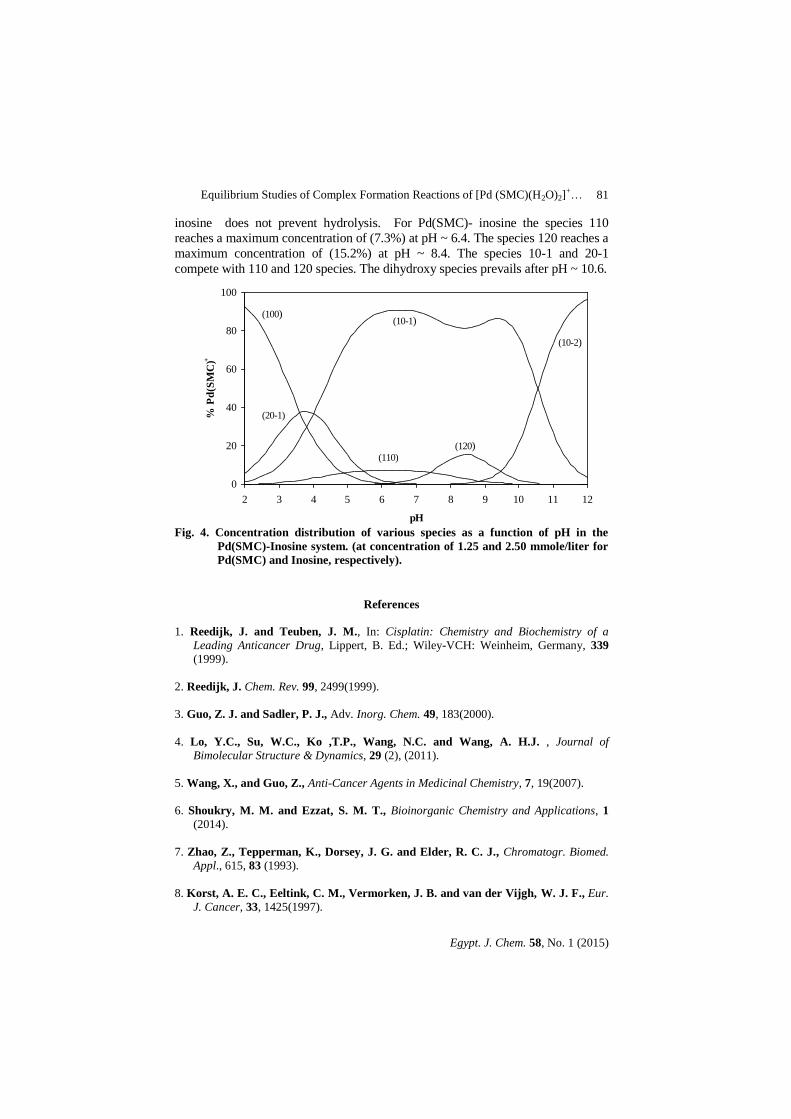
4 Egypt. J. Chem. 58, No. 1, pp. 71- 83 (2015)
#Correspondence should be addressed to Eman M. Shoukry; eman_shoukry 2002@ yahoo.com
C
Equilibrium Studies of Complex Formation
Reactions of [Pd (SMC)(H2O)2]+ with Amino
Acids, Peptides or DNA Constituents
E.M. Shoukry
#, M.F. Amin, A.M. Badawi
*, M.A. Mohamed
and A.G. Ahmed
Department of Chemistry, Faculty of Science, Al-Azhar University
(For Girls) and *Egyptian Petroleum Research Institute, Nasr
City, Egypt.
OMPLEX-FORMATION equilibria have been investigated for
……..[Pd (SMC)(H2O)2]+ where SMC = S-methyl-L-cysteinate, with
amino acids, peptides and DNA constituents. Stoichiometries and
stability constants of the complexes were determined at 37◦C and
constant ionic strength (0.16 M NaNO3). The results showed the
formation of 1:1 complexes with amino acids .Peptides formed both
1:1 complexes and the corresponding deprotonated amide species.
DNA constituents formed both 1:1 and 1:2 complexes .The binding
mode of the ligands containing various functional groups was studied
and the speciation diagrams were evaluated.
Sulfur-containing biomolecules such as cysteine (Cys), methionine (Met),
glutathione (GSH), metallothionein (MT) and albumin play significant roles in
platinum anticancer chemotherapy because of their high affinity to platinum (II)
compounds (1-5)
. The bidentate N, S-complex was found to be promising
cytostatic agent (6)
.Sulfur is involved in the entire metabolic process of platinum
drugs, including reactions prior to cell uptake, deactivation prior to DNA binding
and formation of DNA-adduct, … etc (7)
. On the other hand, the platinum sulfur
interactions can be used to produce favorable effects in the clinical application
of Pt-based drugs. It is possible now to employ sulfur-containing compounds as
chemoprotectants to mitigate the severe toxic side effects of platinum drugs and
some of them have been registered in a number of European countries (8-10)
.
Furthermore, Methionine, cysteine, and pencillamine are believed to be
determinant in the reduction of the nephrotoxicity of cis-platin and other
chemotherapeutic drugs(11)
. A previous investigation (12)
focused on the kinetics
of the interaction of diaqua-( S-methyl-L-cysteinate) palladium(II) with some
DNA constituents. It seemed of interest to extend this work to throw more light
on the speciation of Pd(S-methyl-L-cysteinate) complexes, as a model for the
main metabolite in cancer chemotherapy, in biological fluids where all types of
ligands are present. Furthermore, the results of this study are of interest because
it is possible to make some comparisons with the chemistry of metabolites of
Pt(II) anticancer complexes. The present investigation describes the formation
equilibria involving [Pd(SMC)(H2O)2]+
(SMC = S-methyl-L-cysteinate) and
E.M. Shoukry et al.
Egypt. J. Chem. 58, No. 1 (2015)
72
other ligands such as amino acids, peptides or DNA constituents at 37◦C and
constant ionic strength (0.16 M NaNO3).
Experimental
Materials
K2PdCl4, and S-methyl-L-cysteinate were obtained from Aldrich. The amino
acids and related compounds (glycine, alanine, valine, proline, ethanolamine,
serine, threonine, histidine, histamine, ornithine, lysine, cysteine, methionine ) were
provided by Sigma Chemical Co. The peptides used (glycinamide, glycylglycine,
glycylleucine, glutamine) were all provided by BDH Biochemicals Ltd., Poole,
England. The DNA constituents (Inosine, Inosine-5-monophosphate, uracil,
thymine , uridine and uridine-5-monophosphate) were provided by Sigma
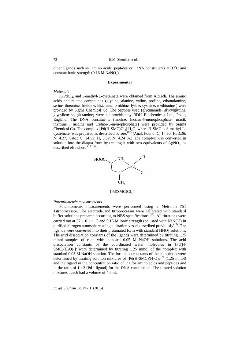
Chemical Co. The complex [Pd(H-SMC)Cl2].H2O, where H-SMC is S-methyl-L-
cysteinate, was prepared as described before.(12)
(Anal. Found: C, 14.60; H, 3.36;
N, 4.27. Calc.: C, 14.52; H, 3.32; N, 4.24 %.) The complex was converted in
solution into the diaqua form by treating it with two equivalents of AgNO3, as
described elsewhere (13–15)
.
Pd
Cl
S
CH3
NH2
Cl
HOOC
[Pd(SMC)Cl2]
Potentiometric measurements Potentiometric measurements were performed using a Metrohm 751
Titroprocessor. The electrode and titroprocessor were calibrated with standard
buffer solutions prepared according to NBS specifications (16)
. All titrations were
carried out at 37 ± 0.1 ∘ C and 0.16 M ionic strength (adjusted with NaNO3) in
purified nitrogen atmosphere using a titration vessel described previously(17)
. The
ligands were converted into their protonated form with standard HNO3 solutions.
The acid dissociation constants of the ligands were determined by titrating 1.25
mmol samples of each with standard 0.05 M NaOH solutions. The acid
dissociation constants of the coordinated water molecules in [Pd(H-
SMC)(H2O)2]2+
were determined by titrating 1.25 mmol of the complex with
standard 0.05 M NaOH solution. The formation constants of the complexes were
determined by titrating solution mixtures of [Pd(H-SMC)(H2O)2]2+
(1.25 mmol)
and the ligand in the concentration ratio of 1:1 for amino acids and peptides and
in the ratio of 1 : 2 (Pd : ligand) for the DNA constituents. The titrated solution
mixtures , each had a volume of 40 ml.
Equilibrium Studies of Complex Formation Reactions of [Pd (SMC)(H2O)2]+…
Egypt. J. Chem. 58, No. 1 (2015)
73
The species formed were characterized by the general equilibrium process
(1), whereas the formation constants for these generalized species are given by
Eq. (2) (charges are omitted for simplicity).
pM + qL + rH M p Lq H r (1)
rHq
Lp
M
]rHqLp[M =β pqr
(2)
where the charges are omitted for simplicity.
M, L and H represent [Pd (SMC)(H2O)2]+, ligand and proton, respectively.
The calculations were performed using the program MINIQUAD-75 (17)
running
on an IBM-486 computer. Stoichiometric and stability constants of the
complexes were determined by trying various possible composition models for
the systems studied. The selected model gave the best statistical fit and was
chemically consistent with the titration data without giving any systematic drift in
the magnitude of various residuals, as described elsewhere (17)
. The stability
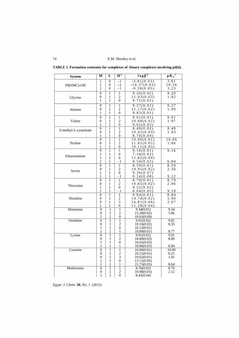
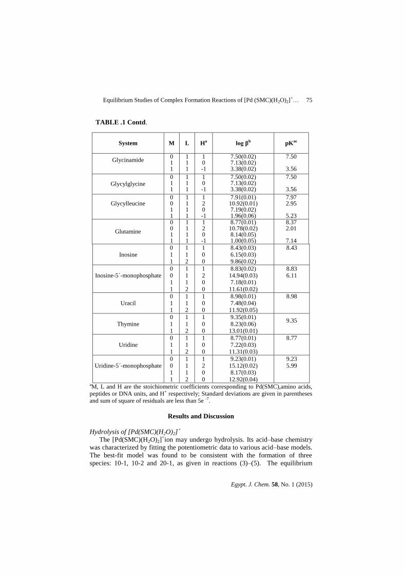
constants of the complexes formed in solution are given in Table 1. The
concentration distribution diagrams were obtained with the program SPECIES (18)
,
taking into account the experimental conditions used.
O P O
O
O
O
OH OH
N
N
N
NH
O
O
OH OH
OHN
N
N
NH
O
NH
O
NH
O
NH
O
NH
O
CH3
O
OH OH
OHN
NH
O
O
O P O
O
O
O
OH OH
N
NH
O
O
Inosine
9
71
3
Inosine-5´-monophosphate
9
71
3
6
Uracil
1
3
2
4
Thymine
1
3
2
4
1
3
2
4
Uridine
1
3
2
4
Uridine-5´-monophosphate
Scheme1. Structural formula of some of the investigated ligands.
E.M. Shoukry et al.
Egypt. J. Chem. 58, No. 1 (2015)
74
TABLE 1. Formation constants for complexes of binary complexes involving pd(ii).
System M L H a l o g β b p K ac
Pd(SMC)-OH
1 1 2
0 0 0
- 1 - 2 - 1
- 3 . 8 1 ( 0 . 0 3 ) - 1 4 . 3 7 ( 0 . 0 3 ) - 0 . 5 8 ( 0 . 0 5 )
3 . 8 1 1 0 . 5 6 3 . 2 3
Glycine
0 0 1
1 1 1
1 2 0
9 . 2 0 ( 0 . 0 2 ) 1 1 . 0 2 ( 0 . 0 3 ) 9 . 7 1 ( 0 . 0 2 )
9 . 2 0 1 . 8 2
Alanine 0 0 1
1 1 1
1 2 0
9 . 2 7 ( 0 . 0 1 ) 1 1 . 1 7 ( 0 . 0 2 ) 9 . 8 3 ( 0 . 0 1 )
9 . 2 7 1 . 9 0
Valine 0 0 1
1 1 1
1 2 0
9 . 0 1 ( 0 . 0 1 ) 1 0 . 9 8 ( 0 . 0 2 ) 9 . 6 5 ( 0 . 0 3 )
9 . 0 1 1 . 9 7
S-methyl-L-cysteinate
0 0 1
1 1 1
1 2 0
8 . 4 9 ( 0 . 0 2 ) 1 0 . 4 2 ( 0 . 0 3 ) 8 . 7 6 ( 0 . 0 4 )
8 . 4 9 1 . 9 3
Proline 0 0 1
1 1 1
1 2 0
1 0 . 0 6 ( 0 . 0 1 ) 1 1 . 8 1 ( 0 . 0 5 ) 1 0 . 1 1 ( 0 . 0 3 )
1 0 . 0 6 1 . 8 6
Ethanolamine
0 1 1 1
1 1 2 1
1 0 0 - 1
9 . 1 6 ( 0 . 0 1 ) 7 . 3 4 ( 0 . 0 2 )
1 1 . 8 2 ( 0 . 0 4 ) 0 . 5 0 ( 0 . 0 2 )
9 . 1 6
6 . 8 4
Serine
0 0 1 1
1 1 1 1
1 2 0 - 1
8 . 5 9 ( 0 . 0 1 ) 1 0 . 9 5 ( 0 . 0 2 ) 9 . 3 6 ( 0 . 0 7 ) 0 . 2 4 ( 0 . 0 8 )
8 . 5 9 2 . 3 6
9 . 1 2
Theronine
0 0 1 1
1 1 1 1
1 2 0 - 1
8 . 7 9 ( 0 . 0 1 ) 1 0 . 8 5 ( 0 . 0 2 ) 9 . 2 2 ( 0 . 0 2 ) 0 . 0 4 ( 0 . 0 5 )
8 . 7 9 2 . 0 6
9 . 1 8
Histidine
0 0 0 1
1 1 1 1
1 2 3 0
8 . 8 4 ( 0 . 0 1 ) 1 4 . 7 4 ( 0 . 0 2 ) 1 6 . 8 1 ( 0 . 0 6 ) 1 1 . 2 8 ( 0 . 0 4 )
8 . 8 4 5 . 9 0 2 . 0 7
Histamine 0 0 1
1 1 1
1 2 0
9.34(0.01) 15.20(0.02) 10.63(0.09)
9.34 5.86
Ornithine
0 0 1 1
1 1 1 1
1 2 0 1
9.81(0.02) 18.16(0.02) 10.12(0.01) 18.89(0.01)
9.81 8.35
8.77
Lysine 0 0 1 1
1 1 1 1
1 2 0 1
9.91(0.02) 18.80(0.03) 10.02(0.02) 18.86(0.03)
9.91 8.89
8.84
Cysteine 0 0 0 1 1
1 1 1 1 1
1 2 3 0 1
10.00(0.01) 18.21(0.01) 19.62(0.01) 13.11(0.05) 21.75(0.03)
10.00 8.21 1.41
8.64
Methionine 0 0 1
1 1 1
1 2 0
8.76(0.02) 10.98(0.03) 8.43(0.04)
8.76 2.12
Equilibrium Studies of Complex Formation Reactions of [Pd (SMC)(H2O)2]+…
Egypt. J. Chem. 58, No. 1 (2015)
75
TABLE .1 Contd.
System
M
L
Ha
log βb
pKac
Glycinamide
0 1 1
1 1 1
1 0 -1
7.50(0.02) 7.13(0.02) 3.38(0.02)
7.50
3.56
Glycylglycine 0 1 1
1 1 1
1 0 -1
7.50(0.02) 7.13(0.02) 3.38(0.02)
7.50
3.56
Glycylleucine
0 0 1 1
1 1 1 1
1 2 0 -1
7.91(0.01) 10.92(0.01) 7.19(0.02) 1.96(0.06)
7.97 2.95
5.23
Glutamine
0 0 1 1
1 1 1 1
1 2 0 -1
8.77(0.01) 10.78(0.02) 8.14(0.05) 1.00(0.05)
8.37 2.01
7.14
Inosine 0 1 1
1 1 2
1 0 0
8.43(0.03) 6.15(0.03) 9.86(0.02)
8.43
Inosine-5΄-monophosphate
0 0 1 1
1 1 1 2
1 2 0 0
8.83(0.02) 14.94(0.03) 7.18(0.01)
11.61(0.02)
8.83 6.11
Uracil 0 1 1
1 1 2
1 0 0
8.98(0.01) 7.48(0.04)
11.92(0.05)
8.98
Thymine 0 1 1
1 1 2
1 0 0
9.35(0.01) 8.23(0.06)
13.01(0.01)
9.35
Uridine 0 1 1
1 1 2
1 0 0
8.77(0.01) 7.22(0.03)
11.31(0.03)
8.77
Uridine-5΄-monophosphate
0 0 1 1
1 1 1 2
1 2 0 0
9.23(0.01) 15.12(0.02) 8.17(0.03)
12.92(0.04)
9.23 5.99
aM, L and H are the stoichiometric coefficients corresponding to Pd(SMC),amino acids,
peptides or DNA units, and H+ respectively; Standard deviations are given in parentheses
and sum of square of residuals are less than 5e -7.
Results and Discussion
Hydrolysis of [Pd(SMC)(H2O)2]+
The [Pd(SMC)(H2O)2]+ion may undergo hydrolysis. Its acid–base chemistry
was characterized by fitting the potentiometric data to various acid–base models.
The best-fit model was found to be consistent with the formation of three
species: 10-1, 10-2 and 20-1, as given in reactions (3)–(5). The equilibrium
E.M. Shoukry et al.
Egypt. J. Chem. 58, No. 1 (2015)
76
constants were determined at temperature 37 °C and at constant 0.16 M ionic
strength (adjusted with NaNO3), and given in Table 1. These values were taken
into account on determining the stability constants of the PdII complexes.
pKa1
[Pd(SMC)(H2O)2]+
[Pd(SMC)(H2O)(OH)] + H+
(3)
100 10-1
pKa2
[Pd(SMC)(H2O)(OH)] [Pd(SMC)(OH)2]-
+ H+
(4)
10-1 10-2
Pd
OH
OH2
Pd
H2O
20-1
+ H2O (5)(SMC)(SMC)
+
[Pd(SMC)(H2O)2]+ + [Pd(SMC)(H2O)(OH)]
logKdimer
10-1100
The µ-hydroxo species (20−1) is assumed to form through dimerization of
the Pd(II) complex via a hydroxo group. The equilibrium constant for the
dimerization reaction (5) can be calculated by Eq. (6) and amounts to 3.23.
log Kdimer = logβ20−1 − logβ10−1 = -0.58− (-3.81) =3.23 (6)
The concentration distribution diagram for [Pd (SMC)(H 2O) 2]+
complex
is shown in Fig. 1. The concentration of the monohydroxo species, 10-1 and the
dimeric species, 20-1, increase with increasing pH. The dimeric species has
maximum concentration of 40% at pH ~ 3.8. The monohydroxo species (10-1) is
the main species in the pH range ~ 4.0-10.6, i.e. it is the main species present
under physiological conditions. A further increase in pH is accompanied by an
increase in the dihydroxo species (10-2), which is the main species above pH
ca.10.6.
Equilibrium Studies of Complex Formation Reactions of [Pd (SMC)(H2O)2]+…
Egypt. J. Chem. 58, No. 1 (2015)
77
0
20
40
60
80
100
2 3 4 5 6 7 8 9 10 11 12
pH
% P
d(S
MC
)+
(10-1)(100)
(10-2)
(20-1)
Fig.1. Concentration distribution of various species as a function of pH in the
Pd(SMC)-OH system at concentration of 1.25 mmole/liter for Pd(SMC).
Complex-formation equilibria involving amino acids
Analysis of the titration data for the Pd(SMC)-amino acid system showed the
formation of 1:1 species. The stability constant of the histidine and ornithine
complexes are higher than those of simple amino acids. This indicates that these
amino acids coordinate via the two nitrogen centers, i.e. imidazole and amino
groups in the case of histidine, and by two amino groups in the case of ornithine.
This is in line with the strong affinity of PdII
for nitrogen donor centers. The
stability constant of the complex with lysine (log β110 =10.02) is a little bit higher
than those of α-amino acids. This may be taken to indicate that lysine most likely
chelates through the α -amino and carboxylate groups (N, O-donor set). The
proline complex has the highest value. This may be due to the highest basicity of
the proline amino group as reflected by the highest pKa value. The stability
constant value of methionine complex (log β 110 = 8.43) is lower than those of
most simple amino acids. This may be explained by the fact that the amino group
of methionine is less basic than those of other amino acids as reflected by pKa
values, (Table 1). S-methyl-L-cysteinate forms a more stable complex than
methionine although the pKa of the former is lower than the latter. This may be
accounted for on the premise that the five-membered chelate ring in the former
complex is energetically more stable than the six-membered chelate ring in the
latter complex. Serine and threonine have an extra binding center on the β-
alcoholate group. This group was reported (19)
to participate in transition metal
ion complex-formation reactions. the potentiometric data is much better fitted
assuming the formation of the complex species 110 and 11–1. The pKa value of
the β- alcoholate group incorporated in the PdII
complex, (logβ110 –log β11–1) is
E.M. Shoukry et al.
Egypt. J. Chem. 58, No. 1 (2015)
78
9.12 and 9.18 for serine and threonine, respectively. In addition, ethanolamine
forms the complex species 110, 120 and 11–1, and the logβ110 value for
ethanolamine is smaller than those for amino acids. This may be due to the
coordination of ethanolamine at low pH through the amino group, while in the
case of serine and threonine the coordination is through amino and carboxylate
groups. The pKa value of the coordinated alcohol group in ethanolamine (6.84) is
smaller than those of serine and threonine. This is attributed to the tendency of
ethanolamine to coordinate through the OH group at lower pH to form a five
member chelate ring. The cysteine complex is significantly more stable than
those of the amino acids (N,O-donor site) and ornithine (N,N donor site). This
indicates that cysteine coordinates via S- and N-donor sites. This is consistent
with the high affinity of both S and N atoms for coordination to the Pd(II) ion.
The concentration distribution diagram (Fig. 2) indicates that serine forms
the species 110 at low pH and predominates between pH ~2.7-9.2 and thus
prevents the hydrolysis of Pd (II), i.e. the species (10-1) and (20-1) are present at
very low concentration. The ionisation of the OH group starts after pH ~7 and
the (11-1) species predominate after pH ~ 9.2.
0
20
40
60
80
100
2 3 4 5 6 7 8 9 10 11 12
pH
% P
d(S
MC
)+
(10-1)
(100)
(10-2)
(20-1)
(110)
(11-1)
Fig. 2. Concentration distribution of various species as a function of pH in the
Pd(SMC) and Serine system. (at concentration of 1.25 mmole/liter for
Pd(SMC) and Serine).
Complex-formation equilibria involving peptide
The complex formation equilibria involving peptides were characterized by
fitting their potentiometric data to various models. The best model was found to
be consistent with the formation of the complexes with stoichiometric
Equilibrium Studies of Complex Formation Reactions of [Pd (SMC)(H2O)2]+…
Egypt. J. Chem. 58, No. 1 (2015)
79
coefficients 110 and 11-1 according to Eq. (7) and (8) (see Table 1).The
complexes are formed by coordination of the amino and carbonyl groups. Upon
deprotonation of the amide group, the coordination sites would switch from
carbonyl oxygen to amide nitrogen. Such changes in coordination centers are
well documented (20)
. The glutamine complex is more stable than the
glycinamide complex. The most likely explanation lies in the fact that
glutaminate carries a negative charge, whereas glycinamide is neutral. The
electrostatic interaction between the glutaminate and the twofold positively
charged metal complex would result in a lowering of free energy of formation.
The pKa values of the amide groups, incorporated in the palladium(II)
complexes (logβ110 - logβ11-1) are in the range of 3.56-7.14. The low pKa
values in the present system are probably due to the high affinity of palladium to
nitrogen-type ligands. It is interesting to note that the pKa value for the
glycinamide complex is lower than those for other peptides. This can be
explained on the premise that the more bulky substituent group on the peptide
may hinder the structural changes when going from the protonated to the
deprotonated complexes. The pKa of the glutamine complex is exceptionally
higher than those of the other peptide complexes. This is due to the formation of
a seven membered chelate ring which is more strained and therefore less
favoured.
Scheme 2. The Complex-formation equilibria for peptides.
The relative magnitudes of the pKa values of the palladium(II) complexes
with peptides have interesting biological implications. Under normal
physiological conditions (pH ca. 7.4), the peptides would coordinate with
[Pd(SMC)(H2O)2]+ in entirely different fashions: glutaminate exists solely in its
protonated form, whereas the other peptides are present entirely in the
deprotonated form. Also, the slight differences in the side chains of the peptides
seem to produce dramatic differences in their behavior towards the palladium
species.
The concentration distribution diagram (Fig. 3) indicates that glycylglycine
forms the complex species (110) at low pH and (11-1) at higher pH. The species
E.M. Shoukry et al.
Egypt. J. Chem. 58, No. 1 (2015)
80
(11-1) is the main species at higher pH and predominantes after pH 4.6 for
glycylglycine.
0
20
40
60
80
100
2 3 4 5 6 7 8 9 10
pH
%P
d(S
MC
)+ (110)
(100)
(10-1)(20-1)
(11-1)
.
Fig. 3. Concentration distribution of various species as a function of pH in the
Pd(SMC)-glycylglycine system. (at concentration of 1.25 mmole/liter for
Pd(SMC) and glycylglycine).
Complex-formation equilibria involving DNA constituents
DNA constituents such as Inosine, Inosine -5-monophosphate, uracil,
thymine,Uridine-5-monphosphate , and uridine form 1 : 1 and 1 : 2 complexes
with[Pd(SMC)]+. Inosine can be protonated at N7 forming a (N1H-N7H)
monocation. The pKa of N1H is 8.43, Table 1. The pKa of N7H is 1.2. (21)
It was
reported (21)
that, in the acidic pH range, N1 remained protonated, while the metal
ion is coordinated to N7. The binding site at the higher pH was disputed and it
was suggested that there is a gradual change from N7- binding to N1-binding
with increase of pH. (21)
. Inosine-5´-monophosphate (5´-IMP) forms a more
stable complex with [Pd(SMC)(H2O)2]+
than that of inosine. The extra
stabilization can be attributed to the triply negatively charged 5´-IMP3-
ion. N3 is
the preferred complexation site of the 4-oxo-pyrimidine derivatives as uracil,
thymine, uridine and uridine-5´-monophosphate. The pKa values of N3H given in
Table 1, are consistent with the literature values(22, 23)
. The stability constants of
the 4-oxo-pyrimidine derivatives are larger than most of DNA constituents due
to their high basicity. The thymine complex is more stable than that of uracil,
probably due to the higher basicity of the N3 site of thymine resulting from the
inductive effect of the extra electron-donating methyl.
The concentration distribution diagrams for Pd(SMC)-inosine, complex, (Fig. 4)
show that the species 20-1, 10-1, 10-2, are the predominated species (above
pH=3). This means that the complex formation of [Pd(SMC)(H2O)2]+ with
Equilibrium Studies of Complex Formation Reactions of [Pd (SMC)(H2O)2]+…
Egypt. J. Chem. 58, No. 1 (2015)
81
inosine does not prevent hydrolysis. For Pd(SMC)- inosine the species 110
reaches a maximum concentration of (7.3%) at pH ~ 6.4. The species 120 reaches a
maximum concentration of (15.2%) at pH ~ 8.4. The species 10-1 and 20-1
compete with 110 and 120 species. The dihydroxy species prevails after pH ~ 10.6.
0
20
40
60
80
100
2 3 4 5 6 7 8 9 10 11 12
pH
% P
d(S
MC
)+
(10-1)(100)
(10-2)
(20-1)
(110)
(120)
Fig. 4. Concentration distribution of various species as a function of pH in the
Pd(SMC)-Inosine system. (at concentration of 1.25 and 2.50 mmole/liter for
Pd(SMC) and Inosine, respectively).
References
1. Reedijk, J. and Teuben, J. M., In: Cisplatin: Chemistry and Biochemistry of a
Leading Anticancer Drug, Lippert, B. Ed.; Wiley-VCH: Weinheim, Germany, 339
(1999).
2. Reedijk, J. Chem. Rev. 99, 2499(1999).
3. Guo, Z. J. and Sadler, P. J., Adv. Inorg. Chem. 49, 183(2000).
4. Lo, Y.C., Su, W.C., Ko ,T.P., Wang, N.C. and Wang, A. H.J. , Journal of
Bimolecular Structure & Dynamics, 29 (2), (2011).
5. Wang, X., and Guo, Z., Anti-Cancer Agents in Medicinal Chemistry, 7, 19(2007).
6. Shoukry, M. M. and Ezzat, S. M. T., Bioinorganic Chemistry and Applications, 1
(2014).
7. Zhao, Z., Tepperman, K., Dorsey, J. G. and Elder, R. C. J., Chromatogr. Biomed.
Appl., 615, 83 (1993).
8. Korst, A. E. C., Eeltink, C. M., Vermorken, J. B. and van der Vijgh, W. J. F., Eur.
J. Cancer, 33, 1425(1997).
E.M. Shoukry et al.
Egypt. J. Chem. 58, No. 1 (2015)
82
9. Hausheer, F. H., Kanter, P., Cao, S., Haridas, K., Seetharamulu, P., Reddy, D.,
Petluru, P., Zhao, M., Murali, D., Saxe, J. D., Yao, S., Martinez, N., Zukowski, A.
and Rustum, Y. M., Semin. Oncol. 25, 584 (1998).
10. Jakupec, M. A., Galanski, M. and Keppler, B. K., In Metal Ions in Biological
Systems, Sigel, A.; Sigel, H. Eds.; Marcel Dekker, Inc.: New York, 42, 179 (2004).
11. Slater, T.F., Ahmed, M., and Ibrahim, S.A., J. Clin.Hemat.Oncol. 7, 534(1977).
12. Bugarˇci´c, D., Shoukry, M. M., and van Eldik, R., J. Chem. Soc., Dalton Trans.
3945 (2002).
13. Theodorou, V., Photaki, I., Hadjiliadis, N., Gellert, R. V., and Bau, R., Inorg.
Chim. Acta, 60, 1(1982).
14. Allain, A., Kubiak, M., Jezowsak-Tezebiatowska, B., Kozlowski, H., and
Glowiak, T., Inorg. Chim. Acta, 46, 127 (1980).
15. Livingstone, S. E., and Nolan, J. D., Inorg. Chime. Acta, 7, 1447( 1968).
16. Bates, R.G., Determination of pH: Theory and Practice, 2nd ed., Wiley Interscience,
New York (1975).
17. Shoukry, M.M. Hosny, W.M. and Khalil, M.M., Transit. Met. Chem. 20, 252
(1995).
18. Pettit, L., Personal Communication, University of Leeds (1993).
19. Shehata, M.R., Shoukry, M. M., Abdel-Shakour F. H., and van Eldik, R., Eur. J.
Inorg. Chem. 3912 (2009).
20. Shehata, M. R., Shoukry, M. M. and Ali, S., Spectrochimica Acta Part A 91, 383
(2012).
21. Shehata , M. R., Transition Metal Chemistry, 26, 198( 2001).
22. Wolfenden, R. V., J. Mol. Biol. 40, 307 (1969).
23. Shehata, M.R., Shoukry M.M., Ragab, M.S., Spectrochimica Acta Part A:
Molecular and Bimolecular Spectroscopy, 96, 809 (2012).
( Received 18 / 1 / 2015,
accepted 28 / 1 / 2015)
Equilibrium Studies of Complex Formation Reactions of [Pd (SMC)(H2O)2]+…
Egypt. J. Chem. 58, No. 1 (2015)
83
دراسة اتزان تكوين المتركبات الناتجة من تفاعل المتراكب
[Pd (SMC)(H2O)2الثنائى+
مع الاحماض الامينية،البيبتيدات او
DNA مكونات
مين ، عبد الفتاح محسن بدوىأ فخرى منى شكرى ، محمد ايمان*
منى ،
مل جمعه احمدأ ولجليل محمد اعبد
جامعة الازهر و–( للبنات)كلية العلوم –ياء قسم الكيم*
البترول معهد بحوث
.مصر -القاهرة - المصرى
المتراكب تفاعل من الناتجة المتركبات تم فى هذا البحث دراسة اتزان تكوين+
] Pd(SMC)(H2O)2 [ ( حيث انSMC = مع ) كبريت مثيل سستين
وتم التعرف على النسب ،DNAونات الاحماض الامينية ، البيبتيدات او مك
الجزيئية لهذه المتراكبات ، وكذلك تم حساب ثوابت الاستقرار لها عند درجة حرارة
73o
اتضح من النتائج تكوين . مولارى من نترات الصوديوم 0..6م وقوة ايونية
مع الاحماض الامينية وتكون البيبتيدات متراكبات .:.متراكبات بالنسبة الجزئية
بالاضافة الى متراكبات منزوعة البروتون من مجموعة الاميد .:.بالنسبة الجزئية
كما تم شرح DNA مع وحدات 1:.و .:.وتكوين متراكبات بالنسب الجزئية
لمرفقات المحتوية على المجموعات الوظيفية المختلفة وحساب طريقة الارتباط ل
.تركيز المتراكبات المتكونة فى المحلول كدالة فى رقم الاس الهيدروجينى للوسط